the rich inner life of the cell nucleus: dynamic
TRANSCRIPT

https://doi.org/10.1007/s12551-020-00761-x
REVIEW
The rich inner life of the cell nucleus: dynamic organization, activeflows, and emergent rheology
Alexandra Zidovska1
Received: 24 August 2020 / Revised: 3 September 2020 / Accepted: 14 September 2020© The Author(s) 2020
AbstractThe cell nucleus stores the genetic material essential for life, and provides the environment for transcription, maintenance,and replication of the genome. Moreover, the nucleoplasm is filled with subnuclear bodies such as nucleoli that areresponsible for other vital functions. Overall, the nucleus presents a highly heterogeneous and dynamic environmentwith diverse functionality. Here, we propose that its biophysical complexity can be organized around three inter-relatedand interactive facets: heterogeneity, activity, and rheology. Most nuclear constituents are sites of active, ATP-dependentprocesses and are thus inherently dynamic: The genome undergoes constant rearrangement, the nuclear envelope flickers andfluctuates, nucleoli migrate and coalesce, and many of these events are mediated by nucleoplasmic flows and interactions.And yet there is spatiotemporal organization in terms of hierarchical structure of the genome, its coherently moving regionsand membrane-less compartmentalization via phase-separated nucleoplasmic constituents. Moreover, the non-equilibrium oractivity-driven nature of the nucleus gives rise to emergent rheology and material properties that impact all cellular processesvia the central dogma of molecular biology. New biophysical insights into the cell nucleus can come from appreciating thisrich inner life.
Keywords Cell nucleus · Chromatin dynamics · Nuclear compartmentalization · Active matter · Rheology
Introduction
The cell nucleus is arguably one of the most importantorganelles in eukaryotic cell, housing the genome thatcontains the genetic blueprint for the entire cell (Albertset al. 2014). The genetic information is stored in the DNAmolecule, which lies at the core of the central dogma ofmolecular biology (Crick 1958, 1970). DNA is transcribedinto RNA, which becomes translated into proteins. Thefirst step of gene expression, transcription, occurs in thecell nucleus assisted by the intricate interplay of molecularmachinery that acts on chromatin, the functional formof DNA inside cells (Van Holde 2012; Alberts et al.2014). In addition, many other DNA transactions occurinside the nucleus such as genome replication prior tocell division or DNA repair to maintain genome integrity.These processes are ATP-dependent and their molecular
� Alexandra [email protected]
1 Center for Soft Matter Research, Department of Physics,New York University, New York, NY, USA
machinery requires direct access to the DNA molecule,leading to a persistent dynamic rearrangement of thegenome. While the biochemistry of these processes hasbeen studied in great detail (Van Holde 2012; Alberts et al.2014), their biophysical mechanisms and implications arefar from understood (Hubner and Spector 2010; Dekkeret al. 2013; Gibcus and Dekker 2013; Bickmore and vanSteensel 2013; Sazer and Schiessel 2018). Moreover, thetimescales and length scales of these processes are directlyinfluenced by the material properties of the nucleus andits constituents, which in turn affect all cellular processesvia the central dogma. For example, the viscosity of thenucleoplasm impacts the rates of molecular and organelletransport inside the nucleus, whereas the persistence lengthof the DNA molecule affects its local organization anddynamics (Milo and Phillips 2015).
In addition to the highly dynamic genome, the nucleuscontains a plethora of smaller structures such as nucleoli,Cajal bodies, PML bodies, and speckles (Misteli andSpector 2011; Alberts et al. 2014). These subnuclear bodiesserve as sites of further essential processes and oftenmigrate and undergo their own dynamic rearrangement orrestructuring, e.g., the coalescence of nucleoli or speckles
/ Published online: 16 October 2020
Biophysical Reviews (2020) 12:1093–1106
(Caragine et al. 2018; 2019; Kim et al. 2019). Thegenome and subnuclear bodies are all immersed in thenucleoplasm, a surrounding fluid rich with proteinaceousmolecular machinery as well as their respective molecularproducts such as RNA. This complex solution of polymersand colloidal particles is confined by the nuclear envelopethat is comprised of a layer of intermediate filaments calledlamins and two lipid bilayers (Alberts et al. 2014). Veryrecently, the nuclear envelope was found to be perpetuallyundulating (Chu et al. 2017).
Overall, the cell nucleus is a rich environment with arich inner life. Its constituents are numerous and diverse,ranging from polymers to colloids, from small moleculesto macromolecules, giving rise to a highly heterogeneoussystem. Strikingly, the nucleus lacks any internal bound-aries, yet its content is evidently functionally organized.Moreover, its organization is dynamical, simultaneouslyaccommodating many orthogonal active (ATP-dependent)processes happening concurrently, and thus giving rise toemergent behaviors and properties. Hence, the cell nucleuspresents a non-equilibrium living system, which defiesprinciples of equilibrium thermodynamics. In this review,we will survey current knowledge about the biophysicalorigins of nuclear organization and heterogeneity, dynam-ics of nuclear constituents, nuclear compartmentalizationvia phase separations, and the emergent rheology of thenucleus.
Nuclear organization and heterogeneity
The major component of the nucleus is the chromatinfiber composed of DNA wrapped around protein particles,nucleosomes, made of the histone proteins and resemblinga beads-on-a-string structure (Alberts et al. 2014). In thehuman genome, about 2 m of DNA are packed insidea nucleus of roughly 10 μm diameter (Fig. 1) (Albertset al. 2014). The chromatin fiber is further folded into a3D conformation, the static structure of which has beenelucidated in great detail by chromosome conformationcapture techniques (e.g., HiC), which measure probabilitiesof specific genomic sequences being in physical proximityof each other (Lieberman-Aiden et al. 2009; Dekkeret al. 2013; Gibcus and Dekker 2013; Bonev and Cavalli2016). HiC revealed that chromatin fiber is hierarchicallyorganized with increasing length scale: First, it makesloops, leading to formation of topologically associateddomains, which are further assembled into A and Bcompartments, corresponding to transcriptionally activeand inactive compartments, respectively, and finally intochromosome territories (Lieberman-Aiden et al. 2009;Cremer and Cremer 2010; Dekker et al. 2013; Gibcusand Dekker 2013; Bonev and Cavalli 2016). Moreover,
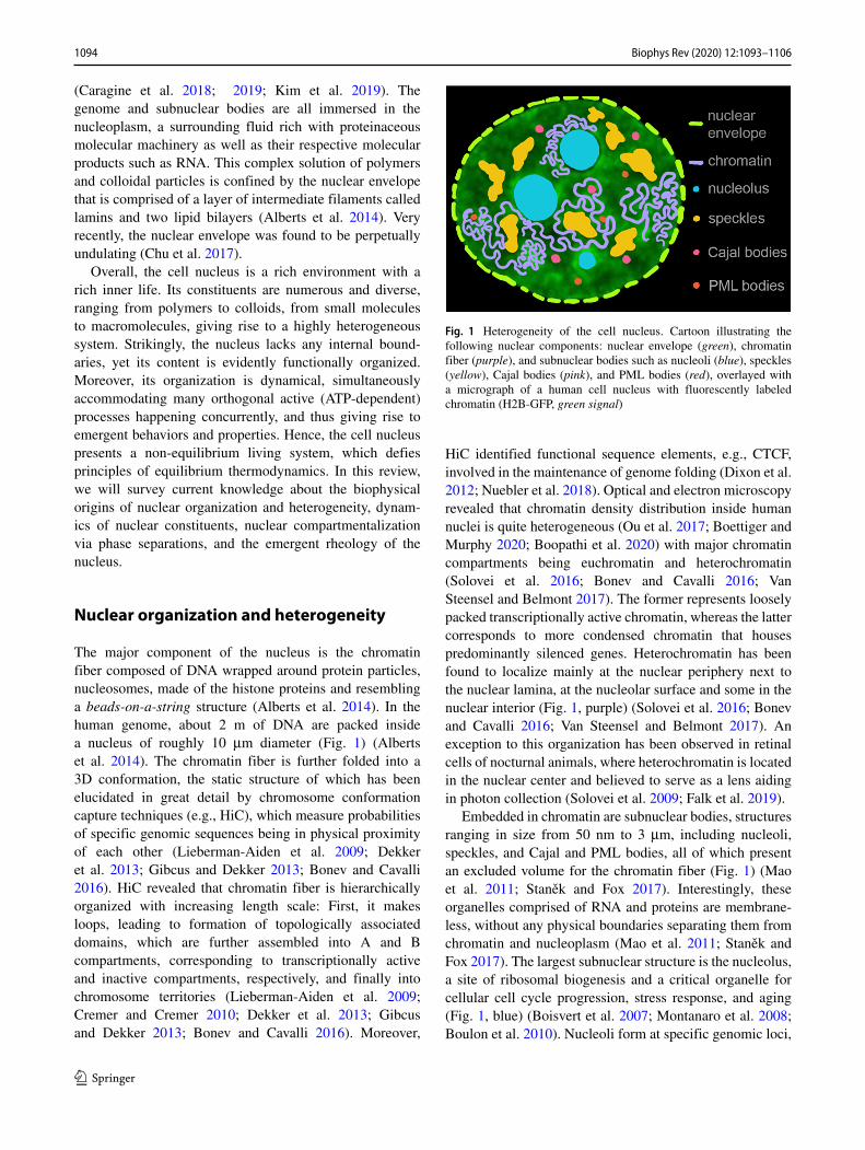
Fig. 1 Heterogeneity of the cell nucleus. Cartoon illustrating thefollowing nuclear components: nuclear envelope (green), chromatinfiber (purple), and subnuclear bodies such as nucleoli (blue), speckles(yellow), Cajal bodies (pink), and PML bodies (red), overlayed witha micrograph of a human cell nucleus with fluorescently labeledchromatin (H2B-GFP, green signal)
HiC identified functional sequence elements, e.g., CTCF,involved in the maintenance of genome folding (Dixon et al.2012; Nuebler et al. 2018). Optical and electron microscopyrevealed that chromatin density distribution inside humannuclei is quite heterogeneous (Ou et al. 2017; Boettiger andMurphy 2020; Boopathi et al. 2020) with major chromatincompartments being euchromatin and heterochromatin(Solovei et al. 2016; Bonev and Cavalli 2016; VanSteensel and Belmont 2017). The former represents looselypacked transcriptionally active chromatin, whereas the lattercorresponds to more condensed chromatin that housespredominantly silenced genes. Heterochromatin has beenfound to localize mainly at the nuclear periphery next tothe nuclear lamina, at the nucleolar surface and some in thenuclear interior (Fig. 1, purple) (Solovei et al. 2016; Bonevand Cavalli 2016; Van Steensel and Belmont 2017). Anexception to this organization has been observed in retinalcells of nocturnal animals, where heterochromatin is locatedin the nuclear center and believed to serve as a lens aidingin photon collection (Solovei et al. 2009; Falk et al. 2019).
Embedded in chromatin are subnuclear bodies, structuresranging in size from 50 nm to 3 μm, including nucleoli,speckles, and Cajal and PML bodies, all of which presentan excluded volume for the chromatin fiber (Fig. 1) (Maoet al. 2011; Stanek and Fox 2017). Interestingly, theseorganelles comprised of RNA and proteins are membrane-less, without any physical boundaries separating them fromchromatin and nucleoplasm (Mao et al. 2011; Stanek andFox 2017). The largest subnuclear structure is the nucleolus,a site of ribosomal biogenesis and a critical organelle forcellular cell cycle progression, stress response, and aging(Fig. 1, blue) (Boisvert et al. 2007; Montanaro et al. 2008;Boulon et al. 2010). Nucleoli form at specific genomic loci,
1094 Biophys Rev (2020) 12:1093–1106
termed nucleolar organizer regions, and remain tetheredto the rDNA genes for their lifetime (McClintock 1934).Moreover, rDNA transcription is closely linked to nucleolarformation, with nucleoli dissolving upon eliminating thisactivity (Grob et al. 2014). The nucleolus has been foundto behave as a liquid droplet and to form via liquid–liquidphase separation of nucleolar proteins and RNA from thenucleoplasm (Brangwynne et al. 2011; Feric et al. 2016;Caragine et al. 2018). Strikingly, its surface exhibits subtlefluctuations in vivo consistent with a liquid droplet of avery low surface tension (Caragine et al. 2018). Other largestructures present in the nucleus and devoid of chromatin arespeckles (Fig. 1, yellow). They are responsible for splicing,the post-transcriptional processing of RNA, and were alsoshown to exhibit liquid-like properties (Marzahn et al.2016; Kim et al. 2019). Smaller structures like Cajal bodies(Fig. 1, pink) and PML bodies (Fig. 1, red) were foundto participate in telomere maintenance and transcriptionalregulation, respectively; however, their full functionalityremains unknown (Platani et al. 2002; Gorisch et al. 2004;Jady et al. 2006).
Chromatin and subnuclear bodies are immersed in thenucleoplasmic fluid, which is aqueous in its nature, enrichedwith nuclear molecular machinery and its products (Albertset al. 2014). The nucleoplasmic composition likely variesin space and time with the progress of nuclear processes(Liang et al. 2009; Dross et al. 2009; Erdel et al. 2015).Thus, in first order, the nuclear content can be viewedas a colloidal suspension containing polydisperse colloidalparticles embedded in a heterogeneous polymer solution ina multicomponent solvent (Fig. 1). To extract the complexbehavior of the system and to understand the biologicalfunction and underlying physics of its components, itsheterogeneity must be taken into account. The stark degreeof heterogeneity of the nuclear content requires detailedapproaches focused on specific nuclear components andtheir behavior as well as mapping of their respectiveinteractions in different local microenvironments across thenucleus.
Dynamics of nucleus and its constituents
Nuclear reorganization and shape fluctuations
To perform their respective biological functions, the nucleusand its constituents have to be highly dynamic, constantlyrearranging and restructuring (Fig. 2a) (Alberts et al. 2014).The nucleus as a whole undergoes a major reorganizationduring the cell cycle. At the beginning of interphase, thetime between two cell divisions, the nuclear envelope formsaround mitotic chromosomes decondensing into looselypacked chromosomes, each of which corresponds to a single
linear polymer and constitutes a chromosome territory(Cremer and Cremer 2010; Alberts et al. 2014). The genomeis then duplicated and later condensed back into mitoticchromosomes facilitating chromosome segregation duringthe cell division (Alberts et al. 2014). During interphase,the size of the nucleus monotonously increases over hours(Chu et al. 2017), while exhibiting small oscillations ofthe nuclear area over minutes (Talwar et al. 2013; Makhijaet al. 2016) and fast undulations, flickering, of the nuclearenvelope over seconds (Chu et al. 2017). The amplitude ofthe nuclear envelope fluctuations (as depicted in Fig. 2e–g) steadily decreases during the interphase and thus canbe utilized as a reliable cell cycle stage indicator in livecells (Chu et al. 2017). The reduction in the nuclear shapefluctuations with progressing cell cycle has been attributedto the increase in the bending rigidity of the nuclearenvelope by the gradual deposition of lamin intermediatefilaments, although a contribution from cell–cycle–specificforces cannot be ruled out (Chu et al. 2017). Finally, asthe cell enters mitosis, the nuclear envelope dissolves, andthe nucleus as an entity ceases to exist (Alberts et al.2014).
Chromatin dynamics
Numerous site-specific DNA transactions such as transcrip-tion, replication, and DNA repair contribute to chromatindynamics during the cell cycle, giving rise to chromatindynamics at different timescales and length scales. Overthe past two decades, chromatin dynamics has been inves-tigated by tracking motions of fluorescently tagged nuclearproteins visualizing structures of interest such as nucleo-somes (Xu et al. 2018; Nagashima et al. 2019; Ashwinet al. 2019), single genes (Marshall et al. 1997; Belmontand Straight 1998; Levi et al. 2005; Chuang et al. 2006;Bronstein et al. 2009; Weber et al. 2012; Chen et al. 2013;Lampo et al. 2016; Germier et al. 2017; Amitai and Hol-cman 2018; Khanna et al. 2019; Vivante et al. 2020), nuclearproteins, enzymes and machineries (Misteli 2001; Carmo-Fonseca et al. 2002; Darzacq et al. 2007; Stixova et al. 2011;Cisse et al. 2013; Hinde et al. 2014; Eaton and Zidovska2020), and subchromosomal foci (Bornfleth et al. 1999;Albiez et al. 2006), as well as entire chromosome territo-ries (Zink et al. 1998; Edelmann et al. 2001). Furthermore,experiments measuring fluorescence recovery after photo-bleaching (Abney et al. 1997; Misteli et al. 2000; Phair andMisteli 2000; Kimura and Cook 2001) and photoactivation(Mora-Bermudez et al. 2007; Wiesmeijer et al. 2008) ofnuclear proteins have revealed their peculiar kinetics. All ofthese approaches have contributed to our understanding ofdynamic processes in the cell nucleus. Chromatin dynam-ics was shown to be mostly subdiffusive to diffusive withoccasional directed motion. While single particle tracking
1095Biophys Rev (2020) 12:1093–1106

Fig. 2 Dynamics of nuclear components. a Cartoon illustratingmotions of different nuclear components: undulations of the nuclearenvelope (gray), coalescence and surface fluctuations of nucleoli(pink), and chromatin dynamics (green), with arrows indicating theirrespective motions. b Micrograph of a human cell nucleus with flu-orescently labeled chromatin (H2B-GFP) and maps of chromatinmotions by Displacement Correlation Spectroscopy (DCS) obtained atc short timescale, �t = 0.25 s, and d long timescale, �t = 10 s.Scale bar, 2 μm. b–d adapted from Zidovska et al. (2013). e Micro-graph of a human cell nucleus with fluorescently labeled chromatin(H2B-GFP) and a cartoon illustrating the chromatin fiber (green) nextto the nuclear envelope (black). Scale bar, 5 μm. f Nuclear contours of
the nucleus from (e) at different times, with insets showing enlargedview of two areas with visible contour fluctuations. g Nuclear shapefluctuations, u2(φ, t), where u(φ, t) is the instantaneous deviation ofthe contour at polar angle φ and time t from the average contour. e–gadapted from Chu et al. (2017). h, j Micrographs of human cell nucleiwith fluorescently labeled chromatin (H2B-GFP, green) and nucleoli(NPM-mApple, red), insets show an enlarged view of the boxed nucle-oli at different times. Scale bar, 5 μm. i, k Contours of nucleoli from(h, j) at different times, with insets highlighting nucleolar surface fluc-tuations (i) and shape changes during nucleolar coalescence (k). h–kadapted from Caragine et al. (2018)
approaches are highly informative (Shukron et al. 2019),reporting about the local chromatin dynamics of a trackedentity, it is unclear how these local motions relate to eachother on a larger, genome-wide scale.
To elucidate the large-scale genome-wide chromatinmotions in vivo, a new spectroscopy-based method Dis-placement Correlation Spectroscopy (DCS) was recentlydeveloped (Zidovska et al. 2013). DCS is a microscopy-based image correlation method, which introduced spa-tiotemporal spectroscopy analysis into the dynamic imagecorrelation processing. It maps chromatin dynamics overtime intervals as shown in Fig. 2b–d, while concurrentlysampling all time intervals accessible by the experiment(Zidovska et al. 2013). Using transgenic histones H2B-GFPas markers of chromatin position and high-resolution spin-ning disc confocal microscopy (Fig. 2b), this noninvasivetechnique enables measurement of chromatin dynamics inreal time across the entire nucleus in live cells, while simul-taneously probing different timescales and length scales
(Fig. 2c–d). Using DCS, chromatin dynamics was found tobe subdiffusive with two distinct time and length scales:(i) fast, local motion, and (ii) slower, coherent motion(Zidovska et al. 2013). While the first had been observedbefore by single particle tracking, the slower, correlatedmotion is new and has major implications for the organi-zation of nuclei on micron-second scales (Zidovska 2020).Importantly, these motions happen in the nucleus con-currently and superposed. Domains of coherent motion(∼ 3–5 μm) reach across chromosome territories, suggest-ing some types of coupling of motion over scales that arehuge compared to individual genes (Zidovska et al. 2013).The discovery of coherent chromatin motion was later cor-roborated by high-resolution imaging of the local motionof single nucleosomes and replication domains and DCS-like spectroscopy analysis in U2OS cells (Nozaki et al.2017; Xiang et al. 2018; Shaban et al. 2018). While the bio-logical role of coherent motion is yet to be uncovered, itleads to physical motion of the entire genome, thus likely
1096 Biophys Rev (2020) 12:1093–1106

impacting gene regulation via local changes in rates andmolecular transport in the nucleus. It may account for appar-ently directed movements of tagged genes that have beenreported in the literature and whose mechanism is unknown(Marshall et al. 1997; Levi et al. 2005; Chuang et al. 2006).These large-scale coupled motions were ATP-dependentand independent of the cytoplasmic cytoskeleton (Zidovskaet al. 2013). Perturbation of major nuclear ATPases such asDNA polymerase, RNA polymerase II, and topoisomeraseII caused local displacements to increase, but eliminatedcoherence, i.e., local motions became uncoupled (Zidovskaet al. 2013; Shaban et al. 2018). These observations revealedcoherent motions to be an emergent property of the activechromatin dynamics, suggesting that gene-level activitymight lead to the nucleus-wide motions (Zidovska 2020).
Motivated by the DCS observations, theoreticalapproaches were developed to further explore the role ofactivity in chromatin dynamics. First, a hydrodynamictheory accounting for active chromatin dynamics withinthe nucleoplasmic fluid was developed (Bruinsma et al.2014). This theory introduces two types of active eventsthat can act on the chromatin fiber: scalar events and vectorevents. Scalar events correspond to local condensation anddecondensation of the chromatin fiber, which can be causedfor example by chromatin remodelers (Bruinsma et al.2014; Racki and Narlikar 2008). Such events do not havea direction, only a magnitude. In contrast, vector eventsrepresent activity induced by nuclear enzymes such asRNA polymerase II, helicase, and topoisimerase II, whichcan be described by a force dipole. A force dipole consistsof two equally large but opposing forces, thus possess-ing both magnitude and a direction. It corresponds to theforce that the enzyme exerts on the chromatin fiber andthe opposing force applied on the surrounding fluid due toNewton’s 3rd law. Thus, the presence of force dipoles leadsto local nucleoplasmic flows, which in turn interact with thechromatin fiber. Moreover, this theory predicts that vectorevents can lead to large-scale fluctuations due to dipolarinteractions, suggesting that collective alignment of forcedipoles can lead to large-scale coherence of chromatindynamics, whereas the scalar events give rise to chromatinconcentration fluctuations at short length scales (Bruinsmaet al. 2014).
The effect of force dipoles on chromatin dynamics withinthe nucleoplasm was further investigated by computationalsimulations, which revealed that extensile (outward) dipolarforces can give rise to the chromatin coherent motion as wellas large-scale nucleoplasmic flows (Saintillan et al. 2018).In contrast, contractile (inwards) dipolar forces led to aseemingly accelerated Brownian dynamics (Saintillan et al.2018). Nucleoplasmic flows due to chromatin activity maylikely contribute to the transport of molecular machinerywithin the nucleus, which would otherwise be diffusion-
limited (Saintillan et al. 2018). Interestingly, hydrodynamic-free approaches accounting for chromatin activity werealso able to reproduce the large-scale chromatin coherenceobserved by DCS (Liu et al. 2018; Shi et al. 2018; Di Pierroet al. 2018). In these models, chromatin fiber conformationis given by a quasi-equilibrium energy landscape orinformed by HiC experiments and activity applied eitherimplicitly via effective temperature in a quasi-equilibrium(Di Pierro et al. 2018) or explicitly via an isotropic noise(Liu et al. 2018). In addition, chromatin dynamics wasfound to resemble glassy behavior with many differenttypes of subdiffusive motion (Shi et al. 2018). Strikingly,while hydrodynamic models suggest a key role of thenucleoplasm in chromatin coherent motion, it is possiblethat in the hydrodynamic-free models the nucleoplasmmay be involved in achieving the preferred chromatinfiber conformations used in those models. Lastly, it isimportant to note that the nucleoplasm itself may need to beconsidered an active fluid, as it contains a wealth of nuclearenzymes and subnuclear bodies, whose potential activitycould contribute to active flows (Zidovska 2020).
Dynamics of subnuclear bodies
Within the dynamic chromatin network, there are subnu-clear bodies such as nucleoli, speckles, and Cajal bodies,which can undergo their own dynamical events. These aremembrane-less structures exhibiting liquid-like behavior.The archetype of these liquid condensates is the nucleo-lus (Fig. 2h, red), which was found quite dynamic in vivo(Brangwynne et al. 2011; Weber and Brangwynne 2015;Caragine et al. 2018, 2019). First, nucleolar formation isnucleated at specific genomic sites (nucleolar organizerregions, NORs), which encode for ribosomal genes (rDNA)(McClintock 1934; Ritossa and Spiegelman 1965; Wallaceand Birnstiel 1966). Nucleoli then form via the liquid–liquidphase separation of nucleolar proteins from the nucleo-plasm (Brangwynne et al. 2011; Berry et al. 2015; Fericet al. 2016). In addition, active recruitment of participat-ing proteins may also play a role (Falahati and Wieschaus2017). Nucleoli remain attached to rDNA for their lifetime;therefore, the number of nucleoli in the nucleus is lim-ited by the number of NORs in the genome (Amenta 1961;Sullivan et al. 2001). Although tethered to chromatin perma-nently in somatic cells, nucleoli exhibit translatory motion,albeit impeded by this attachment (Caragine et al. 2019).The nucleolar number then decreases during the cell cyclevia fusion of smaller nucleoli into larger ones (Caragineet al. 2018, 2019). Interestingly, members of nucleolar pairswere shown to undergo correlated motion if they were inapproach to fuse, while otherwise they exhibited indepen-dent motions (Caragine et al. 2019). A careful inspectionof the kinetics of nucleolar fusion as shown in Fig. 2j–k
1097Biophys Rev (2020) 12:1093–1106

revealed them to be consistent with coalescence of liquiddroplets in a surrounding fluid of higher viscosity. More-over, human nucleoli were shown to exhibit subtle, yetmeasurable surface fluctuations depicted in Fig. 2h–i, con-sistent with liquid droplets of very low surface tension(Caragine et al. 2018). The nucleolar interface was found tobe actively maintained by ATP-dependent processes relatedto chromatin packing and transcription (Caragine et al.2019).
Similarly, other types of liquid condensates in the nucleussuch as speckles (Kim et al. 2019) and Cajal bodies (Plataniet al. 2002; Gorisch et al. 2004; Jady et al. 2006; Schmidtet al. 2016) were also found to move inside the nucleus.Upon transcriptional inhibition, speckles were found tochange their shape on timescales of minutes, move towardseach other in a directionally correlated way, and coalescelike liquid droplets (Kim et al. 2019). Cajal bodies exhibitATP-dependent subdiffusive motion with an intermittentassociation towards the surrounding chromatin (Plataniet al. 2002). Strikingly, dynamics of nuclear bodies is tightlycoupled to active processes in the nucleus, emphasizing thenon-equilibrium nature of their physical behavior.
Nuclear compartmentalization via phaseseparations
The nucleoplasm presents a solvent to polymers such as thechromatin fiber and RNA, as well as to colloidal particles inthe form of liquid condensates and protein aggregates in thenucleus. Thus, it is directly involved in the fluid-mediatedinteractions among the respective nuclear components.Moreover, the nucleoplasm contributes to both nuclearheterogeneity as well as dynamics by carrying the molecularmachinery needed for nuclear processes as well as theirproducts. Inevitably, its composition must vary in spaceand time in vivo. In fact, numerous nucleoplasmic proteinswere shown to phase separate from the nucleoplasm andform liquid condensates via liquid–liquid phase separation(LLPS) as illustrated in Fig. 3a. In addition to nucleoliand speckles, which we discussed earlier, transcriptionmachinery located at active genes (Cho et al. 2018; Guoet al. 2019), DNA-repair machinery at double-strandedDNA breaks (Kilic et al. 2019; Pessina et al. 2019) and HP1proteins at heterochromatin (Strom et al. 2017; Larson et al.2017) were all shown to form liquid-like condensates. Thus,LLPS of various nucleoplasmic components can providenuclear compartments generating local chemical reactorsdedicated to specific biological functions, e.g., ribosomebiogenesis or heterochromatin formation (Fig. 3a).
Presently, it remains an open question how to buildan integrated physical picture including concurrent LLPSof multiple components into their distinct functional
compartments with the nucleoplasm as the surroundingliquid supplying all necessary components. Moreover,other phase-separation driven processes might compete orcomplement each other with nucleoplasmic LLPS. Forexample, while LLPS of heterochromatin HP1 proteins wasshown to drive the formation of heterochromatin in genome(Strom et al. 2017; Larson et al. 2017), computationalsimulations suggested that HP1 association with parts ofthe chromatin fiber could lead to microphase polymerseparation of heterochromatin and euchromatin in theabsence of a solvent and thus without any hydrodynamicinteractions (MacPherson et al. 2018; Falk et al. 2019). Inthese studies, chromatin fiber was considered to be a block-copolymer with different types of monomers comprisingdifferent polymer blocks as well as varying interactionsbetween distinct monomer types, e.g., attraction of HP1-bound monomers (Fig. 3b). To add to this complexity,chromatin fiber itself was shown to be able to undergo localLLPS in the nucleoplasm (Gibson et al. 2019).
Furthermore, it has to be noted that all nuclear con-stituents (chromatin, subnuclear bodies, and nucleoplasm)are non-equilibrium systems containing both active (i.e.,ATP-driven) and passive (i.e., thermally driven) compo-nents. Strikingly, both colloidal and polymer mixtures com-prised of active and passive components were shown tophase separate their active and passive entities (Stenhammaret al. 2015; Smrek and Kremer 2017). In fact, in the caseof polymers, such phase separation was proposed to play arole in the formation of euchromatin and heterochromatin,the respective transcriptionally active and inactive parts ofthe genome (Ganai et al. 2014; Smrek and Kremer 2017;Shi et al. 2018). Hence, the presence of the activity mustalso be considered when interrogating the nucleus and itsorganization and dynamics (Fig. 3c). In the light of theseobservations, it is conceivable that the combined effects ofmicrophase, activity-driven and liquid–liquid phase separa-tions might need to be considered. Moreover, interactionsamong these effects may lead to new physical phenomena.
Emergent nuclear rheology in vivo
Given the heterogeneous, dynamic, and non-equilibriumnature of the nucleus, its material properties are inevitablyhighly complex. Nuclear heterogeneity highlights the com-posite character of the nucleus, while the non-equilibriumdynamics leads to an overall emergent behavior, part ofwhich is its rheology. Elucidating material properties of thenucleus as a whole as well as of its components is crucialfor revealing the biophysical origins of its underlying phys-iology and building a mechanistic picture of the nucleus.
The bulk rheology of the entire nucleus has been probedusing micropipette aspiration (Fig. 4a–b) (Dahl et al. 2004,
1098 Biophys Rev (2020) 12:1093–1106

Fig. 3 Nuclear compartmentalization via phase separations. Schemat-ics illustrating different types of phase separations implicated in thenuclear organization: a liquid-liquid phase separation of liquid com-ponents (liquid A, blue, and liquid B, pink), b microphase separationof chromatin fiber as a block-copolymer (monomer A, gray, andmonomer B, purple), and c activity-driven phase separation due tolocal active processes (active, green, and inactive, red)
2005; Pajerowski et al. 2007) and micromanipulation tech-niques (Fig. 4c–d) (Stephens et al. 2017; Stephens et al.2018), showing a complex viscoelastic behavior of the com-posite nucleus consistent with multiple relaxation processes.Moreover, combined with biochemical and transgenic alter-ations of nuclear components, these techniques revealedmechanical contributions of the nuclear envelope and chro-matin (Dahl et al. 2004, 2005; Pajerowski et al. 2007;Stephens et al. 2017; Stephens et al. 2018). For exam-ple, micropippette aspiration methods in live human stemcells revealed the predominantly elastic contribution ofthe nuclear envelope and chromatin being more viscous(Pajerowski et al. 2007), while the micromanipulation tech-niques revealed that chromatin governs resistance to smallnuclear deformations in isolated nuclei (Stephens et al.2017). These results further demonstrate the criticality ofilluminating mechanical contributions of individual nuclearcomponents.
To explore the rheology of the nuclear interior, passiveand active microrheology approaches were employed. Theformer relies on the injection of nonmagnetic particlesinside the cell nucleus and follows their displacementin time, while the latter uses magnetic particles andmeasures their displacement in response to applicationof a known external magnetic force (Fig. 4e). In bothcases, the displacement of the particle informs on themechanical response of the media surrounding the particle(Fig. 4f–h). Microrheology approaches found the viscosityof the nucleoplasm to be 10 − 103 Pa s and elasticmodulus of 10−1 − 103 Pa (Tseng et al. 2004; de Vrieset al. 2007; Celedon et al. 2011; Hameed et al. 2012).These results range over several orders of magnitude,possibly due to the heterogeneity of the nucleus as wellas differences in probe size. Particles of different sizesare sensitive to different features of the system they areembedded in, and therefore report on the rheology at
different length scales. The heterogeneity of the nucleus,which we discussed earlier, suggests that a position of therheological probe will also impact its readout, reportingon different local microenvironments. Recently, a newway of introducing artificial particles into the nucleuswas developed, using synthetic droplets whose molecularcomponents are expressed by transgenically modified cells(Shin et al. 2018). These components then assemble upona light stimulus into droplets, which can be used asrheological probes (Shin et al. 2018).
In addition, a noninvasive microrheology method wasrecently developed that uses intrinsic dynamics of naturallyoccurring nuclear structures to probe the nuclear rheology(Caragine et al. 2018). This approach uses spontaneousphysiological dynamics of nuclear components such as thenucleolus to probe the material properties of the nucleus andits constituents. Specifically, two types of nucleolar motionswere employed: its surface fluctuations and fusion events.The nucleolar surface fluctuations report on the surfacetension of the nucleolus-nucleoplasm interface (Fig. 2h–i).Analysis of these fluctuations in vivo revealed a surfacetension of ∼ 10−6 N m−1 (Caragine et al. 2018), a surfacetension ∼ 104 times lower than that of a water dropletin air. Such low surface tensions have been measuredfor interfaces in polymer-colloidal mixtures (Aarts et al.2004) and frog oocyte nucleoli (Brangwynne et al. 2011;Feric et al. 2016). Furthermore, a careful observation ofthe kinetics of the nucleolar coalescence revealed whichforces dominate the process (Fig. 2j–k). Specifically, thisis reflected by the growth kinetics of the neck connectingtwo coalescing nucleoli as shown in Fig. 4i–l (Paulsenet al. 2014; Caragine et al. 2018). In the case of nucleoli,the viscous forces caused by the external fluid were foundto oppose the capillary forces driving the coalescence,revealing a viscosity of the surrounding nucleoplasmic fluidto be ∼ 103 Pa s (Caragine et al. 2018).
It has to be noted that in all these techniques above,the materials tested were assumed to be in thermodynamicequilibrium. However, as we discussed earlier, the nucleusis far from equilibrium. Thus, it is important that we treatthe material properties obtained as effective or apparentproperties that the active system appears to have if itwere in equilibrium. Hence, the rheology observed is trulyan emergent rheology. The case in point is the nucleolarsurface, which upon ATP-depletion loses its smoothness,visibly decreasing its surface tension (Caragine et al. 2019).Similarly, the shape of liquid-like speckles dramaticallyalters its aspect ratio upon transcriptional inhibition (Kimet al. 2019). Thus, the non-equilibrium material propertiesare a result of participating active forces leading to differentproperties as would be measured for passive materials.
In light of techniques such as microrheology, methodsfor measuring dynamics inside the cell nucleus—including
1099Biophys Rev (2020) 12:1093–1106
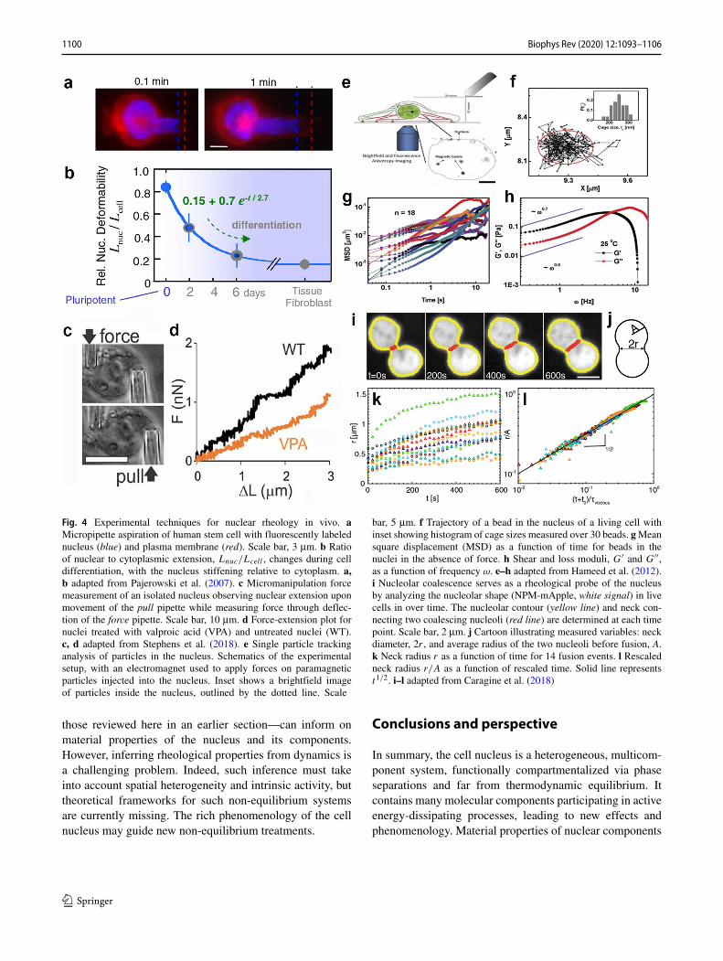
Fig. 4 Experimental techniques for nuclear rheology in vivo. aMicropipette aspiration of human stem cell with fluorescently labelednucleus (blue) and plasma membrane (red). Scale bar, 3 μm. b Ratioof nuclear to cytoplasmic extension, Lnuc/Lcell , changes during celldifferentiation, with the nucleus stiffening relative to cytoplasm. a,b adapted from Pajerowski et al. (2007). c Micromanipulation forcemeasurement of an isolated nucleus observing nuclear extension uponmovement of the pull pipette while measuring force through deflec-tion of the force pipette. Scale bar, 10 μm. d Force-extension plot fornuclei treated with valproic acid (VPA) and untreated nuclei (WT).c, d adapted from Stephens et al. (2018). e Single particle trackinganalysis of particles in the nucleus. Schematics of the experimentalsetup, with an electromagnet used to apply forces on paramagneticparticles injected into the nucleus. Inset shows a brightfield imageof particles inside the nucleus, outlined by the dotted line. Scale
bar, 5 μm. f Trajectory of a bead in the nucleus of a living cell withinset showing histogram of cage sizes measured over 30 beads. g Meansquare displacement (MSD) as a function of time for beads in thenuclei in the absence of force. h Shear and loss moduli, G′ and G′′,as a function of frequency ω. e–h adapted from Hameed et al. (2012).i Nucleolar coalescence serves as a rheological probe of the nucleusby analyzing the nucleolar shape (NPM-mApple, white signal) in livecells in over time. The nucleolar contour (yellow line) and neck con-necting two coalescing nucleoli (red line) are determined at each timepoint. Scale bar, 2 μm. j Cartoon illustrating measured variables: neckdiameter, 2r , and average radius of the two nucleoli before fusion, A.k Neck radius r as a function of time for 14 fusion events. l Rescaledneck radius r/A as a function of rescaled time. Solid line representst1/2. i–l adapted from Caragine et al. (2018)
those reviewed here in an earlier section—can inform onmaterial properties of the nucleus and its components.However, inferring rheological properties from dynamics isa challenging problem. Indeed, such inference must takeinto account spatial heterogeneity and intrinsic activity, buttheoretical frameworks for such non-equilibrium systemsare currently missing. The rich phenomenology of the cellnucleus may guide new non-equilibrium treatments.
Conclusions and perspective
In summary, the cell nucleus is a heterogeneous, multicom-ponent system, functionally compartmentalized via phaseseparations and far from thermodynamic equilibrium. Itcontains many molecular components participating in activeenergy-dissipating processes, leading to new effects andphenomenology. Material properties of nuclear components
1100 Biophys Rev (2020) 12:1093–1106

impact the timescales and length scales of nuclear process,most prominently those of the DNA-related biochemicaltransactions, which constitute an integral part of the centraldogma of molecular biology. Hence, emergent rheology ofthe cell nucleus effectively impacts all cellular processes.
Remarkably, the nucleus is compartmentalized into func-tional regions separating phases of different physical, mate-rial, and chemical properties. These phases serve as sitesof active processes carrying out specific biological func-tions, such as polymerases and transcription factors forminga functional liquid condensates or DNA repair machineryphase separating at DNA double-stranded breaks. Thus, wecan view the nucleus as being actively patterned via phaseseparations and activity-generating local reactors, which inturn contribute to the patterning itself. There is still a longway to go to decouple all of the effects and biological pro-cesses as they occur concurrently and superposed in the cellnucleus. Future in vivo studies are needed to investigatethe biophysical origins of the complex phenomenology ofphysiological behavior of the nucleus. In addition, in vitrostudies might recapitulate its key features and examine itsunderlying mechanisms. Moreover, simultaneous orthogo-nal biochemical and biophysical processes may need to beexplored within their common multicomponent phase dia-gram both in vivo and in vitro. Furthermore, the collectivephenomena that occur in this active system may stronglycontribute to such emergent behavior.
Similar to other living systems, the nucleus presentsan intricate interplay of heterogeneity and non-equili-brium activity posing new challenges for biologists andphysicists alike. It calls for new experimental and analyticalapproaches rooted in soft condensed matter physics,biophysics, and statistical mechanics while connecting tothe biochemistry and molecular biology of the nucleus.Moreover, phenomena found in this system may teach usnew non-equilibrium physics. Such knowledge is criticalalso from biomedical perspective identifying potentialphysical parameters as readouts for diagnostic tools andtherapy design for diseases rooted in malfunctions ofnuclear constituents.
Acknowledgments AZ would like to thank members of the Zidovskalab for fruitful discussions.
Funding This work was supported by the National Institutes of HealthGrant R00-GM104152, the National Science Foundation GrantsCAREER PHY-1554880, CMMI-1762506 and New York UniversityMRSEC DMR-1420073, and NYU Whitehead Fellowship for JuniorFaculty in Biomedical and Biological Sciences.
Compliance with ethical standards
Conflict of interest The author declares that she has no conflict ofinterest.
Open Access This article is licensed under a Creative CommonsAttribution 4.0 International License, which permits use, sharing,adaptation, distribution and reproduction in any medium or format, aslong as you give appropriate credit to the original author(s) and thesource, provide a link to the Creative Commons licence, and indicateif changes were made. The images or other third party material inthis article are included in the article’s Creative Commons licence,unless indicated otherwise in a credit line to the material. If materialis not included in the article’s Creative Commons licence and yourintended use is not permitted by statutory regulation or exceedsthe permitted use, you will need to obtain permission directly fromthe copyright holder. To view a copy of this licence, visit http://creativecommonshorg/licenses/by/4.0/.
TheMichele Auger Award for YoungScientists’ Independent Research
In late 2018, long time Editorial Board Member of Bio-physical Reviews journal, Professor Michele Auger, sadlysuccumbed to illness. As a mark of our respect for Michele,the Biophysical Reviews’ Editorial Board, together with thekind support of Springer-Nature Corporation, created a per-petual memorial award in honor of her life and service. The,‘Michele Auger Award for Young Scientists’ IndependentResearch’, is to be granted each year to a single candidateperforming biophysical research, who at the time of appli-cation is under 40 years of age. The award consists of aplaque and a free personal subscription to the journal alongwith an invitation to submit a single author review article toBiophysical Reviews.
Michele Auger (1963 - 2018)
Professor Michèle Auger – a much admired Editorial Board Member of Biophysical Reviews (2011 – 2018)
Born in GrandMere, Quebec and raised in TroisRivieres,Michele Auger enrolled first in biophysics at the Universitedu Quebec a Trois-Rivieres, only to later transfer tochemistry. After obtaining her B. Sc. in 1985, Michele
1101Biophys Rev (2020) 12:1093–1106

joined the group of Prof. Ian C.P. Smith at the Universityof Ottawa/National Research Council of Canada to pursueher Ph. D. studies in biophysics. After graduating fromthe University of Ottawa in 1990, Michele refined herskills in solid state NMR as a postdoctoral fellow in thegroup of Prof. Robert G. Griffin at the MassachusettsInstitute of Technology. Michele joined the Departmentof Chemistry at the University of Laval in 1991 asan Assistant Professor and recipient of an NSERCWomen’s Faculty Award. She was promoted to AssociateProfessor in 1996 and then Professor in 2000 whereshe remained until 2018. Michele’s research involvedusing solid state NMR to study (i) the interaction ofproteins, peptides and drugs with phospholipid membranes,and (ii) biopolymers such as spider silk. Michele servedinternationally on the Council of the International Unionof Pure and Applied Biophysics from 2011 - 2017 andwas an Editorial Board Member of Biophysical Reviewsjournal from 2011-2018. Brilliant, creative, dedicated to thescientific and academic communities, Michele displayedadmirable professional, ethical and leadership qualities. Sheis remembered as a dedicated, generous, and inspirationalscientist who touched the lives of many through herfriendship, teaching and kindness. (This short foreword isadapted from a longer memorial published on the IUPABNewsletter #70 http://iupab.org/wp-content/uploads/2019/02/IUPAB-news-70-2-1.pdf)
Alexandra Zidovska: Recipientof theMichele Auger Awardfor Young Scientists’ IndependentResearch 2020
Professor Alexandra Zidovska – the winner of the Michèle Auger Award for Young Scien�sts’ Independent Research for 2020.
Prof. Alexandra Zidovska is an experimental physiciststudying physical phenomena in biological systems andmaterials. Her career-long passion for biophysics has itsroots in her undergraduate research work under Prof.Erich Sackmann at the Technical University Munich inGermany, where she earned her Diplom or Master’sDegree in Physics in 2003. She used reflection interferencecontrast microscopy (RICM) and magnetic tweezers tostudy micromechanical properties of the cell membraneand filopodia of macrophages. Her undergraduate workrevealed that the cell membrane of macrophages exhibitsundulations that impede their adhesion, which may becritical for their ability to fight pathogens in human body.For her PhD studies, Alexandra moved on to the Universityof California, Santa Barbara where she worked in thelab of Prof. Cyrus Safinya in the Materials Departmentand graduated in 2008. She credits Prof. Safinya withinstilling in her a sense of taste in problems and how tobring the right tools to the study of these problems. Herdoctoral work focused on studies of the structure-functionrelationship of lipid-DNA self-assemblies used for genedelivery. She investigated liquid crystalline structures oflipid-DNA self-assemblies and phase behavior of exotichighly charged dendrimer lipids using small angle X-rayscattering (SAXS), confocal microscopy and cryogenictransmission electron microscopy (cryoTEM). Her graduatework produced an impressive record of papers in esteemedjournals, including the discovery of a new phase ofliposomes, the block liposomes.
Alexandra then conducted her post-doctoral research atHarvard University, where she worked jointly in the labsof Prof. Timothy Mitchison at Harvard Medical Schooland Prof. David Weitz in the Physics Department. Seekingyet greater challenges in her research, she made a bravechange of direction and began a quest to understandthe physics of the cell nucleus and the genome. Itwas during this time that she developed a new methodDisplacement Correlation Spectroscopy (DCS) for mappingand quantifying chromatin motions simultaneously acrossthe entire cell nucleus. DCS employs high-resolutionspinning disc confocal microscopy and measures chromatindisplacements by image correlation techniques. Becausethe method reveals the nucleus-wide motions maps acrossthe entire temporal spectrum, the method has proven tobe a major leap forward for the field. Indeed, Alexandra’swork revealed a new form of motion of the genome inwhich large regions of chromatin move together coherentlyover extended periods of time. Her work earned her theprestigious Damon Runyon Cancer Research Fellowshipamong other honors.
Since 2014, Alexandra’s work as Assistant Professor ofPhysics and research group leader at New York University’s
1102 Biophys Rev (2020) 12:1093–1106

Center for Soft Matter Research has continued to uncoverthe physics and inner workings of the cell nucleus.She leveraged early-career awards such as The Pathwayto Independence Award from the National Institutes ofHealth and the CAREER Award from the National ScienceFoundation, as well as a New York University WhiteheadFellowship for Junior Faculty in Biomedical and BiologicalSciences, to establish a world-class experimental lab thatin its first years can already proudly claim several majordiscoveries. One line of work follows up on her discoveryof coherent chromatin motion, showing that its originlies in active processes such as transcription occurring atthe single-gene level and whose influence is amplifiedgenome-wide due to hydrodynamic self-interactions of thechromatin fiber. She and her team have also shown thatthe nuclear envelope exhibits persistent flickering motionsthat had gone unnoticed previously and whose origin alsolies in active cellular process. Very recently, the Zidovskalab has shown how the intrinsic motions and coalescenceof subnuclear bodies such as nucleoli can be exploited toinfer material properties of the nuclear environment. Someof these exciting results are reviewed in her accompanyingarticle, which is a stirring and moving account of the cellnucleus that seems to be in perpetual motion.
In an example of life imitating science, the themesof motion, flow, activity, energy and purpose permeateAlexandra’s pursuits outside of the lab. She is a lifelongswimmer who uses her pool time to stoke her competitiveflame, balanced by a healthy dose of flow yoga. She alsogrounds herself through creative outlets such as painting andcooking. Alexandra is also passionately engaged in causesrelated to diversity and inclusion in physics and relatedsciences. Her lab has an impressive record of recruiting,training and promoting women scientists across all levels,and Alexandra is founder and faculty leader of a new group,NYU WiPhy, dedicated to providing a more welcoming andstimulating environment for women and those from otherunderrepresented groups in physics.
References
Aarts DG, Schmidt M, Lekkerkerker HN (2004) Direct visualobservation of thermal capillary waves. Science 304(5672):847–850
Abney JR, Cutler B, Fillbach ML, Axelrod D, Scalettar BA (1997)Chromatin dynamics in interphase nuclei and its implications fornuclear structure. J Cell Biol 137(7):1459–1468
Alberts B, Johnson A, Lewis J, Morgan D, Raff M, Roberts K, Walter P(2014) Molecular biology of the cell. Garland Science, New York
Albiez H, Cremer M, Tiberi C, Vecchio L, Schermelleh L, DittrichS, Kupper K, Joffe B, Thormeyer T, von Hase J, et al. (2006)Chromatin domains and the interchromatin compartment formstructurally defined and functionally interacting nuclear networks.Chrom Res 14(7):707–733
Amenta PS (1961) Fusion of nucleoli in cells cultured from the heartof Triturus viridescens. Anat Rec 139(2):155–165
Amitai A, Holcman D (2018) Encounter times of chromatin lociinfluenced by polymer decondensation. Phys Rev E 97(3):032417
Ashwin S, Nozaki T, Maeshima K, Sasai M (2019) Organization offast and slow chromatin revealed by single-nucleosome dynamics.Proc Natl Acad Sci USA, 116(40):19939–19944
Belmont AS, Straight AF (1998) In vivo visualization of chromosomesusing lac operator-repressor binding. Trends Cell Biol 8(3):121–4
Berry J, Weber SC, Vaidya N, Haataja M, Brangwynne CP (2015)RNA transcription modulates phase transition-driven nuclear bodyassembly. Proc Natl Acad Sc USA 112(38):E5237–E5245
Bickmore WA, van Steensel B (2013) Genome architecture: domainorganization of interphase chromosomes. Cell 152(6):1270–1284
Boettiger A, Murphy S (2020) Advances in chromatin imaging atkilobase-scale resolution. Trends Genet 36(4):273–287
Boisvert F-M, van Koningsbruggen S, Navascues J, Lamond AI (2007)The multifunctional nucleolus. Nat Rev Mol Cell Biol 8(7):574–585
Bonev B, Cavalli G (2016) Organization and function of the 3Dgenome. Nat Rev Genet 17(11):661–678
Boopathi R, Dimitrov S, Hamiche A, Petosa C, Bednar J (2020) Cryo-electron microscopy of the chromatin fiber. Curr Opin Struct Biol64:97–103
Bornfleth H, Edelmann P, Zink D, Cremer T, Cremer C (1999)Quantitative motion analysis of subchromosomal foci in livingcells using four-dimensional microscopy. Biophys J 77:2871–2886
Boulon S, Westman BJ, Hutten S, Boisvert F-M, Lamond AI (2010)The nucleolus under stress. Mol Cell 40(2):216–227
Brangwynne CP, Mitchison TJ, Hyman AA (2011) Active liquid-like behavior of nucleoli determines their size and shape inXenopus laevis oocytes. Proc Natl Acad Sci USA 108(11):4334–4339
Bronstein I, Israel Y, Kepten E, Mai S, Shav-Tal Y, Barkai E, Garini Y(2009) Transient anomalous diffusion of telomeres in the nucleusof mammalian cells. Phys Rev Lett 103:018102
Bruinsma R, Grosberg AY, Zidovska A (2014) Chromatin hydrody-namics. Biophys J 106(9):1871–1881
Caragine CM, Haley SC, Zidovska A (2018) Surface fluctuations andcoalescence of nucleolar droplets in the human cell nucleus. PhysRev Lett 121(14):148101
Caragine CM, Haley SC, Zidovska A (2019) Nucleolar dynamics andinteractions with nucleoplasm in living cells. eLife, 8:e47533
Carmo-Fonseca M, Platani M, Swedlow JR (2002) Macromolecularmobility inside the cell nucleus. Trends Cell Biol 12(11):491–495
Celedon A, Hale CM, Wirtz D (2011) Magnetic manipulation ofnanorods in the nucleus of living cells. Biophys J 101(8):1880–1886
Chen B, Gilbert LA, Cimini BA, Schnitzbauer J, Zhang W, Li G-W, Park J, Blackburn EH, Weissman JS, Qi LS, Huang B (2013)Dynamic imaging of genomic loci in living human cells by anoptimized CRISPR/Cas system. Cell 155(7):1479–1491
Cho W-K, Spille J-H, Hecht M, Lee C, Li C, Grube V, Cisse II(2018) Mediator and RNA polymerase II clusters associate intranscription-dependent condensates. Science 361(6400):412–415
Chu F-Y, Haley SC, Zidovska A (2017) On the origin of shapefluctuations of the cell nucleus. Proc Natl Acad Sci USA114(39):10338–10343
Chuang C-H, Carpenter AE, Fuchsova D, Hohnson T, de LanerolleP, Belmont AS (2006) Long-range directional movement of aninterphase chromosome site. Curr Biol 16(8):825–831
Cisse II, Izeddin I, Causse SZ, Boudarene L, Senecal A, Muresan L,Dugast-Darzacq C, Hajj B, Dahan M, Darzacq X (2013) Real-time
1103Biophys Rev (2020) 12:1093–1106

dynamics of RNA polymerase II clustering in live human cells.Science 341(6146):664–667
Cremer T, Cremer M (2010) Chromosome territories. Cold SpringHarbor Perspectives in Biology 2(3):a003889
Crick F (1970) Central dogma of molecular biology. Nature (London)227(5258):561
Crick FH (1958) On protein synthesis. Symp Soc Exp Biol 12(138):8Dahl KN, Engler AJ, Pajerowski JD, Discher DE (2005) Power-law
rheology of isolated nuclei with deformation mapping of nuclearsubstructures. Biophys J 89(4):2855–2864
Dahl KN, Kahn SM, Wilson KL, Discher DE (2004) The nuclearenvelope lamina network has elasticity and a compressibilitylimit suggestive of a molecular shock absorber. J Cell Sci117(20):4779–4786
Darzacq X, Shav-Tal Y, de Turris V, Brody Y, Shenoy SM, PhairRD, Singer RH (2007) In vivo dynamics of RNA polymerase IItranscription. Nat Struct Mol Biol 14(9):796–806
de Vries AH, Krenn BE, van Driel R, Subramaniam V, KangerJS (2007) Direct observation of nanomechanical properties ofchromatin in living cells. Nano Lett 7(5):1424–1427
Dekker J, Marti-Renom MA, Mirny LA (2013) Exploring the three-dimensional organization of genomes: interpreting chromatininteraction data. Nat Rev Genet 14(6):390–403
Di Pierro M, Potoyan DA, Wolynes PG, Onuchic JN (2018)Anomalous diffusion, spatial coherence, and viscoelasticity fromthe energy landscape of human chromosomes. Proc Natl Acad SciUSA 115(30):7753–7758
Dixon JR, Selvaraj S, Yue F, Kim A, Li Y, Shen Y, Hu M, Liu JS, RenB (2012) Topological domains in mammalian genomes identifiedby analysis of chromatin interactions. Nature 485(7398):376–380
Dross N, Spriet C, Zwerger M, Muller G, Waldeck W, LangowskiJ (2009) Mapping eGFP oligomer mobility in living cell nuclei.PLoS One 4(4):e5041
Eaton JA, Zidovska A (2020) Structural and dynamical signatures oflocal DNA damage in live cells. Biophys J 118(9):2168–2180
Edelmann P, Bornfleth H, Zink D, Cremer T, Cremer C (2001)Morphology and dynamics chromosome territories in living cells.Biochim Biophys Acta 1551(1):M29–39
Erdel F, Baum M, Rippe K (2015) The viscoelastic propertiesof chromatin and the nucleoplasm revealed by scale-dependentprotein mobility. J Phys Condens Matter 27(6):064115
Falahati H, Wieschaus E (2017) Independent active and thermody-namic processes govern the nucleolus assembly in vivo. Proc NatlAcad Sci USA, 114(6):1335–1340
Falk M, Feodorova Y, Naumova N, Imakaev M, Lajoie BR, LeonhardtH, Joffe B, Dekker J, Fudenberg G, Solovei I, Mirny L (2019)Heterochromatin drives compartmentalization of inverted andconventional nuclei. Nature 570:395–399
Feric M, Vaidya N, Harmon TS, Mitrea DM, Zhu L, RichardsonTM, Kriwacki RW, Pappu RV, Brangwynne CP (2016) Coex-isting liquid phases underlie nucleolar subcompartments. Cell165(7):1686–1697
Ganai N, Sengupta S, Menon GI (2014) Chromosome positioning fromactivity-based segregation. Nuc Ac Res 42(7):4145–4159
Germier T, Kocanova S, Walther N, Bancaud A, Shaban HA, SellouH, Politi AZ, Ellenberg J, Gallardo F, Bystricky K (2017) Real-time imaging of a single gene reveals transcription-initiated localconfinement. Biophys J 113(7):1383–1394
Gibcus JH, Dekker J (2013) The hierarchy of the 3D genome. Mol Cell49(5):773–782
Gibson BA, Doolittle LK, Schneider MW, Jensen LE, Gamarra N,Henry L, Gerlich DW, Redding S, Rosen MK (2019) Organizationof chromatin by intrinsic and regulated phase separation. Cell179(2):470–484
Gorisch SM, Wachsmuth M, Ittrich C, Bacher CP, Rippe K,Lichter P (2004) Nuclear body movement is determined bychromatin accessibility and dynamics. Proc Natl Acad Sci USA101(36):13221–13226
Grob A, Colleran C, McStay B (2014) Construction of syntheticnucleoli in human cells reveals how a major functional nucleardomain is formed and propagated through cell division. Genes Dev28(3):220–230
Guo YE, Manteiga JC, Henninger JE, Sabari BR, Dall’Agnese A,Hannett NM, Spille J-H, Afeyan LK, Zamudio AV, Shrinivas K,et al. (2019) Pol II phosphorylation regulates a switch betweentranscriptional and splicing condensates. Nature 572(7770):543–548
Hameed FM, Rao M, Shivashankar G (2012) Dynamics of passive andactive particles in the cell nucleus. PLoS One 7(10):e45843
Hinde E, Kong X, Yokomori K, Gratton E (2014) Chromatin dynamicsduring DNA repair revealed by pair correlation analysis ofmolecular flow in the nucleus. Biophys J 107(1):55–65
Hubner MR, Spector DL (2010) Chromatin dynamics. Annu RevBiophys 39:471–489
Kim J, KY Han NKTH, Belmont AS (2019) Nuclear speckle fusionvia long-range directional motion regulates speckle morphologyafter transcriptional inhibition. J Cell Sci 132(8):jcs226563
Jady BE, Richard P, Bertrand E, Kiss T (2006) Cell cycle-dependentrecruitment of telomerase RNA and Cajal bodies to humantelomeres. Mol Biol Cell 17(2):944–954
Khanna N, Zhang Y, Dudko JSLOK, Murre C (2019) Chromosomedynamics near the sol-gel phase transition dictate the timing ofremote genomic interactions. Nat Commun 10(1):2771
Kilic S, Lezaja A, Gatti M, Bianco E, Michelena J, Imhof R, AltmeyerM (2019) Phase separation of 53BP1 determines liquid-likebehavior of DNA repair compartments. EMBO J 38(16):e101379
Kimura H, Cook PR (2001) Kinetics of core histones in living humancells: little exchange of H3 and H4 and some rapid exchange ofH2B. J Cell Biol 153(7):1341–1354
Lampo TJ, Kennard AS, Spakowitz AJ (2016) Physical modelingof dynamic coupling between chromosomal loci. Biophys J110(2):338–347
Larson AG, Elnatan D, Keenen MM, Trnka MJ, Johnston JB,Burlingame AL, Agard DA, Redding S, Narlikar GJ (2017) Liquiddroplet formation by HP1α suggests a role for phase separation inheterochromatin. Nature 547(7662):236
Levi V, Ruan Q, Plutz M, Belmont AS, Gratton E (2005) Chromatindynamics in interphase cells revealed by tracking in a two-photonexcitation microscope. Biophys J 89(6):4275–4285
Liang L, Wang X, Da X, Chen T, Chen WR (2009) Noninvasivedetermination of cell nucleoplasmic viscosity by fluorescencecorrelation spectroscopy. J Biomed Opt 14(2):024013
Lieberman-Aiden E, Van Berkum NL, Williams L, Imakaev M,Ragoczy T, Telling A, Amit I, Lajoie BR, Sabo PJ, DorschnerMO, et al. (2009) Comprehensive mapping of long-rangeinteractions reveals folding principles of the human genome.Science 326(5950):289–293
Liu L, Shi G, Thirumalai D, Hyeon C (2018) Chain organizationof human interphase chromosome determines the spatiotemporaldynamics of chromatin loci. PLoS Comp Biol 14(12):e1006617
MacPherson Q, Beltran B, Spakowitz AJ (2018) Bottom–up modelingof chromatin segregation due to epigenetic modifications. ProcNatl Acad Sci USA 115(50):12739–12744
Makhija E, Jokhun D, Shivashankar G (2016) Nuclear deformabilityand telomere dynamics are regulated by cell geometric constraints.Proc Natl Acad Sci USA 113(1):E32–E40
Mao YS, Zhang B, Spector DL (2011) Biogenesis and function ofnuclear bodies. Trends Genet 27(8):295–306
1104 Biophys Rev (2020) 12:1093–1106

Marshall W, Straight A, Marko J, Swedlow J, Dernburg A, BelmontA, Murray A, Agard D, Sedat J (1997) Interphase chromosomesundergo constrained diffusional motion in living cells. Curr Biol7(12):930–939
Marzahn MR, Marada S, Lee J, Nourse A, Kenrick S, ZhaoH, Ben-Nissan G, Kolaitis R-M, Peters JL, Pounds S, etal. (2016) Higher-order oligomerization promotes localizationof SPOP to liquid nuclear speckles. EMBO J 35(12):1254–1275
McClintock B (1934) The relation of a particular chromosomalelement to the development of the nucleoli in Zea mays. CellTissue Res 21(2):294–326
Milo R, Phillips R (2015) Cell biology by the numbers. GarlandScience
Misteli T (2001) Protein dynamics: implications for nuclear architec-ture and gene expression. Science 291(5505):843–847
Misteli T, Gunjan A, Hock R, Bustin M, Brown DT (2000) Dynamicbinding of histone H1 to chromatin in living cells. Nature408(6814):877
Misteli T, Spector DL (2011) The nucleus. vol 3, no 2. Cold SpringHarbor Laboratory Press, New York
Montanaro L, Trere D, Derenzini M (2008) Nucleolus, ribosomes, andcancer. Am J Pathol 173(2):301–310
Mora-Bermudez F, Gerlich D, Ellenberg J (2007) Maximal chromo-some compaction occurs by axial shortening in anaphase anddepends on Aurora kinase. Nature Cell Biol 9(7):822
Nagashima R, Hibino K, Ashwin S, Babokhov M, Fujishiro S, ImaiR, Nozaki T, Tamura S, Tani T, Kimura H, et al. (2019) Singlenucleosome imaging reveals loose genome chromatin networksvia active RNA polymerase II. J Cell Biol 218(5):1511–1530
Nozaki T, Imai R, Tanbo M, Nagashima R, Tamura S, Tani T, JotiY, Tomita M, Hibino K, Kanemaki MT, et al. (2017) Dynamicorganization of chromatin domains revealed by super-resolutionlive-cell imaging. Mol Cell 67(2):282–293
Nuebler J, Fudenberg G, Imakaev M, Abdennur N, Mirny LA(2018) Chromatin organization by an interplay of loop extrusionand compartmental segregation. Proc Natl Acad Sci USA115(29):E6697–E6706
Ou HD, Phan S, Deerinck TJ, Thor A, Ellisman MH, O’SheaCC (2017) ChromEMT: visualizing 3D chromatin structureand compaction in interphase and mitotic cells. Science357(6349):eaag0025
Pajerowski JD, Dahl KN, Zhong FL, Sammak PJ, Discher DE (2007)Physical plasticity of the nucleus in stem cell differentiation. ProcNat Acad Sci USA 104(40):15619–15624
Paulsen JD, Carmigniani R, Kannan A, Burton JC, Nagel SR (2014)Coalescence of bubbles and drops in an outer fluid. Nat Commun5:3182
Pessina F, Giavazzi F, Yin Y, Gioia U, Vitelli V, Galbiati A, Barozzi S,Garre M, Oldani A, Flaus A, et al. (2019) Functional transcriptionpromoters at DNA double-strand breaks mediate RNA-drivenphase separation of damage-response factors. Nat Cell Biol,21(10):1286–1299
Phair RD, Misteli T (2000) High mobility of proteins in themammalian cell nucleus. Nature 404(6778):604
Platani M, Goldberg I, Lamond AI, Swedlow JR (2002) Cajal bodydynamics and association with chromatin are ATP-dependent. NatCell Biol 4(7):502
Racki LR, Narlikar GJ (2008) ATP-dependent chromatin remodelingenzymes: two heads are not better, just different. Curr Op Gen Dev18(2):137–144
Ritossa F, Spiegelman S (1965) Localization of DNA complementaryto ribosomal RNA in the nucleolus organizer region of Drosophilamelanogaster. Proc Natl Acad Sci USA 53(4):737–745
Saintillan D, Shelley MJ, Zidovska A (2018) Extensile motor activitydrives coherent motions in a model of interphase chromatin. ProcNatl Acad Sci USA 115(45):11442–11447
Sazer S, Schiessel H (2018) The biology and polymer physics under-lying large-scale chromosome organization. Traffic 19(2):87–104
Schmidt JC, Zaug AJ, Cech TR (2016) Live cell imaging revealsthe dynamics of telomerase recruitment to telomeres. Cell166(5):1188–1197
Shaban HA, Barth R, Bystricky K (2018) Formation of correlatedchromatin domains at nanoscale dynamic resolution duringtranscription. Nuc Ac Res 46(13):e77
Shi G, Liu L, Hyeon C, Thirumalai D (2018) Interphase humanchromosome exhibits out of equilibrium glassy dynamics. NatCommun 9(1):3161
Shin Y, Chang Y-C, Lee DS, Berry J, Sanders DW, Ronceray P,Wingreen NS, Haataja M, Brangwynne CP (2018) Liquid nuclearcondensates mechanically sense and restructure the genome. Cell175(6):1481–1491
Shukron O, Seeber A, Amitai A, Holcman D (2019) Advances usingsingle-particle trajectories to reconstruct chromatin organizationand dynamics. Trends Genet 35(9):685–705
Smrek J, Kremer K (2017) Small activity differences drive phaseseparation in active-passive polymer mixtures. Phys Rev Lett118(9):098002
Solovei I, Kreysing M, Lanctot C, Kosem S, Peichl L, Cremer T,Guck J, Joffe B (2009) Nuclear architecture of rod photoreceptorcells adapts to vision in mammalian evolution. Cell 137(2):356–368
Solovei I, Thanisch K, Feodorova Y (2016) How to rule the nucleus:divide et impera. Curr Opin Cell Biol 40:47–59
Stanek D, Fox AH (2017) Nuclear bodies: news insights into structureand function. Curr Opin Cell Biol 46:94–101
Stenhammar J, Wittkowski R, Marenduzzo D, Cates ME (2015)Activity-induced phase separation and self-assembly in mix-tures of active and passive particles. Phys Rev Lett 114(1):018301
Stephens AD, Banigan EJ, Adam SA, Goldman RD, Marko JF(2017) Chromatin and lamin a determine two different mechanicalresponse regimes of the cell nucleus. Mol Biol Cell 28(14):1984–1996
Stephens AD, Liu PZ, Banigan EJ, Almassalha LM, Backman V,Adam SA, Goldman RD, Marko JF (2018) Chromatin histonemodifications and rigidity affect nuclear morphology independentof lamins. Mol Biol Cell 29(2):220–233
Stixova L, Bartova E, Matula P, Danek O, Legartova S, KozubekS (2011) Heterogeneity in the kinetics of nuclear proteins andtrajectories of substructures associated with heterochromatin.Epigenetics Chromatin 4(1):5
Strom AR, Emelyanov AV, Mir M, Fyodorov DV, Darzacq X, KarpenGH (2017) Phase separation drives heterochromatin domainformation. Nature 547(7662):241
Sullivan GJ, Bridger JM, Cuthbert AP, Newbold RF, BickmoreWA, McStay B (2001) Human acrocentric chromosomes withtranscriptionally silent nucleolar organizer regions associate withnucleoli. EMBO J 20(11):2867–2877
Talwar S, Kumar A, Rao M, Menon GI, Shivashankar G (2013)Correlated spatio-temporal fluctuations in chromatin com-paction states characterize stem cells. Biophys J 104(3):553–564
Tseng Y, Lee JS, Kole TP, Jiang I, Wirtz D (2004) Micro-organizationand visco-elasticity of the interphase nucleus revealed by particlenanotracking. J Cell Sci 117(10):2159–2167
Van Holde KE (2012) Chromatin. Springer Science & Business Media,Berlin
1105Biophys Rev (2020) 12:1093–1106

Van Steensel B, Belmont AS (2017) Lamina-associated domains:links with chromosome architecture, heterochromatin, and generepression. Cell 169(5):780–791
Vivante A, Bronshtein I, Garini Y (2020) Chromatin viscoelasticitymeasured by local dynamic analysis. Biophys J 118(9):2258–2267
Wallace H, Birnstiel M (1966) Ribosomal cistrons and the nucleolarorganizer. Biochim Biophys Acta 114(2):296–310
Weber SC, Brangwynne CP (2015) Inverse size scaling of thenucleolus by a concentration-dependent phase transition. CurrBiol 25(5):641–646
Weber SC, Spakowitz AJ, Theriot JA (2012) Nonthermal ATP-dependent fluctuations contribute to the in vivo motion ofchromosomal loci. Proc Natl Acad Sci USA 109(19):7338–7343
Wiesmeijer K, Krouwels IM, Tanke HJ, Dirks RW (2008) Chromatinmovement visualized with photoactivable GFP-labeled histoneH4. Differentiation 76(1):83–90
Xiang W, Roberti MJ, Heriche J-K, Huet S, Alexander S, EllenbergJ (2018) Correlative live and super-resolution imaging reveals
the dynamic structure of replication domains. J Cell Biol217(6):1973–1984
Xu J, Ma H, Jin J, Uttam S, Fu R, Huang Y, Liu Y (2018)Super-resolution imaging of higher-order chromatin structures atdifferent epigenomic states in single mammalian cells. Cell Rep24(4):873–882
Zidovska A (2020) The self-stirred genome: large-scale chromatindynamics, its biophysical origins and implications. Curr Opin GenDev 61:83–90
Zidovska A, Weitz DA, Mitchison TJ (2013) Micron-scale coherencein interphase chromatin dynamics. Proc Natl Acad Sci USA110(39):15555–15560
Zink D, Cremer T, Saffrich R, Fischer R, Trendelenburg MF, AnsorgeW, Stelzer EH (1998) Structure and dynamics of human interphasechromosome territories in vivo. Hum Genet 102(2):241–251
Publisher’s note Springer Nature remains neutral with regard tojurisdictional claims in published maps and institutional affiliations.
1106 Biophys Rev (2020) 12:1093–1106