the respiratory system is a group of organs that perform some very important tasks in our body
TRANSCRIPT
Introducing The
Respiratory System
The respiratory system is a group of organs that perform some very important tasks in our body.

In the mitrochondria of every cell in our body, a process called cellular respiration is carried out. In cellular respiration, (click) glucose, which is carried by the bloodstream (click) reacts with oxygen, also carried by the bloodstream (click) to produce carbon dioxide, which goes into the bloodstream, (click) water, (click) and energy.
Cellular Respiration
Glucose + oxygen carbon dioxide + water + energy
mitochondrion

The three main functions of our respiratory system are to (click) get oxygen to the bloodstream, which carries it to all body cells, (click) remove carbon dioxide from the bloodstream and our body, (click) and remove some of the excess water from the bloodstream and our body.
gets oxygen to
the bloodstrea
m
The Respiratory System
removes carbon dioxide
from the bloodstrea
m
removes some water
from the bloodstrea
m
Glucose + oxygen carbon dioxide + water + energy

Now we’ll show you the main parts of the respiratory system and briefly explain what each of them does.
The Respiratory
System

The air you breathe in with your nose goes into the nasal cavity. Hairs and mucous in the nose trap some particles, and the air is also warmed and moistened.
nasal cavity

Of course, you can also breathe in air through the mouth or oval cavity.
nasal cavity
oral cavity

Air from both the nasal and oral cavities goes into the pharynx, or throat.
pharynx
nasal cavity
oral cavity

The opening at the top of the windpipe, or trachea is called the glottis
pharynx
nasal cavity
glottis
oral cavity

A flap of tissue called the epiglottis covers the glottis when food is being swallowed. This prevents food from entering the respiratory tract.
epiglottis
pharynx
nasal cavity
glottis
oral cavity

The top part of the trachea is called the larynx. The larynx contains the vocal chords.
epiglottis
pharynx
nasal cavity
glottis
larynx
oral cavity

The trachea is the main tube by which air moves from the pharynx to the lungs. The white rings which surround the trachea are made of cartilidge, a tough type of tissue that is a bit softer than bone, but harder than muscle. These rings prevent the trachea from collapsing.
epiglottis
pharynx
trachea
glottis
larynx
nasal cavity
oral cavity

The trachea branches into two tubes called bronchi. One bronchus goes to each lung.
epiglottis
pharynx
trachea
glottis
larynx
bronchi
nasal cavity
oral cavity

The bronchi branch into many tubes called bronchioles. As they get further from the bronchi, bronchioles get smaller and smaller. Bronchioles carry air to every part of each lung.
epiglottis
pharynx
trachea
glottis
larynx
bronchi
bronchioles
nasal cavity
oral cavity

The rib cage surrounds the lungs. These ribs protect the lungs and also play a role in breathing, as we’ll see later.
epiglottis
pharynx
trachea
glottis
larynx
rib cage
bronchi
bronchioles
nasal cavity
oral cavity

The lungs are the organs in which gas exchange takes place. We have a left lung and a right lung.
epiglottis
pharynx
trachea
lung
glottis
larynx
rib cage
bronchi
bronchioles
nasal cavity
oral cavity
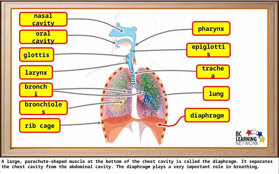
A large, parachute-shaped muscle at the bottom of the chest cavity is called the diaphragm. It separates the chest cavity from the abdominal cavity. The diaphragm plays a very important role in breathing.
epiglottis
pharynx
trachea
lung
diaphragm
glottis
larynx
rib cage
bronchi
bronchioles
nasal cavity
oral cavity

Now we’ll take a closer look at ends of the bronchioles. If we take this little blue square…

And enlarge it, we’ll represent it by this diagram.

These are clusters of tiny air sacs, or aveolar sacs.
alveolar sacs
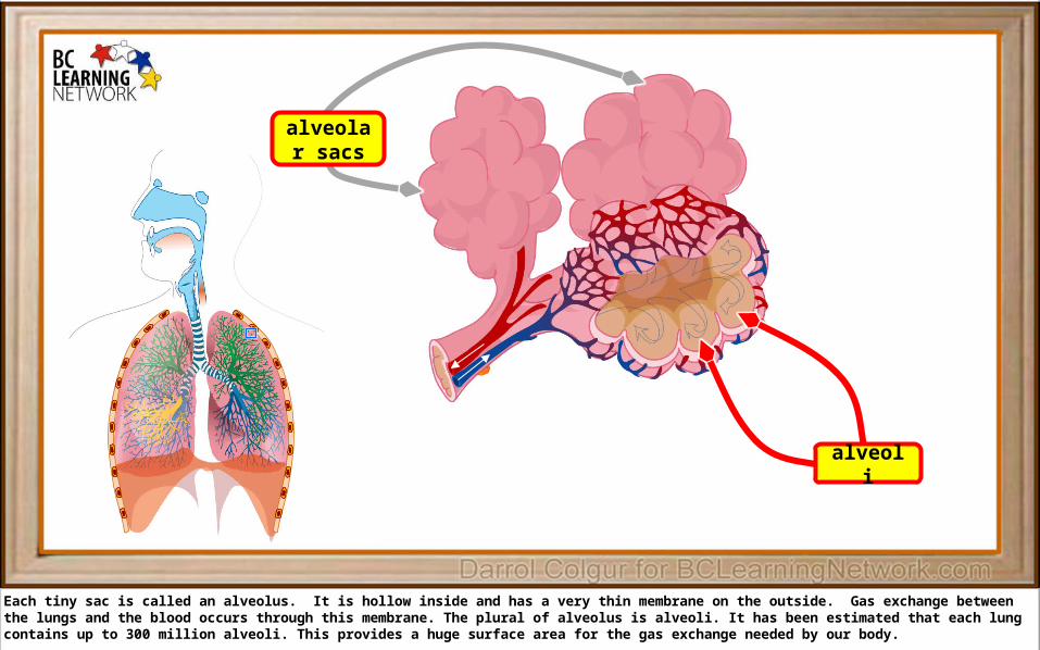
Each tiny sac is called an alveolus. It is hollow inside and has a very thin membrane on the outside. Gas exchange between the lungs and the blood occurs through this membrane. The plural of alveolus is alveoli. It has been estimated that each lung contains up to 300 million alveoli. This provides a huge surface area for the gas exchange needed by our body.
alveoli
alveolar sacs

Air enters and leaves the alveoli through the bronchioles.
air
air
bronchiole alveoli
alveolar sacs

A very dense bed of tiny capillaries surrounds each alveolus. This brings blood into very close contact with the outer membranes of alveoli, through which oxygen and carbon dioxide diffuse.
air
air
bronchiole alveoli
capillaries
alveolar sacs

The blood vessels coloured blue are carrying deoxygenated blood from the heart. As this blood goes through the capillary beds, it gradually absorbs oxygen from the alveoli and becomes red.
air
air
bronchioledeoxygenated blood from the
heart alveoli
capillaries
alveolar sacs

These vessels carry red, or oxygenated blood back to the heart.
air
airoxygenated blood to the
heart
bronchioledeoxygenated blood from the
heart alveoli
capillaries
alveolar sacs

Acknowledgements for Images Used
"Respiratory system complete en" by LadyofHats - The image i did myself as sources i used the books: Sobotta "atlas der anatomie des menschen" ISBN. 3 541 02828 9 , Churchill livingstone "gray's anatomy" ISBN. 0 433 01505 8, Interamericana. McGraw-hill "atlas forografico de anatomia del cuerpo humano" ISBN. 968 25 1677 3. Also used several online diagrams like ([1] and [2]) Image renamed from Image:Respiratory system complete.svg. Licensed under Public Domain via Wikimedia Commons - http://commons.wikimedia.org/wiki/File:Respiratory_system_complete_en.svg#/media/File:Respiratory_system_complete_en.svg