the relationship between time of day, mood, and
TRANSCRIPT
Bowling Green State University Bowling Green State University
ScholarWorks@BGSU ScholarWorks@BGSU
Honors Projects Honors College
Spring 5-3-2016
The Relationship Between Time of Day, Mood, and The Relationship Between Time of Day, Mood, and
Electroencephalography (EEG) Asymmetry Electroencephalography (EEG) Asymmetry
Morgan Tantillo [email protected]
Follow this and additional works at: https://scholarworks.bgsu.edu/honorsprojects
Part of the Other Neuroscience and Neurobiology Commons, Other Psychiatry and Psychology
Commons, and the Other Statistics and Probability Commons
Repository Citation Repository Citation Tantillo, Morgan, "The Relationship Between Time of Day, Mood, and Electroencephalography (EEG) Asymmetry" (2016). Honors Projects. 294. https://scholarworks.bgsu.edu/honorsprojects/294
This work is brought to you for free and open access by the Honors College at ScholarWorks@BGSU. It has been accepted for inclusion in Honors Projects by an authorized administrator of ScholarWorks@BGSU.

1
The Relationship Between Time of Day, Mood, and Electroencephalography (EEG) Asymmetry
MORGAN TANTILLO
HONORS PROJECT
Submitted to the Honors College
Bowling Green State University in partial
Fulfillment of the requirements for graduating with
UNIVERSITY HONORS
May 2nd, 2016
Sherona Garrett-Ruffin, Advisor
Department of Psychology
Hanfeng Chen, Advisor
Department of Mathematics and Statistics

2
Abstract
Previous researchers have had success in finding a correlation between exercise and
an increase in positive mood. Researchers have also found a correlation between
time of day and mood. The current study will explore the relationship between time
of day, mood, and electroencephalography (EEG) asymmetry. The study utilized a
convenient sample of ten undergraduate students at Bowling Green State University.
Participants had baseline EEG recordings taken, and then participated in moderate
exercise, followed by another EEG recording. Participants’ mood was assessed
through a self-reported mood questionnaire before the condition as well as
immediately after. Due to multiple statistical tests, the alpha level for rejection of the
null hypothesis was set at .016. While no statistically significant differences were
found, the difference between baseline EEG asymmetry and post-task EEG
asymmetry approached significance. Specifically, there was greater left hemispheric
activity post-task which is indicative of a more positive mood.

3
INTRODUCTION:
EEG asymmetry and Mood
The purpose of this review is to summarize the research on
electroencephalogram (EEG) asymmetry and mood. Research presented in this
review will help the reader understand the current knowledge on the subject.
An electroencephalogram (EEG) was used to measure electrical activity in
the brain by attaching electrodes to the scalp to detect electrical activity. In regards
to EEG asymmetry and mood, researchers have historically studied the alpha band
(8-13 Hz). In the first recordings taken from the scalp by Berger in the 1920’s, a
wave around 10 cycles per second was observed and later named the alpha rhythm.
This rhythm is used as an indirect measure of mood (Goldman et al. (2012)). Alpha
activity (8-13 Hz) is linked to a wakeful, relaxed and alert state (Goldman et al.
2012). The highest alpha rhythm amplitude is seen when the eyes are closed and the
participant is relaxed. Because of this, the loss of alpha rhythm is used to determine
the first stages of sleep (Goldman et al. 2012). Asymmetry refers to the difference in
electrical activity between the left and right hemispheres. Typically, to assess mood
researchers measure the difference in alpha amplitude between the right and left
hemispheres (Hall (2007); Schneider (2009); Aprea and Tantillo (2015)). A higher
amount and amplitude of alpha activity is indicative of less brain activity, such that a
high alpha reading in the right hemisphere means there is less activity in that area.
In a study using PET scans, researchers found that the thalamus, visual cortex, and
lateral geniculate are involved in generation of the alpha rhythm (Sadato et al.
1998).
To determine the meaning of alpha activity, research regarding alpha activity
and mood was needed. Raymond et al. (2005) completed a neurofeedback study to
investigate the role of the alpha rhythm on mood states. The participant received
positive reinforcement when alpha wave amplitudes decreased in the left

4
hemisphere. It was shown that those who received the positive feedback reported a
significantly higher amount of energy than those who received mock feedback.
Additionally, those who received real feedback showed an increase in positive
mood, as indicated by the Profile of Mood States (POMS) questionnaire, and stated
that they felt more composed, agreeable, elevated and confident after the trial. From
this information, researchers inferred that less alpha rhythm activity in the left
hemisphere correlates with a more positive mood state. Indeed, a robust finding in
the literature is that positive mood is associated with greater left hemispheric
activity (Raymond et al., 2005
Exercise and Mood
Researchers have found positive correlations between exercise and an
individual’s mood. Hall (2007) and Schneider (2009) completed experiments where
the mood was predicted after various exercises based on hemispheric dominance.
The researchers measured EEG asymmetry patterns prior to any exercise, and using
those patterns predicted how participants would feel after moderate and vigorous
exercise. The researchers used an electrocap to record from nine scalp locations.
These locations were left, right, mid-frontal, central, and parietal. In order to reduce
artifacts, Hall measured EOG (electro-oculogram) activity on both eyes. Both Hall
(2007) and Schneider (2009) found a correlation between those who were left-
brain dominant and a more positive mood following the exercise. The next step was
to investigate specific regions of the brain and how activity within them differs with
exercise.
EEG Asymmetry and Exercise
Schneider et al. (2009) conducted an experiment to test the correlation
between frontal cortical asymmetry and response to exercise. The researchers
found that, when moderate exercise was performed, a more positive affect was seen
through more activity in the left hemisphere. Asymmetry was measured using EEG
alpha activity, placing the electrodes at F3 and F4, P3 and P4 using the International
10-20 system. The researcher used a younger population (ages 14-16) because
previous studies linking EEG asymmetry and mood were limited to adults. Their
findings could then be generalized to adolescents, thereby extending the

5
relationship between EEG asymmetry and mood to a different population. The
researchers, however, failed to test EEG asymmetry before each trial. Trials were
about one week apart, and a lot may change for a high school student within a week.
It seems that when they were tested and determined to have, for example, left
hemisphere dominance at the first testing, this could have changed by the next
week. The internal validity of the experiment may have been compromised, because
the researchers did not eliminate the possibility that an outside variable was
affecting the results. To help eliminate this, the trials could have been closer
together, such as a day or two apart to help eliminate the possibility of an outside
event-affecting mood.
In the studies discussed earlier in this review the researchers focused on
mood changes in response to exercise among healthy individuals, which brings up
the issue of whether exercise can be a treatment for individuals with certain mood
disorders, such as depression and anxiety.
Another robust finding in the literature is that exercise reduces depression
(Bercer, 1983, Brown et. al, 1978, Folkins, 1981, Klein et al., 1985, and Jasnoski et.
al, 1988). These studies, however, are dated. More recent studies regarding the
impact of exercise on mood related illnesses are needed. Furthermore, the
researchers did not comment on the time of day that each trial took place. The time
of day that the exercise was completed may mediate or moderate the relationship
between EEG asymmetry and exercise.
Exercise, Mood and Time of Day
Maraki et al. (2005) investigated the effect of an exercise class on mood,
which included investigating differences due to time of day. Subjects for the study
included healthy females, aged 18-45 who did not take part in regular exercise. The
design was a 2 (exercise) x2 (time of day) repeated measures, where each
participants took part in each of the four trials. Trials included a morning control,
morning exercise, evening control, and evening exercise. Control trials consisted of
one hour of rest. Exercise trials consisted of a one-hour aerobic and muscle
conditioning class. Mood was measured throughout the experiment using a Positive
and Negative Affect Schedule (PANAS). Researchers found that an exercise class

6
caused an increase in positive affect and decrease in negative affect. There were no
significant differences found in mood change from morning trials to evening trials.
There is some controversy in the literature regarding whether the effects of exercise
on mood depend on the time of day. Previous researchers (McMurray, Hill & Field
(1990)) reported significant differences. However, Maraki et al. (2005) believe that
the lack of interaction between exercise and time of day is due to differences in the
mood scales used. More research is needed. Additionally, more experiments are
needed to test the effect of time of day on mood, independent of exercise.
Researchers (Egloff et al. 1995) explored how the time of day, as well as day
of the week altered one’s positive mood. To examine this relationship, the
researchers measured the participants’ mood, using three different scales, three
times a day over the course of 7 days. The first scale contained the words active,
attentive, inspired, and interested. The second scale contained the words balanced,
content, at ease and happy. The final scale contained the words afraid, angry,
disgusted, hostile, irritable, jittery, nervous, and shaky. The first scale measured
positive affect activation, while the second scale measured positive affect
pleasantness. The final scale measured negative affect. The participants rated each
word, on a scale of 1 to7, based on how they were feeling at the moment. A one
denoted “not at all” and seven denoted “extremely”. Researchers found that positive
affect pleasantness was at its highest rating in the evening, and lowest in the
morning. Egloff et al. (1995) also found that positive affect activation was highest in
the afternoon. The results of the study indicate that more experiments are needed
regarding the change in positive affect with respect to time of day, possibly using
different scales.
Last semester, I completed a study on the effect of exercise on EEG
asymmetry and mood, using various exercise levels and the Positive and Negative
Affect Schedule (PANAS) (Aprea & Tantillo, 2015). The levels of exercise included no
exercise, and vigorous exercise. After each condition, participants’ EEG asymmetry
and self-reported affect were measured. We found a statistically significant increase
in positive affect following vigorous exercise. Participants also showed more alpha
activity in the right hemisphere following more vigorous exercise, which is
7
indicative of greater left hemispheric activity. This finding was consistent with
previous research linking greater left hemispheric activity with exercise.
Surprisingly, there was no difference in self-reported mood between the groups.
Drawing from my previous work and noting the gap in the literature, I sought to
investigate whether time of day is related to EEG asymmetry. Given the robust
finding that exercise elevates mood and increases left hemispheric activity, I made
exercise a constant and had all participants engage in moderate exercise. Mood will
be operationalized by self-report and EEG asymmetry. The proposed study is a
modified replication of my previous work (Aprea and Tantillo, 2015).
Given the controversy in the literature regarding time of day and mood, I did
not make a directional hypothesis regarding time of day, but instead sought to
determine if there were differences in EEG asymmetry and self-reported mood
between morning and evening exercise conditions.
METHODS
Participants
The sample was a convenient sample, recruited through word of mouth of the
researcher and advisor. Research took place on the Bowling Green State University
campus in Bowling Green, Ohio. Informed consent was obtained from each
participant prior to starting the experiment. The participants consisted of ten
Bowling Green State University undergraduate students. The ages ranged from 18-
23, with a mean age of 20.2. All participants identified as Caucasian, with seven
female and three male students tested. All ten participants were right-hand
dominant. The human subject review board application for the study was approved.
Participants were given an incentive for contributing their time. They were able to
choose between two SONA credits and a fifteen-dollar gift card.
Materials
Positive and Negative Affect Schedule
The Positive And Negative Affect Schedule (PANAS) questionnaire developed
by Watson, D., Clark, L. A., & Tellegan (1998), was used to gauge the mood of each
participant at baseline and following the exercise condition. The PANAS

8
questionnaire consists of two columns of words such as “interested”, “upset”,
“enthusiastic”, and “nervous”. Participants put a number 1 through 5, where 1
denotes “Very slightly or not at all” and 5 denotes “Extremely”. The PANAS yields a
positive affect score and a negative affect score. Scoring was done by taking the
mean of all words reflecting positive affect, and the mean of all words reflecting
negative affect for comparison. Watson (1998) provides a clear definition of positive
affect versus negative affect, and how they differ from each other. A low positive
affect does not reflect the same feelings as a negative affect. The researchers
developed the scale by taking a large number of descriptive words, then testing the
relevance of each word using analyses by Zevon and Tellegen (1982). John R.
Crawford and Julie D. Henry (2004) tested the reliability of the PANAS questionnaire
and found that, as measured by Cronbach’s alpha, the positive affect is .89 and the
negative affect is .85. This shows that the questionnaire is reliable. Waston and Clark
(1988) found that the correlation between the positive affect and negative affect is -
.12-.23, showing that the scales are independent. Convergent correlations were
found to range from .89 to .95, and discriminant correlations ranged from -.02 to -
.18 (Watson and Clark 1988).
Health screening questionnaire
A health-screening questionnaire made by the researcher was also used. This
included questions such as gender, age, and dominant hand and general questions
regarding their health. Questionnaires were not linked to a specific participant, but
rather a number to ensure confidentiality.
Electroencephalogram (EEG)
To measure EEG asymmetry, an electroencephalogram (EEG) machine was
used. BSL Pro 3.7.2 software with a Biopac acquisition unit MP 150 was used. A
Gateway computer was used to run the program and take recordings. The sample
rate for the acquisition unit is 1,000 Hz. The gel used on the electrodes was a Ten20
conductive gel. Nuprep gel was used to abrade the skin to make the best connection
and obtain the best signal from person to electrode to computer. Light abrasion of
the skin where the electrodes were to be placed was done to reduce impedance.
Electrodes were placed using the International 10-20 scale. For each lead, a bipolar
9
montage will be used, with the active electrodes at F3 and F4, reference at Fz and
two ground electrodes on the ears.
The alpha wave (8-13 Hz) was measured in the left and right hemispheres in
each trial. The amplitude of alpha activity in the right hemisphere is compared to
the amplitude of alpha activity measured in the left hemisphere after the
manipulations. Data was log transformed to reduce skewness. The equation used to
determine an asymmetry score is ln (right)- ln (left). EEG readings usually do not
show a normal distribution, so the data is log transformed to meet assumptions for
parametric statistical tests.
An impedance check for each lead was made prior to recording. Impedance
levels were below 10 Kiloohms, with only a 1 to 2-kiloohm difference between the
electrode leads. The experimenter visually inspected the data for artifacts using
manual sliding window of 1 sec to remove amplitudes above 50 microvolts. Less
than 1% of the data was removed due to artifacts.
Procedure
Prior to beginning the experiment, each participant was given a consent form
to sign and return to the researcher. Those who agreed to and signed the consent
form were then set up with the electro-cap and ground electrodes, and gel was
applied in each. As the gel was let to sit, each participant was given a health-
screening questionnaire to fill out. There was no time limit for filling out these
papers, but participants were asked to stay in the room with the researchers for the
duration. Participants were reminded that they could discontinue with the study at
any time without penalty. A mixed between within design was used. The time of day
conditions involved a between groups design. A within-participant design was used
for the EEG recording and mood assessment, where each participant had his or her
baseline measured, and then measured again following moderate exercise.
Participants came to the testing site on one occasion. After completing the consent
forms and health questionnaires, participants’ baseline EEG recording taken for two
minutes. During the recording, participants were asked to focus on a piece of tape
on the wall, and keep relaxed with their eyes open. Participants were told to blink if
needed. Following the baseline recording, participants were asked to walk for 20

10
minutes along a predetermined path around the Psychology Building. After
returning to the testing site 20 minutes later, participants has their EEG recorded
again for two minutes, with the same instructions. Immediately following the
recording, participants were again asked to fill out a PANAS questionnaire. They
were also asked to fill out a manipulation check questionnaire made by the
researcher.
Morning recordings were taken between 8 A.M. and 10 A.M. Night recordings
were taken between 6 P.M. and 8 P.M.
Participants were reminded that they could leave the experiment at any
point without penalty. Each participant was given a debriefing before leaving the
testing area.
Results
EEG Asymmetry
Typically EEG data is very skewed. In order to reduce skewness and meet the
assumptions of parametric tests, the EEG data was log transformed using the
formula ln(right alpha)-ln(left alpha).
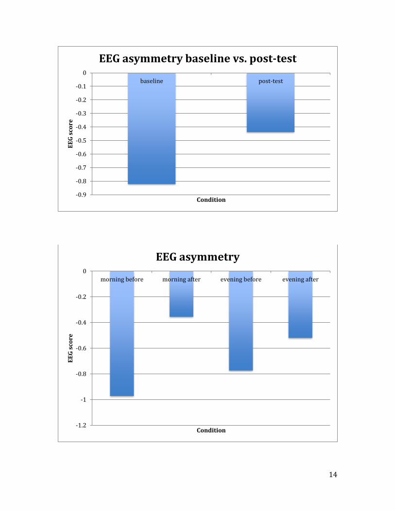
A mixed between-within subjects analysis of variance was conducted to
assess the impact of time of day (morning vs. evening) on participants’ EEG
asymmetry, across two time periods ( baseline and post-task).
There was no significant interaction between time periods and time of day,
Wilks Lambda=.992, F (1,8)=.064, p=.806.
The difference between baseline (M=-1.0005, SD=.9458) and post-task (M=-
.5735 SD=.6657), approached significance, Wilks Lambda=.673, F(1,8)=3.878,
p=.084, at baseline there was more alpha activity in the left hemisphere , which is
indicative of greater right hemispheric activity (i.e. more negative affect)
The difference between morning (M=-.815, SD=.355) and evening (M=-.764,
SD=.355) was not significant, F(1, 8)=.011, p=.921, suggesting no differences
between the morning and evening EEG asymmetry scores.
Positive Affect
11
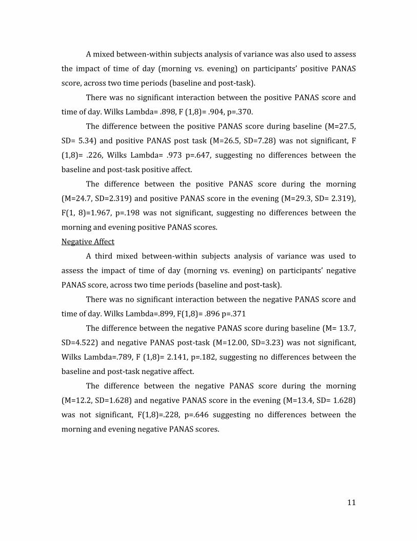
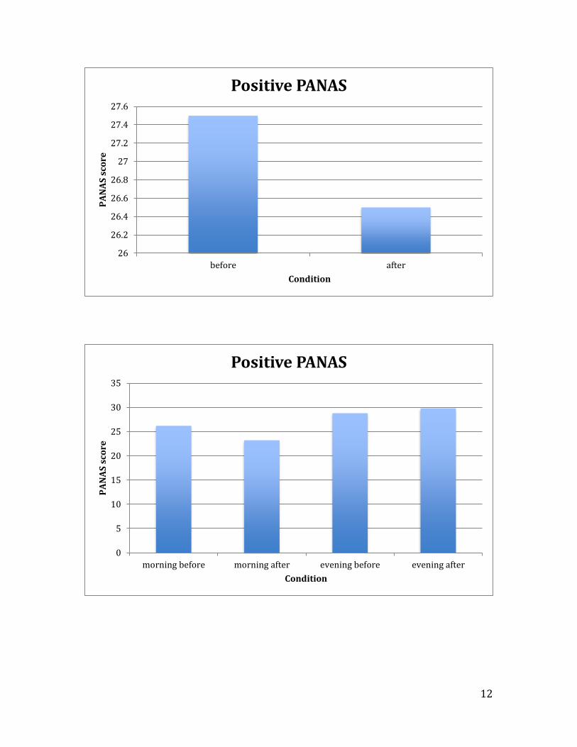
A mixed between-within subjects analysis of variance was also used to assess
the impact of time of day (morning vs. evening) on participants’ positive PANAS
score, across two time periods (baseline and post-task).
There was no significant interaction between the positive PANAS score and
time of day. Wilks Lambda= .898, F (1,8)= .904, p=.370.
The difference between the positive PANAS score during baseline (M=27.5,
SD= 5.34) and positive PANAS post task (M=26.5, SD=7.28) was not significant, F
(1,8)= .226, Wilks Lambda= .973 p=.647, suggesting no differences between the
baseline and post-task positive affect.
The difference between the positive PANAS score during the morning
(M=24.7, SD=2.319) and positive PANAS score in the evening (M=29.3, SD= 2.319),
F(1, 8)=1.967, p=.198 was not significant, suggesting no differences between the
morning and evening positive PANAS scores.
Negative Affect
A third mixed between-within subjects analysis of variance was used to
assess the impact of time of day (morning vs. evening) on participants’ negative
PANAS score, across two time periods (baseline and post-task).
There was no significant interaction between the negative PANAS score and
time of day. Wilks Lambda=.899, F(1,8)= .896 p=.371
The difference between the negative PANAS score during baseline (M= 13.7,
SD=4.522) and negative PANAS post-task (M=12.00, SD=3.23) was not significant,
Wilks Lambda=.789, F (1,8)= 2.141, p=.182, suggesting no differences between the
baseline and post-task negative affect.
The difference between the negative PANAS score during the morning
(M=12.2, SD=1.628) and negative PANAS score in the evening (M=13.4, SD= 1.628)
was not significant, F(1,8)=.228, p=.646 suggesting no differences between the
morning and evening negative PANAS scores.
12
26
26.2
26.4
26.6
26.8
27
27.2
27.4
27.6
before after
PA
NA
S s
core
Condition
Positive PANAS
0
5
10
15
20
25
30
35
morning before morning after evening before evening after
PA
NA
S s
core
Condition
Positive PANAS

13
11
11.5
12
12.5
13
13.5
14
before after
PA
NA
S s
core
Condition
Negative PANAS
0
2
4
6
8
10
12
14
16
morning before morning after evening before evening after
PA
NA
S s
core
Condition
Negative PANAS
14
-0.9
-0.8
-0.7
-0.6
-0.5
-0.4
-0.3
-0.2
-0.1
0
baseline post-test
EE
G s
core
Condition
EEG asymmetry baseline vs. post-test
-1.2
-1
-0.8
-0.6
-0.4
-0.2
0
morning before morning after evening before evening after
EE
G s
core
Condition
EEG asymmetry

15
Discussion
The purpose of the research was to explore the effect of time of day on EEG
asymmetry, positive affect and negative affect. There were no significant differences
between time of day and the dependent measures. Interestingly, the difference
between baseline and post-task EEG asymmetry approached significance. At
baseline, there was more alpha activity in the left hemisphere, which is indicative of
more right hemispheric activity, or more negative affect. This finding is consistent
with the literature. However, there was no difference in self-reported affect between
baseline and post-task.
As with most research, there are strengths and weaknesses to this study. I
will frame the discussion by talking about the four major validities: construct,
internal, external and statistical.
Construct:
EEG recordings were thoroughly analyzed and checked for artifacts. A
manual sliding window of one second was utilized to capture signals above 50µV to
remove artifacts. Less than 1% of the data was removed due to artifacts. The
artifacting was done by the researcher to control for eye blinks, as well as any other
outside signals that could’ve been picked up by the unit. Normally, measuring the
eye blinks using an EOG (electrooculography) would be used to control for this.
Because of the risk of damaging equipment, the decision was made to manually
remove eye blink and other artifacts. Given that the amplitude readings were within
the expected ranges and limited data was removed due to artifacts, I am confident of
validity of the EEG measurements.
The study had good construct validity for the PANAS, as it is a valid and
reliable measure of affect. The study also had good construct validity in how the
independent variable, time of day, was manipulated. Standard protocol was used. All
morning trials took place between the hours of 8 A.M. and 10 A.M., and the evening
trials took place between 6 P.M. and 8 P.M. A limitation and threat to the construct
validity was the manipulation of exercise. The exercise task, which was a constant in
the study, was meant to be moderately intense, yet the manipulation check showed
that all participants found the task to be extremely low level instead.

16
Internal:
I sought to increase internal validity through random assignment, where five
participants were part of the morning trials, and the other five were part of the
evening trials. This was to help ensure groups would be as similar as possible.
However, a major threat to the internal validity is that the experiment was a
between subjects design, rather than a within subjects design. This leaves room for
more significant differences across participants, which could alter the results found.
One important variable that was controlled for within the study was handedness,
where all participants were right-hand dominant. Many other variables were not
controlled for, such as nicotine intake, caffeine intake, and history of psychological
disorders.
External:
The study achieved external validity through a sample that accurately
represents the population of interest. The participants were all between the ages of
18 and 23, which is representative of the majority of Bowling Green State University
undergraduate students. Nevertheless, the results cannot be generalized to other
populations outside of BGSU undergraduate students. Another threat to the
external validity of the experiment is the artificial environment in which testing
took place. In a more natural environment, such as having the participants exercise
outdoors, may result in significant differences between morning and evening
recordings.
Statistical:
As stated previously, there were no statistically significant differences. The
reason for a null results could be due to the small sample size, the variables that
were not controlled for, or too small of a range between morning and evening trials.
Further studies could utilize a larger sample in an effort to obtain significant
results. The study could also alter the times of morning and night recordings, so that
the morning is earlier and evening is much later. Altering the exercise to be sure
that participants felt it was moderately intense could change results, as well as
having them tested in a more natural environment, such as outdoors.
Conclusion

17
Overall, the results found in the current study are consistent with the
literature, in that baseline EEG recordings showed more alpha activity in the left
hemisphere, which is indicative of greater right hemispheric activity and more
negative affect. Results of the effect of time of day were not supported, which may
be due to factors that were not properly controlled for. The sample used and
findings throughout the current experiment are representative of undergraduate
Bowling Green State University students. Further studies should be completed to
explore what was found, and strengthen some of the validities within the current
experiment.

18
Works Cited

19
Bercer, B. G., & Owen, D. R. (1983). Mood alteration with swimming--swimmers
really do "feel better". Psychosomatic Medicine, 45(5), 425
Brown, R. (1978). The prescription of exercise for depression. Physician and
Sportsmedicine, 6, 35-37.
Crawford, J., & Henry, J. (2004). The positive and negative affect schedule (PANAS):
Construct validity, measurement properties and normative data in a large
non-clinical sample. British Journal of Clinical Psychology, 43(Pt 3), 245-265.
Egloff, B., Tausch, A., Kohlmann, C. W., & Krohne, H. W. (1995). Relationships
between time of day, day of the week, and positive mood: Exploring the
role of the mood measure. Motivation and Emotion, 19(2), 99-110.
Folkins, C. H., & Sime, W. E. (1981). Physical fitness training and mental health.
American Psychologist, 36(4), 373-389. doi:10.1037/0003-066X.36.4.373
Goldman, R. I., Stern, J. M., Engel Jr, J., & Cohen, M. S. (2002). Simultaneous
EEG and fMRI of the alpha rhythm. Neuroreport, 13(18), 2487.
Hall, E. E., Ekkekakis, P., & Petruzzello, S. J. (2007). Regional brain activity and
strenuous exercise: Predicting affective responses using EEG asymmetry.
BiologicalPsychology, 75(2), 194-200. doi:10.1016/j.biopsycho.2007.03.002
Hansen, C. J., Stevens, L. C., & Coast, J. R. (2001). Exercise duration and mood state:
How much is enough to feel better? Health Psychology, 20(4), 267-275.
doi:10.1037/0278-6133.20.4.267
Klein, M. H. A comparative outcome study of group psychotherapy vs.
exercise treatments for depression. International Journal of Mental

20
Health.s
Maraki, M., Tsofliou, F., Pitsiladis, Y. P., Malkova, D., Mutrie, N., & Higgins, S.
(2005). Acute effects of a single exercise class on appetite, energy
intake and mood. Is there a time of day effect?. Appetite, 45(3), 272-
278.
Raymond, J., Varney, C., Parkinson, L. A., & Gruzelier, J. H. (2005). The effects
of alpha/theta neurofeedback on personality and mood. Cognitive brain
research, 23(2), 287-292.
Sadato, N., Nakamura, S., Oohashi, T., Nishina, E., Fuwamoto, Y., Waki, A., &
Yonekura, Y. (1998). Neural networks for generation and suppression
of alpha rhythm: a PET study. Neuroreport, 9(5), 893-897.
Schneider, M., Graham, D., Grant, A., King, P., & Cooper, D. (2009). Regional brain
activation and affective response to physical activity among healthy
adolescents. Biological Psychology, 82(3), 246-252.
doi:10.1016/j.biopsycho.2009.08.003
Watson, D., Clark, L. A., & Tellegen, A. (1988). Development and validation of brief
measures of positive and negative affect: The PANAS scales. Journal of
Personality and Social Psychology, 54(6), 1063-1070. doi:10.1037/0022-
3514.54.6.1063

21