the relationship between depression, clinical pain, and experimental pain in a chronic pain cohort
TRANSCRIPT
ARTHRITIS & RHEUMATISMVol. 52, No. 5, May 2005, pp 1577–1584DOI 10.1002/art.21008© 2005, American College of Rheumatology
The Relationship Between Depression, Clinical Pain, andExperimental Pain in a Chronic Pain Cohort
Thorsten Giesecke,1 Richard H. Gracely,2 David A. Williams,2 Michael E. Geisser,2
Frank W. Petzke,3 and Daniel J. Clauw2
Objective. Individuals with chronic pain fre-quently display comorbid depression, but the impact ofsymptoms of depression on pain processing is notcompletely understood. This study evaluated the effectof symptoms of depression and/or clinically diagnosedmajor depressive disorder (MDD) on pain processing inpatients with fibromyalgia (FM).
Methods. Results of quantitative sensory testingand neural responses to equally painful pressure stim-uli (measured by functional magnetic resonance imag-ing [fMRI]) were compared with the levels of symptomsof depression and comorbid MDD among patients withFM.
Results. Neither the level of symptoms of depres-sion nor the presence of comorbid MDD was associatedwith the results of sensory testing or the magnitude ofneuronal activation in brain areas associated with thesensory dimension of pain (primary and secondarysomatosensory cortices). However, symptoms of depres-sion and the presence of MDD were associated with themagnitude of pain-evoked neuronal activations in brainregions associated with affective pain processing (theamygdalae and contralateral anterior insula). Clinicalpain intensity was associated with measures of both thesensory dimension of pain (results of sensory testing)
and the affective dimension of pain (activations in theinsula bilaterally, contralateral anterior cingulate cor-tex, and prefrontal cortex).
Conclusion. In patients with FM, neither theextent of depression nor the presence of comorbid majordepression modulates the sensory-discriminative as-pects of pain processing (i.e., localizing pain and report-ing its level of intensity), as measured by sensory testingor fMRI. However, depression is associated with themagnitude of neuronal activation in brain regions thatprocess the affective-motivational dimension of pain.These data suggest that there are parallel, somewhatindependent neural pain-processing networks for sen-sory and affective pain elements. The implication fortreatment is that addressing an individual’s depression(e.g., by prescribing an antidepressant medication thathas no analgesic properties) will not necessarily have animpact on the sensory dimension of pain.
Major depressive disorder (MDD) is often foundin conjunction with chronic pain, with a prevalence of30–54% among tertiary care patients (1). Hypothesesabout the link between MDD and chronic pain includethe notion that one causes the other, or that a commonunderlying diathesis causes individuals to be more sus-ceptible to both MDD and chronic pain (2). Laboratorystudies assessing this relationship have yielded inconsis-tent results, showing increased experimental painthresholds (3), decreased experimental pain tolerance(4), or no relationship between experimental painthreshold and symptoms of depression (5).
Although the underlying mechanism mediatingthe comorbidity between MDD and chronic pain isunknown, there is support for a biologic model (i.e.,involvement of neurotransmitters such as serotonin,norepinephrine, corticotropin-releasing hormone, andsubstance P in both chronic pain and MDD), as well asa psychosocial model (i.e., association of sadness or
Supported by the Department of the Army (grant DAMD17-00-2-0018), and the NIH (grant 5-R01-AT000004-02 and grant5-M01-RR13297 from the General Clinical Research Center Programof the National Center for Research Resources).
1Thorsten Giesecke, MD: University of Michigan, Ann Ar-bor, and University of Cologne, Cologne, Germany; 2Richard H.Gracely, PhD, David A. Williams, PhD, Michael E. Geisser, PhD,Daniel J. Clauw, MD: University of Michigan, Ann Arbor; 3Frank W.Petzke, MD: University of Cologne, Cologne, Germany.
Address correspondence and reprint requests to Daniel J.Clauw, MD, University of Michigan Health System, Department ofInternal Medicine, Division of Rheumatology, 24 Frank Lloyd WrightDrive, Lobby M, PO Box 385, Ann Arbor, MI 48106. E-mail:[email protected].
Submitted for publication October 25, 2004; accepted inrevised form January 20, 2005.
1577
maladaptive responses such as catastrophizing orlearned helplessness with both chronic pain and MDD).
Pain is a multidimensional experience with bothsensory-discriminative and affective-motivational di-mensions (6,7). Studies investigating the underlying struc-ture of pain descriptors and pain responses consistentlyinclude at least 2 factors that reflect both the sensoryand the affective/evaluative dimensions of pain (8).
Stimulation-induced pain consistently increasesneural activity in a network of brain structures involvedin processing sensation, movement, emotion, and cogni-tion (9–11). Evidence from functional brain imaging andstudies of clinical lesions supports a division betweenbrain regions such as the primary and secondary somato-sensory cortices (S1 and S2) that process the sensory-discriminative dimension of pain (12), and regions suchas the anterior insula and anterior cingulate that processthe affective and evaluative dimensions of pain (13,14).
Brain imaging studies in patients with depressionhave shown reduced cerebral blood flow, specifically inthe prefrontal cortex, anterior cingulate gyrus, anteriortemporal cortex, caudate, putamen, and thalamus (15–18). The decreased blood flow in specific areas can bereversed with antidepressant therapy (19,20).
Fibromyalgia (FM) is a chronic pain syndromewith a prevalence from 0.5% to 4% in industrializedcountries (21). The American College of Rheumatology(ACR) 1990 criteria for the classification of FM requireboth a history of chronic widespread pain and tender-ness to blunt pressure (22). Since these patients oftenhave comorbid depression, FM may be an ideal condi-tion in which to study the relationship between MDD,clinical pain, and experimental pain.
This study evaluated 1) whether higher levels ofsymptoms of depression (or the presence of comorbidmajor depression) are associated with increased sensi-tivity to experimental pressure-induced pain, and 2)which brain areas are involved in mediating the relation-ship between experimental pain, levels of symptoms ofdepression, and clinical pain.
PATIENTS AND METHODS
Patients and groups. The study was conducted in theGeorgetown University Medical Center General Clinical Re-search Center, a tertiary health care facility. Written informedconsent was obtained from all study participants, and the studywas approved by the Georgetown University InstitutionalReview Board. To be included in the FM cohort, patients hadto meet the 1990 ACR criteria for FM (22). Exclusion criteriafor all subjects were severe physical impairment, medicalconditions that were capable of causing patients’ symptoms(e.g., morbid obesity, autoimmune/inflammatory diseases, car-
diopulmonary disorders), uncontrolled endocrine or allergicdisorders (i.e., hyper-/hypothyroidism, diabetes, allergic rhini-tis), malignancy, severe psychiatric illnesses (e.g., schizophre-nia, substance abuse), factors known to affect thehypothalamic-pituitary-adrenal axis or autonomic function(cigarette smoking, daily caffeine intake exceeding the equiv-alent of 2 cups of coffee), and medication usage other thanas-needed analgesics (excluding long-term narcotics) or appro-priate dosages of thyroid hormone.
Fifty-three patients (33 female/20 male) and 42 con-trols (20 female/22 male) were included in the study. Themean � SD age in the patient group and in the control groupwas 42 � 9 years and 38 � 9 years, respectively. Enrolledsubjects were asked to discontinue intake of antidepressantmedications 4 weeks prior to the study (depending on thehalf-life of the drug), but subjects were allowed to takenonsteroidal antiinflammatory medications as analgesics 3days prior to the study. On day 1 of the study, patientscompleted self-report questionnaires, underwent the struc-tured clinical interview, and were familiarized with the pain-testing paradigm. On day 2, experimental pain testing andfunctional magnetic resonance imaging (fMRI) were per-formed.
Clinical pain. Clinical pain was measured using a100-mm visual analog scale (VAS). The 2 anchors for this VASwere 0 � no pain, and 100 � worst pain imaginable. Patientsgave their responses verbally.
Center for Epidemiological Studies Depression Scale(CES-D). The CES-D is a 20-item, self-report questionnaire(23) that assesses symptoms of depression in nonpsychiatricadults. This was administered to patients to measure the extentof their symptoms of depression.
Composite International Diagnostic Interview (CIDI).The CIDI is a standardized instrument for assessment ofmental disorders, with classifications according to the defini-tions and criteria of the International Classification of Dis-eases, Tenth Revision and the Diagnostic and StatisticalManual of Mental Disorders, Fourth Edition (24). Trainedresearch assistants administered the 12-month computer ver-sion of the CIDI.
Sensory testing. Pressure-pain sensitivity was evalu-ated by subjective scaling of both the pain threshold level andmore intense, suprathreshold pressure-pain sensations. Dis-crete 5-second pressure stimuli were applied to the left thumb-nail by a 1-cm2 hard rubber probe. Subjects rated the intensityof pressure-pain sensations using a combined numeric analogdescriptor scale (25). During pain testing, a series of stimuliwas presented in a predictable, ascending manner, beginning at0.5 kg/cm2 and increasing in 0.5-kg/cm2 intervals to toleranceor to a maximum of 10 kg/cm2. Following the ascending series,36 stimuli were delivered at 20-second intervals in randomorder using the multiple random staircase (MRS) method (26).The MRS method determines the stimulus intensities neces-sary to elicit faint (0.5 of 20), moderate (7.5 of 20), and slightlyintense (13.5 of 20) pain ratings. The MRS provides a relativelypure sensory-physiologic measure of experimental pain sensi-tivity (27,28).
Functional imaging. MRI and fMRI scans were per-formed on a 1.5-Tesla vision system (Siemens, Munich, Ger-many). A T1-weighted MRI anatomic scan session (time toecho [TE] 4 msec, time to recovery [TR] 9.7 msec, flip angle12°, 256 � 256-pixel matrix, field of vision [FOV] 256 mm,
1578 GIESECKE ET AL
1-mm3 voxels, acquired noninterleaved in the sagittal direc-tion) was followed by 2 functional scan sessions using multisliceecho-planar imaging fMRI acquisition (TE 40 msec, TR 5seconds, flip angle 90°, 64 � 64-pixel matrix, FOV 192 mm, 50horizontal 3-mm slices). These parameters allowed coverage ofthe entire brain with 3-mm3 voxels.
During each functional scan session, the brain wasscanned at 5-second intervals. Three initial scans allowed forsaturation of the tissue. Starting on the fourth scan, pressurestimuli of 25 seconds’ duration (the “on” condition) werealternated with 25-second resting periods (the “off” condition).Onset and offset of a stimulus was coincident with the begin-ning of a scan, allowing the acquisition of 5 scans during eachof 12 “on” and 12 “off” conditions.
During the “on” conditions, stimuli of varying intensi-ties were presented randomly. These stimulus intensities in-cluded 3 stimuli calibrated to elicit a rating of 13.5 of 20(slightly intense pain). The analysis compared the scans ac-quired during these slightly intense pain conditions to thoseacquired during the “off” condition.
Imaging analysis. Imaging data were analyzed withMEDx 3.4 (Sensor Systems, Sterling, VA). The functionalimages were corrected for head motion and intensity differ-ences, and were spatially smoothed at 6-mm full width at halfmaximum.
The brain volumes collected during “on” conditionswere compared by t-test to the brain volumes collected during“off” conditions. Resultant Z-statistic volumes and mean dif-ferences of the volumes were registered into standardizedspace using the SPM96 echo-planar imaging template andwere then resliced to 2-mm3 voxels.
Results were corrected for multiple comparisons (29).Anatomic regions were identified by inspection of individualfunctional images superimposed on an individual structuralimage, and by conversion of the coordinates to the coordinatesystem of the Talairach-Tournoux atlas and localization usingthis atlas (30) and automated software (31).
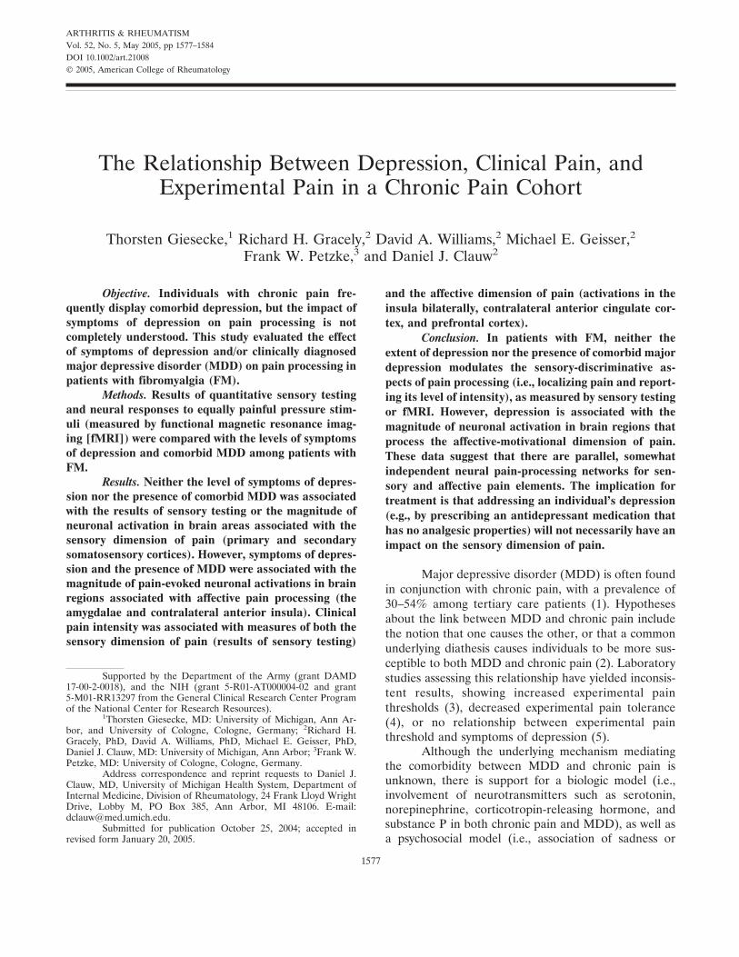
Figure 1. Outline of study design, including main correlational ana-lysis and group comparisons. FM � fibromyalgia; CIDI � CompositeInternational Diagnostic Interview.
Table 1. Demographic and clinical characteristics of the controlsubjects and fibromyalgia (FM) patients*
Controls(n � 42)
FM patients(n � 53)
Sex, no. male/no. female 22/20 20/33Age, years 37.9 � 9.1 42.0 � 8.9CES-D score 6.5 � 6.6 17.1 � 10.6†Clinical pain VAS score 3.3 � 10.1 47.5 � 25.6†Pressure-pain intensity, kg‡
MRS low 1.81 � 1.47 1.05 � 0.73§MRS medium 4.57 � 2.39 2.96 � 1.57¶MRS high 6.99 � 2.97 5.15 � 2.53§
* Except where indicated otherwise, values are the mean � SD.CES-D � Center for Epidemiological Studies Depression Scale;VAS � visual analog scale; MRS � multiple random staircase(method).† P � 0.001 versus controls.‡ Low � intensity needed to elicit first pain (pain threshold); me-dium � intensity needed to elicit moderate pain; high � intensityneeded to elicit slightly intense pain.§ P � 0.05 versus controls.¶ P � 0.01 versus controls.
Table 2. Correlation coefficients for associations between evokedpain sensitivity (MRS method), level of symptoms of depression, andmagnitude of clinical pain in 53 fibromyalgia patients*
MRS pressure-pain intensityVAS pain
scoreLow Medium High
CES-D scorer �0.20 0.01 �0.11 0.26P 0.20 0.97 0.50 0.06
VAS pain scorer �0.20 �0.18 �0.30 –P 0.20 0.26 0.054 –
* Associations are Pearson correlation coefficients. See Table 1 fordefinitions.
Table 3. Association of neuronal activations in brain regions withlevel of symptoms of depression and clinical pain ratings*
Side, region
Coordinates
Correlationwith CES-D
score
Correlationwith VASpain score
x y Z r Z r Z
ContralateralS1 56 �10 46 0.28 2.142 0.20 1.714S2 66 �22 16 0.10 1.050 0.13 1.005ACC 8 36 18 0.10 0.866 0.47† 3.540Anterior insula 32 2 14 0.51† 4.085 – –Anterior insula 34 28 6 – – 0.50† 3.818PFC (BA 10) 32 50 23 0.06 0.547 0.53† 4.156Amygdala 18 0 �10 0.40‡ 3.091 0.13 1.005
IpsilateralS2 �64 �20 14 0.20 1.714 0.21 1.985Cerebellum �34 �68 �30 0.10 1.134 0.10 0.987Amygdala �20 0 �12 0.50† 3.959 0.15 1.050
* Associations are Pearson correlation coefficients and Z statistics.Brain areas are mapped to the coordinate system of the Talairach-Tournoux atlas. S1 � primary somatosensory cortex; S2 � secondarysomatosensory cortex; ACC � anterior cingulate cortex; PFC �prefrontal cortex; BA � Brodmann’s area (see Table 1 for otherdefinitions).† P � 0.05, corrected for multiple comparisons.‡ P � 0.001, uncorrected.
NEURAL ACTIVATIONS IN DEPRESSION AND CHRONIC PAIN 1579
Statistical analysis. Correlations between clinical pain,experimental pain sensitivity, and self-reported symptoms ofdepression were analyzed using Pearson’s correlation coeffi-cients (SPSS for Windows, version 11.0; SPSS, Chicago, IL).For the subset of patients who participated in the fMRIprotocol, correlations in the mean difference of neuronalactivation between no pain and slightly intense pain at eachvoxel of the brain and the extent of self-reported symptoms ofdepression or clinical pain were analyzed using Pearson’scorrelation coefficients, corrected for multiple comparisons.
RESULTS
Fifty-three patients with FM and 42 controlsprovided complete self-report data and participated inthe experimental pain testing. The data from fMRI werecollected on a subset of 30 patients with FM (18
female/12 male), with a mean age of 42 � 10 years. Thedemographic and clinical data are shown in Table 1, andthe study design is shown in Figure 1.
Associations between the magnitude of clinicalpain, experimental pain sensitivity, and symptoms ofdepression for the patient group are shown in Table 2.The magnitude of clinical pain showed only weak corre-lations with self-reported symptoms of depression (r �0.26, P � 0.06) and experimental pain (r � �0.18 to�0.30, P � 0.26–0.054). Pressure-pain thresholds at allpain intensity levels (i.e., mild, moderate, severe pain)were unrelated to self-reported symptoms of depression.
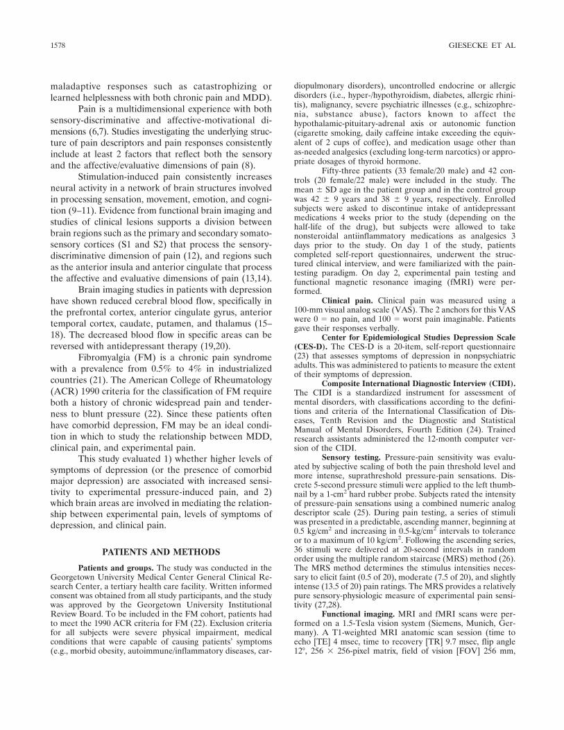
Results of the correlational analyses of self-reported symptoms of depression and neuronal activa-tions are shown in Table 3 and Figure 2. Correlations
Figure 2. Brain regions associated with self-reported symptoms of depression. Pain-evoked neuronalactivations were associated with self-reported level of symptoms of depression (green arrows). Regions ofsignificant correlations are shown in standard space superimposed on a structural T1-weighted magneticresonance image. Images are shown in radiologic view, with the right brain shown on the left. Higher levelsof symptoms of depression were significantly associated with higher neuronal activation in both amygdalae(left image: x/y/z coordinates in Talairach space �20/0/�12 mm and 18/0/�10 mm) and in thecontralateral anterior insula (middle and right images: x/y/z coordinates 32/2/14 mm).
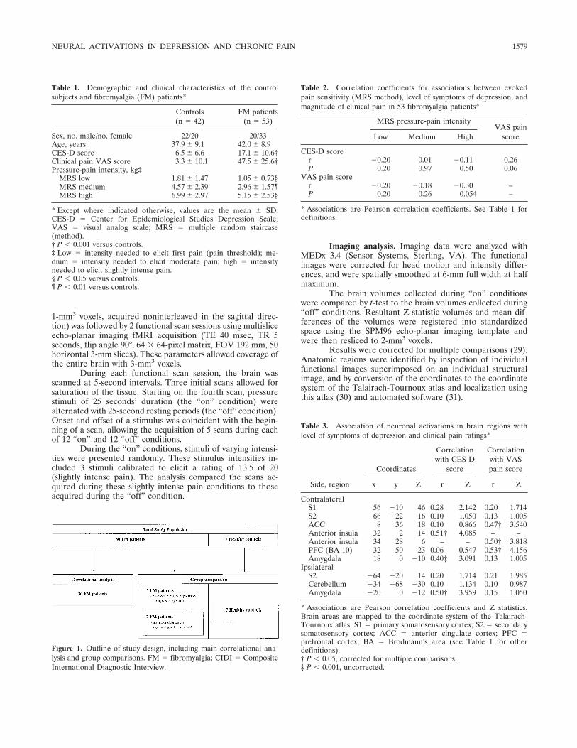
Figure 3. Brain regions associated with self-reported clinical pain score. Pain-evoked neuronal activa-tions were associated with self-reported level of clinical pain (green arrows). Regions of significantcorrelations are shown in standard space superimposed on a structural T1-weighted magnetic resonanceimage. Images are shown in radiologic view, with the right brain shown on the left. Higher levels of clinicalpain were significantly associated with higher neuronal activation in the anterior insula (left image: x/y/zcoordinates in Talairach space 34/28/6 mm), the prefrontal cortex (middle image: x/y/z coordinates32/50/23 mm), and the anterior cingulate (right image: x/y/z coordinates 8/36/18 mm).
1580 GIESECKE ET AL
with neuronal activations in cortical areas that processthe sensory-discriminative dimension of pain (S1 andS2) were not significant. However, CES-D scores weresignificantly correlated with neuronal activations in theamygdalae and contralateral anterior insula.
Similar to the trends observed with the CES-Dscores, clinical pain VAS scores reported by the subsetof 30 FM patients did not correlate with neural activityin the S1 or S2 brain regions. The clinical pain scores,however, did correlate significantly with pain-evokedneural activity in the contralateral anterior insula, ante-rior cingulate cortex, and prefrontal cortex (Table 3 andFigure 3).
Self-reported symptoms of depression cannot be
used to definitively diagnose MDD. In our sample of 30FM subjects who underwent fMRI, a subset of 7 subjectsmet the full criteria for comorbid MDD by structuredclinical interview (i.e., the CIDI). Therefore, in a secondanalysis, we compared fMRI patterns of neuronal acti-vation in these 7 patients with those of 7 age- andsex-matched FM patients without MDD, and with 7 age-and sex-matched healthy control subjects (Figure 1).Consistent with previous studies in FM, both groups ofFM patients, those with and those without MDD, re-quired significantly less pressure to cause slightly intensepain as compared with the healthy controls (FM patientswith no major depression 4.7 kg, FM patients with MDD5.2 kg, controls 6.9 kg; P � 0.001 for both FM groups
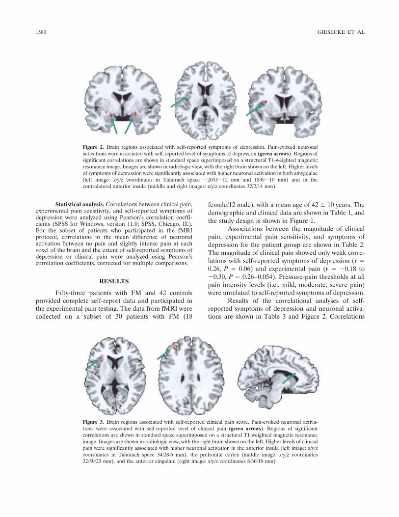
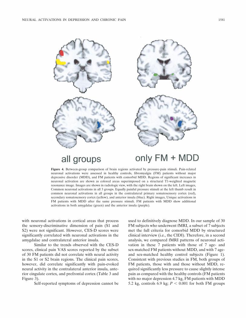
Figure 4. Between-group comparison of brain regions activated by pressure-pain stimuli. Pain-relatedneuronal activations were assessed in healthy controls, fibromyalgia (FM) patients without majordepressive disorder (MDD), and FM patients with comorbid MDD. Regions of significant increases inneuronal activation are shown as colored areas superimposed on a structural T1-weighted magneticresonance image. Images are shown in radiologic view, with the right brain shown on the left. Left images,Common neuronal activations in all 3 groups. Equally painful pressure stimuli at the left thumb result incommon neuronal activations in all groups in the contralateral primary somatosensory cortex (red),secondary somatosensory cortex (yellow), and anterior insula (blue). Right images, Unique activations inFM patients with MDD after the same pressure stimuli. FM patients with MDD show additionalactivations in both amygdalae (green) and the anterior insula (purple).
NEURAL ACTIVATIONS IN DEPRESSION AND CHRONIC PAIN 1581
versus controls) (32,33). These equally painful stimuliresulted in similar neuronal activations in the corticalareas that code for stimulus intensity in all 3 groups, andresulted in unique neuronal activations in both theamygdalae and the contralateral insula in the patientswith MDD, thus confirming the results of the correla-tional analysis involving symptoms of depression, andextending the findings to the clinical diagnosis of MDD(Figure 4).
DISCUSSION
In a cohort of patients with chronic pain who hada confirmed diagnosis of FM, the level of symptoms ofdepression or presence of MDD was not associated witheither subjective pressure-pain sensitivity or neuronalactivations in regions of the brain that are implicated inprocessing the sensory-discriminative dimension of pain(i.e., S1 and S2). In contrast, the presence of MDD orsymptoms of depression was associated with neuronalactivation in brain regions implicated in processing themotivational-affective dimension of pain (i.e., the amyg-dalae and anterior insula). Clinical pain was related toboth the sensory and the affective domains, in that itweakly correlated with sensory testing results and withthe magnitude of neuronal activations in brain areasassociated with affective/integrative aspects of pain pro-cessing (i.e., the insula bilaterally, the contralateralanterior cingulate cortex, and the prefrontal cortex).
These sensory testing and functional imagingdata are consistent with the findings of a number ofother studies suggesting that pain has at least 2 dimen-sions: a sensory-discriminative dimension that identifiesits intensity, quality, and spatiotemporal characteristics,and an affective-motivational dimension that processesits negative valence and unpleasantness (6,7). Thesedata also provide additional evidence that the anteriorinsula may play a critical role in integrating sensory andemotional experiences, since this was the only regionassociated with both symptoms of depression and theclinical pain report (34,35).
Much has been made of the overlap and similar-ities between pain and symptoms of depression, butthese and other data suggest it is also important toidentify pain-processing mechanisms that are indepen-dent of mood. For example, this and other functionalimaging studies in FM suggest that there is objectiveevidence of amplification of the sensory dimension ofpain that is totally independent of mood or emotion(32,36,37). Similar findings of allodynia/hyperalgesiathat are not explained by psychological factors occur in
other “central” pain syndromes, such as irritable bowelsyndrome, temporomandibular disorder, and idiopathiclow back pain (33,38).
The notion that sensory and affective aspects ofpain may be independently processed is not just oftheoretical interest. Dissimilar pharmacologic therapiesmay differentially influence the sensory and affectivedimensions of pain (25). Within the class of antidepres-sants, some are relatively efficacious analgesics (e.g.,tricyclic compounds), whereas others do not function inthis manner (e.g., highly selective serotonin reuptakeinhibitors) (39). The effects of antidepressants on painalso appear to be independent of mood, since 1) anti-depressant effects and analgesic effects frequently occurindependently of each other in clinical trials, and 2)doses of antidepressants necessary to produce analgesiaare, in many cases, lower than the those required to treatMDD (40–44).
These data are consistent with a neuromatrixmodel of pain that applies concepts from cognitiveneuroscience network theory (45). In this model, dimen-sions of the pain experience are the output of a neuralnetwork program, or neuromatrix, which is determinedby genetic influences as well as sensory, cognitive, andaffective experiences unique to each individual. Thistheory maintains that the neuromatrix operates throughparallel distributed processing carried out by somatosen-sory (sensory dimension), limbic (affective dimension),and thalamocortical (evaluative dimension) modulesthat produce distinct, but related dimensions, whichcontribute to a unified pain experience.
The present results support the independence ofmultiple processing networks and confirm that specificcortical regions, particularly the anterior insula, mayintegrate the output of these separate networks andserve to process an individual’s overall sensory/emotional experience. The term interoception has beenused to describe an afferent neural system that isrepresentative of “the material me” that may underliefeelings, emotions, and self-awareness (34). Neural ac-tivity in the anterior insula has consistently been ob-served in studies of pain processing (12,46,47). Whenlesions of the insular region are present, the affectivedimension of pain is altered, whereas the sensory-discriminative dimension is spared; this encompasses adisconnection syndrome called asymbolia for pain (13).Altered neural activity in the insula has not been re-ported in neuroimaging studies of patients with depres-sion, but it has been observed consistently in emotionaltasks with negative affective components, such as tastingsalt (48) and viewing faces of disgust (49). This func-
1582 GIESECKE ET AL
tional characterization is consistent with insular projec-tions to the anterior cingulate and amygdalae, and alsoconsistent with the finding that these regions wereassociated with clinical pain in the present study (50).
This study identified the mediating processesbetween symptoms of depression or MDD and pain, andtheir anatomic correlates in the brain. The design couldnot determine the independent influence of eitherchronic pain or depression on each other. The ideal wayto address the causality between chronic pain and de-pression in future studies is to evaluate patients withchronic pain as they transition between depressed andnondepressed states. The few existing longitudinal stud-ies suggest that there is, at best, only a weak relationshipbetween improvement in MDD and improvement inchronic pain, and that this relationship diminishes oncethe influence of other disease-related variables are con-trolled (51,52). Any preliminary findings on this topicneed replication and extension. In addition, in futurestudies it may be preferable to utilize a longitudinal,within-subject design to examine neural-activation pat-terns in the same individual over time, as they movefrom a depressed to a nondepressed state, or vice versa.
Another potentially fruitful series of studieswould examine how different types of drug or nondrugtherapies impact upon pain processing. For example,previous studies using experimental pain paradigms havesuggested that benzodiazepines and opioids may differ-entially affect sensory or affective-motivational compo-nents of pain, but functional imaging has not been usedto study similar phenomenon (25).
Finally, it is not clear if the findings of the currentstudy apply only to FM or might be seen more broadly inother chronic pain conditions. It is possible that allchronic pain conditions that have a prominent centralelement, i.e., characterized by hyperalgesia/allodynia(e.g., irritable bowel syndrome, low back pain, vulvo-dynia), may show similar features. It is also possible thatthis phenomenon of neural activation in pain processingmight also be noted in more classic peripheral, nocicep-tive pain conditions such as osteoarthritis or rheumatoidarthritis.
In summary, chronic pain, MDD, and otherforms of these conditions frequently coexist. Although itis tempting to lump these constructs together becausethey can co-occur and may have common mechanisms, itmay not be prudent to extrapolate this concept toindividual patients. It appears as though there aredifferent and easily distinguished sensory and affectiveelements to each individual’s pain experience. There arestrong data suggesting that these elements are somewhat
independent of one another and respond differentiallyto both pharmacologic and nonpharmacologic interven-tions. Evaluation of these sensory and affective dimen-sions in patients with chronic pain is likely to improvediagnosis, choice of treatment, and treatment efficacy.
REFERENCES
1. Banks S, Kerns RD. Explaining high rates of depression in chronicpain: a diathesis-stress framework. Psychol Bull 1996;119:95–110.
2. Fishbain DA, Cutler R, Rosomoff HL, Rosomoff RS. Chronicpain-associated depression: antecedent or consequence of chronicpain? A review. Clin J Pain 1997;13:116–37.
3. Davis GC, Buchsbaum MS, Bunney WE. Analgesia to painfulstimuli in affective-illness. Am J Psychiatry 1979;136:1148–51.
4. Zelman DC, Howland EW, Nichols SN, Cleeland CS. The effectsof induced mood on laboratory pain. Pain 1991;46:105–11.
5. Geisser ME, Gaskin ME, Robinson ME, Greene AF. The rela-tionship of depression and somatic focus to experimental andclinical pain in chronic pain patients. Psychol Health 1993;8:405–15.
6. Melzack R, Wall PD. Pain mechanisms: a new theory. Science1965;150:971–9.
7. Melzack R, Casey KL. Sensory, motivational, and central controldeterminants of pain: a new conceptual model. In: Kenshalo D,editor. The skin senses. Springfield (IL): Chas C. Thomas; 1968. p.423–43.
8. Fernandez E, Turk DC. Sensory and affective components of pain:separation and synthesis. Psychol Bull 1992;112:205–17.
9. Casey KL, Minoshima S, Morrow TJ, Koeppe RA. Comparison ofhuman cerebral activation pattern during cutaneous warmth, heatpain, and deep cold pain. J Neurophysiol 1996;76:571–81.
10. Derbyshire SW. Imaging the brain in pain. APS Bulletin 1999;9:7–8.
11. Peyron R, Garcia-Larrea L, Gregoire MC, Costes N, Convers P,Lavenne F, et al. Haemodynamic brain responses to acute pain inhumans: sensory and attentional networks. Brain 1999;122:1765–80.
12. Grant MA, Farrell MJ, Kumar R, Clauw DJ, Gracely RH. FMRIevaluation of pain intensity coding in fibromyalgia patients andcontrols [abstract]. Arthritis Rheum 2001;44 Suppl 9:S394.
13. Berthier M, Starkstein S, Leiguarda R. Asymbolia for pain: asensory-limbic disconnection syndrome. Ann Neurol 1988;24:41–9.
14. Rainville P, Duncan GH, Price DD, Carrier B, Bushnell MC. Painaffect encoded in human anterior cingulate but not somatosensorycortex. Science 1997;277:968–71.
15. Drevets WC, Price JL, Simpson JR Jr, Todd RD, Reich T, VannierM, et al. Subgenual prefrontal cortex abnormalities in mooddisorders. Nature 1997;386:824–7.
16. Bench CJ, Friston KJ, Brown RG, Scott LC, Frackowiak RS,Dolan RJ. The anatomy of melancholia: focal abnormalities ofcerebral blood flow in major depression. Psychol Med 1992;22:607–15.
17. Mayberg HS, Lewis PJ, Regenold W, Wagner HN Jr. Paralimbichypoperfusion in unipolar depression. J Nucl Med 1994;35:929–34.
18. Murata T, Suzuki R, Higuchi T, Oshima A. Regional cerebralblood flow in the patients with depressive disorders. Keio J Med2000;49 Suppl 1:A112–3.
19. Martin SD, Martin E, Rai SS, Richardson MA, Royall R. Brainblood flow changes in depressed patients treated with interper-sonal psychotherapy or venlafaxine hydrochloride: preliminaryfindings. Arch Gen Psychiatry 2001;58:641–8.
20. Davies J, Lloyd KR, Jones IK, Barnes A, Pilowsky LS. Changes inregional cerebral blood flow with venlafaxine in the treatment ofmajor depression. Am J Psychiatry 2003;160:374–6.
NEURAL ACTIVATIONS IN DEPRESSION AND CHRONIC PAIN 1583
21. Wolfe F, Ross K, Anderson J, Russell IJ. Aspects of fibromyalgiain the general population: sex, pain threshold, and fibromyalgiasymptoms. J Rheumatol 1995;22:151–6.
22. Wolfe F, Smythe HA, Yunus MB, Bennett RM, Bombardier C,Goldenberg DL, et al. The American College of Rheumatology1990 criteria for the classification of fibromyalgia: report of theMulticenter Criteria Committee. Arthritis Rheum 1990;33:160–72.
23. Radloff LS. The CES-D scale: a self-report depression scale forresearch in the general population. Applied Psychological Mea-surement 1977;1:385–401.
24. Andrews G, Peters L. The psychometric properties of the com-posite international diagnostic interview. Soc Psychiatry PsychiatrEpidemiol 1998;33:80–8.
25. Gracely RH, Dubner R, McGrath PA. Narcotic analgesia: fentanylreduces the intensity but not the unpleasantness of painful toothpulp sensations. Science 1979;203:1261–3.
26. Gracely RH, Lota L, Walter DJ, Dubner R. A multiple randomstaircase method of psychophysical pain assessment. Pain 1988;32:55–63.
27. Petzke F, Clauw DJ, Ambrose K, Khine A, Gracely RH. Increasedpain sensitivity in fibromyalgia: effects of stimulus type and modeof presentation. Pain 2003;105:403–13.
28. Giesecke T, Williams DA, Harris RE, Cupps TR, Tian X, TianTX, et al. Subgrouping of fibromyalgia patients on the basis ofpressure-pain thresholds and psychological factors. ArthritisRheum 2003;48:2916–22.
29. Worsley KJ, Evans AC, Marrett S, Neelin P. A three-dimensionalstatistical analysis for CBF activation studies in human brain.J Cereb Blood Flow Metab 1992;12:900–18.
30. Talairach J, Tournoux P. Coplanar stereotaxic atlas of the humanbrain. New York: Thieme Medical Publishers; 1988.
31. Lancaster JL, Woldorff MG, Parsons LM, Liotti M, Freitas CS,Rainey L, et al. Automated Talairach atlas labels for functionalbrain mapping. Hum Brain Mapp 2000;10:120–31.
32. Gracely RH, Petzke F, Wolf JM, Clauw DJ. Functional magneticresonance imaging evidence of augmented pain processing infibromyalgia. Arthritis Rheum 2002;46:1333–43.
33. Giesecke T, Gracely RH, Grant MA, Nachemson A, Petzke F,Williams DA, et al. Evidence of augmented central pain process-ing in idiopathic chronic low back pain. Arthritis Rheum 2004;50:613–23.
34. Craig AD. How do you feel? Interoception: the sense of thephysiological condition of the body. Nat Rev Neurosci 2002;3:655–66.
35. Damasio AR, Grabowski TJ, Bechara A, Damasio H, Ponto LL,Parvizi J, et al. Subcortical and cortical brain activity during thefeeling of self-generated emotions. Nat Neurosci 2000;3:1049–56.
36. Gracely RH, Grant MA, Giesecke T. Evoked pain measures infibromyalgia. Best Pract Res Clin Rheumatol 2003;17:593–609.
37. Cook DB, Lange G, Ciccone DS, Liu WC, Steffener J, NatelsonBH. Functional imaging of pain in patients with primary fibromy-algia. J Rheumatol 2004;31:364–78.
38. Naliboff BD, Derbyshire SW, Munakata J, Berman S, MandelkernM, Chang L, et al. Cerebral activation in patients with irritablebowel syndrome and control subjects during rectosigmoid stimu-lation. Psychosom Med 2001;63:365–75.
39. Fishbain D. Evidence-based data on pain relief with antidepres-sants. Ann Med 2000;32:305–16.
40. Salerno SM, Browning R, Jackson JL. The effect of antidepressanttreatment on chronic back pain: a meta-analysis. Arch Intern Med2002;162:19–24.
41. Bair MJ, Robinson RL, Katon W, Kroenke K. Depression andpain comorbidity: a literature review. Arch Intern Med 2003;163:2433–45.
42. Carter GT, Sullivan MD. Antidepressants in pain management.Curr Opin Investig Drugs 2002;3:454–8.
43. O’Malley PG, Jackson JL, Santoro J, Tomkins G, Balden E,Kroenke K. Antidepressant therapy for unexplained symptomsand symptom syndromes. J Fam Pract 1999;48:980–90.
44. O’Malley PG, Balden E, Tomkins G, Santoro J, Kroenke K,Jackson JL. Treatment of fibromyalgia with antidepressants: ameta-analysis. J Gen Intern Med 2000;15:659–66.
45. Rumelhart DE, McClelland JL, and the PDP Research Group.Parallel distributed-processing: explorations in the microstructureof cognition. Cambridge (MA): MIT Press; 1986.
46. Bornhovd K, Quante M, Glauche V, Bromm B, Weiller C, BuchelC. Painful stimuli evoke different stimulus-response functions inthe amygdala, prefrontal, insula and somatosensory cortex: asingle-trial fMRI study. Brain 2002;125:1326–36.
47. Coghill RC, Sang CN, Maisog JM, Iadarola MJ. Pain intensityprocessing within the human brain: a bilateral, distributed mech-anism. J Neurophysiol 1999;82:1934–43.
48. Kinomura S, Kawashima R, Yamada K, Ohno S, Itoh M, YoshiokaS, et al. Functional anatomy of taste perception in the human brainstudied with positron emission tomography. Brain Res 1994;659:263–6.
49. Phillips ML, Young AW, Senior C, Brammer M, Andrew C,Calder AJ, et al. A specific neural substrate for perceiving facialexpressions of disgust. Nature 1997;389:495–8.
50. Mesulam MM, Mufson EJ. Insula of the old world monkey. III.Efferent cortical output and comments on function. J CompNeurol 1982;212:38–52.
51. Newman SP, Fitzpatrick R, Lamb R, Shipley M. The origins ofdepressed mood in rheumatoid arthritis. J Rheumatol 1989;16:740–4.
52. Dickens C, Jayson M, Sutton C, Creed F. The relationshipbetween pain and depression in a trial using paroxetine in sufferersof chronic low back pain. Psychosomatics 2000;41:490–9.
1584 GIESECKE ET AL