the radiology of oral and perioral cysts juan f. yepes, dds, md, mph assistant professor division of...
TRANSCRIPT

The Radiology of
Oral and Perioral Cysts
Juan F. Yepes, DDS, MD, MPHAssistant ProfessorDivision of OD, OM, OMFRDepartment of Oral Health PracticeUKCDODM 820 Spring 2009


Cyst
Pathologic cavity filled with fluid, lined by epithelium,and surrounded by a connective tissue wall.
The cyst fluid either is secreted by the cells liningthe cavity or derives from the surrounding tissueFluid.

Cyst
• Cyst occur more often in the jaws because most cysts originate from the rest of odontogenic epithelium that remain after tooth formation.
• Cyst are radiolucent lesions
• Usually asymptomatic **** (infection)
• Related with missing teeth

Cyst
Radiographic Features:
Location
Cyst may occur centrally in any location in the maxilla or mandible
Cyst are rare in the condyle and the coronoid process
A few cyst arise in the soft tissues of the orofacial region
Periphery RadiolucentWell defined and corticatedHowever a secondary infection can change this

CystRadiographic Features:
Shape
Cyst are usually round or oval, resembling a fluid filled ballon
Some cyst may have a scalloped borders
Internal Structure
Cyst are often totally radiolucent
Long standing cyst may have some calcifications inside
Some cyst have septa

Cyst
Radiographic Features:
Effects on Surrounding Structures
Cyst grow slowly
Sometimes displacement and resorption of the teeth
Cyst can expand the mandible

Cyst
Odontogenic Cyst
Non-Odontogenic Cyst
• Radicular Cyst• Residual Cyst• Dentigerous Cyst• Buccal bifurcation cyst• Odontogenic Keratocyst• Basal cell nevus syndrome• Lateral Periodontal Cyst• Calcifying odontogenic Cyst

The Radiology of Oral and Perioral Cysts
Embryonic epithelial precursor Adult epithelial derivative
Surface epithelium
Rests of Serres Gingival Cyst (Dentigerous Cyst if Crown involved)
Reduced Enamel Epithelium
Dentigerous Cyst
Rest of Malassez Radicular cyst

The Radiology of Oral and Perioral Cysts
Odontogenic Cysts Nonodontogenic Cysts

The Radiology of Oral and Perioral Cysts
Radicular CystDental Cyst

The Radiology of Oral and Perioral Cysts
Radicular Cyst
Periapical cyst, Dental cyst
Radicular cyst is a cyst most likely originated when rest of epithelial cells (Malassez) in the periodontal ligament are stimulated toproliferate and undergo cyst degeneration by inflammatory products from a non-vital tooth.
Most common cyst in the jaws
They arise from non-vital teeth
Asymptomatic, unless secondary infection occurs

The Radiology of Oral and Perioral Cysts
Radicular Cyst
Periapical cyst, Dental cyst
• In most cases the epicenter of the RC is located at the apex
• 60% are found in the maxilla, especially around incisors and canines
• Well defined cortical border, if secondary infected, the inflammatory• reaction may result in loss of the cortical bone
• Internal structure Radiolucent

The Radiology of Oral and Perioral Cysts
Radicular Cyst
Periapical cyst, Dental cyst
Differential Diagnosis
- RO : Granuloma – Cyst – Abscess
- Odontogenic Keratocyst
- Lateral periodontal Cyst

The Radiology of Oral and Perioral Cysts

The Radiology of Oral and Perioral Cysts

The Radiology of Oral and Perioral Cysts

The Radiology of Oral and Perioral Cysts

The Radiology of Oral and Perioral Cysts

The Radiology of Oral and Perioral Cysts

The Radiology of Oral and Perioral Cysts

The Radiology of Oral and Perioral Cysts

The Radiology of Oral and Perioral Cysts

The Radiology of Oral and Perioral Cysts

The Radiology of Oral and Perioral Cysts

The Radiology of Oral and Perioral Cysts

The Radiology of Oral and Perioral Cysts
The Radiology of Oral and Perioral Cysts

............ ...............
.......
.......
....
The Radiology of Oral and Perioral Cysts

The Radiology of Oral and Perioral Cysts

The Radiology of Oral and Perioral Cysts

The Radiology of Oral and Perioral Cysts

The Radiology of Oral and Perioral Cysts
Dentigerous CystFollicular Cyst

The Radiology of Oral and Perioral Cysts
Dentigerous Cyst
A dentigerous cyst is a cyst that forms around the crown ofan unerupted tooth. It begins from accumulation in the layers of reduced enamel epithelium or between the epitheliumAnd the crown of the unerupted tooth.
Second most common cyst in the jaws
They develop around the crown of an unerupted or supernumerarytooth

The Radiology of Oral and Perioral Cysts
Dentigerous Cyst
Radiology Features
• The epicenter is just above the crown of the involved tooth
• The cyst is attaches at the CEJ
• Very often are quite big before diagnosis
• Well defined cortex
• Completely radiolucent

The Radiology of Oral and Perioral Cysts
Dentigerous Cyst
Radiology Features
- DG often displace and resorb adjacent teeth
- Displaces the associated tooth in a apical direction
Differential Diagnosis - Hyperplastic follicle - OKC: Less likely resorb teeth
Attach more apical
- Cystic ameloblastoma

The Radiology of Oral and Perioral Cysts

The Radiology of Oral and Perioral Cysts

The Radiology of Oral and Perioral Cysts

The Radiology of Oral and Perioral Cysts

The Radiology of Oral and Perioral Cysts

The Radiology of Oral and Perioral Cysts

The Radiology of Oral and Perioral Cysts

The Radiology of Oral and Perioral Cysts

The Radiology of Oral and Perioral Cysts

The Radiology of Oral and Perioral Cysts
The Radiology of Oral and Perioral Cysts
The Radiology of Oral and Perioral Cysts

The Radiology of Oral and Perioral Cysts

The Radiology of Oral and Perioral Cysts

The Radiology of Oral and Perioral Cysts

The Radiology of Oral and Perioral Cysts
The Radiology of Oral and Perioral Cysts

The Radiology of Oral and Perioral Cysts

The Radiology of Oral and Perioral Cysts

The Radiology of Oral and Perioral Cysts
KeratocystOdontogenic Keratocyst
OKC
The Radiology of Oral and Perioral Cysts
OKC
Non-inflammatory odontogenic cyst that arises from thedental lamina.
Unlike other cyst, the epithelium of the OKC appears tohave innate growth potential
The epithelium lining is keratinized and thin
Inside the cyst viscous white material “cheese”

The Radiology of Oral and Perioral Cysts
OKC
- OKCs account for above 1/10 of all cyst in the jaws
- Second and third decades with slightly male predilection
- No symptoms until mid size swelling
- High recurrence
- Aspiration Keratin

The Radiology of Oral and Perioral Cysts
OKC
Radiographic Features:
- 90% Posterior body of the mandible- Epicenter superior to the inferior alveaolar canal- Looks very similar to dentigerous cyst- Well corticated and radiolucent- MINIMAL EXPANSION- Resorb teeth bust less than DC

The Radiology of Oral and Perioral Cysts

The Radiology of Oral and Perioral Cysts

The Radiology of Oral and Perioral Cysts

The Radiology of Oral and Perioral Cysts

The Radiology of Oral and Perioral Cysts

The Radiology of Oral and Perioral Cysts

The Radiology of Oral and Perioral Cysts

The Radiology of Oral and Perioral Cysts

The Radiology of Oral and Perioral Cysts

The Radiology of Oral and Perioral Cysts

The Radiology of Oral and Perioral Cysts

The Radiology of Oral and Perioral Cysts


The Radiology of Oral and Perioral Cysts
Nevoid Basal CellCarcinoma Syndrome
Gorlin-Goltz Syndrome

The Radiology of Oral and Perioral Cysts

Basal cell nevus syndrome
Gorlin-Goltz Syndrome
- Multiple basal cell carcinomas of the skin
- Skeletal abnormalities
- Eye abnormalities
- Multiple OKC

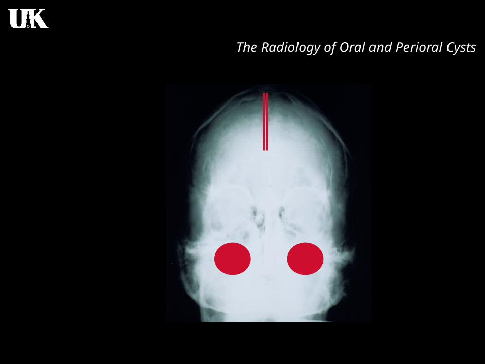
Basal cell nevus syndrome
• Usually after 5 years of age and before 30
• Multiples OKC (mandible)
• High recurrence
• Bifid rib
• Polydactyly
• Temporoparietal bossing
• Calcification of the falx cerebri

The Radiology of Oral and Perioral Cysts

The Radiology of Oral and Perioral Cysts
The Radiology of Oral and Perioral Cysts

The Radiology of Oral and Perioral Cysts

The Radiology of Oral and Perioral Cysts

The Radiology of Oral and Perioral Cysts

The Radiology of Oral and Perioral Cysts
Residual CystRadicular Cyst
Dentigerous Cyst

The Radiology of Oral and Perioral Cysts

The Radiology of Oral and Perioral Cysts

The Radiology of Oral and Perioral Cysts

The Radiology of Oral and Perioral Cysts

The Radiology of Oral and Perioral Cysts

The Radiology of Oral and Perioral Cysts

The Radiology of Oral and Perioral Cysts

The Radiology of Oral and Perioral Cysts

The Radiology of Oral and Perioral Cysts
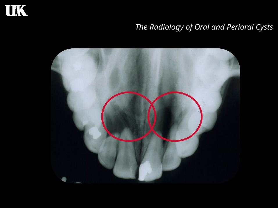
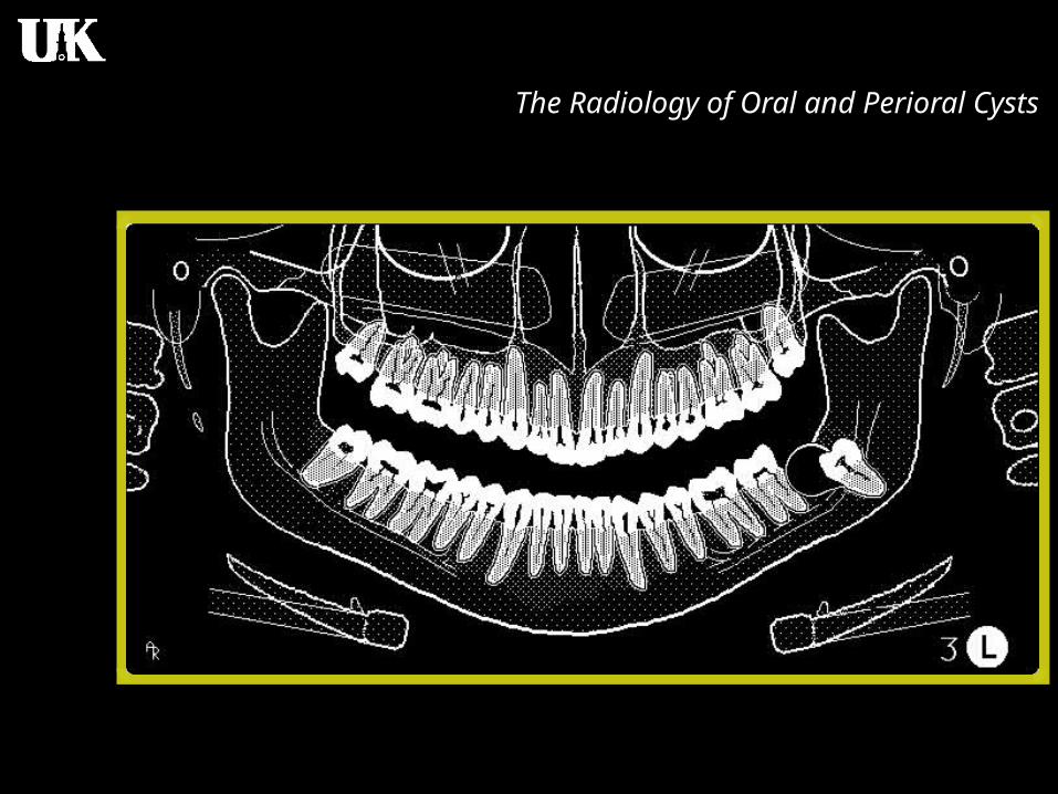
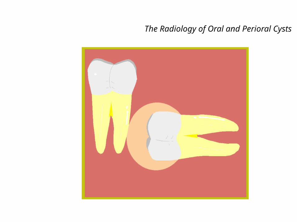
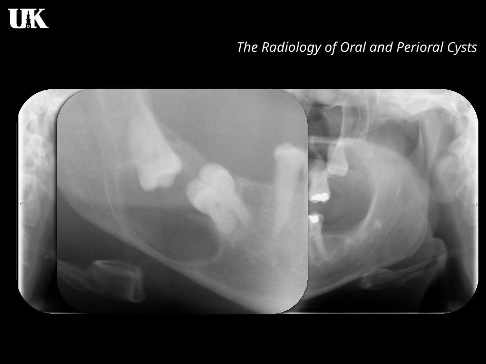
Paradental CystInfected Buccal Cyst

The Radiology of Oral and Perioral Cysts
Paradental Cyst
~0.5% of odontogenic cysts, histopathologically,but not radiographically.
Daley, T.D., Wysocki, G.P., Pringle, G.A. Relative incidenceOf odontogenic tumors and oral and jaw cysts in a Canadianpopulation. Oral Surg oral med Oral Pathol 1994; 77:276-280.

The Radiology of Oral and Perioral Cysts

The Radiology of Oral and Perioral Cysts

The Radiology of Oral and Perioral Cysts

The Radiology of Oral and Perioral Cysts
Lateral PeriodontalCyst

The Radiology of Oral and Perioral Cysts

The Radiology of Oral and Perioral Cysts
Nonodontogenic Cysts

The Radiology of Oral and Perioral Cysts
Incisive CanalCyst
Nasopalatine Canal Cyst

The Radiology of Oral and Perioral Cysts
Nasopalatine Duct Cyst
- Remnants of the nasopalatine duct
- 10% of jaw cyst
- Most cases between 40-50 years old
- Asymptomatic or mild symptoms

The Radiology of Oral and Perioral Cysts
Nasopalatine Duct Cyst
- + frequent complain: small, well defined swelling just posterior to the palatine papilla
- Sometimes (depending of the size) the cyst produces swelling below the maxillary labial frenum
The Radiology of Oral and Perioral Cysts
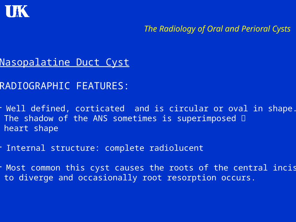
Nasopalatine Duct Cyst
RADIOGRAPHIC FEATURES:
- Well defined, corticated and is circular or oval in shape. The shadow of the ANS sometimes is superimposed heart shape
- Internal structure: complete radiolucent
- Most common this cyst causes the roots of the central incisors to diverge and occasionally root resorption occurs.
The Radiology of Oral and Perioral Cysts
Nasopalatine Duct Cyst
DIFFERENTIAL DIAGNOSIS
-The most common differential diagnosis is a large incisive foramen
- Keep in main: Always compare with old images
- Radicular cyst VIATALITY!!!

The Radiology of Oral and Perioral Cysts


The Radiology of Oral and Perioral Cysts

The Radiology of Oral and Perioral Cysts

The Radiology of Oral and Perioral Cysts

The Radiology of Oral and Perioral Cysts
The Radiology of Oral and Perioral Cysts

The Radiology of Oral and Perioral Cysts

Periapical Radiolucencies

Periapical Radiolucencies
> 10 mm

Periapical Radiolucencies

Periapical Radiolucencies

The Radiology of Oral and Perioral Cysts

The Radiology of Oral and Perioral Cysts

The Radiology of Oral and Perioral Cysts

The Radiology of Oral and Perioral Cysts

The Radiology of Oral and Perioral Cysts
Median Mandibular
Cyst

The Radiology of Oral and Perioral Cysts

The Radiology of Oral and Perioral Cysts
Pseudocystsand Non-Cysts

The Radiology of Oral and Perioral Cysts
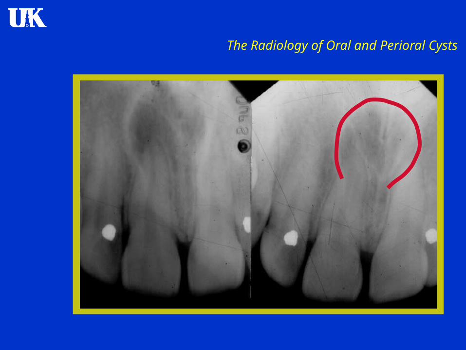
Mucous RetentionPseudocyst
The Radiology of Oral and Perioral Cysts
Serous Retention PseudocystMucous RetentionCystSerous Retention Cyst
Mucosal Cyst

The Radiology of Oral and Perioral Cysts

The Radiology of Oral and Perioral Cysts

The Radiology of Oral and Perioral Cysts

The Radiology of Oral and Perioral Cysts

The Radiology of Oral and Perioral Cysts

The Radiology of Oral and Perioral Cysts

The Radiology of Oral and Perioral Cysts

The Radiology of Oral and Perioral Cysts

The Radiology of Oral and Perioral Cysts
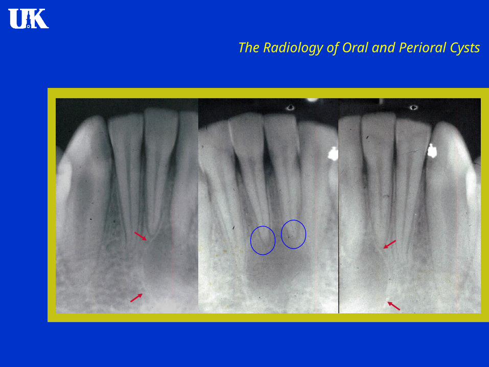
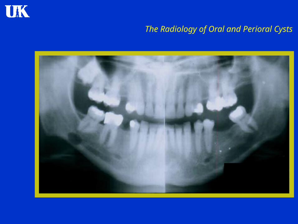
Simple BoneCyst

The Radiology of Oral and Perioral Cysts
Traumatic Bone CystHemorrhagic Bone Cyst
Solitary Bone Cyst

The Radiology of Oral and Perioral Cysts
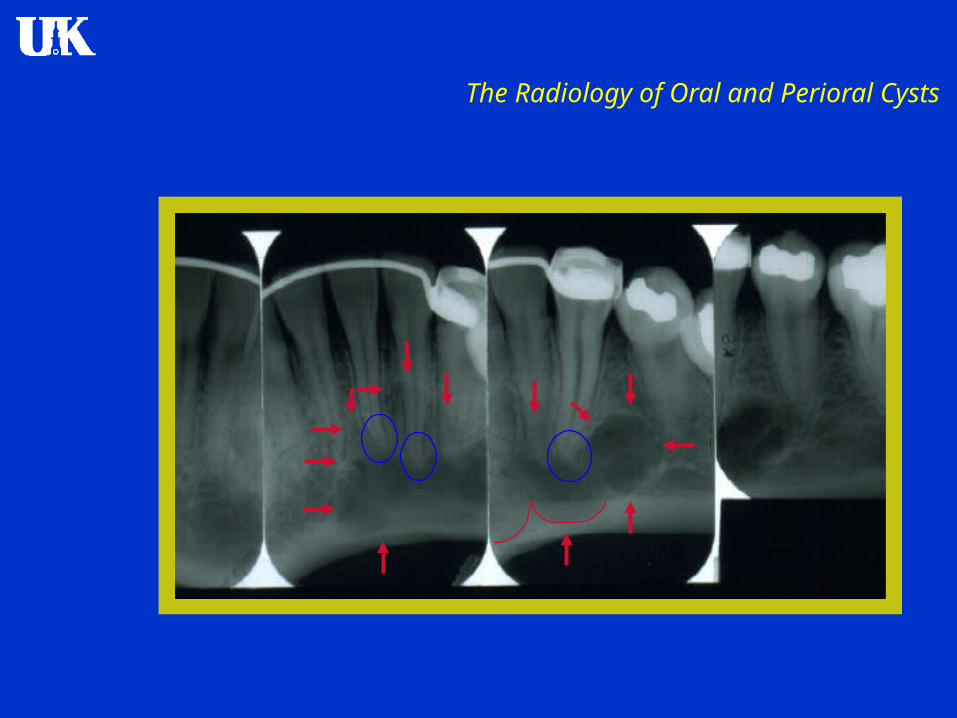
Simple Bone Cyst
- SBC is a cavity within the bone that is lined by connective tissue. It may be empty or it may contain some fluid.
-SBC is not a true cyst
- Etiology: unknown, however probably is a abnormality in the bone metabolism

The Radiology of Oral and Perioral Cysts
Simple Bone Cyst
- SBC’s are very common
- First or second decade of life
- Male predominance 2:1
- Associated with cemento-osseous dysplasia
- Asymptomatic in most cases
- Expansion of the mandible or maxilla: unusual

The Radiology of Oral and Perioral Cysts
Simple Bone Cyst
RADIOGRAPHIC FEATURES
- Almost all SBC’s are found in the mandible
- More often in the posterior body or in the ramus
- Margin: varies, the lesion often scallops between the roots of the teeth
- The borders are better defined in the alveolar process
- Internal structure: Totally radiolucent
- No true septa

The Radiology of Oral and Perioral Cysts
Simple Bone Cyst
RADIOGRAPHIC FEATURES
- In most cases these lesions have not effect on the surrounding teeth.
- Lamina dura usually is intact
- Tendency to grow along the long axis of the mandible
DIFFERENTIAL DIAGNOSIS
- OKC (however….)

The Radiology of Oral and Perioral Cysts

The Radiology of Oral and Perioral Cysts

The Radiology of Oral and Perioral Cysts

The Radiology of Oral and Perioral Cysts
The Radiology of Oral and Perioral Cysts

The Radiology of Oral and Perioral Cysts

The Radiology of Oral and Perioral Cysts

The Radiology of Oral and Perioral Cysts
The Radiology of Oral and Perioral Cysts

The Radiology of Oral and Perioral Cysts

The Radiology of Oral and Perioral Cysts

The Radiology of Oral and Perioral Cysts

The Radiology of Oral and Perioral Cysts
The Radiology of Oral and Perioral Cysts

The Radiology of Oral and Perioral Cysts

The Radiology of Oral and Perioral Cysts
Salivary GlandInclusion Defect
Stafne Defect

The Radiology of Oral and Perioral Cysts
Submandibular Salivary GlandInclusion Defect
Sublingual Salivary GlandInclusion Defect

The Radiology of Oral and Perioral Cysts
Submandibular Stafne DefectSublingual Stafne Defect

The Radiology of Oral and Perioral Cysts

The Radiology of Oral and Perioral Cysts
The Radiology of Oral and Perioral Cysts