the potential and challenges of nanopore · this system used the staphylococcus aureus toxin, ......
TRANSCRIPT

1146 volume 26 number 10 oCTober 2008 nature biotechnology
1Department of Molecular and Cell Biology, Harvard University, Cambridge, Massachusetts 02138, USA. 2Department of Chemistry and Biochemistry, University of California, Santa Cruz, California 95064, USA. 3Department of Physics and Astronomy, University of British Columbia, Vancouver, British Columbia V6T 1Z1, Canada. 4Department of Chemical Biology, Oxford University, Oxford OX1 3TA, UK. 5Foundation for Applied Molecular Evolution, Gainesville, Florida 32604, USA. 6Department of Physics, University of Washington, Seattle, Washington 98195, USA. 7Department of Physics, University of California at San Diego, La Jolla, California 92093, USA. 8Department of Physics, Harvard University, Cambridge, Massachusetts 02138, USA. 9Electronic BioSciences, San Diego, California 92121, USA. 10Department of Bioengineering, University of California at San Diego, La Jolla, California 92093, USA. 11Microchip Biotechnologies Inc., Dublin, California 94568, USA. 12Oak Ridge National Laboratory, Oak Ridge, Tennessee 37831, USA. 13Departments of Physics and Chemistry and the Biodesign Institute, Arizona State University, Tempe, Arizona 85287, USA. 14Department of Physics, Brown University, Providence, Rhode Island 02912, USA. 15Electrical Engineering and Computer Science, Case Western Reserve University, Cleveland, Ohio 44106, USA. 16Biomedical Engineering, Boston University, Boston, Massachusetts 02215, USA. 17NABsys, Inc., Providence, Rhode Island 02906, USA. 18Department of Chemistry, University of North Carolina, Chapel Hill, North Carolina 27599, USA. 19Department of Physics, North Carolina State University, Raleigh, North Carolina 27695, USA. 20Department of Biochemistry, University of British Columbia, Vancouver, British Columbia V6T 1Z3, Canada. 21National Human Genome Research Institute, National Institutes of Health, Bethesda, Maryland 20892, USA. Correspondence should be addressed to D.B.([email protected]).
Published online 9 October 2008; doi:10.1038/nbt.1495
The potential and challenges of nanopore sequencingDaniel Branton1, David W Deamer2, Andre Marziali3, Hagan Bayley4, Steven A Benner5, Thomas Butler6, Massimiliano Di Ventra7, Slaven Garaj8, Andrew Hibbs9, Xiaohua Huang10, Stevan B Jovanovich11, Predrag S Krstic12, Stuart Lindsay13, Xinsheng Sean Ling14, Carlos H Mastrangelo15, Amit Meller16, John S Oliver17, Yuriy V Pershin7, J Michael Ramsey18, Robert Riehn19, Gautam V Soni16, Vincent Tabard-Cossa3, Meni Wanunu16, Matthew Wiggin20 & Jeffery A Schloss21
A nanopore-based device provides single-molecule detection and analytical capabilities that are achieved by electrophoretically driving molecules in solution through a nano-scale pore. The nanopore provides a highly confined space within which single nucleic acid polymers can be analyzed at high throughput by one of a variety of means, and the perfect processivity that can be enforced in a narrow pore ensures that the native order of the nucleobases in a polynucleotide is reflected in the sequence of signals that is detected. Kilobase length polymers (single-stranded genomic DNA or RNA) or small molecules (e.g., nucleosides) can be identified and characterized without amplification or labeling, a unique analytical capability that makes inexpensive, rapid DNA sequencing a possibility. Further research and development to overcome current challenges to nanopore identification of each successive nucleotide in a DNA strand offers the prospect of ‘third generation’ instruments that will sequence a diploid mammalian genome for ~$1,000 in ~24 h.
When a small (~100 mV) voltage bias is imposed across a nanopore in a membrane separating two chambers containing aqueous electrolytes, the resulting ionic current through the pore can be measured with standard electrophysiological techniques. Bearing in mind that the opening and closing of many biological channels depends on relatively small pep-tide moieties physically blocking the channel, one of us (Deamer) at the
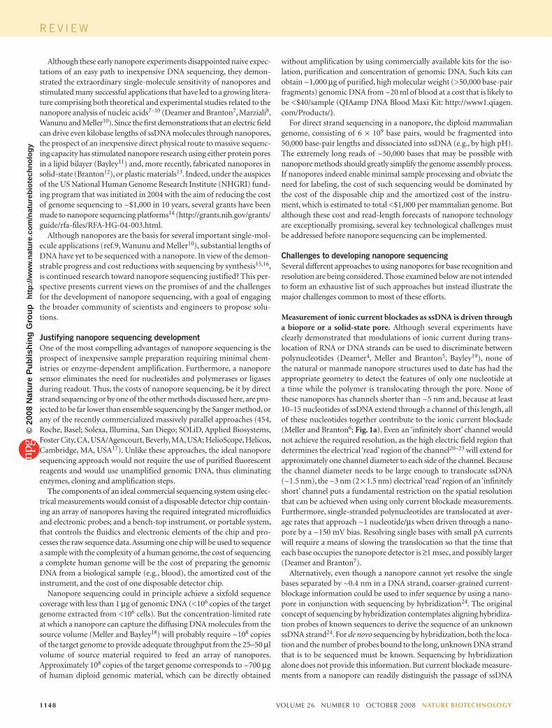
University of California (Santa Cruz) and George Church of Harvard (Cambridge, MA, USA) (personal communication) independently pro-posed that if a strand of DNA or RNA could be electrophoretically driven through a nanopore of suitable diameter, the nucleobases would simi-larly modulate the ionic current through the nanopore. Subsequently, Deamer, Branton and colleagues1 demonstrated that single-stranded DNA (ssDNA) and RNA molecules can be driven through a pore-forming protein and detected by their effect on the ionic current through this nanopore (Fig. 1a). This system used the Staphylococcus aureus toxin, α-hemolysin, the use of which as a biosensor had been pioneered by Bayley and his colleagues2. The Bayley group3 has since shown that an α-hemolysin pore is remarkably stable and remains functional at close to the boiling point of water. Because the inside diameter of the α-hemolysin pore is barely as large as the diameter of a single nucleic acid strand, the results of Deamer, Branton and colleagues1 showed that a nanopore can locally unravel a coiled nucleic acid so that its nucleotides are translocated through the pore in strictly single-file, sequential order. Because the cur-rent of ions through the nanopore is partially blocked by the translocat-ing molecule, each translocating molecule produces a readily detected reduction of the ionic current relative to that which flows through the open, unblocked pore. Given this fact, Deamer, Branton and colleagues1 hypothesized that if each nucleotide in the polymer produced a char-acteristic modulation of the ionic current during its passage through the nanopore, the sequence of current modulations would reflect the sequence of bases in the polymer.
To test this hypothesis, two groups (Deamer4, Meller and Branton5) investigated the current modulations caused by several different RNA and ssDNA polynucleotides. These experiments showed that pore cur-rent is blocked to a substantially greater degree by polyC RNA than by
r e v i e w©
2008
Nat
ure
Pub
lishi
ng G
roup
ht
tp://
ww
w.n
atur
e.co
m/n
atur
ebio
tech
nolo
gy

nature biotechnology volume 26 number 10 oCTober 2008 1147
oligomers (Deamer4), and further measurements using various DNA homopolymers revealed only small ion-current differences (~5% or less) between deoxypurine and deoxypyrimidine oligomers (Meller and Branton5). Single nucleotide discrimination could not be achieved because the ion-current blockades were found to be the consequence of the ~10–15 nucleotides (rather than any single nucleotide) that occupy the membrane-spanning domain of the α-hemolysin pore (Meller and Branton6; Fig. 1a).
polyA RNA, whereas other experiments with RNA molecules containing 30 As followed by 70 Cs revealed that the transition from polyA to polyC segments within a single RNA molecule is readily detectable (Deamer4). Such easily measured distinctions between purine and pyrimidine ribo-nucleotides were unfortunately not as clear for deoxyribonucleotides (Meller and Branton5). In fact, the current level differences that had been observed with RNAs turned out to be a reflection of base stacking and other secondary structural differences between polyA and polyC
.
.
.ATCGGCT...
Open pore
400 sec
Blocked pore
Time
Convert Hybridize Detect
Cur
rent
(pA
)
100
50
0
a
c d
b
A
T
C
G
+
–
Back gate
Tunnelingprobe
Figure 1 Approaches to nanopore sequencing. (a) Strand-sequencing using ionic current blockage. A typical trace of the ionic current amplitude (left) through an α-hemolysin pore clearly differentiates between an open pore (top right) and one blocked by a strand of DNA (bottom right) but cannot distinguish between the ~12 nucleotides that simultaneously block the narrow transmembrane channel domain (red bracket). (b) Exonuclease-sequencing by modulation of the ionic current. An exonuclease (pale blue) attached to the top of an α-hemolysin pore through a genetically encoded (deep blue), or chemical, linker sequentially cleaves dNMPs (gold) off the end of a DNA strand (in this case, one strand of a double-stranded DNA). A dNMP’s identity (A, T, G or C) is determined by the level of the current blockade it causes when driven into an aminocyclodextrin adaptor (red) lodged within the pore. After a few milliseconds, the dNMP is released and exits on the opposite side of the bilayer. (c) Nanopore sequencing using synthetic DNA and optical readout. Each nucleotide in the target DNA that is to be sequenced is first converted into a longer DNA strand composed of pairs of two different code-units (colored orange and blue for illustration); each code-unit is a 12-base-long oligomer. After hybridizing the converted DNA with molecular beacons that are complementary to the code units, these beacons are stripped off using a nanopore. The sequence of the original DNA is read by detecting the discrete short-lived photon-bursts as each oligo is stripped. (d) Strand-sequencing using transverse electron currents. DNA is driven through a nanopore functionalized with embedded emitter and collector tunneling probes (orange) and a back gate (black). The amplitude of the tunneling currents that traverse through the nucleotides is expected to differentiate each nucleobase as the DNA is electrophoretically driven through the pore (arrow).
REV IEW©
2008
Nat
ure
Pub
lishi
ng G
roup
ht
tp://
ww
w.n
atur
e.co
m/n
atur
ebio
tech
nolo
gy
1148 volume 26 number 10 oCTober 2008 nature biotechnology
without amplification by using commercially available kits for the iso-lation, purification and concentration of genomic DNA. Such kits can obtain ~1,000 µg of purified, high molecular weight (>50,000 base-pair fragments) genomic DNA from ~20 ml of blood at a cost that is likely to be <$40/sample (QIAamp DNA Blood Maxi Kit: http://www1.qiagen.com/Products/).
For direct strand sequencing in a nanopore, the diploid mammalian genome, consisting of 6 × 10⁹ base pairs, would be fragmented into 50,000 base-pair lengths and dissociated into ssDNA (e.g., by high pH). The extremely long reads of ~50,000 bases that may be possible with nanopore methods should greatly simplify the genome assembly process. If nanopores indeed enable minimal sample processing and obviate the need for labeling, the cost of such sequencing would be dominated by the cost of the disposable chip and the amortized cost of the instru-ment, which is estimated to total <$1,000 per mammalian genome. But although these cost and read-length forecasts of nanopore technology are exceptionally promising, several key technological challenges must be addressed before nanopore sequencing can be implemented.
Challenges to developing nanopore sequencingSeveral different approaches to using nanopores for base recognition and resolution are being considered. Those examined below are not intended to form an exhaustive list of such approaches but instead illustrate the major challenges common to most of these efforts.
Measurement of ionic current blockades as ssDNA is driven through a biopore or a solid-state pore. Although several experiments have clearly demonstrated that modulations of ionic current during trans-location of RNA or DNA strands can be used to discriminate between polynucleotides (Deamer4, Meller and Branton5, Bayley19), none of the natural or manmade nanopore structures used to date has had the appropriate geometry to detect the features of only one nucleotide at a time while the polymer is translocating through the pore. None of these nanopores has channels shorter than ~5 nm and, because at least 10–15 nucleotides of ssDNA extend through a channel of this length, all of these nucleotides together contribute to the ionic current blockade (Meller and Branton6; Fig. 1a). Even an ‘infinitely short’ channel would not achieve the required resolution, as the high electric field region that determines the electrical ‘read’ region of the channel20–23 will extend for approximately one channel diameter to each side of the channel. Because the channel diameter needs to be large enough to translocate ssDNA (~1.5 nm), the ~3 nm (2 × 1.5 nm) electrical ‘read’ region of an ‘infinitely short’ channel puts a fundamental restriction on the spatial resolution that can be achieved when using only current blockade measurements. Furthermore, single-stranded polynucleotides are translocated at aver-age rates that approach ~1 nucleotide/µs when driven through a nano-pore by a ~150 mV bias. Resolving single bases with small pA currents will require a means of slowing the translocation so that the time that each base occupies the nanopore detector is ≥1 msec, and possibly larger (Deamer and Branton7).
Alternatively, even though a nanopore cannot yet resolve the single bases separated by ~0.4 nm in a DNA strand, coarser-grained current-blockage information could be used to infer sequence by using a nano-pore in conjunction with sequencing by hybridization24. The original concept of sequencing by hybridization contemplates aligning hybridiza-tion probes of known sequences to derive the sequence of an unknown ssDNA strand24. For de novo sequencing by hybridization, both the loca-tion and the number of probes bound to the long, unknown DNA strand that is to be sequenced must be known. Sequencing by hybridization alone does not provide this information. But current blockade measure-ments from a nanopore can readily distinguish the passage of ssDNA
Although these early nanopore experiments disappointed naive expec-tations of an easy path to inexpensive DNA sequencing, they demon-strated the extraordinary single-molecule sensitivity of nanopores and stimulated many successful applications that have led to a growing litera-ture comprising both theoretical and experimental studies related to the nanopore analysis of nucleic acids7–10 (Deamer and Branton7, Marziali8, Wanunu and Meller10). Since the first demonstrations that an electric field can drive even kilobase lengths of ssDNA molecules through nanopores, the prospect of an inexpensive direct physical route to massive sequenc-ing capacity has stimulated nanopore research using either protein pores in a lipid bilayer (Bayley11) and, more recently, fabricated nanopores in solid-state (Branton12), or plastic materials13. Indeed, under the auspices of the US National Human Genome Research Institute (NHGRI) fund-ing program that was initiated in 2004 with the aim of reducing the cost of genome sequencing to ~$1,000 in 10 years, several grants have been made to nanopore sequencing platforms14 (http://grants.nih.gov/grants/guide/rfa-files/RFA-HG-04-003.html.
Although nanopores are the basis for several important single-mol-ecule applications (ref.9, Wanunu and Meller10), substantial lengths of DNA have yet to be sequenced with a nanopore. In view of the demon-strable progress and cost reductions with sequencing by synthesis15,16, is continued research toward nanopore sequencing justified? This per-spective presents current views on the promises of and the challenges for the development of nanopore sequencing, with a goal of engaging the broader community of scientists and engineers to propose solu-tions.
Justifying nanopore sequencing developmentOne of the most compelling advantages of nanopore sequencing is the prospect of inexpensive sample preparation requiring minimal chem-istries or enzyme-dependent amplification. Furthermore, a nanopore sensor eliminates the need for nucleotides and polymerases or ligases during readout. Thus, the costs of nanopore sequencing, be it by direct strand sequencing or by one of the other methods discussed here, are pro-jected to be far lower than ensemble sequencing by the Sanger method, or any of the recently commercialized massively parallel approaches (454, Roche, Basel; Solexa, Illumina, San Diego; SOLiD, Applied Biosystems, Foster City, CA, USA/Agencourt, Beverly, MA, USA; HelioScope, Helicos, Cambridge, MA, USA17). Unlike these approaches, the ideal nanopore sequencing approach would not require the use of purified fluorescent reagents and would use unamplified genomic DNA, thus eliminating enzymes, cloning and amplification steps.
The components of an ideal commercial sequencing system using elec-trical measurements would consist of a disposable detector chip contain-ing an array of nanopores having the required integrated microfluidics and electronic probes; and a bench-top instrument, or portable system, that controls the fluidics and electronic elements of the chip and pro-cesses the raw sequence data. Assuming one chip will be used to sequence a sample with the complexity of a human genome, the cost of sequencing a complete human genome will be the cost of preparing the genomic DNA from a biological sample (e.g., blood), the amortized cost of the instrument, and the cost of one disposable detector chip.
Nanopore sequencing could in principle achieve a sixfold sequence coverage with less than 1 µg of genomic DNA (<106 copies of the target genome extracted from <106 cells). But the concentration-limited rate at which a nanopore can capture the diffusing DNA molecules from the source volume (Meller and Bayley18) will probably require ~108 copies of the target genome to provide adequate throughput from the 25–50 µl volume of source material required to feed an array of nanopores. Approximately 108 copies of the target genome corresponds to ~700 µg of human diploid genomic material, which can be directly obtained
REV IEW©
2008
Nat
ure
Pub
lishi
ng G
roup
ht
tp://
ww
w.n
atur
e.co
m/n
atur
ebio
tech
nolo
gy

nature biotechnology volume 26 number 10 oCTober 2008 1149
difficult, methods are available to systematically encode and substitute each and every nucleotide in the genome with a specific permutation of two different 12-mer oligos (A and B), concatenated in a specific order (AB, BA, AA, BB) that reflects and encodes the nucleotide sequence of the unknown DNA32 (Fig. 1c). This converts the quaternary DNA code of A, T, G and C into a binary code in which each base is represented by a pair of 12-mer oligos (A and B). An automated, massively-parallel pro-cess developed by Lingvitae (Oslo; http://www.lingvitae.com/DPTutorial.php) currently requires ~24 h for the conversion of a complete human genome into a DNA mixture consisting of fragments, each corresponding to a 24-bp segment of the original genome. Work is currently underway to develop inexpensive error-free conversion of longer segments of the original genome and to greatly reduce the conversion time. The conver-sion process does introduce an extra biochemical step, which is not ideal, but it side-steps some of the challenges faced by other approaches and thus simplifies the subsequent sequencing readout.
For readout, this converted DNA mixture is then hybridized with a mixture containing two different ‘molecular beacons’ (Public Health Research Institute, Newark, NJ, USA; http://www.molecular-beacons.org/Introduction.html), each of which is a 12-mer oligo designed to comple-ment either A or B. When free in solution, the molecular beacons pro-duce only a very low background fluorescence because of self-quenching (Fig. 1c). Similarly, when hybridized to the converted DNA, the molecular beacons produce only low background fluorescence because the universal quencher at one end of each beacon is in close proximity to the fluorophore of its nearest neighbor (Fig. 1c). Because the beacons do fluoresce briefly as they are stripped off the complementary converted DNA strand, readout is performed by sequentially stripping off the fluorescent 12-mer oligos one at a time by driving the converted DNA strand through a <2-nm-diameter nanopore (that is, a pore diameter that strips off the complementary, fluo-rescently labeled 12-mer oligos (Branton33)). The original DNA sequence is obtained by determining the color sequence of the photon bursts, where each pair of two successive bursts corresponds to a specific base. With high-density nanopore arrays (Wanunu and Meller34), optical readout can facilitate massive parallelism, and a high resolution electron-multiplying charge-coupled device camera could be used to probe thousands of nano-pores simultaneously. Because the nanopores require no on-chip electrical contacts, surface modification, or mechanisms to regulate the transloca-tion process, improved nanofabrication methods may make it possible to develop such nanopores in very high density arrays. Nevertheless, at this time, fabricating high-density arrays of 1.7- to 2-nm-diameter nanopores remains a substantial challenge.
Measurement of transverse tunneling currents or capacitance as ssDNA is driven through a solid-state nanopore with embedded probes. It has been proposed that tunneling currents through nucle-obases that are driven through a nanopore articulated with tunneling probes may be able to distinguish among the four nucleobases of ssDNA (Fig. 1d)35–39 (Di Ventra35,38). Single bases should be resolved because it is the transverse tunneling current from an emitter probe tip of ≤1-nm diameter that generates the nucleobase-identifying signal rather than the nucleotide occupancy through the entire length of the nanopore channel. Although simulations of attainable base contrast when using tunneling measurements for nucleobase identification have presented encouraging but differing insights into the challenges this approach must address35,38,40–43 (Di Ventra35,38,40,43), the ability of a scanning tunneling microscope (STM) to reveal the atomic scale features of matter is well established44.
As in a STM, electron tunneling currents can be in the nano-ampere range with appropriate probes37,45,46 (Lindsay46). The nanoamp electron currents would make it possible to read the nucleotides at a greater speed
from the passage of double-stranded DNA (dsDNA) in a pore that is large enough to translocate dsDNA25. Because a nanopore is able to discrimi-nate between ssDNA and dsDNA, it may be able to detect and resolve the location and number of oligonucleotide probes that are hybrid-ized to a long translocating ssDNA. Thus, if the standard routines for sequencing by hybridization24 could be enhanced by nanopore-derived information regarding the location and number of double strand regions (that is, bound oligonucleotide probes) on the DNA strands that are to be sequenced, de novo sequencing should be possible. This is the basic concept of hybridization-assisted nanopore sequencing (HANS; Ling26). Current research on the HANS method faces two challenges. Can a nanopore determine the location of a hybridized probe with sufficient accuracy to enhance sequencing by hybridization? What length of DNA sequence can be reliably reconstructed, given the practical limitations of detecting bound probes and locating them precisely on the DNA strand that is to be sequenced?
Measurement of ionic current blockades from individual nucleotides sequentially cleaved off the end of a DNA strand and driven through a biopore. At the time Keller and colleagues27 recognized that it might be possible to sequence single molecules of DNA by identifying the deoxy-nucleoside monophosphates (dNMPs) released by an exonuclease from the end of a DNA or RNA chain, there was no obvious way to identify individual unlabeled bases after their release. Recent work (Bayley28,29) indicates that unlabeled bases can be identified by α-hemolysin when fitted with an aminocyclodextrin adaptor (Bayley28), and methods have now been developed to covalently attach cyclodextrins within the lumen of the α-hemolysin pore (Bayley29). On the basis of this work, Oxford Nanopore Technologies (Oxford, UK) has recently succeeded in cova-lently attaching the aminocyclodextrin adaptor within the α-hemolysin pore (Fig. 1b). When a dNMP is captured and driven through the α-hemolysin-aminocyclodextrin pore in a lipid bilayer membrane, the ionic current through the pore is reduced to one of four levels, each of which reflects which of the four dNMPs—A, T, G or C—is trans-locating. Furthermore, because all four of the ionic current blockage levels are easily distinguishable from the current that flows through the open, unblocked pore, the current traces can provide an accurate count of the total number of dNMPs that have been translocated through the α-hemolysin-aminocyclodextrin pore. For sequencing, it will now be important to assure that 100% of the exonuclease-released dNMPs are captured in the pore and efficiently expelled on the opposite side of the membrane. Because this approach uses the nanopore to iden-tify the released dNMPs, rather than identifying the bases of an intact DNA strand, the strictly single-file, sequential passage of the bases that a nanopore can enforce is lost. It will therefore be especially important to demonstrate that the sequence of independently read dNMPs reflects the order in which the bases are cleaved from the DNA (that is, no over-taking or double counting). Finally, the choice and attachment of the exonuclease to the nanopore must be considered. A genetic construct in which the nuclease and α-hemolysin genes are spliced together might be used or the nuclease might be chemically attached to assure delivery of the released dNMPs into the nanopore. The enzyme should be processive and, for low noise detection, active in high salt. Preferably, the enzyme should digest dsDNA, which is readily produced from genomic DNA and easy to handle.
Nanopore sequencing using converted targets and optical readout. Another readout modality in development for nanopore-based sequenc-ing converts the sequence information of DNA into a two-color scheme that is then optically read (Soni and Meller30, Meller31). Whereas attach-ment of fluorescent probes to each and every nucleotide in DNA is
REV IEW©
2008
Nat
ure
Pub
lishi
ng G
roup
ht
tp://
ww
w.n
atur
e.co
m/n
atur
ebio
tech
nolo
gy

1150 volume 26 number 10 oCTober 2008 nature biotechnology
potential in the pore polarizes the capacitor and voltage fluctuations on the two silicon layers are measured. Simulation results demonstrated that A, C, G and T give distinct capacitance signals and that the instrument can, in principle, resolve single-base substitutions in a DNA strand53. In an early trial of this approach, a voltage signal associated with DNA translocation was detected with one such device, but the time resolution was inadequate to distinguish between nucleotides49. The control of DNA velocity and orientation during translocation is also a major challenge for this approach.
Achieving the promise of long readsOne of the compelling potential advantages of nanopores for sequencing is the promise of long reads. Because the nanopore sensor reads mol-ecules sequentially, base by base, as they thread through the pore, its fundamental strength is that the accuracy of a base call at one instance in time does not depend on the prior history of the system. In principle, the length of DNA that could be read with a nanopore is limited only by the practicalities of avoiding shearing during sample preparation and of limitations yet to be explored with respect to capture and threading of exceptionally long molecules through individual pores. To date, it has been demonstrated that lengths of ssDNA on the order of 25 kb have been threaded through biopores (Meller and Branton, unpublished data) and up to 5.4-kb lengths of ssDNA have been threaded through solid-state nanopores (ref.25, Branton54). A unique feature and promise of nanopore technologies is, therefore, that if a detection scheme is devel-oped that allows reading of a few bases on the fly during unidirectional translocation of the DNA strand through a pore, then the extension of the technology from reading a few bases to reading thousands of bases should be straightforward. Although the expected accuracy of the read is yet unknown, insertions, deletions and other sequence errors will not compromise the read-length as de-phasing is not an issue in indepen-dent single-molecule reads. Sufficient averaging (high sequence coverage) could then reach any desired level of accuracy, as long as sequence errors are random rather than systematically sequence or position dependent.
Furthermore, given the high throughput available and anticipated in short reads from current next-generation instruments, it may be that nanopores will play a role in providing an assembly scaffold of very long reads at low accuracy to facilitate assembly of short read sequences. A
than is possible with the pico-ampere ionic currents that flow through a <3 nm-diameter nanopore. Although this approach using only robust solid-state components and electrical measurements may ultimately be the least expensive and fastest way of sequencing a genome, four major challenges must be addressed (Di Ventra43). First, the voltage bias and solution conditions that optimize contrast between the bases must be determined and maintained to provide unambiguous nucleobase iden-tification; it is difficult to predict beforehand exactly what the electronic response of the detector will be to the different DNA bases, particularly in a fluid system such as is envisioned here. Second, the device must provide a mechanism to assure that each base will assume a reproducible orienta-tion and position on the collector probe while it is being interrogated; tunneling currents are exponentially sensitive to atomic scale changes of orientations and distances. Third, unidirectional translocation of the DNA must be controlled so that each nucleobase remains between the tunneling probes at least 0.1 msec to sample over inevitable noise and molecular motion; this translocation rate will assure that each nucleotide is sampled over a time period that is two orders of magnitude longer than that required for a state-of-the-art preamplifier47 to sense nanoamp cur-rents. And fourth, it remains to be shown whether the transverse current measurements can provide sufficient contrast to not only discriminate between the bases, but also provide a signal characteristic of the gaps between bases that could be used to distinguish each base from the next base in the unknown DNA sequence.
The use of single-walled carbon nanotubes has been proposed (Golovchenko and Branton, unpublished data) as a means of address-ing the second and third challenges above—and possibly even the first challenge—if the carbon nanotube were to be appropriately functional-ized37. Nanotubes bind and orient nucleobases in a specific manner48 and the binding activation enthalpies per base lie in a range that can be modulated by temperature, ionic strength, or a voltage bias so as to con-trol the DNA as it slides on the nanotube (unpublished data).
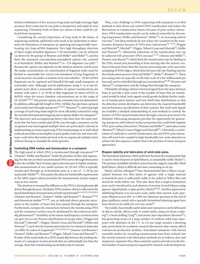
Another inventive solution to the challenge of identifying each base using transverse tunneling currents is to form base-specific hydrogen bonds between chemically modified metal electrodes and the nucleobases in the molecule that is to be sequenced. Ohshiro and Umezawa45 showed that in a STM whose metallic probe is modified with thiol derivatives of adenine, guanine, cytosine, or uracil, tunneling is greatly enhanced between a sample nucleobase and its complementary nucleobase modi-fied metallic probe. Using a cytosine-modified probe, they demonstrated base identification and electrical signals able to distinguish between TTTTTTTTGTTTTTTTTT and TTTTTTTGGTTTTTTTTT. Their work has led Lindsay and colleagues46 to propose a nanopore reader bear-ing pairs of two chemically functionalized probes, one probe of each pair able to couple to the nucleotide’s phosphate moiety while the other probe base-pairs with the nucleobase (Fig. 2). The nucleobases can be identi-fied by the current-distance responses as the DNA moves through the nanopore and past the reader, rather than the tunneling current in a static configuration. The functional groups on each of four such readers— A, C, G or T—would be designed to form a hydrogen-bonded path when the cognate base is translocated through the nanopore between the pair of probes (Lindsay46). Four such readers would be needed to generate a complete sequence, each one reading a duplicate strand. Synchronizing the translocation of four duplicate strands through four readers will pose a major challenge for this approach.
Electrostatic DNA detection and sequencing based on a metal-oxide-silicon capacitor incorporated into the nanopore has also been proposed49–51. Using the electron beam of a transmission electron microscope49,52, a nanopore is fabricated in a membrane consisting of two layers of doped silicon, separated by a 5-nm-thick insulating SiO2 layer. As DNA is translocated through the pore, variation of the electrostatic
Figure 2 A nanopore reader with chemically functionalized probes. As a strand of DNA emerges from a nanopore, a ‘phosphate grabber’ on one functionalized electrode and a ‘base reader’ on the other electrode form hydrogen bonds (light blue ovals) to complete a transverse electrical circuit through each nucleotide as it is translocated through the nanopore.
REV IEW©
2008
Nat
ure
Pub
lishi
ng G
roup
ht
tp://
ww
w.n
atur
e.co
m/n
atur
ebio
tech
nolo
gy
nature biotechnology volume 26 number 10 oCTober 2008 1151
Thus, a key challenge to DNA sequencing with nanopores is to find methods to slow down and control DNA translocation and reduce the fluctuations in translocation kinetics because of pore-surface interac-tions. DNA translocation speeds can be reduced somewhat by decreas-ing temperature (Meller and Branton5, Meller74), or increasing solvent viscosity75, but these methods do not reduce the variations in the trans-location dynamics because of DNA-pore interactions70–73,76 (Wiggin and Marziali70; Marziali71, Wiggin, Tabard-Cossa and Marziali72; Meller and Wanunu70). Substantial reductions of the translocation rate can be achieved with processive DNA enzymes77–79 (Akeson and Meller77; Deamer and Akeson78) which limit the translocation rate by binding to the DNA strand and preventing it from moving into the narrow con-fines of the pore faster than the enzyme processing rate; or by successive unzipping of DNA oligos, which then becomes the rate-limiting step for the translocation process (Soni and Meller30, Meller31, Branton33). These processing rates are typically on the time scale of a few milliseconds per base and can be controlled through ion concentrations78–80 (Deamer and Akeson78), temperature and the voltage bias through the nanopore.
Ultimately, eliciting a distinct electrical signal from the space between bases to provide a clear count of the number of bases that are translo-cated would be ideal. Such signals would greatly facilitate further analy-sis of translocation kinetics and base dwell time distributions so that the detection system developers can determine the required bandwidth and performance specifications of their systems. But until such signals are available, a detailed understanding of, and methods to control, the kinetics of DNA strand translocation through a narrow pore need to be obtained. Fabricating nanopores provides the opportunity for generat-ing nanopores with tailored surface properties that could both regulate DNA-pore surface interaction (Wanunu and Meller81) and reduce noise (Branton82; Tabard-Cossa, Wiggin and Marziali83). Ultimately, a combi-nation of methods to control translocation rate and DNA-pore interac-tion will need to be coupled to high-bandwidth, low-noise detection to achieve the fast sequence analysis that is the promise of many nanopore approaches.
Biopore stability and fabrication of solid-state poresThe hemolysin heptamer, which until now has been the usual protein that is used to form biopores in lipid bilayers, is remarkably stable (Bayley3). The primary instability therefore arises from the support, typically a fluid lipid bilayer, which is difficult and time consuming to set up.
Bayley and his colleagues84 have demonstrated that a bilayer encap-sulated between two thin layers of agarose with a single inserted α-hemolysin pore is sufficiently stable to be sealed in Teflon film and stored for weeks before use. They also show that a single α-hemolysin pore can be introduced in each element of an array of such bilayers using agarose-tipped plastic or glass probes (Bayley85,86). Another approach to stabilizing bilayers is to use nano-scale, rather than micron-scale, aper-tures. Bilayers across 100- to 1,000-nm-diameter apertures at the end of glass capillaries coated with a specially formulated silanizing agent have been shown to be stable for over two weeks87.
Very stable, functionally useful solid-state nanopores can be fabricated in silicon nitride, silicon oxide or metal oxides, using ion beam sculpt-ing56, e-beam drilling (Ling88) and atomic layer deposition (Branton82), but generating arrays of a large number of uniform solid-state nano-pores with diameters in the 1.5- to 2.0-nm range remains a daunting task, particularly for academic research laboratories that cannot afford commercial production facilities. Articulated nanopores with buried nanotube probes for tunneling measurements have been realized, but the current research-scale fabrication methods are so tedious, slow and manpower-expensive they often cannot be used to provide even the lim-ited number of such nanopores required for research-scale development.
hybrid combination of low accuracy long reads and high coverage, high accuracy short reads may be one path to inexpensive and rapid de novo sequencing. Ultimately, both of these two classes of data could be col-lected from nanopores.
Considering the central importance of long reads to the future of sequencing methods, additional work needs to be undertaken to deter-mine the limitations of nanopores in capturing and sequentially trans-locating very long ssDNA fragments. Very high throughput detection of short single-stranded oligomers (<50 nucleotides) can be achieved (as shown by the groups of Branton and Meller5 and Meller55), and for these the measured concentration-normalized capture rate constant in α-hemolysin (Meller and Branton18) is ~5.8 oligomers (sec µM)–1. Because the capture rate depends on the solution molarity and because the molar concentration (or concentration of fragment ends) must be limited to reasonably low wt/vol concentrations of long fragments to avoid excessive viscosities, it remains to be seen whether ~50-kb ssDNA fragments can be captured and threaded through small nanopores at reasonable rates. Although several publications using 3- to 6-nm-di-ameter pores show a reasonable number of capture translocations per minute with native 3- to 10-kb or kbp fragments of native ssDNA or dsDNA when the source chamber concentrations are in the range of 10–20 nM25,56,57, the precise capture rate constants were not determined. In addition, although full-length λ-DNA (48 kbp) has also been captured and translocated through nanopores51,58,59 (Branton58), achieving high coverage of such long reads might be most efficiently achieved by using the recently demonstrated trapping and recapture ability of a nanopore57. The discovery and accompanying theory that show how the same mol-ecule that has been translocated all the way through a nanopore can be recaptured and interrogated multiple times are particularly relevant to implementing accurate sequencing. If the initial passage of an individual molecule provides an incomplete or poor quality read-out, real-time soft-ware could drive that molecule back to be re-sequenced multiple times without having to resample the entire genome.
Controlling DNA motion and translocation in a nanoporeThe high speed at which DNA is translocated through nanopores4,5,56 (Deamer4, Meller and Branton5) holds the promise of ultra-fast sequenc-ing; but the rate at which unconstrained DNA moves through these pores is also the Achilles’ heel of many approaches because it implies unattain-able measurements of very small currents. At 120 mV, DNA is typically translocated through an α-hemolysin pore at a rate of ~1–20 µs per nucleotide (Meller60). This pushes the detector bandwidth requirements to the MHz region which precludes the measurement of pico-ampere steps in ion current.
The situation is worsened by diffusion as the DNA is electrophoretically driven through the pore. Stochastic DNA motion, which is reflected in the broad distribution of transit times in both experi mental1,4–6,25,52,54,56,58,61
(Deamer and Branton1, Deamer4, Meller and Branton5,6, Branton54,58) and theoretical studies19,62–68, can, as indicated above, generate uncer-tainty in the number of bases that have passed through the nanopore. Furthermore, nonspecific interactions between the translocating DNA and the nanopore’s surface may be dominated by discontinuous stick-slip phenomena69. Variability in the nature and frequency of interactions can give rise to non-Poisson distributions of escape times (Wiggin and Marziali70; Marziali71, Wiggin, Tabard-Cossa and Marziali72; Meller and Wanunu73), such that the translocation time for two identical molecules can differ by orders of magnitude1,4–6,51,56,61,72 (Deamer and Branton1; Deamer4; Meller and Branton5,6; Wiggin, Tabard-Cossa and Marziali72). If some of the nucleotides in a DNA strand slip between the probing ele-ments of a nanopore in time periods that are substantially less than the average, these fast-translocating nucleotides may be missed.
REV IEW©
2008
Nat
ure
Pub
lishi
ng G
roup
ht
tp://
ww
w.n
atur
e.co
m/n
atur
ebio
tech
nolo
gy

1152 volume 26 number 10 oCTober 2008 nature biotechnology
4. Akeson, M., Branton, D., Kasianowicz, J.J., Brandin, E. & Deamer, D.W. Microsecond time-scale discrimination among polycytidylic acid, polyadenylic acid, and polyuridylic acid as homopolymers or as segments within single RNA molecules. Biophys. J. 77, 3227–3233 (1999).
5. Meller, A., Nivon, L., Brandin, E., Golovchenko, J. & Branton, D. Rapid nanopore dis-crimination between single oligonucleotide molecules. Proc. Natl. Acad. Sci. USA 97, 1079–1084 (2000).
6. Meller, A., Nivon, L. & Branton, D. Voltage-driven DNA translocations through a nano-pore. Phys. Rev. Lett. 86, 3435–3438 (2001).
7. Deamer, D.W. & Branton, D. Characterization of nucleic acids by nanopore analysis. Acc. Chem. Res. 35, 817–825 (2002).
8. Nakane, J.J., Akeson, M. & Marziali, A. Nanopore sensors for nucleic acid analysis. J. Phys. Condens. Matter 15, R1365–R1393 (2003).
9. Healy, K. Nanopore-based single-molecule DNA analysis. Nanomedicine 2, 459–481 (2007).
10. Wanunu, M. & Meller, A. Single-molecule analysis of nucleic acids and DNA-protein interactions using nanopores. in Single-Molecule Techniques: A Laboratory Manual (eds. Selvin, P. & Ha, T.J.), p 395–420 (Cold Spring Harbor Laboratory Press, Cold Spring Harbor, NY, 2008).
11. Tobkes, N., Wallace, B.A. & Bayley, H. Secondary structure and assembly mechanism of an oligomeric channel protein. Biochemistry 24, 1915–1920 (1985).
12. Li, J. et al. Ion-beam sculpting at nanometre length scales. Nature 412, 166–169 (2001).
13. Harrell, C.C. et al. Resistive-pulse DNA detection with a conical nanopore sensor. Langmuir 22, 10837–10843 (2006).
14. Schloss, J.A. How to get genomes at one ten-thousandth the cost. Nat. Biotechnol. 26, 1113–1116 (2008).
15. Shendure, J. & Hanlee, J. Next-generation DNA sequencing. Nat. Biotechnol. 26, 1135–1145 (2008).
16. Rothberg, J.M. & Leamon, J. The development and impact of 454 sequencing. Nat. Biotechnol. 26, 1117–1124 (2008).
17. Mardis, E.R. The impact of next-generation sequencing technology on genetics. Trends Genet. 24, 133–141 (2008).
18. Meller, A. & Branton, D. Single molecule measurements of DNA transport through a nanopore. Electrophoresis 23, 2583–2591 (2002).
19. Ashkenasy, N., Sanchez-Quesada, J., Bayley, H. & Ghadiri, M.R. Recognizing a single base in an individual DNA strand: a step toward DNA sequencing in nanopores. Angew. Chem. Int. Ed. 44, 1401–1404 (2005).
20. Aksimentiev, A., Heng, J.B., Timp, G. & Schulten, K. Microscopic kinetics of DNA translocation through synthetic nanopores. Biophys. J. 87, 2086–2097 (2004).
21. Aksimentiev, A. & Schulten, K. Imaging α-hemolysin with molecular dynamics: ionic conductance, osmotic permeability, and the electrostatic potential map. Biophys. J. 88, 3745–3761 (2005).
22. Muthukumar, M. & Kong, C.Y. Simulation of polymer translocation through protein channels. Proc. Natl. Acad. Sci. USA 103, 5273–5278 (2006).
23. Liu, H., Qian, S. & Bau, H.H. The effect of translocating cylindrical particles on the ionic current through a nanopore. Biophys. J. 92, 1164–1177 (2007).
24. Drmanac, R. et al. Sequencing by hybridization (SBH): advantages, achievements, and opportunities. Adv. Biochem. Eng. Biotechnol. 77, 75–101 (2002).
25. Fologea, D. et al. Detecting single stranded DNA with a solid state nanopore. Nano Lett. 5, 1905–1909 (2005).
26. Ling, X.S., Bready, B. & Pertsinidis, A. Hybridization-assisted nanopore sequencing of nucleic acids. US patent application no. 2007 0190542 (2007).
27. Jett, J.H. et al. High-speed DNA sequencing: an approach based upon fluorescence detection of single molecules. J. Biomol. Struct. Dyn. 7, 301–309 (1989).
28. Astier, Y., Braha, O. & Bayley, H. Toward single molecule DNA sequencing: direct identification of ribonucleoside and deoxyribonucleoside 5′-monophosphates by using an engineered protein nanopore equipped with a molecular adapter. J. Am. Chem. Soc. 128, 1705–1710 (2006).
29. Wu, H.-C., Astier, Y., Maglia, G., Mikhailova, E. & Bayley, H. Protein nanopores with covalently attached molecular adapters. J. Am. Chem. Soc. 129, 16142–16148 (2007).
30. Soni, G.V. & Meller, A. Progress toward ultrafast DNA sequencing using solid-state nanopores. Clin. Chem. 53, 1996–2001 (2007).
31. Lee, J.W. & Meller, A. Rapid DNA sequencing by direct nanoscale reading of nucleotide bases on individual DNA chains. in Perspectives in Bioanalysis vol. 2, (ed. Mitchelson, K.), 245–263 (Elsevier, Oxford, UK, 2007).
32. Lexow, P. Sequencing method using magnifying tags. US Patent 6,723,513 B2 (2004).
33. Sauer-Budge, A.F., Nyamwanda, J.A., Lubensky, D.K. & Branton, D. Unzipping kinet-ics of double-stranded DNA in a nanopore. Phys. Rev. Lett. 90, 2381011–2381014 (2003).
34. Kim, M.J., Wanunu, M., Bell, D.C. & Meller, A. Rapid fabrication of uniformly sized nanopores and nanopore arrays for parallel DNA analysis. Adv. Mater. 18, 3149–3153 (2006).
35. Zwolak, M. & Di Ventra, M. Electronic signature of DNA nucleotides via transverse transport. Nano Lett. 5, 421–424 (2005).
36. Zikic, R. et al. Characterization of the tunneling conductance across DNA bases. Phys. Rev. E 74, 011919 (2006).
37. Meunier, V. & Krstic, P.S. Enhancement of the transverse conductance in DNA nucle-otides. J. Chem. Phys. 128, 041103 (2008).
38. Lagerqvist, J., Zwolak, M. & Di Ventra, M. Fast DNA sequencing via transverse electronic transport. Nano Lett. 6, 779–782 (2006).
39. Zhang, X.-G., Krstic, P.S., Zikic, R., Wells, J.C. & Fuentes-Cabrera, M. First-principles
There is little doubt that the accelerating rate of discovery in the field of nano-scale electronics and the proven ability of the electronics com-munity to develop mass-production strategies for high-value compo-nents will be able to master the nano-scale science required to fabricate massive nanopore arrays. But until such time as nanopore sequencing in any form is shown to be feasible and valuable, nanopore sequencing researchers face the challenge of using only research-scale facilities rather than those that are to be found, or could be developed, in a specialized, mass-production plant.
For some nanopore applications, the ultimate stable pore is likely to be a hybrid between a solid-state pore and α-hemolysin. This might involve producing a ~5-nm pore in a synthetic membrane such as silicon nitride, then capturing an α-hemolysin heptamer in the pore in the absence of a lipid bilayer. Should this prove possible, the resulting nanopore is likely to be both highly reproducible and indefinitely stable.
ConclusionsSeveral advantages are offered by nanopore sequencing if it can be achieved. The most important are minimal sample preparation, sequence readout that does not require nucleotides, polymerases or ligases, and the potential of very long read-lengths (>10,000–50,000 nt). It follows that a successful nanopore sequencing device will provide a tremendous reduction in costs and might well achieve the $1,000 per mammalian genome goal set by the US National Institutes of Health. The instrument itself will be relatively inexpensive, and the time required for sixfold cov-erage could be as little as one day if 100 nanopores having the required integrated microfluidics and electronic probes can be fabricated into each sequencing chip. But important challenges remain. A substantial short-term challenge is to slow DNA translocation from microseconds per base to milliseconds, and several recent studies indicate that this can be achieved by using DNA-processing enzymes. If a future instrument incorporates the hemolysin heptamer, it will also be necessary to establish a stable support of some kind. Again, there is recent progress toward this end, though in the longer term it seems likely that synthetic solid-state nanopores will be preferred for a commercial instrument. Electronic sensing based on either tunneling probes or a capacitor is being tested for its ability to detect a DNA strand during translocation, but whether this is possible remains to be demonstrated. A continuing concern is that stochastic motion of the DNA molecule in transit will increase signal noise in such a sensor, thereby reducing the potential for single-base resolution. All that said, the advantages of nanopore sequencing are so attractive that work will continue unless a fundamental limitation is dis-covered. So far, no such limitation has emerged, and the progress toward the goal of fast, inexpensive nanopore sequencing has been both impres-sive and encouraging.
AuTHOR COnTRiBuTiOnSD.B. wrote this review, with additions and editorial assistance from D.W.D., A.Marziali. and H.B. S.A.B., T.B., M.D., S.G., A.H., X.H., S.B.J., P.S.K., S.L., X.S.L., C.H.M., A.Meller., J.S.O., Y.V.P., J.M.R., R.R., G.V.S., V.T.-C., M.Wanunu. and M.Wiggin. contributed some of the text and read drafts of the manuscript for accuracy. J.A.S. proposed the idea for the review and read the manuscript for accuracy.
Published online at http://www.nature.com/naturebiotechnology/reprints and permissions information is available online at http://npg.nature.com/reprintsandpermissions/
1. Kasianowicz, J.J., Brandin, E., Branton, D. & Deamer, D.W. Characterization of individual polynucleotide molecules using a membrane channel. Proc. Natl. Acad. Sci. USA 93, 13770–13773 (1996).
2. Braha, O. et al. Designed protein pores as components for biosensors. Chem. Biol. 4, 497–505 (1997).
3. Kang, X.F., Gu, L.-Q., Cheley, S. & Bayley, H. Single protein pores containing molec-ular adapters at high temperatures. Angew. Chem. Int. Edn Engl. 44, 1495–1499 (2005).
REV IEW©
2008
Nat
ure
Pub
lishi
ng G
roup
ht
tp://
ww
w.n
atur
e.co
m/n
atur
ebio
tech
nolo
gy
nature biotechnology volume 26 number 10 oCTober 2008 1153
65. Matysiak, S., Montesi, A., Pasquali, M., Kolomeisky, A.B. & Clementi, C. Dynamics of polymer translocation through nanopores. Theory meets experiment. Phys. Rev. Lett. 96, 118103 (2006).
66. Huopaniemi, I., Luo, K., Ala-Nissila, T. & Ying, S.C. Langevin dynamics simulations of polymer translocation through nanopores. J. Chem. Phys. 125, 124901 (2006).
67. Chen, P. & Li, C.M. Nanopore unstacking of single-stranded DNA helices. Small 3, 1204–1208 (2007).
68. Zhao, X., Payne, C.M., Cummings, P. & Lee, J.W. Single stranded DNA molecules translocation through nanoelectrode gaps. Nanotechnology 18, 424018 (2007).
69. Cheikh, C. & Koper, G. Influence of the stick-slip transition on the electrokinetic behavior of nanoporous material. Physica A 373, 21–28 (2007).
70. Nakane, J., Wiggin, M. & Marziali, A. A nanosensor for transmembrane capture and identification of single nucleic acid molecules. Biophys. J. 87, 615–621 (2004).
71. Tropini, C. & Marziali, A. Multi-nanopore force spectroscopy for DNA analysis. Biophys. J. 92, 1632–1637 (2007).
72. Wiggin, M.W., Tropini, C.T., Tabard-Cossa, V. Jetha, N.N., & Marziali, A. Non-exponential kinetics of DNA escape from α-hemolysin nanopores. Biophys. J. published online, doi:101529/biophysj.108.137760 (5 September 2008).
73. Wanunu, M., Chakrabarti, B., Mathe, J., Nelson, D.R. & Meller, A. Orientation-dependent interactions of DNA with an α-hemolysin channel. Phys. Rev. E 77, 031904 (2008).
74. Mathe, J., Aksimentiev, A., Nelson, D.R., Schulten, K. & Meller, A. Orientation discrimi-nation of single-stranded DNA inside the α-hemolysin membrane channel. Proc. Natl. Acad. Sci. USA 102, 12377–12382 (2005).
75. Fologea, D., Uplinger, J., Thomas, B., McNabb, D.S. & Li, J. Slowing DNA translocation in a solid-state nanopore. Nano Lett. 5, 1734–1737 (2005).
76. Payne, C.M., Zhao, X., Vlcek, L. & Cummings, P. Molecular dynamics simulation of ss-DNA translocation between copper nanoelectrodes incorporating electrode charge dynamics. J. Phys. Chem. B 112, 1712–1717 (2008).
77. Hornblower, B. et al. Single-molecule analysis of DNA-protein complexes using nanop-ores. Nat. Methods 4, 315–317 (2007).
78. Benner, S. et al. Sequence-specific detection of individual DNA polymerase complexes in real time using a nanopore. Nat. Nanotechnol. 2, 718–724 (2007).
79. Cockroft, S.L., Chu, J., Amorin, M. & Ghadiri, M.R. A single-molecule nanopore device detects DNA polymerase activity with single-nucleotide resolution. J. Am. Chem. Soc. 130, 818–820 (2008).
80. Joyce, C.M. & Steitz, T.A. Function and structure relationships in DNA-polymerases. Annu. Rev. Biochem. 63, 777–822 (1994).
81. Wanunu, M. & Meller, A. Chemically-modified solid-state nanopores. Nano Lett. 7, 1580–1585 (2007).
82. Chen, P. et al. Atomic layer deposition to fine-tune the surface properties and diameters of fabricated nanopores. Nano Lett. 4, 1333–1337 (2004).
83. Tabard-Cossa, V., Trivedi, D., Wiggin, M., Jetha, N.N. & Marziali, A. Noise analy-sis and reduction in solid-state nanopores. Nanotechnology 18, 305505–305510 (2007).
84. Kang, X.F., Cheley, S., Rice-Ficht, A.C. & Bayley, H. A storable encapsulated bilayer chip containing a single protein nanopore. J. Am. Chem. Soc. 129, 4701–4705 (2007).
85. Holden, M.A. & Bayley, H. Direct introduction of single protein channels and pores into lipid bilayers. J. Am. Chem. Soc. 127, 6502–6503 (2005).
86. Holden, M.A., Jayasinghe, L., Daltrop, O., Mason, A. & Bayley, H. Direct transfer of membrane proteins from bacteria to planar bilayers for rapid screening by single-channel recording. Nat. Chem. Biol. 2, 314–318 (2006).
87. White, R.J. et al. Single ion-channel recordings using glass nanopore membranes. J. Am. Chem. Soc. 129, 11766–11775 (2007).
88. Storm, A.J., Chen, J.H., Ling, X.S., Zandbergen, H.W. & Dekker, C. Fabrication of solid-state nanopores with single-nanometre precision. Nat. Mater. 2, 537–541 (2003).
transversal DNA conductance deconstructed. Biophys. J. 91, L04–L06 (2006).40. Lagerqvist, J., Zwolak, M. & Di Ventra, M. Influence of the environment and probes on
rapid DNA sequencing via transverse electronic transport. Biophys. J. 93, 2384–2390 (2007).
41. Meng, S., Maragakis, P., Papaloukas, C. & Kaxiras, E. DNA nucleoside interaction and identification with carbon nanotubes. Nano Lett. 47, 45–50 (2007).
42. Xu, M., Endres, R.G. & Arakawa, Y. The electronic properties of DNA bases. Small 3, 1539–1543 (2007).
43. Zwolak, M. & DiVentra, M. Physical approaches to DNA sequencing and detection. Rev. Mod. Phys. 80, 141–165 (2008).
44. Golovchenko, J. The tunneling microscope: a new look at the atomic world. Science 232, 48–53 (1986).
45. Ohshiro, T. & Umezawa, Y. Complementary base-pair-facilitated electron tunneling for electrically pinpointing complementary nucleobases. Proc. Natl. Acad. Sci. USA 103, 10–14 (2006).
46. He, J., Lin, L., Zhang, P. & Lindsay, S. Identification of DNA base-pairing via tunnel-current decay. Nano Lett. 7, 3854–3858 (2007).
47. Michel, B., Novotny, L. & Durig, U. Low-temperature compatible I–V converter. Ultramicroscopy 42–44, 1647–1652 (1992).
48. Hughes, M.E., Brandin, E. & Golovchenko, J.A. Optical absorption of DNA-carbon nano-tube structures. Nano Lett. 7, 1191–1194 (2007).
49. Heng, J.B. et al. Beyond the gene chip. Bell Labs Tech. J. 10, 5–22 (2005).50. Gracheva, M.E. et al. Simulation of the electric response of DNA translocation through
a semiconductor nanopore-capacitor. Nanotechnology 17, 622–633 (2006).51. Sigalov, G., Comer, J., Timp, G. & Aksimentiev, A. Detection of DNA sequences using
an alternating electric field in a nanopore capacitor. Nano Lett. 8, 56–63 (2008).52. Storm, A.J. et al. Fast DNA translocation through a solid-state nanopore. Nano Lett. 7,
1193–1197 (2005).53. Gracheva, M.E., Aksimentiev, A. & Leburton, J.-P. Electrical signatures of single-
stranded DNA with single base mutations in a nanopore capacitor. Nanotechnology 17, 3160–3165 (2006).
54. Fologea, D., Brandin, E., Uplinger, J., Branton, D. & Li, J. DNA conformation and base number simultaneously determined in a nanopore. Electrophoresis 28, 3186–3192 (2007).
55. Mathe, J., Visram, H., Viasnoff, V., Rabin, Y. & Meller, A. Nanopore unzipping of indi-vidual DNA hairpin molecules. Biophys. J. 87, 3205–3212 (2004).
56. Li, J., Gershow, M., Stein, D., Brandin, E. & Golovchenko, J. DNA molecules and con-figurations in a solid-state nanopore microscope. Nat. Mater. 2, 611–615 (2003).
57. Gershow, M. & Golovchenko, J.A. Recapturing and trapping single molecules with a solid state nanopore. Nature Nanotechnology 2, 775–779 (2007).
58. Chen, P. et al. Probing single DNA molecule transport using fabricated nanopores. Nano Lett. 4, 2293–2298 (2004).
59. Smeets, R.M.M. et al. Salt dependence of ion transport and DNA translocation through solid-state nanopores. Nano Lett. 6, 89–95 (2006).
60. Meller, A. Dynamics of polynucleotide transport through nanometer-scale pores. J. Phys. Condens. Matt. 15, R581–R607 (2003).
61. Heng, J.B. et al. Sizing DNA using a nanometer-diameter pore. Biophys. J. 87, 2905–2911 (2004).
62. Lubensky, D.K. & Nelson, D.R. Driven polymer translocation through a narrow pore. Biophys. J. 77, 1824–1838 (1999).
63. Chern, S.S., Cardenas, A.E. & Coalson, R.D. Three-dimensional dynamic Monte Carlo simulations of driven polymer transport through a hole in a wall. J. Chem. Phys. 115, 7772–7782 (2001).
64. Loebl, H.C., Randel, R., Goodwin, S.P. & Matthai, C.C. Simulation studies of poly-mer translocation through a channel. Phys. Rev. E. 67, 041913–041911 - 041913–041915 (2003).
REV IEW©
2008
Nat
ure
Pub
lishi
ng G
roup
ht
tp://
ww
w.n
atur
e.co
m/n
atur
ebio
tech
nolo
gy