the physiology and molecular biology of sponge tissues
TRANSCRIPT

C H A P T E R O N E
A
IS
*{
1C
dvance
SN 00
DepaDeparorres
The Physiology and Molecular
Biology of Sponge Tissues
Sally P. Leys*,1 and April Hill†
Contents
1. Ins in
65-
rtmetmepond
troduction
Marine Biology, Volume 62 # 2012
2881, DOI: 10.1016/B978-0-12-394283-8.00001-1 All righ
nt of Biological Sciences, University of Alberta, Edmonton, Alberta, Canadant of Biology, University of Richmond, Richmond, Virginia, USAing author: Email: [email protected]
Else
ts
3
2. G
eneral Organization of Sponges 52
.1. G ross morphology 52
.2. B ody wall overview 72
.3. C ells, tissues, and regionalization 93. T
he Choanoderm Epithelium 93
.1. O verview of the aquiferous system 93
.2. C hoanocyte structure 103
.3. O rganization of choanocyte chambers—Terminology 123
.4. C hoanocyte function—Feeding 133
.5. C hoanocyte differentiation and turnover 143
.6. C ontrol over flow 164. T
he Pinacoderm Epithelium 184
.1. P inacoderm description and overview of function 184
.2. P inacocytes—Terminology 204
.3. C ilia and flagella—Function and location in the sponge 204
.4. P inacoderm: Role in sealing and osmoregulation 224
.5. C ell adhesion and cell junctions 234
.6. T he basement membrane: Differences amongsponge epithelia
264
.7. P inacoderm function: Biomineralization 274
.8. P inacoderm development 285. T
he Aquiferous System 295
.1. D ifferentiation of porocytes and canals 295
.2. R ole of Wnt in canal differentiation and polarity in sponges 316. E
pithelia as Sensory and Contractile Tissues 326
.1. O verview of sensory and coordinating tissues 326
.2. M olecules involved in coordination and signal transduction 336
.3. G ene expression as an indicator of sensory epithelia 35vier Ltd
reserved.
1
2 Sally P. Leys and April Hill
7. T
issue Formation During Sponge Development 367
.1. O verview of embryogenesis and larval morphogenesisin sponges
367
.2. R egulatory genes in development 377
.3. G ene expression during early embryogenesis 387
.4. G ene expression during gastrulation and formationof larval layers
397
.5. D evelopmental gene expression in larvae 417
.6. G ene expression patterns in juvenile and adult spongetissues and cells
428. T
he Immune System 438
.1. M olecules with a potential role in the immune responsein sponges
439. C
onclusions 44Ackn
owledgements 45Refe
rences 45Abstract
Sponges have become the focus of studies on molecular evolution and the
evolution of animal body plans due to their ancient branching point in the
metazoan lineage. Whereas our former understanding of sponge function was
largely based on a morphological perspective, the recent availability of the first
full genome of a sponge (Amphimedon queenslandica), and of the transcrip-
tomes of other sponges, provides a new way of understanding sponges by their
molecular components. This wealth of genetic information not only confirms
some long-held ideas about sponge form and function but also poses new
puzzles. For example, the Amphimedon sponge genome tells us that sponges
possess a repertoire of genes involved in control of cell proliferation and in
regulation of development. In vitro expression studies with genes involved
in stem cell maintenance confirm that archaeocytes are the main stem cell
population and are able to differentiate into many cell types in the sponge
including pinacocytes and choanocytes. Therefore, the diverse roles of archae-
ocytes imply differential gene expression within a single cell ontogenetically,
and gene expression is likely also different in different species; but what
triggers cells to enter one pathway and not another and how each archaeocyte
cell type can be identified based on this gene knowledge are new challenges.
Whereas molecular data provide a powerful new tool for interpreting sponge
form and function, because sponges are suspension feeders, their body plan
and physiology are very much dependent on their physical environment, and in
particular on flow. Therefore, in order to integrate new knowledge of molecular
data into a better understanding the sponge body plan, it is important to use an
organismal approach. In this chapter, we give an account of sponge body
organization as it relates to the physiology of the sponge in light of new
molecular data. We focus, in particular, on the structure of sponge tissues
and review descriptive as well as experimental work on choanocyte morphology

The Physiology and Molecular Biology of Sponge Tissues 3
and function. Special attention is given to pinacocyte epithelia, cell junctions,
and the molecules present in sponge epithelia. Studies describing the role of
the pinacoderm in sensing, coordination, and secretion are reviewed. A wealth
of recent work describes gene presence and expression patterns in sponge
tissues during development, and we review this in the context of the previous
descriptions of sponge morphology and physiology. A final section addresses
recent findings of genes involved in the immune response. This review is far
from exhaustive but intends rather to revisit for non-specialists key aspects of
sponge morphology and physiology in light of new molecular data as a means
to better understand and interpret sponge form and function today.
Key Words: Functional morphology; sponge physiology; signalling molecules;
cell biology; developmental regulatory molecules
1. Introduction
Sponges are unusual animals which, due to their ancient heritage, canshed light on fundamental questions such as the origin of multicellularity,the evolution of tissues, signaling pathways, body polarity, and coordinationsystems. Sponge body plans are so different from those of other animals thatit is difficult to compare even basic features, yet their molecular frame-work—which was revealed with the first full sponge genome from Amphi-medon queenslandica (Srivastava et al., 2010), as well as from transcriptomicand other gene data from other sponges (Nichols et al., 2006; Harcet et al.,2010)—shows that they have a very similar complement of genes and genepathways to those in other animals. Although at the time of this review wehave only one full sponge genome on which to base our comparison, itappears that some of the molecules that may underlie important morpho-logical innovations in animals might be missing in sponges. Examples are theabsence of sodium channels from the A. queenslandica genome and yet theirpresence in choanoflagellates, the closest unicellular ancestor to animals(Liebeskind et al., 2011), and the presence of most components of thepost-synaptic density in the sponge genome, including the ligand bindingsites, but the absence of the ligands (Sakaraya et al., 2007; Alie and Manuel,2010). This, together with the suggestion that ctenophores might be basal tosponges (Dunn et al., 2008), implies that sponges may not be ‘witnesses tothe pre-history’ of animal systems as was suggested earlier by Pavans deCeccatty (1974a) but may instead have lost complex animal characters, evenneurons, however, unusual that might seem. Increasingly, it becomes clearthat we need to revisit the morphology and physiology of sponges in orderto better understand the relationship between gene and protein, andbetween cell and tissue function.
4 Sally P. Leys and April Hill
Sponges are suspension feeders, with a body plan designed to process asmuch water as possible, or needed, for feeding and respiration. Thus, theevolution of the sponge body plan can be seen as being guided by the fluidenvironment: sponges in low food environments find ways to process morewater (use passive flow, e.g. Maldonado and Young, 1998; Leys et al., 2011)or eat different food as in the case of carnivorous sponges (e.g. Vacelet andBoury-Esnault, 1995); sponges in high food environments, or which haveformed symbioses with microbes, may need to process less water (Weiszet al., 2008).
Since the sponge is essentially a piping system, whose pressure differ-ences are essential to generate the proper flow rates over the filter, at thecellular level modifications to a canal filtration system require constantreadjustments, and therefore, cells and tissues should have the flexibility toadjust and modify the canal system in response to changes in flow tomaintain the correct pressure differential. This dependence on flow pene-trating all regions of the animal means that regional specialization is lessevident than in other animals. In carnivorous sponges, where the tissues arefreed up from that dependence, regionalization is often overt with distinctspatial separation of food capture regions and reproductive regions, separa-tion of male and female gametes; where they are still present, choanocyte(feeding) chambers are also segregated to a small region of the sponge body.But all sponges have some regionalization of tissues, and in many groups,skeletal types are regionalized, while in others, reproductive structures areseparated from choanosomal feeding tissues. Despite the commonality ofthe aquiferous system, there are many ways of building it, and the millenniaduring which sponges have been doing this have generated enough varia-tions on the theme that interpretation of regions as tissues that carry out acommon function requires a good understanding of the cell type, origin,and function. Today, molecular expression data can help fill in the picture.
Simpson’s ‘The cell biology of sponges’ (1984) is an invaluable compre-hensive resource on the structure and function of sponges. It covers a periodof research that used ultrastructure to study the fine details of sponge cellfunction, associations, and lineage, and highlights several areas of uncertainknowledge. Some of these areas we now knowmore about by the use of newtechniques, in particular scanning electron microscopy and immunocyto-chemistry, but also X-ray microtomography, new physiological approaches,and new molecular data. In order to understand what genes and theirproducts do in sponges, we need to have a good idea of sponge cell biologyand physiology. The aim of this review is to re-evaluate aspects of spongestructure and function uponwhichmolecular and physiological data have shednew light. We are selective in our approach and do not intend to try to matchSimpson’s scope, but rather touch on topics in which new advances have beenmade with respect to our understanding of sponge tissues, tissue function,differentiation, and patterning. We first provide an overview of sponge body

The Physiology and Molecular Biology of Sponge Tissues 5
organization and then discuss new interpretations of the cell biology andphysiology of sponges, highlighting, where relevant, new knowledge yieldedby the study of gene expression.
2. General Organization of Sponges
2.1. Gross morphology
Sponges can be massive and spherical, thin and encrusting, tall and tubular,and many variations on these forms. In many texts, one will see spongesdescribed as having ascon, sycon, and leucon grades of structure, termswhich refer to the organization of choanocyte feeding chambers in thebody wall, ascon, a simple tube lined by choanocytes; sycon, a tube withfingers lined by choanocytes; and leucon, with canals leading to sperhical orovoid chambers lined with choanocytes (Ruppert et al., 2004). However,since only Calcarea have all three grades (Fig. 1.1A), and there are manyvariations of leuconoid form in demosponges, hexactinellids, and homo-scleromorph sponges (Fig. 1.1B–D), it is not a very useful way of thinkingabout the body structure. To understand how sponges actually function, weneed to get away from this view, and yet at the same time work towardunderstanding what the implications are of having the variation of grades ofstructure in the Calcarea.
Body form has been most useful for taxonomists, but for the sponge, it isclearly a matter of feeding and excretion and, therefore, water flow. In thecase of hexactinellids, massive incurrent canals penetrate deep into the choa-nosome which can give a syconoid appearance (Fig. 1.1B), and in the caseof homoscleromorph sponges, incurrent canals appear as depressions inthe outer surface and penetrate deep into the wall of the sponge, also givinga syconoid-like, ‘sylleibid’ appearance in section (Fig. 1.1C). In leuconoidforms, where the body wall is filled with many choanocyte chambers (thechoanosome), across the body wall some incurrent canals are short, while therespective excurrent canals are long, and vice-versa for chambers deeper intothe body wall (Fig. 1.1D): that is to say choanocyte chambers lie in parallel,never in series. The grades of structure were originally considered grades ofevolutionary complexity (reviewed in Manuel et al., 2002) presumably sincethe leuconoid sponges would have less issue with filtering the same water.That is, in asconoid Calcarea, flow is unidirectional down the length of a tubelined by potential feeding cells, so it appears that the same flowwould be usedby all cells; in syconoid Calcarea, the water current goes into separate, albeitlarge, chambers, and therefore, fewer cells would compete for the same waterflow; in leuconoid sponges, the flow would be divided up, each portion to befiltered by only very few cells. However, if it were more valuable to be able tocapture a larger item, we might view this in reverse. In this case, larger

A i
ac ch
app ost
d
B
cc
dc
atr
cho
cho
sp
atr
cc
A
C
100 mm10 mm
100mm
150mm 100 mm
50mm
100mm
am
cm
cc
ex
ec
la
cc
cc
exp
ecec
cc
exp
cc
10mm
S
MC
Po
Chc
ICIC
M
M
M
EC
AP
SF
ETMe1
SF
ChC AP
SS
DM
0
ptpt ch
in
in
fc
exam
pt
ch
exex
A
25mm
ms
dm
mspr
pr
apap
R2
R1
R1†s
ex
gmpt
ssac
ac
dm7
dm
Ascon Sycon Sybellid Leuconoid Solenoid
i ii
i ii
i ii
iii
iii
ii iii
iv
vii
v
vi
B
C
D
Figure 1.1 Body structure of sponges. A. Calcarea (i) S. coactum longitudinal section, (ii) S. ra-
phanus cross section, (iii) Leuconia johnstoni transverse section, (iv) Leucosolenia variabilis cross
section, (v) Leucosolenia eleanor cross-section (SEM), (vi) Leucascus roseus transverse section
(SEM), (vii) diagrams illustrating the different body wall structures found in Calcarea.
(i, Eerkes-Medrano and Leys, 2006; ii-iv, from Manuel, 2009; v, Leys, unpublished; vi, vii,
from Cavalcanti and Klautau, 2011). (B) Hexactinellida. (i) Cross section of the body wall of
Rhabdocalyptus dawsoni from a composition of images of wax sections, (ii) Cross-section and
(iii) diagram of the chamber structure in Farrea occa (i, Leys, 1999; ii, iii, Reiswig and Mehl,
6 Sally P. Leys and April Hill

The Physiology and Molecular Biology of Sponge Tissues 7
chambers would allow several cells to cooperatively capture a protist insyconoid sponges. In asconoid sponges, having a large single tube mighteven encourage the entry of unwary intruders which could be disabledusing toxins, allowing the sponge to feed on something much larger than isnormally able to enter via the incurrent flow.
2.2. Body wall overview
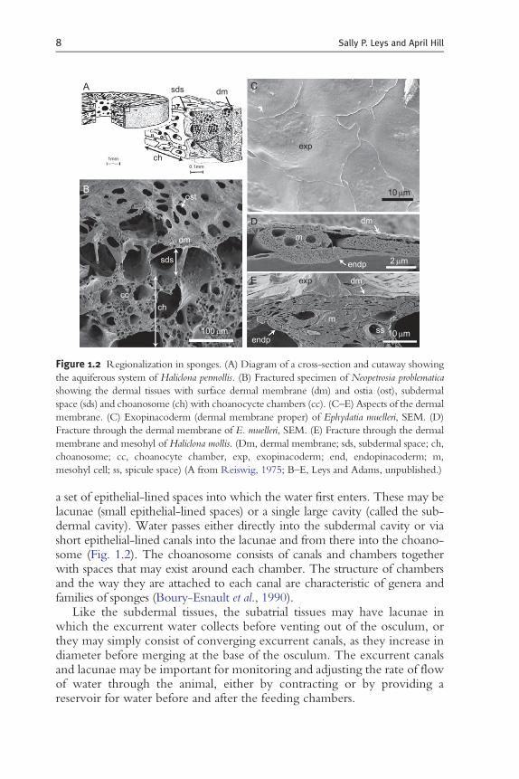
Sponges have quite diverse body constructions. We focus here on thehaplosclerid-type morphology, many aspects of which can be generalized toother groups. The body wall consists of an outer layer (cortex), a canal systemthat brings water into chambers where the pumping and feeding cells are (thechoanosome), and lacunae (epithelial-lined spaces) or canals that bring waterto converge on the excurrent vent or osculum (the atrial tissue) (Fig. 1.2).
The outermost tissue has been called the dermal membrane because earlyresearchers thought the epithelium in all sponges was syncytial (Wilson,1907). Electron microscopy has confirmed that only hexactinellid spongeshave syncytial tissues; however, in some species, boundaries of surface cells arehard to see even by scanning electron microscopy, so it is understandable thatthey were difficult to see by light microscopy (Fig. 1.2C). The dermalmembrane (as used by earlier workers) consists of a thin single layer of cells,pinacocytes (see Section 3.1) that lies on a collagenous middle layer, themesohyl. It is worth noting that some authors used the term dermal mem-brane to refer to the entire outer region of the sponge—whether a singleexopinacoderm, or an exopinacoderm and endopinacoderm with fine meso-hyl inbetween—and others used it to refer to all pinacocytes lining the surfaceand canals of the sponge. Recent workers no longer use this term and referinstead only to the ‘exopinacoderm’ (Boury-Esnault and Rutzler, 1997).
The nature of the sponge surface layers varies greatly. In some sponges, theexopinacoderm covers a cortex—a distinct rind that consists of particular celltypes, spicule types, and occasionally foreign material embedded into the tissue(e.g. Teragawa, 1986). In other sponges, the skin is formed by a thin single layerof exopinacocytes covering amassive collagenousmesohyl, while in others, theskin forms a 3-mm-thin layer over an equally meagre mesohyl, which issandwiched by another epithelium, the endopinacoderm (or in syconoid andasconoid sponges, the choanoderm). Under the exopinacoderm lies the first of
1991). (C) Homoscleromorpha. Cross-sections of (i) Oscarella kamchatkensis, Gazave et al.,
2010; (ii)Corticium candelabrum (cc, choanocyte chamber, la, larva) (courtesy of A. Riesgo). (D)
Demospongiae. (i) Scanning EM and (ii) cross section of Ephydatia muelleri (ec, excurrent canal;
cc, choanocyte chamber; exp, exopinacoderm) (iii). Cross-section of Haliclona elegans showing
the different paths water might take through the sponges. (i and ii, Elliott and Leys, unpub-
lished; iii, Langenbruch and Scalera-Liaci, 1986.
A sds dm
exp
dm
dm
endp
exp
ssm
m
endp
ch
ost
dm
sds
ch
100mm 10mm
2mm
10mm
cc
1mm
0.1mm
B
D
E
C
Figure 1.2 Regionalization in sponges. (A) Diagram of a cross-section and cutaway showing
the aquiferous system of Haliclona permollis. (B) Fractured specimen of Neopetrosia problematica
showing the dermal tissues with surface dermal membrane (dm) and ostia (ost), subdermal
space (sds) and choanosome (ch) with choanocycte chambers (cc). (C–E) Aspects of the dermal
membrane. (C) Exopinacoderm (dermal membrane proper) of Ephydatia muelleri, SEM. (D)
Fracture through the dermal membrane of E. muelleri, SEM. (E) Fracture through the dermal
membrane and mesohyl of Haliclona mollis. (Dm, dermal membrane; sds, subdermal space; ch,
choanosome; cc, choanocyte chamber, exp, exopinacoderm; end, endopinacoderm; m,
mesohyl cell; ss, spicule space) (A from Reiswig, 1975; B–E, Leys and Adams, unpublished.)
8 Sally P. Leys and April Hill
a set of epithelial-lined spaces into which the water first enters. These may belacunae (small epithelial-lined spaces) or a single large cavity (called the sub-dermal cavity). Water passes either directly into the subdermal cavity or viashort epithelial-lined canals into the lacunae and from there into the choano-some (Fig. 1.2). The choanosome consists of canals and chambers togetherwith spaces that may exist around each chamber. The structure of chambersand the way they are attached to each canal are characteristic of genera andfamilies of sponges (Boury-Esnault et al., 1990).
Like the subdermal tissues, the subatrial tissues may have lacunae inwhich the excurrent water collects before venting out of the osculum, orthey may simply consist of converging excurrent canals, as they increase indiameter before merging at the base of the osculum. The excurrent canalsand lacunae may be important for monitoring and adjusting the rate of flowof water through the animal, either by contracting or by providing areservoir for water before and after the feeding chambers.

The Physiology and Molecular Biology of Sponge Tissues 9
2.3. Cells, tissues, and regionalization
The sponge body has functional regions defined by specific tissues and thecell types in them. Tissues are defined after Hyman (1940) as being formedby one or more type of cell and organized so as to form a functioning unit(whole). Most sponge larvae also have well-differentiated regionalizedtissues, and in the larva, there is the additional function of motility anddirection finding, for which regions are specialized (Maldonado andBergquist, 2002), but the types of tissues and cells present in the larva forthe most part take their names (and we infer their function) from those inthe adult. In the adult, sponge tissues include the choanocyte epithelium(choanoderm), including cells supporting the choanocyte chambers; thepinacocyte epithelium (pinacoderm), including dermal tissues, incurrent,and excurrent canals; the skeletal and collagenous support tissues; and theosculum. Here, we focus on the structure and formation of two epithelialtissues only, the choanoderm and pinacoderm. We do not address skeletaltissues or cells of the mesohyl.
3. The Choanoderm Epithelium
3.1. Overview of the aquiferous system
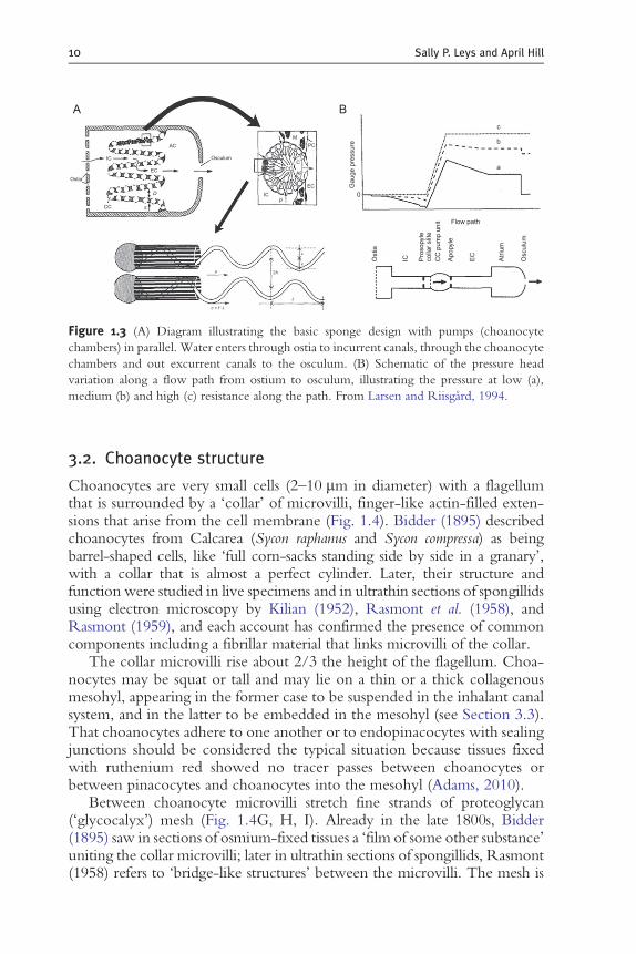
Flow through the sponges is generated by the beating of many thousandsof flagella per cubic millimetre of choanosomal tissue. The beat of theflagellum generates a low pressure at its base, drawing water toward andthrough the collar and from there up along the length of the flagellumaway from the cell body (Larsen and Riisgard, 1994). Many choanocytesin a choanocyte chamber, with collars and flagella facing toward the exit ofthe chamber, generate a current of water through the collar into theirbases, and from there into the chamber and out of the chamber exit. Themanner in which choanocytes are organized into chambers (how manythere are, how many openings there are between them, what the dimen-sions of the collar slits are) and the manner in which chambers areorganized at the end of incurrent canals have been studied in manysponges (e.g., Langenbruch, 1983, 1988; Langenbruch et al., 1985;Langenbruch and Scalera-Liaci, 1986; Langenbruch and Weissenfels,1987; Langenbruch and Jones, 1990; Boury-Esnault, 2006). The dimen-sions of the fine incurrent and excurrent canals are features that determinethe precise resistance over the collar slit in the choanocyte chamber(Larsen and Riisgard, 1994) (Fig. 1.3). The pressure drop must slow theflow sufficiently for choanocytes (or pinacocytes at the entrance to cham-bers) to phagocytose food which is primarily bacteria �/�� to ½ the size ofthe choanocyte.
A
Ostia
Osculum
AC PC
M
C
EC
CCAP
P0
2h
c = f .l
l
m
e
e
a
Flow path
Gau
ge p
ress
ure
Ost
ia
IC Pro
sopy
leco
llar
slite
CC
pum
p un
it
Apo
pyle
EC
Atr
ium
Osc
ulum
b
c
IC
EC
IC
CC
D
S
B
Figure 1.3 (A) Diagram illustrating the basic sponge design with pumps (choanocyte
chambers) in parallel. Water enters through ostia to incurrent canals, through the choanocyte
chambers and out excurrent canals to the osculum. (B) Schematic of the pressure head
variation along a flow path from ostium to osculum, illustrating the pressure at low (a),
medium (b) and high (c) resistance along the path. From Larsen and Riisgard, 1994.
10 Sally P. Leys and April Hill
3.2. Choanocyte structure
Choanocytes are very small cells (2–10 mm in diameter) with a flagellumthat is surrounded by a ‘collar’ of microvilli, finger-like actin-filled exten-sions that arise from the cell membrane (Fig. 1.4). Bidder (1895) describedchoanocytes from Calcarea (Sycon raphanus and Sycon compressa) as beingbarrel-shaped cells, like ‘full corn-sacks standing side by side in a granary’,with a collar that is almost a perfect cylinder. Later, their structure andfunction were studied in live specimens and in ultrathin sections of spongillidsusing electron microscopy by Kilian (1952), Rasmont et al. (1958), andRasmont (1959), and each account has confirmed the presence of commoncomponents including a fibrillar material that links microvilli of the collar.
The collar microvilli rise about 2/3 the height of the flagellum. Choa-nocytes may be squat or tall and may lie on a thin or a thick collagenousmesohyl, appearing in the former case to be suspended in the inhalant canalsystem, and in the latter to be embedded in the mesohyl (see Section 3.3).That choanocytes adhere to one another or to endopinacocytes with sealingjunctions should be considered the typical situation because tissues fixedwith ruthenium red showed no tracer passes between choanocytes orbetween pinacocytes and choanocytes into the mesohyl (Adams, 2010).
Between choanocyte microvilli stretch fine strands of proteoglycan(‘glycocalyx’) mesh (Fig. 1.4G, H, I). Already in the late 1800s, Bidder(1895) saw in sections of osmium-fixed tissues a ‘film of some other substance’uniting the collar microvilli; later in ultrathin sections of spongillids, Rasmont(1958) refers to ‘bridge-like structures’ between the microvilli. The mesh is

A
2 mm
2 mm
E
JC
g
B
as
r
CC
mv gl fcps
asbfbbac
m nbf
F
E
D
C c
as
mt
nu0.2mm 0.1mmn
ac
bb
K LI
F G H
0.1mm
0.1mm
1/10m
1/10m
CB D
2
3
20
9
1 mm
1 mm
3
19
3
2
20
9
9
845
10
5
12
25
Figure 1.4 Choanocyte structure. (A) Choanocyte from Ephydatia muelleri (SEM); (B) Long-
itudinal section through the flagellum of E. fluviatilis TEM; (C) Fluorescence image of two
choanocytes from Ephydatia muelleri showing actin labelled with Bodipy Fl phallacidin (green)
and tubulin labelled with anti-alpha tubulin (red); nuclei are labelled with Hoechst 33342
(blue); (D) Diagram of a choanocyte from Brill, 1973); (E) Choanocytes in Sycon coactum,
Calcarea; (F) Choanocyte from Oscarella lobularis, Homoscleromorph; (G–I) Fibrillar proteo-
glycan mesh between microvilli of Spongilla lacustris, G and H, and between microvilli of Sycon
coactum, I. (J–L) Structure of the flagellum and basal body of a choanocyte inHalisarca dujardini.
(A, C, E, Leys unpublished; B, D, Brill, 1973; F, Boury-Esnault et al., 1984; G, H, Fjerding-
stad, 1961; J–L, from Gonobobleva and Maldonado, 2009).
The Physiology and Molecular Biology of Sponge Tissues 11
better preserved by glutaraldehyde/osmium fixation and has subsequentlybeen found both outside and inside the collar in many sponges. Electronmicrographs show a mesh on the collar in Spongilla lacustris (demosponges,Fjerdingstad, 1961), Sycon (Calcarea, Ledger 1976 in Simpson 1984), andFarrea occa and Rossella racovitzae (hexactinellids, Reiswig and Mehl, 1991;

12 Sally P. Leys and April Hill
Koestler in Leys et al., 2007). The mesh forms spaces of approximately 40square nanometres, and importantly, this is the smallest space (and greatestsurface area) through which the water actually passes in the sponge.
In freshwater demosponges, a layer of glycocalyx also forms what lookslike a gasket—a structure filling in the space between neighbouring collarsapproximately 2–3 mm above the cell’s surface (Weissenfels, 1992). In somemarine demosponges (e.g. Haliclona mollis), a set of cells forms a layer similarto that gasket, sealing the spaces between each of the collars (Adams, 2010)(Fig. 1.5A–C); it is possible this layer of cells is what is referred to as Sollas’membrane (Bidder, 1895), an enigmatic structure whose existence has beendifficult to confirm. This gasket is well known in glass sponges (Hexactinel-lida) where it is called the ‘secondary reticulum’, a layer of syncytial tissuethat rises from the primary reticulum to surround the collar of each collarbody (Mackie and Singla, 1983). The secondary reticulum may play a role inchannelling flow through the collar, as inferred from a study that watched thepassage of micorospheres through sponge fragments using video microscopy(Wyeth et al., 1996; Wyeth, 1999). In Ephydatia fluviatilis, it is suggested tofunction as a ‘one-way valve’ to prevent back flow of water (Langenbruchand Weissenfels, 1987), so in both cases, perhaps it has a similar function.
3.3. Organization of choanocyte chambers—Terminology
There are some 50–200 choanocytes in demosponge chambers (Reiswig,1975; Rasmont and Rozenfeld, 1981), 200–300 in a hexactinellid chamber(Leys et al., 2011), and up to 1000 in a calcareous syconoid chamber (Sycon,Leys, personal observation). Openings to choanocyte chambers are called‘pyles’ (orifice Gr.), and, therefore, entrances are called prosopyles (L.forward of) and exits, apopyles (Gr. away from). Prosopyles can be formedby pseudopodial extensions of choanocytes arranged so as to create a hole
cc
coc
sr
sr
10 mm
A B C
2mm 2mm
mv
Figure 1.5 Gasket of cells surrounding the collars in choanocyte chambers ofHaliclona mollis.
(Adams, 2010). (A) Low and (B,C) high magnification views of the cells that envelop the
collar microvilli.

The Physiology and Molecular Biology of Sponge Tissues 13
(choanocytic prosopyles), or by pinacocytes that contact the choanocytesand form something like a porocyte (pinacocytic prosopyle). Apopyles arelarger and, therefore, formed by several cells, called either ‘cone’ cells orapopylar cells (De Vos et al., 1990), which are interpreted to arise fromchoanoblasts during early chamber formation (Weissenfels, 1981). Unlikechoanocytes, however, which are flagellated, cone cells are ciliated and thecilium beats in a slowwhip-like fashion away from the chamber (Fig. 1.6) (seeSection 4.2). Some sponges lack cone cells and instead several endopinaco-cyte-like cells form a sieve plate at the exit of the chamber (e.g. Tethyawilhelma Nickel, personal communication; SPL, unpublished data).
3.4. Choanocyte function—Feeding
The action of the choanocyte flagellum in generating a low pressure to drawwater through the collar is well described by Simpson (1984) from VanTright (1919) and Kilian (1952). Though the basics are quite clear, exactlyhow the water moves through the collar and chamber is not actuallyknown. For example, while it is understood that water follows a pressuregradient generated by the choanocyte, as noted above, it is suggested thatthere must be one-way valves to prevent water from flushing the other wayunder changes in external pressure (Vogel, 1978). Though potential one-way valves (apopylar cells and a glycocalyx mesh between the collars) havebeen identified by Langenbruch and Weissenfels (1987), it would be diffi-cult to test how they work. Also, whether all water must pass through thecollar filter or whether there are bypass canals is likewise not known. Someauthors suggest that bypass structures exist, for example, to compensate forpressure changes that occur during rhythmic contractions (Nickel et al.,2006), and other authors, using corrosion casts to study the aquiferoussystem, also find links between incurrent and excurrent canals (Bavestrello
ap
A B C
end ci
10mm 1mm
chch
ci
1mm1mm1mm10mm
Figure 1.6 Apopyles and apopylar (cone) cells. (A) Several apopyles (ap) open into an
excurrent canal in Ephydatia muelleri (end, endopinacoderm; chch, choanocyte chambers).
(B, C) Apopylar cells with cilia (ci) which are approximately 4 mm long and show the
characteristic bending of ciliary motion. (Leys, unpublished).
14 Sally P. Leys and April Hill
et al., 1988). However, since using corrosion casts may cause tearing orrupture of the canal system, doubt remains as to whether such bypasses exist,especially when it is considered that these would generate leakage in thepressure system that the choanocyte filter depends upon. Feeding studieswhich show greater than 95% retention of bacteria<1 mm by the sponge donot support the presence of bypass canals in all sponges (Pile et al., 1996;Yahel et al., 2003, 2007).
Food capture occurs at choanocytes, prosopyles, and incurrent canalepithelia (Kilian, 1952; Weissenfels, 1976; Imsiecke, 1993; Leys and Eerkes-Medrano, 2006); even exopinacocytes have been shown capable of uptake oflatex beads (Willenz and Van de Vyver, 1982), so epithelial cells in generalmust be capable of phagocytosis. Generally however, bacteria and otherpicoplankton are captured by pseudopodia at the choanocyte, and once inthat cell, are transferred to amoebocytes for digestion and transfer of foodto other cells in the sponge. The transfer of food between choanocyte andamoebocyte has been shown by video microscopy by Imsiecke (1993),and between amoebocyte and other cells in a suite of studies by Willenz andcolleagues (Willenz and Van de Vyver, 1984, 1986; Willenz et al., 1986).
3.5. Choanocyte differentiation and turnover
Choanocytes are one of the better-understood cells in sponges in terms oftheir origin, function, and differentiation potential. They generate flow,capture food, and can differentiate into other choanocytes, spermatocytes,and even oocytes. Other studies suggest that they can even become archae-ocytes, and in this sense, they are a type of stem cell (Funayama et al., 2010). Incases where sperm arise from choanocytes, usually whole populations ofchoanocytes in chambers undergo this modification, multiplying in number,losing the collar, packaging the nucleic material, and modifying the cell bodyto form spermatic cysts (Boury-Esnault and Jamieson, 1999; Maldonado andRiesgo, 2008). Therefore, choanocytes are at the same time dispensable cellsand have the potential to re-differentiate into gametes.
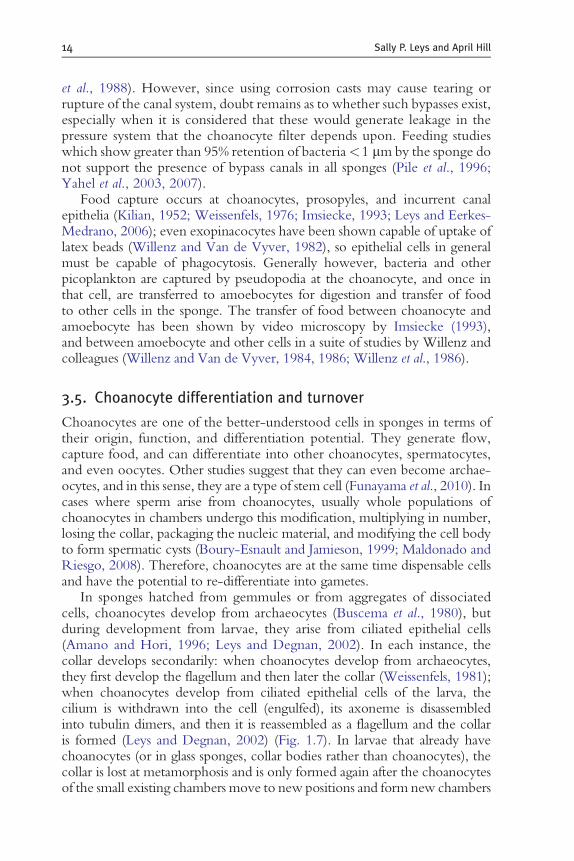
In sponges hatched from gemmules or from aggregates of dissociatedcells, choanocytes develop from archaeocytes (Buscema et al., 1980), butduring development from larvae, they arise from ciliated epithelial cells(Amano and Hori, 1996; Leys and Degnan, 2002). In each instance, thecollar develops secondarily: when choanocytes develop from archaeocytes,they first develop the flagellum and then later the collar (Weissenfels, 1981);when choanocytes develop from ciliated epithelial cells of the larva, thecilium is withdrawn into the cell (engulfed), its axoneme is disassembledinto tubulin dimers, and then it is reassembled as a flagellum and the collaris formed (Leys and Degnan, 2002) (Fig. 1.7). In larvae that already havechoanocytes (or in glass sponges, collar bodies rather than choanocytes), thecollar is lost at metamorphosis and is only formed again after the choanocytesof the small existing chambers move to new positions and form new chambers
A B
10mm
G
D
Fd
d
N
C
C
C
NN
N
g gg g
p
f
f f f
f
f
13
23
C
Figure 1.7 Differentiation of choanocytes. (A) Loss of cilia from the metamorphosing larva
of Amphimedon queenslandica and their disassembly in cells of the post-larva. (B) Fluorescently
labelled ciliated cells of the larva transdifferentiate into choanocytes (arrows) of the juvenile
sponge. (C, D) Collar formation in Tetilla serica: a tube becomes gradually separated into
microvilli. (A, B, Leys and Degnan, 2002; C, D, Watanabe, 1978b).
The Physiology and Molecular Biology of Sponge Tissues 15
(Leys, personal observations). An excellent account of collar formation comesfrom Watanabe (1978a) who showed that in Tetilla serica, new choanocytesform the collar as a membranous tube (essentially a pseudopodial extension)which, once it reaches a 2–3 mm in height, divides into the fingers thatbecome microvilli (Fig. 1.7). Choanocytes can, therefore, dedifferentiateand re-differentiate easily—they are fairly plastic.
Funayama et al. (2005a) took advantage of the feeding behaviour ofchoanocytes to find a molecular marker for the choanocyte cell lineage.They fed the sponge fluorescent plastic microspheres (latex beads), and oncethese were captured by the sponge, they dissociated the sponge tissue andvia fluorescent cell sorting isolated the cell fraction that contained thefluorescent beads. A gene that was expressed highly in mRNA isolatedfrom that fraction was annexin, and via in situ hybridization its expression inchoanocytes and choanocyte-committed archaeocytes was confirmed(Funayama et al., 2005a). Annexin has many roles in cell adhesion, cell

16 Sally P. Leys and April Hill
motility, endocytosis, and vesicle transport, and so its specific expression inchoanocytes may reflect the role of those cells in nutrient uptake andtransport (Funayama et al., 2005a).
The high turnover rate of choanocytes has been known for some time,and in one instance, it has been suggested that spent choanocytes aresloughed off several times a day (De Goeij et al., 2009). Measurements ofthe choanocyte cell cycle range from 20 h for Hymeniacidon synapium(Shore, 1971) to 15 h for Baikalospongia bacillifera (Efremova and Efremov,1979) and 5.4 h for Halisarca caerulea (De Goeij et al., 2009). The latterestimate based on the number of BrdU positive cells in sections of labelledsponges may be on the short side; mammalian intestinal epithelial cellsturnover every 3–60 days (Karam, 1999). Regardless of the precise rate,clearly, choanocytes are replaced frequently compared to other cells in thesponge body, and this has important implications. Firstly, it means there is apopulation of stem cells that replaces them and this appears to be archae-ocytes committed to the choanocyte lineage. In contrast to pinacocytes,choanocytes are continually dividing, as can be shown by BrdU staining ofdividing nuclei (De Goeij et al., 2009; Funayama et al., 2010) and thelabelling of archaeocytes that are committed to be choanocytes as well aschoanocytes themselves with the stem cell marker Piwi (Funayama et al.,2010). Secondly, it implies that either choanocytes are cheap to make or thatpumping is a costly activity and that these cells are destroyed or are easilydamaged. The small size of choanocytes might reflect the need to be able toreplace them rapidly at a minimal expense. Rasmont and Rozenfeld (1981)determined that based on the volumes of each cell type, in E. fluviatilis, asingle archaeocyte could divide into at least 30 choanocytes.
Determining the cost of production of a choanocyte would greatly help usunderstand the exact cost of pumping for the sponge. It has been estimatedthat the cost of pumping amounts to 1% of metabolism (Riisgard et al., 1993;Larsen and Riisgard, 1994). However, recent measurements of oxygen con-sumption in a sponge that went from not pumping to a maximum pumpingrate suggest that the cost of pumping is up to 30% of metabolism (Hadas et al.,2008), and a new study of filtration in glass sponges (Leys et al., 2011) suggeststhis may be due to the resistance generated by the fine dimensions of theglycocalyx mesh on the collar, which was not taken into account earlier.More data on the dimensions of passages through sponges, and of oxygenconsumption in a variety of sponges, will greatly help refine our under-standing of energy expenditure and the cost of filtration for sponges.
3.6. Control over flow
Sponges can control the flow through their aquiferous system, but they dothis in different ways. In glass sponges (Hexactinellida), the flagella stopmoving (‘arrest’) when the sponge is irritated by sediment or there is damage

The Physiology and Molecular Biology of Sponge Tissues 17
to the tissue. This happens by the spread of an action potential through thesyncytial tissues (see below); the flagella only start to beat again after themembrane potential has reset. Other sponges are cellular and as far as weknow are not able to spread action potentials and changes in flow neveroccur within the timeframe that would suggest electrical activity. In thosesponges, the flagella beat without arrest for hours on end or until ‘conditionsare unfavourable’ (Grant, 1825).
Grant was probably one of the first to observe this incessant pumping ofwater and gives a nice account of using a microscope and the reflection ofcandle light to watch the flow pour from the osculum for up to 5 h withoutchange in rate (Grant, 1825). Cellular sponges can contract their canals andclose their ostia and oscula, so flow can be slowed and stopped gradually, butin general, it is understood that the flagella of choanocytes—as do cilia—beatrelentlessly, even if each flagellum might beat at a slightly different rate thanthe others. Observations of flagella beating in intact living tissues (damageddissociated choanocytes have a highly variable beating pattern) indicate thatthe flagella beat in sinusoidal waves in a single plane and each choanocyteflagellum in a chamber beats in a slightly different plane to the others ( Jones,1964; Kilian and Wintermann-Kilian, 1979; Wyeth et al., 1996; Wyeth,1999). Jones (1964) notes that in the calcareous sponge, Leucosolenia, thebeat varies in frequency but is usually too fast to be measured; the same istrue for freshwater demosponges (Kilian and Wintermann-Kilian, 1979); thebeat eventually slows and stops after some hours (Kilian, 1952; Jones, 1964).According to Kilian andWintermann-Kilian (1979), however, inE. fluviatilis,some flagella do occasionally stop beating. While slowing and arrest afterseveral hours of observation can be attributed to damage (light, tearing, lackof oxygen or flow under the microscope slide), it is unclear what themechanism of arrest of an individual flagellum would be.
So far, only glass sponges (hexactinellids) are known to be able to arrest theflagella in an instant (<30 s), and they can do this because they have syncytialtissue which, upon a mechanical or electrical stimulus to the sponge (a strongprod, or damage to the tissue), propagates electrical signals throughout theentire sponge body (Leys and Mackie, 1997). The action potential wouldcause calcium to enter the collar bodies, and the rise in intracellular calciumwould arrest the flagellum (Leys et al., 1999). The flagella beat resumes onlyonce calcium is pumped out of the cell body. In demosponges, homoscler-omorphs, and calcareous sponges, no electrical (rapid) signalling mechanism isknown. If flagella were to stop, as they do in hexactinellids, this implies thatthere must be control over calcium levels in individual choanocytes and it isunclear how that would happen except during contraction of the choano-some. All cellular sponges have been known to be able to contract their tissuesslowly in response to poor water quality, mechanical or chemical irritation,or as a spontaneous rhythm (e.g. Parker, 1910; McNair, 1923; Prosseret al., 1962; Emson, 1966; Prosser, 1967; De Vos and Van de Vyver, 1981;
18 Sally P. Leys and April Hill
Weissenfels, 1990; Nickel, 2004; Elliott and Leys, 2007), and it has now alsobeen shown that the lyssacine hexactinellidOopsacas minuta can also carry outvery slow contractions of the entire body (Nickel, 2010). Contractions arethought to be coordinated by calcium waves that travel through the tissuesbecause contraction amplitude is reduced in the absence of calcium andabolished in the absence of calcium and magnesium (Elliott and Leys, 2010).In other animals, calciumwaves are propagated by a repeated cycle in which atrigger (e.g. mechanical irritation) causes a rise in intracellular calcium [Ca2þ]iwhich causes release of molecules (e.g. glutamate, NO, ATP, or cAMP;Elliott and Leys, 2007; Elliott and Leys, 2010) that bind receptors on neigh-bouring cells triggering a rise in [Ca2þ]i in those cells, and so on until all thecells in a region have experienced a calcium rise (reviewed in Nickel, 2010).During a strong contraction, even whole choanocyte chambers contract andchoanocyte collars and flagella are so compressed that they project out of theexit opening (apopyle) of the chamber (Elliott, 2009); at that point, themotionof the flagella is also noted to have stopped (Weissenfels, 1990).Watermay alsobe slowed from moving through canals by the contraction of sphinctersformed by cells in canals. Reiswig (1971) showed that reduced oscula diametercorrelated with reduced pumping velocity in Tethya crypta, and constrictionof atrial canals corresponded with cessation of pumping in Verongia gigantea.
Control over the flow may be necessary to prevent collapse of the canalsdue to unequal pressure in the incurrent and excurrent canal system. If theincurrent canals clog, then the choanocyte pumps will be unable to bringwater into the chambers, or if the osculum is torn or any part of the excurrentor incurrent canal system is opened by wounding, the pressure drop across thechoanocytes in the chamber presumably will be lost. The sponge must havemechanisms to sense fine changes in the pressure across the entire system andadjust the dimensions of canals by contractions or remodelling. To repair thesystem, changes must occur in the space of minutes. We might, therefore,expect sensory cells in different regions of the aquiferous system, as well as theability of the sponge tissues, to have positional information to determinewhich regions are incurrent and which are excurrent so as to be able toconsistently remodel according to this directional body plan.
4. The Pinacoderm Epithelium
4.1. Pinacoderm description and overview of function
The main function of an epithelium is compartmentalization, to control thesecretion and absorption of water and the transport of chemicals, proteinsand ions between body regions. Epithelia also detect and ward off infection(Van de Vyver, 1970; Muller and Muller, 2003), and provide structuralsupport (Teragawa, 1986), especially when out of water as would be the
The Physiology and Molecular Biology of Sponge Tissues 19
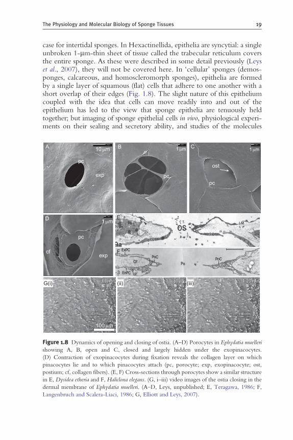
case for intertidal sponges. In Hexactinellida, epithelia are syncytial: a singleunbroken 1-mm-thin sheet of tissue called the trabecular reticulum coversthe entire sponge. As these were described in some detail previously (Leyset al., 2007), they will not be covered here. In ‘cellular’ sponges (demos-ponges, calcareous, and homoscleromorph sponges), epithelia are formedby a single layer of squamous (flat) cells that adhere to one another with ashort overlap of their edges (Fig. 1.8). The slight nature of this epitheliumcoupled with the idea that cells can move readily into and out of theepithelium has led to the view that sponge epithelia are tenuously heldtogether; but imaging of sponge epithelial cells in vivo, physiological experi-ments on their sealing and secretory ability, and studies of the molecules
A B
exp
pc
pc
exp
G(i) (ii) (iii)
cf
D E
F
pcpc
ost
C10mm 1mm
1mm
1mm
1mm
100mm
10mm 1mm 1mm
Figure 1.8 Dynamics of opening and closing of ostia. (A–D) Porocytes in Ephydatia muelleri
showing A, B, open and C, closed and largely hidden under the exopinacocytes.
(D) Contraction of exopinacocytes during fixation reveals the collagen layer on which
pinacocytes lie and to which pinacocytes attach (pc, porocyte; exp, exopinacocyte; ost,
postium; cf, collagen fibers). (E, F) Cross-sections through porocytes show a similar structure
in E, Dysidea etheria and F, Haliclona elegans. (G, i–iii) video images of the ostia closing in the
dermal membrane of Ephydatia muelleri. (A–D, Leys, unpublished; E, Teragawa, 1986; F,
Langenbruch and Scalera-Liaci, 1986; G, Elliott and Leys, 2007).
20 Sally P. Leys and April Hill
present in sponges shows that despite their apparent simplicity, establishedepithelia in sponges can and do form effective barriers (Adams et al., 2010).
4.2. Pinacocytes—Terminology
The sponge surface is formed by a flat cell type called a pinacocyte (literally‘pavement’ cell and pinacoderm, a pavement epithelium). A good descriptionof the pinacoderm is given by Simpson (1984) and De Vos et al. (1991).Pinacocytes are thin (often 1 mm thick except at the nucleus) broad cells (upto 20 mm in diameter) that form a squamous sheet. In many sponges, pina-cocytes are T-shaped, with the nucleus in a cell body that projects down intothe collagenous middle layer. Pinacoderms line the sponge surface and canalsand also line the internal cavity and canals of the larvae of freshwater sponges.
At least six types of pinacocytes can be identified by morphological orpositional differences. Pinacocytes forming the outer surface of the spongeare exopinacocytes, those on internal surfaces (on canals or lacunae) areendopinacocytes, and those that form the base of the sponge and provide theattachment to a surface and which can secrete a massive calcareous skeleton(e.g. Gilis, 2011) are distinguished as basopinacocytes. Furthermore, thepolarity of the aquiferous system defines the prefixes given to pinacocytesbefore (pro) and after (ap) the feeding chambers, hence prosopinacocytesand apopinacocytes (Boury-Esnault and Rutzler, 1997). As more geneexpression data become available, with genes labelling particular regionsof a sponge, it is increasingly important to use terminology that accuratelydefines location. Differences might be expected, for example, in the popu-lations of potassium channels or glutamate receptors on incurrent andexcurrent canal epithelia.
4.3. Cilia and flagella—Function and location in the sponge
An interesting and little discussed feature of sponge pinacocytes is that theycan be ciliated (Boury-Esnault and Rutzler, 1997); Simpson refers to thecilia as flagella (Simpson, 1984, p. 52). There is little (if any) difference in theultrastructure of cilia and flagella and often both organelles are referred to bya single term (usually cilium) (Lindemann and Lesich, 2010). However, ciliaand flagella have fundamentally different patterns of movement—cilia beatin a whip-like manner with active and recovery strokes (or are non-motile,sensory, organelles), whereas flagella beat in sinusoidal waves, and this hasbeen traced to differences in the domains of proteins involved in formingthe axonome (Linck, 1973). Sponges are one of the few animals that havesomatic cells (choanocytes) with flagella; flagella are also found in the flamecells of Platyhelminthes, but it seems that most other metazoans have ciliarather than flagella on somatic cells. Choanocytes can differentiate intosperm, which also possess flagella. In contrast, the sponge larval epithelium

The Physiology and Molecular Biology of Sponge Tissues 21
is formed by ciliated cells, which beat with a whip-like motion in meta-chronal waves, like the ciliated epithelia of other invertebrate larvae; atmetamorphosis of the larva, however, these ciliated cells dedifferentiate,resorb or lose the cilium, and migrate inwards, where they re-differentiateinto choanocytes with a flagellum and a collar (Amano and Hori, 1996; Leysand Degnan, 2002). Not all sponge larvae retain their ciliated cells for use aschoanocytes. In E. fluviatilis, the ciliated epithelial cells are engulfed byamoeboid cells which then spread out to form the new pinacocytes(Wielsputz and Saller, 1990), but since choanocytes are already formed infreshwater sponge larvae presumably, the ciliated epithelial cells are nolonger necessary to make new choanocytes at metamorphosis. It is interest-ing that only freshwater sponges and hexactinellids have choanocyte cham-bers in the larva; in both cases, these are ready to be used once the osculumhas formed after metamorphosis. The two types of sponges share little incommon except perhaps living in low nutrient habitats. If nutrients arelimited, perhaps it is more advantageous to be equipped to feed immediatelyupon metamorphosis rather than use up energy to reshuffle epithelial cellsand organelles into new locations.
As the sponge forms new canals after metamorphosis, ciliated pinaco-cytes arise on newly formed canal epithelia (SPL personal observation).Therefore, pinacocytes must be able to differentiate cilia, but whether allpinacocytes can do this, or whether certain archaeocytes are committed tothe ciliated lineage and differentiate into ciliated pinacocytes is not yetknown. In demosponges, endopinacocytes in the canals may have onecilium or a pair of cilia, for example, in Hippospongia communis and Tethyaaurantium (Simpson, 1984, p. 52; Vacelet et al., 1989; Boury-Esnault et al.,1990, figs. 23 and 24) or Ephydatia muelleri (Elliott, 2009), but all endopi-nacocytes that form the inner lining of the osculum are ciliated (in allsponges observed so far) with one to four cilia per pinacocyte (Ludeman,2010; Leys, personal observation). Cilia also occur on the apopylar cellswhich form a ‘gasket’-like exit to chambers (see Section 3.3). Many authorsrefer to these as flagella, but in E. muelleri, where chambers and canals can beobserved live in thin areas around the periphery of the animals, while theflagella of choanocytes beat in sinusoidal waves, the organelles on theapopylar cells beat like cilia in a whip-like manner (SPL personal observa-tion). Furthermore, using the fluorescent vital dye FM 1-43, which is takenup by channels in the cilium membrane, and using phase contrast micro-scopy to watch their motion cilia in the canals and osculum can be locatedand watched, and these do not ‘beat’ at all but simply vibrate as if in the flowof water (SPL and D. Ludemann, unpublished observations).
In homoscleromorph sponges, all endopinacocytes are ciliated, each withone cilium, and in most species so are exopinacocytes (Boury-Esnault et al.,1984; Maldonado and Riesgo, 2007). But no study has examined whetherthese are motile or non-motile, nor if motile, how they beat. In Calcarea, no

22 Sally P. Leys and April Hill
ciliated pinacocytes have ever been reported, and no cilia have been foundon any part of the syncytial trabecular reticula or dermal membranes ofhexactinellids.
4.4. Pinacoderm: Role in sealing and osmoregulation
In his studies of contraction mechanisms in sponges, Prosser (1967) foundthat marine sponges have an ionic gradient across the tissues, inwardsodium, and outward potassium, such that he anticipated the membranepotential would be�30 to�65 mV. Freshwater sponge pinacocytes have alow osmolarity (12–15 MOsM) and possess contractile vacuoles whichactively excrete water to the outside as external solute concentration falls(Brauer, 1975). These data together suggest that for both marine and fresh-water sponges, pinacocytes differ in ion content from the external environ-ment (salt or freshwater) and the pinacoderm controls the ion content ofthe mesohyl.
Sponge pinacoderms are thin and elastic and are coatedwith a proteoglycanmatrix which has so far prevented intracellular recordings of true membranepotential. But it is still possible to determine the ability of the pinacoderm toseal by studying the transport of ions across the whole epithelium in culture.Adams and colleagues cultured sponge tissues in small cups with a permeablemembrane bottom and recorded the ionic resistance across the sponge epithe-lium (Adams et al., 2010). Using miniature cell cultures, they simultaneouslyrecorded current in the medium above and below the tissue. Cultures thatformed a confluent (continuous) sheet covering all of themembrane in the cuphad a transepithelial resistance similar to that of epithelia in other metazoans(>2 MΩ). Furthermore, for a culture with high resistance, there was nopassage of a small molecular weight tracer across the tissue; if the tissue in thecup was torn, then the tracer was able to pass across the tissues (Adams et al.,2010). The authors considered the resistance was generated across the baso-pinacoderm (the tissue that was directly in contact with the cup membrane),but the endopinacoderm would also contribute to the resistance to passage ofions across the fine mesohyl and between pinacocytes. Therefore, it can beconcluded that the pinacoderm (baso- and endopinacoderm at least, and likelyalso the exopinacoderm) provides good resistance to the passage of ions andcontrols the passage of small molecules across it.
In the context of being able to control cell–cell signalling via the releaseof small molecules, a sealed and stable pinacoderm makes sense. Images ofthe exopinacoderm labelled with a vital fluorescent dye (FM 1–43) also showthat contacts between neighbouring pinacocytes remain stable for at least 1 h(Leys et al., 2009; Adams, 2010). So why is it thought that sponge epithelia aretransient? Statements about the looseness of sponge pinacocytes appear to stemfrom observations of highly mobile cells filmed at the periphery of explantsof sponge tissue (e.g. Bond, 1992), and from sections and scanning electron

The Physiology and Molecular Biology of Sponge Tissues 23
micrographs of fixed tissues that show cells not adherent to one another at thechoanocyte chambers (e.g. fig. 6 in Simpson, 1984), and the supposed entry ofbacteria through endopinacocytes into the mesohyl (Weissenfels, 1976).Whereas the motility of cells in explants is to be expected because thesecells are actively seeking new unexploited areas to establish tissues in, thedirect openings to the mesohyl from incurrent canals are not (Weissenfels,1976, 1983). The images showing openings to those areas were subsequentlyacknowledged to have been caused by damage (Langenbruch andWeissenfels,1987). Tissues that are poorly preserved often reveal openings to the mesohyl,but careful preservation and handling shows the cells in contact. The factthat sponge epithelia require such careful handling in order to preserve well,however, may be taken as some indication of the tenuous adhesion of spongejunctional molecules.
4.5. Cell adhesion and cell junctions
Sponges are almost best known for their ability to be disassociated—usually bybeing forced through fine mesh—and for the individual cells to re-associate ina species-specific manner (Wilson, 1907). Carbohydrate molecules, and spe-cifically large sulphated proteoglycans originally known as ‘aggregation fac-tors’, carry out species-specific adhesion (Fernandez-Busquets and Burger,2003; Carvalho de Souza et al., 2009; Vilanova et al., 2009). The adhesionoccurs at moieties on glycan fingers that extend from massive proteoglycanmolecules attached loosely to the cell surface. Both the attachment to the celland the species-specific recognition with other cells are calcium dependent.Carbohydrates are unusual cell-adhesion molecules, not commonly found inother phyla, although perhaps this mechanism of adhesion has not beenadequately explored in other metazoans (Fernandez-Busquets and Burger,2003), but they have a remarkable capacity for adaptation, and are able afterone interaction to modify the types of ligands so as to recognize secondencounters, that is they are polymorphic (Fernandez-Busquets and Burger,1999). This type of adhesion has been considered a potential ‘first’ adhesionbetween early unicellular animals (Fernandez-Busquets and Burger, 1999;Vilanova et al., 2009).
4.5.1. Molecules in sponge cell junctions—OverviewFrom the genome of the demosponge A. queenslandica, we know that spongeshave molecules that are involved in cell polarity, adhesion and sealing, andin attachment to and communication with the extracellular matrix (Fahey andDegnan, 2010). Transcriptome data from several other sponges are alsobeginning to provide a rich picture of potential epithelial genes in sponges.Therefore, in addition to the carbohydrate type adhesion described abovewhich is involved in species-specific aggregation, sponges produce mem-brane-associated proteins that are used in cell–cell adhesion. Junctions in

24 Sally P. Leys and April Hill
sponges differ from those in other invertebrates and vertebrates, but the detailsof position of the junction, molecules, and proteins involved in each junctiontype are only just being examined (e.g. Leys and Riesgo, 2011).
4.5.2. Adherens junctionsAdherens junctions hold cells together; they usually occur on one side ofa sealing junction and their presence around the apex of the cell is con-sidered an indication of tissue stability. While many authors refer to adher-ing junctions in sponges as desmosomes, strictly speaking desmosomes(macula adherentes) have desmosomal cadherins (desmogleins, desmocol-lins, and plakophilins) and plakoglobins, which are innovations of verte-brates. Invertebrates usually have zonula adherens, a belt-form junction, butin sponges, adherens junctions are usually spot-form junctions, appearing asdense plaques of actin between cells (Bagby, 1965; Pavans de Ceccatty,1981; Boury-Esnault et al., 2003; Elliott and Leys, 2007). Focal adhesions(spot desmosomes) have also been found on the basal epithelial layer ofE. muelleri (Pavans de Ceccatty, 1986) and perhaps are the only real desmo-somal adhesive junction sponges have. However, since none of the proteinsthat make up any of these adherens junctions have been studied, the truenature of sponge adherens junctions remains to be explored.
The principal component of adherens junctions is cadherin, a transmem-brane molecule with extracellular cadherin (EC) repeats, EGF, and LAMGrepeats (altogether called Fat cadherins). Sponges have 17 cadherins (Faheyand Degnan, 2010), and one of these (first reported by Abedin and King,2008) is a classical Fat cadherin, which is usually associated with adherensjunctions. In choanoflagellates, cadherins have binding sites for tyrosine phos-phorylation and are thought to be involved in cell signalling (Abedin andKing, 2008). In metazoans (including sponges), cadherins also have a conservedcytoplasmic domain, which has a binding site for b-catenin that is involved insignalling via the Wnt pathway. Therefore, adhesion and cell–cell signallinglikely went hand-in-hand in the earliest metazoans. One cadherin in A.queenslandica has a hedgehog-like domain (Adamska et al., 2007b), and it issuggested that its expression around the pigmented posterior pole of thesponge larva reflects a role in signalling during the development of polarityin that sponge (Adamska et al., 2007b, 2010) (see Section 7).
Other adhesion molecules present in the sponge genome belong tothe MAGUK family of transmembrane proteins (Sakaraya et al., 2007;de Mendoza et al., 2010; Fahey and Degnan, 2010). There are 10 types ofMAGUKs (de Mendoza et al., 2010), some of which generate the scaffoldthat anchors post-synaptic molecules in their place in nerve cells and othersthat generate tight junctions in vertebrates. Sponges have neither nerves nortight junctions, but given the presence of members of the DLG, MPP, andMAGI super families of MAGUK proteins in sponges, we might expectthese to be involved in cell–cell adhesion and/or signalling at cell junctions.

The Physiology and Molecular Biology of Sponge Tissues 25
In situ hybridization of MAGI RNA in ‘primorphs’ (small sponges grownfrom dissociated and reaggregated cells) suggested expression in both thedermal membrane (exopinacoderm) and the choanoderm of sponge epithe-lia (Adell et al., 2004). Similar hybridization experiments with DLG andseveral of the post-synaptic scaffolding genes in the A. queenslandica larvasuggest a varied expression with some label in the inner cell mass, some in thesub-epithelial layer, and some specifically in a type of globular cell (also calleda mucous cell) that populates the larval epithelium (Sakaraya et al., 2007).
4.5.3. Occluding junctionsOccluding junctions in sponge epithelia are both ‘parallel membrane’ junc-tions (Green and Bergquist, 1979, 1982; Pavans de Ceccatty, 1981; Adams,2010) and septate junctions (tight junctions are considered a chordate inno-vation). The former, in which the plasma membranes of two cells lie parallelto one another for about 500 nmwith only 10–20 nm separation, occlude thepassage of small molecular weight tracers (3H-inulin, 5KD, and rutheniumred, 0.85KD) (Adams et al., 2010). In these junctions, faint septae can some-times be seen in transmission electron micrographs, but generally the occlud-ing region occurs either at the parallel junctions or at regions adjacent to themin which the membrane of one cell appears merged (fused) or in tight contactwith the other cell. Septate junctions have been reported in all sponges (seereview by Leys et al., 2009), but only in calcareous sponges are the septae veryclear with 10 nm spacing between cells and between septae (Ledger, 1975;Green and Bergquist, 1979).
Septate junctions have been best characterized in Drosophila where twotypes are known. Pleated sheet SJs (pSJ) are found in the epidermis, salivarygland, and photoreceptors, while smooth SJ (sSJ) occur in the mid-gut andmalpighian tubules. The ultrastructure of each shows that pSJ have distinctseptae and sSJ lack them. Both junctions have the molecules neuroglian andcontactin and both molecules are needed for barrier function, but only pSJhave neurexin IV (a contactin-associated protein, or Caspr) (Baumgartneret al., 1996; Genova and Fehon, 2003; Faivre-Sarrailh et al., 2004), so thismolecule seems to be associated with the presence of septae but is notnecessary for barrier function (Baumgartner et al., 1996; Baumann, 2001).sSJ are instead characterized by fasciclin III, ankyrin, and a,b-spectrin.
Although the composition of the sponge septate and parallel membranejunctions is not yet known, the A. queenslandica genome does not seem tohave clear orthologs of neuroglian, neurexin IV, fibronectin III, or contactin (Faheyand Degnan, 2010).Neurexin IV, however, was reported inOscarella carmela, ahomoscleromorph (Nichols et al., 2006), and A. queenslandica does have aclaudin gene ortholog which aligns well with sequences from two othersponges as well as Drosophila, nematode, and human sequences (Leys andRiesgo, 2011). Claudins are known from tight junctions, but in the fly, theseproteins are localized to septate junctions and have been shown to play a role

26 Sally P. Leys and April Hill
in occlusion (Asano et al., 2003). Claudins are also present in the genome ofHydra (Chapman et al., 2010) and recent work using GFP-claudins showslabelling around the apex of each cell (Salvenmoser, 2011). It is, therefore,quite possible that the demosponge claudin molecules may also function inocclusion, and that in sponge parallel and/or septate junctions, perhapsclaudins rather than neurexins perform the sealing role (Leys and Riesgo,2011). To better understand how the sponge junction works, antibodylocalization and gene knockdown is needed, as well as further more detailedexploration of other sponge genomes as they become sequenced.
Other molecules found in the A. queenslandica genome and which maybe involved in establishing polarity of the epithelium, or in localizingjunctions to their polarized position in the epithelium, include Scrb, Par,Crumbs, and Patj (Fahey and Degnan, 2010). In other invertebrates, Scrb andPar are localized to septate junctions. Given the very narrow region whichsponge epithelial cells make contact with one-another, it is conceivable thatthese molecules are also present at the sponge junctions, but testing this willrequire antibody localization using immunogold to confirm the position atthe ultrastructural level.
4.6. The basement membrane: Differences amongsponge epithelia
The integrity of the epithelium is achieved not only by adhesion betweencells but also by the support of the extracellular matrix below. Dependingon the location in the sponge body, epithelia have some sort of basal supportprovided by collagen, and most also have an upper cuticle. There aretypically two components to the basement membrane—the lamina reticularis(a sheet of collagens I, II, and V), and the basal lamina (a 50–100 nm layer oftype IV collagen together with laminins and proteoglycans such as nidogen/entactin and perlecan) that lies immediately below, and follows every contourof, the cell membrane. Electron micrographs frequently show a dense mat ofcollagen underlying the sponge epithelium, which may be the equivalent of alamina reticularis. In electron micrographs of homoscleromorph sponges,there is also a fine basal lamina below both the pinacoderm and choanodermand antibodies to type IV collagen raised from that sponge recognize a layerthat is beneath the epithelium (Boute et al., 1996). Recently, type IV collagenthat corresponds well to alpha and beta chains has also been found in thecalcareous sponge Sycon coactum and another homoscleromorph, Corticiumcandelabrum (Leys and Riesgo, 2011). The finding of type IV collagen in Syconis all the more unusual since electron micrographs do not show a basementmembrane structure in any calcareous sponge.
The A. queenslandica genome does not have either type IV collagen or theproteoglycans nidogen or perlecan, and though there are several laminin-related genes, only one (Lam g-like) has a domain architecture sufficiently

The Physiology and Molecular Biology of Sponge Tissues 27
comparable to laminins from bilaterians to be considered proper laminins(Fahey and Degnan, 2010, 2012). Type IV collagen is necessary for basallamina formation since its globular heads link to form a mesh that is attachedto the cell surface by laminins. Although no other demosponge genomes areavailable, searches of other sponge transcriptomes and use of degenerate PCRhas not found conventional type IV collagen in other demosponges. How-ever, it has been shown that demosponges have a related form of thismolecule called ‘spongin short chain collagen’ (SSCC), which shares verysimilar NC1a and NC1b domains with type IV collagen and possesses four ofthe six conserved cysteine molecules typical of type IV collagen (Aouacheriaet al., 2006). SSCC is also predicted to have the same three-dimensionalstructure and therefore to be capable of forming the same interactions withthe epithelium as type IV collagen (Aouacheria et al., 2006). SSCC isexpressed by cells of the basal pinacoderm (Exposito and Garrone, 1990;Exposito et al., 1991). Further support for the idea that the three-dimensionalstructure of SSCC may be similar to type IV collagen comes from a study byDe Goeij et al. (2009) who showed that polyclonal antibodies to type IVcollagen from rabbit also recognize a layer below the choanoderm in thedemosponge H. caerulea. It is possible that these antibodies recognize aconserved 3D structure in the protein despite what would be considered tobe a little conserved amino acid sequence.
4.7. Pinacoderm function: Biomineralization
Biomineralization in sponges—the production of the skeleton—is the sub-ject of several excellent reviews (Uriz et al., 2003; Uriz, 2006; Sethmann andWorheide, 2008; Wang et al., 2010) and is also covered by Wang and co-authors in this issue. Most secretion of mineral and organic skeletons insponges takes place in the mesohyl, but here, we alert the reader to twoinstances in which the pinacoderm functions in biomineralization. The firstis the observation that some spicules are formed by pinacocytes in thehomoscleromorph sponge Corticium candelabrum (Maldonado and Riesgo,2007). This observation may not be so unusual if pinacocytes in homo-scleromorphs give rise to sclerocytes as they are said to do in Calcarea(Simpson, 1984, p. 147), and also to microsclerocytes as they are said todo in some demosponges (Simpson, 1984, p. 178). Maldonado and Riesgo(2007), however, do not think this is the case in C. candelabrum and there-fore this points to an unusual secretory ability of the pinacoderm in thathomoscleromorph sponge. The epithelium also plays a role in biominerali-zation in hypercalcified sponges, both demosponges (e.g. Acanthochaeteteswellsi,Astrosclera willeyana) and Calcarea (e.g. Petrobiona massiliana). The precisemechanism of mineral uptake, transport and secretion is not yet fully under-stood, but histology, histochemistry, and many types of energy X-ray analysesof the basopinacoderm from both demosponges and calcareous sponges

28 Sally P. Leys and April Hill
suggest that minerals (magnesium and/or calcium) are sequestered into vesi-cles in basopinacocytes and secreted as an amorphous material on the basalside of the epithelium (Reitner and Gautret, 1996; Worheide, 1998; Gilis,2011; Gilis et al., 2011). Polymerization (crystallization) of the material belowthe basopinacoderm is promoted by the prior or concurrent secretion of anorganic matrix, which is shown by staining with alcian blue, ruthenium redand acridine orange, to contain highly sulphated or acidic polysaccharides(e.g. glycosaminoglycans) (Gilis, 2011). Together, these materials allow for abiologically controlled crystallization of the minerals moulding them into abasal skeleton and incorporating into it spicules also produced by the sponge.In Astrosclera, biomineralization has also been shown to involve a type ofmesohyl cell, a large vacuolar cell (LVC) (Worheide, 1998), which is foundin the choanosome and ectosome above the calcified skeleton. Demonstra-tion that these cells express the enzyme carbonic anhydrase required forcatalyzing HCO3
� (Jackson et al., 2007) suggests that LVCs are involved inuptake of calcium and its transport to basopinacocytes where secretion of thematrix occurs.
4.8. Pinacoderm development
The origin of pinacocytes is, after more than a century of study, still unclear.Earlier research using a variety of models, such as dissociated tissue formingreaggregates (Wilson and Penney, 1930), dissociated larvae forming juvenilesponges (Borojevic and Levi, 1964, 1965), and explants of adult sponges(Simpson, 1963; Bagby, 1972), all from different species, concluded thatarchaeocytes differentiate into collencytes which become pinacocytes.Galtsoff (1925) who was the first to use aggregates for this, thought thatpinacocytes remained differentiated upon dissociation of a sponge, and simply‘sorted’ out to reform the pinacoderm. Wilson and Penney (1930) concludedthat a type of cell called a ‘grey cell’ with grey granular inclusions, formed thepinacocytes. Bagby (1972) determined that since there were no grey cells inMicrociona archaeocytes must become the pinacocytes. The only ‘live cell’study of this problem was by Simpson (1963), who showed that in explantspinacocytes did not divide to form the pinacoderm, but rather they arosefrom mesohyl cells which differentiated and moved into the pinacoderm.Study of the metamorphosing larva of E. fluviatilis supports the view thatarchaeocytes migrate up into and form the pinacoderm of the juvenile sponge(Boury-Esnault, 1976; Wielsputz and Saller, 1990). So in sum, new pinaco-cytes arise by differentiation of archaeocytes from the mesohyl.
Curiously, a recent study of sponges differentiated from gemmules ofSuberites domuncula suggests the opposite that the expression of genes in thepinacoderm triggers the differentiation of pinacocytes into sclerocytes andmesohyl cells (Schroder et al., 2004). After stimulation of canal formationusing increased temperature and Fe3þ, pinacocytes were the first cells to

The Physiology and Molecular Biology of Sponge Tissues 29
express noggin and silicatein, and subsequently, these genes were expressed incells in the mesohyl (Schroder et al., 2004). The authors concluded that asubpopulation of pinacocytes is able to differentiate into other cell types; butit is also possible that genes expressed by pinacocytes could induce thedifferentiation of cells in the mesohyl into other cell types. Whether cellsfrom the pinacoderm can differentiate into other cells might be studied byusing a fluorescent cell-tracker such as CMFDAwhich incorporates only intocells it has direct contact with; therefore, using a bath application mesohylcells would not be labelled. So far, two studies have used labelling of newnuclei using 5-bromo-20-deoxyruridine (BrDU) to identify stem cells, one inadult specimens of the crevice-dwelling marine sponge Halisarca (De Goeijet al., 2009) and the other in the freshwater sponge E. fluviatilis differentiatingfrom gemmules (Funayama et al., 2010). Although both groups found incor-poration of the label into choanocytes and archaeocytes within the 10-h timeframe of exposure to the drug, neither found the label in pinacocytes.
In sum, pinacocytes do not appear to divide, although this does not meanthey are terminally differentiated. It is possible that some pinacocytes candifferentiate into other cell types, but it is perhaps more likely that theytrigger/stimulate the differentiation of mesohyl cells into other cell types.The latter role is important in the formation of the canals and differentiationof the sponge body plan. Pinacocytes are, if not the first, certainly amongthe first cells to differentiate in the early development of a sponge from alarva or from a gemmule and during aggregation after dissociation of theadult. Pinacocytes line the incipient canal epithelia and are responsible forcontrolling ion exchange across the epithelium into the mesohyl.
5. The Aquiferous System
5.1. Differentiation of porocytes and canals
Ostia—incurrent openings into the canal system—are formed by porocytes,either singly, by having a large ‘hole’ in the middle of the cell, or possibly byseveral meeting up around an ostium to form the pore (Simpson, 1984).Evidence for the latter, however, comes from electron micrographs thatshow several pinacocytes converging on an ostium in a freshwater spongeexopinacoderm. Given that the porocyte lies below the exopinacocytes andbetween the two pinacoderm epithelia, when the pore is open, the cellbody may actually be completely hidden by the two pinacoderms (Fig. 1.8).
Porocytes have been seen to form in as little as 10 min in E. fluviatilis(Weissenfels, 1980) and within 10 h in Leucosolenia variabilis ( Jones, 1961).The former observation might be explained by a porocyte having beenhidden by pinacocytes and opening to reveal itself since videos of openingand closing ostia show this sort of time frame (Elliott and Leys, 2007)

30 Sally P. Leys and April Hill
(Fig. 1.8), but the latter suggests that—if cell division takes 10–20 h—theyprobably arise from mesohyl cells, rather than dividing from other pinaco-cytes. Oscula are also thought to arise initially from a single porocyte(Weissenfels, 1980), but how they coordinate with other porocytes toform a larger osculum is still unclear. In freshwater sponges hatched fromgemmules, the region in which the osculum will differentiate appears first asa bulge, which according to Brien (1932) is caused by pressure from waterpushed by the beat of newly formed chambers; but one wonders how thatpressure is dissipated if there is still no vent or opening that is the osculum.Nevertheless, the movement of fluid over the tissues to generate signals fordifferentiation of tissues is a common theme in animal development, evenin, for example, the nodal cilia.
Much of our understanding of canal formation comes from the observa-tions by Wintermann (1951) and Mergner (1964; 1966) and later byWeissenfels (1981), all of whom found that the excurrent canal system arisesfirst, chambers plug into the canals, and the incurrent canal system joins uplast (and is most plastic). The canals themselves arise from lacunae, smallepithelia-lined cavities within the sponge mesohyl, which move around,often in a clockwise centripetal manner, until they merge with others toform canals (Leys, personal observation). Hans Mergner first experimentedwith the role of the osculum in canal formation and found that if heremoved the osculum (from a small freshwater sponge hatched from gem-mules), it remained differentiated and formed a small ball of exo andendopinacoderm, which he called the ‘oscula ball’. He found that if heinserted this oscula ball anywhere in the sponge canal system it caused a newosculum to form in that spot (Mergner, 1964), and surmised that a substance(‘Induktionstoffes’) from the osculum tissue caused the differentiation ofcanal epithelia and formation of an osculum.
Recent experiments support the idea that osculum sends out a factorcontrolling canal formation. Windsor and Leys (2010) used Mergner’s(1964) experimental approach to remove oscula from the freshwater spongeE. muelleri and place them on the surface of a different sponge, one hatchedfrom the same batch of gemmules. They found that the transplanted osculumnot only attached to the surface of the sponge but also linked up with theexcurrent canal system and in fact drew the entire aquiferous system towardsit, causing the original osculum to cease to function (Windsor and Leys,2010). This seemed to confirm Mergner’s (1966) idea that a substancesecreted by the oscula tissues induces canal formation, competing withgradients secreted by the original host osculum. An alternative hypothesis,however, is that the secreted factor stimulates regeneration, and regenerationtriggers the release of factors that result in oscula formation.Windsor and Leys(2010) proposed that the secreted factor is Wnt protein because soaking thedeveloping sponge in Wnt activators caused a phenotype of multiple osculaand disruption of the aquiferous system.

The Physiology and Molecular Biology of Sponge Tissues 31
5.2. Role of Wnt in canal differentiation and polarityin sponges
To date, Wnt pathway genes have been isolated from at least 10 differentsponges—Amphimedon queenslandica (Adamska et al., 2010; Srivastava et al.,2010), Suberites domuncula and Lubomirskia baicalensis (Adell et al., 2007),Ephydatia muelleri (Windsor and Leys, 2010), Oscarella lobularis (Lapebieet al., 2009), and Sycon ciliatum (Adamska, unpublished); S. coactum, Pseudo-corticium jarrei, Spongilla lacustris and Petrosia ficiformis (Ana Riesgo, PamelaWindsor and Sally Leys, unpublished). Expression of Wnt genes has beenstudied in larvae, young adults and adult sponge tissue, but not in thedifferent developmental stages (larvae, postlarva and adult) of any onespecies. Our understanding the role of Wnt in sponge development andbody plan formation is therefore still patchy.
The most complete studies to date ofWnt gene and ligand expression havebeen carried out in the A. queenslandica sponge larva (Adamska et al., 2010)where it has been shown that one of the three Wnt genes (WntA) has apolarized expression at the posterior pole of the larva together with pigmentcells that form a ring around the posterior pole. WntA expression is juxta-posed with expression of TGFb, which labels the entire anterior portion ofthe larval ciliated epithelium (Adamska et al., 2010). Some Wnt ligands alsohave a polarized expression (e.g. LRP) while the expression of others islocalized more to the outer, middle or inner layers of the larva (Adamskaet al., 2010). Since LiCl experiments have so far not been successful inA. queenslandica, it is unknown what function Wnt has except for a probablerole in guiding anterior and posterior axis formation in the larva.
In the cnidarians Hydra and Nematostella, artificial activation of the Wntpathway causes the formation of ectopic tentacles and multiplication of the‘head’ end (Broun et al., 2005), and gradients ofWnt gene expressionmark theanterior to posterior poles of the polyp (Kusserow et al., 2005). To determinewhether the sponge canal system might develop in a polarized manner bygradients ofWnt gene expression, Windsor and Leys (2010) hatched spongesin a growth medium with drugs that activate the Wnt pathway (lithiumchloride and alsterpaullone) and found that with both drugs the sponges hadmany more oscula than normal sponges, and that the aquiferous system wascompletely malformed and non-functional. Furthermore, in the treatedsponges, there were either fewer ostia and porocytes, or they were completelyabsent. A similar approach was used in Oscarella lobularis by Lapebie et al.(2009). Incubation in azakenpaullone caused the sponge to form many moreostia; oscula were not studied. These results suggest that secretedWnt proteinmay be Mergner’s ‘induktionstoffe’ (Mergner 1966) which polarizes theepithelia during development to determine where ostia and incurrent canalsor oscula and excurrent canals form. In E. fluviatilis, there is also evidence thattheWnt pathway may be involved independently in radial organization of the
32 Sally P. Leys and April Hill
skeleton because sponges grown in BIO or azakenpaullone have a disorga-nized skeleton (Nakata and Funayama, 2011). If production of the skeletonwere necessary for proper organization of aquiferous system, these resultswould make sense together, but since it is also known that normal spongesform in the absence of materials to construct spicules (Weissenfels andLangenbruch, 1985), the simplest explanation is that the Wnt pathway hasmultiple roles in sponge development.
Other genes that may have a role in canal formation include the homeo-box genes Iroquois and NK class. Both show expression around canals or onthe outer epithelium (Perovic et al., 2003; Gazave et al., 2008, see Section 7),but experimental approaches will be needed to determine what role thesegenes might have in actual canal formation.
6. Epithelia as Sensory and Contractile Tissues
6.1. Overview of sensory and coordinating tissues
Sponges lack nerves in the conventional sense; that is, they do not possess adefined cell type that transmits signals using electrical impulses that travel alongthe cell membrane and communicate to other cells via a chemical synapse.This does not mean, however, that sponges do not send either electrical orchemical signals, but rather that the speed at which the signals propagatethrough the sponge is slower than conventional electrical signalling. Further-more, the means by which signals propagate is not necessarily by electricalimpulses, as generally is the case in other animals. Glass sponges propagateelectrical signals that shut down their feeding current (Leys andMackie, 1997;Leys et al., 1999; reviewed in Leys and Meech, 2006; Leys et al., 2007), but sofar, there is no evidence of electrical signalling in cellular sponges. However, atleast one species (Hymedesmia) is capable of dropping a tissue flap over its ostialpore fields in under 1 s (Reiswig, 1979), which indicates that localized, rapid(perhaps electrical) signalling may occur in some species. Signals that are notelectrical impulses would presumably be waves of calcium (propagated, loca-lized increases in calcium concentration that spread through the cell), triggeredby stimuli to the cell membrane, either via receptors on the cell surface or viachannels in the cell membrane. Signals could propagate through cells of theepithelium or cells of the mesohyl, but in recent years, the epithelium hasbecome recognized as the primary conduit of coordination in sponges.
In the 1970s, it was already known that signal transduction largelyoccurred through the epithelia of all sponges, although thick walled spongeswere thought to have single contractile cells or bundles of cells in themesohyl which were considered to play a role in contraction (Pavans deCeccatty, 1974a,b). Viewed live by cine-film or time-lapse digital micro-scopy, amoebocytes in the mesohyl of thin-walled sponges are very active,

The Physiology and Molecular Biology of Sponge Tissues 33
some of the cells move in a highly directional manner, others travel almostin circles (Pavans de Ceccatty, 1969, 1976; Adams, 2010). Collencytes leavea trail of collagen in their wake, sclerocytes sit stationary and take minutes tohours to develop a single spicule, lophocytes cluster around spicules, cellswith inclusions appear stationary, archaeocytes are sluggish and tend to staynear choanocyte chambers.
It is not possible to film cell activity in thick walled sponges, and certainlymesohyl cells may not be equally active in all sponges, or perhaps the activityof cells in the mesohyl depends on the thickness or density of collagen.Ultrastructural studies in the 1960s and 1970s showed cells stretching acrossthe mesohyl and in contact with epithelia at either end, interactions thatare suggestive of cell–cell communication (Pavans de Ceccatty et al., 1970;Pavans de Ceccatty, 1974a,b, 1986). The presence of actin microfilamentsin these cells was taken to indicate they might be contractile; therefore, theywere termed myocytes (Bagby, 1965, 1970). The general view was thatwhere sponge tissues were thin, it was more likely that pinacocytes werecontractile, while in thick tissues, it was more likely that mesohyl cells(myocytes) were contractile. In very thick walled sponges, sphinctersaround canals contract more rapidly than the whole sponge can and reducethe diameter of canals; the contractile cells in sphincters are pinacocytes. Inrecent years, pinacocytes have come to be considered the primary contrac-tile cell (Nickel et al., 2006; Nickel, 2010; Nickel et al., 2011), and actino-cyte substituted as a more generic term (Boury-Esnault and Rutzler, 1997),even though strictly speaking all cells would be expected to have actin.Although it is not possible to view mesohyl cells in thick sponges in vivo,estimates from area projections of contracted and relaxed sponges usingmicroCT scanning suggest that even where the mesohyl is substantial, theepithelium is the primary contractile and conductive pathway (Nickel et al.,2011). Nevertheless, even in thin-walled sponges like Ephydatia in whichperiodic contractions have been studied by time-lapse imaging since the mid-20th century (Pavans de Ceccatty et al., 1960; Pavans de Ceccatty, 1969; DeVos and Van de Vyver, 1981; Weissenfels, 1990), it is still unclear whetherboth pinacoderm and mesohyl cells are involved in contractions, but it islikely that the epithelium is the principal conduit for signal propagationthrough the sponge.
6.2. Molecules involved in coordination and signaltransduction
One of the most central questions in the evolution of animals is when didnervous tissues first arise? Because sponges show propagated signals thattranslate into slow but coordinated behaviour, they appear to bridge the‘gap’ between non-neural and neuronal states. Thus, it would seem thatgenomics and transcriptomics promise to shed much light on the topic of
34 Sally P. Leys and April Hill
neuron origins by revealing what aspects of ion channels, transmitters andsynapses are present in extant species. Molecules identified to date, how-ever, seem to simply support the current idea that sponges have an ‘almostnervous system’ of the type discussed by Pavans de Ceccatty (1974a,b,1979). Nonetheless, it is not yet clear if sponges never fully went downthe path to conventional nerves, or if they have in fact lost some of the keymolecules involved in conventional nervous systems. For example, theAmphimedon sponge genome has genes for calcium and potassium channelsbut no sodium channels (Liebeskind et al., 2011), yet sodium channels seemto be present in choanoflagellates, the unicellular protistan lineage sisterto the Metazoa. The absence of sodium channels in sponges may make sensegiven that physiological experiments on glass sponges (Hexactinellida)suggest they use calcium/potassium impulses (depolarization of the mem-brane by calcium influx and repolarization by potassium) rather thansodium/potassium as do most other animals (Leys et al., 1999). Calciumimpulses are temperature sensitive and have been put forward as one ofthe reasons that glass sponges are restricted in their range to deep, coolerwaters (Leys et al., 2003). The presence of a functional sodium channel inchoanoflagellates, however, prompts speculation that sponges may have lostaspects of a conventional nervous system. TheAmphimedon genome has bothinward rectifying (Kir) and putative votage gatedK
þ channels (Sakaraya et al.,2007; Alie and Manuel, 2010), and expression of Kir channels in the frogoocyte system has shown that sponge Kir channels are strong rectifiers(Tompkins-MacDonald et al., 2009), which indicates that the membranepotential can be quickly returned to normal should it be depolarized. Thesedata suggest that Amphimedon cells might normally depolarize, as if forelectrical signalling.
Most research has focused on seeking evidence for synapses or synapticmolecules in sponges. There is now abundant evidence that many mole-cules known from the post-synaptic scaffold (PSD) are present in Amphi-medon (Sakaraya et al., 2007; Alie and Manuel, 2010; Srivastava et al., 2010),but no work has convincingly shown the presence of a synapse. In situhybridization of PSD genes from Amphimedon (GKAP, GRIP andHOMER) is confusing, showing potential expression in a globular cell inthe sponge epithelium, but also in many other regions of the sponge larva(Sakaraya et al., 2007). Can these all be post-synaptic signaling sites, even inthe supposed receptor cell (the globular cell)? Perhaps the best morpholo-gical evidence for a synaptic-like structure is still found in the electronmicrographs of Pavans de Ceccatty (1959). Because of the lack of recogniz-able landmarks in a sponge, gene expression data are very difficult tointerpret. What is needed now is a merging of the two techniques bymaking antibodies to the proteins coded by the genes involved in thepost-synaptic density for visualization by immunogold electron microscopyto determine whether sponges in fact possess a discrete ‘pseudo-synapse’

The Physiology and Molecular Biology of Sponge Tissues 35
as often suggested, or whether these proteins are distributed more widelyaround the sponge cells.
More progress has been made recently by physiological studies of spongeresponsiveness. Molecules found to be present in the Amphimedon spongegenome do appear to have a specific role in signaling along sponge epithelia.The range of studies that have demonstrated the responsiveness of spongesto different stimuli was recently reviewed by Nickel (2010). Since the early1900s, chemicals as well as mechanical and electrical stimuli were applied tosponges to determine the speed at which they could respond (Parker, 1910;Emson, 1966; Prosser, 1967; Ellwanger and Nickel, 2006). Rates of con-traction were fastest in the osculum (McNair, 1923), but all regions of thesponge have now been shown to contract periodically (Nickel, 2010).
Since the A. queenslandica genome contains several GABA receptors, atleast seven metabotropic glutamate receptors, serotonin, and a host of othertransmitters known from neuronal systems (Sakaraya et al., 2007; Srivastavaet al., 2010), it is expected that demosponges are fully capable of respon-siveness and signaling. In recent years, it has been shown that contractionscan be triggered by bath application of glycine, cAMP and the neurotrans-mitters adrenaline and serotonin, while nitric oxide appears to modulatecontraction intensity (Ellwanger et al., 2004; Ellwanger and Nickel, 2006).Glutamate seems to be particularly important. It is able not only to triggersmall contractions, but at the right concentration it also initiates the propa-gation of a coordinated inflation and contraction (I/C) behaviour, termed a‘sneeze’ because clogging by dye particles also triggers this behaviour, whichthrough several such I/C events manages to rid the chamber of the particles(Elliott and Leys, 2007). It has also been shown that GABA acts antagonis-tically to glutamate to prevent sneezes, which importantly shows that evenwithout conventional nerves, sponges have and use glutaminergic signallingsystems (Elliott and Leys, 2010). Antibodies to GABA receptors recognizepinacocytes in Chondrosia reniformis (Ramoino et al., 2007), but given thatthose antibodies were not raised against sponge molecules, the possibility ofcross-reactivity with other cell surface molecules cannot be ruled out. Useof antibodies raised to specific sequences of glutamate, GABA and otherreceptor and signalling molecules from the sponges themselves will helpdetermine whether sponge incurrent and excurrent endopinacoderms havedifferent receptor populations, or whether all pinacoderms present a uni-form population of receptors.
6.3. Gene expression as an indicator of sensory epithelia
Two sets of genes have been suggested as indicators of sensory cells andtissues in sponges. An Antennapedia (ANTP) NK class homeobox tran-scription factor (HomoNK) has been isolated from Oscarella lobularis and,based on phylogenetic analysis, likened to NK6 and seven genes which in

36 Sally P. Leys and April Hill
vertebrates are expressed in neurosensory cells. In O. lobularis HomoNK isexpressed in the choanocytes of the chambers in both reproductive andnon-reproductive adult tissue, and these, therefore, were tentativelysuggested to have a sensory function (Gazave et al., 2008). Choanocyteswere also suggested as potential mechanosensory cells by Jacobs et al. (2007),but functional data to support this idea still needs to be obtained. A. queen-slandica larvae also express genes of the basic Helix loop Helix (bHLH)family (Simionato et al., 2007), and bHLH as well as the Notch receptorand Delta ligand are expressed in globular cells of the outer epithelium(Richards et al., 2008; see also Section 7.4). Phylogenetic analysis showsA. queenslandica bHLH is ancestral to the Atonal/neurogenin family, andfunctional studies demonstrate that the sponge gene can substitute for thenative gene to trigger neuronal differentiation in Xenopus as well as devel-opment of sensory bristles in Drosophila. Given this finding, the authorsspeculate that the globular cells themselves might be a type of sensory cell inthe sponge larval epithelium (Richards et al., 2008). However, it shouldperhaps be considered that the globular cells play a role in causing thedifferentiation of other, neighbouring cells—perhaps the flask cell—intosensory cells in the epithelium. It would be useful to find a marker forsensory cells in sponges in general and to be able to test this hypothesis usingknock down by RNAi.
7. Tissue Formation During Sponge
Development
7.1. Overview of embryogenesis and larvalmorphogenesis in sponges
Mechanisms of sexual reproduction and larval development are highlyvaried in sponges (see Leys and Ereskovsky, 2006). Gametes are formedfrom archaeocytes or choanocytes, and whereas sperm are released into thewater column, in many cases, oocytes are retained and fertilized in theparent sponge developing into a swimming larva that is released. In fewerknown instances, both eggs and sperm are released into the water column,and in these cases, fertilization is usually (but not always) external. Thedifferent types of development and different types of larvae are reviewedelsewhere (Ereskovsky, 2004, 2005; Leys and Ereskovsky, 2006), but themost studied and probably the best understood modes of developmentinvolve brooded larvae. See Ereskovsky (2005) and Leys and Ereskovsky(2006) for a more comprehensive review of sponge development.
Eight types of larvae have been described in sponges (Maldonado andBergquist, 2002), and these fall into four categories: parenchymella, coeloblas-tula (hollow blastula), amphiblastula, and trichimella. There are other unusual

The Physiology and Molecular Biology of Sponge Tissues 37
types of larvae which differ in construction of cell layers, skeleton, polarity,regionalization, for example, the direct developer of T. serica (Watanabe,1978a,b), the solid larva of Chondrosia (Levi and Levi, 1976), the armouredlarva of Alectona (Vacelet, 1999), and while these lack specific names, they areeach very different in morphology from the other four categories.
Sponges develop through cleavage to form a solid or hollow blastula.Cellular differentiation begins after approximately the 64 cell stage to formmicromeres and macromeres which sort, often by cellular migration, toposition micromeres on the outside and macromeres on the inside, or bycentrifugal movement of all cells to generate a hollow larva. Other mechan-isms of sorting include inversion of a hollow blastula in syconoid Calcaronea.The result is a layered and/or polarized larva. For the parenchymella, embry-ogenesis results in a layered larva in which there is an outer pseudostratifiedciliated epithelium (the nuclei lie at different heights in the epithelium), aninner cell mass with amoeboid cells and often also with spicules, and a thinsub-epithelial layer of cells that lie perpendicular to the other layers therebyforming a sort of belt or girdle around the larva. The coeloblastula is a hollowlarva that has a single layer of ciliated cells, and within that layer some othercells either at the middle of the larva (e.g. the cinctoblastula of homoscler-omorphs) or at the posterior pole (e.g. calcinean calcareous sponge larvae).The amphiblastula is also formed by a single layer of cells, but only theanterior half is ciliated, while the posterior half large has large globular cells.Amphiblastulae larvae also have some maternal cells which migrate in fromthe parent mesohyl at the time of inversion. The trichimella larva is somewhatunusual in being syncytial. Blastomeres fuse during early development toform the future trabecular reticulum and that tissue envelops cellular regions.The trichimella has precociously formed flagellated chambers and multici-liated cells whose cilia pierce the smooth syncytial outer tissue.
The sorting of cellular material that occurs during sponge embryogenesishas been likened to gastrulation (Leys and Degnan, 2002) although bydefinition gastrulation involves the formation of the feeding epithelium,and in sponges, this is, therefore, a two-step process, the first involvingpositioning the cells that will do the feeding (that is those that will generatethe feeding current and do much of the particle capture) into a tissue, andthe second involving moving those cells to a region in which they can feed;the latter step usually occurs at metamorphosis (Leys, 2004).
7.2. Regulatory genes in development
The ‘genetic toolkit’ present in sponge genomes that is presumably respon-sible for the organization of sponge development is largely absent fromother non-metazoan genomes including choanoflagellates. This findingsupports the notion that ontogenetic development originated at the baseof the metazoans and that sponge genetics will play an important role in our

38 Sally P. Leys and April Hill
understanding of metazoan development in general (Srivastava et al., 2010),and sponge developmental processes in particular. Two major groups ofgene families are generally considered when discussing the genetic program-ming of animal development and include the molecules involved in signaltransduction pathways and the host of factors that influence transcription ofdevelopmentally important genes.
Generally speaking there are seven major cell–cell signalling pathwaysthat control the developmental programmes of bilaterian animals: Wnt,TGF-b, Hedgehog, receptor tyrosine kinase, nuclear receptor, Notch, and JAK/STAT (e.g. reviewed in Barolo and Posakony, 2002). Sponge genomes areknown to possess ligands and receptors for the majority of these pathways.The components of arguably the most prominent developmental controlpathways, Wnt, TGF-beta and Notch-delta appear to be complete in A.queenslandica (Adamska et al., 2011), while important components of theHedgehog, receptor tyrosine kinase, nuclear receptors and JAK/STAT signalingpathways are also found in Porifera (Muller, 2003; Wiens et al., 2003;Larroux et al., 2006; Nichols et al., 2006; Adamska et al., 2007b).
Many transcription factor genes that play important roles in develop-mental processes in animals are present in sponges. Here, we will confineour discussion to those transcription factors and signal pathway genes forwhich we have some expression or functional data in tissue formation orfunction. A more complete description of classes of sponge transcriptionfactors can be found elsewhere (e.g. Larroux et al., 2008; Degnan et al.,2009; Srivastava et al., 2010).
7.3. Gene expression during early embryogenesis
Little is presently known about the earliest molecular factors that set thedevelopmental programme in fertilized eggs leading to proper cleavage andblastula formation in sponges. This is partially due to the difficulty ofobtaining eggs, sperm, and/or early embryos in most species. A host ofgenes are known to be expressed in cleavage and/or blastula stage embryosof A. queenslandica, and these include components of the Wnt signal trans-duction pathway: WntA, Frizzled (FzdA), Secreted Frizzled Related Proteins(SFRPA, C, D), Lrp5/6, GSK3, APC, Axin, Dishevelled, b-catenin, Tcf andGroucho. In most cases, the expression patterns of these genes appear insubsets of micromeres distributed throughout the embryo (including thefollicle envelope) with some genes demonstrating apparently higher expres-sion levels than others. Secreted Frizzled Related Protein, SFRPD, however,seems to be a marker for the follicle membrane surrounding embryos andis not expressed in the embryo proper. Interestingly, another Secreted FrizzledRelated Protein, SFRPC, is expressed only in the inner part of the blastulaembryo (and is excluded from the follicle layer). Lrp5/6 is also expressedin the follicle layer of early embryos and does not become expressed in

The Physiology and Molecular Biology of Sponge Tissues 39
the embryo until movements of gastrulation (Adamska et al., 2010). Anothersignalling pathway gene, TGF-b, is expressed in cleavage stage embryos ofA. queenslandica in small micromeres that are distributed uniformly through-out the embryo; expression is subsequently confined to the outer region ofthe blastula (Adamska et al., 2007a).
Several transcription factors are known to be expressed in A. queen-slandica embryos: these include PaxB, POU, Lim3, Bsh, Prox2,NK2, Sox (B,C and F), Fox (J and L1), Tbx (A and B), Mef2, HNF4, Ets (Larroux et al.,2006) and NF-kB (Gauthier and Degnan, 2008). Two of those genes, Bshand NF-kB, are expressed in subsets of micromeres at cleavage; NF-kBexpression, however, is strongest in granular cells of the post-cleavageembryo (Larroux et al., 2006; Gauthier and Degnan, 2008). Given the factthat most of the genes studied thus far in embryos are from one species(A. queenslandica) and that most reveal expression patterns in ‘subsets ofmicromeres’, it is difficult to draw any conclusions about the role of thesemolecules in early development. For two of these transcription factors,PaxB and Six1/2, expression in embryos and larvae has been partiallyevaluated in the demosponge Chalinula loosanoffi. PaxB expression levelsduring early developmental stages (embryos through to metamorphosinglarvae) are very low. PaxB transcripts could not be detected by in situhybridization in pre-release larvae for this species (Hill et al., 2010). Thesedata are consistent with what has been observed for A. queenslandica PaxB(Larroux et al., 2006). Six1/2 transcripts are evident throughout all devel-opmental stages. In blastula/early gastrula stage embryos, Six1/2 expressionis restricted to the outer epithelial layer (Hill et al., 2010). Interestingly,Six1/2 expression is found in the choanosome of adult tissue in this species.These data might fit well with what is known of demosponge larvalmetamorphosis where the ciliated outer epithelial cells of the larvae endup migrating inward to form choanocytes (e.g. Leys and Degnan, 2002). Inthis scenario, Six1/2 could be a marker of that process. More detailedanalysis of gene expression during sponge embryogenesis from more speciesas well as experimental manipulation of gene expression at early develop-mental stages will be necessary.
7.4. Gene expression during gastrulation and formationof larval layers
In A. queenslandica, Wnt expression patterns during gastrulation and forma-tion of larvae indicate a role in anterior–posterior axial polarity as Wnt-expressing cells become progressively confined to the posterior pole duringgastrulation. The expression of Wnt later coincides with the region wherepigment cells will form a spot and ring at the posterior end of the larvae(Adamska et al., 2007a). Other members of the Wnt pathway are alsoexpressed during gastrulation and larval development; this work is detailed

40 Sally P. Leys and April Hill
in Adamska et al. (2010). The expression of the genes encoding Wntpathway receptors, cytoplasmic domains and nuclear components of thepathway discussed earlier (e.g. Frizzled,GSK3, b-catenin) becomes localizedto the outer layer of the early gastrula stage embryo, and it is suggested thatthe Wnt pathway is activated in a gradient from posterior to anterior(Adamska et al., 2010, see also Section 4.2). It is also likely that the Wntpathway plays a role in pigment cell migration because the pigmented ringmarking the posterior of the A. queenslandica larvae becomes established atthe same stages at which Wnt is expressed at the posterior pole. Functionaland/or larval manipulation experiments would help support this hypothesis.
At ‘gastrulation’ (migration of cells to form the two layers of the larva) ofA. queenslandica,TGF-b is expressed in the outer layer with expression domainsat both poles of the larva. Later in development, however, TGF-b expressionbecomes restricted to an anterior pole domain suggesting a role for this path-way in establishing axial polarity (antero-posterior) along with the Wnts(Adamska et al., 2007a). Hedgehog (Hh) family molecules are also foundin sponges, however, the Hh protein (named Hedgling in A. queenslandica)lacks important features of true metazoan Hh genes (Adamska et al., 2007b).Nonetheless, Hedgling is expressed in a sharp narrow band around the devel-oping pigment spot and ring in A. queenslandica larvae and the expressionof Wnt, TGF-b, and Hedgling molecules form a series of concentric, partiallyoverlapping domains around the forming ring, which might indicate anancient co-expression cassette (Adamska et al., 2011) along with their probableroles in the establishment of axial polarity.
In A. queenslandica, the Notch receptor and the Delta ligand are co-expressed in the outer layer of the developing larva, and subsequently incells of the larval subepithelia. Later, Notch remains expressed in the sub-epithelial layer and the Delta ligand becomes highly expressed in globularcells that are located beyond the subepithelia (Richards et al., 2008).Richards et al. (2008) propose that Notch-Delta signalling plays a role inglobular cell genesis and that the globular cells of these larvae may be relatedto eumetazoan neurons. Along with Notch and Delta, expression of thetranscription factor bHLH is evident in the outer layer of the gastrulatingembryo in subpopulations of cells (before the subepithelial layer forms). Atthe early pigment ring stage, bHLH is restricted to the subepithelial layeralong with Notch and Delta and remains expressed in this layer along withNotch even later in larval development (Richards et al., 2008). Interestingly,A. queenslandica bHLH demonstrates proneural activities in both Xenopus andDrosophila when the sponge gene is over-expressed in these organisms. LikeXenopus neurogenin, A. queenslandica bHLH can induce ectopic expressionof Xenopus N-tubulin and Delta and can activate Xenopus Notch. In flies,A. queenslandica bHLH can induce formation of ectopic sensory bristles(Richards et al., 2008). These experiments indicate a conservation of impor-tant amino acid residues related to bHLH function as found in genes such as

The Physiology and Molecular Biology of Sponge Tissues 41
atonal and neurogenin in bilaterians. These data support the idea that a func-tional Notch-Delta signalling pathway was also likely to have been present inUrmetazoa (Gazave et al., 2009). However, these authors’ notion that expres-sion ofDelta,Notch and bHLH during globular cell genesis in A. queenslandicais suggestive of bilaterian neurogenesis needs considerable further support.
Two other transcription factors have been evaluated during A. queen-slandica gastrulation and larval development. NF-kB shows post-cleavageexpression broadly, but especially in large granulated cells. Later, during celldifferentiation and migration, these granular cells migrate to the outer layerand the expression of NF-kB persists in these cells in the outer and sub-epithelial layers. Expression is also observed in the so-called ‘flask’ cells thatare ciliated and interspersed through the columnar epithelia. By late larvalstages, expression of NF-kB is confined to flask cells of the subepitheliallayer along with some expression observed in the inner cell mass (Gauthierand Degnan, 2008). The role that this transcription factor plays in spongedevelopment is unclear, but the expression patterns are dynamic and insome cases cell specific, indicating that this could also serve as a good markerfor flask cells during late larval development. It should be mentioned thatthe repeated expression of genes in ‘flask’ (globular) cells could reflect a‘stem cell’ like property of these cells, but the possibility that those cellsare ‘sticky’ and collect mRNA transcripts indiscriminately must also beconsidered.
The transcription factor A. queenslandica Bsh is expressed during gastrula-tion in a subset of cells in the outer layer and the inner cell mass. Later,however, during gastrulation, the expression of this gene is restricted to cellsof the inner cell mass. Close inspection reveals that at least some of the cellswithin the inner cell mass that express Bsh are the larval sclerocytes (Larrouxet al., 2006). Bar (Bar-/Bsh) expression has also been studied in Halichondriawhere developmental regulation was observed during larval development(Hill et al., 2004), and in this species, Bar expression is also restricted to theinner cell mass of larvae (Holstien et al., 2010) and associated with scler-ocytes (Hill, pers. obs.). It would be interesting to use functional analysis todetermine if sponge Bar genes play roles in the specification of both larvaland/or adult sclerocytes.
7.5. Developmental gene expression in larvae
In addition to the molecules discussed above, a few other developmentalgenes have been evaluated for expression in mature sponge larvae. InA. queenslandica, the BZIP transcription factor is expressed in two popula-tions of cells of the inner cell mass. One pattern of expression is observed ina subset of cells that is directly adjacent to the boundary of the inner cellmass and subepithelial layer (making a ring pattern around the cell division).The other set of cells that strongly express BZIP are in the very centre of the

42 Sally P. Leys and April Hill
inner cell mass (Larroux et al., 2006). This finding indicates that there maybe some cell specification/differentiation within the inner cell mass of thelarvae. The metazoan-specific transcription factorHNF4 is also expressed inA. queenslandica larvae where it is restricted to the outer epithelial layer andlocalized in small ciliated columnar cells (not in the larger epithelial cells likethe mucous or flask cells, and also not around the pigment ring) (Larrouxet al., 2006). Thus, the aforementioned transcription factors do not haveoverlapping expression in mature larvae and at the least could be used alongwith Bar as markers for the inner cell mass and outer epithelial layers.
The T-box family of transcription factors has multiple representatives inPorifera (Adell et al., 2003; Manuel et al., 2004; Larroux et al., 2006, 2008). InHalichondria, larval expression patterns for five family members have beenevaluated (Holstien et al., 2010). For two of the T-box transcription factors(Tbx4/5 and TbxPor), expression profiles in free-swimming larvae revealpotential involvement in axial polarity.TbxPor is expressed at both the anteriorand posterior poles. At the posterior pole, expression is evident and forms agradient that seems to be strongest in the most posterior cells, whereas in theanterior region, expression is observed in distinct cells along the subepitheliallayer and extends around the lateral sides of the larvae (possibly in ‘flask’ cells ofthe subepithelium). Tbx4/5 also shows expression concentrated at both polesof the larvae. Given that some T-box transcription factors are known to bedownstream targets of Wnt signaling it will be interesting to determinewhether or not this regulatory connection and function (i.e. establishment/maintenance of axial polarity) is utilized in sponges.
7.6. Gene expression patterns in juvenile and adult spongetissues and cells
As is the case in other metazoans, many of the genes expressed during earlydevelopment are also utilized in forming and maintaining the tissues of theadult sponge. Many of the transcription factors discussed above are alsoexpressed during various stages of metamorphosing, juvenile and adultsponges (e.g. Seimiya et al., 1997; Coutinho et al., 2003; Adell and Muller,2004; Hill et al., 2004, 2010; Larroux et al., 2006). However, little is knownabout their tissue-specific expression patterns or the roles they play in shapingadult sponge body plans.
In the demosponge S. domuncula, the homeobox transcription factor Iroquoisis expressed in cells adjacent to canals (probably endopinacocytes) that form theepithelium of the canal structure. This gene is also turned on during theformation of canals in developing sponge aggregates (primorphs) (Perovicet al., 2003). In Chalinula loosanoffi, the transcription factors PaxB and Six1/2are also transcribed in pinacodermal cells (i.e. pinacodermal lining of the atrialopening of the oscular chimney) as well as in the immediately adjacentchoanosome (Hill et al., 2010). In E. muelleri, PaxB and Six1/2 are both

The Physiology and Molecular Biology of Sponge Tissues 43
expressed in multiple cell types, and most notably in endopinacocytes of thecanals. PaxB and Six1/2 are also both expressed in cells or small groups of cellsthat are at the junction between choanocyte chambers. Given the placement,shape and occurrence of these cells, it is likely that they are archeocyte cells thatare specified to develop into choanocytes. Very low levels of PaxB and Six1/2are found in fully formed choanocyte cells (April Hill, personal observation).Recent data indicate that when PaxB or Six1/2 expression is knocked-downby RNAi, the resulting sponges have poorly formed canals with missingpinacocytes (Hill, personal observation). Thus, it seems likely that one rolefor Iroquois, PaxB and Six1/2may be in the development of the endopinaco-cytes of adult sponges.
In the homoscleromorph sponge, Oscarella lobularis, an NK homeoboxtranscription factor is expressed strictly in the choanocyte cells of thechambers in both reproductive and non-reproductive adult tissue (Gazaveet al., 2008). In the freshwater sponge E. fluviatilis, two additional genesare also known to be expressed in choanocytes. While these genes arenot formally transcription factors or signal transduction pathway genes,they merit mention here. The annexin gene is expressed in choanocyte-committed archeocytes and chamber-forming choanocytes (Funayamaet al., 2005a; see also Section 3.5). Additionally, the stem cell marker Piwiis expressed in archeocytes and choanocytes of Ephydatia and studies fromthis group suggest that choanocytes might represent ancestral-type stem cells(Funayama et al., 2010; see also Section 5.4).
8. The Immune System
8.1. Molecules with a potential role in the immuneresponse in sponges
A host of genes that are involved in the immune system function in bilateriansare present in sponge genomes. Importantly, these include genes that playroles in cell adhesion and possibly allorecognition (i.e. aggregation factors; seeMuller, 2003), as well as a host of metazoan specific factors such as moleculesin the Toll-like receptor (TLR) and interleukin1 receptor (IL-1R) pathways,macrophage expressed gene 1 (MPEG1), interferon regulatory factor-like proteinsand MDA-5-like RNA helicases regulated upon exposure to LPS in pri-morphs (Schroder et al., 2003). Thus, it may be that these lectin proteinsare involved in sponge bacterial defense.
One of the most important and best-characterized sets of pathwaysinvolved in innate immune defense are the TLR and interleukin 1 (IL-1R)receptor pathways. First isolated and reported for sponges in S. domuncula,TLR transcripts are expressed constitutively in epithelial layers of the spongesurface (Wiens et al., 2007). In an earlier study, Wiens et al. (2005) found
44 Sally P. Leys and April Hill
that myeloid differentiation factor 88 (MyD88), a protein containing a Toll/interleukin-1 receptor domain and a death domain, is up-regulated whensponges are exposed to LPS. Another gene of the macrophage-expressedprotein class is also expressed highly at the surface in the sponges after LPStreatment. These data indicate that sponges may have a system in place torecognize Gram-negative bacteria (Wiens et al., 2005). Additional workshowed that caspase gene product is increased in S. domuncula during treatmentwith LPS, and that when recombinant TLR was added, it could prevent theinduced expression of caspase (Wiens et al., 2007). These data support thehypothesis that an active TLR pathway is utilized in sponges. Recent sequen-cing of the A. queenslandica genome revealed that although a conventionalTLR or IL-1R receptor is not present, there are two highly-related receptorsthat contain IL-1R-like immunoglobulins (Ig) extracellular domains and anintracellular TLR-like (Toll/interleukin 1) receptor/resistance domain (calledIgTlRs) (Gauthier et al., 2010). Most of the other members of the conservedpathway (e.g. Tollip, MyD88, Pellino) are also present in this demosponge(Gauthier et al., 2010). The authors of the latter study correctly point out thatthe Toll pathway is also known to play important roles in animal developmentand their study clearly demonstrates expression of IgTlR, MyD88, Tollip andPellino orthologs during larval morphogenesis. Interestingly, IgTlR, Tollip andPellino are expressed in the globular cells of the larvae (Nehyba et al., 2009;Gauthier et al., 2010; Srivastava et al., 2010).
One possible marker in sponges of cells involved in innate immunity isthe lectin gene. In E. fluviatilis, this gene is not expressed early in spongedevelopment, but is expressed at late stages in large, differentiated cells thatare distributed throughout the sponge body and localized beneath epithelialcells (Funayama et al., 2005b). The E. fluviatilis lectin gene is most similar to alectin gene isolated in S. domuncula called GBP (galactose-binding protein). TheS. domuncula GBP gene is able to bind to lipopolysaccharides (like thebacterial endotoxin LPS) and can inhibit bacterial growth (Schroder et al.,2003). Indeed, S. domuncula GBP is up-regulated upon exposure to LPS inprimorphs (Schroder et al., 2003). Thus it may be that these lectin proteinsare involved in sponge bacterial defense. Studies determining whether ornot the TLR and interleukin 1 receptor pathways play roles in immunity aswell as development inA. queenslandicawill help clarify the function of thesepathways in innate immunity.
9. Conclusions
Whether or not sponges represent the oldest extant phylogenetic line-age, they are clearly a crucial group of ancient non-bilaterian animals whosestructure and physiology will shed light on the evolution, development and
The Physiology and Molecular Biology of Sponge Tissues 45
molecular genetics of all animals. Here, we try to revisit the comparativemorphology and physiology of sponges in light of a growing body of data thatreveals the molecular machinery underlying cell and tissue function in theseanimals. Important advances have been made in understanding the spongeepithelium, cell adhesion and junctions, differentiation of the aquiferoussystem, coordination and contraction, as well as possible aspects of a sensorysystem. A host of signaling pathway components and other genetic factorshave also been uncovered leading to a more complete assessment of themolecules that play roles in sponge development and function. However, itis clear that there is much more work to be done on every level (molecular,cellular, physiological) before we can understand more fully the origin ofmulticellularity, the evolution of tissues, signal pathways, body polarity andcoordination systems in the sponges and, indeed, across the animals.
ACKNOWLEDGEMENTS
We thank P. Willenz, A. Riesgo, M. Hill, N. Boury-Esnault and R. Brown for comments.This work was supported by grants from the Natural Sciences and Engineering ResearchCouncil (Canada) (SPL) and from the National Science Foundation and the Jeffress Memor-ial Trust (USA) (ALH).
REFERENCESAbedin, M., and King, N. (2008). The premetazoan ancestry of cadherins. Science 319,
946–948.Adams, E. D. M. (2010). Physiology and morphology of epithelia in the freshwater demos-
ponge, Spongilla lacustris MSc thesis, University of Alberta.Adams, E. D. M., Goss, G., and Leys, S. (2010). Freshwater sponges have functional, sealing
epithelia with high transepithelial resistance and negative transepithelial potential. PublicLibrary of Science One 5, e15040.
Adamska, M., Degnan, S. M., Green, K. M., Adamski, M., Craigie, A., Larroux, C., andDegnan, B. M. (2007a). Wnt and TGF-b expression in the sponge Amphimedon queen-slandica and the origin of metazoan embryonic patterning. Public Library of Science One 2,e1031.
Adamska, M., Matus, D. Q., Adamski, M., Green, K., Rokhsar, D. S., Martindale, M. Q.,and Degnan, B. M. (2007b). The evolutionary origin of hedghog proteins. CurrentBiology 17, R836–R837.
Adamska, M., Larroux, C., Adamski, M., Green, K., Lovas, E., Koop, D., Richards, G. S.,Zwafink, C., and Degnan, B. M. (2010). Structure and expression of conservedWnt pathway components in the demosponge Amphimedon queenslandica. Evolution &Development 12, 494–518.
Adamska, M., Degnan, B. M., Green, K., and Zwafink, C. (2011). What sponges can tell usabout the evolution of developmental processes. Zoology 114, 1–10.
Adell, T., and Muller, W. E. G. (2004). Isolation and characterization of five Fox (Forkhead)genes from the sponge Suberites domuncula. Gene 334, 35–46.
Adell, T., Grebenjuk, V. A., Wiens, M., and Muller, W. E. G. (2003). Isolation andcharacterization of two T-box genes from sponges, the phylogenetically oldest metazoantaxon. Development Genes and Evolution 213, 421–434.
46 Sally P. Leys and April Hill
Adell, T., Gamulin, V., Perovic-Ottstadt, S., Wiens, M., Korzhev, M., Muller, I. M., andMuller, W. E. G. (2004). Evolution of metazoan cell junction proteins: The scaffoldprotein MAGI and the transmembrane receptor tetraspanin in the demosponge Suberitesdomuncula. Journal of Molecular Evolution 59, 41–50.
Adell, T., Thakur, A., and Muller, W. (2007). Isolation and characterization of Wnt path-way-related genes from Porifera. Cell Biology International 31, 939–949.
Alie, A., and Manuel, M. (2010). The backbone of the post-synaptic density originated in aunicellular ancestor of choanoflagellates and metazoans. BMC Evolutionary Biology 10, 34.
Amano, S., and Hori, I. (1996). Transdifferentiation of larval flagellated cells to choanocytesin the metamorphosis of the demosponge Haliclona permollis. The Biological Bulletin 190,161–172.
Aouacheria, A., Geourjon, C., Aghajari, N., Navratil, V., Deleage, G., Lethias, C., andExposito, J.-Y. (2006). Insights into early extracellular matrix evolution: Spongin shortchain collagen-related proteins are homologous to basement membrane Type IV col-lagens and form a novel family widely distributed in invertebrates. Molecular Biology andEvolution 23, 2288–2302.
Asano, A., Asano, K., Sasaki, H., Furuse, M., and Tsukita, S. (2003). Claudins in Caenor-habditis elegans: Their distribution and barrier function in the epithelium. Current biology:CB 13, 1042–1046.
Bagby, R. M. (1965). The fine structure of myocytes in the sponges Microciona prolifera (Ellisand Solander) and Tedania ignis (Duchassaing and Michelotti). Journal of Morphology 118,167–182.
Bagby, R. M. (1970). The fine structure of pinacocytes in the marine sponge Microcionaprolifera (Ellis and Solander). Cell and Tissue Research 105, 579–594.
Bagby, R. M. (1972). Formation and differentiation of the upper pinacoderm in reaggrega-tion masses of the sponge Microciona prolifera (Ellis and Solander). The Journal of Experi-mental Zoology 180, 217–225.
Barolo, S., and Posakony, J. W. (2002). Three habits of highly effective signaling pathways:Principles of transcriptional control by developmental cell signaling.Genes & Development16, 1167–1181.
Baumann, O. (2001). Posterior midgut epithelial cells differ in their organization of the mem-brane skeleton from other Drosophila epithelia. Experimental Cell Research 270, 176–187.
Baumgartner, S., Littleton, J. T., Broadie, K., Bhat, M. A., Harbecke, R., Lengyel, J. A.,Chiquet-Ehrismann, R., Prokop, A., and Bellen, H. J. (1996). A Drosophila neurexin isrequired for septate junction and blood-nerve barrier formation and function. Cell 87,1059–1068.
Bavestrello, G., Burlando, B., and Sara, M. (1988). The architecture of the canal systemsof Petrosia ficiformis and Chondrosia reniformis studied by corrosion casts (Porifera, Demos-pongiae). Zoomorphology 108, 161–166.
Bidder, G. (1895). The collar cells of Heterocoela. The Quarterly Journal of Microscopical Science38, 9–43.
Bond, C. (1992). Continuous cell movements rearrange anatomical structures in intactsponges. The Journal of Experimental Zoology 263, 284–302.
Borojevic, R., and Levi, C. (1964). Etude au microscope electronique des cellules del’eponge Ophlitaspongia seriata (Grant) au cours de la reorganisation apres dissociation.Zeitschrift fur Zellforschung und Mikroskopische Anatomie 64, 708–725.
Borojevic, R., and Levi, C. (1965). Morphogenese experimentale d’une eponge a partir decellules de la larve nageante dissociee. Zeitschrift fur Zellforschung 68, 57–69.
Boury-Esnault, N. (1976). Morphogenese experimentale des papilles inhalantes de l’epongePolymastia mamillaris (Muller). Archives de zoologie experimentale et generale 117, 181–196.
Boury-Esnault, N. (2006). Systematics and evolution of Demospongiae. Canadian Journal ofZoology 84, 205–224.
The Physiology and Molecular Biology of Sponge Tissues 47
Boury-Esnault, N., and Jamieson, B. G. M. (1999). Porifera. In “Reproductive Biology ofInvertebrates” (B. G. M. Jamieson, ed.), pp. 1–20. Oxford and IBH Publishing Co. Pvt.Ltd, New Delhi, Vol. 9.
Boury-Esnault, N., and Rutzler, K. (1997). Thesaurus of Sponge Morphology. SmithsonianInstitution, Washington.
Boury-Esnault, N., De Vos, L., Donadey, C., and Vacelet, J. (1984). Comparative study ofthe choanosome of Porifera: The Homoscleromorpha. Journal of Morphology 180, 3–17.
Boury-Esnault, N., De Vos, L., Donadey, C.Vacelet, J. et al. (1990). Ultrastructure ofchoanosome and sponge classification. In “New Perspectives in Sponge Biology”(K. Rutzler, ed.), pp. 237–244. Smithsonian Institution Press, Washington, DC.
Boury-Esnault, N., Ereskovsky, A., Bezac, C., and Tokina, D. (2003). Larval develop-ment in the Homoscleromorpha (Porifera, Demospongiae). Invertebrate Biology 122,187–202.
Boute, N., Exposito, J. Y., Boury-Esnault, N., Vacelet, J., Nor, N., Miyazaki, K.,Yoshizato, K., and Garrone, R. (1996). Type IV collagen in sponges, the missing linkin basement membrane ubiquity. Biologie Cellulaire 88, 37–44.
Brauer, E. B. (1975). Osmoregulation in the fresh water sponge, Spongilla lacustris. The Journalof Experimental Zoology 192, 181–192.
Brien, P. (1932). Contribution a l’etude de la regeneration naturelle chez les spongillidae.Spongilla lacustis (L.), Ephydatia fluviatilis (L.).Archives Zoologie Experimentale et Generale 74,461–506.
Brill, B. (1973). Untersuchungen zur Ultrastruktur der Choanocyte von Ephydatia fluviatilisL. Zeitschrift fur Zellforschung und mikroskopische Anatomie 14, 231–245.
Broun, M., Gee, L., Reinhardt, B., and Bode, H. R. (2005). Formation of the headorganizer in hydra involves the canonical Wnt pathway. Development 132, 2907–2916.
Buscema, M., De Sutter, D., and Van de Vyver, G. (1980). Ultrastructural study ofdifferentiation processes during aggregation of purified sponge archaeocytes. WilhelmRoux’s Archives 188, 45–53.
Carvalho de Souza, A., Ganchev, D., Snel, M., van der Eerden, J., Vliegenthart, J., andKamerling, J. (2009). Adhesion forces in the self-recognition of oligosaccharide epitopesof the proteoglycan aggregation factor of the marine sponge Microciona prolifera. Glyco-conjugate Journal 26, 457–465.
Cavalcanti, F., and Klautau, M. (2011). Solenoid: A new aquiferous system to Porifera.Zoomorphology 130, 255–260.
Chapman, J. A., Kirkness, E. F., Simakov, O., Hampson, S. E., Mitros, T., Weinmaier, T.,Rattei, T., Balasubramanian, P. G., Borman, J., Busam, D., Disbennett, K.Pfannkoch, C.et al. (2010). The dynamic genome of Hydra. Nature 464, 592–596.
Coutinho, C. C., Fonseca, R.N.,Mansure, J. J. C., and Borojevic, R. (2003). Early steps in theevolution of multicellularity: Deep structural and functional homologies among homeoboxgenes in sponges and higher metazoans.Mechanisms of Development 120, 429–440.
De Goeij, J. M., De Kluijver, A., Van Duyl, F. C., Vacelet, J., Wijffels, R. H., DeGoeij, A. F. P. M., Cleutjens, J. P. M., and Schutte, B. (2009). Cell kinetics ofthe marine sponge Halisarca caerulea reveal rapid cell turnover and shedding. The Journalof Experimental Biology 212, 3892–3900.
de Mendoza, A., Suga, H., and Ruiz-Trillo, I. (2010). Evolution of the MAGUK proteingene family in premetazoan lineages. BMC Evolutionary Biology 10, 93.
De Vos, L., and Van de Vyver, G. (1981). Etude de la contraction spontanee chez l’eponged’eau douce Ephydatia fluviatilis cultivee en vitro. Annales de la societe Royale zoologique deBelgique 111, 21–31.
De Vos, L., Boury-Esnault, N., and Vacelet, J. (1990). The apopylar cell of sponges.In “New Perspectives in Sponge Biology” (K. Ruetzler, ed.), pp. 153–158. SmithsonianInstitution Press, Washington, DC.

48 Sally P. Leys and April Hill
De Vos, L., Ruetzler, K., Boury-Esnault, N., Donadey, C., and Vacelet, J. (1991). Atlas ofSponge Morphology. Smithsonian Institution, Washington, DC.
Degnan, B. M., Vervoort, M., Larroux, C., and Richards, G. S. (2009). Earlyevolution of metazoan transcription factors. Current Opinion in Genetics & Development19, 591–599.
Dunn, C. W., Hejnol, A., Matus, D. Q., Pang, K., Browne, W. E., Smith, S. A., Seaver, E.,Rouse, G. W., Obst, M., Edgecombe, G. D., Sorensen, M. V., Haddock, S. H. D.,Schmidt-Rhaesa, A., Okusu, A., Mobjerg Kristensen, R., Wheeler, W. C.,Martindale, M. Q., and Giribet, G. (2008). Broad phylogenomic sampling improvesresolution of the animal tree of life. Nature 452, 745–749.
Eerkes-Medrano, D., and Leys, S. P. (2006). Ultrastructure and embryonic development ofa syconoid calcareous sponge. Invertebrate Biology 125, 177–194.
Efremova, S. M., and Efremov, V. I. (1979). Proliferation cellulaire chez la larvae nageantede l’eponge d’eau douce: Baikalospongia bacillifera (Dybowski). Colloque Internationauxdu CNRS Biologie des spongiaires 291, 59–65.
Elliott, G. R. D. (2009). The cytology and physiology of coordinated behaviour in afreshwater sponge, Ephydatia muelleri PhD thesis, University of Alberta.
Elliott, G. R. D., and Leys, S. P. (2007). Coordinated contractions effectively expel waterfrom the aquiferous system of a fresh water sponge. The Journal of Experimental Biology210, 3736–3748.
Elliott, G. R. D., and Leys, S. P. (2010). Evidence for glutamate, GABA and NO incoordinating behaviour in the sponge, Ephydatia muelleri (Demospongiae, Spongillidae).The Journal of Experimental Biology 213, 2310–2321.
Ellwanger, K., and Nickel, M. (2006). Neuroactive substances specifically modulate rhyth-mic body contractions in the nerveless metazoon Tethya wilhelma (Demospongiae,Porifera). Frontiers in Zoology 3, 710.1186/1742-9994-1183-1187.
Ellwanger, K., Brummer, F., and Nickel, M. (2004). Glutamate, GABA and serotonin inducecontractions in the sponge Tethya wilhelma (Porifera: Demospongiae). In “Jahrestagung derDeutschen Zoologischen Gesellschaft, Abstractband” (R. Kinzelbach, ed.), p. 157. Zool-ogisches Institut der Universitat Rostock, Rostock.
Emson, R. H. (1966). The reactions of the sponge Cliona celata to applied stimuli. Compara-tive Biochemistry and Physiology 18, 805–827.
Ereskovsky, A. V. (2004). Comparative embryology of Sponges and its application for poriferanphylogeny. In “Sponge Science in the New Millennium” (M. Pansini, R. Pronzato,G. Bavestrello and R.Manconi, eds), pp. 301–318. Bolletin dei Musei e Instituto Biologia,Universitaire de Genova, Genova 68.
Ereskovsky, A. V. (2005). Comparative Embryology of Sponges. Sanct-Petersburg Uni-versity Press, St. Petersburg.
Exposito, J.-Y., and Garrone, R. (1990). Characterization of a fibrillar collagen genein sponges reveals the early evolutionary appearance of two collagen gene families.Proceedings of the National Academy of Sciences of the United States of America 87, 6669–6673.
Exposito, J.-Y., Le Guellec, D., Lu, Q., and Garrone, R. (1991). Short chain collagens insponges are encoded by a family of closely related genes. The Journal of Biological Chemistry266, 21923–21928.
Fahey, B., and Degnan, B. M. (2010). Origin of animal epithelia: Insights from the spongegenome. Evolution & Development 12, 601–617.
Fahey, B., and Degnan, B. M. (2012). Origin and Evolution of Laminin Gene FamilyDiversity. Molecular Biology and Evolution. doi:10.1093/molbev/mss060.
Faivre-Sarrailh, C., Banerjee, S., Li, J., Hortsch, M., Laval, M., and Bhat, M. A. (2004).Drosophila contactin, a homolog of vertebrate contactin, is required for septate junctionorganization and paracellular barrier function. Development 131, 4931–4942.

The Physiology and Molecular Biology of Sponge Tissues 49
Fernandez-Busquets, X., and Burger, M. M. (1999). Cell adhesion and histocompatibilityin sponges. Microscopy Research and Technique 44, 204–218.
Fernandez-Busquets, X., and Burger, M. M. (2003). Circular proteoglycans fromsponges: First members of the spongican family. Cellular and Molecular Life Sciences60, 88–112.
Fjerdingstad, E. J. (1961). The ultrastructure of choanocyte collars in Spongilla lacustris (L.).Zeitschrift fur Zellforschung 53, 645–657.
Funayama, N., Nakatsukasa, M., Hayashi, T., and Agata, K. (2005a). Isolation of thechoanocyte in the fresh water sponge, Ephydatia fluviatilis and its lineage marker, Efannexin. Development, Growth & Differentiation 47, 243–253.
Funayama, N., Nakatsukasa, M., Kuraku, S., Takechi, K., Dohi, M., Iwabe, N., Miyata, T.,and Agata, K. (2005b). Isolation of Ef silicatein and Ef lectin as molecular markers ofsclerocytes and cells involved in innate immunity in the freshwater sponge Ephydatiafluviatilis. Zoological Science 22, 1113–1122.
Funayama, N., Nakatsukasa, M., Mohri, K., Masuda, Y., and Agata, K. (2010). Piwiexpression in archeocytes and choanocytes in demosponges: Insights into the stem cellsystem in demosponges. Evolution & Development 12, 275–287.
Galtsoff, P. S. (1925). Regeneration after dissociation (an experimental study on sponges) I.Behavior of dissociated cells of Microciona prolifera under normal and altered conditions.Journal of Experimental Zoology 42, 183–221.
Gauthier, M., and Degnan, B. (2008). The transcription factor NF-kB in the demospongeAmphimedon queenslandica insights on the evolutionary origin of the Rel homologydomain. Development Genes and Evolution 218, 23–32.
Gauthier, M. E. A., Du Pasquier, L., and Degnan, B. M. (2010). The genome of the spongeAmphimedon queenslandica provides new perspectives into the origin of Toll-like andinterleukin 1 receptor pathways. Evolution & Development 12, 519–533.
Gazave, E., Lapebie, P., Renard, E., Vacelet, J., Rocher, C., Ereskovsky, A. V.,Lavrov, D. V., and Borchiellini, C. (2010). Molecular Phylogeny Restores the Supra-Generic Subdivision of Homoscleromorph Sponges (Porifera, Homoscleromorpha).Public Library of Science One 5, e14290.
Gazave, E., Lapebie, P., Renard, E., Bezac, C., Boury-Esnault, N., Vacelet, J., Perez, T.,Manuel, M., and Borchiellini, C. (2008). NK homeobox genes with choanocyte-specificexpression in homoscleromorph sponges.Development Genes and Evolution 218, 479–489.
Gazave, E., Lapebie, P., Richards, G., Brunet, F., Ereskovsky, A., Degnan, B.,Borchiellini, C., Vervoort, M., and Renard, E. (2009). Origin and evolution of theNotch signalling pathway: An overview from eukaryotic genomes. BMC EvolutionaryBiology 9, 249.
Genova, J. L., and Fehon, R. G. (2003). Neuroglian, Gliotactin, and the Naþ/Kþ ATPaseare essential for septate junction function in Drosophila. The Journal of Cell Biology 161,979–989.
Gilis. (2011). Biomineralization of Basal Skeletons in Recent Hypercalcified Sponges PhDthesis, Universite Libre de Bruxelles.
Gilis, M., Grauby, O., Willenz, P., Dubois, P., Legras, L., Heresanu, V., and Barronnet, A.(2011). Multi-scale mineralogical characterization of the hypercalcified sponge Petrobionamassiliana (Calcarea, Calcaronea). Journal of Structural Biology 176, 315–329.
Gonobobleva, E., and Maldonado, M. (2009). Choanocyte ultrastructure in Halisarca dujar-dini (Demospongiae, Halisarcida). Journal of Morphology 270, 615–627.
Grant, R. (1825). Observations and experiments on the structure and functions of thesponge. Edinburgh Philosophical Journal 8, 94–107.
Green, C. R., and Bergquist, P. R. (1979). Cell membrane specialisations in the Porifera.Colloques Internationaux du CNRS Biologie des Spongiaires 291, 153–158.

50 Sally P. Leys and April Hill
Green, C. R., and Bergquist, P. R. (1982). Phylogenetic relationships within the inverteb-rata in relation to the structure of septate junctions and the development of occludingjunctional types. Journal of Cell Science 53, 279–305.
Hadas, E., Ilan, M., and Shpigel, M. (2008). Oxygen consumption by a coral reef sponge.The Journal of Experimental Biology 211, 2185–2190.
Harcet, M., Roller, M., Cetkovic, H., Perina, D., Wiens, M., Muller, W. E. G., andVlahovicek, K. (2010). Demosponge EST sequencing reveals a complex genetic toolkitof the simplest metazoans. Molecular Biology and Evolution 27, 2747–2756.
Hill, A., Tetrault, J., and Hill, M. (2004). Isolation and expression analysis of a poriferan Antp-class Bar-/Bsh-like homeobox gene. Development Genes and Evolution 214, 515–523.
Hill, A., Boll, W., Ries, C., Warner, L., Osswalt, M., Hill, M., and Noll, M. (2010). Originof pax and six gene families in sponges: Single PaxB and Six1/2 orthologs in Chalinulaloosanoffi. Developmental Biology 343, 106–123.
Holstien, K., Rivera, A., Windsor, P., Ding, S., Leys, S., Hill, M., and Hill, A. (2010).Expansion, diversification, and expression of T-box family genes in Porifera.DevelopmentGenes and Evolution 220, 251–262.
Hyman, L. H. (1940). The Invertebrates. McGraw-Hill, New York.Imsiecke, G. (1993). Ingestion, digestion, and egestion in Spongilla lacustris (Porifera,
Spongillidae) after pulse feeding with Chlamydomonas reinhardtii. Zoomorphology 113,233–244.
Jackson, D. J., Macis, L., Reitner, J., Degnan, B. M., and Worheide, G. (2007). SpongePaleogenomics Reveals an Ancient Role for Carbonic Anhydrase in Skeletogenesis.Science 316, 1893–1895.
Jacobs, D., Nakanishi, N., Yuan, D., Camara, A., Nichols, S., and Hartenstein, V. (2007).Evolution of sensory structures in basal metazoa. Integrative and Comparative Biology 47,712–723.
Jones, W. C. (1961). Properties of the wall of Leucosolenia variabilis. The Quarterly Journal ofMicroscopical Science s3-102, 543–550.
Jones, W. C. (1964). Photographic records of living oscular tubes of leucosolenia variabilis I.The choanoderm boundary, the choanocytes and the pore arrangement. Journal of theMarine Biological Assocation of the United Kingdom 44, 67–85.
Karam, S. (1999). Lineage commitment and maturation of epithelial cells in the gut. Frontiersin Bioscience 4, d286–d298.
Kilian, E. F. (1952). Wasserstromung und Nahrungsaufnahme beim SusswasserschwammEphydatia fluviatilis. Zeitschrift fur vergleichende Physiology 34, 407–447.
Kilian, E. F., and Wintermann-Kilian, G. (1979). Mouvement cellulaire et contractionchez Spongilla lacustris et Ephydatia fluviatilis. Colloques internationaux du CNRS 291,137–143.
Kusserow, A., Pang, K., Sturm, C., Hrouda, M., Lentfer, J., Schmidt, H. A., Technau, U.,von Haeseler, A., Hobmayer, B., Martindale, M. Q., and Holstein, T. W. (2005).Unexpected complexity of theWnt gene family in a sea anemone. Nature 433, 156–160.
Langenbruch, P. F. (1983). Body structure of marine sponges I. Arrangement of theflagellated chambers in the canals system of Reniera sp.. Marine Biology 75, 319–325.
Langenbruch, P. F. (1988). Body structure of marine sponges V. Structure of choanocytechambers in some Mediterranean and Caribbean haplosclerid sponges (Porifera).Zoomorphology 108, 13–21.
Langenbruch, P.-F., and Jones, W. C. (1990). Body structure of marine sponges. VI.Choanocyte chamber structure in the Haplosclerida (Porifera, Demospongiae) and itsrelevance to the phylogenesis of the group. Journal of Morphology 204, 1–8.
Langenbruch, P. F., and Scalera-Liaci, L. (1986). Body structure of marine sponges IV.Aquiferous system and choanocyte chambers in Haliclona elegans (Porifera, Demospon-giae). Zoomorphology 106, 205–211.

The Physiology and Molecular Biology of Sponge Tissues 51
Langenbruch, P. F., and Weissenfels, N. (1987). Canal systems and choanocyte chambers infreshwater sponges (Porifera, Spongillidae). Zoomorphology 107, 11–16.
Langenbruch, P. F., Simpson, T. L., and Scalera-Liaci, L. (1985). Body structure of marinesponges III. The structure of choanocyte chambers in Petrosia ficiformis (Porifera, Demos-pongiae). Zoomorphology 105, 383–387.
Lapebie, P., Gazave, E., Ereskovsky, A., Derelle, R., Bezac, C., Renard, E., Houliston, E.,and Borchiellini, C. (2009). WNT/b-Catenin signalling and epithelial patterning in thehomoscleromorph sponge Oscarella. Public Library of Science One 4, e5823.
Larroux, C., Fahey, B., Liubicich, D., Hinman, V., Gauthier, M., Gongora, M., Green, K.,Woerheide, G., Leys, S. P., and Degnan, B. M. (2006). Developmental expressionof transcription factor genes in a demosponge: Insights into the origin of metazoanmulticellularity. Evolution & Development 8, 150–173.
Larroux, C., Luke, G. N., Koopman, P., Rokhsar, D. S., Shimeld, S. M., and Degnan, B. M.(2008). Genesis and expansion of metazoan transcription factor gene classes. MolecularBiology and Evolution 25, 980–996.
Larsen, P. S., and Riisgard, H. U. (1994). The sponge pump. Journal of Theoretical Biology168, 53–63.
Ledger, P. W. (1975). Septate junctions in the calcareous sponge Sycon ciliatum. Tissue & Cell7, 13–18.
Levi, C., and Levi, P. (1976). Embryogenese de Chondrosia reniformis (Nardo), demospongeovipare, et transmission des bacteries symbiotiques. Annales des Sciences Naturelles, Zoologieet Biolologie animale 18, 367–380.
Leys, S. P. (1999). The choanosome of hexactinellid sponges. Invertebrate Biology 118,221–235.
Leys, S. P. (2004). Gastrulation in sponges. In “Gastrulation: From Cells to Embryo”(C. Stern, ed.), pp. 23–31. Cold Spring Harbor Press, New York.
Leys, S. P., and Degnan, B. M. (2002). Embryogenesis and metamorphosis in a haploscleriddemosponge: Gastrulation and transdifferentiation of larval ciliated cells to choanocytes.Invertebrate Biology 121, 171–189.
Leys, S., and Eerkes-Medrano, D. (2006). Feeding in a calcareous sponge: Particle uptake bypseudopodia. The Biological Bulletin 211, 157–171.
Leys, S. P., and Ereskovsky, A. (2006). Embryogenesis and larval differentiation in sponges.Canadian Journal of Zoology 84, 262–287.
Leys, S. P., and Mackie, G. O. (1997). Electrical recording from a glass sponge. Nature 387,29–30.
Leys, S. P., and Meech, R. W. (2006). Physiology of coordination in sponges. CanadianJournal of Zoology 84, 288–306.
Leys, S., and Riesgo, A. (2011). Epithelia: An evolutionary novelty of metazoans.Journal of Experimental Zoology (Molecular and Developmental Evolution). doi:10.1002/jez.b.21442.
Leys, S. P., Mackie, G. O., and Meech, R. W. (1999). Impulse conduction in a sponge.The Journal of Experimental Biology 202, 1139–1150.
Leys, S. P., Mackie, G., and Meech, R. W. (2003). Effect of temperature on impulseconduction in the sponge Rhabdocalyptus dawsoni. Proceedings of the PhysiologicalSociety, Cambridge.
Leys, S. P., Mackie, G. O., and Reiswig, H. (2007). The biology of glass sponges. Advancesin Marine Biology 52, 1–145.
Leys, S. P., Nichols, S. A., and Adams, E. D. M. (2009). Epithelia and integration in sponges.Integrative and Comparative Biology 49, 167–177.
Leys, S., Yahel, G., Reidenbach, M. A., Shavit, U., Tunnicliffe, V., and Reiswig, H. (2011).The sponge pump: The role of current induced flow in the design of the sponge bodyplan. Public Library of Science One 6, (12), e27787.

52 Sally P. Leys and April Hill
Liebeskind, B. J., Hillis, D. M., and Zakon, H. H. (2011). Evolution of sodium channelspredates the origin of nervous systems in animals. Proceedings of the National Academy ofSciences 108, 9154–9159.
Linck, R. W. (1973). Chemical and structural differences between cilia and flagella from thelamellibranch mollusc, Aequipecten irradians. Journal of Cell Science 12, 951–981.
Lindemann, C. B., and Lesich, K. A. (2010). Flagellar and ciliary beating: The proven andthe possible. Journal of Cell Science 123, 519–528.
Ludeman, D. (2010). Origin of Sensory Cells within the Metazoa: Do Sponges have SensoryCilia? Honours thesis, University of Alberta.
Maldonado, M., and Bergquist, P. R. (2002). Phylum Porifera. In “Atlas of MarineInvertebrate Larvae” (C. M. Young, M. A. Sewell and M. E. Rice, eds), pp. 21–50.Academic Press, San Diego.
Maldonado, M., and Riesgo, A. (2007). Intra-epithelial spicules in a homosclerophoridsponge. Cell and Tissue Research 328, 639–650.
Maldonado, M., and Riesgo, A. (2008). Reproductive output in a Mediterranean popula-tion of the homosclerophoridCorticium candelabrum (Porifera, Demospongiae), with noteson the ultrastructure and behavior of the larva. Marine Ecology 29, 298–316.
Maldonado, M., and Young, C. (1998). Limits on the bathymetric distribution of keratosesponges: A field test in deep water. Marine Ecology Progress Series 174, 123–139.
Manuel, M. (2009). Early evolution of symmetry and polarity in metazoan body plans.Comptes Rendus Biologies 332, 184–209.
Manuel, M., Borojevic, R., Boury-Esnault, N., and Vacelet, J. (2002). Class CalcareaBowerbank, 1864. (J. N. A. Hooper and R. W. M. Van Soest, eds), Systema Porifera:A Guide to the Classification of Sponges, pp. 1103–1110. Kluwer Academic/PlenumPublishers, New York, vol. 2.
Manuel, M., Le Parco, Y., and Borchiellini, C. (2004). Comparative analysis of BrachyuryT-domains, with the characterization of two new sequences, from a hexactinellid and acalcisponge. Gene 340, 291–301.
McNair, G. T. (1923). Motor reactions of the fresh-water sponge Ephydatia fluviatilis. TheBiological Bulletin 44, 153–166.
Mergner, H. (1964). Uber die inducktion neuer oscularrohre bei Ephydatia fluviatilis. Roux’sArchives of Developmental Biology 155, 9–128.
Mergner, H. (1966). Zum Nachweis der Artspezifitat des Induktionsstoffes bei Oscularrohr-neubildungen von Spongilliden. Verhandlungen der Deutschen Zoologischen Gesellschaft 30,522–564 (supplementband).
Muller, W. E. G. (2003). The origin of metazoan complexity: Porifera as integrated animals.Integrative and Comparative Biology 43, 3–10.
Muller,W. E. G., andMuller, I. M. (2003). Origin of themetazoan immune system: Identificationof the molecules and their functions in sponges. Integrative and Comparative Biology 43, 281–292.
Nakata, Y. and Funayama, N. (2011). Investigation of the molecular mechanism that recruitspicules to their holding-up points. In: Bosch T, Holstein TW, editors. Searching forEve: basal metazoans and the evolution of multicellular complexity Tutzing, Germany.
Nehyba, J., Hrdlickova, R., and Bose, H. R. (2009). Dynamic evolution of immune systemregulators: The history of the interferon regulatory factor family. Molecular Biology andEvolution 26, 2539–2550.
Nichols, S. A., Dirks, W., Pearse, J. S., and King, N. (2006). Early evolution of animal cellsignaling and adhesion genes. Proceedings of the National Academy of Science of United States ofAmerica 103, 12451–12456.
Nickel, M. (2004). Kinetics and rhythm of body contractions in the sponge Tethya wilhelma(Porifera: Demospongiae). The Journal of Experimental Biology 207, 4515–4524.
Nickel, M. (2010). Evolutionary emergence of synaptic nervous systems: What can we learnfrom the non-synaptic, nerveless Porifera? Invertebrate Biology 129, 1–16.
The Physiology and Molecular Biology of Sponge Tissues 53
Nickel, M., Donath, T., Schweikert, M., and Beckmann, F. (2006). Functionalmorphology of Tethya species (Porifera): 1. Quantitative 3D-analysis of Tethyawilhelma by synchrotron radiation based X-ray microtomography. Zoomorphology125, 209–223.
Nickel, M., Scheer, C., Hammel, J. U., Herzen, J., and Beckmann, F. (2011). The contractilesponge epithelium sensu lato—Body contraction of the demosponge Tethya wilhelma ismediated by the pinacoderm. The Journal of Experimental Biology 214, 1692–1698.
Parker, G. H. (1910). The reactions of sponges with a consideration of the origin of thenervous system. The Journal of Experimental Zoology 8, 765–805.
Pavans de Ceccatty, M. (1959). Les structures cellulaires de type nerveux chez Hippospongiacommunis LMK. Annales des Sciences Naturelles, Zoologie et Biolologie animale 12, 105–112.
Pavans de Ceccatty, M. (1969). Les systemes des activites motrices, spontanees et provoqueesdes Eponges.Comptes Rendus de l’Academie de Sciences Paris, Sciences de la vie 269, 596–599.
Pavans de Ceccatty, M. (1974a). Coordination in sponges. The foundations of integration.American Zoologist 14, 895–903.
Pavans de Ceccatty, M. (1974b). The origin of the integrative systems: A change in viewderived from research on coelentrates and sponges. Perspectives in Biology and Medicine 17,379–390.
Pavans de Ceccatty, M. (1976). Cellular movements and pathways of coordination in thesponges. Bulletin de la Societe de Zoologie Francaise 100, 154.
Pavans de Ceccatty, M. (1979). Cell correlations and integration in Sponges. ColloquesInternationaux du CNRS Biologie des Sponiaires 291, 123–135.
Pavans de Ceccatty, M. (1981). Demonstration of actin filaments in sponge cells. Cell BiologyInternational Reports 5, 945–952.
Pavans de Ceccatty, M. (1986). Cytoskeletal organization and tissue patterns of epitheliain the sponge Ephydatia muelleri. Journal of Morphology 189, 45–65.
Pavans de Ceccatty, M., Cargouil, M., and Coraboeuf, E. (1960). Les reactions motrices del’eponge Tethya lyncurium (Lmk.) a quelques stimulations experimentales (I). Vie et Milieu11, 594–600.
Pavans de Ceccatty, M., Thiney, Y., and Garrone, R. (1970). Les bases ultrastructurales descommunications intercellulaires dans les oscules de quelques eponges. Zoological Society ofLondon 25, 449–466.
Perovic, S., Schroder, H. C., Sudek, S., Grebenjuk, V. A., Batel, R., Stifanic, M.,Muller, I. M., and Muller, W. E. G. (2003). Expression of one sponge Iroquois homeoboxgene in primorphs from Suberites domuncula during canal formation. Evolution & Develop-ment 5, 240–250.
Pile, A. J., Patterson, M. R., and Witman, J. D. (1996). In situ grazing on plankton <10umby the boreal sponge Mycale lingua. Marine Ecology Progress Series 141, 95–102.
Prosser, C. L. (1967). Ionic analysis and effects of ions on contractions of sponge tissues. Zeitvergleichliche Physiologie 54, 109–120.
Prosser, C. L., Nagai, T., and Nystrom, R. A. (1962). Oscular contractions in sponges.Comparative Biochemistry and Physiology 6, 69–74.
Ramoino, P., Gallus, L., Paluzzi, S., Raiteri, L., Diaspro, A., Fato, M., Bonanno, G.,Tagliafierro, G., Ferretti, C., and Manconi, R. (2007). The GABAergic-like system inthe marine demosponge Chondrilla nucula.Microscopy Research and Technique 70, 944–951.
Rasmont, R., Bouillon, J., Castiaux, P., and Vandermeerssche, G. (1958). Ultra-structure ofthe choanocyte collar cells in freshwater sponges. Nature 181, 58–59.
Rasmont, R., and Rozenfeld, F. (1981). Etude micro-cinematographique de la formationdes chambres choanocytaires chez une eponge d’eau douce. Annales de la Societe RoyaleZoologique de Belge 111, 33–44.
Rasmont, R., Bouillon, J., Castiaux, P., and Vandermeerssche, G. (1958). Ultra-structureof the choanocyte collar cells in freshwater sponges. Nature 181, 58–59.
54 Sally P. Leys and April Hill
Reiswig, H. M. (1971). In situ pumping activities of tropical demospongiae.Marine Biology 9,38–50.
Reiswig, H. M. (1975). The aquiferous systems of three marine demospongiae. Journal ofMorphology 145, 493–502.
Reiswig, H. M. (1979). A new sponge with rapid contraction systems. In “Annual Meetingof the Canadian Society of Zoology. Programme abstracts, pp.83a Laval University,Quebec.”
Reiswig, H. M., and Mehl, D. (1991). Tissue organization of Farrea occa (Porifera,Hexactinellida). Zoomorphology 110, 301–311.
Reitner, J., and Gautret, P. (1996). Skeletal formation in the modern but ultraconservativechaetetid sponge Spirastrella (Acanthochaetetes) wellsi (Demospongiae, Porifera). Facies 34,193–208.
Richards, G. S., Simionata, E., Perron, M., Adamska, M., Vervoort, M., and Degnan, B. M.(2008). Sponge genes provide new insight into the evolutionary origin of the neurogeniccircuit. Current Biology 18, 1156–1161.
Riisgard, H., Thomassen, S., Jakobsen, H., Weeks, J., and Larsen, P. (1993). Suspensionfeeding in marine sponges Halichondria panicea and Haliclona urceolus: Effects of tempera-ture on filtration rate and energy cost of pumping. Marine Ecology Progress Series 96,177–188.
Ruppert, E. E., Fox, R. S., and Barnes, R. D. (2004). Invertebrate Zoology: A FunctionalEvolutionary Approach. Thomson Brooks/Cole, Belmont, USA.
Sakaraya, O., Armstrong, K. A., Adamska, M., Adamski, M., Wang, I., Tidor, B.,Degnan, B. M., Oakley, T. H., and Kosik, K. S. (2007). A post-synaptic scaffold at theorigin of the animal kingdom. Public Library of Science One 2, e506. doi: 510.1371/journal.pone.0000506.
Salvenmoser, W. (2011). New aspects of cellular ultrastructure in Hydra. In: Bosch T,Holstein TW, editors. Searching for Eve: basal metazoans and the evolution of multi-cellular complexity Tutzing. Germany .
Schroder, H. C., Ushijima, H., Krasko, A., Gamulin, V., Thakur, N. L., Diehl-Seifert, B.,Muller, I. M., and Muller, W. E. G. (2003). Emergence and disappearance of an immunemolecule, an antimicrobial lectin, in basal metazoa. The Journal of Biological Chemistry 278,32810–32817.
Schroder, H. C., Perovic-Ottstadt, S., Wiens, M., Batel, R., Muller, I. M., andMuller, W. E. G. (2004). Differentiation capacity of epithelial cells in the sponge Suberitesdomuncula. Cell and Tissue Research 316, 271–280.
Seimiya, M., Watanabe, Y., and Kurosawa, Y. (1997). Identification of POU-class homeo-box genes in a freshwater sponge and the specific expression of these genes duringdifferentiation. European Journal of Biochemistry 243, 27–31.
Sethmann, I., and Worheide, G. (2008). Structure and composition of calcareous spongespicules: A review and comparison to structurally related biominerals. Micron 39,209–228.
Shore, R. E. (1971). Growth and renewal studies of the choanocyte population in Hyme-niacidon sinapium (Porifera: Demospongiae) using colcemid and 3-H thymidine. TheJournal of Experimental Zoology 177, 359–363.
Simionato, E., Ledent, V., Richards, G., Thomas-Chollier, M., Kerner, P., Coornaert, D.,Degnan, B., and Vervoort, M. (2007). Origin and diversification of the basic helix-loop-helix gene family in metazoans: Insights from comparative genomics. BMC EvolutionaryBiology 7, 33.
Simpson, T. L. (1963). The biology of the marine sponge Microciona prolifera (Ellis andSolander) I. A study of cellular function and differentiation. Journal of Experimental Zoology154, 135–151.
Simpson, T. L. (1984). The Cell Biology of Sponges. Springer Verlag, New York.

The Physiology and Molecular Biology of Sponge Tissues 55
Srivastava, M., Simakov, O., Chapman, J., Fahey, B., Gauthier, M., Mitros, T.,Richards, G. S., Conaco, C., Dacre, M., Hellsten, U., Larroux, C.Putnam, N. et al.(2010). The Amphimedon queenslandica genome and the evolution of animal complexity.Nature 466, 720–727.
Teragawa, C. K. (1986). Sponge dermal membrane morphology: Histology of cell-mediatedparticle transport during skeletal growth. Journal of Morphology 190, 335–347.
Tompkins-MacDonald, G. J., Gallin, W. J., Sakarya, O., Degnan, B., Leys, S. P., andBoland, L. M. (2009). Expression of a poriferan potassium channel: Insights into theevolution of ion channels in metazoans. The Journal of Experimental Biology 212, 761–767.
Uriz, M.-J. (2006). Mineral skeletogenesis in sponges. Canadian Journal of Zoology 84,322–356.
Uriz, M. J., Turon, X., Becerro, M. A., and Agell, G. (2003). Siliceous spicules and skeletonframeworks in sponges: Origin, diversity, ultrastructural patterns, and biological func-tions. Microscopy Research and Technique 62, 279–299.
Vacelet, J. (1999). Planktonic armoured propagules of the excavating sponge Alectona(Porifera: Demospongiae) are larvae: Evidence from Alectona wallichii and A. mesatlanticasp. nov. Memoirs of the Queensland Museum 44, 627–642.
Vacelet, J., and Boury-Esnault, N. (1995). Carnivorous sponges. Nature 373, 333–335.Vacelet, J., Boury-Esnault, N., De Vos, L., and Donadey, C. (1989). Comparative study of the
choanosome of Porifera: II. The Keratose sponges. Journal of Morphology 201, 119–129.Van de Vyver, G. (1970). La non confluence intraspecifique chez les spongiaires et la notion
d’individu. Annales d’embryologie et de morphogenese 3, 251–262.Van Tright, H. (1919). Contribution to the physiology of the fresh-water sponges
(Spongillidae). Tijdschrift voor Diergeneeskunde 17, 1–220.Vilanova, E., Coutinho, C. C., and Mourao, P. A. S. (2009). Sulfated polysaccharides from
marine sponges (Porifera): An ancestor cell–cell adhesion event based on thecarbohydrate–carbohydrate interaction. Glycobiology 19, 860–867.
Vogel, S. (1978). Evidence for one-way valves in the water-flow system of sponges. TheJournal of Experimental Biology 76, 137–148.
Wang, X., Hu, S., Gan, L., Wiens, M., and Muller, W. E. G. (2010). Sponges (Porifera) asliving metazoan witnesses from the Neoproterozoic: Biomineralization and the conceptof their evolutionary success. Terra Nova 22, 1–11.
Watanabe, Y. (1978a). Structure and formation of the collar in choanocytes of Tetilla serica(Lebwohl), Demosponge. Development, Growth & Differentiation 20, 70–91.
Watanabe, Y. (1978b). The Development of Two Species of Tetilla (Demosponge). NaturalScience Report, Ochanomizu University, vol. 29, pp. 71–106.
Weissenfels, N. (1976). Bau und Funktion des Susswasserschwamms Ephydatia fluviatilis L.(Porifera). III. Nahrungsaufnahme, Verdauung und Defakation. Zoomorphologie 85, 73–88.
Weissenfels, N. (1980). Bau und Funktion des Susswasserschwamms Ephydatia fluviatilis L.(Porifera). VII. Die Porocyten. Zoomorphologie 95, 27–40.
Weissenfels, N. (1981). Bau und Funktion des Susswasserschwamms Ephydatia fluviatilits L.(Porifera) VIII. die Entstehung und Entwicklung der Kragengeisselkammern und ihreVerbindung mit dem ausfuhrenden Kanalsystem. Zoomorphology 98, 35–45.
Weissenfels, N. (1983). Bau und Funktion des Subwasserschwamms Ephydatia fluviatilis(Porifera) X. Der Nachweis des offenen Mesenchyms durch Verfutterung vonBackerhefe (Saccharomyces cerevisiae). Zoomorphology 103, 15–23.
Weissenfels, N. (1990). Condensation rhythm of fresh-water sponges (Spongillidae,Porifera). European Journal of Cell Biology 53, 373–383.
Weissenfels, N. (1992). The filtration apparatus for food collection in freshwater sponges(Porifera, Spongillidae). Zoomorphology 112, 51–55.
Weissenfels, N., and Langenbruch, P. (1985). Langzeitkulturen von Subwasserschwammen(Porifera, Spongillidae) unter Laborbedingungen. Zoomorphology 105, 12–15.

56 Sally P. Leys and April Hill
Weisz, J., Lindquist, N., and Martens, C. (2008). Do associated microbial abundances impactmarine demosponge pumping rates and tissue densities? Oecologia 155, 367–376.
Wielsputz, C., and Saller, U. (1990). The metamorphosis of the parenchymula-larva ofEphydatia fluviatilis (Porifera, Spongillidae). Zoomorphology 109, 173–177.
Wiens, M., Batel, R., Korzhev, M., and Muller, W. E. G. (2003). Retinoid X receptor andretinoic acid response in the marine sponge Suberites domuncula. The Journal of ExperimentalBiology 206, 3261–3271.
Wiens, M., Korzhev, M., Krasko, A., Thakur, N. L., Perovic-Ottstadt, S., Breter, H. J.,Ushijima, H., Diehl-Seifert, B., Muller, I. M., and Muller, W. E. G. (2005). Innateimmune defense of the sponge Suberites domuncula against bacteria involves a MyD88-dependent signaling pathway. The Journal of Biological Chemistry 280, 27949–27959.
Wiens, M., Korzhev, M., Perovic-Ottstadt, S., Luthringer, B., Brandt, D., Klein, S., andMuller, W. E. G. (2007). Toll-Like receptors are part of the innate immune defensesystem of sponges (Demospongiae: Porifera).Molecular Biology and Evolution 24, 792–804.
Willenz, P., and Van de Vyver, G. (1982). Endocytosis of latex beads by the exopinacodermin the fresh water sponge Ephydatia fluviatilis: An in vitro and in situ study in SEM andTEM. Journal of Ultrastructure Research 79, 294–306.
Willenz, P., and Van de Vyver, G. (1984). Ultrastructural localization of lysosomal digestionin the fresh water sponge Ephydatia fluviatilis. Journal of Ultrastructure Research 87, 13–22.
Willenz, P., and Van de Vyver, G. (1986). Ultrastructural evidence of extruding exocytosisof residual bodies in the freshwater sponge Ephydatia fluviatilis. Journal of Morphology 190,307–318.
Willenz, P., Vray, B., Maillard, M. P., and Van de Vyver, G. (1986). A quantitative study ofthe retention of radioactively labeled E. coli by the freshwater sponge Ephydatia fluviatilis.Physiological and Biochemical Zoology 59, 495–504.
Wilson, H. V. (1907). On some phenomena of coalescence and regeneration in sponges. TheJournal of Experimental Zoology 5, 245–258.
Wilson, H. V., and Penney, J. T. (1930). The regeneration of sponges (Microciona) fromdissociated cells. The Journal of Experimental Zoology 56, 72–134.
Windsor, P. J., and Leys, S. P. (2010). Wnt signaling and induction in the sponge aquiferoussystem: Evidence for an ancient origin of the organizer. Evolution & Development 12,484–493.
Wintermann, G. (1951). Entwicklungsphysiologische Untersuchungen an Susswassersch-wammen. Zoologische Jahrbuecher Abteilung fuer Anatomie und Ontogenie der Tiere 71,428–486.
Worheide, G. (1998). The reef cave dwelling ultraconservative corraline demospongeAstrosclera willeyana Lister 1900 from the Indo-Pacific. Facies 38, 1–88.
Wyeth, R. C. (1999). Video and electron microscopy of particle feeding in sandwich culturesof the hexactinellid sponge Rhabdocalyptus dawsoni. Invertebrate Biology 118, 236–242.
Wyeth, R. C., Leys, S. P., and Mackie, G. O. (1996). Use of sandwich cultures for the studyof feeding in the hexactinellid sponge Rhabdocalyptus dawsoni (Lambe, 1892). ActaZoologica 77, 227–232.
Yahel, G., Sharp, J. H., Marie, D., Hase, C., and Genin, A. (2003). In situ feeding andelement removal in the symbiont-bearing sponge Theonella swinhoei: Bulk DOC is themajor source for carbon. Limnology and Oceanography 48, 141–149.
Yahel, G., Whitney, F., Reiswig, H. M., Eerkes-Medrano, D. I., and Leys, S. P. (2007). Insitu feeding and metabolism of glass sponges (Hexactinellida, Porifera) studied in a deeptemperate fjord with a remotely operated submersible. Limnology and Oceanography 52,428–440.