the path to visualization of walking myosin v by high
TRANSCRIPT

REVIEWARTICLE
The path to visualization of walking myosinV by high-speed atomic force microscopy
Noriyuki Kodera & Toshio Ando
Received: 27 December 2013 /Accepted: 7 May 2014 /Published online: 18 June 2014# The Author(s) 2014. This article is published with open access at Springerlink.com
Abstract The quest for understanding the mechanism ofmyosin-based motility started with studies on muscle contrac-tion. From numerous studies, the basic frameworks for thismechanism were constructed and brilliant hypotheses wereput forward. However, the argument about the most crucialissue of how the actin–myosin interaction generates contrac-tile force and shortening has not been definitive. To increasethe “directness of measurement”, in vitro motility assays andsingle-molecule optical techniques were created and used.Consequently, detailed knowledge of the motility of musclemyosin evolved, which resulted in provoking more argumentsto a higher level. In parallel with technical progress, advancesin cell biology led to the discovery of many classes of myo-sins. Myosin V was discovered to be a processive motor,unlike myosin II. The processivity reduced experimental dif-ficulties because it allowed continuous tracing of the motoraction of single myosin V molecules. Extensive studies ofmyosin V were expected to resolve arguments and build aconsensus but did not necessarily do so. The directness ofmeasurement was further enhanced by the recent advent ofhigh-speed atomic force microscopy capable of directly visu-alizing biological molecules in action at high spatiotemporal
resolution. This microscopy clearly visualized myosin V mol-ecules walking on actin filaments and at last provided irrefut-able evidence for the swinging lever-arm motion propellingthe molecules. However, a peculiar foot stomp behavior alsoappeared in the AFM movie, raising new questions of thechemo-mechanical coupling in this motor and myosin motorsin general. This article reviews these changes in the researchof myosin motility and proposes new ideas to resolve thenewly raised questions.
Keywords Myosin . Actin . Muscle .Motor proteins .
High-speedAFM . Imaging
Introduction
Motions in biological systems are vital phenomena. Theyinspire our curiosity to understand how motion is made pos-sible. Skeletal muscles exhibit striking physiological action(i.e. contraction) that is visible to the naked eye and is dailyexperienced by ourselves. It was, therefore, natural that re-search into biological motility started with the analysis ofmuscles. Biochemical studies of muscle proteins revealed thatthe molecular entities responsible for generating contractileforce and shortening comprise just three: myosin, actin, andATP. Each head of the double-headed myosin hydrolyzes ATPinto ADP and inorganic phosphate (Pi). The ATPase rate isvery low when myosin is alone but is markedly accelerated byits interaction with actin, where the chemical energy liberatedby ATP hydrolysis is converted into mechanical work. Thisenergy conversion has most attracted our curiosity and thusbeen extensively studied using physiological, biophysical,biochemical, structural biological, and theoretical methods.It is noteworthy that several experimental techniques havebeen applied or created to study muscle contraction, and laterused in other fields of biological science. Results obtained in
N. Kodera : T. AndoBio-AFM Frontier Research Center, Kanazawa University,Kanazawa 920-1192, Japan
T. Ando (*)Department of Physics, College of Science and Engineering,Kanazawa University, Kakuma-machi, Kanazawa 920-1192, Japane-mail: [email protected]
N. KoderaPREST, Japan Science and Technology Agency, 4-1-8 Honcho,Kawaguchi 332-0012, Japan
T. AndoCREST, Japan Science and Technology Agency, 4-1-8 Honcho,Kawaguchi 332-0012, Japan
Biophys Rev (2014) 6:237–260DOI 10.1007/s12551-014-0141-7

each discipline have built important frameworks and hypoth-eses for the mechanism of muscle contraction. For example:(1) the relationship between load, speed of shortening, and therate of heat production in muscle contraction (Hill 1938,1964); (2) the filament sliding mechanism in which muscleshortens as a result of the mutual sliding of thin filamentscontaining actin relative to thick filaments containing myosin(Huxley 1953; Huxley and Hanson 1954; Huxley andNiedergerke 1954); (3) the generation of force by myosincross-bridges that extend from the myosin filaments to theadjacent actin filaments in the overlap zone in the sarcomere(Huxley 1957a, b); (4) the hypothesis of swinging lever-armas powerstroke (Huxley 1969); (5) the concept of a workingstroke of ∼10 nm derived from the swinging lever-arm hy-pothesis and from the observation of tension recovery follow-ing a quick release applied to contracting muscle fibers(Huxley and Simmons 1971); and (6) the kinetic scheme ofthe actomyosin ATPase reaction (Lymn and Taylor 1971).These ideas were integrated into a general consensus on themuscle contraction mechanism. Nonetheless, a clue to greaterclarification of the chemo-mechanical reaction in actomyosinunexpectedly emerged from the discovery of non-musclemyosins together with the creation of single-molecule bio-physical techniques.
Currently, there are 35 (or more) distinct classes of myosinsdesignated I to XXXV (Berg et al. 2001; Thompson andLangford 2002; Richards and Cavalier-Smith 2005; Fothet al. 2006; Odronitz and Kollmar 2007). They constitute thesuperfamily of actin-based motors that play crucial roles indynamic cellular processes. In the initial attempt to find non-muscle myosins, first a single-headed myosin (Pollard andKorn 1973) and subsequently a traditional double-headedmyosin (Maruta and Korn 1977) were isolated fromAcanthamoeba. Later, the former was called “myosin I” andthe latter “myosin II”. Thereafter, the Roman numerals havebeen given according to the chronological order of discovery.Among the 35 classes of myosins, myosin V (Mehta et al.1999; Sakamoto et al. 2000), VI (Rock et al. 2001; Nishikawaet al. 2002), VII (Yang et al. 2006), X (Nagy et al. 2008; Riccaand Rock 2010; Sun et al. 2010), and XI (Tominaga et al.2003; Hachikubo et al. 2007) are known to be processivemotors, i.e. a single molecule undergoes multiple catalyticcycles and mechanical advances before dissociation from anactin filament. This processivity promoted the study of myo-sin motility because it enabled us to continuously trace themotor action of individual molecules. Note that not all iso-forms in each of these classes are processive. For example, theyeast myosin V, Myo2p (Reck-Peterson et al. 2001), and theChara myosin XI (Kimura et al. 2003) are not processive,although Myo2p is switched to a processive motor whentropomyosin is bound to actin filaments (Hodges et al. 2012).
In parallel with the progress of non-muscle myosin studies,fluorescence microscopy (see review by Joo et al. 2008) and
optical trap nanometry (see review by Neuman and Block2004) were developed to observe the dynamic action of pro-teins at the single molecule level. These techniques assistedthe discovery of several properties of myosin V (hereafterreferred to as M5) including three-to-five successive stepadvances under load with the ∼36-nm step size (Mehta et al.1999), processive runs of several micrometers under no load(Sakamoto et al. 2000), load-dependent stepping kinetics(Veigel et al. 2002), the hand-over-hand manner of this move-ment (Forkey et al. 2003; Yildiz et al. 2003), and the tightrelationship between the catalytic cycle and the 36-nm ad-vance (Sakamoto et al. 2008). These discoveries led to acomprehensive view for the action of myosin motors. None-theless, a complete consensus has not been reached and moredetails of the motor action and the underlying chemo-mechanical coupling have remained elusive.
Looking back on the long history of myosin motility stud-ies, we notice that the gap between experimental data andconclusions derived therefrom has been slowly reducing. Thisis because the level of “directness of measurement” has beenincreasing. Before the single-molecule era, we had to infer theaction of individual molecules from experimental dataensemble-averaged over many molecules. Several details ofthe action of myosin were hidden in the ensemble-averagemeasurements on bulk samples. However, even when single-molecules were observed, their level of directness was notperfect because the subset of observed molecular events de-pends on the site where an optical probe is placed while theentire molecular structure remained invisible. Myosin hasbeen studied by X-ray crystallography, NMR, and electronmicroscopy, but these structures are limited to static snapshotsand the simultaneous assessment of structure and dynamicsseemed impossible.
High-speed atomic force microscopy (HS-AFM), whichstarted to be developed around 1993 to overcome this limita-tion, is now sufficiently advanced (Ando et al. 2008) to allowus to video-record the structure and dynamics of functioningbiomolecules at single-nanometer resolution, withoutdisturbing their function. This new microscopy was appliedto M5 (Kodera et al. 2010). The obtained image data revealeddetails of its molecular action, leading not only to the corrob-oration of known or inferred behavior of the motor but also tonew discoveries (Kodera et al. 2010). In particular, it was asurprise to discover that the tension responsible for forwardmovement can be generated without transitioning through anADP–Pi-bound state, meaning that no chemical energy inputis required for the tension generation. Moreover, the lever-armswing (powerstroke) by the leading head spontaneously oc-curs when the trailing head detaches, thus demonstrating thatno chemical energy input is required for the lever-arm swingeither. These findings appear inconsistent with a popularscheme put forward for the chemo-mechanical reaction inmuscle contraction (e.g., Geeves and Holmes 1999; Goldman
238 Biophys Rev (2014) 6:237–260

2004). Thus, we now need to develop a unified motor mech-anism that can be applied to all classes of myosin motors.
This review does not cover the entire range of myosinmotors but focuses on studies of the motor mechanism, par-ticularly those performed with the use of single-moleculetechniques and our recent HS-AFM study of M5 motility.Readers are referred to reviews on muscle contraction(Geeves and Holmes 1999; Huxley 2000, 2004; Goldman2004), on structure–function relationships of myosins(Ruppel and Spudich 1996; Sweeney and Houdusse 2010a),and on the cellular functions of various myosins (Hartmanet al. 2011; Coluccio 2008).
General aspects of myosin
The myosin superfamily is largely categorized into the mono-meric and dimeric types (Pecham 2011), although some my-osins can adopt both forms depending on the environment(Park et al. 2006; Yu et al. 2009; Tokuo et al. 2007; Umekiet al. 2011) and both types of isoforms sometimes exist in thesame class (e.g., monomeric yeast Myo4p and dimeric yeastMyo2p belonging to class V; Bookwalter et al. 2009). In stabledimeric myosins, the N-terminal head is followed by a coiled-coil tail domain for dimerization, while in monomeric myo-sins it is followed by a tail domain or a stable, single α-helical(SAH) domain (Knight et al. 2005) and then by a tail domain.The C-terminal tail end generally serves as a cargo-bindingdomain (Fig. 1). In this regard, dimeric myosin II is ratherunique. It does not possess a cargo-binding domain and as-sembles into bipolar thick filaments mediated by a longcoiled-coil tail. The tail and cargo-binding domains havediverse amino acid sequences, depending on the intracellularfunction of myosin (e.g., Sellers 2000), while the head regioncomprising a motor domain and a neck domain is generallycommon. The motor domain contains two functional sites, theactin-binding and nucleotide-binding sites which communi-cate with each other. The neck domain comprises a variablenumber of IQ motifs that bind calmodulin or myosin lightchains. The neck is connected to the so-called converter regionin the head, and these two as a single entity is called the lever-arm. When the SAH domain follows the neck, it is alsoincluded in the lever-arm because it functionally extends thelength of the canonical lever-arm (Baboolal et al. 2009). Theheavy chain in the head comprises three parts (25, 50, and 20–23 kDa) aligned in this order from the N-terminus that wererevealed by limited tryptic cleavage of subfragment-1 (S-1) ofskeletal myosin II (Mornet et al. 1979; Mornet et al. 1981).These three parts connected to two surface loops are arrangedas shown in Fig. 2 in the three-dimensional (3D) structure ofthe head (Rayment et al. 1993a; Houdusse et al. 2000). This3D arrangement of the three parts is largely common to allclasses of myosin. The most prominent feature is the presence
of a large cleft dividing the 50-K subdomain into lowerand upper 50-K subdomains. The outer cleft regions ofboth lower and upper 50-K subdomains bind to actinmainly through loops. The ATP-binding site is locatedat the inner cleft region, around which all the three partsface each other. Interestingly, the 3D structure of thecatalytic core is very similar to the entire motor domainstructure of kinesin despite complete dissimilarity in theamino acid sequence, suggesting that they share a similarforce-generating strategy (Kull et al. 1996).
Single-headed myosin S-1 binds to actin at a givenangle relative to the actin filament. The bound single head(both motor and neck domains) often extends towards theplus end of actin at least in the nucleotide-free and ADP-bound conditions (Rayment et al. 1993b; Jontes et al.1995). This orientation towards the plus end of actin iscalled “arrowhead orientation” (Huxley 1963). Among allmembers of the myosin superfamily, only myosin VI isknown to move towards the minus (barbed) end of actin(Wells et al. 1999). The motor domain of a single-headedmyosin VI construct also binds to actin in the arrowheadorientation. However, its neck domain is largely kinkedrelative to the motor domain, so that the neck domainorients slightly towards the minus end of actin (Wellset al. 1999). The swinging lever-arm hypothesis presumesthat the lever-arm rotates so that a minute change in themotor domain is amplified to a large displacement of thelever-arm’s distal end. When this presumption is applied tomyosin VI, its reversal movement must be achieved by therotation of the lever-arm in the direction opposite to that ofthe other myosins (Wells et al. 1999). This is consistentwith the kinked neck domain of myosin VI and also withthe reversal movement of a myosin I construct with anartificial neck which is kinked towards the reverse-arrowhead orientation (Tsiavaliaris et al. 2004).
The actomyosin ATPase reaction proceeds as shown inFig. 3. Immediately after binding to ATP, the actin-boundhead detaches from actin, followed by quick hydrolysis ofthe bound ATP to ADP–Pi. The ADP–Pi-bound head has alow affinity for actin (yet a somewhat stronger affinity than theATP-bound head). When the ADP–Pi-bound head is attachedto actin, the bound Pi dissociates from the head, followed bythe formation of a strongly-bound complex of A–M–ADP (Aand M denote actin and myosin head, respectively), and thenby ADP dissociation. The nucleotide-free head also has astrong affinity for actin. These complexes with strong affini-ties are called “rigor complexes”. Thus, a myosin head hastwo states with respect to its affinity for actin, the weak- andstrong-binding states. The main role of actin in the ATPasereaction is to accelerate the otherwise very slow Pi and ADPdissociation from a myosin head. The degree of accelerationfor Pi dissociation and ADP dissociation varies among theclasses of myosin. The processivity occurs in dymeric
Biophys Rev (2014) 6:237–260 239

myosins when the lifetime of A–M–ADP predominates overthe other states (De La Cruz et al. 1999).
In the prevailing view on myosin motility, the chemo-mechanical coupling is considered to occur as follows (seeFig. 3). The myosin head undergoes conformational changesupon ATP binding and hydrolysis to ADP–Pi. Myosin thenswings back the lever-arm (“recovery stroke”) (Geeves andHolmes 1999). The angle change between the motor and neckdomains caused by this recovery stroke facilitates the bindingof the head to actin in the reverse arrowhead orientation (notethat the motor domain still binds to actin in the arrowheadorientation). The A–M–ADP–Pi state, often termed as the“pre-powerstroke state”, is considered to be a higher-energystate. Upon Pi release, tension for driving the lever-arm swingis generated, the lever-arm actually swings to execute apowerstroke, and then a lower-energy state of A–M–ADP(the post-powerstroke state) is formed. Thus, in this view,the chemical energy liberated by ATP hydrolysis is usedmainly in the Pi-release step.
Progress of motility assays
The situation before the single-molecule era
Almost the entire enzymatic kinetics of actomyosin has beenobtained from muscle myosin. In most of kinetics measure-ments, soluble forms of myosin, S-1 and heavy meromyosin(HMM), were used to simplify interpretations of the data.Then, the enzymatic kinetics and the structural characteristicsof myosin were combined to understand how the myosinmotor might work. Although the overall feature of the enzy-matic kinetics was successfully revealed, no mechanical con-straints were imposed to myosin molecules interacting withactin filaments in vitro. Therefore, it was difficult to deducethe most important characteristic of the mechano-enzyme,namely how does mechanical strain imposed onmyosin affectthe ATPase reaction? Moreover, the whole cycle of theATPase reaction could not be tracked down in one measure-ment because the initial synchrony amongmolecules becomes
Fig. 1 Schematic showing domain organization of myosin molecule.The tail part is variable and contains an α-helical coiled-coil, a SAHdomain, or a chain segment with a variable amino-acid sequence,
depending on the myosin class or isoform. When a SAH domain followsthe neck, it is included in the lever-arm because it functionally extendsthe canonical lever-arm
Fig. 2 Crystal structure ofskeletal myosin II S-1. The heavychain of myosin is shown as aribbon diagram, while two lightchains [i.e. essential light chain(ELC) and regulatory light chain(RLC)] are shown as a spherediagram. The 25-, 50-, and 20-kDa regions are colored green,red, and blue, respectively. Theconverter domain in the 20-kDaregion is additionally coloredwith orange, after which IQmotifs follow
240 Biophys Rev (2014) 6:237–260

obscured by the stochastic nature of the reaction. Therefore,many kinetic constants derived for the entire reaction schemewere obtained in separate experiments where the reactionswere started with different initial states. In some cases, itwas difficult to prepare adequate initial states. As such, aquantitative reaction scheme was obtained by “pasting”manyresults together that were not necessarily obtained under sim-ilar solution conditions.
The mechanical behavior of myosin interacting with actinfilaments could not be observed in solution experiments. Toacquire this information, muscle fibers and myofibrils weresubjected to X-ray diffraction (Huxley et al. 1981, 1983),fluorescence polarization (Nihei et al. 1974; Borejdo andPutnam 1977), electron paramagnetic resonance (EPR) spec-troscopy (Thomas and Cooke 1980), and force analyses (Fordet al. 1977). At a very early stage intrinsic tryptophan fluores-cence or fluorescent ATP analogues were used for fluores-cence polarization studies (dos Remedios et al. 1972a, b).Later, fluorescence and spin polarization probes were intro-duced to a specific cysteine, SH1, in the motor domain. Thiswas possible because SH1 is most reactive of all myosincysteines (Sekine and Kielly 1964) and indeed of all proteinsin skinned muscle fibers (Borejdo and Putnam 1977), andtherefore could be selectively labeled. Unfortunately, it laterbecame known that the SH1-labeled myosin does not functionas a motor (Root and Reisler 1992; Marriott and Heidecker1996; Bobkov et al. 1997). Extensive muscle fiber X-raydiffraction studies were performed but no clear evidence forcross-bridge movement was obtained. In the light of numer-ous spectroscopic and structural studies, the massive part of anactin-bound myosin head proximal to actin was suggested toremain unchanged during the powerstroke, whereas the distalneck region with smaller mass than the proximal one wassuggested to change its orientation (Cooke 1986). In this caseit would be difficult to distinctly detect this change in the fiber
diffraction patterns. Moreover, the time in which a myosinhead interacts with an actin is very short in the ATPase cycle,making it difficult to record the dynamics of cross-bridges.
The molecular structures of myosin heads in differentnucleotide conditions were successfully revealed atsubmolecular level using electron microscopy (e.g., Walkerand Trinick 1988; Katayama 1989; Pollard et al. 1993; Takácset al. 2010) and at the atomic level using X-ray crystallogra-phy (e.g., Rayment et al. 1993a; Fisher et al. 1995;Dominguez et al. 1998; Houdusse et al. 2000). However, asa matter of course, these structures were static snapshots.Moreover, even with the current state of technology, it is stilldifficult to obtain the atomic structure of actin-bound myosinhead, particularly the structure in the power-stroking state(Rayment et al. 1993b; Lorenz and Holmes 2010).
In vitro motility assays
To overcome some of the above adversities, experimentaltechniques called in vitro motility assays were developed inthe early 1980s, as advanced versions of previous methodsdeveloped in the 1970s (Oplatka and Tirosh 1973; Kuroda andKamiya 1975). Dark-field microscopy was performed thatdetected the movement of fibrils that were squeezed out ofthe giant alga Nitella cell, washed with buffer, and mixed withATP (Higashi-Fujime 1980). Next, fluorescence microscopywas conducted using a more purified system where skeletalmyosin-coated fluorescent beads were observed to slide on asubstratum of polar arrays of actin cables derived from giantalga, Nitella (Fig. 4a) (Sheetz and Spudich 1983). The move-ment was ATP-dependent and blocked by inactivation of themyosin heads using N-ethylmaleimide. This study revealedthat the bipolar thick filament structure is not necessary forgenerating force and movement. Remarkably, this assay sys-tem was much simpler and more robust than previous in vitro
Fig. 3 Scheme of muscle actomyosin ATPase reaction including struc-tural model. + indicates the plus end of actin. Boxes with solid lines andbroken lines represent the strong- and weak-affinity states, respectively.The powerstroke is considered to occur mainly in the transition from the
A–M–ADP–Pi state to the A–M–ADP state. The recovery stroke isconsidered to occur in the transition from the M–ATP state to the M–ADP–Pi state
Biophys Rev (2014) 6:237–260 241
motility assay systems (Oplatka and Tirosh 1973; Kuroda andKamiya 1975; Higashi-Fujime 1980; Yano et al. 1982), andthis facilitated quantitative analyses of the myosin motility.Using fluorescently labeled single actin filaments, their ther-mal bending motion was observed to increase in the presenceof HMM and ATP (Yanagida et al. 1984). Also was demon-strated that the sliding movement of individual actin filamentson myosin-coated surfaces occurred at velocities similar tothose recorded in muscles (Fig. 4b) (Spudich et al. 1985).Moreover, actin filaments were observed to slide on one-headed myosin filaments, indicating that a two-headed struc-ture is not needed for motility (Harada et al. 1987). This resultwas reinforced by the observation of actin filamentsgliding over myosin S1-coated surfaces (Toyoshimaet al. 1987). Thus, motor activity inherent in the headwas established, which ruled out some models of musclecontraction.
In vitro motility studies on chemo-mechanical relationship
Understanding how actomyosin ATPase kinetics determinesthe mechanical performance of this motor system (magnitudeof generated force and sliding velocity) became a central focusof muscle research. Even in the in vitro motility assays, singleactin filament sliding and the translational motion of myosin-coated beads are propelled by multiple myosin heads. There-fore, the chemo-mechanical relationship was not straightfor-ward. Nonetheless, the in vitro motility assays made theanalysis more precise and more extensive than previous as-says where muscle fibers were studied. The in vitro motilityassays allowed us to investigate actomyosin motility underwide varieties of solution conditions with myosins and actinsfrom diverse species and cell types. The velocity of actinfilament sliding over myosin-coated surfaces and the velocityof myosin-coated beads along actin filaments were shown tobe analogous to the speed of unloaded shortening of musclefibers (Sheetz et al. 1984; Spudich et al. 1985; Kron andSpudich 1986; Homsher et al. 1992). Despite the reportedanalogy, many studies using different types ofmyosin II foundno direct correlation between the steady-state actomyosinATPase activity and the rate of movement measured by thein vitro motility assays (Umemoto et al. 1989; Lowey et al.1993; Uyeda et al. 1994; Vale et al. 1984; Higashi-Fujime1991). Nor was direct correlation observed using differentnucleotide triphosphates (NTPs) condition (Shimizu et al.1991; Higashi-Fujime and Hozumi 1996). In these studies,one concentration of actin and one concentration of NTPswere used in the measurements of the enzymatic activities.In similar studies where the actin concentration was variedand a fixed concentration of NTPs was used, a moderatecorrelation was shown between the motor activity and thesubstrate turnover rate (Pate et al. 1993a; Regnier et al. 1998).
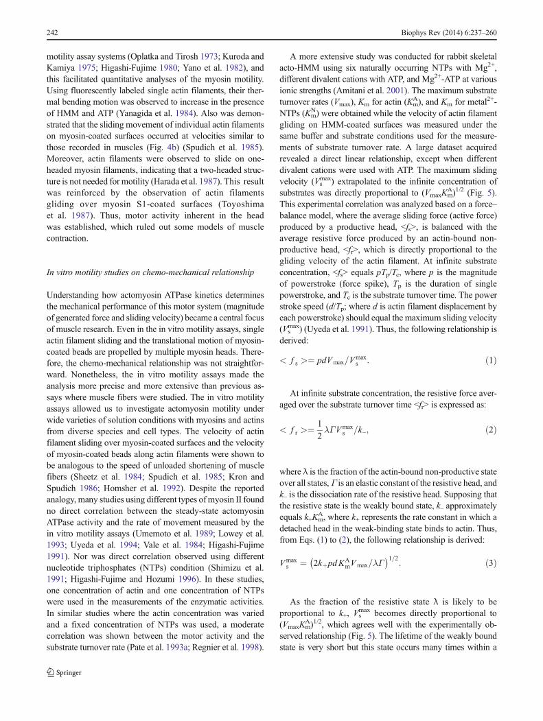
A more extensive study was conducted for rabbit skeletalacto-HMM using six naturally occurring NTPs with Mg2+,different divalent cations with ATP, and Mg2+-ATP at variousionic strengths (Amitani et al. 2001). The maximum substrateturnover rates (Vmax), Km for actin (Km
A), and Km for metal2+-NTPs (Km
N) were obtained while the velocity of actin filamentgliding on HMM-coated surfaces was measured under thesame buffer and substrate conditions used for the measure-ments of substrate turnover rate. A large dataset acquiredrevealed a direct linear relationship, except when differentdivalent cations were used with ATP. The maximum slidingvelocity (Vs
max) extrapolated to the infinite concentration ofsubstrates was directly proportional to (VmaxKm
A)1/2 (Fig. 5).This experimental correlation was analyzed based on a force–balance model, where the average sliding force (active force)produced by a productive head, <fs>, is balanced with theaverage resistive force produced by an actin-bound non-productive head, <fr>, which is directly proportional to thegliding velocity of the actin filament. At infinite substrateconcentration, <fs> equals pTp/Tc, where p is the magnitudeof powerstroke (force spike), Tp is the duration of singlepowerstroke, and Tc is the substrate turnover time. The powerstroke speed (d/Tp; where d is actin filament displacement byeach powerstroke) should equal the maximum sliding velocity(Vs
max) (Uyeda et al. 1991). Thus, the following relationship isderived:
< f s >¼ pdVmax=Vmaxs : ð1Þ
At infinite substrate concentration, the resistive force aver-aged over the substrate turnover time <fr> is expressed as:
< f r >¼ 1
2λΓVmax
s =k–; ð2Þ
where λ is the fraction of the actin-bound non-productive stateover all states, Γ is an elastic constant of the resistive head, andk– is the dissociation rate of the resistive head. Supposing thatthe resistive state is the weakly bound state, k– approximatelyequals k+Km
A, where k+ represents the rate constant in which adetached head in the weak-binding state binds to actin. Thus,from Eqs. (1) to (2), the following relationship is derived:
Vmaxs ¼ 2kþpdKA
mVmax=λΓ� �1=2
: ð3Þ
As the fraction of the resistive state λ is likely to beproportional to k+, Vs
max becomes directly proportional to(VmaxKm
A)1/2, which agrees well with the experimentally ob-served relationship (Fig. 5). The lifetime of the weakly boundstate is very short but this state occurs many times within a
242 Biophys Rev (2014) 6:237–260

substrate turnover cycle. Thus, the weakly bound state be-comes a predominant resistive state. This force balance modelcould also be extended to the case where actin filamentsmoved at various substrate concentrations by including theresistive force produced by rigor heads; thus, it could derivethe dependence of sliding velocity on the substrate concentra-tion. Its derivation showed that the dependence of the slidingvelocity on the substrate concentration [S] followed a modi-fied Michaelis–Menten equation of Vs=Vs
max/{1+(KmN/[S])n}
(here, 1 < n < 2), which again agreed well with the
experimentally obtained dependence under various conditions(Amitani et al. 2001).
Step size
The next fundamental question to tackle experimentallywas the translocation distance that is propelled by actin—myosin interaction during an ATPase cycle (i.e. step size).The swinging lever-arm hypothesis predicts the size to be∼10 nm. However, as mentioned above, the in vitro actingliding or myosin-bead gliding assay is not a single-molecule assay. Moreover, the duration of the workingstroke that occurs in an ATPase cycle has to be measuredto estimate the step size but cannot be measured directly.Consequently, various values were reported for the stepsize: ∼8 nm (Toyoshima et al. 1990), ∼10 nm (Pate et al.1993b), 10–28 nm (Uyeda et al. 1991), and >60 nm(Yanagida et al. 1985; Harada et al. 1990). To resolve thislarge discrepancy, the step size had to be directly measuredat nanometer and millisecond spatiotemporal resolution.This requirement gave birth to the so-called “single-mole-cule measurement assays” capable of observing and manip-ulating single biological molecules in action.
Single-molecule measurements
Optical tweezers are typically used in single moleculemeasurements. A micrometer-sized dielectric particlesuch as a polystyrene bead is trapped by a focused laserbeam (Ashkin and Dziedzic 1985; Neuman and Block
Fig. 4 Schematics showingin vitro motility assay systems foractomyosin. a Myosin-coatedbead assay. The myosin-coatedfluorescent beads are subjected tothe polar arrays of actin cablesnaturally formed on chloroplastdof the alga Nitella, and movementof the beads are observed under aflorescent microscope. b Actinfilament gliding assay. Myosinmolecules are attached to thesurface of a nitrocellulose-coatedcoverslip and gliding motion ofthe fluorescently labeled actinfilaments are observed under afluorescence microscope
Fig. 5 Relationships between the maximum sliding velocity (Vsmax) and
the actomyosin NTPase kinetics parameters, Km for actin (i.e. KmA) and
maximumNTPase rate Vmax. The nucleotide substrates used are indicatedby the side of the corresponding data plots
Biophys Rev (2014) 6:237–260 243

2004) and a trapped particle can be freely moved bychanging the focal position of the laser beam. The posi-tion of the trapped particle in the XY plane perpendicularto the laser beam can be measured with ±<1 nm preci-sion. For the trapped particle, the focused laser beamworks as a 3D spring with a spring constant of 0.01–10 pN/nm. When the particle is displaced by an externalforce in the XY plane, the magnitude of the externalforce applied can therefore be measured at high preci-sion. This optical trap nanometry is now routinely usedto study biological molecules (Svoboda and Block 1994;Mehta et al. 1998; Wang 1999; Bustamante et al. 2000;Kuo 2001). Step size and the force produced by a singlemyosin head were directly measured using a dual-beamlaser trap (Finer et al. 1994; Molloy et al. 1995; Guilfordet al. 1997; Tanaka et al. 1998; Veigel et al. 1999). Eachend of an actin filament was attached to a particle. Thetwo particles are caught in the dual-beam laser trap, andthen the actin filament is allowed to interact with asingle myosin molecule immobilized on a surface(Fig. 6a). The measured step size ranged between 3.5and 15 nm but the extent of discrepancy between labo-ratories was much smaller than previous estimates madeusing actin gliding assays. It was pointed out byYanagida’s group that the measured step size tended tobe affected by the interaction angle between myosin andactin. With careful design, the measured step size wasshown to be dependent on the interaction angle (Tanakaet al. 1998). When an actin filament was aligned parallelwith a myosin filament with a low myosin head density,a relatively large step size of 10–15 nm was obtained(Tanaka et al. 1998).
A very fine glass micro-needle was also used byYanagida’s group as a force transducer for single moleculemechanical measurements. A single actin filament wasattached to the free end of a micro-needle and brought intocontact with an immobilized single myosin molecule. Thedisplacement of the micro-needle caused by the myosin–actin interaction was measured (Kishino and Yanagida1988; Ishijima et al. 1991; Ishijima et al. 1996). Using alow needle stiffness (0.09 pN nm−1) and near zero load, theaverage of single displacement spikes was ∼20 nm(Ishijima et al. 1996). However, the spatiotemporal resolu-tion was not high enough to precisely detect the displace-ment spikes. Then, an experimental system was furtherdeveloped (Fig. 6b); a ZnO crystal whisker with a tip radiusof ∼15 nm was attached to a fine glass needle mounted on athree-dimensional piezoelectric actuator. A single myosinS-1 fluorescently labeled at the regulatory light chain wasattached to the end of the ZnO whisker. The molecule wasbrought into contact with actin bundles formed by chickengizzard α-actinin, where the actin filaments were aligned inanti-parallel (Meyer and Aebi 1990). Using a glass needle
with very small stiffness (0.01–0.03 pN nm−1), the spatialresolution of 2.0 nm and the temporal resolution of <0.2 mswere obtained (Kitamura et al. 1999, 2005). Using thistechnique, an average step size of ∼13 nm was measured,but surprisingly several (2–5) 5.5-nm successive substepswere also detected within each step. This size, 5.5 nm,coincides with the distance between adjacent actin mono-mers in a filament. More surprisingly, the dwell betweensubsteps was independent of the ATP concentration (0.1and 1 μM). Therefore, it was concluded that the multiplesubsteps produced by a single myosin head occurred duringjust one cycle of ATP hydrolysis. This conclusion wasreinforced by theoretical studies (Terada et al. 2002;Takano et al. 2010). However, the experimental result hasnever been reported by other groups.
Total internal reflection fluorescence microscopy(TIRFM) has also been used for single-molecule measure-ments (Park et al. 2007; Murcia et al. 2007; Joo et al. 2008).When an incident laser light is totally reflected at the glass–water interface, an evanescent field is generated that decaysexponentially from the interface, penetrating to a depth of∼100 nm into the solution. Since only fluorophores locatedin the evanescent field are excited, the background lightlevel is very low, allowing the visualization of singlefluorophores (Fig. 6c). Using a Cy3-labeled ATP, the dy-namic events of its binding to and dissociation from singlemyosin S-1 molecules were visualized (Funatsu et al.1995). The dissociation rate estimated from the lifetime ofthe bound Cy3-nucleotide agreed with the biochemicallymeasured rate of Cy3-ATP turnover by S-1. This successopened up a new opportunity to simultaneously measurethe chemical and mechanical events in single myosin mol-ecules. Using one-headed skeletal muscle myosin assem-bled with myosin rod and a setup where TIRFM and a dual-beam laser trap were combined, mechanical events of ac-tin–myosin dissociation/force generation (displacement)and chemical events of binding/dissociation of Cy3-ATP(or Cy3-ADP) were simultaneously observed (Ishijimaet al. 1998). The timing of Cy3-ATP binding well coincidedwith the timing of myosin head dissociation from actin.However, in more than 50 % of 85 detected events, theactin filament was displaced 0.3–1.8 s after the dissociationof Cy3-nucleotide from the myosin head. This surprisingresult suggested that myosin has a hysteresis or memorystate in which the chemical energy liberated by ATP hydro-lysis is stored even after ADP dissociation. However,photobleaching of bound Cy3-nucleotide also looks like itis dissociated from the myosin head. Therefore, their inter-pretation could not be statistically verified. The step sizemeasured in this study (∼15 nm) was consistent with theirpreviously reported values.
Single-molecule measurements quantified other mechanicalproperties of actin, myosin, and their interactions. The tensile
244 Biophys Rev (2014) 6:237–260

and torsional rigidity of a single actin filament was deter-mined using a glass micro-needle technique (Kishino andYanagida 1988) and optical tweezers (Tsuda et al. 1996),respectively. The unbinding force between an actin filamentand a single myosin head in the nucleotide-free conditionwas ∼9 pN using an optical tweezers method where an actinfilament was pulled approximately along its length(Fig. 6d) (Nishizaka et al. 1995). It was also measured tobe ∼15 pN with AFM, where a single myosin head waspulled in the direction perpendicular to the actin filament(Fig. 6e) (Nakajima et al. 1997).
As described above, no complete consensus was reachedamong research groups regarding the myosin step size takenfor each ATP hydrolysis, and hence also as to the swinging
lever hypothesis. The discrepancy was due partly to a rela-tively small step size occurring in a short time that wastechnically difficult to measure.
Myosin V
The technical difficulty mentioned above was significantlyremoved by the discovery of M5 that possesses unique struc-tural and biochemical features. M5 is involved in organelleand mRNA transports as well as in membrane trafficking. Itscellular functions and regulations are well described else-where (Reck-Peterson et al. 2000; Taylor 2007; Sellers and
Fig. 6 Schematics showing typical single-molecule measurement sys-tems for studying myosin–actin interactions. a Dual-beam laser trapassay. Each end of an actin filament is attached to a plastic bead, andthe beads are trapped by the focused laser beams. The trapped actinfilament is brought to a single myosin molecule attached to a silica bead.b Mechanical measurement system with the use of a thin glass micro-needle as a force sensor. A ZnO crystal whisker is attached to the end of aglass micro-needle (not shown in this schematic). Fluorescently labeledsingle myosin S-1 is attached to the apex of the ZnO crystal whisker via aspecific binding of biotin-streptavidin. The tip is brought onto actinbundles formed by α-actinin. The displacements of the S-1 resulting from
the interaction with actin are monitored by the detection of glass micro-needle deflection. c TIRFM experiment for visualizing individual ATPturnovers by single myosin S-1. The light chain of S-1molecule is labeledwith Cy5 (indicated by the red star) for the confirmation of whether or notthe observed ATP turnovers are those by a single S-1 molecule. OnceCy3-ATP binds to the S-1, the large Brownian motion of the Cy3-ATPstops and therefore its fluorescent spot becomes visible. d Single beamassay for the measurement of unbinding force of a single myosin–actinbond. e AFM measurement of rupture force and rupture distance of asingle actin–myosin bond
Biophys Rev (2014) 6:237–260 245
Knight 2007; Sellers andWeisman 2008), so here we focus onits motor mechanism.
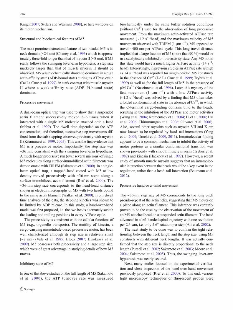
Structural and biochemical features of M5
The most prominent structural feature of two-headedM5 is itsneck domain (∼24 nm) (Cheney et al. 1993) which is approx-imately three-fold longer than that of myosin II (∼8 nm). IfM5really follows the swinging lever-arm hypothesis, a step sizemarkedly larger than that of muscle myosin II should beobserved. M5 was biochemically shown to dominate in a highactin-affinity state (ADP-bound state) during its ATPase cycle(De La Cruz et al. 1999), in stark contrast with muscle myosinII where a weak affinity sate (ADP–Pi-bound state)dominates.
Processive movement
A dual-beam optical trap was used to show that a suspendedactin filament successively moved 3–6 times when itinteracted with a single M5 molecule attached onto a bead(Mehta et al. 1999). The dwell time depended on the ATPconcentration, and therefore, successive step movements dif-fered from the sub-stepping observed previously with myosinII (Kitamura et al. 1999, 2005). This was the first evidence thatM5 is a processive motor. Importantly, the step size was∼36 nm, consistent with the swinging lever-arm hypothesis.A much longer processive run (over several microns) of singleM5 molecules along surface-immobilized actin filaments wasdemonstrated with TIRFM (Sakamoto et al. 2000). In a single-beam optical trap, a trapped bead coated with M5 at lowdensity moved processively with ∼36-nm steps along asurface-immobilized actin filament (Rief et al. 2000). The∼36-nm step size corresponds to the head-head distanceshown in electron micrographs of M5 with two heads boundto the same actin filament (Walker et al. 2000). From dwelltime analyses of the data, the stepping kinetics was shown tobe limited by ADP release. In this study, a hand-over-handmodel was first proposed, i.e. the two heads alternately switchthe leading and trailing positions in every ATPase cycle.
The processivity is consistent with the cellular functions ofM5 (e.g., organelle transports). The motility of kinesin, acargo-carrying microtubule-based processive motor, has beenwell characterized although its step size is relatively small(∼8 nm) (Vale et al. 1985; Block 2007; Hirokawa et al.2009). M5 possesses both processivity and a large step size,which were of great advantage in studying details of how M5moves.
Inhibitory state of M5
In one of the above studies on the full length ofM5 (Sakamotoet al. 2000), the ATP turnover rate was measured
biochemically under the same buffer solution conditions(without Ca2+) used for the observation of long processivemovement. From the maximum actin-activated ATPase ratemeasured (∼1.2 s−1/head) and the maximum velocity of M5movement observed with TRIFM (1 μm s−1), M5 appeared totravel ∼400 nm per ATPase cycle. This long travel distanceimplied that a large fraction of M5 (more than 90%) would bein a catalytically inhibited or low-activity state. AnyM5 not inthis state would have a much higher ATPase activity (14 s−1/head). Interestingly, in previous studies an ATPase rate as highas 14 s−1/head was reported for single-headed M5 constructsin the absence of Ca2+ (De La Cruz et al. 1999; Trybus et al.1999) as well as for the full length of M5 in the presence ofμM Ca2+ (Nascimento et al. 1996). Later, this mystery of thefast movement (1 μm s−1) with a low ATPase activity(∼1.2 s−1/head) was solved by a finding that M5 often takesa folded conformational state in the absence of Ca2+, in whichthe C-terminal cargo-binding domains bind to the heads,resulting in the inhibition of the ATPase and motor activities(Wang et al. 2004; Krementsov et al. 2004; Li et al. 2006; Liuet al. 2006; Thirumurugan et al. 2006; Olivares et al. 2006).Also, several other myosins such as myosin VII and X arenow known to be regulated by head–tail interactions (Yanget al. 2009; Umeki et al. 2009, 2011). Intramolecular foldingappears to be a common mechanism to inhibit the activity ofmotor proteins as a similar conformational transition wasshown previously with smooth muscle myosin (Trybus et al.1982) and kinesin (Hackney et al. 1992). However, a recentstudy of smooth muscle myosin suggests that an intramolec-ular interaction between the heads would be a key event in theregulation, rather than a head–tail interaction (Baumann et al.2012).
Processive hand-over-hand movement
The ∼36-nm step size of M5 corresponds to the long pitchpseudo-repeat of the actin helix, suggesting that M5moves ona plane along an actin filament. This inference was certainlyproven to be the case by the observation of the movement ofan M5-attached bead on a suspended actin filament. The beadadvanced in a left-handed spiral trajectory with one revolutionper 2.3 μm, i.e. only 5.6° rotation per step (Ali et al. 2002).
The next study to be done was to confirm the tight rela-tionship between the neck length and the step size, using M5constructs with different neck lengths. It was actually con-firmed that the step size is directly proportional to the necklength (Purcell et al. 2002; Sakamoto et al. 2003; Moore et al.2004; Sakamoto et al. 2005). Thus, the swinging lever-armhypothesis was nearly secured.
Next, many studies focused on the experimental verifica-tion and close inspection of the hand-over-hand movementpreviously proposed (Rief et al. 2000). To this end, variouslight microscopy techniques or fluorescent probes were
246 Biophys Rev (2014) 6:237–260

introduced to TIRFM: fluorescence imaging with one nano-meter accuracy (FIONA) (Yildiz et al. 2003), fluorescencepolarization (Forkey et al. 2003), two-color imaging with Q-dots (Warshaw et al. 2005), single-molecule high-resolutioncolocalization of fluorescent dyes (Churchman et al. 2005), acombination of FIONA and fluorescence polarization (Syedet al. 2006), and defocused orientation and position imaging(Toprak et al. 2006). All these studies revealed that in fact M5walks in a hand-over-hand manner. Not only translationalposition changes of the two heads but also angular changesof the lever-arm relative to actin were observed where onlyone of the two lever-arms was labeled with a fluorescentprobe. The value of the angle change (∼60°) was also consis-tent with a hand-over-hand movement (Forkey et al. 2003;Syed et al. 2006; Toprak et al. 2006).
These studies supported the hand-over-hand model butonly the dwell states were observed when both heads werebound to actin. Therefore, little was known about the fleetingintermediate between the dwell states because the time reso-lution was low (∼33 ms or longer). To increase the timeresolution to 1 ms or less, the following imaging methodswere employed: dark field imaging of a 40-nm gold nanopar-ticle attached to one of the lever-arms (Dunn and Spudich2007) and recording of light-scattering from a 200-nm M5-coated latex bead traveling in an interference fringe pattern(Cappello et al. 2007). Alternatively, the action of walkingM5was slowed down by attaching a large fluorescent microtubuleto one of the lever-arms and observed by conventional fluo-rescence microscopy (Shiroguchi and Kinosita 2007). Allthree studies revealed that the detached head underwent ex-tensive rotational Brownian motion before landing on a siteahead of the leading head, suggesting that the detached head isunlikely to interact with actin during the Brownian motion.Moreover, it was suggested that the Brownian motion is likelyto be biased forward by the orientational change (swing) of theactin-bound leading head, by which the detached head even-tually finds an actin ahead of the leading head to become anew leading head. Thus, a clearer picture of the processivehand-over-hand movement was obtained.
Asymmetry of enzymatic kinetics between two heads
The processive hand-over-hand movement suggests that onlythe trailing head can detach from actin. If so, there are twopossibilities: (Case 1) ADP dissociation followed by ATPbinding predominantly occurs at the trailing head, so that theADP dissociation would be accelerated at the trailing head orretarded at the leading head; (Case 2) the ATP-binding ratewould be enhanced at the trailing head or suppressed at theleading head. Between these possibilities, Case 1 appearedmore likely as the ATPase rate is limited at the ADP releasestep. In either case, the kinetics asymmetry between the twoheads would be engendered by head-to-head communications
through intramolecular strain. This kinetics asymmetry wasaddressed by solution kinetics studies and optical-trapnanometry studies. In the solution kinetics studies variouskinetics constants were compared for S-1 and HMM con-structs of M5, although the two heads of M5-HMM do notnecessarily interact with the same actin filament in solution(Rosenfeld and Sweeney 2004; Forgacs et al. 2008). Thesestudies suggested retardation of ADP release from the leadinghead by ∼45-fold (Rosenfeld and Sweeney 2004) or up to250-fold (Forgacs et al. 2008). The latter study used newfluorescent ADP and ATP analogues (deac-aminoADP anddeac-aminoATP) (Webb et al. 2004) whose emission intensityincreases by 20-fold upon binding to the active site of M5(Forgacs et al. 2006). In the dual-beam trap experiments, theactin filament that was interacting with a surface-attachedsingle-headed M5 molecule was pulled forward or backwardto apply a force to the actin-bound head (Purcell et al. 2005;Veigel et al. 2002, 2005). The forward and backward forceswere hypothesized to simulate forces experienced by thetrailing or the leading head, respectively. Different designsof optical trap nanometry were also used to study the coordi-nation between actin-bound two heads (Uemura et al. 2004;Clemen et al. 2005; Gebhardt et al. 2006; Oguchi et al. 2008).Most of these studies suggested that the intramolecular strainwould coordinate two heads by decelerating ADP releasefrom the leading head, whereas some studies suggested accel-erated ADP release at the trailing head (Veigel et al. 2002,2005), as proposed by a study of processive run of M5perturbed by varying the nucleotide content (Baker et al.2004).
In two-headed bound M5, the neck of the trailing headtakes the arrowhead orientation, whereas the neck of theleading head takes the reverse arrowhead orientation. Theleading head conformation must be mechanically distortedbecause single-headed M5 binds to an actin filament in anarrowhead orientation. In fact, electron micrographs of two-headed bound M5 showed that the leading neck was curvedslightly outwards whereas the trailing neck was straight(Walker et al. 2000; Burgess et al. 2002; Oke et al. 2010).This conformational distortion seems consistent with the sug-gestion that the coordination of M5 stepping is performed bystrain-mediated inhibition of ADP release from the leadinghead.
Finally, using two-color TIRFM, Sakamoto et al. (2008)simultaneously observed the stepping motion of fluores-cently labeled M5 and the binding and dissociation of thefluorescent nucleotide (deac-aminoADP or deac-aminoATP). This observation directly revealed the prefer-ential ADP dissociation from the trailing head, which wasfollowed by a ∼36-nm step triggered by ATP binding. Italso revealed that every M5 molecule always keeps at leastone nucleotide (ADP in the leading head) during itsstepping, even at low ATP concentrations.
Biophys Rev (2014) 6:237–260 247

Discrepancies and unsolved questions in myosin V motility
The above description suggests there is now a consensus onhow M5 steps. In every ATP hydrolysis cycle, the leadinghead swings to propel M5 forwards by ∼36 nm (Sakamotoet al. 2008). This large stride is realized by the long lever-armsof M5 (Purcell et al. 2002; Sakamoto et al. 2003; Moore et al.2004; Sakamoto et al. 2005). The two heads alternately switchbetween the leading and trailing roles so that M5 walks hand-over-hand (Yildiz et al. 2003; Forkey et al. 2003; Warshawet al. 2005; Churchman et al. 2005; Syed et al. 2006; Topraket al. 2006). This style of movement is made possible by theintramolecular strain-mediated retardation of ADP releasefrom the leading head (Rosenfeld and Sweeney 2004;Purcell et al. 2005; Forgacs et al. 2008; Oguchi et al. 2008;Sakamoto et al. 2008).
Nonetheless, several observations that are difficult to rec-oncile with this generally agreed view have been reported,mostly from the Yanagida lab. A recombinant two-headedM5with a single IQmotif on each neck (∼4 nm long) was reportedto move processively with ∼36-nm steps (Tanaka et al. 2002).It was claimed that the long-neck domain was not essential forboth large step size and processivity of M5 but the motordomain alone determined the processivity and the large stepsize. It was also reported that single-headed M5 constructsunderwent multiple successive large (∼32-nm) steps on anactin filament suspended by a dual-beam optical trap(Watanabe et al. 2004). They also underwent directional dif-fusion on surface-immobilized actin bundles with ∼5.5-nmsubsteps to develop an average displacement of ∼20 nm,which was independent of the neck length (2IQ and 6IQmotifs) (Okada et al. 2007), as previously observed withsingle-headed myosin II (Kitamura et al. 1999). Likewise,myosin VI with its short neck similar to myosin II, wasreported to move with ∼36-nm steps (Rock et al. 2001;Nishikawa et al. 2002). However, among these reported irrec-oncilable conclusions, only the large step size of myosin VIwas resolved. The SAHs were found in the proximal taildomain of myosin VI, which contribute to the lever-armlength (Baboolal et al. 2009; Spudich and Sivaramakrishnan2010; Sweeney and Houdusse 2010b). The other discrepantresults are still incomprehensible (Cyranoski 2000; Geeves2002).
Even if the above general view of how M5 steps iscompletely right, there remain fundamental questions to solve.How is the chemical energy liberated by ATP hydrolysis usedto generate the tension for forward movement? How are thetension generation and lever-arm swing coupled to the ATPasecycle? If they are coupled, which chemical transitions areinvolved in these mechanical events? These fundamentalquestions have long been explored in muscle contractionstudies and some are considered to be resolved. Are thereany inconsistencies when this consensus view is applied to
the motility of M5? Numerous single-molecule measurementsof M5 stepping have not answered these important questions.
The discrepancies mentioned above, as well as the remain-ing unsolved fundamental questions, have certainly proventhat the level of “directness of measurement” is not yet highenough even with single-molecule optical measurements. Pro-teins molecules themselves are indeed invisible in these mea-surements, which can never be overcome even withdiffraction-breaking super-resolution fluorescence microsco-py (Egner and Hell 2005; Huang et al. 2009).
HS-AFM
HS-AFM has overcome this technical limitation. Biologicalmolecules can be directly observed at sub-molecular spatialand sub-100 ms temporal resolution without disturbing theirfunction. In this section, we briefly summarize the principle ofAFM and the current state of HS-AFM performance (Fig. 7).Techniques involved in the HS-AFM instrument are welldescribed elsewhere (Ando et al. 2001; Ando et al. 2008;Ando 2012). See other reviews for protocols of HS-AFMimaging of protein molecules (Uchihashi et al. 2012) andvarious application studies of HS-AFM (Ando et al. 2014).
Principles of AFM imaging
In AFM (Binnig et al. 1986), a sharp tip attached to the freeend of a cantilever traces the sample to acquire its surfacetopography while the sample stage is scanned laterally. AFMcan observe any object on a substrate at single-nanometer (orhigher) resolution under a wide range of environments (vac-uum, air, and liquids). Topography images of biological sam-ples can be acquired under physiological buffer conditions,without fixing, staining and labeling the sample (Engel andMüller 2000; Müller and Dufrêne 2008). Among variousimaging modes, the tapping mode (Zhong et al. 1993) is mostsuited for biological samples. In this mode, the cantilever isexcited to oscillate in the Z-direction at or near its first reso-nant frequency. Because of this oscillation, the cantilever tip isbrought intermittently into contact with the sample surface,which can eliminate lateral friction force and hence minimizedeformation of fragile samples. The reduction of cantileveroscillation amplitude upon tip-sample contact is measured andthen the sample stage is finally moved in the Z-direction torestore the amplitude to its set point using a feedback control.This series of operations is repeated many times for differentsample surface points during lateral scanning of the samplestage. Consequently, the 3D movement of the sample stageapproximately traces the sample surface, and hence, a topog-raphy image of the sample can be constructed from the electricsignal used to drive the scanner in the Z-direction.
248 Biophys Rev (2014) 6:237–260

Current state of HS-AFM
With conventional AFM instruments it takes at least 30 s tocapture an image. This slow rate is due mainly to the slowresponse of the mechanical devices (i.e. cantilevers and Z-scanner), whereas many interesting biological phenomenaoccur much faster. Various efforts have been made to mark-edly increase the imaging rate of tapping-mode AFM over thepast 20 years. Small cantilevers, fast scanners, and fast ampli-tude detectors were developed to build the first prototypicinstruments (Viani et al. 2000; Ando et al. 2001). The im-provement of these devices and creation of new techniques forvibration damping and fast feedback control produced thecurrent HS-AFM (Ando et al. 2008). The highest possibleimaging rate is a function of the speed of an AFM instrumentas well as the imaging conditions and the sample fragility(Uchihashi et al. 2012; Ando et al. 2013). For 50- to 200-nmscan ranges, sufficient to image biological molecules, HS-AFM can capture an image within 30–80 ms without
disturbing their biological function. The spatial resolution ofHS-AFM is now comparable with that of conventional slowAFM even with high-bandwidth detection of the cantileveroscillation amplitude and fast scanning of the sample stage(∼2 nm for XYand ∼0.1 nm for Z in the best case). HS-AFMhas already visualized dynamic biomolecular phenomena (seereviews in Ando 2012; Ando et al. 2013, 2014) and evendynamic phenomena in live cells (Yamashita et al. 2012;Watanabe et al. 2013).
Walking M5 filmed by high-speed AFM
Substrate surface and observation of unidirectional movement
M5-HMMwas directly imaged walking along actin filamentsusing HS-AFM (Kodera et al. 2010). Here, partially biotinyl-ated actin filaments were immobilized on a surface where
Fig. 7 Schematic of HS-AFM system. The system includes an originalinverted optical microscope. The objective lens with a long workingdistance that is a part of the OBD detector is also used to view thecantilever and sample stage via a digital camera. The glass slide, to whichthe cantilever holder and the liquid cell are attached, is placed on theoptical microscope stage. A cantilever chip is held in the holder so that itstip points upward (opposite to the way in conventional AFM). Thesample stage, attached to the Z-scanner and facing downward, is placedover the cantilever. An incident laser beam passing through the objectivelens is focused onto the small cantilever, and the light reflected back fromthe cantilever is collected and collimated by the same objective lens andguided to the quadrant-cell Si PIN photodiode. The incident and reflectedlaser beams are separated using the quarter-wavelength (λ/4) plate and thepolarization beam splitter. A cantilever immersed in a buffer solution is
oscillated with small amplitude (1–2 nm) by the excitation with anexcitation piezo at the first resonant frequency of the cantilever (0.6–1.2 MHz in water). The cantilever oscillation amplitude is maintainedconstant by feedback control. A counterbalancing method is employed tothe Z-scanner to minimize unwanted vibrations; two peazoactuators areattached to the supporting base at its counter sides and simultaneouslydisplaced in the same distance, in the counter directions. The scan signalsfor the X- and Y-scanner are output from the computer through the DAconverters. The output from the active Q-control damper is recorded asthe sample height through the AD converter. The active Q-control damperis constructed with an LRC circuit whose transfer function is very similarto that of the Z-scanner. Its electrical output behaves in a way similar tothe Z-scanner displacement
Biophys Rev (2014) 6:237–260 249

streptavidin was dispersed at low density on mica-supportedplanar lipid bilayer containing electrically neutral phospho-lipids and a biotinylated lipid (Fig. 8a). M5-HMM was neverbound directly to the lipid bilayer surface and only interactedwith the immobilized actin filaments and moved thereon. Themoving M5 was frequently oriented perpendicular to thesubstrate surface so that its characteristic lateral topographywas only occasionally observed. Adding a positively chargedlipid to the bilayer solved this problem.
In the presence of 1–2 μM ATP, successive AFM imagescaptured at 7 fps clearly showed M5-HMM movingprocessively with discrete ∼36-nm steps (Fig. 8b). The two-headed bound M5-HMM exhibited unique structural features(Fig. 8c). The junction of the neck with the motor domainappears smooth in the leading head but is always V-shaped inthe trailing head because the neck regions emerge from dif-ferent parts of the motor domain. The short coiled-coil tail wasmostly tilted towards the minus end of actin. These structuralfeatures, which are totally consistent with an electron micro-scopic observation (Burgess et al. 2002), can be used todetermine the actin polarity when bound M5-HMM is station-ary. In addition, the leading head always assumed a straightconformation (slightly curved outwards), which agreed withthe straight-neck model proposed for walking M5 (Forkeyet al. 2003; Syed et al. 2006; Toprak et al. 2006) but disagreedwith the bent-neck model (Walker et al. 2000; Burgess et al.2002; Snyder et al. 2004; Oke et al. 2010).
The positively charged lipid in the lipid bilayer slightlyretarded the translocation velocities. When it was absent, thevelocities at various ATP concentrations were similar to thosemeasured by fluorescence microscopy under the same buffersolution condition (Forkey et al. 2003; Baker et al. 2004),indicating negligible effects of the tip-sample interaction onthe motor activity. However, molecular behavior during a stepcould not be resolved as it completed very fast.
Visualization of stepping behavior
To slow the step, more streptavidin molecules were placed onthe lipid bilayer surface as moderate obstacles to the walkingof M5-HMM (Fig. 9a), which resolved the stepping process(Fig. 9b). Upon detachment of the trailing head from actin, theleading head appeared to spontaneously rotate from the re-verse arrowhead orientation towards the arrowhead orienta-tion. Before completing this rotation, the leading head washalted for a moment by colliding with a streptavidin moleculeplaced on the way of its natural path. In this halt state, thedetached trailing head was displaced forward, positionedmostdistant from the actin filament (thus, the two heads werealigned nearly straight, pointing opposite directions), andslightly rotated around the neck-neck junction (the secondframe in Fig. 9b). Then, the leading head overcame the obsta-cle and further rotated forwards. Immediately after this the
trailing head bound to a forward site of the actin filament tobecome a new leading head, completing one step. Thus,dynamic processes in the forward step were directly visual-ized. The observed rotation of the leading head is exactly theswinging lever-arm motion itself initially proposed for thepowerstroke of muscle myosin (Huxley 1969). Note thatbefore completing a step the detached trailing head neverinteracted with actin but passively moved forwards drivenby the rotating leading head. This ruled out some models ofM5motility such as the ‘inchworm’-like model considered forkinesin (Hua et al. 2002) and the “biased diffusion” modelproposed for single-headed myosin II (Kitamura et al. 1999)and single-headedM5 (Okada et al. 2007). M5 strictly follow-ed the hand-over-hand mechanism. In contrast, the otherprocessive myosins VI and X, which function not only as acargo transporter but also as a structural anchor in cells, werereported to move irregularly, namely inchworm-like stepping,backward stepping, and forward hand-over-hand stepping(Yildiz et al. 2004; Sun et al. 2007; Nishikawa et al. 2010;Ricca and Rock 2010; Sun et al. 2010).
Foot stomp and unwinding of coiled-coil tail
Interestingly, in the two-headed bound state during the dwell,the motor domain of the leading head frequently exhibitedbrief dissociation and reassociation on the same actin filament,while the molecule remained at approximately the same posi-tion on the filament (Fig. 10a). Similarly, the motor domain ofthe trailing head exhibited a brief translocation by ∼±5 nmalong the actin filament. We termed these behaviors “footstomp”. The foot stomp was more frequently observed at theleading head than at the trailing head (by a ratio of approxi-mately 3:1). Although not well documented, a foot-stomp-likebehavior was previously suggested in fluorescence microsco-py observations of walking M5 molecules (Syed et al. 2006;Shiroguchi and Kinosita 2007). More recently, the foot stompwas further confirmed by the observation of walking M5 byhigh-speed single-molecule polarized fluorescence microsco-py (Beausang et al. 2013). Thus, the foot stomp is an inherentbehavior of this motor.
The foot stomp at the leading head raises an importantquestion as to the chemo-mechanical coupling in M5. Thebriefly detached leading head does not carry bound Pi becausePi has already been released from the leading head followingits initial attachment to actin (De La Cruz et al. 1999; Olivareset al. 2006). Nevertheless, the detached leading head with itsbound ADP reattached to actin while still in the reversearrowhead orientation. It then swung forward followingtrailing head detachment. This fact was important because itindicated that tension generation for forward movement canoccur without transitioning through the ADP–Pi bound state,i.e. it can occur directly in the ADP–bound state. Thus, the
250 Biophys Rev (2014) 6:237–260

Fig. 8 HS-AFM imaging ofwalking M5-HMM. a Schematicshowing assay system used forthe HS-AFM imaging. bSuccessive HS-AFM imagesshowing unidirectionalprocessive movement of M5-HMM observed in 1 μM ATP.Frame rate, 7 fps; Scan area,130×65 nm2 with 80×40 pixels;scale bar 30 nm. The verticaldashed lines show the centers ofmass of the motor domains, andthe plus sign indicates the plusend of actin. c Schematic showingtwo-headed bound M5-HMM toactin. The corresponding AFMmovies can be seen at thefollowing web site: http://www.nature.com/nature/journal/v468/n7320/extref/nature09450-s2.mov
Fig. 9 Stepping behavior of M5-HMM visualized by HS-AFM. aSchematic explaining the HS-AFM images shown in (b). bSuccessive HS-AFM images thatresolved the stepping behavior ofM5-HMM in 1 μM ATP. Framerate, 7 fps; scan area, 150×75 nm2
with 80×40 pixels; scale bar50 nm. The swinging lever-arm ishighlighted with the thin whitelines. The vertical dashed lines in(a) and (b) represent the centers ofmass of the motor domains, andthe plus sign indicates the plusend of actin. The correspondingAFM movies can be seen at thefollowing web site: http://www.nature.com/nature/journal/v468/n7320/extref/nature09450-s3.mov
Biophys Rev (2014) 6:237–260 251

tension generation for forward movement does not seem torequire chemical energy be supplied by ATP hydrolysis.
The asymmetry of foot stomp frequency between the twoheads suggested their actin-affinity difference, consistent witha biochemically measured result (Olivares et al. 2006). Theleading head binds to actin in the unnatural orientation (i.e.reverse arrowhead orientation), and hence, pays an energycost by distorting the neck conformation, resulting in a loweraffinity for actin. The distortion of the leading head is likely tobe the source for tension generation for forward movement.This was reinforced by the following observation; when thetwo-heads were bound to actin in 1 mMADP, the short coiled-coil tail was sometimes unwound, immediately after which themonomerized leading head rotated towards the arrowheadorientation, similar to the swinging lever-arm motion ob-served when M5-HMM was walking forwards (Fig. 10b).
Flexibility of neck-motor domain junction
Under nucleotide-free conditions, the leading head frequentlyexhibited a sharply bent conformation (Fig. 10c) that wasnever observed in the presence of ATP or in the presence of
1 mM ADP. This bent conformation suggested that the neck-motor domain junction is less flexible in the nucleotide-freehead than in the nucleotide-bound head. To examine thisissue, the arrowhead orientation angle of single-headed M5relative to the actin filament was measured under thenucleotide-free and ADP-bound conditions. In nucleotide-free solution, the angle was ∼34°, whereas in ADP the anglewas distributed widely, peaked at ∼29° and ∼51° in the pro-portion of approximately 6 to 1 (supplementary data inKodera et al. 2010), whichmay be relevant to the two differentADP-bound states in equilibrium as reported for myosin II(Geeves 1989; Geeves and Holmes 1999), M5 (Robblee et al.2005; Hannemann et al. 2005; Oguchi et al. 2008; Jacobs et al.2011), and myosin VI (Robblee et al. 2005). Therefore, ADPbinding to the head makes the hinge of the neck–motordomain junction flexible. Because of the rigid hinge of thenucleotide-free head, the leading neck in the reverse arrow-head orientation tends to be sharply bent to release the largestrain accumulated therein. This rigid hinge was also support-ed by the observation that both heads are rarely bound toadjacent actin subunits but only in the nucleotide-free condi-tion (Fig. 10d). In the electron micrographs of actomyosin II
Fig. 10 Unique molecular behaviors of M5-HMM visualized by HS-AFM. a Successive HS-AFM images showing a foot stomp event thatoccurred at the leading-head (ATP, 1 μM; frame rate, 7 fps; scan area,150×75 nm2 with 80×40 pixels; scale bar 50 nm). The leading-headdetachment is marked with the light-blue arrowheads. The correspondingAFM movie can be seen at the following website: http://www.nature.com/nature/journal/v468/n7320/extref/nature09450-s3.mov b HS-AFMimages before (upper panel) and after (lower panel) unwinding ofcoiled-coil tail observed in 50 μM ADP. The corresponding AFMmovie can be seen at the following website: http://www.nature.com/nature/journal/v468/n7320/extref/nature09450-s5.mov c HS-AFM
image showing the leading head with a sharply bent conformationobserved in the nucleotide-free condition. The corresponding AFMmovie can be seen at the following website: http://www.nature.com/nature/journal/v468/n7320/extref/nature09450-s6.mov d HS-AFMimage showing nucleotide-free M5-HMM with heads bound to adjacentactin subunits (upper panel) and its illustration (lower panel). Imagingconditions for (b), (c) and (d) are as followings: frame rate, 3 fps; scanarea, 100×100 nm2 with 80×80 pixels; scale bar 30 nm. The verticaldashed lines represent the centers of mass of the motor domains, and theplus signs indicate the plus ends of actin
252 Biophys Rev (2014) 6:237–260

and actomyosin VI complexes, this conformation was fre-quently seen, suggesting they have rigid hinges (Craig et al.1980; Nishikawa et al. 2002).
The flexibility of the neck-motor domain junction observedfor the actin-bound M5 head with ADP does not seem suffi-cient to account for the two-headed binding in ADP becausethe leading head binding in the reverse-arrowhead orientationrequires the junction to bend by at least ∼60°, even when thecontribution of the neck domain’s flexibility is considered.This apparently restricted flexibility suggests that the actin-unbound head with ADP is more flexible than the actin-boundone. In the other words, actin binding may make the neck-motor domain junction less flexible. This plausible flexibilitychange seems an excellent strategy for facilitating both actinbinding and the generation of enough tension to execute apowerstroke. Very recently, such an actin-binding effect on theflexibility was proposed for muscle myosin (Billington et al.2014).
Asymmetry of ADP dissociation rate
The straight and sharply bent conformations of the leadinghead depend on the presence or absence of bound ADP,respectively, meaning that one can judge whether or not theleading head contains ADP by just looking at its conforma-tion. From the proportion and lifetime of the straight confor-mation as a function of ADP concentration, the rate constantsof ADP binding/dissociation kinetics at the leading head wereestimated. The ADP dissociation rate constant was 0.1 s−1, i.e.one ADP is released from the leading head every 10 s, onaverage. However, M5 walks many steps during 10 s. Thus,from the structural point of view, it was confirmed that thesequential events of ADP release, the subsequent ATP bindingand the resulting head dissociation take place solely at thetrailing head, which is the basis for the processive hand-over-hand movement (Rosenfeld and Sweeney 2004; Purcell et al.2005; Veigel et al. 2005; Forgacs et al. 2008; Oguchi et al.2008; Sakamoto et al. 2008).
New questions
The swinging lever-arm hypothesis is no longer a hypothesis.HS-AFM visualized its occurrence with irrefutable clearness.However, from HS-AFM observations of M5-HMMinteracting with actin, questions were raised about thechemo-mechanical coupling in this motor as well as myosinmotors in general. The prevailing coupling mechanism hadbeen modeled in the context of swinging lever-arm motion(Fig. 3). In this model, the structural states are putativelytightly coupled to the nucleotide states of the motor domain(Fig. 3). In the M–ADP–Pi state, the head takes the pre-
powerstroke conformation caused by recovery stroke of thelever-arm, which facilitates binding of the head to actin in thereverse arrowhead orientation. Coupled to Pi release from theactin-bound head, the intramolecular tension is considered tobe generated just once by the pre-powerstroke to post-powerstroke transition. However, the observation with HS-AFM of walking M5-HMM showed that the tension respon-sible for the lever-arm swing can be generated directly by theADP-bound leading head (after foot stomping). In addition, itwas shown that two-headed bound M5-HMM with ADPalone can generate sufficient tension to cause occasional un-winding of the short coiled-coil tail, after which the lever-armswings. Thus, the recovery stroke or the pre-powerstrokeconformation thought to occur uniquely in the ADP–Pi-boundhead does not seem necessary for its binding to actin andlever-arm swing. More seriously, the chemical energy liberat-ed by ATP hydrolysis does not seem to be used for theprocesses of recovery stroke, tension generation, and lever-arm swing, suggesting that M5 would step forward withoutchemical energy input once the trailing head detaches fromactin.
This new idea of “no chemical energy usage” in theseprocesses would, however, inevitably lead us to encounter a“perpetuum mobile problem”. Even in ADP or in thenucleotide-free condition, foot stomp occurs at both leadingand trailing heads of two-headed bound M5-HMM, with ahigher frequency in the leading head than in the trailing head.When the trailing head detaches from actin, the molecule takesa forward step by the spontaneous swing of the leading head.On the other hand, when the leading head detaches from actin,the molecule cannot take a backward step because the trailinghead is bound to actin in a stable orientation (i.e. the arrow-head orientation). Thus, using only thermal energy, M5 wouldstep forward many times (albeit very slowly), even with lessfrequent occurrence of foot stomps at the trailing head. This isa forbidden perpetuum motion. However, in thisgedankenexperiment, positional fluctuations of thesingle-headed bound molecule due to the flexible neck-motor domain junction are not considered. Taking thisinto account, we can expect a backward step after detach-ment of the leading head from actin, albeit with a lowprobability. The ratio of foot stomp occurrence betweenthe leading and trailing heads was observed to be 4:1 inthe presence of 1 mM ADP. However, the foot stompevents at the trailing head mostly occurred as a brief ±5-nm translocation. Therefore, when this brief translocationis omitted and only the events of brief detachment fromactin are counted as foot stomp, the ratio of foot stompoccurrence between the two heads becomes much larger.Thus, the principle of detailed balance holds between theforward step after trailing head detachment and the back-ward step after leading head detachment, resulting in nonet movement of the molecule.
Biophys Rev (2014) 6:237–260 253

From numerous X-ray crystallography and electron mi-croscopy studies, there is no doubt that myosin changes itsconformation upon binding nucleotides and takes a pre-powerstroke-like conformation in the ADP–Pi-bound state(Fisher et al. 1995; Smith and Rayment 1996; Dominguezet al. 1998; Houdusse et al. 2000; Burgess et al. 2002;Coureux et al. 2004; Volkmann et al. 2005). These staticstructures have been used to construct the prevailing modelof the chemo-mechanical coupling (Fig. 3) assuming there is atight one-to-one relationship between the chemical and con-formational states. In reality, the relationship is not so tight.Myosin takes two (or multiple) conformations even under agiven nucleotide condition. They can go back and forth but thedynamic equilibrium is biased to one side depending on thenucleotide condition. The HS-AFM observations of M5-HMM indicates that structural fluctuations indeed exist andtherefore two-headed binding occurs even in the ADP-boundand nucleotide free conditions, consistent with other studies(Walker et al. 2000; Rosenfeld and Sweeney 2004; Olivareset al. 2006).
The flexibility of myosin head conformation raises a ques-tion whether the chemical energy of ATP hydrolysis is reallyused to change the conformation. In the prevailing view, themyosin head with a strained (tension-generating) conforma-tion in the pre-powerstroke state is achieved using the chem-ical energy of ATP, which is finally released by the swinginglever arm motion. However, the pre-powerstroke and post-powerstroke conformations are in dynamic equilibrium withno marked energy barrier. Thus, one can conclude as follows:although the conformational change of myosin head causedby recovery stroke is an effective strategy for facilitating itsbinding to actin in the reverse arrowhead orientation, theenergy required for producing a pre-powerstroke conforma-tion and the tension-generating state is much less than that tobe liberated by ATP hydrolysis. Therefore, the conformationalchange can occur thermally without chemical energy input. Atfirst glance this new view seems inconsistent with the recentreport (Shiroguchi et al. 2011) in which the energy barrierbetween the pre-powerstroke state conformation (formed im-mediately after ATP addition) and post-powerstroke confor-mation (formed in the absence of nucleotides) was estimatedto be 5.2 kBT (kB, Bolzmann constant; T, room temperature inKelvin). For this estimation, they monitored the angular dis-tributions of a large bead duplex attached to the motor domainof a single-headed M5 construct, while the molecule wasimmobilized to a surface of a 50-nm bead by the neck region.This estimated energy barrier seems too large to be achievedthermally. However, the angle distribution measurement didnot include the contribution of the flexible neck region to theangle fluctuations, because the neck region was attached to asurface. If it was included, the energy barrier could be smaller.Moreover, the observed angular distributions only reflect pro-jections in the image plane. Therefore, the distributions are
underestimated and hence the energy barrier is overestimated.In fact, from solution kinetics measurements of muscle myo-sin ATPase reaction, the free energy change in the recoverystroke-involved transition from M–ATP to M–ADP–Pi isestimated to be 2 kBT (Howard 2001). Moreover, the attach-ment of a large bead duplex to the head would significantlyimpede the otherwise frequent thermal transitions between thepre-powerstroke and post-powerstroke states.
Let us perform another gedankenexperiment in the pres-ence of ADP alone. Suppose each head ofM5 is labeled with achromophore. Upon light absorption, the chromophore on thebound head will alter its conformation so that it dissociatesfrom actin. When a light pulse is illuminated only at thetrailing head of the bound M5, it spontaneously steps forwardafter detachment of the trailing head. By repeated light illu-mination only at the trailing head, the molecule would movefor a long distance unidirectionally.
It seems plausible that a large fraction of the chemicalenergy liberated by ATP hydrolysis is used elsewhere andcould be used to detach the actin-bound nucleotide-freehead from actin. How much energy is required? As men-tioned in “Single-molecule measurements”, the unbinding(rupture) force was previously measured to be ∼15 pN byAFM (Nakajima et al. 1997). The rupture distance was alsoestimated to be 1.7–2.5 nm in this study. This distance isunusually long compared to 0.23 nm for antigen–antibody(Hinterdorfer et al. 1996), 0.15–0.3 nm for streptavidin–biotin (Yuan et al. 2000), and 0.05–0.3 nm for α-actinin–actin (Miyata et al. 1996). However, it is consistent withthe fact that actin–myosin interfaces contain several bondsformed by flexible loops (Milligan 1996; Kabsch et al.1990). From the values of rupture force and rupture dis-tance, the energy required to rupture the actin–myosinrigor bond is 6–9kBT, which is 30–45 % of the chemicalenergy of ATP hydrolysis (∼20kBT). This mechanicallyestimated value is approximately consistent with the valuesof 9 and 10kBT estimated biochemically for the free energychanges in the dissociation A–M → A + M and the ATP-induced dissociation A–M + T → A + M–T, respectively(Howard 2001). One may argue against this statement. Forinstance, ATP is not yet hydrolyzed in the actin–myosindissociation step, and hence the energy of ATP is not yetliberated in this step. However, the account balance ofenergy should be considered for the whole cycle of theATPase reaction. Therefore, unlike chemical cleavage re-actions, in the enzyme-catalyzed reaction cycle the ATP-binding energy can be used as a large part of the energyliberated by ATP hydrolysis. The role of the sequentialATPase reaction process after ATP binding and head dis-sociation from actin would lie in ensuring the unidirection-al progression of the mechanical process. For this assur-ance, only a small fraction of ATP energy, comparable tothermal energy, is sufficient.
254 Biophys Rev (2014) 6:237–260

Conclusions
We have shown how our understanding of the myosin motormechanism has advanced with increasing level of directnessof measurement. When the level of directness is low, argu-ments can continue without resolution. For example, in theefforts to provide evidence for or against the swinging lever-arm hypothesis, experimental data on the structure and dy-namic behavior of myosin were obtained separately. Detailedbut static structural data have often given the impression thatthere is a tight relationship between structural and chemicalstates. The single-molecule biophysical studies of the dynamicbehavior of myosin, on the other hand, have revealed a subsetof dynamic molecular events that depends on where theoptical probe is placed, and hence other subsets are missed.Therefore, constructing models for the myosin motor mecha-nism on this basis is reminiscent of a jigsaw puzzle withmissing pieces that have to be filled by speculation andhypotheses. Directly observing the structural dynamics anddynamic processes of M5-HMM by HS-AFM has significant-ly reduced the missing parts and revealed a picture that ap-pears similar to but is largely different from that envisagedpreviously. Surprisingly, it revealed that generation of tensionresponsible for forward movement and the following lever-arm swing can occur without through the ADP–Pi bound stateand therefore the mechanical events do not require the input ofchemical energy. Thus, the chemical energy of ATP is mainlyused for the detachment of the trailing head from actin. Oncedetached, the forward swing of the leading head and theresulting forward step of the molecule occur spontaneouslywithout chemical energy input. Of course, this issue of energyusage by M5 should be further corroborated in more directways. Moreover, this new principle of energy usage needs tobe confirmed for other myosin motors as well as for othermechano-enzymes. This important task remains to beaccomplished.
Acknowledgments We thank Prof. Cristobal dos Remedios for readingour manuscript and giving valuable comments. This work was supportedby JST/CREST (to TA), JST/PRESTO (to NK), and KAKENHI from theMinistry of Education, Culture, Sports, Science and Technology, Japan(#20221006 and #24227005 to TA).
Compliance with Ethical Guidelines The authors declare that ourexperiments described in this manuscript comply with the current lawsof Japan where they were performed.
Conflict of interest Noriyuki Kodera and Toshio Ando declare thatthey have no conflict of interest.
Human and Animal Studies This article does not contain any studieswith human or animal subjects performed by the any of the authors.
Open Access This article is distributed under the terms of the CreativeCommons Attribution License which permits any use, distribution, and
reproduction in any medium, provided the original author(s) and thesource are credited.
References
Ali MY, Uemura S, Adachi K, Itoh H, Kinosita K Jr, Ishiwata S D2002]Myosin V is a left-handed spiral motor on the right-handed actinhelix. Nat Struct Biol 9:464–467
Amitani I, Sakamoto T, Ando T D2001] Link between the enzymatickinetics and mechanical behavior in an actomyosin motor.Biophys J 80:379–397
Ando T, Kodera N, Takai E, Maruyama D, Saito K, Toda A D2001] Ahigh-speed atomic force microscope for studying biological macro-molecules. Proc Natl Acad Sci USA 98:12468–12472
Ando T, Uchihashi T, Fukuma T D2008] High-speed atomic force mi-croscopy for nano-visualization of dynamic biomolecular processes.Prog Surf Sci 83:337–437. The HS-AFM technology wasestablished and opened a new avenue to directly visualize biologicalmolecules in action at high spatiotemporal resolution
Ando T D2012] High-speed atomic force microscopy coming of age.Nanotechnology 23:062001
Ando T, Uchihashi T, Kodera N D2013] High-speed AFM and applica-tions to biomolecular systems. Annu Rev Biophys 42:393–414
Ando T, Uchihashi T, Scheuring S D2014] Filming biomoleculear processesby high-speed atomic force microscopy. Chem Rev 114:3120–3188
Ashkin A, Dziedzic JM D1985] Observation of radiation-pressure trap-ping of particles by alternating light beams. Phys Rev Lett 54:1245–1248
Baboolal TG, Sakamoto T, Forgacs E,White HD, Jackson SM, Takagi Y,Farrow RE, Molloy JE, Knight PJ, Sellers JR, Peckham M D2009]The SAH domain extends the functional length of the myosin lever.Proc Natl Acad Sci USA 106:22193–22198
Baker JE, Krementsova EB, Kennedy GG, Armstrong A, Trybus KM,Warshaw DM D2004]Myosin V processivity: multiple kinetic path-ways for head-to-head coordination. Proc Natl Acad Sci USA 101:5542–5546
Baumann BAJ, Taylor DW, Huang Z, Tama F, Fagnant PM, Trybus KM,Taylor KA D2012] Phosphorylated smooth muscle heavy meromy-osin shows an open conformation linked to activation. J Mol Biol415:274–287
Beausang JF, Shroder DY, Nelson PC, Goldman YE D2013] Tilting andwobble of myosin V by high-speed single-molecule polarized fluo-rescence microscopy. Biophys J 104:1263–1273
Berg JS, Powell BC, Cheney RE D2001]Amillennial myosin census.MolBiol Cell 12:780–794
Billington N, Revill DJ, Burgess SA, Chantler PD, Knight PJ D2014]Flexibility within the heads of muscle myosin-2 molecules. J MolBiol 426:894–907
Binnig G, Quate CF, Gerber C D1986] Atomic force microscope. PhysRev Lett 56:930–933
Block SM D2007] Kinesin motor mechanics: binding, stepping, tracking,gating, and limping. Biophys J 92:2986–2995
Bobkov AA, Bobkova EA, Homsher E, Reisler E D1997] Activation ofregulated actin by SH1-modified myosin subfragment 1.Biochemistry 36:7733–7738
Bookwalter CS, Lord M, Trybus KM D2009] Essential features of theclass Vmyosin from budding yeast for ASH1mRNA transport. MolBiol Cell 20:3414–3421
Borejdo J, Putnam S D1977] Polarization of fluorescence from singleskinned glycerinated rabbit psoas fibers in rigor and relaxation.Biochim Biophys Acta 459:578–595
Burgess S,WalkerM,Wang F, Sellers JR,White HD, Knight PJ, Trinick JD2002] The prepower stroke conformation of myosin V. J Cell Biol159:983–991
Biophys Rev (2014) 6:237–260 255

Bustamante C, Macosko JC, Wuite GJ D2000] Grabbing the cat by the tail:manipulating molecules one by one. Nat RevMol Cell Biol 1:130–136
Cappello G, Pierobon P, Symonds C, Busoni L, Gebhardt JC, Rief M,Prost J D2007] Myosin V stepping mechanism. Proc Natl Acad SciUSA 104:15328–15333
Cheney RE, O’Shea MK, Heuser JE, Coelho MV, Wolenski JS,Espreafico EM, Forscher P, Larson RE, Mooseker MS D1993]Brain myosin-V is a two-headed unconventional myosin with motoractivity. Cell 75:13–23
Churchman LS, Okten Z, Rock RS, Dawson JF, Spudich JA D2005]Single molecule high-resolution colocalization of Cy3 and Cy5attached to macromolecules measures intramolecular distancesthrough time. Proc Natl Acad Sci USA 102:1419–1423
Clemen AE, Vilfan M, Jaud J, Zhang J, Barmann M, Rief M D2005]Force-dependent stepping kinetics of myosin-V. Biophys J 88:4402–4410
Coluccio LM D2008]Myosins: a superfamily of molecular motors. Vol. 7,proteins and cell regulations series. Springer, Dordrecht
Cooke R D1986] The mechanism of muscle contraction. CRC Crit RevBiochem 21:53–118
Coureux PD, Sweeney HL, Houdusse A D2004] Three myosin V struc-tures delineate essential features of chemo-mechanical transduction.EMBO J 23:4527–4537
Craig R, Szent-Gyorgyi AG, Beese L, Flicker P, Vibert P, Cohen C D1980]Electron microscopy of thin filaments decorated with a Ca2+-regu-lated myosin. J Mol Biol 140:35–55
Cyranoski D D2000] Toshio Yanagida: swimming against the tide. Nature408:764–766
De La Cruz EM, Wells AL, Rosenfeld SS, Ostap EM, Sweeney HLD1999] The kinetic mechanism of myosin V. Proc Natl Acad SciUSA 96:13726–13731
Dominguez R, Freyzon Y, Trybus KM, Cohen C D1998] Crystalstructureof a vertebrate smooth muscle myosin motor domain andits complex with the essential light chain: visualization of the pre–power stroke state. Cell 94:559–571
dos Remedios CG, Millikan RG, Morales MF D1972a] Polarization oftryptophan fluorescence from single striated muscle fibers. A mo-lecular probe of contractile state. J Gen Physiol 59:103–120
dos Remedios CG, Yount RG,MoralesMF D1972b] Individual states in thecycle of muscle contraction. Proc Natl Acad Sci USA 69:2542–2546
Dunn AR, Spudich JA D2007] Dynamics of the unbound head duringmyosin V processive translocation. Nat Struct Mol Biol 14:246–248
Egner A, Hell SW D2005] Fluorescence microscopy with super-resolvedoptical sections. Trends Cell Biol 15:207–215
Engel A, Müller DJ D2000] Observing single biomolecules at work withthe atomic force microscope. Nat Struct Biol 7:715–718
Finer JT, Simmons RM, Spudich JA D1994] Single myosin moleculemechanics: piconewton forces and nanometre steps. Nature 368:113–119
Fisher AJ, Smith CA, Thoden JB, Smith R, Sutoh K, Holden HM,Rayment I D1995] X-ray structures of the myosin motor domain ofDictyostelium discoideum complexed with MgADPBeFx andMgADPAlF4
–. Biochemistry 34:8960–8972Ford LE, Huxley AF, Simmons RM D1977] Tension responses to sudden
length change in stimulated frog muscle fibers near slack length. JPhysiol 269:441–515
Foth BJ, Goedecke MC, Soldati D D2006] New insights into myosinevolution and classification. Proc Natl Acad Sci USA 103:3681–3686
Forgacs E, Cartwright S, Kovacs M, Sakamoto T, Sellers JR, Corrie JE,Webb MR, White HD D2006] Kinetic mechanism of myosinV-S1using a new fluorescent ATP analogue. Biochemistry 45:13035–13045
Forgacs E, Cartwright S, Sakamoto T, Sellers JR, Corrie JE, Webb MR,White HD D2008] Kinetics of ADP dissociation from the trail andlead heads of actomyosin V following the power stroke. J BiolChem 283:766–773
Forkey JN, Quinlan ME, Shaw MA, Corrie JE, Goldman YE D2003]Three-dimensional structural dynamics of myosin V by single-mol-ecule fluorescence polarization. Nature 422:399–404
Funatsu T, Harada Y, Tokunaga M, Saito K, Yanagida T D1995] Imagingof single fluorescent molecules and individual ATP turnovers bysingle myosin molecules in aqueous solution. Nature 374:555–559
Gebhardt JC, Clemen AE, Jaud J, Rief M D2006]Myosin-V is a mechan-ical ratchet. Proc Natl Acad Sci USA 103:8680–8685
Geeves MA D1989] Dynamic interaction between actin and myosinsubfragment 1 in the presence of ADP. Biochemistry 28:5864–5871
Geeves MA, Holmes KC D1999] Structural mechanism of muscle con-traction. Annu Rev Biochem 68:687–728
Geeves MA D2002] Stretching the lever-arm theory. Nature 415:129–131Goldman YE D2004] Muscle contraction. In: Hackney DD, Tamanoi F
Deds] Energy coupling and molecular motors, vol. 23, the enzymeseries. Elsevier, New York, pp 1–53
Guilford WH, Dupuis DE, Kennedy G, Wu J, Patlak JB, Warshaw DMD1997] Smooth muscle and skeletal muscle myosins produce similarunitary forces and displacements in the laser trap. Biophys J 72:1006–1021
Hachikubo Y, Ito K, Schiefelbein J, Manstein DJ, Yamamoto K D2007]Enzymatic activity and motility of recombinant arabidopsis myosinXI, MYA1. Plant Cell Physiol 48:886–891
Hackney DD, Levitt JD, Suhan J D1992] Kinesin undergoes a 9 S to 6 Sconformational transition. J Biol Chem 267:8696–8701
Hannemann DE, Cao W, Olivares AO, Robblee JP, De La Cruz EMD2005] Magnesium, ADP, and actin binding linkage of myosin V:evidence for multiple myosin V-ADP and actomyosin V-ADP states.Biochemistry 44:8826–8840
Harada Y, Noguchi A, Kishino A, Yanagida T D1987] Sliding movementof single actin filaments on one-headed myosin filaments. Nature326:805–808
Harada Y, Sakurada K, Aoki T, Thomas DD, Yanagida T D1990]Mechanochemical coupling in actomyosin energy transduction stud-ied by in vitro movement assay. J Mol Biol 216:49–68
Hartman MA, Finan D, Sivaramakrishnan S, Spudich JA D2011]Principles of unconventional myosin function and targeting. AnnuRev Cell Dev Biol 27:133–155
Higashi-Fujime S D1980] Active movement in vitro of bundle of micro-filaments isolated from Nitella cell. J Cell Biol 87:569–578
Higashi-Fujime S D1991] Reconstitution of active movement in vitrobased on the actin-myosin interaction. Int Rev Cytol 125:95–138
Higashi-Fujime S, Hozumi T D1996] The mechanism for mechanochem-ical energy transduction in actin-myosin interaction revealed byin vitro motility assay with ATP analogues. Biochem Biophys ResCommun 221:773–77810
Hill AV D1938] The heat of shortening and the dynamic constants ofmuscle. Proc R Soc Lond B 126:136–195
Hill AV D1964] The effect of load on the heat of shortening of muscle.Proc R Soc Lond B 159:297–318
Hinterdorfer P, Baumgartner W, Gruber HJ, Schilcher K, Schindler HD1996] Detection and localization of individual antibody-antigenrecognition events by atomic force microscopy. Proc Natl AcadSci USA 93:3477–3481
Hirokawa N, Noda Y, Tanaka Y, Niwa S D2009] Kinesin superfamilymotor proteins and intracellular transport. Nat RevMol Cell Biol 10:682–696
Hodges AR, Krementsova EB, Bookwalter CS, Fagnant PM, SladewskiTE, Trybus KM D2012] Tropomyosin is essential for processivemovement of a class V myosin from budding yeast. Curr Biol 22:1410–1416
Homsher E, Wang F, Sellers JR D1992] Factors affecting movement of F-actin filaments propelled by skeletal muscle heavymeromyosin. AmJ Physiol 262:C714–C723
Houdusse A, Szent-Györgyi AG, Cohen C D2000] Three conformationalstates of scallop myosin S1. Proc Natl Acad Sci USA 97:11238–11243
256 Biophys Rev (2014) 6:237–260

Howard J D2001] ATP hydrolysis. In: Howard J Ded] Mechanics ofmotor proteins and the cytoskeleton. Signauer, Sunderland,pp 229–244
Hua W, Chung J, Gelles J D2002] Distinguishing inchworm and hand-over-hand processive kinesin movement by neck rotation measure-ments. Science 295:844–848
Huang B, Bates M, Zhuang X D2009] Super-resolution fluorescencemicroscopy. Annu Rev Biochem 78:993–1016
Huxley AF, Niedergerke R D1954] Structural changes in muscle duringcontraction: interference microscopy of living muscle fibres. Nature173:971–973
Huxley AF D1957a] Muscle structure and theories of contraction. ProgBiophys Biophys Chem 7:255–318
Huxley AF, Simmons RM D1971] Proposed mechanism of force gener-ation in striated muscle. Nature 233:533–538
Huxley AF D2000] Mechanics and models of the myosin motor. PhilosTrans R Soc Lond B 355:433–440
Huxley HE D1953] Electron microscope studies of the organisation of thefilaments in striated muscle. Biochim Biophys Acta 12:387–394
Huxley HE, Hanson J D1954] Changes in the cross-striations of muscleduring contraction and stretch and their structural interpretation.Nature 173:973–976
Huxley HE D1957b] The double array of filaments in cross-striatedmuscle. J Biophys Biochem Cytol 3:631–648
Huxley HE D1963] Electronmicroscope studies on the structure of naturaland synthetic protein filaments from striated muscle. J Mol Biol 7:281–308
Huxley HE D1969] Themechanism ofmuscular contraction. Science 164:1356–1365
Huxley HE, Simmons RM, Faruqi AR, Kress M, Bordas J, Koch MHD1981]Millisecond time-resolved changes in X-ray reflections fromcontracting muscle during rapid mechanical transients, recordedusing synchrotron radiation. Proc Natl Acad Sci USA 78:2297–2301
Huxley HE, Simmons RM, Faruqi AR, Kress M, Bordas J, Koch MHD1983] Changes in the X-ray reflections from contracting muscleduring rapid mechanical transients and their structural implications.J Mol Biol 169:469–506
Huxley HE D2004] Recent X-ray diffraction studies of muscle contractionand their implications. Philos Trans R Soc Lond B 359:1879–1882
Ishijima A, Doi T, Sakurada K, Yanagida T D1991] Sub-piconewton forcefluctuations of actomyosin in vitro. Nature 352:301–306
Ishijima A, Kojima H, Higuchi H, Harada Y, Funatsu T, Yanagida TD1996] Multiple- and single-molecule analysis of the actomyosinmotor by nanometer-piconewton manipulation with a microneedle:unitary steps and forces. Biophys J 70:383–400
Ishijima A, Kojima H, Funatsu T, Tokunaga M, Higuchi H, Tanaka H,Yanagida T D1998] Simultaneous observation of individual ATPaseand mechanical events by a single myosin molecule during interac-tion with actin. Cell 92:161–171
Jacobs DJ, Trivedi D, David C, Yengo CM D2011] Kinetics andthermodyamics of the rate limiting conformational change in theactomyosin V mechanochemical cycle. J Mol Biol 407:716–730
Jontes JD, Wilson-Kubalek EM, Milligan RA D1995] A 32° tail swing inbrush border myosin I on ADP release. Nature 378:751–753
Joo C, Balci H, Ishitsuka Y, Buranachai C, Ha T D2008] Advances insingle-molecule fluorescence methods for molecular biology. AnnuRev Biochem 77:51–76
Kabsch W, Mannherz HG, Suck D, Pai EF, Holmes KC D1990] Atomicstructure of the actin: DNase I complex. Nature 347:37–44
Katayama E D1989] The effects of various nucleotides on the structure ofactin-attached myosin subfragment-1 studied by quick-freeze deep-etch electron microscopy. J Biochem 106:751–770
Kimura Y, Toyoshima N, Hirakawa N, Okamoto K, Ishijima A D2003] Akinetic mechanism for the fast movement of Chara myosin. J MolBiol 328:939–950
Kishino A, Yanagida T D1988] Force measurements by microma-nipulation of a single actin filament by glass needles. Nature334:74–76
Kitamura K, Tokunaga M, Iwane AH, Yanagida T D1999] A singlemyosin head moves along an actin filament with regular steps of5.3 nanometres. Nature 397:129–134
Kitamura K, Tokunaga M, Esaki S, Hikikoshi Iwane A, Yanagida TD2005] Mechanism of muscle contraction based on stochastic prop-erties of single actomyosin motors observed in vitro. Biophysics 1:1–19
Knight PJ, Thirumurugan K, Xu Y, Wang F, Kalverda AP, Stafford WFIII, Sellers JR, PeckhamM D2005] The predicted coiled-coil domainof myosin 10 forms a novel elongated domain that lengthens thehead. J Biol Chem 280:34702–34708
Kodera N, Yamamoto D, Ishikawa R, Ando T D2010] Video imaging ofwalking myosin V by high-speed atomic force microscopy. Nature468:72–75. Dynamic behavior of myosin V interacting with actinwas visualized byHS-AFM. The molecular movies not only providedcorroborative visual evidence for previously speculated or demon-strated molecular behavior Dincluding lever-arm swing], but alsorevealed more detailed behavior of the molecules that led to acomprehensive understanding of the motor mechanism
Krementsov DN, Krementsova EB, Trybus KM D2004] Myosin V: reg-ulation by calcium, calmodulin, and the tail domain. J Cell Biol 164:877–886
Kron SJ, Spudich JA D1986] Fluorescent actin filaments move on myosinfixed to a glass surface. Proc Natl Acad Sci USA 83:6272–6276
Kull FJ, Sablin EP, Lau R, Fletterick RJ, Vale RD D1996]Crystal structureof the kinesinmotor domain reveals a structural similarity tomyosin.Nature 380:550–555
Kuo SC D2001] Using optics to measure biological forces and mechanics.Traffic 2:757–763
Kuroda K, Kamiya N D1975] Active movement of Nitella chloroplastsin vitro. Proc Jpn Acad 51:774–777
Li XD, Jung HS, Mabuchi K, Craig R, Ikebe M D2006] The globular taildomain of myosin Va functions as an inhibitor of the myosin Vamotor. J Biol Chem 281:21789–21798
Liu J, Taylor DW, Krementsova EB, Trybus KM, Taylor KA D2006]Three-dimensional structure of the myosin V inhibited state bycryoelectron tomography. Nature 442:208–211
Lorenz M, Holmes KC D2010] The actin-myosin interface. Proc NatlAcad Sci USA 107:12529–12534
Lowey S, Waller GS, Trybus KM D1993] Skeletal muscle myosin lightchains are essential for physiological speeds of shortening. Nature365:454–456
Lymn RW, Taylor EW D1971] Mechanism of adenosine triphosphatehydrolysis by actomyosin. Biochemistry 10:4617–4624
Marriott G, Heidecker M D1996] Light-directed generation of the actin-activated ATPase. Activity of caged heavy meromyosin.Biochemistry 35:3170–3174
Maruta H, Korn ED D1977] Purification from Acanthamoeba castellaniiof proteins that induce gelation and syneresis of F-actin. J BiolChem 252:399–402
Mehta AD, Finer JT, Spudich JA D1998] Use of optical traps in single-molecule study of nonprocessive biological motors. MethodsEnzymol 298:436–459
Mehta AD, Rock RS, Rief M, Spudich JA, Mooseker MS, Cheney RED1999] Myosin-V is a processive actin-based motor. Nature 400:590–593
Meyer RK, Aebi U D1990] Bundling of actin filaments by α-actinindepends on its molecular length. J Cell Biol 110:2013–2024
Milligan RA D1996] Protein-protein interactions in the rigor actomyosincomplex. Proc Natl Acad Sci USA 93:21–26
Miyata H, Yasuda R, Kinoshita K Jr D1996] Strength and lifetime of thebond between actin and skeletal muscle and alpha-actinin studied withan optical trapping technique. Biochim Biophys Acta 1290:83–88
Biophys Rev (2014) 6:237–260 257

Molloy JE, Burns JE, Kendrick-Jones J, Tregear RT, White DC D1995]Movement and force produced by a single myosin head. Nature 378:209–212
Moore JR, Krementsova EB, Trybus KM,WarshawDM D2004]Does themyosin V neck region act as a lever? J Muscle Res Cell Motil 25:29–35
Mornet D, Pantel P, Audemard E, Kassab R D1979] The limited trypticcleavage of chymotryptic S-1: an approach to the characterization ofthe actin site in myosin heads. Biochem Biophys Res Commun 89:925–932
Mornet D, Bertrand R, Pantel P, Audemard E, Kassab R D1981] Structureof the actin–myosin interface. Nature 292:301–306
Müller DJ, Dufrêne YF D2008] Atomic force microscopy as a multifunc-tional molecular toolbox in nanobiotechnology. Nat Nanotechnol 3:261–269
Murcia MJ, Garg S, Naumann CA D2007] Single-molecule fluorescencemicroscopy to determine phospholipid lateral diffusion. MethodsMol Biol 400:277–294
Nagy S, Ricca BL, NorstromMF, Courson DS, Brawley CM, SmithbackPA, Rock RS D2008] A myosin motor that selects bundled actin formotility. Proc Natl Acad Sci USA 105:9616–9620
Nakajima H, Kunioka Y, Nakano K, Shimizu K, Seto M, Ando T D1997]Scanning force microscopy of the interaction events between asingle molecule of heavy meromyosin and actin. BiochemBiophys Res Commun 234:178–182
Nascimento AA, Cheney RE, Tauhata SB, Larson RE, Mooseker MSD1996] Enzymatic characterization and functional domain mappingof brain myosin-V. J Biol Chem 271:17561–17569
Neuman KC, Block SM D2004] Optical trapping. Rev Sci Instrum 75:2787–2809
Nihei T, Mendelson RA, Botts J D1974] Use of fluorescence polarizationto observe changes in attitude of S-1 moieties in muscle fibers.Biophys J 4:236–242
Nishikawa S, Homma K, Komori Y, Iwaki M, Wazawa T, HikikoshiIwane A, Saito J, Ikebe R, Katayama E, Yanagida T, IkebeM D2002]Class VI myosin moves processively along actin filaments back-ward with large steps. Biochem Biophys Res Commun 290:311–317
Nishikawa S, Arimoto I, Ikezaki K, Sugawa M, Ueno H, Komori T,Iwane AH, Yanagida T D2010] Switch between large hand-over-hand and small inchworm-like steps in myosin VI. Cell 142:879–888
Nishizaka T, Miyata H, Yoshikawa H, Ishiwata S, Kinosita K Jr D1995]Unbinding force of a single motor molecule of muscle measuredusing optical tweezers. Nature 377:251–254
Odronitz F, Kollmar M D2007] Drawing the tree of eukaryotic life basedon the analysis of 2,269 manually annotated myosins from 328species. Genome Biol 8:R196
Oguchi Y, Mikhailenko SV, Ohki T, Olivares AO, De La Cruz EM,Ishiwata S D2008] Load-dependent ADP binding to myosins V andVI: implications for subunit coordination and function. Proc NatlAcad Sci USA 105:7714–7719
Okada T, Tanaka H, Iwane AH, Kitamura K, IkebeM, Yanagida T D2007]The diffusive search mechanism of processive myosin class-V mo-tor involves directional steps along actin subunits. BiochemBiophysRes Commun 354:379–384
Oke OA, Burgess SA, Forgacs E, Knight PJ, Sakamoto T, Sellers JR,White H, Trinick J D2010] Influence of lever structure on myosin 5awalking. Proc Natl Acad Sci USA 107:2509–2514
Olivares AO, Chang W, Mooseker MS, Hackney DD, De La Cruz EMD2006] The tail domain of myosin Va modulates actin binding to onehead. J Biol Chem 281:31326–31336
Oplatka A, Tirosh R D1973] Active streaming in actomyosin solutions.Biochim Biophys Acta 305:684–688
Park H, Ramamurthy B, Travaglia M, Safer D, Chen L-Q,Franzini-Armstrong C, Selvin PR, Sweeney HL D2006] Full-
length myosin VI dimerizes and moves processively alongactin filaments upon monomer clustering. Mol Cell 21:331–336
Park H, Toprak E, Selvin PR D2007] Single-molecule fluorescence tostudy molecular motors. Q Rev Biophys 40:87–111
Pate E, Franks-Skiba K, White H, Cooke R D1993a] The use of differingnucleotides to investigate cross-bridge kinetics. J Biol Chem 268:10046–10053
Pate E, White H, Cooke R D1993b] Determination of the myosin step sizefrom mechanical and kinetic data. Proc Natl Acad Sci USA 90:2451–2455
Pecham M D2011] Coiled coils and SAH domains in cytoskeletal molec-ular motors. Biochem Soc Trans 39:1142–1148
Pollard TD, Korn ED D1973] Acanthamoeba myosin. I isolation fromAcanthamoeba castellanii of an enzyme similar to muscle myosin. JBiol Chem 248:4682–4690
Pollard TD, Bhandari D, Maupin P, Wachsstock D, Weeds AG, Zot HGD1993] Direct visualization by electron microscopy of the weaklybound intermediates in the actomyosin adenosine triphosphatasecycle. Biophys J 64:454–471
Purcell TJ, Morris C, Spudich JA, Sweeney HL D2002] Role of the lever-arm in the processive stepping of myosin V. Proc Natl Acad SciUSA 99:14159–14164
Purcell TJ, Sweeney HL, Spudich JA D2005] A force-dependent statecontrols the coordination of processive myosin V. Proc Natl AcadSci USA 102:13873–13878
Rayment I, Rypniewski WR, Schmidt-Bäse K, Smith R, Tomchick DR,Benning MM, Winkelmann DA, Wesenberg G, Holden HMD1993a] Three-dimensional structure of myosin subfragment-1: amolecular motor. Science 261:50–58
Rayment I, Holden HM,Whittaker M, Yohn CB, LorenzM, Holmes KC,Milligan RA D1993b] Structure of the actin-myosin complex and itsimplications for muscle contraction. Science 261:58–65
Reck-Peterson SL, Provance DW Jr, Mooseker MS, Mercer JA D2000]Class V myosins. Biochim Biophys Acta 1496:36–51
Reck-Peterson SL, TyskaMJ, Novick PJ,MoosekerMS D2001] The yeastclass V myosins, Myo2p andMyo4p, are nonprocessive actin-basedmotors. J Cell Biol 153:1121–1126
Regnier M, Lee DM, Homsher E D1998] ATP analogs and musclecontraction: mechanics and kinetics of nucleoside triphosphate bind-ing and hydrolysis. Biophys J 74:3044–3058
Ricca BL, Rock RS D2010] The stepping pattern of myosin X is adaptedfor processive motility on bundled actin. Biophys J 99:1818–1826
Richards TA, Cavalier-Smith T D2005]Myosin domain evolution and theprimary divergence of eukaryotes. Nature 436:1113–1118
Rief M, Rock RS, Mehta AD, Mooseker MS, Cheney RE, Spudich JAD2000] Myosin-V stepping kinetics: a molecular model forprocessivity. Proc Natl Acad Sci USA 97:9482–9486
Robblee JP, Cao W, Henn A, Hannemann DE, De La Cruz EM D2005]Thermodynamics of nucleotide binding to actomyosin V and VI: apositive heat capacity change accompanies strong ADP binding.Biochemistry 44:10238–10249
Rock RS, Rice SE, Wells AL, Purcell TJ, Spudich JA, Sweeney HLD2001]Myosin VI is a processive motor with a large step size. ProcNatl Acad Sci USA 98:13655–13659
Root DD, Reisler E D1992] Cooperativity of thiol-modified myosinfilaments: ATPase and motility assays of myosin function.Biophys J 63:730–740
Rosenfeld SS, Sweeney HL D2004] A model of myosin V processivity. JBiol Chem 279:40100–40111
Ruppel KM, Spudich JA D1996] Structure-function analysis of themotor domain of myosin. Annu Rev Cell Dev Biol 12:543–573
Sakamoto T, Amitani I, Yokota E, Ando T D2000] Direct observation ofprocessive movement by individual myosin V molecules. BiochemBiophys Res Commun 272:586–590
258 Biophys Rev (2014) 6:237–260

Sakamoto T, Wang F, Schmitz S, Xu Y, Xu Q, Molloy JE, Veigel C,Sellers JR D2003] Neck length and processivity of myosin V. J BiolChem 278:29201–29207
Sakamoto T, Yildiz A, Selvin PR, Sellers JR D2005] Step-size is deter-mined by neck length in myosin V. Biochemistry 44:16203–16210
Sakamoto T, Webb MR, Forgacs E, White HD, Sellers JR D2008] Directobservation of the mechanochemical coupling in myosin Va duringprocessive movement. Nature 455:128–132
Sekine T, Kielly WW D1964] The enzymic properties of N-ethylmaleimidemodified myosin. Biochim Biophys Acta 81:336–345
Sellers JR D2000]Myosins: a diverse superfamily. Biochim Biophys Acta1496:3–22
Sellers JR, Knight PJ D2007] Folding and regulation inmyosins II andV. JMuscle Res Cell Motil 28:363–370
Sellers JR, Weisman LS D2008] Myosin V. In: Coluccio Ded] Myosins: asuperfamily of molecular motors, vol. 7, proteins and cell regula-tions series. Springer, Dordrecht, pp 289–323
Sheetz MP, Spudich JA D1983] Movement of myosin-coated fluorescentbeads on actin cables in vitro. Nature 303:31–35. The first simpleand robust in vitro actomyosin motility assay system was developed,which opened a new opportunity to study the motor system quanti-tatively under diverse conditions, and moreover, led to the creationof single-molecule biophysics.
Sheetz MP, Chasan R, Spudich JA D1984] ATP-dependent movement ofmyosin in vitro: characterization of a quantitative assay. J Cell Biol99:1867–1871
Shimizu T, Furusawa K, Ohashi S, Toyoshima YY, Okuno M, Malik F,Vale RD D1991] Nucleotide specificity of the enzymatic and motileactivities of dynein, kinesin, and heavy meromyosin. J Cell Biol112:1189–1197
Shiroguchi K, Kinosita K Jr D2007] Myosin V walks by lever action andBrownian motion. Science 316:1208–1212
Shiroguchi K, Chin HF, Hannemann DE, Muneyuki E, De La Cruz EM,Kinosita K Jr D2011] Direct observation of the myosin Va recoverystroke that contributes to unidirectional stepping along actin. PLoSBiol 9:e1001031
Smith CA, Rayment I D1996] X-ray structure of the magnesiumDII]·ADP·vanadate complex of the Dictyostelium discoideummyosin motor domain to 1.9 Å resolution. Biochemistry 35:5404–5417
Snyder GE, Sakamoto T, Hammer JA 3rd, Sellers JR, Selvin PR D2004]Nanometer localization of single green fluorescent proteins: evi-dence that myosin V walks hand-over-hand via telemark configura-tion. Biophys J 87:1776–1783
Spudich JA, Kron SJ, Sheetz MP D1985] Movement of myosin-coatedbeads on oriented filaments reconstituted from purified actin. Nature315:584–586
Spudich JA, Sivaramakrishnan S D2010]Myosin VI: an innovative motorthat challenged the swinging lever-arm hypothesis. Nat Rev MolCell Biol 11:128–137
Sun Y, Schroeder HW 3rd, Beausang JF, Homma K, Ikebe M, GoldmanYE D2007] Myosin VI walks “wiggly” on actin with large andvariable tilting. Mol Cell 28:954–964
Sun Y, Sato O, Ruhnow F, Arsenault ME, Ikebe M, Goldman YE D2010]Single-molecule stepping and structural dynamics of myosin X. NatStruct Mol Biol 17:485–491
Svoboda K, Block SM D1994] Biological applications of optical forces.Annu Rev Biophys Biomol Struct 23:247–285
Sweeney HL, Houdusse A D2010a] Structural and functional in-sights into the myosin motor mechanism. Annu Rev Biophys39:539–557
Sweeney HL, Houdusse A D2010b] Myosin VI rewrites the rules formyosin motors. Cell 141:573–582
Syed S, Snyder GE, Franzini-Armstrong C, Selvin PR, Goldman YED2006] Adaptability of myosin V studied by simultaneous detectionof position and orientation. EMBO J 25:1795–1803
Takács B, Billington B, Gyimesi M, Kintses B, Knight PJ, Málnási-Csizmadiaa A, Kovács M D2010] Myosin complexed with ADP andblebbistatin reversibly adopts a conformation resembling the startpoint of the working stroke. Proc Natl Acad Sci USA 107:6799–6804
Takano M, Terada TP, Sasai M D2010] Unidirectional Brownian motionobserved in an in silico single molecule experiment of an actomy-osin motor. Proc Natl Acad Sci USA 107:7769–7774
Tanaka H, Ishijima A, HondaM, Saito K, Yanagida T D1998] Orientationdependence of displacements by a single one-headed myosin rela-tive to the actin filament. Biophys J 75:1886–1894
TanakaH, HommaK, Iwane AH,Katayama E, IkebeR, Saito J, YanagidaT, Ikebe M D2002] The motor domain determines the large step ofmyosin-V. Nature 415:192–195
Taylor KA D2007] Regulation and recycling of myosin V. Curr Opin CellBiol 19:67–74
Terada TP, Sasai M, Yomo T D2002] Conformational change of theactomyosin complex drives the multiple stepping movement. ProcNatl Acad Sci USA 99:9202–9206
Thirumurugan K, Sakamoto T, Hammer JA 3rd, Sellers JR, Knight PJD2006] The cargo-binding domain regulates structure and activity ofmyosin 5. Nature 442:212–215
Thomas DD, Cooke R D1980] Orientation of spin-labeled myosin headsin glycerinated muscle fibers. Biophys J 32:891–906
Thompson RF, Langford GM D2002] Myosin superfamily evolutionaryhistory. Anat Rec 268:276–289
Tokuo H, Mabuchi K, Ikebe M D2007] The motor activity of myosin-Xpromotes actin fiber convergence at the cell periphery to initiatefilopodia formation. J Cell Biol 179:229–238
Tominaga M, Kojima H, Yokota E, Orii H, Nakamori R, Katayama E,Anson M, Shimmen T, Oiwa K D2003] Higher plant myosin XImoves processively on actin with 35 nm steps at high velocity.EMBO J 22:1263–1272
Toprak E, Enderlein J, Syed S, McKinney SA, Petschek RG, Ha T,Goldman YE, Selvin PR D2006] Defocused orientation and positionimaging DDOPI] of myosin V. Proc Natl Acad Sci USA 103:6495–6499
Toyoshima YY, Kron SJ, McNally EM, Niebling KR, Toyoshima C,Spudich JA D1987] Myosin subfragment-1 is sufficient to moveactin filaments in vitro. Nature 328:536–539
Toyoshima YY, Kron SJ, Spudich JA D1990] The myosin step size:measurement of the unit displacement per ATP hydrolyzed in anin vitro assay. Proc Natl Acad Sci USA 87:7130–7134
Trybus KM, Huiatt TW, Lowey S D1982] A bent monomeric conforma-tion of myosin from smooth muscle. Proc Natl Acad Sci USA 79:6151–6155
Trybus KM, Krementsova E, Freyzon Y D1999] Kinetic characterizationof a monomeric unconventional myosin V construct. J Biol Chem274:27448–27456
Tsiavaliaris G, Fujita-Becker S, Manstein DJ D2004]Molecular engineer-ing of a backwards-moving myosin motor. Nature 427:558–561
Tsuda Y, Yasutake H, Ishijima A, Yanagida T D1996] Torsional rigidity ofsingle actin filaments and actin-actin bond breaking force undertorsion measured directly by in vitro micromanipulation. Proc NatlAcad Sci USA 93:12937–12942
Uchihashi T, Kodera N, Ando T D2012] Guide to video recording ofstructure dynamics and dynamic processes of proteins by high-speedatomic force microscopy. Nat Protoc 7:1193–1206
Uemura S, Higuchi H, Olivares AO, De La Cruz EM, Ishiwata S D2004]Mechanochemical coupling of two substeps in a single myosin Vmotor. Nat Struct Mol Biol 11:877–883
Umeki N, Jung HS,Watanabe S, Sakai T, Li XD, Ikebe R, Craig R, IkebeM D2009] The tail binds to the head-neck domain, inhibiting ATPaseactivity of myosin VIIA. Proc Natl Acad Sci USA 106:8483–8488
Umeki N, Jung HS, Sakai T, Sato O, Ikebe R, Ikebe M D2011]Phospholipid-dependent regulation of the motor activity of myosinX. Nat Struct Mol Biol 18:783–788
Biophys Rev (2014) 6:237–260 259

Umemoto S, Bengur AR, Sellers JR D1989] Effect of multiple phosphor-ylations of smooth muscle and cytoplasmic myosins on movementin an in vitro motility assay. J Biol Chem 264:1431–1436
Uyeda TQ, Warrick HM, Kron SJ, Spudich JA D1991] Quantized veloc-ities at lowmyosin densities in an in vitro motility assay. Nature 352:307–311
Uyeda TQ, Ruppel KM, Spudich JA D1994] Enzymatic activities corre-late with chimaeric substitutions at the actin-binding face of myosin.Nature 368:567–569
Vale RD, Szent-Gyorgyi AG, Sheetz MP D1984] Movement of scallopmyosin on Nitella actin filaments: regulation by calcium. Proc NatlAcad Sci USA 81:6775–6778
Vale RD, Reese TS, Sheetz MP D1985] Identification of a novel force-generating protein, kinesin, involved in microtubule-based motility.Cell 42:39–50
Veigel C, Coluccio LM, Jontes JD, Sparrow JC, Milligan RA, Molloy JED1999] The motor protein myosin-I produces its working stroke intwo steps. Nature 398:530–533
Veigel C,Wang F, BartooML, Sellers JR,Molloy JE D2002] The gated gaitof the processive molecular motor, myosin V. Nat Cell Biol 4:59–65
Veigel C, Schmitz S, Wang F, Sellers JR D2005] Load-dependent kinetics ofmyosin-V can explain its high processivity. Nat Cell Biol 7:861–869
VianiMB, Pietrasanta LI, Thompson JB, Chand A, Gebeshuber IC, KindtJH, Richter M, Hansma HG, Hansma PK D2000] Probing protein-protein interactions in real time. Nat Struct Biol 7:644–647
VolkmannN, LiuH, Hazelwood L,Krementsova EB, Lowey S, TrybusKM,Hanein D D2005] The structural basis of myosin V processive move-ment as revealed by electron cryomicroscopy. Mol Cell 19:595–605
Walker ML, Trinick J D1988] Visualization of domains in native andnucleotide-trapped myosin heads by negative staining. J MuscleRes Cell Motil 9:359–366
Walker ML, Burgess SA, Sellers JR, Wang F, Hammer JA 3rd, Trinick J,Knight PJ D2000] Two-headed binding of a processive myosin to F-actin. Nature 405:804–807
Wang F, Thirumurugan K, Stafford WF, Hammer JA, Knight PJ, SellersJR D2004] Regulated conformation of myosin V. J Biol Chem 279:2333–2336
Wang MD D1999] Manipulation of single molecules in biology. CurrOpin Biotechnol 10:81–86
WarshawDM,KennedyGG,Work SS, Krementsova EB, Beck S, TrybusKM D2005] Differential labeling of myosin V heads with quantumdots allows direct visualization of hand-over-hand processivity.Biophys J 88:L30–L32
WatanabeH, Uchihashi T, Kobashi T, Shibata M, Nishiyama J, Yasuda R,Ando T D2013] Wide-area scanner for high-speed atomic forcemicroscopy. Rev Sci Instrum 84:053702
Watanabe T, Tanaka H, Iwane AH, Maki-Yonekura S, Homma K,Inoue A, Ikebe R, Yanagida T, Ikebe M D2004] A one-headedclass V myosin molecule develops multiple large Dapproximately32-nm] steps successively. Proc Natl Acad Sci USA 101:9630–9635
Webb MR, Reid GP, Munasinghe VR, Corrie JE D2004] A series ofrelated nucleotide analogues that aids optimization of fluorescencesignals in probing the mechanism of P-loop ATPases, such asactomyosin. Biochemistry 43:14463–14471
Wells AL, Lin AW, Chen LQ, Safer D, Cain SM, Hasson T, CarragherBO, Milligan RA, Sweeney HL D1999]Myosin VI is an actin-basedmotor that moves backwards. Nature 401:505–508
Yamashita H, Taoka A, Uchihashi T, Asano T, Ando T, Fukumori YD2012] Single-molecule imaging on living bacterial cell surface byhigh-speed AFM. J Mol Biol 422:300–309
Yanagida T, Nakase M, Nishiyama K, Oosawa F D1984] Direct observa-tion of motion of single F-actin filaments in the presence of myosin.Nature 307:58–60
Yanagida T, Arata T, Oosawa F D1985] Sliding distance of actin filamentinduced by a myosin crossbridge during one ATP hydrolysis cycle.Nature 316:366–369
Yang Y, Kovács M, Sakamoto T, Zhang F, Kiehart DP, Sellers JR D2006]Dimerized Drosophila myosin VIIa: a processive motor. Proc NatlAcad Sci USA 103:5746–5751
Yang Y, Baboolal TG, Siththanandan V, ChenM,Walker ML, Knight PJ,Peckham M, Sellers JR D2009] A FERM domain autoregulatesDrosophila myosin 7a activity. Proc Natl Acad Sci USA 106:4189–4194
Yano M, Yamamoto Y, Shimizu H D1982] An actomyosin motor. Nature299:557–559
Yildiz A, Forkey JN, McKinney SA, Ha T, Goldman YE, Selvin PRD2003] Myosin V walks hand-over-hand: single fluorophore imag-ing with 1.5-nm localization. Science 300:2061–2065
Yildiz A, Park H, Safer D, Yang Z, Chen LQ, Selvin PR, Sweeney HLD2004] Myosin VI steps via a hand-over-hand mechanism with itslever-arm undergoing fluctuations when attached to actin. J BiolChem 279:37223–37226
Yu C, Feng W, Wei Z, Miyanoiri Y, Wen W, Zhao Y, Zhan M D2009]Myosin VI undergoes cargo-mediated dimerization. Cell 138:537–548
Yuan C, Chen A, Kolb P, Moy VT D2000] Energy landscape ofstreptavidin-biotin complexes measured by atomic force microsco-py. Biochemistry 39:10219–10223
Zhong Q, Inniss D, Kjoller K, Elings VB D1993] Fractured polymer silicafiber surface studied by tapping mode atomic-force microscopy.Surf Sci 290:L688–L692
260 Biophys Rev (2014) 6:237–260