the parp inhibitor azd2461 provides insights into the...
TRANSCRIPT
Therapeutics, Targets, and Chemical Biology
The PARP Inhibitor AZD2461 Provides Insightsinto the Role of PARP3 Inhibition for BothSynthetic Lethality and Tolerability withChemotherapy in Preclinical ModelsLenka Oplustil O'Connor1, Stuart L. Rulten2, Aaron N. Cranston3, Rajesh Odedra1,Henry Brown1, Janneke E. Jaspers4,5, Louise Jones3, Charlotte Knights3, Bastiaan Evers5,Attilla Ting1, Robert H. Bradbury1, Marina Pajic4, Sven Rottenberg4, Jos Jonkers5,David Rudge1, Niall M.B. Martin3, Keith W. Caldecott2, Alan Lau1, and Mark J. O'Connor1
Abstract
The PARP inhibitor AZD2461 was developed as a next-gener-ation agent following olaparib, the first PARP inhibitor approvedfor cancer therapy. In BRCA1-deficient mouse models, olaparibresistance predominantly involves overexpression of P-glycopro-tein, so AZD2461 was developed as a poor substrate for drugtransporters. Here we demonstrate the efficacy of this compoundagainst olaparib-resistant tumors that overexpress P-glycoprotein.In addition, AZD2461 was better tolerated in combination withchemotherapy than olaparib in mice, which suggests thatAZD2461 could have significant advantages over olaparib in theclinic. However, this superior toxicity profile did not extend to
rats. Investigations of this difference revealed a differentialPARP3 inhibitory activity for each compound and a higherlevel of PARP3 expression in bone marrow cells from mice ascompared with rats and humans. Our findings have implica-tions for the use of mouse models to assess bone marrowtoxicity for DNA-damaging agents and inhibitors of the DNAdamage response. Finally, structural modeling of the PARP3-active site with different PARP inhibitors also highlights thepotential to develop compounds with different PARP familymember specificity profiles for optimal antitumor activity andtolerability. Cancer Res; 76(20); 6084–94. �2016 AACR.
IntroductionInhibitors of theDNAdamage response (DDR)offer an exciting
opportunity to identify targeted cancer therapies (1–3). In addi-tion to enhancing the effectiveness of DNA-damaging che-motherapies and ionizing radiation (IR) treatment, DDR inhibi-tors have potential for single-agent activity in specific tumorgenetic backgrounds based on the principle of synthetic lethality(4). This was first exemplified by inhibitors of the DDR protein
PARP in breast cancer associated (BRCA)-deficient genetic back-grounds that are associated with a high lifetime risk of breast andovarian cancer (5, 6).
The mechanism for this single-agent activity has been linkedto the role of PARP in the repair of DNA single-strand anddouble-strand breaks (SSB and DSB; refs. 7–10). After inhibitortreatment, PARP is trapped onto unrepaired SSB, resulting in aprotein–DNA adduct (11) that impedes replication fork pro-gression, leading to replication fork collapse and generationof the more genotoxic DSBs. These DSBs would normally berepaired by the homologous recombination repair (HRR) path-way (12), in which BRCA1 and BRCA2 genes play pivotalroles (13). However, in tumors with HRR-defective back-grounds (e.g., because of BRCA deficiency), error-prone DNArepair pathways are utilized (9, 10, 14), resulting in accumu-lation of genomic instability, chromosomal aberrations, andsubsequently, cancer cell death.
Olaparib (AZD2281), an oral potent inhibitor of PARP activity(15), is the first PARP inhibitor to gain regulatory approval (16,17). Olaparib demonstrates low nanomolar activity againstPARP1 and PARP2 enzymes and weak enzyme activity againsttankyrase-1 (15). Most detectable poly(ADP) ribosylation inmammalian cells is attributed to PARP1 (18), the key PARPprotein involved in SSB repair, with PARP2 recognizing gaps andflap structures (19).
Olaparib has demonstrated single-agent antitumor activity inpatients with both BRCA-mutant ovarian (20) and breast cancer(21), as well as in the broader serous ovarian cancer patientpopulation, where patients without BRCAmutations have gained
1AstraZeneca, Alderley Park, Macclesfield, United Kingdom. 2GenomeDamage and Stability Centre, University of Sussex, Falmer, Brighton,United Kingdom. 3KuDOS Pharmaceuticals Ltd, Cambridge, UnitedKingdom. 4Division of Molecular Oncology, The Netherlands CancerInstitute, Amsterdam, the Netherlands. 5Division of Molecular Pathol-ogy, The Netherlands Cancer Institute, Amsterdam, the Netherlands.
Note: Supplementary data for this article are available at Cancer ResearchOnline (http://cancerres.aacrjournals.org/).
Current address for L. Oplustil O'Connor and M.J. O'Connor: AstraZeneca,Cambridge, United Kingdom; current address for S. Rottenberg: Institute ofAnimal Pathology, Vetsuisse Faculty, University of Bern, Bern, Switzerland; andcurrent address for M. Pajic, Personalised Cancer Therapeutics, Garvan Instituteof Medical Research, The Kinghorn Cancer Centre, Darlinghurst, New SouthWales, Australia.
Corresponding Author:Mark J. O'Connor, Oncology iMED, AstraZeneca, Hodg-kin Building (B900), Chesterford Research Park, Little Chesterford, SaffronWalden, CB10 1XL, United Kingdom. Phone: 44-79-1959-6445; Fax: 44-16-2551-5182; E-mail: [email protected]
doi: 10.1158/0008-5472.CAN-15-3240
�2016 American Association for Cancer Research.
CancerResearch
Cancer Res; 76(20) October 15, 20166084
on June 2, 2018. © 2016 American Association for Cancer Research. cancerres.aacrjournals.org Downloaded from
Published OnlineFirst August 22, 2016; DOI: 10.1158/0008-5472.CAN-15-3240
clinical benefit (22, 23). In this latter population, olaparib dem-onstrated a significant increase in progression-free survival (PFS)comparedwithplacebo inpatientswithhigh-grade serous ovariancancer in a maintenance setting. Olaparib was well tolerated as asingle agent and cessation of treatment was primarily due totumor progression, most likely as a consequence of emergingtumor resistance.
Resistance mechanisms associated with olaparib in the clin-ical setting are poorly understood. Preclinically, reactivation ofBRCA2 gene reading frames by secondary mutations (24) andloss of 53BP1 (25, 26) or REV7 (27) in BRCA1;p53-deficientcancer cells have been identified in BRCA-deficient tumors. Lossof 53BP1 causing resistance to PARP inhibition has beendemonstrated in BRCA1-deficient mouse mammary tumors(28) and is suggested to result from 53BP1-deficient KB1PMcells partially restoring HR-mediated DNA repair as evidencedby the presence of DNA damage-induced RAD51 foci (28).Another mechanism of olaparib resistance is overexpression ofAbcb1a and Abcb1b genes encoding the mouse drug effluxtransporter P-glycoprotein (P-gp), for which olaparib is asubstrate. Overexpression of the Abcb1a and Abcb1b genes hasbeen described in both BRCA1;p53-deficient (29) and BRCA2;p53-deficient (30) mouse mammary tumors. While the clinicalsignificance of P-gp–based resistance for olaparib is unclear, itmay be of relevance in cancers that are more commonlyassociated with high P-gp levels, for example, colorectal cancersand acute leukemias.
Here, we characterize a next-generation PARP inhibitor,AZD2461, derived from the same chemical series as olaparib,which retains the same level of anticancer cell potency (in vitro andin vivo) but is differentiated from olaparib in terms of sensitivityto drug resistance mechanisms and PARP inhibitor profile.
Materials and MethodsOlaparib and AZD2461 PARP inhibitor compounds
The PARP inhibitor olaparib (AZD2281, KU-59436) has beendescribed previously (15). AZD2461 synthesis is described in theinternational patent WO2009/093032, specifically compoundnumbers 2b and47. Formulation of compounds for in vivo studiesis provided in the Supplementary Material.
Cell line and culture methodsAll human cancer cells apart from those stated below were
obtained from ATCC (2005–2010). SUM1315MO2 andSUM149PT were purchased from Asterand plc (2005–2010),human cervical cell lines HeLa KB31 and KBA1 cell lines wereobtained from DSMZ (2005–2010). All cell lines were authenti-cated by AstraZeneca using short tandem repeat (STR) profiling.Cell lineswere grown in culturemedia conditions described in theSupplementary Material and maintained as recommended.BRCA1 mutation status of breast cell lines has been describedpreviously (31).
Alkaline comet assayThe alkaline comet assay was conducted in A549 cells as
described in the Supplementary Material. SSBs (tail moments)were analyzed and scored from 100 cells per experiment usingComet Assay IV (Perceptive Instruments; ref. 32). Two-wayANOVA was carried out on the mean values for each time pointfrom replicate experiments.
ImmunofluorescenceFor gH2AX analysis, a marker of DSB damage, A549 cells were
preincubated with DMSOor PARP inhibitors, and then irradiated(2Gy). Cells were then incubated for specific times to allow repairand immunostained for gH2AX foci, which were counted micro-scopically. For more details, see the Supplementary Material.
PARP1, PARP2, and tankyrase-isolated enzyme in vitro assaysThe activity (IC50) of PARP inhibitors was assessed against
purified PARP1, PARP2, and tankyrase enzymes in vitro asdescribed previously (15).
PARP3 Bio-NAD in vitro assayPARP3 protein was prepared as described previously (33).
Ribosylation reaction mix was incubated with Bio-NADþ (Trevi-gen) and Sau3A-cut peGFP-C1 plasmid (Clontech). Productswere separated by SDS-PAGE, blotted onto nitrocellulose mem-brane, and signal detected using Streptavidin-HRRP (GE Health-care). A detailed protocol of the assay is described in the Supple-mentary Material.
Clonogenic and cell proliferation assaysCell suspensions were prepared and seeded in recommended
dilutions into 6-well plates in triplicate overnight as describedin the Supplementary Material. Plates were incubated witholaparib and AZD2461 (0–10 mmol/L) until colonies of >50formed. Colonies were then visualized by Giemsa staining andscored using Colcount software (Oxford Optronix; see Supple-mentary Material). Cellular IC50 values were determined foreach cell line using Microsoft Excel and ID-BS XLfit (v4.2.2)charting application.
For sulphorhodamine B (SRB) assay, cells were seeded into 96-well plates (6,000 cells/well) in triplicate and preincubated withAZD2461 or olaparib (0–200 nmol/L) for 1 hour prior to addi-tion ofmethylmethanesulfonate (MMS; 0–15 mg/mL). Cells wereincubated, fixed, and stained as described in the SupplementaryMaterial. Absorbance was determined at 564 nmol/L. Data arepresented as percentage cell growth relative to untreated control.Cellular PF50 (potentiation factor at 50% cell survival) wascalculated as the ratio of the IC50 for MMS alone versus MMS incombination with single concentrations of PARP inhibitors.
Rodent experimentsBrca1D5-13/D5-13;p53D2-10/D2-10 mammary tumors were generat-
ed in K14cre;Brca1F5-13/F5-13;p53F2-10/F2-10 mice and genotyped asdescribed previously (34). Orthotopic transplantations of tumorfragments into syngeneic animals and caliper measurements ofmammary tumors have been previously reported, along withgeneration of the olaparib-resistant tumor T6-28 (29). Treatmentswere started when tumor volume reached about 200 mm3 (V ¼length � width2/2). Olaparib [50 mg/kg intraperitoneally (i.p.)],tariquidar (2 mg/kg i.p.), or AZD2461 (100 mg/kg orally) weregiven daily. Tariquidar was administered 30 minutes beforeolaparib.
Mice (CD-1 Nude Foxn1nu, Charles River Laboratories) wereorally dosed once daily for 5 days with the combination therapiestemozolomide (50 mg/kg) plus either olaparib (10 mg/kg) orAZD2461 (10 mg/kg) followed by once-daily dosing of olaparibor AZD2461 for an additional 2 days. Rats (HsdHan:RNU-Foxn1rnu) were dosed orally once daily for 5 days with single
Characterization of the PARP Inhibitor AZD2461
www.aacrjournals.org Cancer Res; 76(20) October 15, 2016 6085
on June 2, 2018. © 2016 American Association for Cancer Research. cancerres.aacrjournals.org Downloaded from
Published OnlineFirst August 22, 2016; DOI: 10.1158/0008-5472.CAN-15-3240

agents or combination therapies (temozolomide 50 mg/kg, ola-parib 10 and 20 mg/kg, or AZD2461 10 and 20 mg/kg). Duringthe dosing phase, animals were weighed daily. Mice were culledfor bone marrow analysis as described in the SupplementaryMaterial. All experimental procedures were carried out accordingto current UK Home Office regulations.
Flow cytometry analysis of bone marrow cellsBone marrow cells were prepared from rodent femurs as
described in the Supplementary Material and samples analyzedon a flow cytometer (FACSCanto, BD Biosciences) using forward(FSC-H) and side scatter (SSC-H) on linear scales.
RNA isolation and RT-PCRTotal RNA was extracted from fresh bone marrow of 6 indi-
vidual mice (CD-1 Nude Foxn1nu), 6 rats (HsdHan:RNU-Foxn1rnu), and human bone marrow mononuclear cells from sixdifferent individuals (Lonza 2M-125C) using RNeasy Mini kit(Qiagen) according to the manufacturers' instructions (see Sup-plementary Material including assessment of RNA purity andconcentration). Human, rat, and mouse PARP1, 2, and 3sequences were aligned using the Megalign module in DNAstarto facilitate the design of primers and probes for TaqMan andSYBRGreen qRT-PCR assays in areas of sequence identity. Primersand probes were custom or catalog ordered from Life Technolo-gies (Invitrogen; see Supplementary Material for sequences andqRT-PCR assay analysis).
Results and DiscussionDevelopment of the next-generation PARP inhibitor, AZD2461
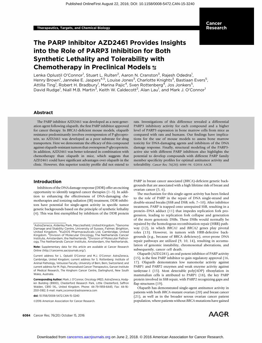
We set about identifying a compound with similar efficacy toolaparib butwithout thepotential liability associatedwithbeing asubstrate for the P-gp (MDR1) transporter. Triaging of com-pounds followingmedicinal chemistry (see SupplementaryMate-rial) identified two series of compounds around the phthalazi-none core of olaparib. AZD2461 (Fig. 1A) was identified as theoptimal compound from the Piperidine Ether series (see inter-national patent WO2009/093032, specifically compound num-ber 2b and47) and is a potent inhibitor of bothPARP1andPARP2with IC50 values of 5 nmol/L and 2 nmol/L, respectively, com-parable with olaparib (5 nmol/L and 1 nmol/L, respectively). Thelack of selectivity between PARP1 and PARP2 may be a positivefeaturewhen attempting to inhibit SSB repair, as PARP2 also playsan important role in this pathway (35). Both AZD2461 andolaparib were effective at inhibiting formation of cellular poly(ADP-ribose) (PAR) polymers following treatment with 10mmol/L hydrogenperoxide (Supplementary Fig. S1). The alkalineCOMET assay, where the length of the comet tail momentrepresents the degree of unrepaired SSBs following induction ofDNA damage by IR, was used to confirm that observed PARinhibition translated into inhibition of SSB repair (Fig. 1B). Inaddition, AZD2461 potentiated the antiproliferation effect of theDNA-damaging alkylating agent MMS, which induces SSBs (Fig.1C; Supplementary Table S1). Collectively, these data confirmthat AZD2461 is as effective at inhibiting SSB repair as olaparib.
Activity against BRCA-deficient tumors through syntheticlethality is a key component of olaparib's developmental pro-gram. Thus, we sought to determine the potency of single-agentAZD2461 inBRCA-deficient cancer cells. In vitro clonogenic assayswere performed with AZD2461 and olaparib against a panel of
breast cancer tumor cells with either mutant (MDA-MB-436,SUM1315MO2, and SUM149PT) or wild-type (WT; T47D,BT549, and MDA-MB-231) BRCA1 gene status. Both olapariband AZD2461 exhibited similar pharmacokinetic and pharma-codynamic profiles (see SupplementaryMaterial and Supplemen-tary Fig. S2) and significant potency as single agents in the BRCA1-mutant breast cancer cell lines but not in the BRCA1WT cell lines(Fig. 1D).
Assessment of the in vitro permeability and efflux of AZD2461was undertaken in the human intestinal-derived cell line CaCo-2(see SupplementaryMaterial and Supplementary Table S2) and inthe matched cell lines KBA1, a genetically modified version ofHeLa that overexpresses high levels of P-gp (36), and KB31,whichdoes not overexpress P-gp (Fig. 2A). While this functional assaydoes not distinguish between saturation of the P-gp pumps versuslow binding, it does discriminate between compounds that arehighly effluxed and those that are not. Using these assays, we wereable to distinguish low versus highly effluxed compounds, whileaddition of the P-gp inhibitor verapamil provided further evi-dence that efflux was occurring through a P-gp mechanism. Thedata in Fig. 2A show that, in contrast to olaparib, AZD2461 hassimilar activity between KBA1 cells and matched WT KB31 cellsand addition of verapamil to KBA1 cells showed little effect on thecellular activity of AZD2461, indicating that AZD2461 is signif-icantly less prone to P-gp–mediated efflux mechanisms thanolaparib. Similar data supporting a lack of P-gp liability inAZD2461 were obtained in the colorectal cancer cell line HCT-15, which expresses high levels of endogenous P-gp (Supplemen-tary Fig. S3 and Supplementary Table S3).
To assess AZD2461 activity in a more clinically relevant BRCA-mutant background and acquired resistant setting, we used in vitroand in vivomodels where prolonged olaparib treatment had led toresistance and high P-gp expression levels. The BRCA2-deficientmouse breast cancer line KB2P3.4 was generated from a BRCA2-deficientmousemammary tumor that demonstrated sensitivity toolaparib treatment (37). This cell line was treated in culture witholaparib for 2 months to induce the olaparib-resistant lineKB2P3.4R, and overexpression of P-gp was confirmed usingimmunofluorescence with a P-gp antibody (Fig. 2B). Treatmentof the parental KB2P3.4 with AZD2461 resulted in a similarresponse to olaparib (Fig. 2B). However, unlike olaparib,AZD2461 was also effective in the KB2P3.4R cell line. Consistentwith this difference being based on P-gp, olaparib sensitivity inKB2P3.4R cells was restored on cotreatment with tariquidar, anew generation P-gp inhibitor.
Increased expression ofAbcb1a andAbcb1b encoding themousedrug efflux transporter P-gp contributes to olaparib resistance inBRCA1;p53-deficient mouse mammary tumors (29). To deter-mine whether AZD2461 could overcome olaparib resistancein vivo, small tumor fragments of an olaparib-resistant tumor(T6-28) exhibiting an 80-fold increased expression ofAbcb1bweretransplanted into syngeneic WT female mice and treated witholaparib, combination olaparib and tariquidar, or AZD2461. Asexpected, olaparib resistance was successfully overcome by tar-iquidar pretreatment (Fig. 2C). Tumors were sensitive toAZD2461 in the absence of tariquidar, consistent with the ideathat spontaneous BRCA1;p53-defective mammary tumors, inwhich resistance is caused by increased P-gp-mediated drug efflux,remain sensitive to AZD2461.Moreover, in a separate study, long-term AZD2461 treatment in the BRCA1;p53-defective mousetumor model suppressed development of drug resistance (28).
Oplustil O'Connor et al.
Cancer Res; 76(20) October 15, 2016 Cancer Research6086
on June 2, 2018. © 2016 American Association for Cancer Research. cancerres.aacrjournals.org Downloaded from
Published OnlineFirst August 22, 2016; DOI: 10.1158/0008-5472.CAN-15-3240
Together, these data demonstrate that AZD2461 is a potentinhibitor of PARP1 and PARP2 that can provide effectiveinhibition of SSB repair and has significant single-agent activityin BRCA-deficient cancer cells, comparable with olaparib.Moreover, we have shown that AZD2461 is a poor substratefor P-gp and has activity in olaparib-resistant cancer cells thatoverexpress P-gp in vitro and is capable of antitumor activity invivo in olaparib-resistant tumors where resistance is based onoverexpression of P-gp. Accordingly, the goal to develop afollow-on compound to olaparib, that has comparable activitybut without P-gp liability, has been successful. AZD2461,therefore, represents an excellent preclinical tool to study PARPinhibitor resistance and may help identify additional mechan-isms of PARP inhibitor resistance.
AZD2461 is as efficacious as, andbetter tolerated than, olaparibin combination with temozolomide in a mouse xenograftmodel
In addition to the utility of PARP inhibitors as single agents,there is a strong rationale for combination with DNA-damagingchemotherapies, such as temozolomide or camptothecins thatinduce SSBs (38, 39). As a single agent, synthetic lethality ofolaparib relies on endogenously generated SSBs and the inability
of BRCA mutation or other HRR deficiency to repair the ensuingDSBs, ultimately resulting in cancer cell death. In combinationwith DNA-damaging chemotherapies, the number of SSBs gen-erated by the chemotherapy agent is much larger than those thatoccur endogenously (40). If repair is prevented (e.g., by PARPinhibitor treatment), cell death can be induced even when theHRR pathway is functional, purely because the tolerated DNAdamage threshold is exceeded. This has two important implica-tions: first, combination of a PARP inhibitor and SSB-inducingagent can result in killing HRR-proficient as well as HRR-deficientcancer cells; second, potential for damage in normal tissue com-partments is increased, as evidenced in the increased hematologictoxicities observed when PARP inhibitors and alkylating agentsare combined (41).
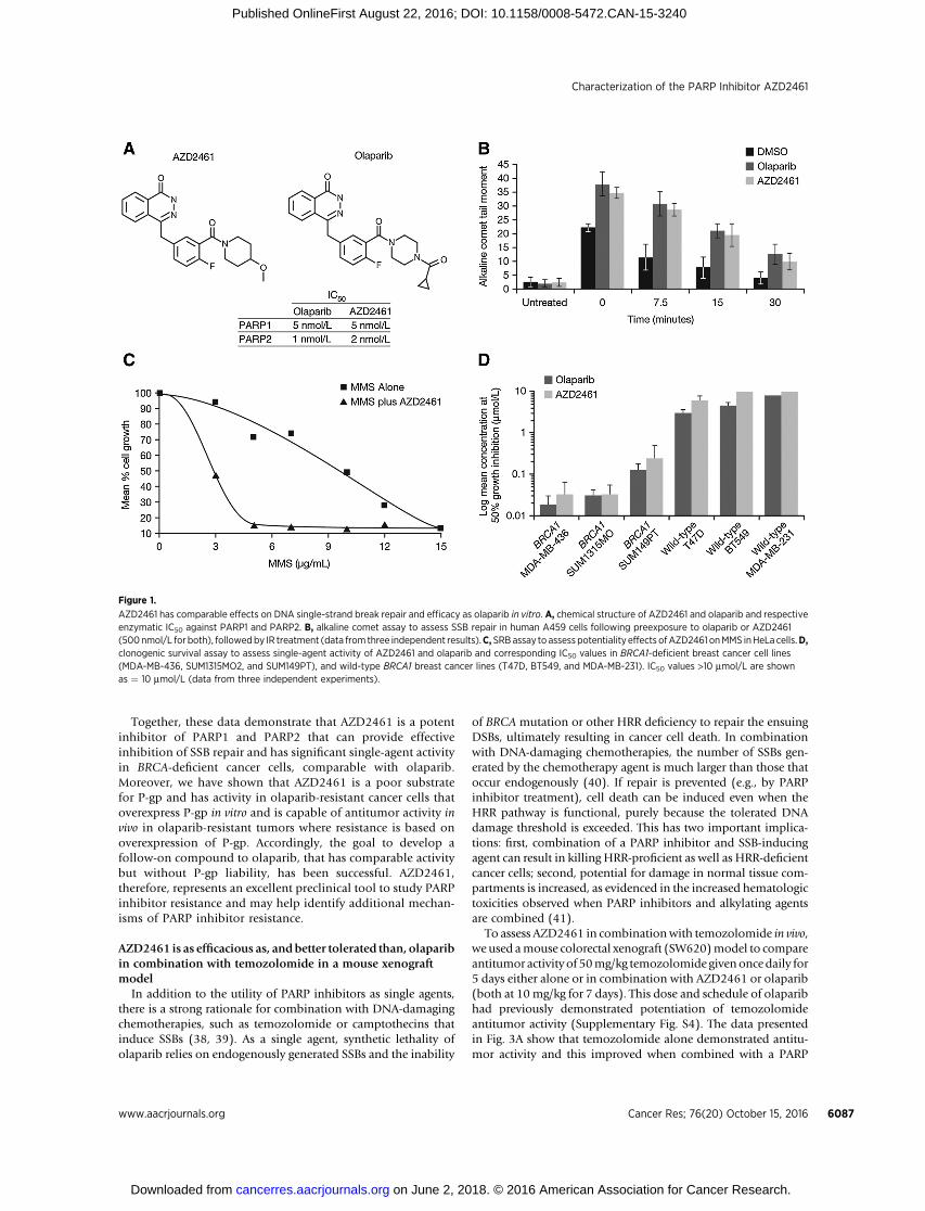
To assess AZD2461 in combinationwith temozolomide in vivo,we used amouse colorectal xenograft (SW620)model to compareantitumor activity of 50mg/kg temozolomide givenoncedaily for5 days either alone or in combination with AZD2461 or olaparib(both at 10mg/kg for 7 days). This dose and schedule of olaparibhad previously demonstrated potentiation of temozolomideantitumor activity (Supplementary Fig. S4). The data presentedin Fig. 3A show that temozolomide alone demonstrated antitu-mor activity and this improved when combined with a PARP
Figure 1.
AZD2461 has comparable effects on DNA single-strand break repair and efficacy as olaparib in vitro. A, chemical structure of AZD2461 and olaparib and respectiveenzymatic IC50 against PARP1 and PARP2. B, alkaline comet assay to assess SSB repair in human A459 cells following preexposure to olaparib or AZD2461(500nmol/L for both), followedby IR treatment (data from three independent results).C,SRBassay to assesspotentiality effects ofAZD2461 onMMS inHeLa cells.D,clonogenic survival assay to assess single-agent activity of AZD2461 and olaparib and corresponding IC50 values in BRCA1-deficient breast cancer cell lines(MDA-MB-436, SUM1315MO2, and SUM149PT), and wild-type BRCA1 breast cancer lines (T47D, BT549, and MDA-MB-231). IC50 values >10 mmol/L are shownas ¼ 10 mmol/L (data from three independent experiments).
Characterization of the PARP Inhibitor AZD2461
www.aacrjournals.org Cancer Res; 76(20) October 15, 2016 6087
on June 2, 2018. © 2016 American Association for Cancer Research. cancerres.aacrjournals.org Downloaded from
Published OnlineFirst August 22, 2016; DOI: 10.1158/0008-5472.CAN-15-3240

Figure 2.
AZD2461 overcomes P-gp–associated resistanceto olaparib. A, activity of AZD2461 and olaparib inmatched cell lines, KBA1 cells (a geneticallymodified version of HeLa that overexpresseshigh levels of P-gp) and KB31 cells.B, immunofluorescence staining for P-gp andrelative growth inhibition following treatment witholaparib or AZD2461 with and without tariquidar inthe parental KB2P3.4 BRCA2�/� mouse cellline and an acquired olaparib-resistant cloneKB2P3.4R. C, response of the olaparib-resistantBrca1D5-13/D5-13;p53D2-10/D2-10 tumor T6-28 toAZD2461. Animals carrying transplanted tumorswere treated daily with 0.5% HPMC (vehicle)10 mL/kg orally, AZD2461 100 mg/kg orally, andolaparib 50 mg/kg i.p. with and without tariquidar2 mg/kg i.p. (tumor volume day 0, 100%).
Oplustil O'Connor et al.
Cancer Res; 76(20) October 15, 2016 Cancer Research6088
on June 2, 2018. © 2016 American Association for Cancer Research. cancerres.aacrjournals.org Downloaded from
Published OnlineFirst August 22, 2016; DOI: 10.1158/0008-5472.CAN-15-3240
inhibitor. This difference was statistically significant (P <0.001) at day 55 (when the temozolomide group was culled);the effect between the combination groups (i.e., temozolo-mide plus olaparib or AZD2461) was not statistically signif-
icant either at day 55 (P ¼ 0.53) or when the study wasstopped at day 73 (P ¼ 0.57). Both combination treatmentsconferred considerable delay in tumor regrowth comparedwith temozolomide alone.
Figure 3.
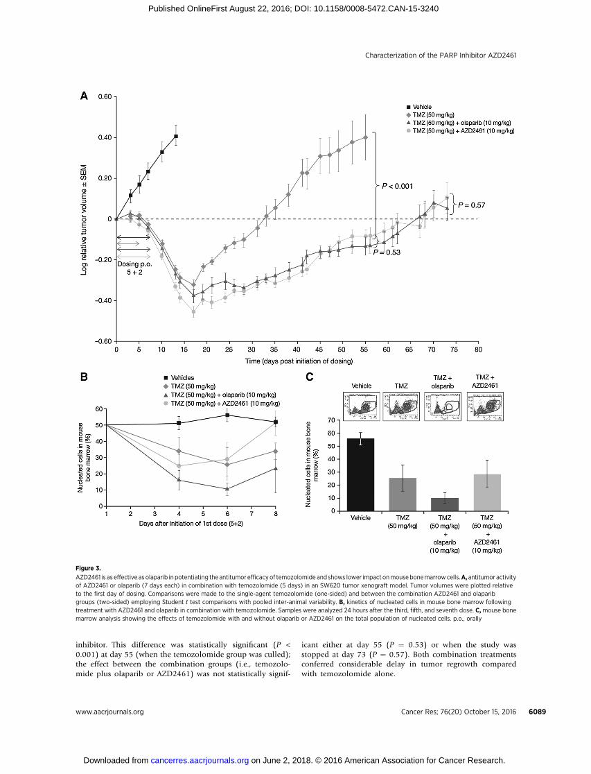
AZD2461 is as effective as olaparib in potentiating the antitumor efficacyof temozolomide and shows lower impact onmousebonemarrowcells.A, antitumor activityof AZD2461 or olaparib (7 days each) in combination with temozolomide (5 days) in an SW620 tumor xenograft model. Tumor volumes were plotted relativeto the first day of dosing. Comparisons were made to the single-agent temozolomide (one-sided) and between the combination AZD2461 and olaparibgroups (two-sided) employing Student t test comparisons with pooled inter-animal variability. B, kinetics of nucleated cells in mouse bone marrow followingtreatment with AZD2461 and olaparib in combination with temozolomide. Samples were analyzed 24 hours after the third, fifth, and seventh dose. C,mouse bonemarrow analysis showing the effects of temozolomide with and without olaparib or AZD2461 on the total population of nucleated cells. p.o., orally
Characterization of the PARP Inhibitor AZD2461
www.aacrjournals.org Cancer Res; 76(20) October 15, 2016 6089
on June 2, 2018. © 2016 American Association for Cancer Research. cancerres.aacrjournals.org Downloaded from
Published OnlineFirst August 22, 2016; DOI: 10.1158/0008-5472.CAN-15-3240
We examined body weight loss as a gross indicator of tolera-bility and myelosuppression as a more clinically relevant indica-tor of combinatorial toxicity. Body weights of mice decreasedrelative to their weight at the start of the experiment but recoveredquickly after the dosingwasfinished;weight losswas greater in thetwo combination groups versus the temozolomide alone group(Supplementary Fig. S5).
To assess the impact of the combinations on bone marrowpopulations, we examined cohorts of mice given the samedose regimens as used in the combination efficacy study. Micewere culled at time points across the dosing phase to create atime course in which the impact of treatments on bonemarrow cells could be assessed (Fig. 3B and C). Flow cyto-metry identified three distinct white blood cell populations(lymphocytes, monocytes, and neutrophils). Temozolomidealone led to a reduction in the total number of white bloodcells in bone marrow with the nadir after the last dose anda failure to return to starting levels at day 8. Temozolomidein combination with olaparib led to a significantly greaterimpact on bone marrow (a nadir at day 6 and a worse state ofrecovery at day 8).
An unexpected finding, however, was that combinationAZD2461 and temozolomide did not result in the same severityof bone marrow effects as the olaparib combination. The nadir
was not statistically different from temozolomide alone andthere was a good recovery by day 8. Although lower doses ofolaparib could result in less bone marrow toxicity, these arealso likely to be less efficacious, as demonstrated by Supple-mentary Fig. S4, which shows that doses �3 mg/kg of olaparibin combination with temozolomide (50 mg/kg) do not confer astatistically significant benefit over temozolomide alone (MTD68 mg/kg).
Collectively, these data suggest that AZD2461 might havetwo potential advantages over olaparib. First, while having asimilar level of antitumor activity, AZD2461 did not have thesame level of P-gp liability as olaparib; second, AZD2461appeared better tolerated in combination with a DNA-damag-ing chemotherapy. The basis for this latter observation, though,cannot be attributed to PARP1/PARP2 inhibition, as bothtreatments are similar in this respect, suggesting an as yetunidentified mechanism.
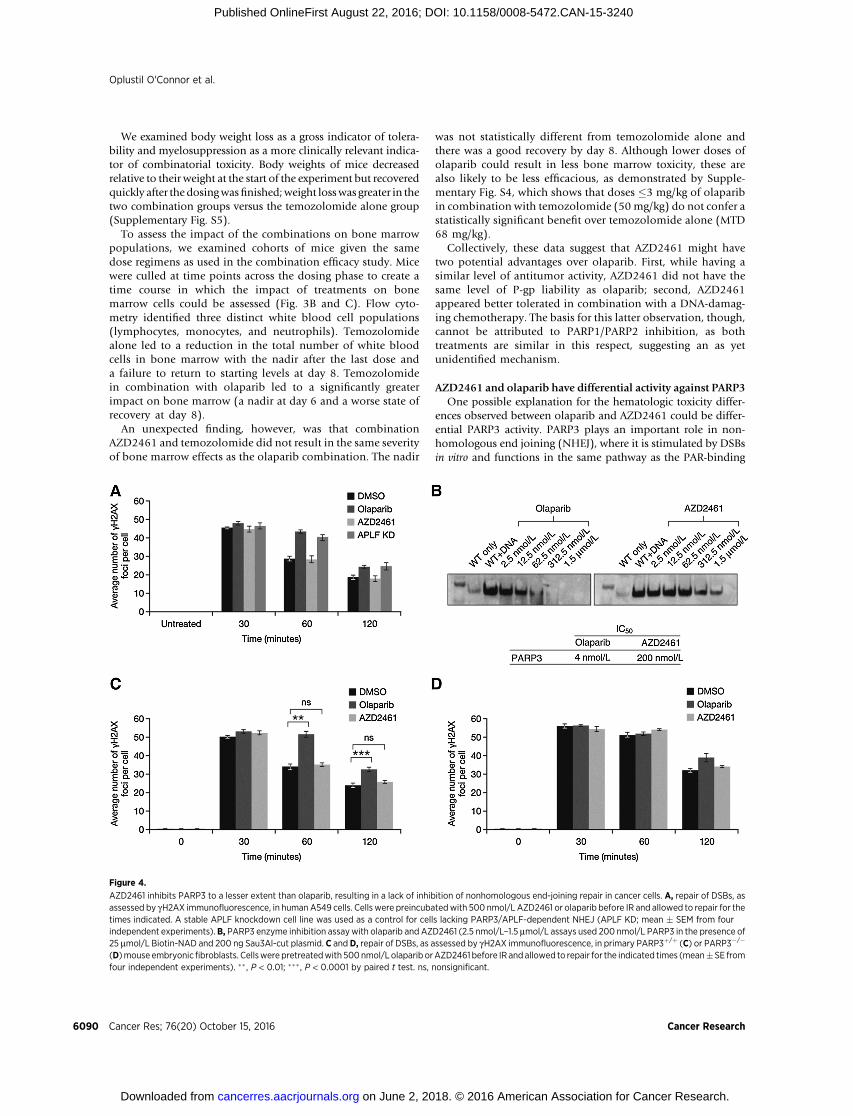
AZD2461 and olaparib have differential activity against PARP3One possible explanation for the hematologic toxicity differ-
ences observed between olaparib and AZD2461 could be differ-ential PARP3 activity. PARP3 plays an important role in non-homologous end joining (NHEJ), where it is stimulated by DSBsin vitro and functions in the same pathway as the PAR-binding
Figure 4.
AZD2461 inhibits PARP3 to a lesser extent than olaparib, resulting in a lack of inhibition of nonhomologous end-joining repair in cancer cells. A, repair of DSBs, asassessed by gH2AX immunofluorescence, in humanA549 cells. Cells were preincubatedwith 500 nmol/L AZD2461 or olaparib before IR and allowed to repair for thetimes indicated. A stable APLF knockdown cell line was used as a control for cells lacking PARP3/APLF-dependent NHEJ (APLF KD; mean � SEM from fourindependent experiments).B, PARP3 enzyme inhibition assaywith olaparib and AZD2461 (2.5 nmol/L–1.5 mmol/L assays used 200 nmol/L PARP3 in the presence of25 mmol/L Biotin-NAD and 200 ng Sau3Al-cut plasmid. C and D, repair of DSBs, as assessed by gH2AX immunofluorescence, in primary PARP3þ/þ (C) or PARP3�/�
(D)mouse embryonicfibroblasts. Cellswerepretreatedwith 500nmol/L olaparib orAZD2461 before IR andallowed to repair for the indicated times (mean�SE fromfour independent experiments). �� , P < 0.01; ��� , P < 0.0001 by paired t test. ns, nonsignificant.
Oplustil O'Connor et al.
Cancer Res; 76(20) October 15, 2016 Cancer Research6090
on June 2, 2018. © 2016 American Association for Cancer Research. cancerres.aacrjournals.org Downloaded from
Published OnlineFirst August 22, 2016; DOI: 10.1158/0008-5472.CAN-15-3240

protein APLF to accelerate chromosomal DSB repair (33). Fol-lowing IR treatment, gH2AX can be used as a marker of DSBdamage (42, 43). The repair of DSBs and the decline in gH2AXover time following IR was abrogated by either the loss of PARP3,APLF, or treatment with the PARP inhibitor KU-58948, which isrelated chemically to olaparib (33).
The link between PARP3, NHEJ, and mouse bone marrowwas highlighted in studies of murine hematopoietic stem andmultipotent progenitor cells (HSPC), which were shown to bemore resistant to IR-induced damage than more differentiatedprogenitor cells (44). This difference was based on both anincreased resistance to apoptosis and the ability to repair DNAby NHEJ. The NHEJ pathway is a lower fidelity alternative toHRR DSB repair, and mice bone marrow stem cells appear toutilize NHEJ while human HSPCs undergo apoptosis inresponse to DNA damage (45). It has been suggested thatthese differences may reflect the different challenges faced bymammals with diverse life spans and ages of reproductivematurity (46).
To investigate whether differential PARP3 activity couldprovide the basis for the differential hematologic toxicityobserved with olaparib and AZD2461 in mice, we looked atthe effect of these inhibitors on gH2AX following IR treatment(Fig. 4A). Unlike olaparib, which results in persistence ofgH2AX following IR to the same degree as APLF knockdown,AZD2461 had no effect on gH2AX dynamics. To confirmwhether this observation was due to PARP3 inhibition, wecarried out PARP3 enzyme inhibition assays where PARP3auto-ADP-ribosylation activity was assessed following the addi-tion of increasing doses (2.5 nmol/L–1.5 mmol/L) of olaparibor AZD2461. Fig. 4B shows that AZD2461 did not inhibitPARP3 to the same extent as olaparib. There was a 50-folddifference in PARP3 inhibitory activity, with the IC50 value forPARP3 being 4 nmol/L for olaparib and 200 nmol/L forAZD2461 (Fig. 4B). The data in Fig. 4A, C, and D are consistentwith (although not proof of) this difference at the enzyme levelbeing translated into a failure to inhibit NHEJ DSB repair,based on gH2AX kinetics.
Increased bone marrow tolerability of AZD2461 incombination with temozolomide is mouse specificand is not seen in rat models
The finding that AZD2461 does not inhibit PARP3, coupledwith previous studies that suggest mouse bone marrow HSPCspreferentially useNHEJwhendealingwithDNAdamage, suggestsour observation of better tolerability of chemotherapy combina-tion could be specific tomice and not translate into humans. Priorto initiating clinical trials to assess combination tolerability, werepeated the experiment in Fig. 3 in an athymic ratmodel. First, weassessed PARP3 levels (and compared with PARP1 and PARP2levels) in HSPCs from mice, rats, and humans (Fig. 5 andSupplementary Figs. S6 and S7). PARP3 levels were about 3.7times higher in mice than in rats (Fig. 5A), consistent with use ofNHEJ as a primary repair mechanism. Because of a lack of thesame sequence identity, it was not possible to directly comparelevels of PARP3 expression across mice, rats, and human bonemarrow cells using the TaqMan probe-based assay. However,using SYBR Green dye detection RT-PCR, we designed specificprimers for human and rat PARP3 and demonstrated that therelative level of PARP3 expression is similar between rat andhuman bone marrow cells (Fig. 5B).
To increase confidence around the finding of better hemato-logic tolerability, we repeated the temozolomide combinationstudy in Fig. 3C in an athymic ratmodel (Fig. 5C). Contrary to theresults inmice, AZD2461 was no better tolerated than olaparib interms of bone marrow toxicity (Fig. 5C) or total body weight lossin rats (Supplementary Fig. S8).
Figure 5.
PARP3 levels are significantly higher in mouse but not rat or human bonemarrow cells and, consistent with this, is a lack of differential bonemarrow toxicity between AZD2461 and olaparib in rats. A, PARP1, PARP2,and PARP3 gene expression in mouse and rat bone marrow cells usingcustomized TaqMan assays (means difference for rat versus mouse,PARP1 ¼ 0.1517, P¼ 0.0632; PARP2¼�0.3883, P ¼ 0.0318; PARP3¼�3.717,P ¼ 0.0169; n ¼ 6 biological samples). B, PARP3 gene expression in rat andhuman bone marrow cells measured using SYBR Green RT-PCR assay (ratversus human PARP3 means difference ¼ �0.01548, P ¼ 0.9232;n ¼ 6 biological samples). Box-and-whiskers graphs show mean from threeexperimental repeats. C, female athymic rat bone marrow analysis(day 5 of treatment) using flow cytometry analysis as in Fig. 3C. Graph showsmean � SD of total nucleated cell population (% of parent) in individualtreatment groups (n ¼ 3).
Characterization of the PARP Inhibitor AZD2461
www.aacrjournals.org Cancer Res; 76(20) October 15, 2016 6091
on June 2, 2018. © 2016 American Association for Cancer Research. cancerres.aacrjournals.org Downloaded from
Published OnlineFirst August 22, 2016; DOI: 10.1158/0008-5472.CAN-15-3240
Similar data were obtained in male athymic rats (data notshown). This difference could not be explained by pharmaco-kinetic differences as these are comparable in mice and rats [10mg/kg in both cases giving an area under the curve (AUCt) of1–2 mmol � h/L].
We have provided an explanation for the better tolerability ofAZD2461 compared with olaparib observed in mice by pro-viding data showing the differential PARP3 activity and thedifferent impact of the two compounds on NHEJ DSB repair. Todate, technical challenges around the isolation and analysis ofrat HSPCs have prevented us from directly demonstrating thatrats are different from mice in utilizing NHEJ. Therefore, ourexplanation for the better tolerability of AZD2461 comparedwith olaparib in mice being attributable to NHEJ DSB repairmechanisms is based on indirect evidence and we cannot ruleout alternative reasons for combination toxicity differencesobserved between the two rodent species. However, the clinicalexperience of combining PARP inhibitors with temozolomide
(41) does argue that rat and not mouse is the better predictivemodel.
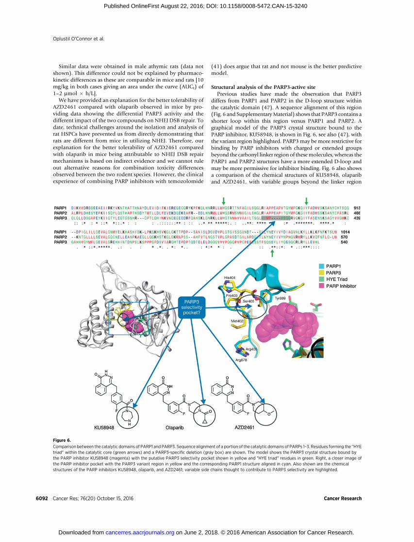
Structural analysis of the PARP3-active sitePrevious studies have made the observation that PARP3
differs from PARP1 and PARP2 in the D-loop structure withinthe catalytic domain (47). A sequence alignment of this region(Fig. 6 and SupplementaryMaterial) shows that PARP3 contains ashorter loop within this region versus PARP1 and PARP2. Agraphical model of the PARP3 crystal structure bound to thePARP inhibitor, KU58948, is shown in Fig. 6, see also (47), withthe variant region highlighted. PARP3may be more restrictive forbinding by PARP inhibitors with charged or extended groupsbeyond the carbonyl linker region of thesemolecules, whereas thePARP1 and PARP2 structures have a more extended D-loop andmay be more permissive for inhibitor binding. Fig. 6 also showsa comparison of the chemical structures of KU58948, olapariband AZD2461, with variable groups beyond the linker region
Figure 6.
Comparison between the catalytic domains of PARP1 and PARP3. Sequence alignment of a portion of the catalytic domains of PARPs 1–3. Residues forming the "HYEtriad" within the catalytic core (green arrows) and a PARP3-specific deletion (gray box) are shown. The model shows the PARP3 crystal structure bound bythe PARP inhibitor KU58948 (magenta) with the putative PARP3 selectivity pocket shown in yellow and "HYE triad" residues in green. Right, a closer image ofthe PARP inhibitor pocket with the PARP3 variant region in yellow and the corresponding PARP1 structure aligned in cyan. Also shown are the chemicalstructures of the PARP inhibitors KU58948, olaparib, and AZD2461; variable side chains thought to contribute to PARP3 selectivity are highlighted.
Oplustil O'Connor et al.
Cancer Res; 76(20) October 15, 2016 Cancer Research6092
on June 2, 2018. © 2016 American Association for Cancer Research. cancerres.aacrjournals.org Downloaded from
Published OnlineFirst August 22, 2016; DOI: 10.1158/0008-5472.CAN-15-3240
highlighted. This illustrates that derivatives of olaparib could begenerated for selectivity for or against PARP3 by targeting thevariable region in the PARP3 D-loop structure.
The demonstration of differential PARP3 activity of AZD2461compared with olaparib has two important ramifications. First,we are just beginning to understand the biological roles of thedifferent PARP family members and their interplay. For example,this study demonstrates that AZD2461 could represent a usefultool to distinguish cellular functions of PARPs 1–3 inDNA repair.Specifically, previous in vitro studies have shown that PARP3preparations can activate PARP1, and it has been suggested thatPARP3 plays a role in regulating the DNA damage responsethrough PARP1 activation (48). However, our study shows thatin cells treated with AZD2461, PARP3 is likely active in the NHEJpathway, even in the absence of detectable PARP1 activity, sug-gesting PARP1 and PARP3 may play independent roles in chro-mosomal DNA repair. Second, the structural similarity betweenolaparib and AZD2461 that results in this observed differentialactivity against PARP3 has allowed a rationalization of the mech-anistic basis of this difference in specificity via modeling of thePARP inhibitors in the PARP3-active site.
Increased understanding of inhibitor–PARP family memberinteractions, as demonstrated by an independent study (49), willalso facilitate our understanding of the PARP family biology.Together, advances in both the ability to generate inhibitorswith aparticular PARP specificity profile, as has been suggested recently(50), along with an understanding of the biological roles of thedifferent PARP family members, should provide the opportunityto generate inhibitors with an optimal PARP inhibitory profile tomaximize antitumor activity and therapeutic index.
Disclosure of Potential Conflicts of InterestNo potential conflicts of interest were disclosed.
Authors' ContributionsConception and design: L. Oplustil O'Connor, S.L. Rulten, A. Ting, S. Rotten-berg, D. Rudge, N.M.B. Martin, K.W. Caldecott, A. Lau, M.J. O'Connor
Development of methodology: A.N. Cranston, H. Brown, S. Rottenberg,J. Jonkers, M.J. O'ConnorAcquisition of data (provided animals, acquired and managed patients,provided facilities, etc.): L. Oplustil O'Connor, S.L. Rulten, A.N. Cranston,R. Odedra, J.E. Jaspers, L. Jones, C. Knights, B. Evers, M. Pajic, S. Rottenberg,J. JonkersAnalysis and interpretation of data (e.g., statistical analysis, biostatistics,computational analysis): L. Oplustil O'Connor, S.L. Rulten, A.N. Cranston,R. Odedra, H. Brown, B. Evers, A. Ting, R.H. Bradbury, M. Pajic, S. Rottenberg,J. Jonkers, N.M.B. Martin, A. Lau, M.J. O'ConnorWriting, review, and/or revision of the manuscript: L. Oplustil O'Connor,A.N. Cranston, R. Odedra, C. Knights, A. Ting, R.H. Bradbury, S. Rottenberg,J. Jonkers, K.W. Caldecott, A. Lau, M.J. O'ConnorAdministrative, technical, or material support (i.e., reporting or organizingdata, constructing databases): M.J. O'ConnorStudy supervision: S.L. Rulten, A. Lau, M.J. O'ConnorOther (his laboratory conducted some of the PARP3 experiments):K.W. Caldecott
AcknowledgmentsTariquidar was a kind gift of Dr. Susan Bates from the NIH. The authors
would like to thank StephenMoore, James Harrison, Gabrielle Grundy, AntonyOliver, Cydney Morgan and Eva Schut for their technical support. Editorialsupport was provided by Kerry Acheson, PhD, of iMed Comms, Macclesfield,UK, and Martin Goulding, PhD, of Mudskipper Business Ltd., funded byAstraZeneca.
Grant SupportThis workwas supported by grants from the EuropeanCommunity's Seventh
Framework Programme (FP7/2007-2013) under grant agreement no. HEALTH-F2010-259893 (L. Oplustil O'Connor and M.J. O'Connor); the NetherlandsOrganization for Scientific Research (NWO-Toptalent021.002.104 to J.E. Jas-pers; NWO-VIDI-91711302 to S. Rottenberg) and the Dutch Cancer Society(NKI 2007-3772 and NKI 2011-5220 to J. Jonkers and S. Rottenberg), and theCR-UK project grant, C6563/A1307 (S.L. Rulten and K.W. Caldecott). The workwas also supported by AstraZeneca.
The costs of publication of this articlewere defrayed inpart by the payment ofpage charges. This article must therefore be hereby marked advertisement inaccordance with 18 U.S.C. Section 1734 solely to indicate this fact.
Received December 17, 2015; revised July 20, 2016; accepted July 26, 2016;published OnlineFirst August 22, 2016.
References1. Curtin NJ. DNA repair dysregulation from cancer driver to therapeutic
target. Nat Rev Cancer 2012;12:801–17.2. Lord CJ, Ashworth A. The DNA damage response and cancer therapy.
Nature 2012;481:287–94.3. O'Connor MJ, Martin NM, Smith GC. Targeted cancer therapies based on
the inhibition of DNA strand break repair. Oncogene 2007;26:7816–24.4. KaelinWG Jr. The concept of synthetic lethality in the context of anticancer
therapy. Nat Rev Cancer 2005;5:689–98.5. Bryant HE, Schultz N, Thomas HD, Parker KM, Flower D, Lopez E, et al.
Specific killing of BRCA2-deficient tumours with inhibitors of poly(ADP-ribose) polymerase. Nature 2005;434:913–7.
6. Farmer H, McCabe N, Lord CJ, Tutt AN, Johnson DA, Richardson TB, et al.Targeting the DNA repair defect in BRCA mutant cells as a therapeuticstrategy. Nature 2005;434:917–21.
7. D'Amours D, Desnoyers S, D'Silva I, Poirier GG. Poly(ADP-ribosyl)-ation reactions in the regulation of nuclear functions. Biochem J 1999;342:249–68.
8. Lindahl T, Satoh MS, Poirier GG, Klungland A. Post-translational modi-fication of poly(ADP-ribose) polymerase induced by DNA strand breaks.Trends Biochem Sci 1995;20:405–11.
9. Ceccaldi R, Liu JC, Amunugama R, Hajdu I, Primack B, Petalcorin MI, et al.Homologous-recombination-deficient tumours are dependent onPoltheta-mediated repair. Nature 2015;518:258–62.
10. Mateos-Gomez PA, Gong F, Nair N,Miller KM, Lazzerini-Denchi E, Sfeir A.Mammalian polymerase theta promotes alternative NHEJ and suppressesrecombination. Nature 2015;518:254–7.
11. Murai J, Huang SY, Das BB, Renaud A, Zhang Y, Doroshow JH, et al.Trapping of PARP1 and PARP2 by clinical PARP inhibitors. Cancer Res2012;72:5588–99.
12. Moynahan ME, Jasin M. Mitotic homologous recombination maintainsgenomic stability and suppresses tumorigenesis. Nat Rev Mol Cell Biol2010;11:196–207.
13. Venkitaraman AR. Cancer suppression by the chromosome custodians,BRCA1 and BRCA2. Science 2014;343:1470–5.
14. Patel AG, Sarkaria JN, Kaufmann SH. Nonhomologous end joiningdrives poly(ADP-ribose) polymerase (PARP) inhibitor lethality inhomologous recombination-deficient cells. Proc Natl Acad Sci U S A2011;108:3406–11.
15. Menear KA, Adcock C, Boulter R, Cockcroft XL, Copsey L, Cranston A, et al.4-[3-(4-cyclopropanecarbonylpiperazine-1-carbonyl)-4-fluorobenzyl]-2H-phthalazin-1-one: a novel bioavailable inhibitor of poly(ADP-ribose)polymerase-1. J Med Chem 2008;51:6581–91.
16. EuropeanMedicines Agency (EMA). Lynparza recommended for approv-al in ovarian cancer; 2014. Available from:http://www.ema.europa.eu/ema/index.jsp?curl¼pages/news_and_events/news/2014/10/news_de-tail_002196.jsp&mid¼WC0b01ac058004d5c1.
Characterization of the PARP Inhibitor AZD2461
www.aacrjournals.org Cancer Res; 76(20) October 15, 2016 6093
on June 2, 2018. © 2016 American Association for Cancer Research. cancerres.aacrjournals.org Downloaded from
Published OnlineFirst August 22, 2016; DOI: 10.1158/0008-5472.CAN-15-3240

17. US Food and Drug Administration (FDA). FDA News release: FDAapproves Lynparza to treat advanced ovarian cancer; 2014. Availablefrom:http://www.fda.gov/NewsEvents/Newsroom/PressAnnouncements/ucm427554.htm.
18. Mullins DW Jr, Giri CP, Smulson M. Poly(adenosine diphosphate-ribose)polymerase: the distribution of a chromosome-associated enzyme withinthe chromatin substructure. Biochemistry 1977;16:506–13.
19. Fisher AE, Hochegger H, Takeda S, Caldecott KW. Poly(ADP-ribose)polymerase 1 accelerates single-strand break repair in concert with poly(ADP-ribose) glycohydrolase. Mol Cell Biol 2007;27:5597–605.
20. Audeh MW, Carmichael J, Penson RT, Friedlander M, Powell B, Bell-McGuinn KM, et al. Oral poly(ADP-ribose) polymerase inhibitor olaparibin patients with BRCA1 or BRCA2mutations and recurrent ovarian cancer:a proof-of-concept trial. Lancet 2010;376:245–51.
21. Tutt A, Robson M, Garber JE, Domchek SM, Audeh MW, Weitzel JN, et al.Oral poly(ADP-ribose) polymerase inhibitor olaparib in patients withBRCA1 or BRCA2 mutations and advanced breast cancer: a proof-of-concept trial. Lancet 2010;376:235–44.
22. Gelmon KA, Tischkowitz M, Mackay H, Swenerton K, Robidoux A, TonkinK, et al. Olaparib in patients with recurrent high-grade serous or poorlydifferentiated ovarian carcinoma or triple-negative breast cancer: a phase 2,multicentre, open-label, non-randomised study. Lancet Oncol 2011;12:852–61.
23. Ledermann J, Harter P, Gourley C, Friedlander M, Vergote I, Rustin G, et al.Olaparib maintenance therapy in platinum-sensitive relapsed ovariancancer. N Engl J Med 2012;366:1382–92.
24. Edwards SL, Brough R, Lord CJ, Natrajan R, Vatcheva R, Levine DA, et al.Resistance to therapy caused by intragenic deletion in BRCA2. Nature2008;451:1111–5.
25. Bouwman P, Aly A, Escandell JM, PieterseM, Bartkova J, van der GuldenH,et al. 53BP1 loss rescues BRCA1 deficiency and is associated with triple-negative and BRCA-mutated breast cancers. Nat Struct Mol Biol 2010;17:688–95.
26. Bunting SF, Callen E, Wong N, Chen HT, Polato F, Gunn A, et al. 53BP1inhibits homologous recombination in Brca1-deficient cells by blockingresection of DNA breaks. Cell 2010;141:243–54.
27. XuG, Chapman JR, Brandsma I, Yuan J,MistrikM, Bouwman P, et al. REV7counteracts DNA double-strand break resection and affects PARP inhibi-tion. Nature 2015;521:541–4.
28. Jaspers JE, Kersbergen A, Boon U, Sol W, van Deemter L, Zander SA, et al.Loss of 53BP1 causes PARP inhibitor resistance in Brca1-mutated mousemammary tumors. Cancer Discov 2013;3:68–81.
29. Rottenberg S, Jaspers JE, Kersbergen A, van der Burg E, Nygren AO, ZanderSA, et al.High sensitivity of BRCA1-deficientmammary tumors to thePARPinhibitor AZD2281 alone and in combination with platinum drugs. ProcNatl Acad Sci U S A 2008;105:17079–84.
30. Hay T, Matthews JR, Pietzka L, Lau A, Cranston A, Nygren AO, et al. Poly-(ADP-ribose) polymerase-1 inhibitor treatment regresses autochthonousBrca2/p53-mutant mammary tumors in vivo and delays tumor relapse incombination with carboplatin. Cancer Res 2009;69:3850–5.
31. Elstrodt F, Hollestelle A, Nagel JH, Gorin M, Wasielewski M, van denOuwelandA, et al. BRCA1mutation analysis of 41humanbreast cancer celllines reveals three new deleterious mutants. Cancer Res 2006;66:41–5.
32. BreslinC,Clements PM, El-Khamisy SF, PetermannE, IlesN,Caldecott KW.Measurement of chromosomal DNA single-strand breaks and replicationfork progression rates. Methods Enzymol 2006;409:410–25.
33. Rulten SL, Fisher AE, Robert I, ZumaMC, RouleauM, Ju L, et al. PARP-3 andAPLF function together to accelerate nonhomologous end-joining. MolCell 2011;41:33–45.
34. Liu X, HolstegeH, van der GuldenH, Treur-MulderM, Zevenhoven J, VeldsA, et al. Somatic loss of BRCA1 and p53 inmice induces mammary tumorswith features of human BRCA1-mutated basal-like breast cancer. Proc NatlAcad Sci U S A 2007;104:12111–6.
35. Ame JC, Rolli V, Schreiber V, Niedergang C, Apiou F, Decker P, et al. PARP-2, a novel mammalian DNA damage-dependent poly(ADP-ribose) poly-merase. J Biol Chem 1999;274:17860–8.
36. Choi KH, Chen CJ, Kriegler M, Roninson IB. An altered pattern ofcross-resistance in multidrug-resistant human cells results from spon-taneous mutations in the mdr1 (P-glycoprotein) gene. Cell 1988;53:519–29.
37. Evers B, Drost R, Schut E, de Bruin M, van der Burg E, Derksen PW, et al.Selective inhibition of BRCA2-deficient mammary tumor cell growth byAZD2281 and cisplatin. Clin Cancer Res 2008;14:3916–25.
38. Javle M, Curtin NJ. The role of PARP in DNA repair and its therapeuticexploitation. Br J Cancer 2011;105:1114–22.
39. Murai J, Zhang Y, Morris J, Ji J, Takeda S, Doroshow JH, et al. Rationale forpoly(ADP-ribose) polymerase (PARP) inhibitors in combination therapywith camptothecins or temozolomide based on PARP trapping versuscatalytic inhibition. J Pharmacol Exp Ther 2014;349:408–16.
40. Plummer ER, Middleton MR, Jones C, Olsen A, Hickson I, McHugh P,et al. Temozolomide pharmacodynamics in patients with metastaticmelanoma: DNA damage and activity of repair enzymes O6-alkylgua-nine alkyltransferase and poly(ADP-ribose) polymerase-1. Clin CancerRes 2005;11:3402–9.
41. Plummer R, Lorigan P, Evans J, StevenN,MiddletonM,Wilson R, et al. Firstand final report of a phase II study of the poly(ADP-ribose) polymerase(PARP) inhibitor, AG014699, in combination with temozolomide (TMZ)in patients with metastatic malignant melanoma (MM). J Clin Oncol24:18s, 2006 (suppl; abstr 8013).
42. BonnerWM, Redon CE, Dickey JS, Nakamura AJ, SedelnikovaOA, Solier S,et al. GammaH2AX and cancer. Nat Rev Cancer 2008;8:957–67.
43. Riballo E, KuhneM,RiefN,DohertyA, SmithGC,RecioMJ, et al. Apathwayof double-strand break rejoining dependent upon ATM, Artemis, andproteins locating to gamma-H2AX foci. Mol Cell 2004;16:715–24.
44. MohrinM, Bourke E, Alexander D,WarrMR, Barry-Holson K, Le BeauMM,et al. Hematopoietic stem cell quiescence promotes error-prone DNArepair and mutagenesis. Cell Stem Cell 2010;7:174–85.
45. Milyavsky M, Gan OI, Trottier M, Komosa M, Tabach O, Notta F, et al. Adistinctive DNA damage response in human hematopoietic stem cellsreveals an apoptosis-independent role for p53 in self-renewal. Cell StemCell 2010;7:186–97.
46. Lane AA, Scadden DT. Stem cells and DNA damage: persist or perish? Cell2010;142:360–2.
47. Lehti€o L, Jemth AS, Collins R, Loseva O, Johansson A, Markova N, et al.Structural basis for inhibitor specificity in human poly(ADP-ribose) poly-merase-3. J Med Chem 2009;52:3108–11.
48. Loseva O, Jemth AS, Bryant HE, Schuler H, Lehtio L, Karlberg T, et al.PARP-3 is a mono-ADP-ribosylase that activates PARP-1 in the absenceof DNA. J Biol Chem 2010;285:8054–60.
49. Wahlberg E, Karlberg T, Kouznetsova E, Markova N, Macchiarulo A,Thorsell AG, et al. Family-wide chemical profiling and structuralanalysis of PARP and tankyrase inhibitors. Nat Biotechnol 2012;30:283–8.
50. PapeoG, Posteri H, BorghiD, Busel AA, Caprera F, Casale E, et al. Discoveryof 2-[1-(4,4-difluorocyclohexyl)piperidin-4-yl]-6-fluoro-3-oxo-2,3-dihy-dro-1H-isoindole-4-carboxamide (NMS-P118): a potent, orally available,and highly selective PARP-1 inhibitor for cancer therapy. J Med Chem2015;58:6875–98.
Cancer Res; 76(20) October 15, 2016 Cancer Research6094
Oplustil O'Connor et al.
on June 2, 2018. © 2016 American Association for Cancer Research. cancerres.aacrjournals.org Downloaded from
Published OnlineFirst August 22, 2016; DOI: 10.1158/0008-5472.CAN-15-3240
2016;76:6084-6094. Published OnlineFirst August 22, 2016.Cancer Res Lenka Oplustil O'Connor, Stuart L. Rulten, Aaron N. Cranston, et al. Chemotherapy in Preclinical ModelsPARP3 Inhibition for Both Synthetic Lethality and Tolerability with The PARP Inhibitor AZD2461 Provides Insights into the Role of
Updated version
10.1158/0008-5472.CAN-15-3240doi:
Access the most recent version of this article at:
Material
Supplementary
http://cancerres.aacrjournals.org/content/suppl/2016/08/20/0008-5472.CAN-15-3240.DC1
Access the most recent supplemental material at:
Cited articles
http://cancerres.aacrjournals.org/content/76/20/6084.full#ref-list-1
This article cites 47 articles, 15 of which you can access for free at:
Citing articles
http://cancerres.aacrjournals.org/content/76/20/6084.full#related-urls
This article has been cited by 2 HighWire-hosted articles. Access the articles at:
E-mail alerts related to this article or journal.Sign up to receive free email-alerts
Subscriptions
Reprints and
To order reprints of this article or to subscribe to the journal, contact the AACR Publications Department at
Permissions
Rightslink site. Click on "Request Permissions" which will take you to the Copyright Clearance Center's (CCC)
.http://cancerres.aacrjournals.org/content/76/20/6084To request permission to re-use all or part of this article, use this link
on June 2, 2018. © 2016 American Association for Cancer Research. cancerres.aacrjournals.org Downloaded from
Published OnlineFirst August 22, 2016; DOI: 10.1158/0008-5472.CAN-15-3240