the of biological chemistry vol. no. 12, issue...
TRANSCRIPT

THE JOURNAL OF BIOLOGICAL CHEMISTRY Q 1984 by The American Society of Biologicai Chemists, Inc.
Vol. 259, No. 12, Issue of June 25, pp. 7453-7453.1984 Printed in U.S.A.
A Monoclonal Antibody Recognizes an 0-Acylated Sialic Acid in a Human Melanoma-associated Ganglioside*
(Received for publication, November 16, 1983)
David A. ChereshSg, Ajit P. VarkitTII, Nissi M. VarkiS, William B. Stallcup**, Joel Levine**, and Ralph A. Reisfeld$ From the $Department of Immunology, Scripps Clinic and Research Foundation, La Jolla, California 92037, lIThe Cancer Center, ~ n i ~ e r s ~ t y of CaZi~ornia at Sun Diego, San Diego, Ca l i fo rn~ 92037, and the **Molecular ~eurobiology ~ b o r a ~ o ~ , Salk I ~ t ~ t u t e , La Jolfa, Ca~ifornia 92037
Monoclonal antibody D l . 1 originally prepared against the B49 cell line derived from a rat brain tumor was shown to react with a ganglioside present in fetal rat brain. We have found that this antigen is also present in human malignant melanoma tumors as well as many melanoma cell lines. The ganglioside from human melanoma cell lines migrates between G M ~ and GMz on one-dimensional thin layer chromatography. Analysis by two-dimensional thin layer chromatogra- phy with intermediate ammonia treatment suggests that the ganglioside contains one or more base-labile 0-acyl esters. Mild base hydrolysis under conditions known to remove 0-acyl esters results in complete loss of antigenic reactivity. Thus, the alkali-labile moiety is a critical component of the epitope recognized by the antibody. Analysis of the sialic acids of total ganglio- sides from [6-sHJgluc~mine- labe l~ melanoma cells showed that approximately 10% of these molecules are 0-acylated. Similar analysis of the purified ganglioside showed that greater than 30% of the sialic acids co- migrated with authentic 9-0-acetyl-N-acetylneur- aminic acid. The antibody did not cross-react with normal human skin melanocytes nor with any of a large number of normal human adult and fetal tissues. The antibody also did not react with numerous other malig- nant cell lines studied. These findings suggest that the antigenic epitope defined by antibody D l . 1 contains an 0-acylated sialic acid and may arise from aberrant 0- acetylation occurring in human malignant melanoma ceIIs.
Monoclonal antibodies to tumor-associated antigens have facilitated the characterization of molecular differences be- tween tumors and normal cells and significantly advanced an understanding of the functional role of some of these antigens. However, most information gained thus far involves either protein or glycoprotein antigens, as they can be readily im- munoprecipitated from tumor cell lysates.
Recent technological advances have made it possible to use
* This work was supported in part by United States Public Health Service Grants CA28420, NS16112, and GM32373 from the National Institutes of Health. This is Scripps Publication 3277-1”. The costs of publication of this article were defrayed in part by the payment of page charges. This article must therefore be hereby marked ‘‘aduertisement” in accordance with 18 U.S.C. Section 1734 solely to indicate this fact.
$ Recipient of National Institutes of Health Fellowship Award
11 Faculty Fellow of the John A. and George L. Hartford Founda- 1F32 CA07544-01.
tion.
monoclonal antibodies for the characterization of complex carbohydrate antigens on tumor cell surfaces. Several inves- tigators have produced monoclonal antibodies directed to the carbohydrate portion of tumor cell-associated glycolipids (1- 8). Monoclonal antibodies to the melanoma-associated gan- glioside, GDzl (7) and G,, (2, 3), have been described. There are also descriptions of anti-glycolipid antibodies reactive with various other neoplastic tissues, including colon carci- noma (1) and neuroblastoma (5). In some cases, the antigenic epitopes defined by these antibodies have only subtle differ- ences between them involving a single sugar residue or the type of sugar linkage.
The gangliosides are glycolipids that contain sialic acids. The elegant work of Schauer and others (9) has now clearly shown that the sialic acids are an extremely diverse group of sugars. This diversity is mostly generated by different types of 0-substitutions (usually 0-acetyl esters) at the 4-, 7-, 8-, and 9-hydroxyl positions of the parent molecule, neuraminic acid. Recent work indicates that the sialic acids of some gangliosides have 0-acylation. This may have been missed by earlier studies because of the extreme lability of the 0-acyl esters.
Recently, Levine et al. (10) described a monoclonal antibody D1.l prepared against the rat B49 cell line which specifically recognizes a ganglioside on developing rat embryonic neu- roectoderm. In this report, we now demonstrate that the ganglioside recognized by monoclonal antibody D1.1 is also found on human melanoma cells and contains an alkali- sensitive antigenic epitope which involves an 0-acylated sialic acid residue.
EXPERIMENTAL PROCEDURES~
RESULTS
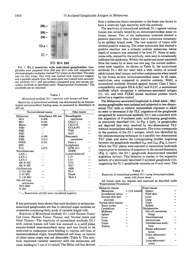
Serological Reactivity of Monoclonal Antibody Dl.1 with a Ganglioside Common to Rat Brain Tumor and Human Mela- noma Cell Lines-As shown in Fig. 1, monoclonal antibody D1.l specifically recognizes a ganglioside in B49 and M14 cells which migrates on thin layer chromatography between Gllll and GM%. The M14 cells express this antigen as a doublet.
Gangliosides are termed according to the nomenclature as previ- ously described by Svennerholm (39).
“Experimental Procedures” are presented in miniprint a t the end of this paper. Miniprint is easily read with the aid of a standard magnifying glass. Full size photocopies are available from the Journal of Biological Chemistry, 9650 Rockville Pike, Bethesda, MD 20814. Request Document No. 83M-3269, cite the authors, and include a check or money order for $2.00 per set of photocopies. Full size photocopies are ais0 included in the microfilm edition of the Journal that is available from Waverly Press.
7453
7454 0-Acylated Ganglioside Antigen in Melanoma
GM3-
GMZ- GM1-
GDla -
GTc - ST0 M14 849
FIG. 1. D1.l reactivity with individual gangliosides. Gan- gliosides were prepared from B49 and M14 cells and separated by chromatography on plastic-backed TLC plates as described. The plate was cut into strips. One strip was stained with resorcinol reagent, and a parallel sample from the same plate was treated with monoclo- nal antibody D1.l and peroxidase-conjugated goat anti-mouse sec- ondary antibody as described under "Experimental Procedures." The standards are as indicated.
TABLE I Monoclonal antibody Dl.1 reactivity with human ceU lines
Reactivity of monoclonal antibody was determined by an enzyme- linked immunosorbent binding assay as measured by absorbance at 492 nm.
Neuroectodermal turnom
Melanoma Absorbance 492 nm M14 0.206 M21 0.121 Melur 0.648 FM3 FM5 FM7 FM8 0.100 FM9 0.212 FOSS 0.121 MS1 FL2 0.074
0 - - -
-
Neuroblastoma LAN-1 - LAN-2 - LAN-5 - SK-N-MC - SK-N-SH -
Glioblastoma U-373 MG - U-87 MG - U-138 MG
Oat cell lung -
NCI-H69 T-293
carcinoma - -
a -, Reactivities 50.020 were consider
Other tumors ~~
lronchogenic carcinoma
CALU-1 CALU-6 UCLA-P3 T291 A427
'ancreatic
Panc-1 .ymphoma
HSBR
L10 L14 LG2 3017 4098 PL3 Daudi Molt-4
carcinoma
HPB-A11
,ymphoblastoid
negative.
It has previously been shown that such doublets in melanoma- associated gangliosides are due to identical sugar moieties on ceramides containing fatty acids of variable length (19).
Reactivity of Monoclonal Antibody D1.1 with Human Tumor Cell Lines, Human Tumor Tissues, and Normal Adult and Fetal Tissues-The reactivity of monoclonal antibody D1.l with various human cell lines was assessed in a solid phase enzyme-linked immunosorbent assay and was found to be restricted to melanoma since binding to various cell lines of neuroectodermal origin, lymphoblastoid cell lines, or tumors of other tissue origin was not detectable (Table I). The anti- body expressed variable reactivity with the melanoma cell lines, binding to 7 out of 11 tested. The Melur cell line derived
from a melanoma lesion metastatic to the brain was shown to have a relatively high reactivity with the antibody.
The reactivity of monoclonal antibody D1.l against various tissues was initially tested by an immunoperoxidase assay on frozen tissues. Two of six melanomas screened showed a positive reactivity. One of these was a melanoma metastatic to an axillary lymph node. The vast majority of tumor cells showed positive staining. The other melanoma that showed a positive reaction was a primary nodular melanoma, whose depth of invasion was assessed to be level IV. Nests of mela- noma cells, stained by the antibody, were seen to occasionally infiltrate the epidermis. Within the epidermal areas separated from the tumor by at least one rete peg, the normal melano- cytes were negative. As shown in Table 11, the monoclonal antibody D1.l did not react with a wide variety of normal adult tissues, fetal tissues, and other malignancies when tested by the frozen section immunoperoxidase assay. In all cases, reactivities were compared to positive controls, W632, a monoclonal antibody directed against human Class I histo- compatibility antigens HLA-A,B,C and 9.2.27, a monoclonal antibody which recognizes a melanoma-associated antigen (11, 12), and with P3X63 mouse myeloma protein which served as a negative control.
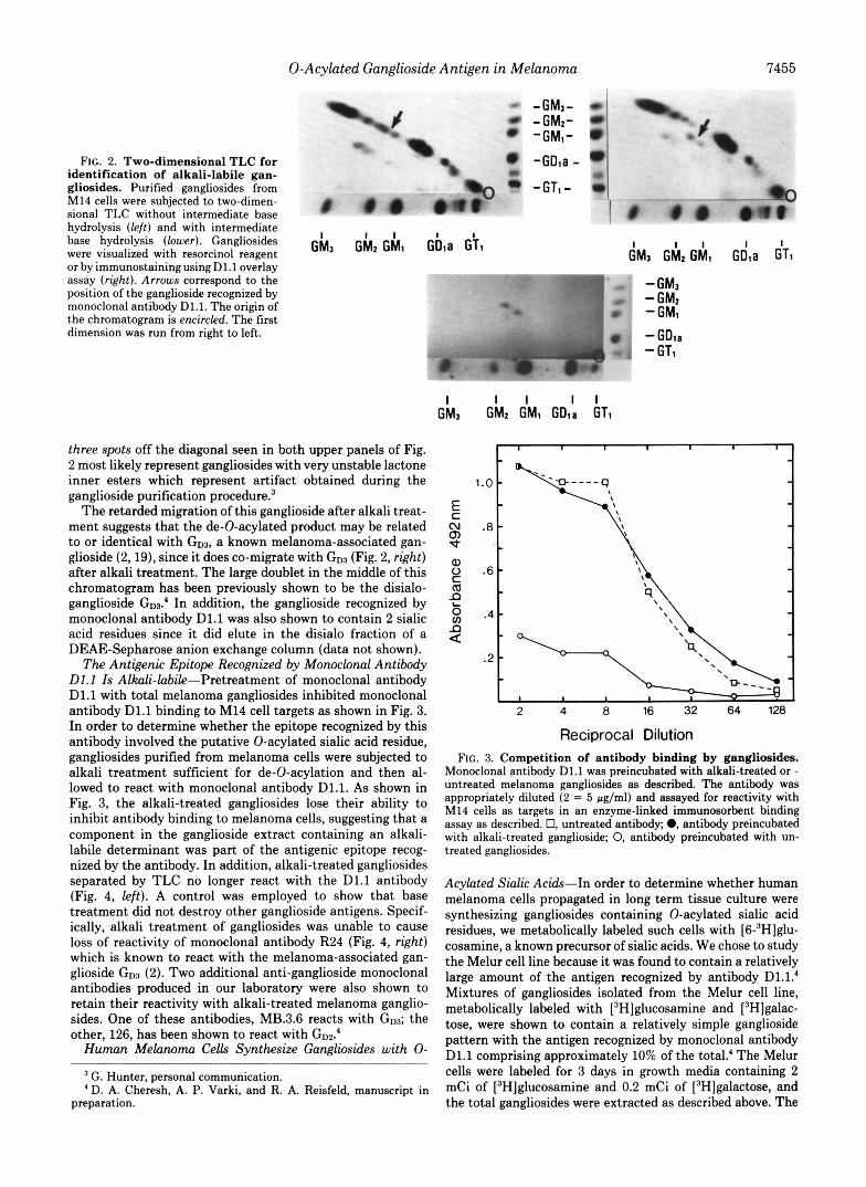
The Melanoma-associated Ganglioside Is Alkali-labile-"el- anoma gangliosides were isolated and subjected to two-dimen- sional TLC with or without intermediate exposure to alkali in order to determine if the TLC migration of the ganglioside recognized by monoclonal antibody D1.l was consistent with the migration of 0-acylated sialic acid-bearing gangliosides, as previously described (15). In Fig. 2 (left), 14 gangliosides are depicted that were resolved by two-dimensional TLC without intermediate alkali treatment. The arrow corresponds to the position of the D1.l antigen, which was identified by the immunostaining technique on a parallel two-dimensional TLC plate and shows the corresponding doublet migrating between the ganglioside standard G M l and G M 2 (Fig. 2, lower). When the TLC plates were exposed to ammonium hydroxide vapors prior to initiation of migration in the second dimension (Fig. 2, right), the D1.l ganglioside demonstrated retarded migration (arrow). This behavior is similar to the migration pattern of a previously described 0-acylated ganglioside (15), suggesting the D1.1 ganglioside contains an 0-acyl ester. The
TABLE I1 Reactivity of monoclonal antibody Dl.1 using immunoperoxidase
assay with frozen tissue All tissues were cut, stained, and examined as described under
"Experimental Procedures." Melanotic tissues
Melanoma + (2/6 tested Intradermal nevus - Normal skin mel- -
anocytes Normal adult tissues
Brain cortex - Cerebellum - Midbrain - Spleen - Liver - Lung - Colon - Kidney -
'eta1 tissues Spleen - Fetal skin - Liver Lung - Colon - Kidney
Oat cell carcinoma -
Neuroblastoma - Astrocytoma - Glioblastoma multi- - Breast adenocarci- - Colon adenocarci- -
Lung adenocarci- -
-
ialignancies
(lung)
-
form
noma
noma
noma
0-Acylated Ganglioside Antigen in Melanoma 7455
FIG. 2. Two-dimensional TLC for identification of alkali-labile gan- gliosides. Purified gangliosides from M14 cells were subjected to two-dimen- sional TLC without intermediate base hydrolysis (left) and with intermediate base hydrolysis (lower). Gangliosides were visualized with resorcinol reagent G h 3 G h z G h l Gbla - or by immunostainingusingD1.1 overlay assay (right). Arrows correspond to the position of the ganglioside recognized by monoclonal antibody D1.l. The origin of the chromatogram is encircled. The first dimension was run from right to left.
three spots off the diagonal seen in both upper panels of Fig. 2 most likely represent gangliosides with very unstable lactone inner esters which represent artifact obtained during the ganglioside purification procedure?
The retarded migration of this ganglioside after alkali treat- ment suggests that the de-0-acylated product may be related to or identical with G D ~ , a known melanoma-associated gan- glioside (2,19), since it does co-migrate with G D ~ (Fig. 2, right) after alkali treatment. The large doublet in the middle of this chromatogram has been previously shown to be the disialo- ganglioside GD~.' In addition, the ganglioside recognized by monoclonal antibody D1.l was also shown to contain 2 sialic acid residues since it did elute in the disialo fraction of a DEAE-Sepharose anion exchange column (data not shown).
The Antigenic Epitope Recognized by Monoclonal Antibody Dl.1 Is Alkali-labile-Pretreatment of monoclonal antibody D1.l with total melanoma gangliosides inhibited monoclonal antibody D1.l binding to M14 cell targets as shown in Fig. 3. In order to determine whether the epitope recognized by this antibody involved the putative 0-acylated sialic acid residue, gangliosides purified from melanoma cells were subjected to alkali treatment sufficient for de-0-acylation and then al- lowed to react with monoclonal antibody D1.1. As shown in Fig. 3, the alkali-treated gangliosides lose their ability to inhibit antibody binding to melanoma cells, suggesting that a component in the ganglioside extract containing an alkali- labile determinant was part of the antigenic epitope recog- nized by the antibody. In addition, alkali-treated gangliosides separated by TLC no longer react with the D1.l antibody (Fig. 4, left). A control was employed to show that base treatment did not destroy other ganglioside antigens. Specif- ically, alkali treatment of gangliosides was unable to cause loss of reactivity of monoclonal antibody R24 (Fig. 4, right) which is known to react with the melanoma-associated gan- glioside G D ~ (2). Two additional anti-ganglioside monoclonal antibodies produced in our laboratory were also shown to retain their reactivity with alkali-treated melanoma ganglio- sides. One of these antibodies, MB.3.6 reacts with G D ~ ; the other, 126, has been shown to react with GD~.'
Human Melanoma Cells Synthesize Gangliosides with 0-
G. Hunter, personal communication. 'D. A. Cheresh, A. P. Varki, and R. A. Reisfeld, manuscript in
preparation.
I ' 1 I 1 I , I
L
1.0 -
.8 -
.6 -
.4 - n a
t l \ 'n"-h " - I 1 I I "
2 4 8 16 32 64 128
.2 p-<\\ D","
2 4 8 16 32 64 128
Reciprocal Dilution FIG. 3. Competition of antibody binding by gangliosides.
Monoclonal antibody D1.l was preincubated with alkali-treated or - untreated melanoma gangliosides as described. The antibody was appropriately diluted (2 = 5 pg/ml) and assayed for reactivity with M14 cells as targets in an enzyme-linked immunosorbent binding assay as described. 0, untreated antibody; 0, antibody preincubated with alkali-treated ganglioside; 0, antibody preincubated with un- treated gangliosides.
Acylated Sialic Acids-In order to determine whether human melanoma cells propagated in long term tissue culture were synthesizing gangliosides containing 0-acylated sialic acid residues, we metabolically labeled such cells with [6-3H]glu- cosamine, a known precursor of sialic acids. We chose to study the Melur cell line because it was found to contain a relatively large amount of the antigen recognized by antibody D1.l.' Mixtures of gangliosides isolated from the Melur cell line, metabolically labeled with [3H]glucosamine and [3H]galac- tose, were shown to contain a relatively simple ganglioside pattern with the antigen recognized by monoclonal antibody D1.l comprising approximately 10% of the total.' The Melur cells were labeled for 3 days in growth media containing 2 mCi of [3H]glucosamine and 0.2 mCi of [3H]galactose, and the total gangliosides were extracted as described above. The

7456
GM1-
GMz-
GM1-
GDla-
GT1-
1
0-Acylated Ganglioside Antigen in Melanoma
A B C D E A B C D FIG. 4. Effects of alkali treatment on antibody binding to
gangliosides. Melanoma gangliosides were separated by TLC, and strips of the plates were cut and either sprayed with resorcinol reagent or overlaid with various anti-ganglioside antibodies for serological detection of antibody reactivity as described under "Experimental Procedures." Left: lane A, ganglioside standards; lane B, M14 ganglio- sides (lanes A and B were visualized with resorcinol reagent); lane C, untreated M14 gangliosides; lane D, alkali-treated M14 gangliosides; lane E, non-alkali-treated control M14 gangliosides (lanes C-E were allowed to react with monoclonal antibody D1.l in the overlay assay. Right: lane A, ganglioside standards; lane B, M14 gangliosides (lanes A and B were visualized with resorcinol reagent); lane C, alkali- treated M14 gangliosides; lane D, non-alkali-treated control M14 gangliosides (lanes C and D were allowed to react with monoclonal antibody R24 previously shown to recognize melanoma ganglioside G D ~ (2).
sialic acids were released and purified as described under "Experimental Procedures." The conditions for release and purification were chosen to maximize recovery while mini- mizing losses of 0-acyl groups (18). Approximately 30% of the total radioactivity was recovered in the Dowex 3-X4A eluate. These released purified sialic acids were then subjected to paper chromatography with an internal ['4C]N-acetylneur- aminic acid standard. As shown in Fig. 5, (upper), approxi- mately 87% of the [3H]sialic acid released co-migrated with the [14C]N-acetylneuraminic acid, while the remainder (13%) migrated faster in the same position as the N-acetyl-9-0- acetylneuraminic acid standard. When treated with base un- der conditions known to de-0-acylate sialic acids, the faster migrating peak almost completely disappeared (3% remain- ing), accompanied by a corresponding increase in the 'H,/"C ratio of the major N-acylneuraminic acid peak. This strongly suggests that approximately 10% of the sialic acids contained in total gangliosides synthesized by these cells are 0-acylated. In similar studies of the biosynthesis of sialic acids in murine Friend erythroleukemia cells, a radioactive peak with identical migration has been more completely characterized as being 9- 0-acetyl-N-acetylneuraminic acid.5 This finding increases the likelihood that alkali lability of the ganglioside recognized by antibody D1.l is attributable to the presence of 0-acylated sialic acids.
The Ganglioside Recognized by Monoclonal Antibody Dl.1 Contains an 0-Acylated Sialic Acid-In order to more directly demonstrate the presence of 0-acylated sialic acids in the ganglioside in question, an aliquot containing 2 x lo6 cpm of total gangliosides was chromatographed on one-dimensional TLC, and the region corresponding to the D1.1 ganglioside was scraped out and eluted. When an aliquot of the purified material was run on TLC and then studied by autoradiogra- phy, a double band was seen, corresponding to the location of
A. Varki and S. Diaz, submitted for publication.
NsuSGc + NeuSAc + tNeu5.9Ac2
I e BASE
! II I I
CMS FROM ORIGIN FIG. 5. Paper chromatography of ['Hlsialic acids from hu-
man melanoma cell gangliosides. Sialic acids were purified from the total gangliosides of [6-3H]glucosamine-labeled Melur melanoma cells and subjected to paper chromatography along with an internal [14C]N-acetylneuraminic acid standard, as described. The sample was spotted on the paper with (lower) or without (upper) prior base treatment to remove 0-acyl esters. The positions of migration of the following standards are indicated: NeAGc, N-glycolylneuraminic acid; NedAc, N-acetylneuraminic acid; and Neu5,9Ac2, 9-0-acetyl- N-acetylneuraminic acid.
the ganglioside recognized by the D1.l antibody (data not shown).
Sialic acids were released from this ganglioside, purified, and subjected to paper chromatography as described above. As shown in Fig. 6, 32% of the total 'H cpm migrated ahead of the [14C]N-acetylneuraminic acid standard. This peak was eliminated by prior de-0-acylation with base (Fig. 6, lower), with a corresponding increase in the 'H/"C ratio in the non- 0-acetylated peak. This directly demonstrates that the D1.l ganglioside contains an 0-acylated sialic acid. Similar results were obtained with neuraminidase-released sialic acids.
DISCUSSION
Gangliosides are ubiquitous molecules present in the mem- branes of all eukaryotic cells. They have been implicated in a variety of cellular functions, including cell-cell adhesion and communication, as well as cell-substrate interactions (21-23). Gangliosides have been found to serve as putative cell mem- brane receptors for hormones (24, 25), toxins (26, 27), and extracellular matrix components (28,29). Their role as devel- opmental antigens, particularly in tissues of neuroectodermal origin, is currently being examined by numerous investigators. The application of monoclonal antibody production for the isolation and characterization of ganglioside antigens has aided significantly to delineate their roles in various biological processes. The ganglioside antigen defined by the monoclonal antibody D1.l has been recently shown to be expressed on the surfaces of developing rat neuroectoderm (10). This anti-

0-Acylated Ganglioside Antigen in Melanoma 7457
CMS FROM ORIGIN
FIG. 6. Paper chromatography of sialic acids from purified ganglioside. The radiolabeled ganglioside recognized by the D1.l antibody was purified by preparative TLC. The sialic acids were released from the ganglioside, purified, and subjected to paper chro- matography as described with (lower) or without (upper) prior base treatment. The standards are N - a c e t y ~ e ~ ~ i n ~ c acid (Neu5Ac) and 9-0-acetyl-N-acetylneuraminic acid (Neu5,9Acz).
gen was shown to be a ~fferentiation marker since its expres- sion disappears during neural differentiation.
We have presented evidence that in human tissue the antigen defined by monoclonal antibody D1.l has a high degree of specificity for melanoma. Evidence for in vivo expression of this antigen has been documented by immuno- histological staining patterns of tissue sections. In addition to its serological specificity, the D1.l antibody is the first de- scribed which recognizes an alkali-labile ganglioside. Our evidence clearly indicates that the epitope recognized by monoclonal antibody DL1 includes the 0-acyl group. In con- trast, the serological reactivity of three additional antibodies directed to gangliosides on melanoma cells did not show any loss in reactivity when tested on the same alkali-treated ganglioside preparations. Preliminary evidence presented here suggests that the ganglioside recognized by monoclonal anti- body D1.l may be an 0-acylated version of the disialoganglio- side GD3 since after de-0-acylation, it co-migrates with GD3. In addition, the ganglioside elutes from a DEAE-Sepharose anion exchange column in the disialo fraction. However, further structural analysis will be necessary to confirm this hypothesis.
Alkali-labile gangliosides have only recently been recog- nized (9, 30, 32). Older methods for the preparation of gan- gliosides (saponification of contaminating acidic lipids) un- doubte~y resulted in destruction of the alkali-labile 0-acyl esters. Recently, however, several 0-acylated gangliosides have been reported. The presence of 9-0-acetylated sialic acids in total brain gangliosides from several vertebrate spe-
cies was first reported by Haverkamp et al. (33). The structure of one 0-acetylated ganglioside from mouse brain is definitely known (30). Several other 0-acetylated gangliosides have recently been found in mouse thymus gangliosides. In this case, it was assumed that the alkali-labile nature of the gangliosides was due to the presence of 0-acetylated sialic acids (32).
In the present study, the two-~mensional TLC with inter- mediate alkali hydrolysis as described by Sonnino et af. (15) has been a useful tool for the analysis of these substituted molecules. In our hands, this technique has allowed us to demonstrate that the TLC migration of the ganglioside de- fined by monoclonal antibody D1.1 behaves similarly to those previously described as having an 0-acylated sialic acid resi- due. Since the ganglioside in question was such a minor component of the total gangliosides, we chose to use a radio- labeling method to demonstrate that it contained an O-ac- ylated sialic acid. Total labeled gangliosides from a melanoma cell line contained about 10% 0-acylated sialic acid. When radiolabeled D1.l antigen was purified from this mixture and subjected to similar analysis, it was found to contain -30% base-labile sialic acids. For reasons discussed in detail else- where (20), the 30% value is likely to be an underestimate. Therefore, these data are most compatible with one of two sialic acids in the ganglioside being 0-acylated. Additional studies will be required to define this further.
Since the base-labile sialic acid co-migrated with the 9-0- acetyl-N-acetylneuraminic acid, it is likely to be an O-acety- lated sialic acid. However, the present data do not allow us to exclude other base-labile 0-acyl groups, such as 0-lactyl es- ters, that have been reported in the sialic acids (9). Previous experiments have shown that prolonged treatment with Vibrio cholerae neuraminidase is required to completely destroy the antigenicity of the ganglioside, both in rat brain and in the B49 cells? This partial resistance to the Vibrio neuraminidase is in keeping with the fact that side chain (7/8/9) mono-0- acetylated sialic acids are relatively more resistant to release by this enzyme (9).
There are other known causes for base-labile groups in glycolipids. Inner lactone esters can occur in the sialic acids, either naturally (34), or as an artifact induced during purifi- cation. This appears unlikely because similar peaks were not seen in identical analyses of radiolabeled sialic acids from many other metabolically labeled tumor cell lines! Further- more, the sialic acids of the ganglioside in question are sus- ceptible to release by Arthrobacter urenfaciens neuraminidase, which will release 0-acetylated sialic acids (18) but would not be expected to release sialic acid lactones. 0-Acyl groups have also been reported in the simple neutral glycolipid galactosyl- ceramide (35). The ganglioside described here is considerably larger. However, we cannot absolutely rule out the possibility of such substitutions occurring in ~ ~ i ~ i o n to the substitution of the sialic acids.
Although the epitope defined by monoclonal antibody D1.l involves an 0-acylated sialic acid, several facts suggest it is a co-determinant and that this sialic acid residue is not the entire epitope responsible for antibody recognition. First, this antibody was raised by immunization of BALB/c mice, which are known to have extensive 0-acetylation of the sialic acids of their red blood cell membranes (31). In addition, murine erythroleukemia cells, which are known to have 0-acetylated sialic acids on their surface: do not show any reaction with this antibody (data not shown). Finally, the antibody fails to react with another alkali-labile ganglioside present on the
J. Levine, unpubIished observations. . _ ~ " _ _ . _ _ _ _ _

7458 0-Acylated GangZioside
same melanoma cells (see Fig. 2). Although sialic acids more often serve to mask underlying
antigens, there are several examples in which they can be antigenic themselves. The human M and N blood groups are partly determined by sialic acids (36). There are naturally occurring human monoclonal IgMs from patients with Wal- denstrom’s macroglobulinemia that react with antigens con- taining sialic acid as a determinant (37). Polyclonal antibodies to sialyloligosaccharides coupled to proteins have been suc- cessfully raised (38). Sialic acid is an integral component of the antigenic epitope recognized by several monoclonal anti- bodies raised against tumor gangliosides from colon carci- noma (I), neuroblastoma (5), and melanoma (2). In none of these cases was there any indication of 0-acylation of sialic acids. As far as we are aware, this is the first example of a monoclonal antibody directed against an alkali-labile ganglio- side. Furthermore, the epitope recognized includes the 0-acyl group itself. Further studies are currently underway to define the complete structure of this ganglioside.
0-Acylation of sialic acids appears to be a tissue- and species-specific characteristic (9). However, it is currently unclear whether 0-acylation in a given cell type is a general characteristic of all sialylated compounds produced by that cell or whether it is restricted to specific sialic acid residues. In a study of sialic acids in mouse red cells, different sialogly- coproteins were found to have similar extents of 0-acetylation (31). This suggested that 0-acetylation may not be very rigorously regulated with regard to its distribution on different sialic acid residues. However, in this study we found that of several gangliosides produced by a monoclonal population of tumor cells in culture, only one or two appeared to be alkali- labile. There is no evidence in any biological system for different sialyltransferases inserting 0-acylated and non-0- acylated sialic acids. Therefore, it appears likely that in mel- anoma cells, the 0-acylation reaction is a carefully regulated event that occurs after the sialylation of gangliosides, result- ing in selective 0-acylation of only certain sialic acid residues. Consequently, quantitative or qualitative differences in spe- cific 0-acyltransferases may cause the appearance of new antigens during the process of malignant transformation. Such antigens provide potentially useful molecular markers for the study of these human tumor cells.
A c ~ n o ~ ~ d g ~ n ~ s - W e wish to thank Dm. R. K. Yu (Yale Univer- sity) and J. Sundsmo (Scripps Clinic and Research Foundation) for their helpful discussions and Laura Wolff, Stephanie Singer, and Sandra Diaz for their technical assistance, and Bonnie Filiault for her diligent secretarial assistance.
REFERENCES 1. Magnani, J. L., Nilsson, B., Brockhaus, M., Zoph, F., Steplewski,
Z., Koprowski, H., and Ginsburg, V. (1982) J. Biol. Chem. 2 5 7 , 14365-14369; (1979) Somatic Cell Genet. 5,957-972
2. Pukel, C . S., Lloyd, K. P., Travassos, L. R., Dippold, W. G., Oettgen, H. F., and Old, L. J. (1982) J. Exp. Med. 155, 1133- 1137
3, Yeh, M-Y., Hellstrom, I., Abe, K., Hakomori, S., and Hellstrom, K. E. (1982) Int. J. Cancer 29,269-275
Antigen in Melanoma
4. Wiels, J., Fellous, M., and Tursz, T. (1981) Proc. Natl. Acad. Sci.
5. Eisenbarth, G. S., Walsh, F. S., and Nirenberg, M. (1979) P m . Nutl. Acad. Sci. U. S. A. 76,4913-4917
6. Brockhaus, M., Magnani, J. L., Blaszczyk, M., Steplewski, Z., Koprowski, H., Karlsson, K-A., Larson, G., and Ginsburg, V. (1981) J. Bwl. Chem. 2 5 6 , 13223-13225
7. Cahan, L. D., Irie, R. F., Singh, R., Cassidenti, A., and Paulson, J. C. (1982) Proc. Natl. Acad. Sci. U. S. A. 79 , 7629-7633
8. Hakomori, S., Patterson, C. M., Nudelman, E., and Sekiguchi, K. (1983) J. Biol. Chem. 258, 11819-11822
9. Schauer, R. (1982) Adu. Carbohydr. Chem. Biochem. 40,131-234 10. Levine, J. M., Beasley, L., and Stallcup, W. B. (1984) J. Neuro-
11. Bumol, T. F., and Reisfeld, R. A. (1982) Proc. Natl. Acad. Sei. U.
12. Harper, J. R., Bumol, T. F., and Reisfeld, R. A. (1982) Hybridoma
13. Ledeen, R. W., and Yu, R. K. (1982) Methods Enzymol. 83,139-
14. Jourdian, G. W., Dean, L., and Roseman, S. (1971) J. Biol. Chem.
15. Sonnino, S., Ghidoni, R., Chigorno, V., Masserini, M., and Tet-
16. Taylor, C. R. (1978) Arch Puthot. Lab. Med. 102,113-121 17. Magnani, J. L., Smith, D. F., and Ginsburg, V. (1980) Anal.
18. Varki, A., and Diaz, S. (1984) Anal, Biochem. 137,236-247 19. Nudelman, E., Hakomori, S., Kannagi, R., Levery, S., Yeh, M-
Y., Hellstrom, K. E., and Hellstrom, I. (1982) J. Bwl. Chem.
U. S. A. 78,6485-6488
chem., in press
S. A. 7 9 , 1245-1249
1,423-432
191
246,430-435
tamanti, G. (1983) Anal. Biochem. 128,104-114
Biochem. 109,399-402
257,12752-12756 20. Varki, A., and Diaz, S. (1983) J. BioL Chem. 258, 12465-12471 21. Blackburn, C. C., and Schnaar, R. L. (1983) J. Biol. Chem. 2 5 8 ,
22. Schnaar, R. L., Weigel, P. H., Kuhlenschmidt, M. S., Lee, Y. C., and Roseman, S. (1978) J. BwL Chem. 253,7940-7951
23. Hatten, M. E. (1981) J. Cell Bwl. 8 9 , 5 4 4 1 24. Kato, I., and Naiki, M. (1976) Infect. Zmmun. 13, 289-291 25. Mullin, B. R., Fishman, P. H., Lee, G., Aloj, S. M., Ledley, F. D.,
Winand, R. J., Kohn, L. D., andBrady, R. 0. (1976) Proc. Nutl. Acad. Sci. U. S. A. 7 3 , 8 4 2 4 6
1180-1188
26. Van Henyningen, W. E. (1974) Nature (Lond.) 249,415-417 27. Holmgren, J., Lonnrofh, I., and Svennerholm, L. (1973) Infect.
28. Perkins, R. M., Kellic, S., Patel, B., and Critchley, D. R. (1982)
29. Kleinman, H. K., Martin, G. R., and Fishman, P. H. (1979) Proc.
30. Ghidoni, R., Sonnino, S., Tettamanti, G., Baumann, N., Reuter,
31. Varki, A., and Kornfeid, S. (1980) J. Exp. Med. 152,532-544 32. Schwarting, G. A., and Gajewski, A. (1983) J. Biol. Chem. 2 5 8 ,
33. Haverkamp, J., Veh, R. W., Sander, M., Schaur, R., Kamerling, J. P., and Vliegenth~, G. F. (1977) ~ o p ~ - S ~ ~ r ’ s Physiol. Chem. 358,1609-1612
34. Gross, S. K., Williams, M. A., and McCluer, R. H. (1980) J. Neurochem. 34,1351-1361
35. Yasugi, E., Kasama, T., Kojima, H., and Yamakawa, T. (1983) J. Biochem. (Tokyo) 9 3 , 1595-1599
36. Sadler, J. E., Paulson, J. C., and Hill, R. L. (1979) J. Biol. Chem.
37. Tsai, C-M., Zopf, D. A., Yu, R. K., Wistar, R., and Ginsburg, V.
38. Smith, D. F., and Ginsburg, V. (1980) J. Biol. Chem. 255.55-59 39. Svennerholm, L. (1963) J. Neurocfaem. 10.613-623
Zmmun. 8,208-214
Exp. Cell Res. 141,231-243
Nut[. Acad. Sci. U. S. A. 7 6 , 3367-3371
G., and Schauer, R. (1980) J. Biol. Chem. 255,6990-6995
5893-5898
254,2112-2119
(1977) Proc. Nutl. Acad. Sei. U. S. A. 74 , 4591-4594

0-Acylated Ganglioside Antigen in Melanoma 7459
Tissues: YOWOns Of f m h , mmal and ulignant tissue were a b t a i n d from the surgical M o g y oepa~tmtmt of the Ida n. 6reea Hospttal of Scrlpps Clinic. specimtns were embedded i n Tissue Tek medium (%icntific Products1 and frozen i n bIDcks i n isopentans a t ltpuld nitrogen temperawre. were thcn s t o m at -7O'C.