the occipital lateral plate mesoderm is a novel source for
TRANSCRIPT

The occipital lateral plate mesoderm is a novel source for vertebrate neck musculature Article
Published Version
Theis, S., Patel, K., Valasek, P., Otto, A., Pu, Q., Harel, I., Tzahor, E., Tajbakhsh, S., Christ, B. and Huang, R. (2010) Theoccipital lateral plate mesoderm is a novel source for vertebrate neck musculature. Development, 137 (17). pp. 2961-2971. ISSN 1477-9129 Available at https://centaur.reading.ac.uk/23593/
It is advisable to refer to the publisher’s version if you intend to cite from the work. See Guidance on citing .Published version at: http://dev.biologists.org/content/137/17/2961.long
Publisher: Company of Biologists
All outputs in CentAUR are protected by Intellectual Property Rights law, including copyright law. Copyright and IPR is retained by the creators or other copyright holders. Terms and conditions for use of this material are defined in the End User Agreement .
www.reading.ac.uk/centaur
CentAUR

Central Archive at the University of Reading Reading’s research outputs online

2961RESEARCH ARTICLE
INTRODUCTIONThe innovation that permitted free movement of the headindependently of the body is considered to be a landmarkadaptation in vertebrates, facilitating predatory avoidance and preycapture. Free movement of the head was achieved by thedevelopment of the neck. For this milestone to be reached, the bodyplan underwent two major modifications: firstly, anterior vertebraereduced or lost their ability to form ribs; and secondly, new skeletalmuscles developed that enabled a turning and bending motion ofthe head. The muscle that evolved to exert these movements ishighly conserved in jawed vertebrates and is called the cucullarismuscle (Kuratani, 2008). The mammalian homologue of thecucullaris muscle is divided dorsoventrally into the larger dorsallypositioned trapezius and a comparatively smaller portion, thesternocleidomastoid muscle (Williams, 1995).
The origin of the cucullaris muscle has been the subject ofconsiderable debate (Krammer et al., 1987). Origins as diverse asthe branchial arches, somites and the lateral plate mesoderm haveall been proposed, evidenced often by innervation (Addens, 1933;Allis, 1897; Couly et al., 1993; Derjugin, 1908; Edgeworth, 1911;Edgeworth, 1926; Favaro, 1903; Fuerbringer, 1897; Gegenbaur,1898; Harrison, 1918; Huang et al., 1997; Huang et al., 2000;Lewis, 1910; Luther and Lubosch, 1938; Noden, 1983; Noden andFrancis-West, 2006; Piekarski and Olsson, 2007).
The head versus trunk origin of the cucullaris muscle will haveconsiderable bearing on the molecular mechanism used during itsdevelopment. The origins of myogenic cells that form head and
trunk muscles differ greatly, as do the source of their connectivetissues. Head muscles develop from unsegmented head mesodermor the splanchnic mesoderm (Noden and Francis-West, 2006;Nathan et al., 2008). Neural crest cells form the connective tissueof these muscles (Noden and Trainor, 2005; Matsuoka et al., 2005;Noden and Francis-West, 2006). By contrast, trunk muscles arederived from the somites and their connective tissue forms locallyor from lateral plate mesoderm (Christ et al., 1974; Christ et al.,1982).
Following the advent of robust lineage-tracing techniques, it isbelieved that trunk muscles, including those of the neck andtongue, are derived from the paraxial mesoderm (somites), whereashead muscles have multiple origins, such as the prechordal,paraxial and splanchnic mesoderm regions, which are all locatedanterior to the somites (Harel et al., 2009). Apart from differencesof head and trunk muscle development based on origin, recentpublications document that they form according to distinctmolecular programmes (Tzahor et al., 2003; Nathan et al., 2008;Harel et al., 2009; Mootoosamy and Dietrich, 2002; Sambasivan etal., 2009). Although the molecular end points of both myogenicprogrammes are the same, culminating in the expression of musclestructural proteins, they differ significantly during their earlyphases of development. In the trunk, Pax3 is expressed inproliferating progenitor cells and is downregulated as MyoD isupregulated (Amthor et al., 1999). By contrast, Pax3 is notexpressed in head myogenic precursors, a role enacted by a suiteof transcription factors, including Tbx1, MyoR, Pitx2, Isl1 andCapsulin (Hacker and Guthrie, 1998; Mootoosamy and Dietrich,2002; Harel et al., 2009; Sambasivan et al., 2009). The head andtrunk myogenic programmes also display distinct outcomes inresponse to individual signalling molecules. For example, trunkmyogenesis is promoted by the action of Wnt and Shh. By contrast,Wnt and Shh have been shown to inhibit head myogenesis. In thehead, the repression mediated by Wnt molecules (and othermolecules) needs to be lifted in order for differentiation to proceed,a process facilitated by the secretion of signalling molecule
Development 137, 2961-2971 (2010) doi:10.1242/dev.049726© 2010. Published by The Company of Biologists Ltd
1Institute of Anatomy, University of Freiburg, Freiburg i. Br., 79104, Germany.2School of Biological Sciences, University of Reading, Reading, RG6 6AJ, UK.3Department of Biological Regulation, Weizmann Institute, Rehovot, 76100, Israel.4Department of Developmental Biology, Institute Pasteur, Paris, 75724, France.5Institute of Anatomy, University of Bonn, Bonn, 53115, Germany.
*Author for correspondence ([email protected])
Accepted 16 June 2010
SUMMARYIn vertebrates, body musculature originates from somites, whereas head muscles originate from the cranial mesoderm. Neckmuscles are located in the transition between these regions. We show that the chick occipital lateral plate mesoderm hasmyogenic capacity and gives rise to large muscles located in the neck and thorax. We present molecular and genetic evidence toshow that these muscles not only have a unique origin, but additionally display a distinct temporal development, forming laterthan any other muscle group described to date. We further report that these muscles, found in the body of the animal, developlike head musculature rather than deploying the programme used by the trunk muscles. Using mouse genetics we reveal thatthese muscles are formed in trunk muscle mutants but are absent in head muscle mutants. In concordance with this conclusion,their connective tissue is neural crest in origin. Finally, we provide evidence that the mechanism by which these neck musclesdevelop is conserved in vertebrates.
KEY WORDS: Muscle development, Myogenesis, Skeletal, Somite, Vertebrate, Chick, Quail
The occipital lateral plate mesoderm is a novel source forvertebrate neck musculatureSusanne Theis1,2, Ketan Patel2,*, Petr Valasek2, Anthony Otto2, Qin Pu5, Itamar Harel3, Eldad Tzahor3,Shahragim Tajbakhsh4, Bodo Christ1 and Ruijin Huang5
DEVELO
PMENT

2962
antagonists produced by neural crest cells (Tzahor et al., 2003). Inaddition, these cells provide the support tissue for the muscle. Thetiming of muscle differentiation in the head is also distinct, takingplace later than in the trunk (Noden et al., 1999). Thereforeprofiling of genes and identifying the origin of the connectivetissues are likely to yield telling clues as to the character of aparticular myogenic programme.
In this study we report that the cucullaris muscle of birds and itsmammalian homologues develop by deploying the head myogenicprogramme. This conclusion is reached from our studies on thecucullaris muscle showing that: (1) the majority of its myofibresdevelop from a non-somitic source; (2) its molecular profile issimilar to that of other head muscles; (3) its connective tissue isderived from neural crest. Most significantly, our workexperimentally demonstrates a new source of skeletal muscle – theoccipital lateral plate mesoderm – as the major source of thecucullaris muscle.
MATERIALS AND METHODSPreparation of embryosAll experiments were performed on Gallus gallus domesticus chickenembryos unless stated otherwise. Lineage-tracing experiments wereperformed with either quail (Coturnix coturnix japonica) or transgenicchick embryos expressing cytoplasmic GFP under control of the beta-actinpromoter (Dr H. Sang, Roslin Institute, UK). Eggs were incubated at 38°Cand 80% humidity.
Grafting procedureIn order to avoid cross-contamination of somitic and lateral platemesoderm tissues during grafting, an alternative to our previous methods(Huang et al., 1997) was used in this study. Here we firstly removed donormaterial using the Chapman easy culture method to isolate embryos fromthe eggshell (Chapman et al., 2001). Somites were then prepared firstly bymaking a precise incision with a sharpened tungsten needle between thesomites and the medial edge of the lateral plate mesoderm. Subsequently,an incision was made between the somites and neural tube. Finally, thedissected tissue was briefly incubated in Dispase I (Sigma, 1 U/ml) toisolate whole somites free of mesenchymal contamination. A minimum offive procedures for each individual somite were performed. For lateral platemesoderm transplantation, a strip of tissue 1.5 somites wide from quail orGFP+ embryos was transplanted homotopically into the wild-type hosts(n>20). Grafting into the host was performed according to a modifiedprotocol of Huang et al. (Huang et al., 1997), but with the omission of sub-endodermal ink injection. A minimum of five procedures was performedfor homotopic regionalised lateral plate mesoderm grafts. Heterotopictransplantations were performed either by replacing chick lateral platemesoderm adjacent to somites 1-3 of a Hamburger-Hamilton stage 9 (HH9) embryo with quail lateral plate mesoderm originating adjacent to somites10-12 from a HH 11 embryo (caudal to cranial graft, n10). Alternatively,chick lateral plate mesoderm adjacent to somites 21-23 of a HH 14 embryowas replaced with quail lateral plate mesoderm originating adjacent tosomites 1-3 from a HH 9 embryo (cranial to caudal graft, n7).
ImmunohistochemistryQuail/chick chimeras were fixed in 4% PFA/PBS and processed for paraffinsectioning (10 m). Grafted material was detected with QCPN and MF20(Developmental Studies Hybridoma Bank, DSHB) and subtype-specific(IgG1 or IgG2b) goat-anti-mouse DyLight antibodies (JacksonImmunoResearch). Alternatively, QCPN was detected using goat-anti-mouseAP conjugated antibody. Paraffin sections (10 m) from GFP/WT chimeras,fixed for 2 hours in 4% PFA/PBS, were subjected to immunohistochemistryusing MF20 and the anti-GFP rabbit polyclonal antibodies (Torrey PinesBiolabs) and detected using goat-anti-mouse IgG 594 and goat-anti-rabbit488 antibodies (Jackson ImmunoResearch). Paraffin embedded tissue formyosin heavy chain (MyHC) expression (antibody MF20), was subjected toantigen retrieval by boiling in citrate buffer (0.1 M, pH 6) for 10 minutes.Non-specific binding was blocked in 10% fetal calf serum (FCS).
Pax3Cre:RosaSTOP/YFP embryos were harvested at 15.5 days post-coitum(dpc), fixed in 4% PFA/PBS for 2 hours and processed for paraffinsectioning (7 m). Immunohistochemistry was performed using MF20 andbiotinylated anti-GFP antibodies (Abcam, UK). Secondary detection wasperformed using goat-anti-mouse IgG 488 and streptavidin 594 antibodies(Jackson ImmunoResearch).
Pax3Sp/Sp:Myf5nlacZ/nlacZ and Tbx1–/– embryos were harvested at 13.5,13.75 or 14.5 dpc, fixed for 1 hour in 4% PFA/PBS and stored in 1%PFA/PBS until processing for paraffin sectioning (7 m). MF20 antibodywas used for primary detection. Secondary antibody conjugated withhorseradish peroxidase (1:200, Sigma) was applied for 1.5 hours at roomtemperature. The antibody localisation was determined using DABstaining. Sections were counterstained with haematoxylin.
Wnt1Cre/R26RlacZ embryos were harvested 13.5 dpc and processed asdescribed by Valasek et al. (Valasek et al., 2010). Turtle (Trachemysscripta) embryos were fixed in 4% PFA/PBS, processed for paraffinsectioning (10 m) and subjected to MyHC immunohistochemistry usingMF20 and goat-anti-mouse IgG 594 (Jackson ImmunoResearch). Imageswere captured using a Zeiss fluorescence microscope.
Whole-mount in situ hybridisationWhole-mount in situ hybridisation was performed according to Nieto et al.(Nieto et al., 1996). Sections of whole mounts after in situ hybridisationwere generated after embedding samples in 4% agarose for vibratomesectioning (55 m).
RESULTSThe majority of the cucullaris muscle is derivedfrom the lateral plate mesoderm and not fromsomitesThe cucullaris muscle in birds has been previously reported to derivefrom the somites (Couly et al., 1993; Huang et al., 1997; Huang etal., 2000; Noden, 1983). More specifically, Huang et al. (Huang etal., 1997; Huang et al., 2000) found a contribution from the first threesomites, whereas Noden (Noden, 1983) reported contributions fromsomites 1-4. In this study, we homotopically transplanted,individually, the first seven somites from HH 9 quail embryos(before neural crest migration) free of contaminating tissues(facilitated through the use of Dispase I) into chick hosts and foundthat somitic contribution to the cucullaris muscle was relativelyminor (Fig. 1A-A�). Quantification of the contribution from somite1 to the cucullaris revealed that approximately 3.6% of fibrescontained graft-derived cells (Fig. 1C). A similar number of fibreswas found to develop from somites 2 and 3. The third, fourth andfifth somites contributed to the hypoglossus anlagen and did notcontribute to the cucullaris muscle (data not shown). Furthermore,somites 6-7 did not contribute to the cucullaris muscle (data notshown) in agreement with Huang et al. (Huang et al., 2000).
We hypothesised that results from previous studies reporting asomitic origin for this muscle (Huang et al., 1997; Huang et al., 2000)could have arisen as a result of contamination by lateral tissue. To thisend we transplanted quail lateral plate mesoderm adjacent to theoccipital somites 1-3 into a chick host. In contrast to transplantationof somites, we found very high densities of tissue originating fromquail in the cucullaris muscle (Fig. 1B-B�). Quantification of thecontribution from the lateral plate mesoderm revealed thatapproximately 90% of the fibres were quail in origin (Fig. 1C).
Next, we determined whether the lateral plate mesoderm actuallyformed skeletal muscle in the cucullaris, as opposed to othertissues. This was addressed using two approaches. Firstly weidentified numerous lateral-plate-grafted quail nuclei located withinMyHC-expressing fibres (Fig. 1D-D�). In addition we used thenewly developed ubiquitously expressing cytoplasmic GFP chickline as a source of donor tissue and found that homotopic lateral
RESEARCH ARTICLE Development 137 (17)
DEVELO
PMENT

plate mesoderm grafts at level of somites 1-3 gave rise to fibres thatco-expressed GFP and MyHC (Fig. 1E-E�). Importantly, thesomatopleure transplants did not give rise to any of the tissues thatwe showed to develop from the somites – for example, deep neckmuscles or the vertebrae. These findings allowed us to concludethat transplanted somatopleure tissue was free of somiticcontamination. These results show that the lateral plate mesodermhas myogenic capacity and is a major contributor to the formationof the cucullaris muscle.
Mapping of the myogenic territory in the occipitallateral plate mesodermOur work suggests that a relatively small population of cellsadjacent to somites 1-3 give rise to the large cucullaris muscle. Wemade use of the superficial localisation of the muscle and lineage-
tracing properties afforded by the ubiquitously expressing GFPchick line that allows visualisation of the transplanted cells inwhole mount to study the development of this anlagen.
Firstly, a detailed contribution of specific regions of the lateralplate mesoderm to the cucullaris muscle was determined bytransplanting tissue adjacent to somites 1-2, 2-3 and 4-5. Transplantsof lateral plate mesoderm lateral to somites 1-2 as well as 2-3 gaverise to all parts of the cucullaris muscle identified through theexpression of MyHC (Fig. 2A,B,F-I�). In addition we detected acontribution to six deeper muscle bands, to date not described in theliterature (Fig. 2B,H-H�). A labelled deeper band of muscle possiblyrepresenting part of the cucullaris cervicis muscle was also labelled(Fig. 2I-I�). By contrast, lateral plate mesoderm adjacent to somites4-5 failed to give rise to cells of the cucullaris muscle but insteadformed tissue anterior to the wing (Fig. 2C).
2963RESEARCH ARTICLENovel source of skeletal muscle
Fig. 1. Cucullaris muscle develops predominantly from the occipital lateral plate mesoderm. (A)Schematic representation of homotopicsomite graft at HH 9. (A�,A�) Homotopic transplantation of quail somite 1 into chick host at HH 9 and processed at HH 34 (day 8). (A�)Low-magnification image showing relative distribution of quail cells in the cucullaris muscle area. Quail cells were found predominantly in the skin andmore centrally in cartilage of the skull (QCPN+ cells, white arrows; cucullaris muscle, red arrows). (A�)High magnification of the cucullaris muscleafter somite 1 transplantation showing sparse QCPN+ cells (green arrowheads) in the cucullaris muscle but distinct from the MyHC+ territory.(B)Schematic representation of homotopic lateral plate mesoderm graft at HH 9. (B�)Low-magnification image of homotopic transplantation oflateral plate mesoderm at somite level 1-3 into chick host at HH 9 and processed at day 8, showing high density of QCPN+ cells in the cucullaris (redarrows) and not in axial muscles (blue arrows). (B�)High-magnification image showing QCPN+ cells tightly associated with MyHC+ fibres within thecucullaris muscle (green arrowheads), but absent in the surrounding connective tissue. (C)Quantification of somitic and lateral plate origincontributions to the cucullaris muscle. Four somite 1 transplants were analysed and found to give an average of 3.60±2.57% somite-derived cells inthe cucullaris muscle that express MyHC. By contrast, 87.22±4.70% of fibres in the cucullaris muscle were derived from the lateral plate mesoderm(n4, P<0.001 by t-test). (D-E�) High-magnification images of cucullaris myofibres after lateral plate mesoderm transplantation adjacent to somitelevel 1-3 generated using the quail/chick procedure (D-D�) or GFP-WT chick transplants (E-E�). (D-D�) QCPN+ quail-derived nuclei are present in theMyHC+ cucullaris myofibres. (E-E�) Lateral plate mesoderm-derived GFP expression overlapped with MyHC.
DEVELO
PMENT

2964
Non-cell autonomous muscle development ofoccipital lateral plate mesodermWe further investigated the myogenic properties of the lateral platemesoderm adjacent to somites 1-3 to determine firstly whether localcues could instruct any lateral plate mesoderm to adopt a myogenicfate and secondly whether this tissue had an inherent myogeniccapacity. We firstly asked whether the environment adjacent tosomites 1-3 could pattern any lateral plate mesoderm to developmyogenic capacity. To this end, lateral plate mesoderm originatingadjacent to somites 10-12 was transplanted to the level of somites 1-3. Although transplanted cells were detected in the cranial region,they did not incorporate into the cucullaris muscle (Fig. 2D,D�).These results suggest that local cues are not sufficient to pattern non-myogenic lateral plate mesoderm to promote muscle formation.
Next we heterotopically transplanted lateral plate adjacent tosomites 1-3 to the level of 21-23 of a HH 15 embryo. Afterincubation until day 9 we determined whether the caudally
transplanted lateral plate could give rise to skeletal muscle. Again,although we found traces of grafted quail tissues, they did not co-express MyHC. Therefore lateral plate mesoderm adjacent to somites1-3 does not have cell-autonomous myogenic capacity (Fig. 2E,E�).
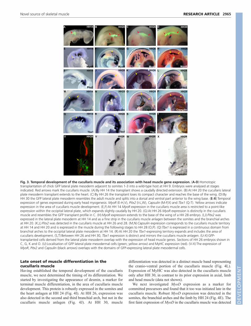
Temporal development of the cucullaris muscleindicates a relatedness to head musculatureNext we determined the temporal expansion of the lateral platemesoderm lateral to somites 1-3. Transplantations were performed atHH 9. Extension of the transplanted region was detectable by HH 14(Fig. 3A). By HH 20, the transplanted tissue had doubled its size tosix somites in length and had reached the base of the heart anlagen(Fig. 3B). The transplanted GFP population lost its compactcharacter and reached, as a band, the anterior limb base by HH 26(Fig. 3C). A morphological form resembling the adult muscle wasnot realised until HH 30, when transplanted tissue divided into aventral and dorsal part just anterior to the wing base (Fig. 3D).
RESEARCH ARTICLE Development 137 (17)
Fig. 2. Lateral plate mesoderm adjacent to somite level1-3 gives rise to the cucullaris muscle. Homotopicaltransplantations of GFP chick lateral plate mesoderm intowild-type hosts were performed at HH 9 and analysed at HH32-34 (day 7-8). (A)Lateral plate mesoderm adjacent tosomites 1-2 labels the whole cucullaris muscle. (B)Lateralplate mesoderm adjacent to somites 2-3 labels the wholecucullaris muscle as well as six muscle bands (redarrowheads). (C)Lateral plate mesoderm adjacent to somites4-5 gives rise to cells anterior to the forelimb base, but doesnot label the cucullaris muscle. (F-I�) Detailed analysis ofmyogenic capacity of occipital lateral plate mesodermadjacent to somites 2-3 at levels shown in B (yellow arrows).(F-I)GFP transplanted tissue. (F�-I�) MyHC expression.(F�-I�) Overlay of GFP transplanted tissue and MyHCexpression. (F-F�) The cucullaris muscle originates lateral tothe ear anlagen as a compact short muscle band (yellowarrowheads). (G-G�) The cucullaris muscle at upper necklevel (yellow arrowheads). (H-H�) The cucullaris capitismuscle is present directly under the skin (yellowarrowheads). Three of the six deeper muscle bands are alsovisible (red arrowheads). (I-I�) At limb level, a part of thecucullaris muscle lies in between two muscle sheets (yellowarrowheads). (D)Schematic for caudal to cranial heterotopictransplantation of lateral plate mesoderm from somite level10-12 to somite level 1-3. (D�)Cucullaris muscle is notpopulated by quail-derived QCPN+ nuclei (arrows).(E)Schematic for cranial to caudal heterotopictransplantation of lateral plate mesoderm from somite level1-3 to somite level 21-23. (E�)Quail-derived nuclei arefound between trunk muscle fibres, but not within MyHC+
muscle fibres (compare with Fig. 1B� for positivecontribution).
DEVELO
PMENT
Late onset of muscle differentiation in thecucullaris muscleHaving established the temporal development of the cucullarismuscle, we next determined the timing of its differentiation. Westarted by investigating the appearance of desmin, a marker forterminal muscle differentiation, in the area of cucullaris muscledevelopment. This protein is robustly expressed in the somites andthe heart anlagen at HH 24 (Fig. 4I). At HH 26, expression wasalso detected in the second and third branchial arch, but not in thecucullaris muscle anlagen (Fig. 4J). At HH 30, muscle
differentiation was detected in a distinct muscle band representingthe cranio-ventral portion of the cucullaris muscle (Fig. 4L).Expression of MyHC was also detected in the cucullaris muscleonly after HH 30, in contrast to its prior expression in axial, limband head muscle (data not shown).
We next investigated MyoD expression as a marker forcommitted precursors and found that it too was initiated late in thecucullaris muscle. Robust MyoD expression was detected in thesomites, the branchial arches and the limb by HH 24 (Fig. 4E). Thefirst faint expression of MyoD in the cucullaris muscle was detected
2965RESEARCH ARTICLENovel source of skeletal muscle
Fig. 3. Temporal development of the cucullaris muscle and its association with head muscle gene expression. (A-D)Homotopictransplantation of chick GFP lateral plate mesoderm adjacent to somites 1-3 into a wild-type host at HH 9. Embryos were analysed at stagesindicated. Red arrows mark the cucullaris muscle. (A)By HH 14 the transplant shows a caudally directed extension. (B)At HH 20 the cucullaris lateralplate mesoderm transplant extends to the heart. (C)By HH 26 the transplant loses its compact character and reaches the base of the wing. (D)ByHH 30 the GFP lateral plate mesoderm resembles the adult muscle and splits into a dorsal and ventral part anterior to the wing base. (E-X)Temporalexpression of genes expressed during early head myogenesis: MyoR (E-H,V), Pitx2 (I-L,W), Capsulin (M-P,X) and Tbx1 (Q-T). Yellow arrows indicateexpression in the area of cucullaris muscle development. (E,F)At HH 14 MyoR expression in the cucullaris muscle area is restricted to a point-likeexpression within the occipital lateral plate, which expands slightly caudally by HH 20. (G)At HH 26 MyoR expression is distinctly in the cucullarismuscle and resembles the GFP transplant profile in C. (H)MyoR expression extends to the base of the wing of a HH 28 embryo. (I,J)Pitx2 wasexpressed in the lateral plate mesoderm at HH 14 and as a fine strip in the cucullaris muscle anlagen between the somites and the branchial archesat HH 20. (K,L)Pitx2 was detected in the cucullaris muscle at HH 26 and 28. (M,N)Capsulin expression corresponds to the cucullaris muscle territoryat HH 14 and HH 20 and is expressed in the muscle during the following stages to HH 28 (O,P). (Q)Tbx1 is expressed in a continuous domain frombranchial arches to the occipital lateral plate mesoderm at HH 14. (R)At HH 20 the Tbx1-expressing territory expands and includes the area ofcucullaris development. (S,T)Between HH 26 and HH 30, Tbx1 expression is distinct and mirrors the cucullaris muscle anlagen. (U-X)GFP-transplanted cells derived from the lateral plate mesoderm overlap with the expression of head muscle genes. Sections of HH 26 embryos shown inC, G, K and O. (U)Localisation of GFP lateral plate mesodermal cells (green; yellow arrow) and MyHC expression (red). (V-X)The expression ofMyoR, Pitx2 and Capsulin (black arrows) overlaps with the domains of GFP-expressing lateral plate mesodermal cells.
DEVELO
PMENT

2966
at HH 26 (Fig. 4F). By HH 30 the MyoD expression in this domainhad elongated along the full extent of the neck, and broadened tocover its entire lateral side (Fig. 4H). In summary, the onset ofcucullaris muscle differentiation is initiated later than in thesomites, branchial arches and limbs.
The trunk myogenic programme is not employedin the formation of the cucullaris muscleAs the cucullaris muscle develops adjacent to the somites andreaches far into the body, we investigated the expression of Pax3,Pax7 and Myf5 as evidence for the involvement of the trunkmyogenic programme during its formation. Pax3 was readilydetectable in the dermomyotomes (HH 14), limb muscle precursors(HH 20 onwards) and the hypoglossal cord at HH 20 (see Fig. S1E,Fin the supplementary material). Importantly, we never detected theexpression of Pax3 in the cucullaris muscle at HH 28 (see Fig. S1Hin the supplementary material), a stage when MyoD is robustlyexpressed in this tissue (Fig. 4G). Furthermore more we were notable to detect clear (non-background) expression of Pax3 in thecucullaris at later stages (HH 30-32, data not shown). Likewise, Pax7was not found during early cucullaris development but was faintlyexpressed at HH 28 (see Fig. S1I-L in the supplementary material).Similarly, Myf5 expression was initiated late in the cucullaris muscle(HH 26) compared with somites, branchial arches and the limbs (seeFig. S1C in the supplementary material). The lack of the earlyspecification markers Pax3, Pax7 and Myf5 indicates that thecucullaris muscle develops out of phase with the trunk myogenicprogramme.
Genes regulating head muscle formation areexpressed in the cucullaris muscleThe late expression of differentiation markers and the absence ofgenes involved in trunk myogenesis raised the possibility that thecucullaris muscle develops according to a head muscle programme.MyoR, Pitx2, Capsulin and Tbx1 were used to mark early headmuscle precursors. In contrast to the lack of early trunk muscle
markers, MyoR was expressed in a small spot-like area of thecranial cucullaris muscle anlagen at HH 14 (Fig. 3E). By HH 26robust MyoR expression in the cucullaris muscle anlagen wasclearly evident (Fig. 3G,V) and overlapped with the lineage-tracingprofiles of GFP lateral plate mesoderm homotopically transplantedadjacent to somites 1-3 at HH 9 (Fig. 3C,U). At HH 30 MyoRexpression in the cucullaris muscle covered the entire lateral neckand reached into the back beyond the base of the limb (Fig. 3H).Pitx2 was detected symmetrically in a very fine stripe in thecucullaris muscle anlagen between the hypaxial domain of thesomites and the branchial arches at HH 20 (Fig. 3J). Thetranscription factor was distinctly expressed in the cucullarismuscle anlagen at HH 26 and HH 28 (Fig. 3K,L,W). Capsulin wasexpressed in the area of cucullaris muscle development at HH 14(Fig. 3M). At HH 26 the gene was distinctly expressed in thecucullaris muscle (Fig. 3O,X). The expression of Tbx1 was foundto mark the cucullaris muscle from the time of its early anlagen tothe fully formed muscle; initiated at HH 14 in a territory both ofthe muscle cells and in a prospective caudal region (Fig. 3Q). ByHH 20 the expression of Tbx1 had increased (Fig. 3R). At HH 26the Tbx1 expression mirrored the cucullaris muscle anlagen (Fig.3S compared with 3C).
In summary, head-muscle-related genes, such as MyoR, Pitx2,Capsulin and Tbx1, are expressed in the cucullaris muscle territoryfrom its earliest time of development.
Genetic evidence for the deployment of the headdevelopmental programme during cucullarismuscle developmentIn order to substantiate our findings and to determine whether themechanisms identified in the chick were conserved in mammals,we turned to the power afforded by mouse genetics. Given ourfindings that the avian homologues of the trapezius andsternocleidomastoid muscles develop late compared with othermuscle groups we first established when these muscles were clearlyidentifiable in the mouse. Our in situ and immunohistological
RESEARCH ARTICLE Development 137 (17)
Fig. 4. Late myogenic differentiation characteristics of the cucullaris muscle. Yellow arrows mark the cucullaris muscle anlagen. (A-D)MyoRexpression is used as a reference for the cucullaris muscle. (A)MyoR is expressed in the branchial arches and the cranial area of cucullaris muscledevelopment at HH 24. (B)Distinct expression in the cucullaris muscle anlagen extends caudally towards the base of the wing at HH 26. (C)MyoRexpression has reached the wing base at HH 28. (D)MyoR is expressed along the entire length of the cucullaris muscle from just caudal to the earanlagen beyond the limb at HH 30. (E)MyoD is expressed in the branchial arches, the somites and the limbs, but is not detected in the cucullarismuscle area at HH 24. (F)MyoD is expressed in the cucullaris muscle anlagen and reaches the forelimb at HH 26. (G)MyoD expression in thecucullaris muscle is upregulated at HH 28. (H)Expression of MyoD is present throughout the muscle and covers the lateral neck at HH 30.(I,J)Desmin is expressed in the somites and the heart anlagen at HH 24 and 26 and in the branchial arches at HH 26, but is not present in thecucullaris muscle anlagen. (K)Faint expression in the cucullaris muscle anlagen is observed at HH 28 in the ventral portion of the muscle. (L)Robustexpression is detected in the cranial portion of the muscle at HH 30.
DEVELO
PMENT

analysis revealed that both muscles are formed before 11.0 dpc inthe mouse (data not shown). Therefore in this study we analysedembryos at stages considerably after this point to avoid the issue ofdelayed development.
We have so far established that the cucullaris muscle is late todifferentiate, reminiscent to head rather than trunk musculature.Furthermore key genes (Pax3/7), which are normally expressedduring trunk muscle development, were not expressed during
2967RESEARCH ARTICLENovel source of skeletal muscle
Fig. 5. Mammalian trapezius and sternocleidomastoid muscle development is Pax3 independent and their connective tissue is derivedfrom neural crest. (A-D�) YFP expression controlled by Pax3Cre was used to identify Pax3 lineage cells (red) and compared to MyHC expression(green). Yellow arrows point at the corresponding muscle fibres in the Pax3Cre:RosaSTOP/YFP image and the MyHC image. (A)The sternocleidomastoidmuscle at inner ear level shows muscle fibres predominantly negative for the reporter gene. (A,A�) High-magnification images show YFP expressionbetween the myofibres but is seldom found in the muscle (yellow arrow). (B,B�) The trapezius muscle at scapula level displays a predominantly Pax3-negative developmental history (yellow arrow). By contrast, connective tissue between the myofibres is positive. (C,C�) The eye muscle ispredominantly negative for a Pax3 history (yellow arrow) in the myofibres but connective tissue is positive for the transcription factor. (D,D�) Limblevel muscle fibres are positive for a developmental history of Pax3 (yellow arrows), whereas the connective tissue is negative. (E,F)Comparison ofMyHC expression in wild type and Pax3Sp/Sp:Myf5nlacZ/nlacZ at 13.75 dpc. (E)Wild type shows a trapezius (arrow) at the level of the scapula.(F)Trapezius is present in Pax3Sp/Sp:Myf5nlacZ/nlacZ (arrow) at the level of the scapula. (G,H)Wnt1Cre/R26RlacZ mice were analysed for a contribution ofneural crest to the cucullaris muscle homologues at 13.5 dpc. (G)Robust Cre activity was detected throughout head tissues. (H)Strong Cre activity ispresent in the trapezius muscle (black arrows) at scapula level. (I)Chick/quail chimera of neural tube transplant at somite level 1-3. Cells of quailorigin are located throughout the cucullaris connective tissue. (J-M�) The involvement of head muscle associated genes in the development of thetrapezius. (J)Both the cranial and the caudal part of the trapezius muscle are labelled in a 13.5 dpc Isl1Cre;R26R mouse embryo. Arrows indicateacromio-trapezius or spino-trapezius. (J�)Sections of a 13.5 dpc Isl1Cre;R26R mouse embryo in which the trapezius muscle overlies the scapula,showing beta-galactosidase activity (arrow). (K,L)The entire trapezius muscle is absent in 13.5 and 14.5 dpc Tbx1–/– embryos (arrow). Thesurrounding trunk musculature is unaffected. (K�,L�) Sections showing absence of the trapezius muscle overlying the scapula in the Tbx1–/– animals(arrow). (M,M�) The position of the acromio-trapezius and spino-trapezius is shown in a wild-type littermate of L. AT, acromio-trapezius;H, humerus; IE, inner ear; SC, scapula; SP, spino-trapezius.
DEVELO
PMENT

2968
myogenesis of the cucullaris muscle. To firmly establish the lackof the involvement of Pax3 in cucullaris muscle development andto homologise between birds and mammals, we examinedPax3Cre:RosaSTOP/YFP mice. This line expresses Cre-recombinaseunder the endogenous Pax3 promoter, which ultimately initiatesYFP expression from a floxed Rosa allele. YFP fluorescence marksall cells with a past or present history of Pax3 expression. Skeletalmuscle in these mice was detected using a MyHC antibody.MyHC+ sternocleidomastoid myofibres, as well as trapeziusmyofibres, were negative for YFP, whereas expression wasdetected between the myofibres in 15.5 dpc embryos (Fig. 5A,B).Most trunk muscles showed YFP activity throughout the tissue(Fig. 5D), whereas its activity was absent from myofibres in headmuscles (Fig. 5C). These results confirm the expression profilingdata by providing genetic evidence that the cucullaris muscle andits homologues develop in a Pax3-independent manner. Theindependence of the cucullaris muscle group from the trunkmyogenic programme was established by examining myogenesisin the Pax3Sp/Sp:Myf5nlacZ/nlacZ. This double knockout lacks all trunkand limb muscles (Tajbakhsh et al., 1997). Remarkably, both thetrapezius (Fig. 5E,F) and sternocleidomastoid (data not shown)were present in the double mutant.
Our expression data suggest that the mechanism regulating thecucullaris homologues resembles that of head musculature. Recentwork has shown that a subset of head muscles develop through aprogramme requiring the transcription factor Isl1 (Nathan et al.,2008). We investigated whether the trapezius and thesternocleidomastoid muscles had a developmental history for theexpression of this gene. We were able to show that both thetrapezius and the sternocleidomastoid muscles expressed thistranscription factor using the Isl1cre/R26R line (Fig. 5J,J�).Furthermore we examined Tbx1–/– mice that have previously beenshown to be severely deficient in head muscles (Kelly et al., 2004).Examination of the trunk of these animals at 13.5 dpc revealed theabsence of both the trapezius and sternocleidomastoid muscles(Fig. 5K,K�). To exclude the possibility that the absence of thesemuscles was due to late development we also examined themutants at a later stage (14.5 dpc). Even at this advanced stage ofdevelopment, we found that both the trapezius andsternocleidomastoid muscles were absent in the Tbx1–/– (Fig. 5L-M�), confirming and extending previous results of Kelly et al.(Kelly et al., 2004).
In conclusion, these results suggest that the cucullaris muscledevelops according to the head and not the trunk myogenicprogramme.
Neural crest origin of the cucullaris muscleconnective tissueThe cucullaris muscle did not express Pax3 during itsdevelopment, and its mammalian homologues were shown to belargely negative for a history of Pax3 expression. However, wefound Pax3-lineage-derived expression in between themyofibres, indicating a somitic or neural-crest-derivedconnective tissue. Connective tissue of head muscles is knownto be derived from neural crest. By contrast, the connectivetissue of trunk muscles is derived from local mesoderm (Nodenand Trainor, 2005; Noden and Francis-West, 2006; Matsuoka etal., 2005). To determine whether neural crest contributes toconnective tissue, including the endomyosium of cucullarismuscle homologues in mammals, we investigatedWnt1Cre/R26RlacZ mice. This reporter line marks neural crestcells, but the reporter protein is not expressed in somite-derived
cells (Jiang et al., 2000). Sections of 13.5 dpc mice showed thatthe endomysial connective tissue of the trapezius muscle isderived from the Wnt1 lineage, indicating that it is neural crestin origin (Fig. 5G,H).
We established the origin of the connective tissue for thecucullaris muscle by homotopically transplanting quail dorsalneural tube at the level of somites 1-3 into chick hosts.Immunohistochemistry of day 8 chimeras showed that cellsexpressing the nuclear antigen QCPN were found between MyHC+
myofibres (Fig. 5I).In conclusion, we confirm and extend the findings of Matsuoka
et al. (Matsuoka et al., 2005) that neural crest cells contribute to theendomysial connective tissue of the cucullaris muscle of birds andits mammalian homologues.
Conserved mechanism for cucullaris muscledevelopmentThe cucullaris muscle is a highly conserved muscle in highervertebrates. We have shown that it not only has a unique origin butalso develops late compared with all other muscle groups. Todetermine whether the latter feature is conserved in vertebrates, weexplored the temporal development of the cucullaris muscle in theturtle. At stage 15 we found differentiated muscle in the head, trunkand limb. Significantly at this stage, the cucullaris muscle did notexpress MyHC (Fig. 6A,A�). By contrast, the cucullaris musclewas clearly evident at stage 17 (Fig. 6B,B�). We conclude that thedifferentiation of the turtle cucullaris muscle takes place later thanhead, trunk and limb muscles – a situation similar to that in birds.
DISCUSSIONThe lateral plate mesoderm has myogenicpropertiesIn this study we have shown that the lateral plate mesoderm hasmyogenic properties and is the major source of the cucullarismuscle. We show that the occipital lateral plate mesoderm, incontrast to other lateral plate regions, possesses the unique propertyof forming skeletal muscle. The cucullaris muscle and itsmammalian homologues extend from the neck, reaching far intothe trunk. In birds, this muscle fails to express genes associated
RESEARCH ARTICLE Development 137 (17)
Fig. 6. Conserved mechanism of cucullaris development in theturtle. MyHC expression in the turtle at stages 15 and 17. Yellowarrows identify the cucullaris muscle. (A,B)Whole embryo morphology.(A�,B�) Sections showing MyHC expression (red) and the nuclear stainDAPI (blue). (A�)At stage 15, differentiated axial muscles are presentbut the cucullaris is not detected. (B�)The cucullaris muscle expressesMyHC at stage 17.
DEVELO
PMENT

with the trunk myogenic programme, but does express markerslinked to the head myogenic programme, differentiates late duringembryogenesis, and has connective tissue originating from theneural crest. These findings led us to propose that cucullaris muscledevelopment is regulated according to the head myogenicprogramme.
The origin of the cucullaris muscle and its mammalianhomologues – the trapezius and the sternocleidomastoid muscle –have been the subject of much debate over the past 100 years. Adiverse range of tissues has been postulated to give rise to theseevolutionarily conserved muscles. Classical histology examinationin salmon and flying fish by Harrison (Harrison, 1895) anddiscussed in 1918 (Harrison, 1918) and 1908 (Derjugin, 1908) ledto the notion that skeletal muscle associated with the pectoral finoriginated from the dorsal lateral plate mesoderm. However,contemporary experiment-based studies underpinned by robustlineage-tracing techniques showed that the cucullaris developsfrom the occipital somites (Couly et al., 1993; Noden, 1983; Huanget al., 1997; Huang et al., 2000). Our study shows that the majorityof the cucullaris muscle develops from the lateral plate mesoderm.
At this point it is worth comparing the outcomes of this studywith those from previous investigations seeking to establish theorigin of the cucullaris. In this study we show, throughquantification analysis coupled with lineage-tracing techniques,that the majority (approximately 90%) of cucullaris myofibresoriginate from the lateral plate mesoderm, with only a minorcontribution (3.6%) from each of somites 1-3. In previous studiesthe origin of this muscle was reported to be from the somites, butit was noted that some successful somite grafts gave a very minorcontribution (similar to those seen here), whereas others showedgreater contribution (Huang et al., 1997; Huang et al., 2000). Webelieve that findings from previous studies can be reconciled withthe present data simply by acknowledging that in our previousstudies (Huang et al., 1997; Huang et al., 2000) and that of Noden(Noden, 1983) lateral plate mesoderm tissue was present in thesomitic grafts. Examination of our previous grafting protocols inlight of our present studies shows that efforts aimed at extractingentire somites would have included small amounts of lateral platemesoderm in the quail donor tissue – a tissue believed not to havemyogenic properties. Indeed, Noden magnanimously states that inhis study, lateral plate mesoderm was more than likely to be presentin the somite grafts (Noden, 1983). Our conclusion that themajority of the cucullaris muscle is non-somitic in origin, reachedusing an alternative tissue preparation approach to minimise lateralplate mesoderm contamination, is reinforced by two findingsreported herein: (1) the extremely low level of Pax3/7 expressionduring chick cucullaris embryogenesis; (2) the near absence ofmuscle cells with a present or past history of Pax3 expression inthe mouse trapezius.
The caudal myogenic boundary within this tissue lies at the levelof somite 3, as transplantation of lateral plate mesoderm adjacentto somites 4-5 failed to give rise to the muscle. Transplantation ofa smaller portion of the contributing lateral plate mesoderm gaverise to myogenic tissue along the entire length of the cucullarismuscle, suggesting that there is little, if any, anteroposteriorcompartmentalisation.
Our results provide experimental evidence that the occipitallateral plate mesoderm is a source of skeletal muscle and confirmssuggestions made to this end by Harrison (Harrison, 1895;Harrison, 1918) and Derjugin (Derjugin, 1908) in their histologicalexamination of the somatopleura development of fish. Asintriguingly, our work shows also that the lateral plate mesoderm
in this region of the embryo forms predominantly skeletal muscle.This finding raises the issue of how the cucullaris lateral platemesoderm gained myogenic capacity. The paraxial mesoderm ofthe trunk is initially segmented as epithelial somites, and contraststhe mesenchymal organisation of both the lateral plate and headmesoderm (Christ and Ordahl, 1995). Indeed the head mesodermand the lateral plate are continuous, and this observation could beincorporated into one of two simple models to explain themyogenic origin of the cucullaris muscle. One possibility is thatcells of the head mesoderm spread caudally into the area lateral tothe first three somites. Alternatively, the occipital lateral platemesoderm could adopt the molecular myogenic programme due toa caudal shift of the head myogenic programme. Boundary shiftsresulting in the acquisition of novel tissue characteristics arecommon within the animal kingdom. For instance, in snakes acranial shift of the trunk molecular boundary has been proposed toresult in the loss of forelimbs and a development of ribs in thecervical region (Cohn and Tickle, 1999).
Connective tissue is either derived locally from the mesoderm(for trunk muscles) or from the lateral plate mesoderm (for limbmuscles) (Christ et al., 1974; Chevallier et al., 1977; Christ et al.,1982). By contrast, all head muscle connective tissue originatesfrom the neural crest (Noden and Trainor, 2005; Noden andFrancis-West, 2006; Matsuoka et al., 2005). In this study we foundthat the connective tissue of the cucullaris muscle group of bothbirds and mammals originates from the neuroectoderm. Theseresults confirm (in mammals) and extend (in birds) the findings ofMatsuoka et al. (Matsuoka et al., 2005). Summarising the dataregarding the origin of the myogenic and connective tissuecomponents of the cucullaris muscle we show that the head muscleprogramme is far more extensively deployed than previouslythought.
Cucullaris progenitor proliferation is notcontrolled by Pax3/7, but by genes expressedduring head progenitor proliferationOur lineage-tracing studies in chicks showed that the lateral platemesoderm movement starts at HH 14, but that myogenicdifferentiation is not initiated until HH 26. We considered that theintervening period may be required to generate the number of cellsneeded to form a large muscle like the cucullaris. In the trunk,proliferation is mainly driven by Pax3 and Pax7, a process thatsimultaneously suppresses differentiation (Amthor et al., 1999;Amthor et al., 1998). However, neither of these genes wasexpressed in the developing cucullaris muscle. Our molecularanalysis was confirmed using a genetic approach in mice throughthe deployment of the Pax3Cre:RosaSTOP/YFP and thePax3Sp/Sp:Myf5nlacZ/nlacZ lines (Engleka et al., 2005). We found thatmyoblasts of the trapezius and sternocleidomastoid muscles werenegative for a recent or past history for Pax3 expression. Theseresults are in keeping with our lineage-tracing experimentsinvolving somite transplantations. Indeed, these results offer amechanistic explanation of the presence of the large muscles foundin the forelimb region of the Pax3Sp/Sp:Myf5nlacZ/nlacZ [see Fig. 2Hin Tajbakhsh et al. (Tajbakhsh et al, 1997)]. We show that thetrapezius and sternocleidomastoid muscles (which develop througha head myogenic programme) are present in thePax3Sp/Sp:Myf5nlacZ/nlacZ, which lacks all trunk and limb muscles.This is concordant with our findings that connective tissue betweenthe myofibres was positive and derived from the Pax3 neurallineage, confirming our previous result of the neural crest lineagetracing in the chick.
2969RESEARCH ARTICLENovel source of skeletal muscle
DEVELO
PMENT

2970
In contrast to finding that the trunk precursor cell markers arenot expressed in the cucullaris muscle at the time of proliferation,we found that genes thought to carry out a similar role in the headsuch as MyoR, Tbx1 and Capsulin were robustly expressedthroughout the development of the muscle (Bothe and Dietrich,2006). Furthermore, the expression of the head precursor markersmatches the movement of the cucullaris anlagen as shown in thetemporal lineage-tracing studies. The role of the head muscleprogramme in the development of neck muscles was confirmedby examining the trunk of Tbx1–/– mice. These animals failed toform either the trapezius or the sternocleidomastoid muscle (seeFig. 5), confirming and extending the results of Kelly et al. (Kellyet al., 2004). In addition, we found that the trapezius alsodisplayed a molecular history for the expression of Isl1, a generecently implicated in the ontogeny of specific head muscles (seeFig. 5) (Nathan et al., 2008). We conclude that the cucullarismuscle and its mammalian homologues use the same myogenicprogramme as that employed in the formation of headmusculature.
Maintaining an extended phase of progenitorproliferation may be necessary for the extensionof the cucullaris muscle into the trunkAn intriguing feature of the cucullaris muscle and its mammalianhomologues is that it is derived from a small area lateral to the firstthree somites, yet eventually it extends over 14 segments, a processthat is not completed until relatively late during embryogenesis(day 8 in chick). By this stage all other major muscle groups (axial,branchial arch and limb muscles) are at an advanced stage ofdifferentiation. This late differentiation was found to beevolutionarily conserved, as we show that the cucullaris of turtlesalso differentiates after axial, head and limb muscles.
During cucullaris muscle development, an extended period oftime for proliferation may be required to generate an adequatenumber of cells to form the muscle. This phase of proliferation maybe especially important for cucullaris muscle development, as themuscle starts off from a small population of mesenchymal cells thatreside under the occipital ectoderm.
In this study we found that the cucullaris muscle displays theexceptional property of employing the head myogenic programme,despite the fact that a considerable part of the muscle lies withinthe trunk territory. The development of the cucullaris muscle at thetransition of the head-trunk territory has an interesting consequencefor its differentiation. The development of muscles in the trunk andhead differ in that they show very different responses to the sameenvironmental cues. For example, muscle development issupported in the trunk by either Wnt or Shh. However, the samesignalling molecule suppresses head muscle development (Tzahoret al., 2003). Release of the myogenic suppression in the head hasbeen shown to be a result of the production of signalling proteinantagonists secreted by the cranial neural crest. Assimilating theresults generated in the present study in consideration of thedifferences apparent in the regulation of head versus trunkmyogenic programmes, we propose the following scenario for thedevelopment of the cucullaris muscle. Wnts from the occipitalneural tube and ectoderm suppress myogenesis of cells residing inthe occipital lateral plate mesoderm. Under this influence theyproliferate and express Tbx1, MyoR and Capsulin. As the pool ofcucullaris progenitors expands, it extends caudally into the trunk.Here the cucullaris precursors are in close proximity withprogenitor cells of trunk musculature. However, these twomyogenic populations respond differentially when exposed to
influential signalling cues. In the presence of Wnts, the trunkprecursors initiate differentiation, whereas the cucullaris precursorsrespond by proliferating. Again similar to the situation in the head,we suggest that the key to initiating differentiation is to antagonisethe action of the environmental signalling molecules and that thisaction could be executed by the neural crest.
Evolution of the cucullaris muscleAn evolutionary explanation of how the cucullaris muscle wastransformed from a relatively short muscle (in sharks) into an entitystretching into the trunk still remains unknown. The short cucullarismuscle in the shark moves the gill arches, but also attaches to thepectoral girdle. The sharks’ ancestral crossopterygian relatives,such as the Eusthenopteron and Sauripterus, possessed a neckgirdle, which linked the pectoral girdle to the base of the skull. Theneck girdle was dermal in origin and consisted of four membranousbones (post-temporal, supracleithrum, cleithrum and clavicle)(DePalma, 2008). During evolution, the size of the neck girdlecomponents was reduced or lost and the skull became free of itsattachments to the pectoral girdle. The advantage for animals withthis modification was increased mobility of the head; however, atthe cost of a destabilized head. We suggest that in the absence ofthe neck girdle, the cucullaris muscle expanded caudally – a featureseen during its development in birds – to provide both stability forthe head (similar to that played by postural muscles) whilepermitting mobility. The unique developmental origin of thismuscle will be valuable in order to identify its precise homologuesin lower vertebrates, a prerequisite towards gaining a fullerunderstanding of the evolution of the neck (Kuratani, 2008).
AcknowledgementsWe thank E. Zelzer for Pax3Cre/LoxYFP embryos; A. P. McMahon, P. Soriano,and H. Sucov for providing the Wnt1Cre and R26R mouse lines; M. Grim andE. Krejci for processing the Wnt1cre/R26RlacZ mice; H. Sang (Roslin Institute)for providing the GFP chick eggs; G. Frank, U. Baur, U. Pein, L. Koschny, G.Dumas and S. Bauerkamper for technical support; D. Junghans and G. Lukefor helpful comments; and the anonymous reviewers for constructivecomments that have significantly improved our manuscript. We thank theDeutsche Forschungsgemeinschaft for financial support (DFG 729/5-1 toR.H.).
Competing interests statementThe authors declare no competing financial interests.
Supplementary materialSupplementary material for this article is available athttp://dev.biologists.org/lookup/suppl/doi:10.1242/dev.049726-/DC1
ReferencesAddens, L. (1933). The motor nuclei and roots of the cranial and first spinal nerves
of vertebrates: I. Introduction. Cyclostomes. Z. Anat. Entwickl.-Gesch. 101, 307-410.
Allis, E. J. (1897). The cranial muscles and cranial and first spinal nerves in Amiacalva. J. Morphol. 12, 487-808.
Amthor, H., Christ, B., Weil, M. and Patel, K. (1998). The importance of timingdifferentiation during limb muscle development. Curr. Biol. 8, 642-652.
Amthor, H., Christ, B. and Patel, K. (1999). A molecular mechanism enablingcontinuous embryonic muscle growth-a balance between proliferation anddifferentiation. Development 126, 1041-1053.
Bothe, I. and Dietrich, S. (2006). The molecular setup of the avian headmesoderm and its implication for craniofacial myogenesis. Dev. Dyn. 235, 2845-2860.
Chapman, S. C., Collignon, J., Schoenwolf, G. C. and Lumsden, A. (2001).Improved method for chick whole-embryo culture using a filter paper carrier.Dev. Dyn. 220, 284-289.
Chevallier, A., Kieny, M. and Muager, A. (1977). Limb-somite relationship:origin of the limb musculature. J. Embryol. Exp. Morphol. 41, 245-258.
Christ, B. and Ordahl, C. P. (1995). Early stages of chick somite development.Anat. Embryol. 191, 381-396.
Christ, B., Jacob, H. J. and Jacob, M. (1974). Origin of wing musculature.Experimental studies on quail and chick embryos. Experientia 30, 1446-1449.
RESEARCH ARTICLE Development 137 (17)
DEVELO
PMENT

Christ, B., Jacob, H. J., Jacob, M. and Wachtler, F. (1982). On the origin,distribution and determination of avian limb mesenchymal cells. Prog. Clin. Biol.Res. 110, 281-291.
Cohn, M. J. and Tickle, C. (1999). Developmental basis of limblessness and axialpatterning in snakes. Nature 399, 474-479.
Couly, G. M., Coltey, P. M. and Le Douarin, N. M. (1993). The triple origin ofskull in higher vertebrates: a study in quail-chick chimeras. Development 117,409-429.
DePalma, A. F. (2008). The classic. Origin and comparative anatomy of thepectoral limb. Surgery of the shoulder. Clin. Orthop. Relat. Res. 466, 531-542.
Derjugin, K. (1908). Die Entwicklung der Brustflossen und des Schultergürtels beiExocoetus volitans. Zeitschr. F. Wiss. Zool. 91, 559-598.
Edgeworth, F. (1911). On the morphology of the cranial muscles in somevertebrates. Q. J. Microsc. Sci. 56, 167-316.
Edgeworth, F. (1926). On the development of the coraco-branchiales andcucullaris in Scyllium canicula. J. Anat. 60, 289-308.
Engleka, K. A., Gitler, A. D., Zhang, M., Zhou, D. D., High, F. A. and Epstein,J. A. (2005). Insertion of Cre into the Pax3 locus creates a new allele of Splotchand identifies unexpected Pax3 derivatives. Dev. Biol. 280, 396-406.
Favaro, G. (1903). Ricerche intorno allo sviluppo dei muscoli dorsali, laterali eprevertebrali negli amnioti. Arch. Ital. Anat. Embriol. 2, 518-577.
Fuerbringer, M. (1897). Ueber die spino-occipialen Nerven der Selachier andHolocephalen und ihre vergleichende Morphologie. Festschr. Gegenbaur. 3, 349-788.
Gegenbaur, C. (1898). Vergleichende Anatomie der Wirbeltiere mitBeruecksichtigung der Wirbellosen. Leipzig: Engelmann.
Hacker, A. and Guthrie, S. (1998). A distinct developmental programme for thecranial paraxial mesoderm in the chick embryo. Development 125, 3461-3472.
Harel, I., Nathan, E., Tirosh-Finkel, L., Zigdon, H., Guimaraes-Camboa, N.,Evans, S. M. and Tzahor, E. (2009). Distinct origins and genetic programs ofhead muscle satellite cells. Dev. Cell 16, 822-832.
Harrison, R. G. (1895). Die Entwickelung der unpaaren und paarigen Flossen derTeleostier. Arch. F. Mikr. Anat. 46, 500-578.
Harrison, R. G. (1918). Experiments on the development of the fore limb ofAmblystoma, a self-differentiating equipotential system. J. Exp. Zool. 25, 413-461.
Huang, R., Zhi, Q., Ordahl, C. P. and Christ, B. (1997). The fate of the first aviansomite. Anat. Embryol. 195, 435-449.
Huang, R., Zhi, Q., Patel, K., Wilting, J. and Christ, B. (2000). Contribution ofsingle somites to the skeleton and muscles of the occipital and cervical regionsin avian embryos. Anat. Embryol. 202, 375-383.
Jiang, X., Rowitch, D. H., Soriano, P., McMahon, A. P. and Sucov, H. M.(2000). Fate of the mammalian cardiac neural crest. Development 127, 1607-1616.
Kelly, R. G., Jerome-Majewska, L. A. and Papaioannou, V. E. (2004). Thedel22q11.2 candidate gene Tbx1 regulates branchiomeric myogenesis. Hum.Mol. Genet. 13, 2829-2840.
Krammer, E. B., Lischka, M. F., Egger, T. P., Riedl, M. and Gruber, H. (1987).The motoneuronal organization of the spinal accessory nuclear complex. Adv.Anat. Embryol. Cell Biol. 103, 1-62.
Kuratani, S. (2008). Evolutionary developmental studies of cyclostomes and theorigin of the vertebrate neck. Dev. Growth Differ. 50, S189-S194.
Lewis, W. (1910). Die Entwicklung des Muskelsystems. In Handbuch derEntwicklungsgeschichte des Menschen. Vol. 1 (ed. F. Keibel and F. Mall), pp.457-526. Leipzig: Hirzel.
Luther, A. and Lubosch, W. (1938). Muskeln des Kopfes: Viscerale Muskulatur. InHandbuch der verleichenden Anatomie der Wirbeltiere. Vol. 5 (ed. E. G. L. Bolk,E. Kallius and W. Lubosch), pp. 467-470, 1011-1106. Berlin: Urban undSchwarzenberg.
Matsuoka, T., Ahlberg, P. E., Kessaris, N., Iannarelli, P., Dennehy, U.,Richardson, W. D., McMahon, A. P. and Koentges, G. (2005). Neural crestorigins of the neck and shoulder. Nature 436, 347-355.
Mootoosamy, R. C. and Dietrich, S. (2002). Distinct regulatory cascades for headand trunk myogenesis. Development 129, 573-583.
Nathan, E., Monovich, A., Tirosh-Finkel, L., Harrelson, Z., Rousso, T., Rinon,A., Harel, I., Evans, S. M. and Tzahor, E. (2008). The contribution of Islet1-expressing splanchnic mesoderm cells to distinct branchiomeric muscles revealssignificant heterogeneity in head muscle development. Development 135, 647-657.
Nieto, M. A., Patel, K. and Wilkinson, D. G. (1996). In situ hybridization analysisof chick embryos in whole mount and tissue sections. Methods Cell Biol. 51,219-235.
Noden, D. M. (1983). The embryonic origins of avian cephalic and cervical musclesand associated connective tissues. Am. J. Anat. 168, 257-276.
Noden, D. M. and Trainor, P. A. (2005). Relations and interactions betweencranial mesoderm and neural crest populations. J. Anat. 207, 575-601.
Noden, D. M. and Francis-West, P. (2006). The differentiation andmorphogenesis of craniofacial muscles. Dev. Dyn. 235, 1194-1218.
Noden, D. M., Marcucio, R., Borycki, A. G. and Emerson, C. P., Jr (1999).Differentiation of avian craniofacial muscles: I. Patterns of early regulatory geneexpression and myosin heavy chain synthesis. Dev. Dyn. 216, 96-112.
Piekarski, N. and Olsson, L. (2007). Muscular derivatives of the cranialmostsomites revealed by long-term fate mapping in the Mexican axolotl (Ambystomamexicanum). Evol. Dev. 9, 566-578.
Sambasivan, R., Gayraud-Morel, B., Dumas, G., Cimper, C., Paisant, S.,Kelly, R. G. and Tajbakhsh, S. (2009). Distinct regulatory cascades governextraocular and pharyngeal arch muscle progenitor cell fates. Dev. Cell 16, 810-821.
Tajbakhsh, S., Rocancourt, D., Cossu, G. and Buckingham, M. (1997).Redefining the genetic hierarchies controlling skeletal myogenesis: Pax-3 andMyf-5 act upstream of MyoD. Cell 89, 127-138.
Tzahor, E., Kempf, H., Mootoosamy, R. C., Poon, A. C., Abzhanov, A., Tabin,C. J., Dietrich, S. and Lassar, A. B. (2003). Antagonists of Wnt and BMPsignaling promote the formation of vertebrate head muscle. Genes Dev. 17,3087-3099.
Valasek, P., Theis, S., Krejci, E., Grim, M., Maina, F., Shwartz, Y., Otto, A.,Huang, R. and Patel, K. (2010). Somitic origin of the medial border of themammalian scapula and its homology to the avian scapula blade. J. Anat. 216,482-488.
Williams, P. L. (1995). Gray’s Anatomy. London: Churchill Livingstone.
2971RESEARCH ARTICLENovel source of skeletal muscle
DEVELO
PMENT