the neural tube origin of ventral root sheath cells in the
TRANSCRIPT

Development 101, 247-254 (1987)Printed in Great Britain © The Company of Biologists Limited 1987
247
The neural tube origin of ventral root sheath cells in the chick embryo
E. R. LUNN1, J. SCOURFIELD1, R. J. KEYNES1 and C. D. STERN2
^Department of Anatomy, Downing Street, Cambridge CB2 3DY, UK^Department of Human Anatomy, South Parks Road, Oxford OX1 3QX, UK
Summary
The embryonic origin of peripheral nerve Schwann/sheath cells is still uncertain. Although the neuralcrest is known to be an important source, it is notclear whether the ventral neural tube also contributesa progenitor population for motor axons. We haveused the techniques of immunohistochemistry, elec-tron microscopy and quail-chick grafting to examinethis problem. Immunohistochemistry with mono-clonal antibody HNK-1 identified a cluster of immu-noreactive cells in the sclerotome, at the site of thefuture ventral root. With the electron microscope,nucleated cells could be seen breaching the basallamina of the neural tube, exclusively in the region ofthe ventral root and preceding axon outgrowth. Aftergrafting a length of crest-ablated quail neural tube inplace of host chick neural tube, a population of quail
cells was found localized to the ventral root exitzone, associated with the ventral root axons. Takentogether, these observations support the possibility ofa neural tube origin for ventral root sheath cells,although we found no evidence for a more extensivemigration of these cells. The ventral root cells sharecertain phenotypic traits, such as HNK-1 immuno-reactivity, with neural-crest-derived Schwann cells,but are not necessarily identical to them. We arguethat while they may help motor axons to exit theneural tube at the correct position, they are unlikelyto guide axons beyond the immediate vicinity of theneural tube.
Key words: ventral root, sheath cells, Schwann cells,chick embryo, neural tube.
Introduction
The embryonic origin of Schwann or sheath cells wasthe subject of much debate in the early part of thiscentury, when three cell populations, the neural tube,the neural crest and the mesoderm, were consideredpossible sources. A mesodermal origin was rejectedat an early stage, after Harrison (1904, 1906) showedthat removal of the neural crest in frog embryos led tomotor nerves devoid of sheath cells. Subsequentexperiments produced conflicting results. Kuntz(1922), for example, claimed that removal of theneural crest and dorsal neural tube in frog and chickembryos did not cause loss of motor nerve Schwanncells, and proposed a ventral neural tube origininstead. This was supported by Raven (1937), usingxenoplastic transplantation in amphibian embryos.Detwiler (1937), staining the amphibian neural crestwith vital dyes, once again proposed a predominantlyneural crest origin, but suggested (as had Harrison,1924) that the ventral neural tube may make anadditional contribution later in development. Jones
(1939), who studied sections, stained with haema-toxylin and eosin, of both normal and crest-ablatedchick embryos, decided that dorsal root ganglionSchwann cells have a neural crest origin whereasventral root Schwann cells emigrate from the neuraltube.
In his book 'The Neural Crest', Horstadius (1950)summarized all this by remarking that 'evidently theproblem of the origin of the sheath cells of Schwann isnot solved yet'. With the advent of autoradiography,Weston (1963) again noted the possibility of a dualorigin of Schwann cells and pointed out that theycould yet turn out to be solely of neural tube origin.The debate then appears to have been abandonedwithout any definite conclusion having been drawn.With the exception of one brief report (Wachtler,1985), recent accounts nevertheless state thatSchwann/sheath cells are all of neural crest origin anddo not consider the possibility that motor axonSchwann/sheath cells could have a neural tube origin.The issue is not a trivial one. For example, if thelineage of all motor axon sheath cells is different from

248 E. R. Lunn, J. Scourfield, R. J. Keynes and C. D. Stern
that of sensory axon sheath cells, we might expectthem to differ in other respects, such as theproduction of molecules with guidance or trophicfunctions.
In a study using monoclonal antibodies that recog-nize both neural crest cells and Schwann/sheath cells,we observed a population of immunoreactive cellslocated at the ventral root in crest-ablated chickembryos (Rickmann, Fawcett & Keynes, 1985). Wetherefore decided to re-examine the origin of sheathcells, using immunohistochemistry, electron mi-croscopy and the quail-chick grafting method (LeDouarin, 1973) in the hope that these comparativelyrecent techniques might resolve some of the olduncertainties.
Materials and methods
ImmunohistochemistryTransverse sections often normal embryos, stage 17 (Ham-burger & Hamilton, 1951), were prepared and processedfor indirect immunoperoxidase staining with monoclonalantibody HNK-1 (Becton Dickinson anti Leu-7), accordingto the protocol of Rickmann et al. (1985). Briefly, embryoswere aldehyde/immersion fixed and embedded in 20 %bovine serum albumin (hardened in aldehyde fixative),after which 50-100,um sections were cut on a freezingmicrotome. These were then stained by an avidin-bio-tin-peroxidase procedure using HNK-1, intensified withosmium tetroxide and embedded in Spurr's resin, afterwhich semithin transverse sections were cut with an ultra-microtome.
Electron microscopyFive stage-17 embryos were fixed for 4 h in 2 % glutaralde-hyde and 2 % formaldehyde (in 0-1 M-Pipes buffer contain-ing 1-5 % sucrose, pH 7-2, at 4°C). Specimens were washedfor 24 h in Pipes buffer and then postfixed in 1% osmiumtetroxide. Block staining was performed in a saturatedsolution of uranyl acetate in maleate buffer (160mOs),followed by dehydration through alcohols. TAAB resin wasused for embedding, after which thin sections (40-60 nm)were cut on a Huxley MK1 ultramicrotome, mounted oncopper grids and double stained with uranyl acetate andlead citrate. Sections were viewed in a Philips EM 300 at80 kV.
Chick-quail chimaerasFertile hens' and quails' eggs were incubated at 38°C tostages 12-13. Since motor axons first emerge from theneural tube opposite the wing bud between stages 16 and 17(Keynes & Stern, 1984), no ventral roots had formed indonor or host embryos prior to operation. In the trunkregion, neural crest migration is first observed three somitescranial to the most recently formed somite (Rickmann etal.1985); neural tube in donor and host embryos was removedopposite the most caudal somites and cranial segmentalplate, before the beginning of crest emigration. The lengthof tissue removed was equivalent to four somites.-
The host hens' eggs were prepared as follows: a windowwas cut with a scalpel blade and the embryo floated up tothe level of the shell by adding calcium- and magnesium-free Tyrode's solution (CMF), to which had been added asolution of lOOOOi.u. ml"1 penicillin and 1 mgmT1 Strepto-mycin in 0-9% saline (Sigma) to a final dilution of 1:100.0-1 ml of ink solution (Pelikan Fount India, diluted 1 in 10with CMF) was injected into the sub-blastodermic space sothat the embryo could be seen against a dark background.A rim of silicone grease was then placed around the edgesof the window and a drop of CMF made to cover theembryo. Visibility was significantly enhanced with tangen-tial fibre-optic illumination (Hara, 1970). The vitellinemembrane was peeled back over the most caudal somitesand cranial segmental plate, and the neural tube andnotochord were excised using a Week microsurgical knife.0-1 % trypsin (Difco, in CMF) was sometimes used to helpseparate the notochord from the endoderm.
The donor quail grafts were prepared from embryos ofthe same developmental stage as the chick hosts. Eachembryo was pinned out in a Sylgard dish with its ventralside uppermost, immersed in 0-1 % trypsin in CMF and theendoderm peeled off. Neural tube and notochord (of thesame craniocaudal level and length as that excised from thehost) were easily separated from the somites and segmentalplate. The trypsin was replaced by CMF and the dorsal halfof the neural tube was then cut off with a knife to removethe neural crest. The notochord was used as a marker forthe ventral side of the grafted neural tube. It was essentialto remove the host notochord, for it has been shown that anadditional notochord can lead to abnormalities of ventralroot emergence (van Straaten et al. 1985). The graft wastransferred to the host with a micropipette, placed inposition in its normal orientation and 1 -5 ml of albumen waswithdrawn to bring the embryo down into the egg onceagain. The egg was then sealed with PVC tape andincubated at 38°C for a further 2-4 days.
Chimaeric embryos were fixed in Zenker's solution,dehydrated through alcohols and wax-embedded. Theblocks were sectioned transversely at 7^m, stained byFeulgen's method (Le Douarin, 1973) and mounted inPermount (Fisher).
Results
Immunohistochemistry
During the earliest stages of neural crest cell mi-gration in the chick embryo, HNK-1 can be used todistinguish crest cells from the surrounding somite-derived cells, by virtue of its selective binding toneural crest cells (Tucker et al. 1984; Rickmann et al.1985). By this means, it has been shown that crestcells are confined to the cranial half-sclerotome asthey pass through the segmental mesoderm (Rick-mann et al. 1985). Motor axons are also restricted tothis part of the sclerotome as they grow out from theventral neural tube. They leave the neural tube in apunctuated manner, growing first from cell bodiessited opposite cranial half-sclerotome, and later from

Origin of ventral root sheath cells 249
. •»
Fig. 1. A composite of transverse semithin sections through the caudal (A) and cranial (B) half-sclerotomes of a wingsegment in a stage-17 embryo, stained with HNK-1. (A) In the caudal half-sclerotome, immunoreactive cells arerestricted to two areas: immediately dorsal to the sclerotome, adjacent to dorsal neural tube, and as a distinct clusterassociated with the site of the future ventral root (arrow). Note the absence of immunoreactive cells within the neuraltube. (B) In the cranial half-sclerotome, immunoreactive neural crest cells are now widespread, ventral to thedermomyotome. A cluster of HNK-1-positive cells can again be seen in association with the ventral root region of theneural tube (arrow).
those opposite caudal half-sclerotome (Keynes &Stern, 1984). It was of interest to see whether HNK-1-positive cells were associated with motor axons at thepoint of axon emergence from the neural tube. Intransverse sections through the caudal halves of wingsomites of stage-17 embryos, where the neural crest isunable to migrate, a distinct cluster of HNK-1-positive cells was seen adjacent to the neural tube, atthe point of future axon emergence (Fig. 1A). Insections through the cranial halves of these samesomites, HNK-1-positive cells were widespread in thesclerotome (cf. Rickmann et al. 1985). As in thecaudal halves, they were clustered around the ventralroot zone of the neural tube (Fig. IB). All the cellswithin the neural tube in this region were themselvesHNK-1 negative (Fig. 1A,B).
Electron microscopyFurther evidence for a neural tube origin of these cellclusters was sought by electron microscopy of trans-verse sections through the wing region of normal
stage-17 embryos. In the caudal half-sclerotome,nucleated cells were seen breaching the basal laminaof the neural tube, again exclusively in the region ofthe future ventral root (Fig. 2A). They were some-times associated with filopodial processes presumedto be derived from motoneurones. Sections throughthe cranial half-sclerotome showed the samephenomenon, with, as expected, the additional ap-pearance of fully emergent axon profiles (Fig. 2B).
Chick-quail chimaerasWhile the observations described above suggestedthat the ventral neural tube does contribute a popu-lation of axon-associated cells, it remained possiblethat the ventral cell clusters were nevertheless de-rived from the neural crest. Chick-quail chimaeraswere therefore constructed to see whether quail cellsemigrated from a grafted, crest-ablated neural tubealong with motor axons. Fifteen grafted embryossurvived to stages 20-26, when they were .assessed byFeulgen staining, the quail cells being distinguishable

250 E. R. Lunn, J. Scourfield, R. J. Keynes and C. D. Stern
L- _ . , * • .
Fig. 2. (A) Electron micrograph ofa transverse section through theventral root region of the neuraltube (left) and adjacent caudal half-sclerotome (right) of the wingregion in a stage-17 chick embryo.Dorsal is uppermost. The basallamina on the surface of the neuraltube is arrowed, and is breached bya nucleated cell (n), whosecytoplasm extends within theconfines of the neural tube,x 18 750.
by their prominent nucleolar staining (Le Douarin,1973). Although ventral root cells were first detect-able at stage 17 (see above), they were assessed in thechimaeras at later stages so that their migrationdistance could be estimated simultaneously. Eightembryos were subsequently excluded from the analy-sis, because only a few quail cells were found to bepresent, or the graft was malpositioned or severelykinked. In the seven remaining grafts, the spinal cordwas usually incompletely formed, since only theventral portion of the neural tube had been grafted.In some cases, however, the host neural tube andcrest had restored the missing dorsal part of the graftor had displaced part of the graft.
Contamination of the grafts by donor neural crestor somite cells, due to incomplete removal of thecrest or imperfect dissection of the donor neural tube,was also anticipated. The presence or otherwise ofquail neural crest could be determined by studyingthe dorsal root ganglia; if these contained quail cells itwas assumed that quail crest was present in that area.Donor somite cell contamination was assessed by thepresence or absence of quail cells isolated within thehost sclerotome. Using these" criteria, the averagelength of each graft that was free of contaminatingcells was 74-0 % of the total graft length (Table 1).
In contamination-free sections, quail cells werefrequently associated with emerging ventral root

Origin of ventral root sheath cells 251
2B
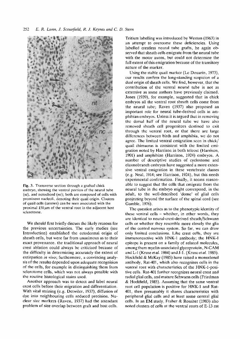
axons in the sclerotome immediately external to theneural tube; cells were seen, on average, in 62-0 % ofthese sections (Table 1). At all the stages examined,the quail cells extended, at most, about 150/zm intothe sclerotome (Fig. 3). The ventral root axons couldbe identified clearly with phase-contrast optics and noquail cells were associated with their more distalregions.
Where regeneration by the host neural tube hadoccurred, cells of host origin were associated muchmore extensively with the motor axons derived fromthe graft. The ventral roots were also thicker in theseareas, presumably reflecting a greater degree of axonoutgrowth under these conditions.
Fig. 2. (B) Ventral root exit zone oppositecranial half-sclerotome of a stage-17 chickembryo, wing region. Same orientation asFig. 2A. The basal lamina of the neural tube(arrowed) is breached by numerous parallelaxon profiles. X21000.
Discussion
Since Harrison's (1904, 1906) original experimentsthere has been little doubt that Schwann/sheath cellscan arise from the neural crest. A recent demon-stration of this has come from the experiments of LeLievre, Schweizer, Ziller & Le Douarin (1980), whografted fragments of quail neural crest into chickembryos, between the somites and the neural tube,and showed that quail cells subsequently came to linethe nerve fibres of the spinal roots. Over the years,however, there has been disagreement as to whetherthe ventral neural tube also makes a contribution,leading Weston (1963) to point out that sheath cellscould be entirely of neural tube origin.
Table 1. Results of the quail-chick chimaeras
A
Graft
1234567
B
Stageexamined
20232426262626
CGraftlength(>m)
497644210490700413in
D% Sections free
of contaminatingcells
88-774-276-791-457 069-560-8
x = 74-0% ±4-5
E% Contamination-free
sections with quailcells at ventral root
65030-482-656-391-285-423-7
x = 62-0% ±9-4
Column B shows the stage at which each grafted embryo was fixed, sectioned and stained; grafts were transplanted at stages 12-13(see Materials and methods). Column C shows the final length of each graft, calculated as the product of the number of sectionscontaining grafted quail neural tube cells and the section thickness (7/im). Column D shows the percentage of sections found to be freeof contaminating cells, as determined by the absence of quail cells in the dorsal root ganglia and the absence of isolated clusters ofquail cells in the host sclerotome. Column E shows the percentage of contamination-free sections that contained quail cells localized tothe ventral root. For columns D and E, the mean percentage ± S.E.M. (X) is also given
252 E. R. Lunn, J. Scourfield, R. J. Keynes and C. D. Stern
» >
Fig. 3. Transverse section through a grafted chickembryo, showing the ventral portion of the neural tube(m), and notochord (nc); both are composed of cells withprominent nucleoli, denoting their quail origin. Clustersof quail cells (arrows) can be seen associated with theproximal 150 fim of the ventral root in the adjacent hostsclerotome.
We should first briefly discuss the likely reasons forthe previous uncertainties. The early studies (seeIntroduction) established the ectodermal origin ofsheath cells, but were far from unanimous as to theirexact provenance, the traditional approach of neuralcrest ablation could always be criticized because ofthe difficulty in determining accurately the extent ofextirpation in vivo; furthermore, a convincing analy-sis of the results depended upon adequate recognitionof the cells, for example in distinguishing them fromsclerotome cells, which was not always possible withthe routine histological stains used.
Another approach was to detect and label neuralcrest cells before their migration and differentiation.With vital staining (e.g. Detwiler, 1937), diffusion ofdye into neighbouring cells reduced precision. Nu-clear size markers (Raven, 1937) had the attendantproblem of size overlap between graft and host cells.
Tritium labelling was introduced by Weston (1963) inan attempt to overcome these deficiencies. Usinglabelled crestless neural tube grafts, he again ob-served that sheath cells emigrate from the neural tubewith the motor axons, but could not determine thefull extent of this emigration because of the transitorynature of the marker.
Using the stable quail marker (Le Douarin, 1973),our results confirm the long-standing suspicion of adual origin of sheath cells. We find, however, that thecontribution of the ventral neural tube is not asextensive as some authors have previously claimed.Jones (1939), for example, suggested that in chickembryos all the ventral root sheath cells come fromthe neural tube; Raven (1937) also proposed animportant role for neural tube-derived cells in am-phibian embryos. Unless it is argued that in removingthe dorsal half of the neural tube we have alsoremoved sheath cell progenitors destined to exitthrough the ventral root, or that there are largedifferences between birds and amphibia, we do notagree. The limited ventral emigration seen in chick/quail chimaeras is consistent with the limited emi-gration noted by Harrison in both teleost (Harrison,1901) and amphibian (Harrison, 1924) embryos. Anumber of descriptive studies of cyclostome andelasmobranch embryos have suggested a more exten-sive ventral emigration in these vertebrate classes(e.g. Neal, 1914; see Harrison, 1924), but this needsexperimental confirmation. Finally, it seems reason-able to suggest that the cells that emigrate from theneural tube in the embryo might correspond, in theadult, to the well-described 'dome' of glial cellsprojecting beyond the surface of the spinal cord (seeGamble, 1976).
The question arises as to the phenotypic identity ofthese ventral cells - whether, in other words, theyare identical to neural-crest-derived sheath/Schwanncells or whether they resemble more closely the gliaof the central nervous system. So far, we can drawonly limited conclusions. Like crest cells, they areimmunoreactive with HNK-1 antibody; the HNK-1epitope is present on a family of related molecules,among them myelin-associated glycoprotein, N-CAMand L-l (Kruseef al. 1984) and J-l. (Krusee/a/. 1985).Hockfield & McKay (1985) have raised a monoclonalantibody, Rat-401, which also recognizes cells in theventral root with characteristics of the HNK-1-posi-tive cells. Rat-401 further recognizes neural crest andradial glial cells, and mature Schwann cells (Friedman& Hockfield, 1985). Assuming that the same ventralroot cell population is positive for HNK-1 and Rat-401, then presumably it shares characteristics withperipheral glial cells and at least some central glialcells. In an EM study, Fraher & Rossiter (1983) alsonoted clusters of cells at the ventral roots of E-13 rat

Origin of ventral root sheath cells 253
embryos, with characteristics 'resembling those ofastrocytes in the adjacent CNS-PNS transition zone'.
Cell processes emerging at the chick embryo ven-tral root are immunoreactive for both N-CAM (Tos-ney, Watanabe, Landmesser & Rutishauser, 1986)and Ng-CAM (Thiery, Delouv6e, Grumet & Edel-man, 1985). While these processes may belong exclus-ively to motoneurones, the possibility does arise thatthe ventral root sheath cells are also positive for N-CAM and Ng-CAM.
The final question concerns the function of thesecells during neural development. With electron mi-croscopy, nucleated cells were seen breaching thebasal lamina of the neural tube even prior to axonoutgrowth, opposite caudal half-sclerotome andlocalized to the site of the future ventral root. Inan EM study of amphibian embryos, Nordlander,Singer, Beck & Singer (1981) also noted the presenceof nucleated cells breaching the neural tube basallamina at the ventral root. Nordlander et al. (1981)and Hockfield & McKay (1985) have suggested thatnon-neuronal cells emerging from the ventral rootmight guide motor axons to the periphery. Certainlyit is possible that the ventral root cells direct motoraxons to exit from the neural tube at the correctposition. Since we cannot tell whether the first cellprocesses to breach the basal lamina surrounding theneural tube are these sheath cells or, alternatively,growth cone filopodia, it is difficult to be definite onthis point. However, our observations do suggest thatif these cells guide motor axons, they are unlikely todo so beyond the immediate vicinity of the neuraltube, in view of their limited range of migration. Itseems equally plausible that their primary role is oneof local trophic support and myelination for motoraxons.
We thank Marie Watkins and Jeremy Skepper for experttechnical assistance.
References
DETWILER, S. R. (1937). Application of vital dyes to thestudy of sheath cell origin. Proc. Soc. exp. Biol. Med.37, 380-382.
FRAHER, J. P. & ROSSITER, J. P. (1983). Cell clusters onfetal rat ventral roots: prenatal development. /. Anat.136, 111-128.
FRIEDMAN, B. & HOCKFIELD, S. J. (1985). Monoclonalantibody Rat-401 identifies developing Schwann cells.Soc. Neurosci. Abstr. 11, 291-10.
GAMBLE, H. J. (1976). In The Peripheral Nerve (ed. D. N.Landon), pp. 330-354. London: Chapman & Hall.
HAMBURGER, V. & HAMILTON, H. L. (1951). A series ofnormal stages in the development of the chick embryo.J. Morph. 88, 49-92.
HARA, K. (1970). "Dark-field" illumination for micro-surgical operations on chick blastoderms in vitro.Mikroskopie 26, 61-63.
HARRISON, R. G. (1901). Uber die histogenese desperipheren nervensystems bei Salmo salar. Archiv f.mikr. Anat. 57, 354-444.
HARRISON, R. G. (1904). Neue Versuche undBeobachtungen uber die Entwicklung der peripherenNerven der Wirbeltiere. Sitzungsber. Niederrh. Ges.Natur. u. Heilkunde zu Bonn S 55-62.
HARRISON, R. G. (1906). Further experiments on thedevelopment of peripheral nerves. Am. J. Anat. 5,121-131.
HARRISON, R. G. (1924). Neuroblast versus sheath cell inthe development of peripheral nerves. /. comp. Neurol.37, 123-206.
HOCKFIELD, S. & MCKAY, D. G. (1985). Identification ofmajor cell classes in the developing mammaliannervous system. J. Neurosci. 5, 3310-3328.
HORSTADIUS, S. (1950). The Neural Crest. OxfordUniversity Press.
JONES, D. S. (1939). Studies on the origin of sheath cellsand sympathetic ganglia in the chick. Anat. Rec. 73,343-359.
KEYNES, R. J. & STERN, C. D. (1984). Segmentation inthe vertebrate nervous system. Nature, Lond. 310,786-789.
KRUSE, J., KEILHAUER, G., FAJSSNER, A., TIMPL, R. &
SCHACHNER, M. (1985). The Jl glycoprotein - a novelnervous system cell adhesion molecule of theL2/HNK1 family. Nature, Lond. 316, 146-148.
KRUSE, J., MAIHAMMER, R., WERNICKE, H., FAISSNER, A.,SOMMER, I., GORIDIS, C. & SCHACHNER, M. (1984).Neural cell adhesion molecules and myelin-associatedglycoprotein share a common carbohydrate moietyrecognised by monoclonal antibodies L2 and HNK-1.Nature, Lond. 311, 153-155.
KUNTZ, A. (1922). Experimental studies on thehistogenesis of the sympathetic nervous system. J.comp. Neurol. 34, 1-26.
LE DOUARIN, N. M. (1973). A Feulgen-positive nucleolus.Expl Cell Res. 77, 459-468.
LE LIEVRE, C. S., SCHWEIZER, G. G., ZILLER, C. M. & LE
DOUARIN, N. M. (1980). Restrictions of developmentalcapabilities in neural crest derivatives as tested by invivo transplantation experiments. Devi Biol. 77,362-378.
NEAL, H. V. (1914). Trie morphology of the eye musclenerves. J. Morph. 25, 1-187.
NORDLANDER, R. H., SINGER, J. F., BECK, R. & SINGER,
M. (1981). An ultrastructural examination of earlyventral root formation in Amphibia. J. comp. Neurol.199, 535-551.
RAVEN, C. P. (1937). Experiments on the origin of sheathcells and sympathetic neuroblasts in amphibia. J. comp.Neurol. 67, 221-280.
RICKMANN, M., FAWCETT, J. W. & KEYNES, R. J. (1985).The migration of neural crest cells and the growth ofmotor axons through the rostral half of the chicksomite. J. Embryol. exp. Morph. 90, 437—455.

254 E. R. Lunn, J. Scourfield, R. J. Keynes and C. D. Stern
THIERY, J-P., DELOUVEE, A., GRUMET, M. & EDELMAN,G. M. (1985). Initial appearance and regionaldistribution of the neuron-glia cell adhesion moleculein the chick embryo. J. Cell Biol. 100, 422-456.
TOSNEY, K. W., WATANABE, M., LANDMESSER, L. &RUTISHAUSER, U. (1986). The distribution of NCAM inthe chick hindlimb during axon outgrowth andsynaptogenesis. Devi Biol. 114, 437-452.
TUCKER, G. C , AOYAMA, H., LIPINSKI, M., TURSZ, T. &THIERY, J.-P. (1984). Identical reactivity of monoclonalantibodies HNK-1 and NC-1: conservation invertebrates on cells derived from the neuralprimordium and on some leukocytes. CellDifferentiation 14, 223-230.
VAN STRAATEN, H. W. M., THORS, F., WIERTZ-HOESSELS,
L., HEKKING, J. & DRUKKER, J. (1985). Effect ofnotochordal implant on the early morphogenesis of theneural tube and neuroblasts; histometrical andhistological results. Devi Biol. 110, 247-254.
WACHTLER, F. (1985). Uber den moglichen doppeltenUrsprung von Schwannschen Zellen. Verh. Anat. Ges.79, 603-604.
WESTON, J. A. (1963). A radioautographic analysis of themigration and localisation of trunk neural crest cells inthe chick. Devi Biol. 6, 279-310.
{Accepted 8 June 1987)