the neural substrate of human empathy: effects of ......the neural substrate of human empathy:...
TRANSCRIPT

The Neural Substrate of Human Empathy: Effectsof Perspective-taking and Cognitive Appraisal
Claus Lamm1,3, C. Daniel Batson2, and Jean Decety1,3
Abstract
& Whether observation of distress in others leads to empathicconcern and altruistic motivation, or to personal distress andegoistic motivation, seems to depend upon the capacity forself–other differentiation and cognitive appraisal. In this experi-ment, behavioral measures and event-related functional mag-netic resonance imaging were used to investigate the effectsof perspective-taking and cognitive appraisal while participantsobserved the facial expression of pain resulting from medicaltreatment. Video clips showing the faces of patients were pre-sented either with the instruction to imagine the feelings ofthe patient (‘‘imagine other’’) or to imagine oneself to be inthe patient’s situation (‘‘imagine self’’). Cognitive appraisal wasmanipulated by providing information that the medical treat-ment had or had not been successful. Behavioral measuresdemonstrated that perspective-taking and treatment effective-
ness instructions affected participants’ affective responses tothe observed pain. Hemodynamic changes were detected inthe insular cortices, anterior medial cingulate cortex (aMCC),amygdala, and in visual areas including the fusiform gyrus.Graded responses related to the perspective-taking instructionswere observed in middle insula, aMCC, medial and lateral pre-motor areas, and selectively in left and right parietal cortices.Treatment effectiveness resulted in signal changes in the peri-genual anterior cingulate cortex, in the ventromedial orbito-frontal cortex, in the right lateral middle frontal gyrus, and inthe cerebellum. These findings support the view that humans’responses to the pain of others can be modulated by cognitiveand motivational processes, which influence whether observ-ing a conspecific in need of help will result in empathic con-cern, an important instigator for helping behavior. &
INTRODUCTION
Empathy refers to the capacity to understand and re-spond to the unique affective experiences of anotherperson (Decety & Jackson, 2004; Batson, Fultz, &Schoenrade, 1987). This psychological construct de-notes, at a phenomenological level of description, asense of similarity between the feelings one experiencesand those expressed by others. Despite the variousdefinitions of empathy among psychologists, there isbroad agreement on three primary components: (1) anaffective response to another person, which some be-lieve entails sharing that person’s emotional state; (2) acognitive capacity to take the perspective of the otherperson; and (3) some monitoring mechanisms that keeptrack of the origins (self vs. other) of the experiencedfeelings. Depending on how empathy is triggered, theautomatic tendency to mimic the expressions of others(bottom-up processing) and the capacity for the im-aginative transposing of oneself into the feeling andthinking of another (top-down processing) may bedifferentially involved. It also seems likely that bothprocesses rely upon, to some extent, neural mechanisms
that are involved when the self experiences emotion. Itis not plausible, however, that this sharedness is abso-lute. A complete overlap between self and other repre-sentations would produce distress and hamper theability to toggle between self and other perspectives.
In recent years, there has been a growing interest inresearch on the neural mechanisms that mediate empa-thy, particularly following the target article by Prestonand de Waal (2002), in which they reviewed an impres-sive array of evidence in support of the perception–actionmodel and its fundamental role in social interaction. Thismodel posits that perception of emotion activates theneural mechanisms that are responsible for the genera-tion of emotions. Such a system prompts the observer toresonate with the emotional state of another individual,with the observer activating the motor representationsand associated autonomic and somatic responses thatstem from the observed target (i.e., a sort of inversemapping). For instance, a handful of functional magneticresonance imaging (fMRI) studies have shown that theobservation of pain in others is mediated by several brainareas that are implicated in processing the affective andmotivational aspects of pain (see Jackson, Rainville, &Decety, 2006, for a review). In one study, participantsreceived painful stimuli in some trials and, in other trials,observed a signal that their partner, who was present in
1INSERM Unit 280, France, 2University of Kansas, 3Universityof Chicago
D 2007 Massachusetts Institute of Technology Journal of Cognitive Neuroscience 19:1, pp. 42–58
the same room, would receive the same stimuli (Singeret al., 2004). The anterior medial cingulate cortex (aMCC;Vogt, 2005), the anterior insula, and the cerebellum wereactivated during both conditions. Similar results were re-ported by Morrison, Lloyd, di Pellegrino, and Roberts(2004), who applied a moderately painful pinprick stim-ulus to the fingertips of their participants, and—in asecond condition—showed them a video clip showinganother person undergoing similar stimulation. Both con-ditions resulted in common hemodynamic activity inpain-related areas of the right cingulate cortex. In con-trast, the primary somatosensory cortex showed signifi-cant activations in response to tactile stimuli only, but notto visual stimuli. The different response patterns in thetwo areas are consistent with the role of the aMCCin coding the motivational-affective dimension of pain,which is associated with the preparation of behavioralresponses to aversive events (Vogt, 2005; Paus, 2001). Inanother study, participants were shown photographs de-picting right hands and feet in painful or neutral everyday-life situations, and were asked to imagine the level of painthat these situations would produce ( Jackson, Meltzoff,& Decety, 2005). Significant activation in regions in-volved in the network processing the affective aspect ofpain, notably the aMCC and the anterior insula, was de-tected. Moreover, the level of activity within the aMCCwas strongly correlated with participants’ mean ratings ofpain attributed to the different situations. These resultslend support to the idea that common neural circuits areinvolved in representing one’s own and others’ affectivepain-related states. Recently, Singer and colleagues (2006)demonstrated that the hemodynamic response in thiscircuit is modulated by learned social preferences, espe-cially in male participants.
Imagining how another person feels and how onewould feel in a particular situation requires distinctforms of perspective-taking that likely carry differentemotional consequences (Batson, Early, & Salvarini,1997). Research in social psychology (e.g., Batson et al.,2003; Underwood & Moore, 1982) has documented thisdistinction by showing that the former may evoke em-pathic concern (defined as an other-oriented responsecongruent with the perceived distress of the person inneed), whereas the latter induces both empathic con-cern and personal distress (i.e., a self-oriented aversiveemotional response). In a recent fMRI study, partici-pants were shown pictures of people with their hands orfeet in painful or nonpainful situations with the instruc-tion to imagine themselves or to imagine another in-dividual experiencing these situations ( Jackson, Brunet,Meltzoff, & Decety, 2006). Both the self-perspective andthe other-perspective were associated with activation inthe neural network involved in pain processing, includ-ing the parietal operculum, the aMCC, and the anteriorinsula. These results reveal the similarities in neuralnetworks representing first-person and third-person in-formation, which is consistent with the shared represen-
tations account of social interaction (Decety & Grezes,2006; Decety & Sommerville, 2003). In addition, theself-perspective yielded higher pain ratings and involvedthe pain matrix (Derbyshire, 2000) more extensively inthe secondary somatosensory cortex, the posterior partof the anterior cingulate cortex (ACC), and the middleinsula. These results highlight important differences be-tween the self- and other-perspectives. For instance,although the anterior insula is activated both when par-ticipants imagine their own and when they imagineanother’s pain, nonoverlapping clusters can be identi-fied within the middle insula. Likewise, both self- andother-perspectives are associated with a common sub-area in the aMCC, but the self-perspective selectivelyactivated another part of this region.
Finally, being aware of one’s own emotions and feel-ings enables us to reflect on them. Among variousemotion regulation strategies when observing a target inpain, reappraisal by denial of relevance (i.e., taking a de-tached observer position), by implicitly or explicitly gen-erating an image of the observing self which is unaffectedby the target, is known to reduce the subjective experi-ence of anxiety, sympathetic arousal, and pain reactivity(Kalisch et al., 2005). Such a strategy is likely to play animportant role in preventing empathic overarousal (thinkabout a psychotherapist and his/her client). fMRI studieshave identified a limited number of regions in the antero-lateral prefrontal and medial prefrontal/orbito-frontalcortices that mediate such function (Kalisch et al., 2005;Ochsner, Bunge, Gross & Gabrieli, 2002).
The goal of the present experiment was to assess therespective contribution of the processes that mediateempathy: affective sharing, perspective-taking, and cog-nitive appraisal. We exposed participants to video clipsshowing the faces of persons who were described aspatients suffering a neurological disease affecting theiraudition. As a cover story, participants were told thatpatients underwent a sound therapy supposed to im-prove their medical status; however, as this therapyinvolved being stimulated with sounds of a certainfrequency, patients had to suffer great pain duringtreatment. Participants were requested to watch thevideos adopting two different perspectives, that is, ei-ther imagining how they themselves would feel if theywere in the place of the other (imagine self ), orimagining how the other feels (imagine other). Inaddition, participants were told that the video clipshad been shot from two groups of individuals. In onegroup, patients got better after treatment, whereaspatients from the other group did not benefit from thattreatment. This manipulation was performed to elicitdifferent cognitive appraisals by observers watchingidentical stimuli, but with knowledge of different impli-cations. It was anticipated that witnessing another per-son suffering and knowing that the treatment had notbeen effective would increase emotional distress in theobserver (and vice versa). During scanning, participants
Lamm, Batson, and Decety 43

had to rate intensity and unpleasantness of pain imag-ined when watching the video clips. After the scan-ning session, additional behavioral data, including anemotional response scale and two memory tests, werecollected. It was anticipated that the neural networkinvolved in the processing of the affective and motiva-tional aspects of pain (MCC/ACC and insula) would notonly be activated by the perception of pain in others,but also be modulated by the perspective-taking in-structions as well as by the cognitive appraisal resultingfrom knowledge about the current state of the patients.Notably, different aspects of the aMCC/ACC and insulawere expected to be differentially associated with thesetwo factors. Further, if imagining self in pain leads tomore personal distress than imagining other, one mayanticipate stronger signal increase in the amygdala forthe former than for the latter. In addition, the process ofself–other differentiation during perspective-taking wasexpected to selectively activate left and right temporo-parietal areas (Decety & Grezes, 2006).
METHODS
Participants
Seventeen right-handed healthy volunteers (8 women),aged between 18 and 31 years (mean = 23.5 years,SD = 4.4), participated in the main experiment of thisstudy. They gave informed written consent and were paidfor their participation. No subject had any history ofneurological, major medical, or psychiatric disorder. Thestudy was approved by the local Ethics Committee andwas conducted in accordance with the Declaration ofHelsinki. From a pretest sample of 64 candidates, we se-lected those having at least moderately high scores onthe Empathic Concern Scale of the Interpersonal Reac-tivity Index (IRI; Davis, 1996), and an IRI PerspectiveTaking score of at least 11. This was done to exclude par-ticipants with low empathy and perspective-taking abil-ities (see Results). In order to reduce social desirability,study-compliant responding, and priming effects, ques-tionnaires were completed without information about thepurpose of the study several weeks before the study. In
addition, 111 volunteers participated in behavioral experi-ments designed for stimulus selection and validation.
Stimulus Preparation and Validation
Two types of stimuli were generated for this study:aversive sounds and video clips showing persons listen-ing to these sounds. Sounds and video clips werevalidated in two independent behavioral experiments.Thirty aversive sounds were composed by mixingthree highly dissonant tone pairs in a frequency rangefrom 1300 to 11,000 Hz (to minimize interference withMR gradient noise). Sounds were composed usingS_TOOLS-STx (v3.6.1; Acoustics Research Institute ofthe Austrian Academy of Sciences, Vienna, Austria) andwere digitally amplified to yield sound pressure levels ofapproximately 95 dB(A). Affective reactions to thesesounds were evaluated using the SAM manikin approach(Bradley & Lang, 2000), and sounds with an averageunpleasantness rating of �8 were selected for furtheruse (with ratings ranging from 1 = very pleasant to 9 =very unpleasant). Video clips (without sound) showingthe face of individuals listening to these sounds were re-corded from 50 healthy individuals (targets), who wereeither professional actors or experienced pantomimeplayers (26 women, age range: 18–37). Video clips wereshot from a frontal view using a digital color camcorder,were centered on the target’s nose, and showed thewhole head and parts of the shoulders. Targets wereinstructed to direct their gaze at a point approximately50 cm below the camcorder lens to avoid direct eyecontact. In order to imply that videos had been taken ina hospital environment, videos were taken against a lightblue background curtain (as used in hospitals), andtargets were wearing a white medical blouse and audio-metric headphones. Targets were instructed to empha-size their painful response to the sounds in order toyield facial expressions of strong pain. Video clips wereedited to show the transition from a neutral facial ex-pression to the painful reaction resulting from soundpresentation (Figure 1).
Video clips in which targets displayed brow lowering,orbit tightening, and either cursing or pressing of the
Figure 1. Sample frames
extracted from a video clipused in this study showing
the transition from neutral
to painful facial expression
triggered by the presentationof an aversive, painful sound.
44 Journal of Cognitive Neuroscience Volume 19, Number 1
lips, or mouth opening or stretching, were selected forfurther analysis, as these movements have consistentlybeen attributed to the facial expression of pain (e.g.,Craig, Prkachin, & Grunau, 2001). Only video clipsdisplaying a natural pain response were selected (al-though recent evidence documents that the deliberateexaggeration of pain does not yield unrealistic facialexpressions; Prkachin, 2005). This selection procedureyielded 105 video clips that were shown to a sample of94 healthy participants (67 women, age range: 18–54),who rated the pain experienced by targets on a 7-pointLikert-type scale ranging from ‘‘not painful at all’’ to‘‘extremely painful.’’ The resulting mean ratings rangedbetween M = 2.568 and M = 6.274 for the 105 videoclips (mean and standard deviation of all clips: M =4.593, SD = 0.966). Fifty-five different clips of 24 targets(12 women) with the highest pain ratings were selectedfor the fMRI study. The mean rating of these clips wasM = 5.419 (SD = 0.493, range: M = 4.579 – 6.274).
Experimental Procedure
Using a standardized written and verbal instructionprocedure, participants were informed that they wouldwatch video clips of patients experiencing painful audi-tory stimulation due to medical treatment. According toinstructions, the patients were suffering from a neuro-logical disease (Tinnitus aurium) that had been treatedusing a new therapy. The new therapy required repeatedstimulation of patients by sounds of specific frequenciesand amplitudes, resulting in great pain. As this newtherapy was being used for the first time, some of thepatients benefited from it, whereas others did not.Participants were instructed to watch the video clipsadopting either of two perspectives (imagine self vs.imagine other), and were told to which treatment group(effective vs. not-effective treatment) each patient be-longed. A sample of the sounds was played to partic-ipants, pointing out that the pain evoked in patients wasconsiderably stronger due to their neurological illness.Before scanning, participants performed several practicetrials to familiarize them with the experimental design aswell as with the button box used for responding.
A 2 � 2 factorial design with factors perspective-taking(levels: imagine self vs. imagine other) and treatment ef-fectiveness (levels: treatment effective vs. not-effective)was implemented. A mixed blocked/event-related presen-tation mode was used for stimulus presentation. Beforeeach block, an instruction screen was shown that indi-cated the perspective to adopt, and whether the patientsto be shown belonged to the effective or to the not-effective treatment group. Each block consisted of fourvideo clips (i.e., trials) of four different patients, showingthe transition from a neutral facial expression (0.5 sec)to the expression of strong pain triggered by auditorystimulation (3 sec). The last video clip of each block hadto be evaluated in terms of intensity and unpleasantness
of pain (see Behavioral Measurements section). In orderto control for the intensity of visual stimulation, scram-bled static images with a centered fixation dot wereshown during intertrial intervals (ITIs). Mean ITI durationwas 6 sec, and ITIs were randomly jittered to reducestimulus predictability and to allow efficient event-relatedsignal estimation (Donaldson & Buckner, 2001).
Four consecutive fMRI runs comprising five blockseach were performed, with the sequence of blocks beingpseudorandomized and counterbalanced across partic-ipants, and with no condition being repeated more thanonce per run. In each condition, different video clips ofthree male and female patients were shown. A patientshown in one condition was never shown in any of theother conditions. Some of the clips were repeated (notmore than once) to yield a final number of 20 trials percondition. Video clips of conditions had equal meanratings and standard deviations, and identical ITI distri-butions. Assignment of patients to conditions was coun-terbalanced across participants. After each run, a shortbreak was provided to participants, and before startingthe next run, perspective-taking and emotion regulationinstructions were briefly recapitulated verbally.
In addition to the empathy-related fMRI runs, a local-izer task was performed to identify the sensory and affec-tive neural network activated by the first-hand experienceof painful auditory stimulation. Aversive sounds werepresented in an ON/OFF block design with 9 ON and10 OFF epochs (duration 6 and 16 sec, respectively). Aftereach ON epoch, participants had to evaluate the intens-ity and unpleasantness of the pain evoked by the sound.
Behavioral Measures
A variety of behavioral measures was employed to in-vestigate the effects of experimental manipulations andto assess the relationships between personal traits andneural activity. In the scanner, intensity and unpleas-antness of the imagined pain were rated on a 4-pointscale ranging from ‘‘no pain’’ to ‘‘worst imaginablepain.’’ Mean ratings of conditions were analyzed usinga 2 � 2 repeated-measures analysis of variance (ANOVA),with factors perspective taking and treatment effective-ness. After scanning, participants were submitted to arecognition memory test, a forced-choice memory test,and a behavioral experiment assessing self-reportedemotional responses. They were also extensively de-briefed using a structured semistandardized interview.In the recognition memory test, 52 static photos of faceswere presented. Half of them depicted patients that hadbeen shown during MRI scanning, and half of them werefalse targets. Participants were asked to decide whetherthe person on the photo was one of the patients shownduring MRI scanning. Photos were edited to contain onlythe faces and not the entire heads of targets in order toavoid recognition by nonfacial characteristics such ashair color, hair cut, characteristic ears, foreheads, or the
Lamm, Batson, and Decety 45

like. In the forced-choice memory test, participants hadto determine for each of the 24 patients to whichtreatment group he/she belonged. Data of the twomemory tests were analyzed using correct recognitionor correct classification rates as dependent variables. Itwas predicted that the perspective-taking instructionswould lead to a self-referential memory effect (Rogers,Kuipers & Kirker, 1977), that is, better recognition andclassification rates should be associated with patientsviewed using the self-perspective.
Emotional responses in the four experimental con-ditions were assessed using a procedure developed byBatson, Early, et al. (1997). Participants were shown fourvideo clips per condition, and rated the degree to whichthey experienced 14 emotional states (e.g., alarmed,concerned, compassionate, distressed) while watchinga clip (1 = not at all, 8 = extremely). In addition,intensity and unpleasantness of pain were evaluated inthe same way as in the MR scanner, but using an 8-pointrating scale. Ratings of emotional states were aggregatedby calculating empathic concern and personal distressindices (see Batson, Early, et al., 1997, for details).Indices were analyzed using repeated-measures ANOVAswith the factors index, perspective, and treatment effec-tiveness. We predicted that imagining how oneselfwould feel in the place of the patient would lead tohigher personal distress, whereas imagining how theother felt would trigger more empathic concern. Behav-ioral data (including pretest data) were analyzed usingSPSS 12.0.1 (SPSS, Chicago, IL, USA), and significancewas defined as p � .05.
Finally, participants completed four dispositionalmeasures: the IRI (Davis, 1996), Empathy Quotient[EQ] (Baron-Cohen & Wheelwright, 2004), EmotionalContagion Scale [ECS] (Doherty, 1997), and EmotionRegulation Scale [ERS] (Gross & John, 2003). The IRI isprobably the most widely used self-report measure ofdispositional empathy. Its four subscales (empathic con-cern, perspective taking, fantasy scale, and personaldistress) assess different aspects of empathic responses.The EQ is a recently developed and well-validatedquestionnaire tapping cognitive empathy, emotionalreactivity to others, and social skills. It was used as analternative assessment of dispositional empathy. TheECS assesses the susceptibility to other’s emotions fromafferent feedback generated by mimicry. We expectedsuch mimicry during watching the facial expression ofpain. Finally, two different strategies of emotion regula-tion—emotion suppression and emotion reappraisal—were assessed using the ERS.
fMRI Data Acquisition and Analysis
MRI was performed using a whole-body 1.5-T SiemensSonata scanner (Siemens, Erlangen, Germany). Func-tional images were acquired using an echo-planar imag-ing (EPI) sequence (echo time TE = 60 msec, repetition
time TR = 1990 msec, flip angle = 908, 21 axial sliceswith 4.5 mm slice thickness and 0.45 mm gap, in-planeresolution = 3.6 � 3.6 mm2, 64 � 64 matrix, FOV =230 � 230 mm2). Images were acquired using an as-cending interleaved sequence with no temporal gapbetween consecutive image acquisitions. The influenceof in-plane susceptibility gradients in orbito-frontal re-gions was reduced by orienting image slices accordingto recommendations by Deichmann et al. (2003). FourfMRI runs with 162 image acquisitions were performedto investigate hemodynamic responses related to empa-thy, and one run with 158 images was performed forthe localizer task. The first nine scans of each run servedto achieve steady-state magnetization conditions andwere discarded from analyses.
Stimulus presentation and response collection wereperformed using the Presentation software (Neurobeha-vioural Systems, Albany, CA, USA), with block onsets beingtemporally synchronized with functional magnetic reso-nance image acquisition. Visual stimuli were presentedusing a back-projection system, with video clips sub-tending a visual angle of 9.478 � 7.568. MR-compatibleheadphones (CONFON HP-SI01; MR Confon GmbH, Mag-deburg, Germany) were used to present auditory stimuliat sound pressure levels of approximately 95 dB(A). Abutton box consisting of four buttons pressed using thedominant right hand recorded the responses of subjects.
Image processing was carried out using SPM2 (Well-come Department of Imaging Neuroscience, London,UK), implemented in MATLAB 6.5 (Mathworks, Sher-born, MA, USA). Preprocessing included slice-timing cor-rection, correction for head motion (realignment to firstimage volume), normalization to the EPI template pro-vided in SPM2, and smoothing using a 6-mm full-widthhalf-maximum isotropic Gaussian kernel. Event-relatedresponses were assessed by setting up fixed-effectsgeneral linear models for each subject. Regressors ofinterest modeling the four experimental conditions,the instruction display, and the evaluation epochswere set up, and regressors were convolved with acanonical hemodynamic response function (hrf ) andtheir temporal and dispersion derivatives. The latterwere incorporated into the model to account for poten-tial timing differences in the (neural and hemodynamic)response to the video stimuli (Friston et al., 1998).Hemodynamic responses in the localizer task weremodeled using the canonical hrf only. Fixed-effectsmodels incorporated a high-pass filter with a frequencycutoff at 128 sec. Following model estimation, contrastswere calculated for each subject to assess differencesbetween factor levels (Self > Other, Other > Self, Effec-tive > Not-effective, Not-effective > Effective, positiveand negative interaction). In addition, signal changes inrelationship to the inherently modeled baseline wereassessed. The resulting contrast images, containing pa-rameter estimates for each of the three basis functions,were entered into second-level random effects repeated
46 Journal of Cognitive Neuroscience Volume 19, Number 1

measures ANOVAs. Nonsphericities of ANOVAs wereaccounted for by using Greenhouse–Geisser correction,as implemented in SPM2. F-contrasts incorporating allthree basis parameters as well as T-contrasts assessingthe parameter estimates for the canonical hrf only werecomputed. As analyses assessing derivatives did not yieldrelevant effects, only the results of the T-contrasts will bereported here. Activity common to the observation ofpain in others and the first-hand experience of pain wereanalyzed using a masking analysis. This analysis con-sisted of a random-effects t test for the contrast Watch-ing Pain > Baseline that was masked (inclusively) by thecontrast Sound > Baseline. For analyses of activitydifferences between factor levels, a voxel-level thresholdof p = .001 (uncorrected) and a spatial extent thresholdof k = 5 was chosen. The contrast Watching Pain >Baseline was thresholded at p = .00001 (uncorrected),k = 20, and a threshold of p = .0001 (uncorrected),k = 20 was used for the sound localizer task, as the latterhad lower power due to fewer stimulus repetitions andfewer image acquisitions. Choice of thresholds wasbased upon former studies of our group using similartask manipulations ( Jackson et al., 2005, 2006), as wellas upon exploratory data analyses. Note also that similarthresholds were used in the region-of-interest (ROI)analyses of Botvinick et al. (2005) and Singer et al.(2004). In addition, for analyses focusing on the moresubtle differences between factor levels, the thresholdwas lowered to p = .005 to assess whether there wasbelow threshold activation in a priori defined regions in-volved in the perception of pain and in emotion regula-tion. Anatomic and Brodmann’s area labeling of activityclusters was performed using the Anatomy Toolbox(v1.0; Eickhoff et al., 2005), Anatomic Automatic Label-ing [AAL] (Tzourio-Mazoyer, Landeau, Papathanassiou,Crivello, Etard et al., 2002), and the Talairach Demondatabase (http://ric.uthscsa.edu/projects/tdc). Nomen-clature for activations in the cingulate cortex is basedon a recent review by Vogt (2005). In addition to thewhole-brain analyses, an ROI analysis of amygdala ac-tivity was performed using the MarsBaR toolbox, v0.38(www.sourceforge.net/projects/marsbar). This analysisextracted parameter estimates of activity in the left andright amygdala (structurally defined with ROIs providedin the MarsBaR toolbox) to analyze them using repeated-measures ANOVA.
In order to assess the relationship between behavioraldata and brain activity, random-effects correlation anal-yses were performed. Scores on the Empathic Concernsubscale of the IRI, the EQ questionnaire, and thenormalized values of the empathic concern index werecorrelated with parameter estimates of the contrastOther > Baseline. In addition, Self > Baseline wascorrelated with the IRI Personal Distress subscale andnormalized personal distress index values, and ECSscores were correlated with Watching Pain > Baseline.A rather liberal significance threshold of p = .001 (un-
corrected) and k = 5 was selected for these analyses. Inorder to avoid an abundance of false positives associatedwith the multitude of analyses, significant correlationswere only interpreted if they were located in a prioridefined regions of the pain matrix (Derbyshire, 2000).
RESULTS
Dispositional Measures
Table 1 compares responses to the dispositional mea-sures with published normative data. This comparisonshows that IRI and EQ scores were slightly lower, butclearly within the range of the norm values—despite thefact that we had preselected subjects to exclude thosewith low empathic concern and perspective-taking abil-ities. Thus, albeit empathic concern in our final samplewas not above average, the truncated range should bekept in mind when comparing our results to otherstudies. Emotional contagion was noticeably lower thanin the norm population. Emotion regulation by means ofreappraisal was higher than in the norm population,whereas emotion suppression was slightly lower. Notethough that we are comparing Anglo-American normpopulations with a French sample filling out Frenchtranslations of the questionnaires.
Behavioral Data
Due to equipment failure, the behavioral data of oneparticipant were not available. Ratings acquired duringMR scanning revealed significant effects of the perspec-tive-taking and the treatment-effectiveness factors. Painintensity was significantly affected by treatment effective-ness [main effect of the effectiveness factor, F(1,15) =6.059, p = .026, h2 = 0.288], with pain intensity ratingsbeing higher when the treatment was not effective[M(Effective) = 3.116, M(Not-effective) = 3.398]. Per-spective-taking did not significantly affect the ratings(no main effect of factor perspective taking, p = .350),and the interaction term also was not significant( p = .869). A similar result was obtained with theunpleasantness ratings [main effect of treatment effec-tiveness, F(1,15) = 37.31, p < .001, h2 = 0.713; M(Ef-fective) = 2.814, M(Not-effective) = 3.567; other effects,p > .381]. Evaluations collected during the postscanningbehavioral experiment indicated that watching patientsundergoing ineffective treatment resulted in higherunpleasantness [main effect of treatment effectiveness,F(1,15) = 6.534, p = .022, h
2 = 0.303; other effects,p > .611]. No significant results were obtained for painintensity ratings ( p > .455 for all effects). Ratings of theaversive sounds during the localizer task yielded a meanintensity rating of 2.931 (SD = 0.563) and a meanunpleasantness rating of 2.872 (SD = 0.666).
The recognition memory test revealed that patientsviewed with the self-perspective were remembered better
Lamm, Batson, and Decety 47

[main effect of perspective taking, F(1,15) = 4.623, p =.048, partial h2 = 0.236; M(Self ) = 34.375, SD = 15.478;M(Other) = 27.083, SD = 13.088]. Treatment effec-tiveness did not have a significant effect on recogni-tion rates ( p = .485), nor was the interaction termsignificant ( p = .736). In addition, patients displayingstronger pain were more likely to be recognized (as indi-cated by a significant correlation between the perceivedintensity of pain determined in the pretests and thepercentage of correct hits, r = .429, p = .004).The forced-choice memory test showed that the self-perspective led to a higher percentage of correct classi-fications [main effect of perspective taking, F(1,14) =4.421, p = .054, h
2 = 0.24; M(Self ) = 34.444, SD =15.387; M(Other)= 24.444, SD = 17.385]. Neither thetreatment-effectiveness main effect ( p = .815) nor theinteraction term was significant ( p = .526). The maineffect of perspective-taking missed the chosen thresholdbecause one participant showing stereotyped responsebehavior had to be excluded from the analysis, resulting ina reduction of degrees of freedom; note though thatestimated effect size (partial h2) is slightly higher than inthe recognition memory test. Correlation with perceivedpain intensity was not significant (r = .191, p = .220).
Analysis of the behavioral experiment performed afterscanning confirmed our predictions concerning theeffects of perspective-taking on empathic concern andpersonal distress. Empathic concern was considerablystronger when participants focused on the feelings ofthe other, whereas adopting the self-perspective led tostronger personal distress [interaction between indicesand perspective factor, F(1,15) = 16.715, p = .001,partial h
2 = 0.527; Figure 2A]. In addition, personaldistress was generally more pronounced if the treatmentwas not effective [main effect of treatment effectiveness,F(1,15) = 10.103, p = .006, partial h2 = 0.402]. Theseeffects were additionally modulated by the treatmenteffectiveness manipulation: Whereas the treatment out-come had almost no modulating effect on empathicconcern for the self-perspective, adopting the other-perspective strongly increased personal distress whenthe treatment was not effective [three-way interaction,F(1,15) = 5.884, p = .028, partial h2 = 0.282; Figure 2B].
The semistandardized interviews performed duringexperimental debriefing revealed that participants wereable to differentiate the four different conditions, and thatthere were no suspicions concerning the authenticity ofthe cover story. The majority of participants reportedreacting to both patient groups in similar ways, butthat they used reappraisal strategies when watching pa-tients undergoing effective treatment. For example, self-reassuring statements such as ‘‘the patient is in pain, buthe/she will be OK soon’’ were used. During the other-condition, participants adopted a more other-orientedperspective and focused more on the facial expressionsof the patients than during the self-perspective. Notably,14 subjects reported overt facial mimicry while watchingT
ab
le1
.M
ean
Sco
res
and
Sta
nd
ard
Devi
atio
ns
for
the
Dis
po
siti
on
alM
eas
ure
s
Inte
rper
son
al
Rea
ctiv
ity
Ind
ex(I
RI)
Em
oti
on
Reg
ula
tio
nQ
ues
tio
nn
air
e
PT
EC
PD
FS
Em
pa
thy
Qu
oti
ent
Em
oti
on
al
Co
nta
gio
nSc
ale
Rea
pp
rais
al
Sup
pre
ssio
n
Sam
ple
(n=
17)
Mean
(SD
)16.3
5(2
.91)
19.7
6(1
.95)
9.9
4(6
.15)
17.1
2(5
.07)
38.1
3(9
.98)
43.5
6(3
.37)
31.3
5(4
.23)
13.5
9(6
.26)
No
rmat
ive
Dat
aaM
ean
(SD
)17.3
7(4
.79)
20.3
6(4
.02)
10.8
7(4
.78)
No
tav
aila
ble
41.8
b/4
7.2
c
(11.2
b/1
0.2
c)
54.3
(8.1
)27.6
b/2
7.6
6c
(5.6
4b/6
.12c
)14.5
6b/1
2.5
6c
(4.4
4b/4
.72c
)
Th
eta
ble
pro
vid
es
resu
lts
for
the
sam
ple
inve
stig
ated
ino
ur
stu
dy,
and
pu
blis
hed
no
rmat
ive
valu
es.
PT
=p
ers
pect
ive
tak
ing;
EC
=em
pat
hic
con
cern
;P
D=
pers
on
ald
istr
ess
;F
S=
fan
tasy
.M
axim
um
Sco
res:
IRI
Su
bsc
ales
=28;
Em
pat
hy
Qu
oti
en
t=
80;
Em
oti
on
alC
on
tagi
on
=60;
Reap
pra
isal
=42;
Su
pp
ress
ion
=28.
aN
orm
ativ
ed
ata
deri
ved
and
tran
sfo
rmed
tosu
msc
ore
sfr
om
:IR
I(B
elli
ni,
Bai
me,
&Sh
ea,
2002)
;E
Q(B
aro
n-C
oh
en
&W
heelw
righ
t,2004);
EC
S(D
oh
ert
y,199
7);
ER
S(G
ross
&Jo
hn
,2003)
.bM
ale
sam
ple
.cF
em
ale
sam
ple
.
48 Journal of Cognitive Neuroscience Volume 19, Number 1

the videos, with reports of mimicry being stronger in theself-perspective in eight of these subjects.
Network of Areas Involved in the Observationof Pain
Observation of pain expressed by the patients activateda widely distributed network of brain regions, reflectingthe sensory, cognitive–motor, and affective processingof the stimuli (Figure 3). Clusters comprising (bilater-ally) medial and lateral occipital cortex, including thefusiform gyrus, indicate the visual processing of thestimuli. Activity in bilateral anterior and in the leftmiddle insula, aMCC, thalamus, basal ganglia (pallidum
and caudate nucleus), and bilateral periamygdalar re-gion reveals the affective response to the observationof pain. Additional significant clusters were detected inmotor control-related regions, such as the cingulate andsupplementary motor area (CMA/SMA) and the lateralprecentral gyrus, as well as in temporo-parietal andlateral prefrontal areas (Brodmann’s areas 8 and 9).
Analysis of the localizer task resulted in bilateralhemodynamic changes in the posterior superior tempo-ral gyrus, comprising the Heschl gyrus and overlappingwith the probabilistic cytoarchitectonic maps of theprimary auditory cortices provided in the AnatomyToolbox (Morosan et al., 2001). Also, activity was de-tected bilaterally in the anterior insula, in the left middle
Figure 2. Mean values for
the empathic concern and
personal distress indices.
(A) Note that adoptingthe self-perspective elicits
higher personal distress,
whereas the other-perspectivetriggers higher empathic
concern. (B) This effect is
modulated by the treatment
effectiveness factor (effectivevs. not-effective treatment).
See text for further details.
Figure 3. Significant
hemodynamic response to
the observation of patientsexpressing pain (Watching
Pain > Baseline). Activation
was detected in the neuralnetwork involved in the
sensory (FFG = fusiform
gyrus, MOG = middle
occipital gyrus) and theaffective processing of pain
(insula, aMCC). Results are
superimposed on axial (z =
�16, z = 6), coronal ( y =�70), and sagittal sections
(x = 0) of the single-subject
structural MNI MRI template(used in all figures and
displayed in neurological
convention). Threshold p =
.00001 (uncorrected), k = 20.
Lamm, Batson, and Decety 49

insula, in several subclusters in the aMCC, in the rostralthalamus, in a mesencephalic cluster containing thecorpus geniculatum mediale and the periaqueductalgray, and in areas involved in motor control (pallidum
and caudate nucleus, SMA, CMA, and right precentralgyrus; Table 2 and Figure 4).
The masking analysis revealed a vastly overlappingneural network, including several subclusters in bilateralanterior and left middle insula, a cluster in the aMCC andthe right amygdala, as well as in the SMA, CMA, and rightprecentral gyrus (see Table 2 and Figure 5).
Responses Related to Perspective-takingand Treatment Effectiveness
Contrasting the self with the other conditions revealeddifferent responses in a number of brain regions involv-ed in pain processing, perspective-taking, and agency(see Table 3, Figures 6 and 7). The contrast Self > Otherrevealed stronger responses with the self-perspectivein bilateral insula, left supramarginal gyrus (BA 40), leftmiddle frontal gyrus (Brodmann’s area 9), and in sev-eral areas involved in motor control such as the SMA,the right dorsal premotor cortex (lateral BA 6), the pu-tamen, and the caudate nucleus. Note that clusters inthe insula are located in an area classified as the mid-dle (dysgranular) insula (Mesulam & Mufson, 1982) and
Table 2. Common Hemodynamic Responses during theObservation of Patients Expressing Pain and the First-handExperience of Pain (Masking Analysis, Voxel Threshold:p = .00001, Uncorrected; Cluster Size Threshold: k = 20;Masking Threshold: p = .001, Uncorrected)
MNICoordinates
Brain Region L/R/M t Value x y z
Middle Temporal Gyrus L 9.45 �52 �2 �16
x Temporal Pole of STG L 8.49 �38 2 �20
x STG L 6.47 �48 �8 �12
SMA M 8.84 0 6 62
x SMA/CMA M 7.09 0 14 50
Insula R 7.17 42 �2 �18
Anterior Insula R 9.66 34 18 6
Anterior Insula L 8.66 �38 18 4
x Anterior Insula L 7.87 �36 26 4
x IFG L 7.86 �32 26 �10
STG R 8.18 50 �44 18
x STG R 6.90 40 �42 4
aMCC L 7.98 �10 12 40
Orbital part of IFG R 7.35 44 20 �16
x Temporal Pole of STG R 6.38 50 6 �14
Olfactory Bulb/Gyrus Rectus R 7.09 22 10 �16
x Amygdala R 6.98 26 0 �22
Stereotactic coordinates and t values are provided for the local voxelmaxima in the respective cluster. x = subpeaks of a cluster; L = lefthemisphere; R = right hemisphere; M = medial activation; IFG =inferior frontal gyrus; STG = superior temporal gyrus; aMCC =anterior medial cingulate cortex; SMA = supplementary motor area;CMA = cingulate motor area.
Figure 4. Significant hemodynamic response to painful auditory
stimulation (Sound > Baseline). Results are superimposed on asagittal section (x = 3). PAG = periaqueductal gray. Threshold
p = .0001 (uncorrected), k = 20.
Figure 5. Brain areas
commonly activated by the
observation and the first-hand experience of pain
(masking analysis). Results are
superimposed on sagittal (x =�6, x = 0) and axial sections
(z = 4), showing shared neural
activation in areas coding the
motivational–affective aspectsof pain. Threshold p = .00001
(uncorrected), k = 20.
Threshold for the mask:
p = .001 (uncorrected).
50 Journal of Cognitive Neuroscience Volume 19, Number 1
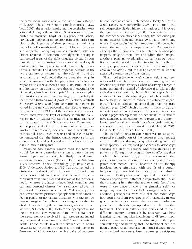
are clearly distinct from the more rostral clusters ofthe contrast Watching Pain > Baseline. When loweringthe threshold to p = .005, two additional clusters wereidentified in the aMCC (MNI coordinates of clustermaxima: x = 0, y = 16, z = 24; t = 3.28; and x = �6,y = 4, z = 40; t = 3.25). Also, although the size of theclusters in the middle insula increased considerably, noadditional clusters were identified in the anterior orposterior parts of the insular cortex. The reverse con-trast (Other > Self ) revealed significant clusters in theright superior and right inferior parietal lobe (Table 3).
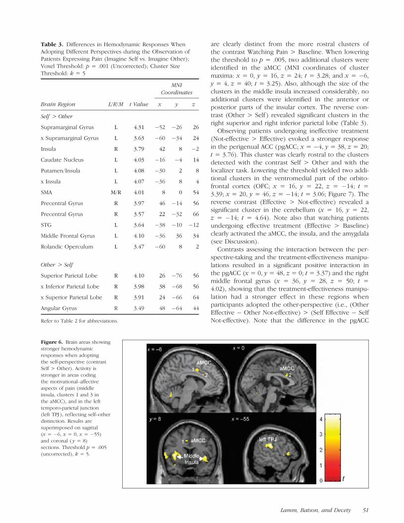
Observing patients undergoing ineffective treatment(Not-effective > Effective) evoked a stronger responsein the perigenual ACC (pgACC; x = �4, y = 38, z = 20;t = 3.76). This cluster was clearly rostral to the clustersdetected with the contrast Self > Other and with thelocalizer task. Lowering the threshold yielded two addi-tional clusters in the ventromedial part of the orbito-frontal cortex (OFC; x = 16, y = 22, z = �14; t =3.39; x = 20, y = 46, z = �14; t = 3.06; Figure 7). Thereverse contrast (Effective > Not-effective) revealed asignificant cluster in the cerebellum (x = 16, y = 22,z = �14; t = 4.64). Note also that watching patientsundergoing effective treatment (Effective > Baseline)clearly activated the aMCC, the insula, and the amygdala(see Discussion).
Contrasts assessing the interaction between the per-spective-taking and the treatment-effectiveness manipu-lations resulted in a significant positive interaction inthe pgACC (x = 0, y = 48, z = 0; t = 3.37) and the rightmiddle frontal gyrus (x = 36, y = 28, z = 50; t =4.02), showing that the treatment-effectiveness manipu-lation had a stronger effect in these regions whenparticipants adopted the other-perspective (i.e., (OtherEffective � Other Not-effective) > (Self Effective � SelfNot-effective). Note that the difference in the pgACC
Table 3. Differences in Hemodynamic Responses WhenAdopting Different Perspectives during the Observation ofPatients Expressing Pain (Imagine Self vs. Imagine Other);Voxel Threshold: p = .001 (Uncorrected); Cluster SizeThreshold: k = 5
MNICoordinates
Brain Region L/R/M t Value x y z
Self > Other
Supramarginal Gyrus L 4.31 �52 �26 26
x Supramarginal Gyrus L 3.63 �60 �34 24
Insula R 3.79 42 8 �2
Caudate Nucleus L 4.03 �16 �4 14
Putamen/Insula L 4.08 �30 2 8
x Insula L 4.07 �36 8 4
SMA M/R 4.01 8 0 54
Precentral Gyrus R 3.97 46 �14 56
Precentral Gyrus R 3.57 22 �32 66
STG L 3.64 �38 �10 �12
Middle Frontal Gyrus L 4.10 �36 36 34
Rolandic Operculum L 3.47 �60 8 2
Other > Self
Superior Parietal Lobe R 4.10 26 �76 56
x Inferior Parietal Lobe R 3.98 38 �68 56
x Superior Parietal Lobe R 3.91 24 �66 64
Angular Gyrus R 3.49 48 �64 44
Refer to Table 2 for abbreviations.
Figure 6. Brain areas showing
stronger hemodynamic
responses when adoptingthe self-perspective (contrast
Self > Other). Activity is
stronger in areas coding
the motivational–affectiveaspects of pain (middle
insula, clusters 1 and 3 in
the aMCC), and in the left
temporo-parietal junction(left TPJ ), ref lecting self–other
distinction. Results are
superimposed on sagittal(x = �6, x = 0, x = �55)
and coronal ( y = 8)
sections. Threshold p = .005
(uncorrected), k = 5.
Lamm, Batson, and Decety 51

reflects a difference in relative deactivation that was morepronounced with the other-perspective [mean param-eter estimates: M(Other Effective) = �4.15, M(OtherNot-effective) = 0.4, M(Self Effective) = �0.48, M(SelfNot-effective) = �3.4].
The ROI analysis of amygdala activity revealed astronger response in the amygdala with the self-perspec-tive [trend-like main effect for the perspective factor,F(1,16) = 4.38, p = .053, partial h2 = 0.215], with thisdifference being slightly more pronounced in the lefthemisphere [trend-like interaction Perspective � Hemi-sphere, F(1,16) = 3.077, p = .099, partial h2 = 0.161;Figure 8].
Correlation of Brain Activity with BehavioralData and Dispositional Measures
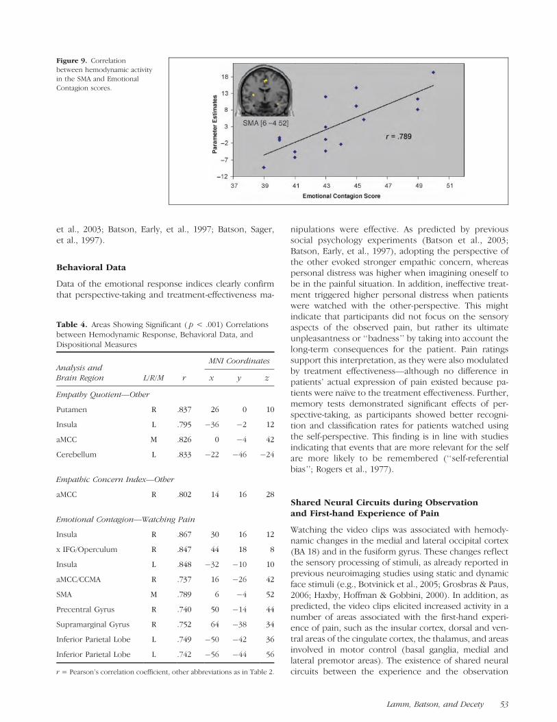
Analysis of the EQ scores revealed significant correla-tions in the right putamen, the left posterior/middleinsula, the aMCC, and the left cerebellum. Scores of theempathic concern index correlated with a similar clusterin the aMCC. No significant correlation was observed forthe Empathic Concern scale of the IRI, the personaldistress index, and the Personal Distress scale of the IRI.Scores of the Emotional Contagion scale correlated withactivity in brain regions involved in affective pain pro-cessing (insula and aMCC) and movement control (SMA,lateral premotor area; Table 4, Figure 9). In addition,two clusters in the left and right parietal cortex closeto the ones revealed by the Self–Other contrasts indi-
cated stronger hemodynamic responses with higherEmotional Contagion scores in these regions.
DISCUSSION
There are good reasons to posit that witnessed pain inothers may result in anxiety, promoting at least cautiousapproach behavior or general threat-defense mecha-nisms (MacDonald & Leary, 2005). The affective experi-ence of pain signals an aversive state and motivatesbehavior to terminate, reduce, or escape exposure tothe source of the noxious stimulation (Price, 1999).Indeed, negative feelings triggered by pain usually mo-tivate organisms to avoid dangerous stimuli and moveaway from danger. However, the observation of pain inothers may also instigate an altruistic motivation to helpthe other, which is quite different from the egoisticmotivation to reduce personal distress.
The aim of our study was to investigate the effects ofperspective-taking and cognitive appraisal on the behav-ioral and neural correlates of pain observation. To thisend, participants watched video clips of patients display-ing an aversive emotional response due to painfulauditory stimulation under different conditions. Usinga number of both state and trait behavioral measuresand event-related fMRI, we were able to demonstratedistinct behavioral and neural responses that are ingood agreement with both empirical findings and theo-retical concepts concerning the promotion of empathicemotion and, ultimately, altruistic motivation (Batson
Figure 7. Brain areas showing
stronger hemodynamic
responses when observing
patients who did not benefitfrom the painful sound
treatment (Not-effective >
Effective). Results aresuperimposed on sagittal
sections. Threshold p = .005
(uncorrected), k = 5.
Figure 8. Stronger
hemodynamic responses inleft and right amygdala when
subjects adopted the self-
perspective (contrast Self >Other). Left: t values from the
contrast Self > Other, overlaid
on a coronal section of the
single-subject structural MNIMRI template. Right: mean
(±SE) parameter estimates
for signal changes in the
whole amygdala.
52 Journal of Cognitive Neuroscience Volume 19, Number 1
et al., 2003; Batson, Early, et al., 1997; Batson, Sager,et al., 1997).
Behavioral Data
Data of the emotional response indices clearly confirmthat perspective-taking and treatment-effectiveness ma-
nipulations were effective. As predicted by previoussocial psychology experiments (Batson et al., 2003;Batson, Early, et al., 1997), adopting the perspective ofthe other evoked stronger empathic concern, whereaspersonal distress was higher when imagining oneself tobe in the painful situation. In addition, ineffective treat-ment triggered higher personal distress when patientswere watched with the other-perspective. This mightindicate that participants did not focus on the sensoryaspects of the observed pain, but rather its ultimateunpleasantness or ‘‘badness’’ by taking into account thelong-term consequences for the patient. Pain ratingssupport this interpretation, as they were also modulatedby treatment effectiveness—although no difference inpatients’ actual expression of pain existed because pa-tients were naıve to the treatment effectiveness. Further,memory tests demonstrated significant effects of per-spective-taking, as participants showed better recogni-tion and classification rates for patients watched usingthe self-perspective. This finding is in line with studiesindicating that events that are more relevant for the selfare more likely to be remembered (‘‘self-referentialbias’’; Rogers et al., 1977).
Shared Neural Circuits during Observationand First-hand Experience of Pain
Watching the video clips was associated with hemody-namic changes in the medial and lateral occipital cortex(BA 18) and in the fusiform gyrus. These changes reflectthe sensory processing of stimuli, as already reported inprevious neuroimaging studies using static and dynamicface stimuli (e.g., Botvinick et al., 2005; Grosbras & Paus,2006; Haxby, Hoffman & Gobbini, 2000). In addition, aspredicted, the video clips elicited increased activity in anumber of areas associated with the first-hand experi-ence of pain, such as the insular cortex, dorsal and ven-tral areas of the cingulate cortex, the thalamus, and areasinvolved in motor control (basal ganglia, medial andlateral premotor areas). The existence of shared neuralcircuits between the experience and the observation
Figure 9. Correlation
between hemodynamic activity
in the SMA and Emotional
Contagion scores.
Table 4. Areas Showing Significant ( p < .001) Correlationsbetween Hemodynamic Response, Behavioral Data, andDispositional Measures
MNI CoordinatesAnalysis andBrain Region L/R/M r x y z
Empathy Quotient—Other
Putamen R .837 26 0 10
Insula L .795 �36 �2 12
aMCC M .826 0 �4 42
Cerebellum L .833 �22 �46 �24
Empathic Concern Index—Other
aMCC R .802 14 16 28
Emotional Contagion—Watching Pain
Insula R .867 30 16 12
x IFG/Operculum R .847 44 18 8
Insula L .848 �32 �10 10
aMCC/CCMA R .737 16 �26 42
SMA M .789 6 �4 52
Precentral Gyrus R .740 50 �14 44
Supramarginal Gyrus R .752 64 �38 34
Inferior Parietal Lobe L .749 �50 �42 36
Inferior Parietal Lobe L .742 �56 �44 56
r = Pearson’s correlation coefficient, other abbreviations as in Table 2.
Lamm, Batson, and Decety 53

of pain is specifically corroborated by the maskinganalysis. This analysis relies upon a localizer task thatinduces affective responses similar to the ones experi-enced by the patients shown. Masking revealed that theobservation of pain in others, and its first-hand experi-ence, activated a largely overlapping neural network. It isworth noting that this overlap did not include areascoding the sensory aspects of pain, as neither theauditory cortex nor the primary or secondary somato-sensory cortices were activated. Common activation wasconfined to areas involved in the motivational–affectivedimension of pain processing, such as the anteriorinsula, the aMCC, and the amygdala, as well as to areasinvolved in motor control. This confirms several recentfMRI studies ( Jackson et al., 2005, 2006; Singer et al.,2004, 2006; Botvinick et al., 2005; Morrison et al., 2004),which indicate that ACC and anterior insula activityduring the observation of pain is related to the affectiveaspects of pain processing rather than to its sensory-discriminative aspects. This interpretation gets addition-al support by neurophysiological evidence suggestingthat the anterior dysgranular part of the insula playsa central role in mediating subjective feeling states, pos-sibly conveyed via mechanisms of interoceptive aware-ness (Critchley, Wiens, Rotshtein, Ohman, & Dolan,2005; Craig, 2002). It is also worth noting that electricalstimulation of the posterior part of the insula, but not ofthe anterior part, evokes painful sensations (Ostrowskyet al., 2002). Altogether, these findings are in agreementwith the proposal that indirect pain representations(as elicited by the observation or imagination of painin others) partially overlap with, but are neverthelessqualitatively different from, first-hand experiences ofpain (Craig, 1968).
Correlation of Brain Activity with BehavioralData and Dispositional Measures
Correlation of hemodynamic activity with behavioraldata and dispositional measures further supports thehypothesis that the affective network of pain processingis specifically involved in the perception of pain inothers. Subjects scoring higher on EQ showed strongeractivity in the left middle insula, the cingulate cortex,and the striatum, and higher empathic concern indiceswere associated with a stronger hemodynamic responsein the aMCC. A similar result was reported by Singer et al.(2004) for the bilateral anterior insula and the aMCCusing the Empathic Concern scale of the IRI. However,no significant correlations with empathic concern werefound in our study. Note though that empathic concerncorrelated with EQ scores only weakly (r = .351) in oursample, and that our range of IRI scores was consider-ably smaller due to the preselection of participants withhigh EC scores (17–24 as opposed to 12–24 in Singeret al., 2004).
Further, emotional contagion scores indicated brain–behavior correlations in similar areas, as well as in areasinvolved in motor control (i.e., SMA/CMA, dorsal andventral precentral gyrus, and posterior parietal cortex).These areas belong to a circuit involved in the prepara-tion and planning of self-generated motor action. Cor-relation of emotion contagion scores with activity inthese motor areas might thus reflect the ‘‘inverse map-ping’’ mechanism posited by the perception–actionaccount of empathy, which assigns a primary role tomotor mimicry and emotional contagion (Preston & deWaal, 2002). Such a mechanism may be triggered byovertly or covertly mirroring in the self the facial painexpressions displayed by the target. Indeed, commentsin debriefing indicated that some subjects used overtmimicry, especially in the self condition. Another possi-bility is that witnessing pain in others automaticallyprompts motor responses to withdraw oneself frompain. These two responses tap similar neural mecha-nisms and are difficult to separate experimentally.
Effect of Perspective-taking
Imagining the self and imagining the other in pain ac-tivate similar neural mechanisms. However, a completeblurring of self and other would be detrimental and isnot the purpose of empathy. Therefore, activation of ad-ditional neural mechanisms is needed to distinguish theself from other. This distinction has been associated withthe sense of agency (i.e., the feeling of being causallyinvolved in an action), which relies on the comparisonbetween self-generated and externally produced signals.Neuroscience research has provided clues to the exis-tence of a cerebral network specifically devoted to thisdistinction. Attribution of an action to another agent hasbeen associated with increased activity in the right pari-etal cortex (e.g., Farrer et al., 2003). The inferior parietalcortex is a multisensory integration area that is ideallysuited to detect distinctions between self-generatedand external signals. Interestingly, perspective-taking in-structions in our study resulted in the activation of dis-tinct subregions of the left and right parietal cortex.The self-perspective elicited higher activity in the leftparietal cortex, whereas the right parietal cortex was se-lectively involved when the other-perspective was adopt-ed. This pattern of activity is consistent with the majorrole of inferior posterior parietal areas in self-agency andperspective-taking (Decety & Grezes, in press; Blanke &Arzy, 2005; Decety & Sommerville, 2003; Ruby & Decety,2003). Accumulating evidence from neuroimaging stud-ies and lesion studies in neurological patients indicatesthat the right inferior parietal cortex has a critical func-tion in the distinction between self-produced actionsand actions generated by others ( Jackson & Decety,2004; Blakemore & Frith, 2003). Importantly, this is alsotrue when behavior is merely mentally simulated. For
54 Journal of Cognitive Neuroscience Volume 19, Number 1
example, imagining somebody else performing an action(Ruby & Decety, 2001) or experiencing an emotion(Ruby & Decety, 2004), as opposed to imaginingperforming the action or experiencing the emotiononeself, revealed a very similar modulation of left- versusright-hemispheric parietal hemodynamic activities. Arecent fMRI study of social perception and empathydemonstrated that activity in the inferior parietal cortexwas negatively associated with the degree of overlapbetween self and other, and that less self–other over-lap led to increased accuracy during social perception(Lawrence et al., 2006).
Further, the self-perspective led to higher activity inbrain areas involved in the affective response to threat orpain, such as the amygdala, the insula, and the aMCC.This is consistent with the idea that personal distress canbe elicited by imagine-self instructions, as first demon-strated in social psychology studies (e.g., Batson, Early,et al., 1997). Moreover, such a finding is in goodagreement with another fMRI study that used a similarperspective-taking manipulation and demonstrated thatthe first-person perspective taps into affective processesto a greater extent than the more detached third-personperspective ( Jackson et al., 2006). The amygdala plays acritical role in fear-related behaviors, such as the evalu-ation of actual or potential threats (LeDoux, 2000).Interestingly, the amygdala receives nociceptive infor-mation from the spino-parabrachial pain system and theinsula, and its activity appears closely tied to the contextand level of aversiveness of the stimuli (Zald, 2003).Imagining oneself to be in a painful and potentiallydangerous situation thus might trigger a stronger fearfuland/or aversive response than imagining someone elseto be in the same situation. Higher activity in the middleinsula may reflect the sensory aspects evoked by theimagination of pain. A meta-analysis of imaging studiesreporting insular activations (Wager & Feldman Barrett,2004) suggests that the middle part of the insula plays arole in coding the sensory–motor aspects of painfulstimulation. Importantly, this region has strong con-nections with the basal ganglia (Chikama, McFarland,Amaral & Haber, 1997), in which activity was also higherwhen adopting the self-perspective (Table 3). Takentogether, activity in this part of the insula possiblyreflects the simulation of the sensory aspects of thepainful experience. This simulation might both lead tothe mobilization of motor areas (including the SMA) inorder to prepare defensive or withdrawal behavior, andto interoceptive monitoring associated with autonomicchanges evoked by this simulation process (Critchleyet al., 2005).
When lowering the threshold, two additional clusterswere detected in the aMCC, a region involved in pro-cessing the affective, evaluative, and attentional aspectsof pain perception (Peyron, Laurent, & Garcia-Larrea,2000). In addition, activity in this region has been relatedto the monitoring of autonomic function (Critchley et al.,
2003). We thus suggest that the self-perspective resultsin the evaluation of the affective, autonomic, and moti-vational consequences obtained from the imagination ofa painful experience, in line with the evocation ofpersonal distress. This interpretation is also in line witha review of Bush, Posner, and Luu (2000), labeling thisarea as the ‘‘cognitive division’’ of the ACC.
Effect of Cognitive Appraisal
Humans have the striking capacity to regulate theiremotions (Beer & Heerey, 2003; Tice, Bratslavsky &Baumeister, 2001). This capacity involves the initiationof new or the alteration of ongoing emotional responsesthrough the action of regulatory processes (Ochsner &Gross, 2005). We suggest that such regulatory processesplay an important role when observing distress inothers, as they enable us to show supportive behavioreven in potentially dangerous or harmful situations. Themajority of neuroimaging studies on emotion regulationhave provided explicit instructions as to how partici-pants should reappraise or suppress elicited emotions(see Ochsner & Gross, 2005, for review). In contrast, wepresented information designed to affect cognitive ap-praisal, as our aim was to create a situation as close aspossible to an everyday context. It was anticipated thatwitnessing another person suffering and knowing thathis/her treatment had not been effective would increasethe emotional response in the observer. Conversely,knowing that a treatment had been beneficial for thepatient was expected to elicit down-regulation of theperceptually triggered affective response. Both the be-havioral and hemodynamic data supported this hypoth-esis. Behavioral data showed higher pain intensity andunpleasantness ratings when the treatment had notbeen effective. This finding was paralleled by activitydifferences in a number of brain regions involved inaffective coding and emotion regulation, such as rostraland perigenual ACC and the ventromedial OFC. Notealso that observing ineffectively treated patients trig-gered strong activation in the aMCC, the insula, andthe amygdala, indicating an affective response. Based onself-report and behavioral data, we suggest that thisresponse was regulated (reappraised) via top-downmechanisms such as focusing on the long-term conse-quences of the treatment.
A recent review of cingulate cortex functions in painand emotion indicates that a subregion in the rostralACC is involved in coding fearful responses (Vogt, 2005).Similarly, Bush et al. (2000) labeled this part of the ACCas its ‘‘affective division,’’ in contrast to the moreposterior-dorsal ‘‘cognitive division’’ (see above). Activ-ity in this area may hence be related to a strongerdefensive response in cases where the treatment hadno benefit, and thus, the patient’s overall situation wasperceived as being more unpleasant or distressing (asindicated by the behavioral data). Alternatively and more
Lamm, Batson, and Decety 55

speculatively, ACC activity may have resulted from ananger-related reaction triggered by the fact that the pa-tient had to suffer pain without benefiting from it. Thatparticipants experienced more anger when watchingpatients undergoing ineffective treatment is—at leastindirectly—indicated by significantly higher scores forthe adjective ‘‘upset’’ on the emotional response scalewhen the therapy was not effective [t(15) = 2.695, p =.027, h2 = .326]. Note also that self-generation of angeractivates a similar part of the ACC (Damasio et al., 2000).
While watching the videos, the participants in ourstudy had to consider whether the overall effect of thepainful treatment was positive or negative. This manip-ulation of the context in which pain occurred differen-tially elicited OFC activity, which plays an important rolein the evaluation of positive and negative reinforce-ments, and in the motivational and emotional aspectsof social behavior (Rolls, 2004). The OFC is also involvedin emotion reappraisal, as attending to a negativelyvalenced picture evokes stronger activity in the ventro-medial OFC than reappraising that picture in a way thatit no longer elicited a negative response (Ochsner et al.,2002). Similarly, watching patients undergoing ineffec-tive treatment was associated with higher activity in thisregion, whereas effective treatment was associated witha decreased OFC response. Note that the OFC was activein both conditions (data not shown). Activity in the OFCthus might reflect differing requirement to evaluate theoverall positive and negative aspects of the presentedstimuli. This top-down process might operate upon thevisually conveyed information about the affective state ofthe patients. Interestingly, watching effective versusineffective treatment patients did not modulate activityin either the visual–sensory areas or in the insula (evenwhen the threshold was lowered to p = .05). Thissuggests that the two patient groups were differentiatedafter perceiving their emotional reactions (but notnecessarily after participants reacted emotionally them-selves), and that top-down mechanisms did not operateon perceptual processing at an early stage. Keep inmind, however, that this finding might be influencedby the requirement for participants to evaluate the painof the patients.
The interaction between perspective-taking and treat-ment effectiveness yielded increased activity in thepgACC and the middle frontal gyrus. Involvement ofthe pgACC most likely indicates a difference in perspec-tive-taking requirements, which are more complex whenimagining the feelings of patients who—in contrast tothe observer—do not know the ultimate outcome of thetreatment. In fact, increased activity in the rostral cingu-late and the paracingulate cortex when performingmentalizing tasks has reliably been found (see Gallagher& Frith, 2003, for review). Thus, we speculate that thestronger deactivation with the other-perspective indi-cates a stronger requirement to inhibit mentalizing,which might be counterproductive in a case where
participants have to focus on the long-term consequen-ces of the treatment and not the immediate affectivereaction of the patient. The middle frontal gyrus, on theother hand, may be associated with emotion down-regulation, as indicated by a recent fMRI study onemotion regulation (Ochsner et al., 2004). However,studies using explicit emotion regulation instructionsshow stronger involvement of lateral prefrontal corticalregions than our study. This highlights the role of theseregions in deliberately exerting top-down control,whereas appraisal-based regulation might be supportedby brain structures involved in relatively automatic as-sessment of reward properties (such as the OFC).
Conclusion
Our findings are consistent with the view that humans’responses to the pain of others can be modulated bothby cognitive and motivational processes. These process-es are likely to influence whether observing a conspe-cific in need of help will result in empathic concern, animportant instigator of helping behavior, or personaldistress. These two types of affective responses arequalitatively distinct and have different motivationalconsequences. Empathic concern may instigate an altru-istic motivation to help the other; personal distress mayproduce an egoistic motivation to reduce personaldistress (Batson et al., 1987). It was thus important todemonstrate both the similarities in the neural networksunderlying the sharing of pain with others and thespecific mechanisms that permit distinguishing the selffrom others, which is critical for the experience of em-pathy (Decety & Hodges, 2006; Decety & Lamm, 2006;Decety, 2005). Such an experience cannot be identicalto the actual perception of pain because personal andvicarious experiences differ neurophysiologically asdemonstrated by our behavioral and fMRI data (as alsodemonstrated by Craig, 1968, for autonomic nervoussystem measurements). Indeed, in order for the sub-jective experience to be labeled empathy, the observermust recognize that the emotion she/he is experiencingis a response to the other’s emotional state. Finally, ourresults demonstrate that both bottom-up (automatic)and top-down (controlled) processes interact to pro-duce the experience of empathy. Knowledge about thecontext in which the pain experience occurs providesimportant clues to the role of top-down cognitive ap-praisal in the regulation of the vicarious affective painreaction.
Acknowledgments
This study was supported by a fellowship from the Fondationde Recherche Medicale (FRM) to C. L. We thank the AcousticsResearch Institute of the Austrian Academy of Sciences forproviding the software S_TOOLS-STx, v3.6.1, and two anony-mous reviewers for helpful comments on the manuscript.
56 Journal of Cognitive Neuroscience Volume 19, Number 1

Reprint requests should be sent to Jean Decety, Social Cog-nitive Neuroscience, Department of Psychology, The Universityof Chicago, 5848 S University Avenue, Chicago, IL 60637, USA,or via e-mail: [email protected].
REFERENCES
Baron-Cohen, S., & Wheelwright, S. (2004). The EmpathyQuotient: An investigation of adults with Asperger syndromeor high functioning autism, and normal sex differences.Journal of Autism and Developmental Disorders, 34,163–175.
Batson, C. D., Early, S., & Salvarini, G. (1997). Perspectivetaking: Imagining how another feels versus imagining howyou would feel. Personality & Social Psychology Bulletin,23, 751–758.
Batson, C. D., Fultz, J., & Schoenrade, P. A. (1987). Distressand empathy: Two qualitatively distinct vicarious emotionswith different motivational consequences. Journal ofPersonality, 55, 19–39.
Batson, C. D., Lishner, D. A., Carpenter, A., Dulin, L.,Harjusola-Webb, S., Stocks, E. L., et al. (2003). ‘‘. . . As youwould have them do unto you’’: Does imagining yourselfin the other’s place stimulate moral action? Personality &Social Psychology Bulletin, 29, 1190–1201.
Batson, C. D., Sager, K., Garst, E., Kang, M., Rubchinsky, K.,& Dawson, K. (1997). Is empathy-induced helping due toself–other merging? Journal of Personality and SocialPsychology, 73, 495–509.
Beer, J. S., & Heerey, E. A. (2003). The regulatory functionof self-conscious emotion: Insight from patients withorbitofrontal lesions. Journal of Personality and SocialPsychology, 85, 594–604.
Bellini, L. M., Baime, M., & Shea, J. A. (2002). Variation ofmood and empathy during internship. Journal of theAmerican Medical Association, 287, 3143–3146.
Blakemore, S.-J., & Frith, C. D. (2003). Self-awareness andaction. Current Opinion in Neurobiology, 13, 219–224.
Blanke, O., & Arzy, S. (2005). The out-of-body experience:Disturbed self-processing at the temporo-parietal junction.The Neuroscientist, 11, 16–24.
Botvinick, M., Jha, A. P., Bylsma, L. M., Fabian, S. A., Solomon,P. E., & Prkachin, K. M. (2005). Viewing facial expressionof pain engages cortical areas involved in the directexperience of pain. Neuroimage, 25, 312–319.
Bradley, M. M., & Lang, P. J. (2000). Affective reactions toacoustic stimuli. Psychophysiology, 37, 204–215.
Bush, G., Luu, P., & Posner, M. I. (2000). Cognitive andemotional influences in anterior cingulate cortex. Trendsin Cognitive Sciences, 4, 215–222.
Chikama, M., McFarland, N. R., Amaral, D. G., & Haber,S. N. (1997). Insular cortical projections to functional regionsof the striatum correlate with cortical cytoarchitectonicorganization in the primate. Journal of Neuroscience, 15,9686–9705.
Craig, A. D. (2002). Opinion: How do you feel? Interoception:The sense of the physiological condition of the body.Nature Reviews Neuroscience, 3, 655–666.
Craig, K. D. (1968). Physiological arousal as a function ofimagined, vicarious, and direct stress experiences. Journalof Abnormal Psychology, 73, 513–520.
Craig, K. D., Prkachin, K. M., & Grunau, R. V. E. (2001). Thefacial expression of pain. In D. C. Turk & R. Melzack (Eds.),Handbook of pain assessment (2nd ed., pp. 153–169).New York: Guilford.
Critchley, H. D., Mathias, C. J., Josephs, O., O’Doherty, J.Zanini, S., Dewar, B.-K., et al. (2003). Human cingulate
cortex and autonomic control: Converging neuroimagingand clinical evidence. Brain, 126, 2139–2152.
Critchley, H. D., Wiens, S., Rotshtein, P., Ohman, A., & Dolan,R. D. (2005). Neural systems supporting interoceptiveawareness. Nature Neuroscience, 7, 189–195.
Damasio, A. R., Grabowski, T. J., Bechara, A., Damasio, H.,Ponto, L. L. B., Parvizi, J., et al. (2000). Subcortical andcortical brain activity during the feeling of self-generatedemotions. Nature Neuroscience, 3, 1049–1056.
Davis, M. H. (1996). Empathy: A social psychologicalapproach. Madison, WI: Westview Press.
Decety, J. (2005). Perspective taking as the royal avenueto empathy. In B. F. Malle & S. D. Hodges (Eds.), Otherminds: How humans bridge the divide between self andother (pp. 143–157). New York: Guilford Publications.
Decety, J., & Grezes, J. (2006). The power of simulation:Imagining one’s own and other’s behavior. Brain Research,1079, 4–14.
Decety, J., & Hodges, S. D. (2006). The social neuroscience ofempathy. In P. A. M. Lange (Ed.), Bridging social psychologybenefits of transdisciplinary approaches (pp. 103–109).Mahwah, NJ: Erlbaum.
Decety, J., & Jackson, P. L. (2004). The functional architectureof human empathy. Behavioral and CognitiveNeuroscience Reviews, 3, 71–100.
Decety, J., & Lamm, C. (2006). Human empathy through thelens of social neuroscience. The Scientific World Journal, 6,1146–1163.
Decety, J., & Sommerville, J. A. (2003). Shared representationsbetween self and others: A social cognitive neuroscienceview. Trends in Cognitive Sciences, 7, 527–533.
Deichmann, R., Gottfried, J. A., Hutton, C., & Turner, R.(2003). Optimized EPI for fMRI studies of the orbitofrontalcortex. Neuroimage, 19, 430–441.
Derbyshire, S. W. G. (2000). Exploring the pain ‘‘neuromatrix’’.Current Review of Pain, 4, 467–477.
Doherty, R. W. (1997). The emotional contagion scale: Ameasure of individual differences. Journal of NonverbalBehavior, 21, 131–154.
Donaldson, D. I., & Buckner, R. L. (2001). Effectiveparadigm design. In P. Jezzard, P. M. Mathews, & S. M. Smith(Eds.), Functional MRI: An introduction to methods(pp. 177–195). Oxford: Oxford University Press.
Eickhoff, S., Stephan, K. E., Mohlberg, H., Grefkes, C., Fink,G. R., Amunts, K., et al. (2005). A new SPM toolbox forcombining probabilistic cytoarchitectonic maps andfunctional imaging data. Neuroimage, 25, 1325–1335.
Farrer, C., Franck, N., Georgieff, N., Frith, C.D., Decety, J.,& Jeannerod, M. (2003). Modulating the experience ofagency: A positron emission tomography study.Neuroimage, 18, 324–333.
Friston, K. J., Fletcher, P., Josephs, O., Holmes, A. P.,Rugg, M. D., & Turner, R. (1998). Event-related fMRI:Characterizing differential responses. Neuroimage, 7,30–40.
Gallagher, H. L., & Frith, C. D. (2003). Functional imagingof ‘theory of mind’. Trends in Cognitive Sciences, 7,77–83.
Grosbras, M.-H., & Paus, T. (2006). Brain networks involvedin viewing angry hands or faces. Cerebral Cortex, 16,1087–1096.
Gross, J. J., & John, O. P. (2003). Individual differences intwo emotion regulation processes: Implications for affect,relationships, and well-being. Journal of Personality andSocial Psychology, 85, 348–362.
Haxby, J. V., Hoffman, E. A., & Gobbini, M. I. (2000). Thedistributed human neural system for face perception.Trends in Cognitive Sciences, 4, 223–233.
Lamm, Batson, and Decety 57

Jackson, P. L., Brunet, E., Meltzoff, A. N., & Decety, J. (2006).Empathy examined through the neural mechanismsinvolved in imagining how I feel versus how you wouldfeel pain: An event-related fMRI study. Neuropsychologia,44, 752–761.
Jackson, P. L., & Decety, J. (2004). Motor cognition: Anew paradigm to study self–other interactions. CurrentOpinion in Neurobiology, 14, 1–5.
Jackson, P. L., Meltzoff, A. N., & Decety, J. (2005). Howdo we perceive the pain of others: A window into theneural processes involved in empathy. Neuroimage, 24,771–779.
Jackson, P. L., Rainville, P., & Decety, J. (2006). To what extentdo we share the pain of others? Insight from the neuralbases of pain empathy. Pain, 125, 5–9.
Kalisch, R., Wiech, K., Critchley, H. D., Seymour, B., O’Doherty,J. P., Oakley, D. A., et al. (2005). Anxiety reduction throughdetachment: Subjective, physiological, and neural effects.Journal of Cognitive Neuroscience, 17, 874–883.
Lawrence, E. J., Shaw, P., Giampietro, V. P., Surguladze, S.,Brammer, M. J., & David, A. S. (2006). The role of ‘sharedrepresentations’ in social perception and empathy:An fMRI study. Neuroimage, 29, 1173–1184.
LeDoux, J. E. (2000). Emotion circuits in the brain. AnnualReviews of Neuroscience, 23, 155–184.
MacDonald, G., & Leary, M. R. (2005). Why does socialexclusion hurt? The relationship between social painand physical pain. Psychological Bulletin, 131, 202–223.
Mesulam, M. M., & Mufson, E. J. (1982). Insula of the oldworld monkey: III. Efferent cortical output and commentson function. Journal of Comparative Neurology, 212,38–52.
Morosan, P., Rademacher, J., Schleicher, A., Amunts, K.,Schormann, T., & Zilles, K. (2001). Human primaryauditory cortex: Cytoarchitectonic subdivisions andmapping into a spatial reference system. Neuroimage,13, 684–701.
Morrison, I., Lloyd, D., di Pellegrino, G., & Robets, N. (2004).Vicarious responses to pain in anterior cingulate cortex:Is empathy a multisensory issue? Cognitive & AffectiveBehavioural Neuroscience, 4, 270–278.
Ochsner, K. N., Bunge, S. A., Gross, J. J., & Gabrieli, J. D. E.(2002). Rethinking feelings: An fMRI study of cognitiveregulation of emotion. Journal of Cognitive Neuroscience,14, 1215–1229.
Ochsner, K. N., & Gross, J. J. (2005). The cognitive controlof emotion. Trends in Cognitive Sciences, 9, 242–249.
Ochsner, K. N., Ray, R. D., Cooper, J. C., Robertson, E. R.,Chopra, S., Gabrieli, J. D. E., et al. (2004). For betteror for worse: Neural systems supporting the cognitivedown- and up-regulation of negative emotion. Neuroimage,23, 483–499.
Ostrowsky, K., Magnin, M., Ryvlin, P., Isnard, J., Guenot, M.,& Mauguiere, F. (2002). Representation of pain andsomatic sensation in the human insula: A study of responsesto direct electrical cortical stimulation. Cerebral Cortex,12, 376–385.
Paus, T. (2001). Primate anterior cingulate cortex: Where
motor control, drive and cognition interface. NatureReviews Neuroscience, 2, 417–424.
Peyron, R., Laurent, B., & Garcia-Larrea, L. (2000). Functionalimaging of brain responses to pain. A review andmeta-analysis (2000). Neurophysiologie Clinique, 30,263–288.
Preston, S. D., & de Waal, F. B. M. (2002). Empathy: Itsultimate and proximate bases. Behavioural and BrainSciences, 25, 1–72.
Price, D. D. (1999). Psychological mechanisms of pain andanalgesia. Seattle, WA: IASP Press.
Prkachin, K. M. (2005). Effects of deliberate control onverbal and facial expressions of pain. Pain, 114, 328–338.
Rogers, T. B., Kuipers, N. A., & Kirker, W. S. (1977).Self-reference and the encoding of personal information.Journal of Personality and Social Psychology, 35, 677–688.
Rolls, E. T. (2004). The functions of the orbitofrontal cortex.Brain and Cognition, 55, 11–29.
Ruby, P., & Decety, J. (2001). Effect of subjective perspectivetaking during simulation of action: A PET investigation ofagency. Nature Neuroscience, 4, 546–550.
Ruby, P., & Decety, J. (2003). What you believe versus whatyou think they believe: A neuroimaging study of conceptualperspective-taking. European Journal of Neuroscience,17, 2475–2480.
Ruby, P., & Decety, J. (2004). How would you feel versushow do you think she would feel? A neuroimaging studyof perspective-taking with social emotions. Journal ofCognitive Neuroscience, 16, 988–999.
Singer, T., Seymour, B., O’Doherty, J., Kaube, H., Dolan,R. J., & Frith, C. D. (2004). Empathy for pain involves theaffective but not the sensory components of pain. Science,303, 1157–1161.
Singer, T., Seymour, B., O’Doherty, J. P., Stephan, K. E.,Dolan, R. J., & Frith, C. D. (2006). Empathic neural responsesare modulated by the perceived fairness of others. Nature,439, 466–469.
Tice, D. M., Bratslavsky, E., & Baumeister, R. F. (2001).Emotional distress regulation takes precedence overimpulse control: If you feel bad, do it. Journal of Personalityand Social Psychology, 80, 53–67.
Tzourio-Mazoyer, N., Landeau, B., Papathanassiou, D.,Crivello, F., Etard, O., Delcroix, N., et al. (2002). Automatedanatomical labelling of activations in SPM using amacroscopic anatomical parcellation of the MNI MRIsingle subject brain. Neuroimage, 15, 273–289.
Underwood, B., & Moore, B. (1982). Perspective-taking andaltruism. Psychological Bulletin, 91, 143–173.
Vogt, B. A. (2005). Pain and emotion interactions in subregionsof the cingulate gyrus. Nature Reviews Neuroscience, 6,533–544.
Wager, T. D., & Feldman Barrett, L. (2004). From affect tocontrol: Functional specialization of the insula in motivationand regulation. Retrieved from www.apa.org/psycextra/ on24 December 2005.
Zald, D. H. (2003). The human amygdala and the emotionalevaluation of sensory stimuli. Brain Research Reviews, 41,88–123.
58 Journal of Cognitive Neuroscience Volume 19, Number 1