the nervous system - alderman science pagealderman.weebly.com/uploads/8/1/0/7/8107206/ch_7_c.pdfby...
TRANSCRIPT
PowerPoint® Lecture Slide Presentation
by Patty Bostwick-Taylor,
Florence-Darlington Technical College
Copyright © 2009 Pearson Education, Inc., publishing as Benjamin Cummings
PART C 7
The Nervous
System

Copyright © 2009 Pearson Education, Inc., publishing as Benjamin Cummings
Protection of the Central Nervous System
Scalp and skin
Skull and vertebral column
Meninges
Cerebrospinal fluid (CSF)
Blood-brain barrier

Copyright © 2009 Pearson Education, Inc., publishing as Benjamin Cummings
Protection of the Central Nervous System
Figure 7.17a

Copyright © 2009 Pearson Education, Inc., publishing as Benjamin Cummings
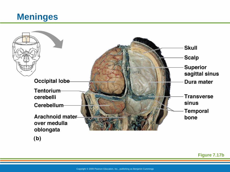
Meninges
Dura mater
Double-layered external covering
Periosteum—attached to inner surface of
the skull
Meningeal layer—outer covering of the
brain
Folds inward in several areas

Copyright © 2009 Pearson Education, Inc., publishing as Benjamin Cummings
Meninges
Arachnoid layer
Middle layer
Web-like
Pia mater
Internal layer
Clings to the surface of the brain
Copyright © 2009 Pearson Education, Inc., publishing as Benjamin Cummings
Meninges
Figure 7.17b
Copyright © 2009 Pearson Education, Inc., publishing as Benjamin Cummings
Cerebrospinal Fluid (CSF)
Similar to blood plasma composition
Formed by the choroid plexus
Forms a watery cushion to protect the brain
Circulated in arachnoid space, ventricles, and
central canal of the spinal cord

Copyright © 2009 Pearson Education, Inc., publishing as Benjamin Cummings
Figure 7.18a–b
Ventricles and Location of
the Cerebrospinal Fluid

Copyright © 2009 Pearson Education, Inc., publishing as Benjamin Cummings
Ventricles and Location of
the Cerebrospinal Fluid
Figure 7.18c

Copyright © 2009 Pearson Education, Inc., publishing as Benjamin Cummings
Hydrocephalus in a Newborn
Hydrocephalus
CSF accumulates and exerts pressure on the
brain if not allowed to drain
Figure 7.19
Copyright © 2009 Pearson Education, Inc., publishing as Benjamin Cummings
Blood-Brain Barrier
Includes the least permeable capillaries of the
body
Excludes many potentially harmful substances
Useless as a barrier against some substances
Fats and fat soluble molecules
Respiratory gases
Alcohol
Nicotine
Anesthesia

Copyright © 2009 Pearson Education, Inc., publishing as Benjamin Cummings
Traumatic Brain Injuries
Concussion
Slight brain injury
No permanent brain damage
Contusion
Nervous tissue destruction occurs
Nervous tissue does not regenerate
Cerebral edema
Swelling from the inflammatory response
May compress and kill brain tissue
Copyright © 2009 Pearson Education, Inc., publishing as Benjamin Cummings
Cerebrovascular Accident (CVA)
Commonly called a stroke
The result of a ruptured blood vessel supplying a
region of the brain
Brain tissue supplied with oxygen from that blood
source dies
Loss of some functions or death may result
Copyright © 2009 Pearson Education, Inc., publishing as Benjamin Cummings
Alzheimer’s Disease
Progressive degenerative brain disease
Mostly seen in the elderly, but may begin in
middle age
Structural changes in the brain include abnormal
protein deposits and twisted fibers within neurons
Victims experience memory loss, irritability,
confusion, and ultimately, hallucinations and
death

Copyright © 2009 Pearson Education, Inc., publishing as Benjamin Cummings
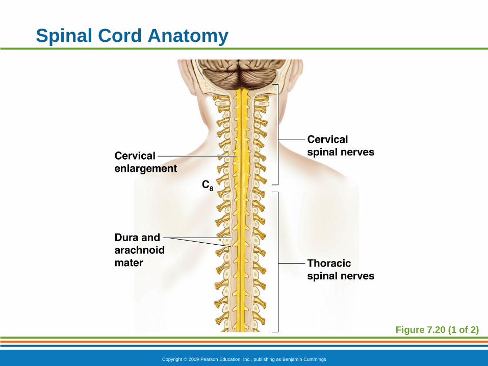
Spinal Cord
Extends from the foramen magnum of the skull to
the first or second lumbar vertebra
31 pairs of spinal nerves arise from the spinal
cord
Cauda equina is a collection of spinal nerves at
the inferior end
Copyright © 2009 Pearson Education, Inc., publishing as Benjamin Cummings
Spinal Cord Anatomy
Figure 7.20 (1 of 2)

Copyright © 2009 Pearson Education, Inc., publishing as Benjamin Cummings
Spinal Cord Anatomy
Figure 7.20 (2 of 2)
Copyright © 2009 Pearson Education, Inc., publishing as Benjamin Cummings
Spinal Cord Anatomy
Internal gray matter is mostly cell bodies
Dorsal (posterior) horns
Anterior (ventral) horns
Gray matter surrounds the central canal
Central canal is filled with cerebrospinal
fluid
Exterior white mater—conduction tracts
Dorsal, lateral, ventral columns

Copyright © 2009 Pearson Education, Inc., publishing as Benjamin Cummings
Spinal Cord Anatomy
Figure 7.21

Copyright © 2009 Pearson Education, Inc., publishing as Benjamin Cummings
Spinal Cord Anatomy
Meninges cover the spinal cord
Spinal nerves leave at the level of each vertebrae
Dorsal root
Associated with the dorsal root ganglia—
collections of cell bodies outside the
central nervous system
Ventral root
Contains axons

Copyright © 2009 Pearson Education, Inc., publishing as Benjamin Cummings
Pathways Between Brain and Spinal Cord
Figure 7.22

Copyright © 2009 Pearson Education, Inc., publishing as Benjamin Cummings
Peripheral Nervous System (PNS)
Nerves and ganglia outside the central nervous
system
Nerve = bundle of neuron fibers
Neuron fibers are bundled by connective tissue
Copyright © 2009 Pearson Education, Inc., publishing as Benjamin Cummings
PNS: Structure of a Nerve
Endoneurium surrounds each fiber
Groups of fibers are bound into fascicles by
perineurium
Fascicles are bound together by epineurium

Copyright © 2009 Pearson Education, Inc., publishing as Benjamin Cummings
PNS: Structure of a Nerve
Figure 7.23
Copyright © 2009 Pearson Education, Inc., publishing as Benjamin Cummings
PNS: Classification of Nerves
Mixed nerves
Both sensory and motor fibers
Sensory (afferent) nerves
Carry impulses toward the CNS
Motor (efferent) nerves
Carry impulses away from the CNS

Copyright © 2009 Pearson Education, Inc., publishing as Benjamin Cummings
PNS: Cranial Nerves
12 pairs of nerves that mostly serve the head and
neck
Only the pair of vagus nerves extend to thoracic
and abdominal cavities
Most are mixed nerves, but three are sensory only

Copyright © 2009 Pearson Education, Inc., publishing as Benjamin Cummings
PNS: Cranial Nerves
I Olfactory nerve—sensory for smell
II Optic nerve—sensory for vision
III Oculomotor nerve—motor fibers to eye
muscles
IV Trochlear—motor fiber to eye muscles
Copyright © 2009 Pearson Education, Inc., publishing as Benjamin Cummings
PNS: Cranial Nerves
V Trigeminal nerve—sensory for the face; motor
fibers to chewing muscles
VI Abducens nerve—motor fibers to eye muscles
VII Facial nerve—sensory for taste; motor fibers
to the face
VIII Vestibulocochlear nerve—sensory for
balance and hearing

Copyright © 2009 Pearson Education, Inc., publishing as Benjamin Cummings
PNS: Cranial Nerves
IX Glossopharyngeal nerve—sensory for taste;
motor fibers to the pharynx
X Vagus nerves—sensory and motor fibers for
pharynx, larynx, and viscera
XI Accessory nerve—motor fibers to neck and
upper back
XII Hypoglossal nerve—motor fibers to tongue
Copyright © 2009 Pearson Education, Inc., publishing as Benjamin Cummings
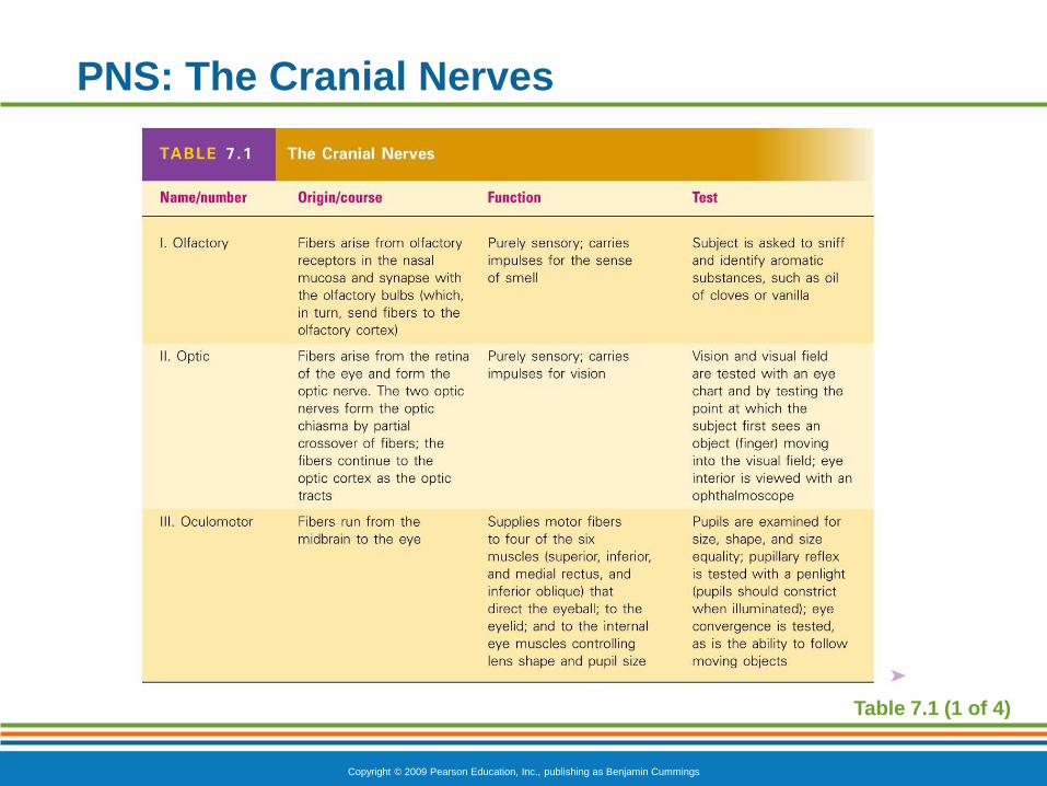
PNS: The Cranial Nerves
Table 7.1 (1 of 4)

Copyright © 2009 Pearson Education, Inc., publishing as Benjamin Cummings
PNS: The Cranial Nerves
Table 7.1 (2 of 4)

Copyright © 2009 Pearson Education, Inc., publishing as Benjamin Cummings
PNS: The Cranial Nerves
Table 7.1 (3 of 4)

Copyright © 2009 Pearson Education, Inc., publishing as Benjamin Cummings
PNS: The Cranial Nerves
Table 7.1 (4 of 4)

Copyright © 2009 Pearson Education, Inc., publishing as Benjamin Cummings
PNS: Distribution of Cranial Nerves
Figure 7.24