the muscular system - univerzita...
TRANSCRIPT

General myology
© David Kachlík 30.9.2015

Function of the muscle tissue
• generation of movements
• stabilization of the position of the body
• control of the volume of the organs
– smooth muscle - sphincters
• motion of the substances in the body
– blood, lymph, urine, air, food and fluids, sperm
• generation of body heat
– voluntary and involuntary contractions of skeletal
striated muscle© David Kachlík 30.9.2015

Characteristics of the muscle tissue
• electrical irritability (excitability)
– ability of the skeletal muscle to respond to stimuli
– skeletal muscle contracts as a result of nerve irritation
• contractility
– ability of contraction
• extensibility
– ability to extend without tissue damage
• elasticity
– ability to return to the original shape after being extended
© David Kachlík 30.9.2015

Types of the muscle tissue
• smooth muscle tissue
(textus muscularis levis)
• striated muscle tissue
(textus muscularis striatus)
• cardiac striated mucle tissue
(textus muscularis striatus cardiacus)
© David Kachlík 30.9.2015

Smooth muscle tissue
• smooth muscle cell (myocytus levis)
• nucleus is in the centre of the cell
• in sarcoplasm there are longitudinally placed
contractable myofibrils made of myofilaments
(myofilamenta)
– myofilamentum crassum
– myofilamentum tenue
• even at rest it is in a mild contractile state – tonus
• doesn´t become fatigued
• inervated by automonic system is not under
voluntary control © David Kachlík 30.9.2015

Eis, Jelínek, Špaček, Histopatologický atlas, Praha 2006
Smooth muscle
© David Kachlík 30.9.2015

Eis, Jelínek, Špaček, Histopatologický atlas, Praha 2006
Smooth muscle – electronmicroscopical picture
© David Kachlík 30.9.2015

Skeletal striated muscle Imyofibre (myofibra)
• elementary structural unit
• multinucleated
• thickness: 10–100 µm
• length: mm – cm
• origin: merging of elongated mononuclear cells
(myoblasts) → myotubes (nuclei inside, myofibrils at
the surface) → conversion to myofibres (nuclei at the
surface, myofibrils inside)
• sarcolemma on the surface
– striated in the microscope
• lighter and darker sections© David Kachlík 30.9.2015

Skeletal striated muscle II
• myoglobin (pigment causing red colouring)
• fibres
– quick
• quickly fatigued
• light (white)
• in superficial layers
– slow
• more resistant to tiredness
• dark (red)
• in deeper layer
• inervated by cranial and spinal nerves
– without inervation non-functional and atrophies© David Kachlík 30.9.2015

Myofilaments and sarcomeres
• thick (myosin) and thin (actin) myofilaments (myofilamenta) overlap so they create annealing
• sarcomere (sarcomerum)
– contractile muscle units of skeletal and cardiac muscle
– reach from one Z-disc to another
© David Kachlík 30.9.2015
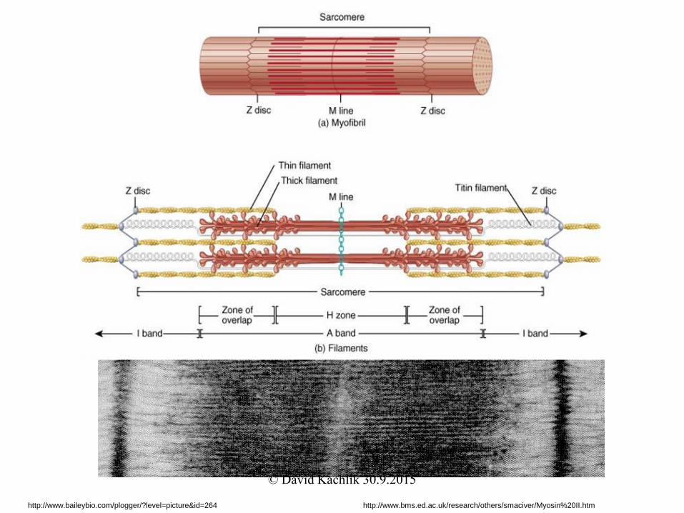
http://www.baileybio.com/plogger/?level=picture&id=264 http://www.bms.ed.ac.uk/research/others/smaciver/Myosin%20II.htm
© David Kachlík 30.9.2015

Eis, Jelínek, Špaček, Histopatologický atlas, Praha 2006
Skeletal striated muscle – longitudinal section
© David Kachlík 30.9.2015

Eis, Jelínek, Špaček, Histopatologický atlas, Praha 2006
Skeletal striated muscle – transverse section
© David Kachlík 30.9.2015

Cardiac striated muscle
• microscopically looks like a net
• similar to skeletal muscle tissue
• fibres connected by diagonal plasmatic bridges
• intercalated disc (discus intercalaris)
– divides cardiac muscle tissue into mononuclear segments
(cells)
• nucleus in the middle of the cell
• sarcolemma on the surface
• conducting system of the heart (complexus stimulans
cordis)
– modified cells of the cardiac muscle are specialized to generate
impulses and to conduct them© David Kachlík 30.9.2015

Eis, Jelínek, Špaček, Histopatologický atlas, Praha 2006
Cardiac muscle tissue
© David Kachlík 30.9.2015

Eis, Jelínek, Špaček, Histopatologický atlas, Praha 2006
Cardiac muscle tissue
intercalated disc
© David Kachlík 30.9.2015
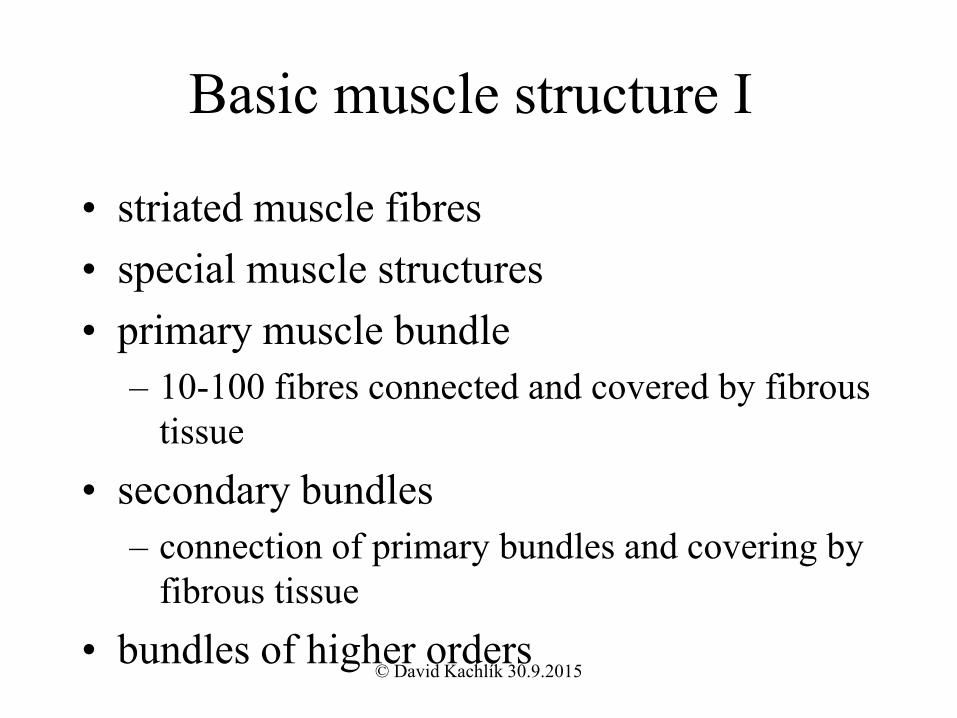
Basic muscle structure I
• striated muscle fibres
• special muscle structures
• primary muscle bundle
– 10-100 fibres connected and covered by fibrous
tissue
• secondary bundles
– connection of primary bundles and covering by
fibrous tissue
• bundles of higher orders© David Kachlík 30.9.2015
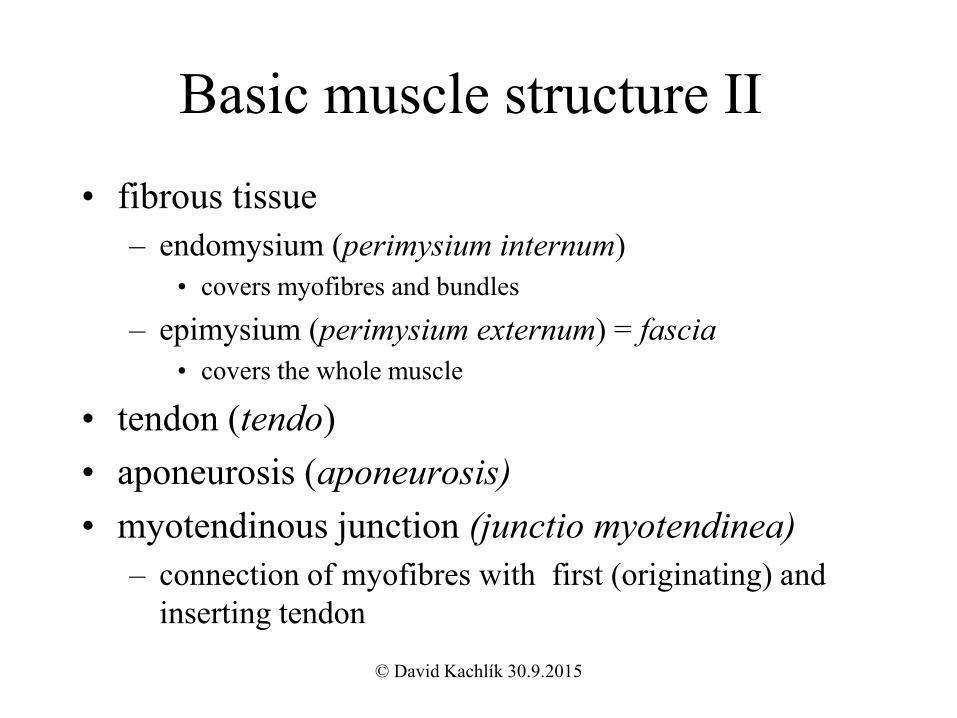
Basic muscle structure II
• fibrous tissue
– endomysium (perimysium internum)
• covers myofibres and bundles
– epimysium (perimysium externum) = fascia
• covers the whole muscle
• tendon (tendo)
• aponeurosis (aponeurosis)
• myotendinous junction (junctio myotendinea)
– connection of myofibres with first (originating) and
inserting tendon
© David Kachlík 30.9.2015
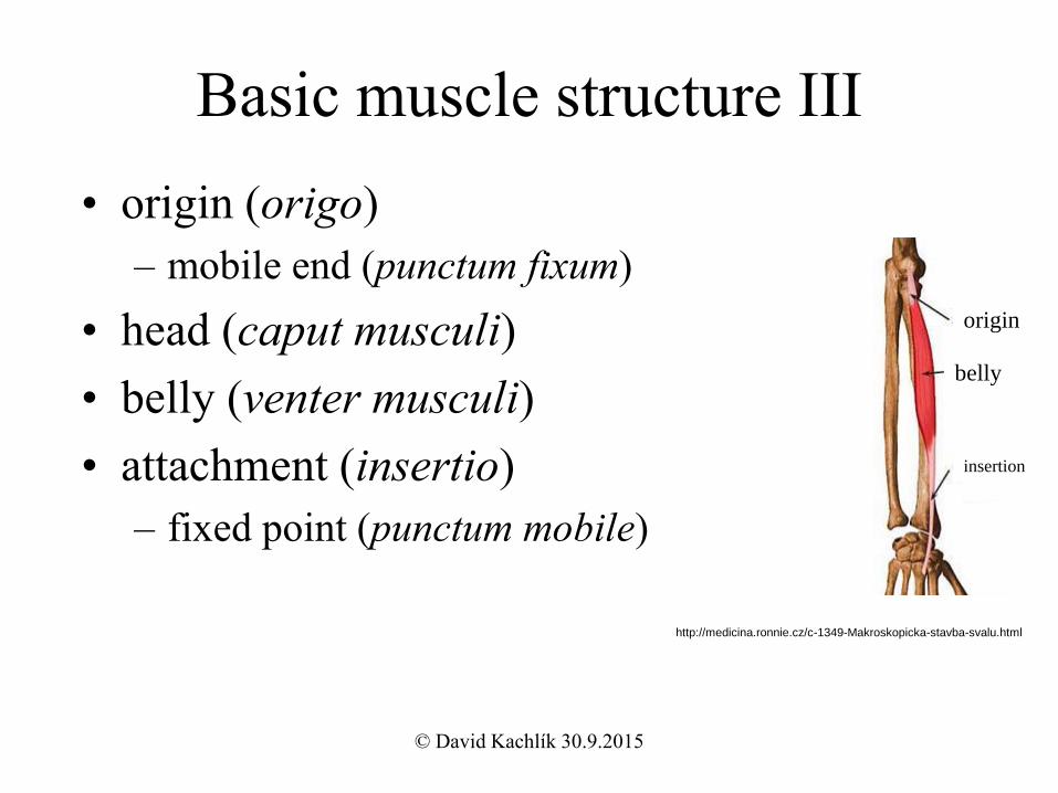
Basic muscle structure III
• origin (origo)
– mobile end (punctum fixum)
• head (caput musculi)
• belly (venter musculi)
• attachment (insertio)
– fixed point (punctum mobile)
http://medicina.ronnie.cz/c-1349-Makroskopicka-stavba-svalu.html
origin
belly
insertion
© David Kachlík 30.9.2015

External muscle shape• fusiform muscle (m. fusiformis)
• two-headed muscle (m. biceps)
• three-headed muscle (m. triceps)
• four-headed muscle (m. quadriceps)
• flat muscle (m. planus)
• two-bellied muscle (m. biventer)
• orbicular muscle (m. orbicularis)
• pennate muscle (m. pennatus)
– semipennate muscle (m. semipennatus)
– multipennate muscle (m. multipennatus)
– diagonally directed bundles face towards the inserting tendon© David Kachlík 30.9.2015

Čihák R., Anatomie 1, Grada Publishing a.s. 2001
© David Kachlík 30.9.2015
Muscle contraction
• velocity: in tens of miliseconds
• contraction
– isotonic
• length changed, inner tonus unchanged
• concentric (shortening of a muscle)
• excentric (lengthening of a muscle)
– isometric
• length unchanged, muscle belly tonus changed
© David Kachlík 30.9.2015

• spasm – involuntary contraction of one muscle
• cramp – painful spasm
• tetanus – multiple spasms of skeletal muscles
• tic – involuntary twiches of muscles, usually under
voluntary control
• tremor – rhythmical, involuntary contractions of opposite
groups of muscles
• fasciculations – involuntary, short twiches on motor unit
visible under the skin
• fibrilace – spontaneous contractions of fibres of one
muscle that aren´t visible under the skin
Abnormal contraction
© David Kachlík 30.9.2015

Function of a muscle I
• agonists
– in the same direction acting muscles
• antagonists
– counteracting muscles
• synergists
– muscles participating in one movement (working
together)
• main (principal) muscle
– one out of the group of synergists
• auxiliary (accesory) muscles
– they act together with the principal muscle© David Kachlík 30.9.2015

Function of a muscle II
• fixative (stabilizing) muscles
– firm up the part of the body, that causes the
movement
• neutralizing muscles
– neutralize unwanted directions of movements
• one-jointed muscles
– they´re causing the movement only in 1 joint
• double-jointed (multiple-jointed) muscles
– they act mainly in the joint closest to the insertion© David Kachlík 30.9.2015

Function of a muscle III
• flexor (m. flexor)
– makes the angle in the joint
more acute
• extensor (m. extensor)
– makes the angle in the joint
more obtuse
• adductor (m. adductor)
– moves the bone medially
• abductor (m. abductor)
– moves the bone laterally
• rotator (m. rotator)
– turns the bone around its long
axis
• levator (m. levator)
– lifts up a part of the body
• depressor (m. depressor)
– drops down a part of the body
• pronator (m. pronator)
– helps with pronation
• supinator (m. supinator)
– helps with supination
• opponens (m. opponens)
– places the thumb against other
fingers
• sphincter (m. sphincter)
• dilator (m. dilatator)© David Kachlík 30.9.2015

Function of a muscle IV
• tonus
– maintains the permanent tension of the muscle
– maintains the correct position of the joints and
parts of the body
– decreases in sleep and narcosis
• postural muscles
– ensure upright posture of the body
© David Kachlík 30.9.2015
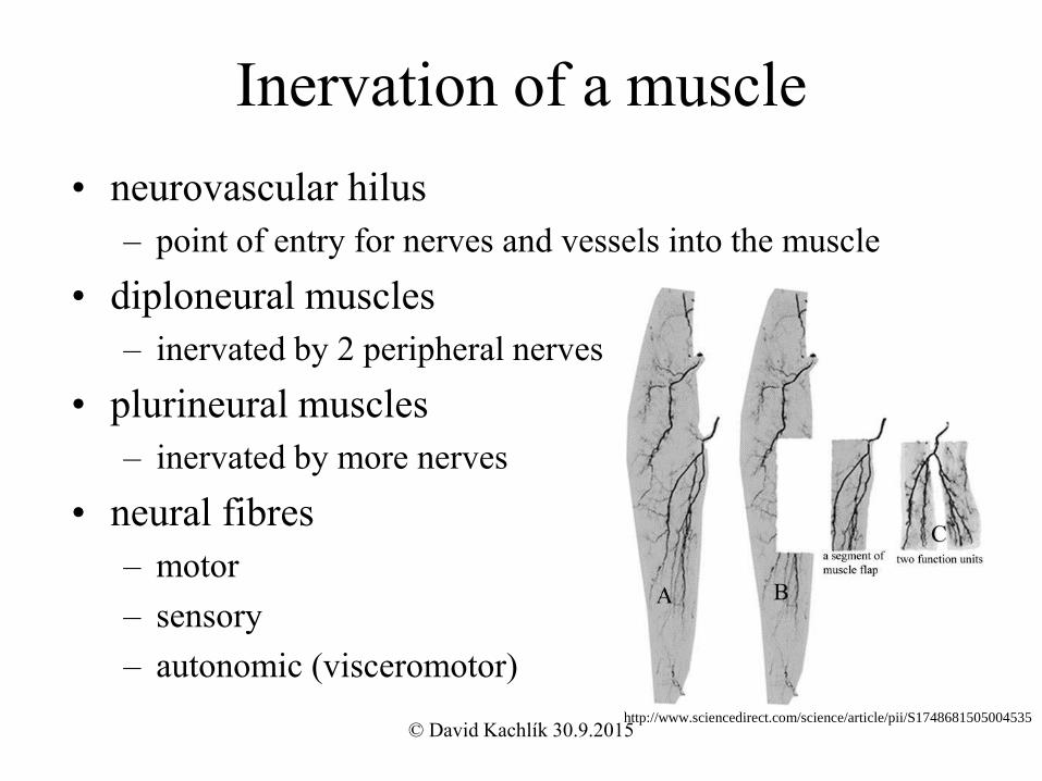
Inervation of a muscle
• neurovascular hilus
– point of entry for nerves and vessels into the muscle
• diploneural muscles
– inervated by 2 peripheral nerves
• plurineural muscles
– inervated by more nerves
• neural fibres
– motor
– sensory
– autonomic (visceromotor)
http://www.sciencedirect.com/science/article/pii/S1748681505004535© David Kachlík 30.9.2015

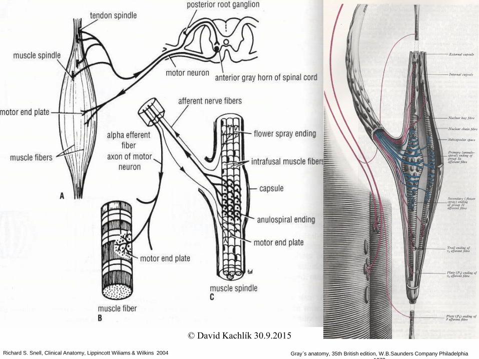
Motor inervation
• somatomotor fibres are axons of motoneurons of
anterior spinal horns (α-motoneurons)
• neuromuscular synapse (synapsis neuromuscularis)
– termination of an axon on the surface of myofibre
• motor unit
– group of myofibres inervated by 1 motoneuron
– simple, rough movements – big motor unit (150 myofibres)
– delicate, precise movements – small motor unit (8–15
myofibres)
© David Kachlík 30.9.2015
Richard S. Snell, Clinical Anatomy, Lippincott Wiliams & Wilkins 2004 Gray´s anatomy, 35th British edition, W.B.Saunders Company Philadelphia
1973
© David Kachlík 30.9.2015

Muscle spindle
Fusus neuromuscularis
= intrafusal muscle fibres
• specialized organ of perception in the muscle =
proprioception
• consists of several myofibres
• surrounded by fibrous capsule
• inervated by fibres of γ-motoneurons
– informations about length change to the spinal cord
• detects also difference of grade of contraction of
extra– and intrafusal fibres
© David Kachlík 30.9.2015

doc. MUDr. Josef Zámečník, Ústav patologie a molekulární medicíny 2.LF UK a FN v Motole
Muscle spindle – transverse section
© David Kachlík 30.9.2015

doc. MUDr. Josef Zámečník, Ústav patologie a molekulární medicíny 2.LF UK a FN v Motole
Muscle spindle – transverse section
© David Kachlík 30.9.2015

Fig. 14-5. Neuromuscular spindle.
Cross section. Juxta-equatorial region.
Front leg muscle. Newborn rat. E.M.
x1900. 1. Extrafusal skeletal muscle
fibers. 2. Lumen of capillary. 3. Outer
capsule. 4. Periaxial space. 5. Inner
capsule. 6. Myelinated nerve fibers. 7.
Nuclear chain muscle. 8. Nuclear bag
muscle fibers.
J.A.G. Rhodin: Histology. A text and atlas. 1974
Fig. 14-4. Neuromuscular spindle.
Cross section. Equatorial region. Triceps
surae muscle. Rat embryo, 2 days before
birth. E.M. x1200. (From: J.H. Levy, 1972)
1. Extrafusal skeletal muscle fibers. 2.
Lumen of blood capillary. 3. Outer
capsule. 4. Periaxial space. 5. Inner
capsule. 6. Myelinated nerve fibers. 7.
Intrafusal nuclear chain muscle fibers. 8.
Nuclear bag muscle fibers. 9. Fibroblasts.
10. Loose connective tissue.
© David Kachlík 30.9.2015

J.A.G. Rhodin: Histology. A text and atlas. 1974
Fig. 14-6. Neuromuscular spindle.
Cross section. Juxta-equatorial region.
Enlargement of rectangle in Fig. 14-5.
E.M. x4500. 1. Part of extrafusal muscle
fiber. 2. Lumen of capillary. 3. Sheet-like
cells of outer capsule. 4. Myelinated nerve
fibers. 5. Periaxial space. 6. Inner
capsule. 7. Nucleus of fibroblast. 8.
Nucleus of Schwann cell. 9. Nucleus of
nuclear bag fiber. 10. Polar ends of
nuclear chain fibers. 11. Satellite muscle
cell. 12. Sensory nerve endings.
Fig. 14-7. Sensory nerve ending.
Neuromuscular spindle. Enlargement of
rectangle in Fig. 14-6. E.M. x60,000. 1.
Myofilaments of intrafusal nuclear chain
muscle fiber. 2. Sensory nerve ending. 3.
Mitochondria. 4. Membranous contact
between sarcolemma and cell membrane of
nerve ending. 5. Zonula adherens junction.
6. External (basal) lamina.
© David Kachlík 30.9.2015

J.A.G. Rhodin: Histology. A text and atlas. 1974
Fig. 14-18. Motor nerve
endings. Skeletal muscle.
Gold chloride method. Human.
L.M. x200. 1. Striated muscle
fibers. 2. Bundle of myelinated
nerve fibers. 3. Motor end
plates.
Fig. 14-19. Motor end plates.
Gold chloride method. Skeletal
muscle. Human. L.M. x600. 1.
Efferent nerve fibers. 2. Motor
end plate in surface view. 3.
Motor end plate in side view. 4.
Striated skeletal muscle fibers.
Fig. 14-20. Motor end plate. Skeletal muscle.
Enlargement of area approximately similar to
rectangle in Fig. 14-19. Rat. E.M. x30,000. 1.
Efferent nerve process (axon). 2. Myelin sheath.
3. Successive laminae of the myelin sheath
terminate as cytoplasmic swellings. 4.
Cytoplasm of Schwann cell (neurolemma). 5.
Endoneurial connective tissue. 6. Cytoplasm of
teloglial cell (Schwann cell). 7. Axon terminal
end (end knob). 8. Synaptic trough (recess). 9.
Mitochondria. 10. Subneural, synaptic clefts. 11.
Nucleus of skeletal muscle cell. 12. Myofibrills.
13. External (basal) lamina.
© David Kachlík 30.9.2015

J.A.G. Rhodin: Histology. A text and atlas. 1974
Fig. 14-21. Detail of motor end plate.
Skeletal muscle. Rat. E.M. x60,000. 1.
Axon terminal. 2. Cytoplasm of muscle
cell (sole plate). 3. Mitochondria. 4. Flat
synaptic vesicles. 5. Round synaptic
vesicles. 6. Presynaptic membrane. 7.
Synaptic cleft with glycoprotein material.
8. Postsynaptic, junctional folds. 9.
Subneural primary and secondary clefts.
10. Postsynaptic sarcolemma. 11.
Synaptic vesicles fusing with presynaptic
membrane. 12. Microtubules. 13.
Ribosomes.
Figs. 14-22. and 14-23. Detail of
synaptic junctions of motor end
plate. Skeletal muscle. Rat. E.M.
x124,000. 1. Presynaptic cell membrane
of axon terminal. 2. Synaptic cleft. 3.
Postsynaptic sarcolemma. 4. Round
(excitatory?) electron-lucent, synaptic
vesicles, bounded by a delicate,
trilaminar membrane. 5. Flat
(inhibitory?) synaptic vesicles.
© David Kachlík 30.9.2015
Vascular supply of a muscle
• entry and exit of the vessels in muscle
neurovascular hilus
• flowing blood volume depends on the
activity of the muscle
• poor supply for the tendons
– blood and lymph vessels of their own
– branches of the vessels for the muscle,
periosteum, tendons
© David Kachlík 30.9.2015

Growth and regeneration of a muscle
• lengthwise growth
– growth of length of myofibres at their ends
• widthwise growth
– myofibres are getting thicker
• from birth the amount of myofibres is
unchanged
• cells are not regularly renewed
regeneration practically can´t occur© David Kachlík 30.9.2015

Special muscle structures I
• fascia (= perimysium externum)
– fibrous envelope of muscle or muscle group
– barrier for spreading of inflammation in that
specific area
• osteofascial septum (= septum
osteofasciale)
– fascial divider from the superficial fascia to
the periosteum
– separates the space for muscle groups –
compartment (compartimentum)
https://www2.aofoundation.org/wps/portal/!ut/p/c0/
© David Kachlík 30.9.2015

Compartment syndrome
• in the first hours after injury of the muscle or fractures of
the long bones of the thigh, leg, arm or forearm
• increase of pressure in the fascial space (intramuscular
bleeding, oedema) → compression of the vessels →
ischemia with possible necrosis
• treatment is mostly surgical – fasciotomy or
dermatofasciotomy
– fascial incision (together with skin) for decreasing the pressure in the fascial
space
• Volkmann´s contracture
– flectional contracture of the hand and fingers as a result of postischemic
fibrous degeneration of muscle bellies of the flexors, sometimes also
extensors
© David Kachlík 30.9.2015
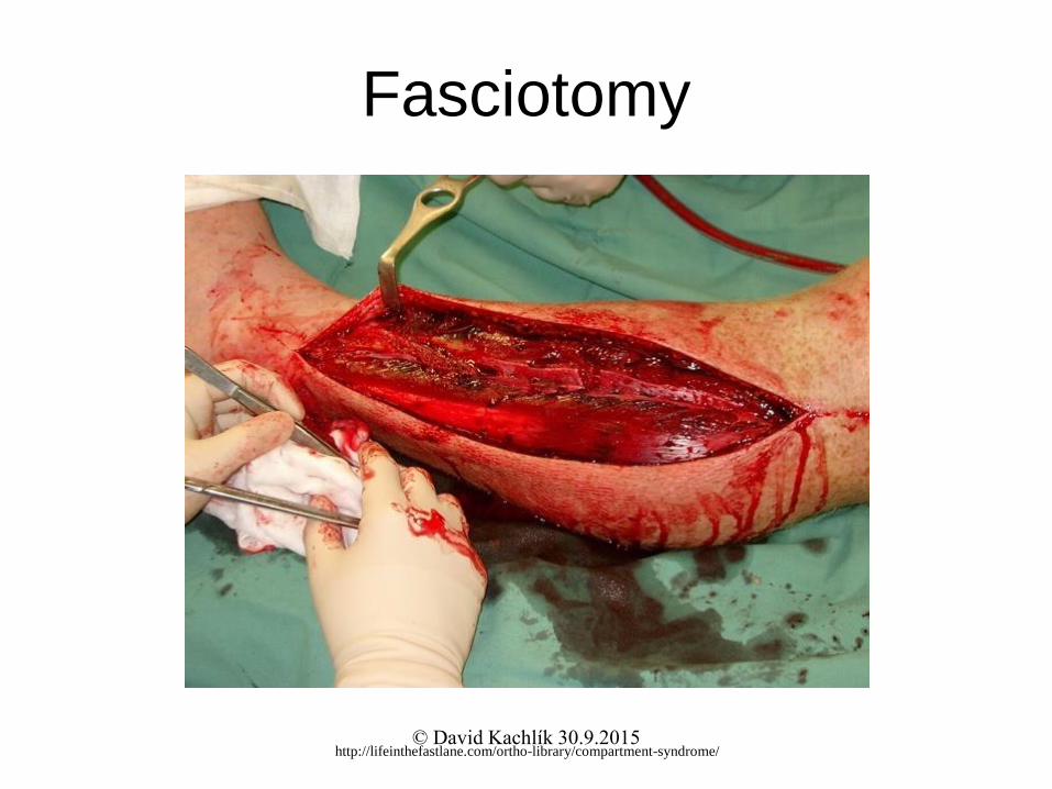
Fasciotomy
http://lifeinthefastlane.com/ortho-library/compartment-syndrome/© David Kachlík 30.9.2015

Special muscle structures II• tendon (tendo)
– strip of tough fibrous connective tissue composed of bundles of collagenous
fibrils
– connects the muscle to the bone
– peritenonium internum (covers the bundles)
– peritenonium externum (consistent envelope on the surface of the tendon)
• aponeurosis (aponeurosis)
– flat tendon
– mutually crossing bundles in layers
• tendinous sheath (vagina tendinum)
– space along the tendon lined by synovial membrane
– vagina fibrosa: surrounds the vagina synovialis, holds the tendons to the bone
– vagina synovialis
• epitenonium: inner layer (covers the tendon)
• peritenonium: outer layer
• mesotenonium: mutual switching of both previous things
http://www.zoology.ubc.ca/~biomania/tutorial/bonejt/jt01ac03.htm
© David Kachlík 30.9.2015
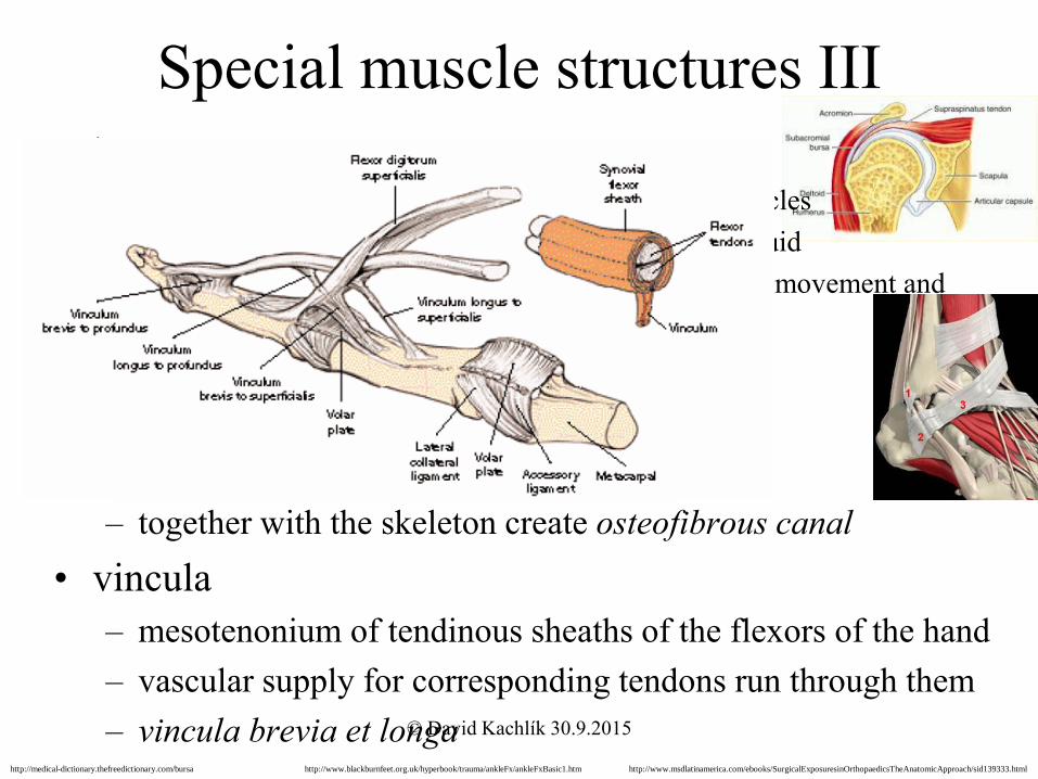
Special muscle structures III
• bursae mucosae– pouches in the vincinity of the joints, tendons and muscles
– lined by synovial membrane, filled up with synovial fluid
– reduction of rubbing in places exposed to considerable movement and
pressure
• retinacula
– strengthened stripes of the superficial fascia
– tie the inserting tendons to the bone
– together with the skeleton create osteofibrous canal
• vincula
– mesotenonium of tendinous sheaths of the flexors of the hand
– vascular supply for corresponding tendons run through them
– vincula brevia et longahttp://www.msdlatinamerica.com/ebooks/SurgicalExposuresinOrthopaedicsTheAnatomicApproach/sid139333.htmlhttp://www.blackburnfeet.org.uk/hyperbook/trauma/ankleFx/ankleFxBasic1.htmhttp://medical-dictionary.thefreedictionary.com/bursa
© David Kachlík 30.9.2015

Development – muscles of the trunk
• from dermatomyotoms (part of the somites)
– epaxial unit
• maintains the segmental arrangement
• inervation: posterior rami of the spinal nerves
• deep muscles of the back
– hypaxial unit
• merges into larger units
• inervation: anterior rami of the spinal nerves
• lateral and anterior muscles of the body wall
– border: fascia thoracolumbalis – lamina media© David Kachlík 30.9.2015

Myoseptum horizontale
http://en.wikipedia.org/wiki/Epaxial_and_hypaxial_muscles
http://www.sharksinfo.com/lateral-line.html
© David Kachlík 30.9.2015

Development – muscles of the limbs
• migration of hypaxial myogenic cells from
ventrolateral margin of the dermatomyotom to the
limb bud
• secondary extension of the beginnings to the trunk
– spinohumeral muscles
– thoracohumeral muscles
• inervation: anterior rami of the spinal nerves
– form the plexuses
• plexus brachialis (brachial plexus) – C5-8, T1
• plexus lumbalis (lumbar plexus) – T12, L1-4
• plexus sacralis (sacral plexus) – L4-5, S1-3
© David Kachlík 30.9.2015

EMG (electromyography)
• detection of the superficial muscle or the intramuscular
activity
• detects the change of electrical potential
• diagnostics for various muscle and neural malfunctions
http://biomech.ftvs.cuni.cz/pbpk/kompendium/biomechanika/experiment_metody_emg.php
http://www.fsps.muni.cz/inovace-SEBS-ASEBS/elearning/biomechanika/vyzkumne-metody-v-biomechanice© David Kachlík 30.9.2015

Functional muscle test
• informs us about the muscle strength
• helps to assess the extent and location of the impairment
• analysis and examination of performance for the whole
movement
• assessment – 6 grades
• 0 – no sign of contraction
• 1 – twich (not enough to do the move)
• 2 – very weak (movement in the whole extent, doesn´t overcome the
resistance of the tested part of the body)
• 3 – weak (overcomes the gravity)
• 4 – good (overcomes medium-sized outer resistance)
• 5 – normal (very good function)
© David Kachlík 30.9.2015

Rigor mortis
• is state of muscle rigidity, that begins 3-4 hours after death and lasts approx. 24 hours
• after death Ca2+ ions leave the sarcoplasmatic reticulum and enable the myosin heads to bond to the actin fibres
• since the synthesis of ATP has ceased, actin bridges don´t separate until the time when proteolytic enzymes start to eliminate the cells
http://tincconsconwa.blogspot.cz/2010/01/rigor-mortis.html© David Kachlík 30.9.2015
• skeletal muscle tissue starts to be replaced
by fibrous and fatty tissue around the age of
30
• reflexes slowdown, loss of flexibility and
decrease of strength
• change of muscle fibres from quick to slow
Growing old and musle tissue
© David Kachlík 30.9.2015

Enthesopathy
• illness of muscle and tendinous insertions
• usually caused by repeated overstraining
• e.g. tennis elbow
http://www.fyzioterapie-stepankavojtova.cz/bolestivyloket.htmlhttp://compex.zdravi-cz.eu/tenisovy-loket.php
© David Kachlík 30.9.2015