the muscular system approximately 40% of your body weight is your muscle. 1. functions origin...
TRANSCRIPT

The Muscular SystemThe Muscular SystemApproximately 40% of your body weight is your muscle.
1. Functions
Muscles produce movement. When muscle contracts , it pulls insertion bone near originorigin bone. Movement occurs at joint between origin and insertioninsertion.
OriginOrigin – The bone that moves less, provides the area of attachment for the end of the muscle called the origin.

InsertionInsertion – the movable bone provides the
surface for the muscle’s insertion.
e.g. biceps : origin at the joint of humerus and
scapula, insert on radius.
triceps : origin at humerus, scapula and
clavicle, inserts on ulna.a. Biceps and triceps work in opposing pairs in an
antagonistic system.
b. Groups of muscles usually contract to produce a single movement – synergistic pattern.
e.g. extension of lower legs is by rectus
femoris, gracilis and sartorius.

Reference: http://www.ultranet.com/~jkimball/BiologyPages/M/Muscles.html#Anatomy_of_Skeletal_Muscle
http://www.lrn.org/Content/Lessons/muscle.html#overview
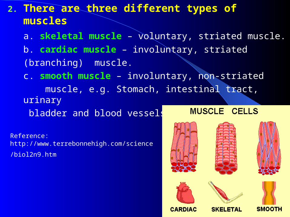
2. There are three different types of muscles
a. skeletal muscle – voluntary, striated muscle.
b. cardiac muscle – involuntary, striated
(branching) muscle.
c. smooth muscle – involuntary, non-striated
muscle, e.g. Stomach, intestinal tract, urinary
bladder and blood vessels.
Reference:
http://www.terrebonnehigh.com/science/biol2n9.htm

Skeletal Muscle ContractionSkeletal Muscle ContractionSkeletal muscles contain thousands of muscle fibers (muscle cells). Each fiber consists of finer threadlike structures called myofibrils. Myofibrils contain two kinds of protein strands: thick filament, myosin, with side projecting cross-bridge. Thinner filament, actin. Repeating bands of actin and myosin translate into light – dark repeating unit that gives skeletal muscle its striped appearance.Dark line (Z) line between each repeating unit is defined as sacromere that is the fundamental unit of muscle contraction.

References:
http://www.uoguelph.ca/zoology/devobio/210labs/sketchmuscle1.html http://www.lrn.org/Content/Lessons/muscle.html#overview

Reference:
http://members.shaw.ca/bodybuilding/Muscles/structure.html
Muscle StructureMuscle Structure

Reference: http://www.ultranet.com/~jkimball/BiologyPages/M/Muscles.html#Anatomy_of_Skeletal_Muscle
http://www.lrn.org/Content/Lessons/muscle.html#overview

The Sliding-Filament ModelThe Sliding-Filament Model
Reference: http://www.ultranet.com/~jkimball/BiologyPages/M/Muscles.html#Anatomy_of_Skeletal_Muscle

Role of Calcium in Muscle Excitation and Contraction Coupling
http://cwx.prenhall.com/bookbind/pubbooks/martinidemo/chapter10/medialib/CH10/html/ch10_4_1.html
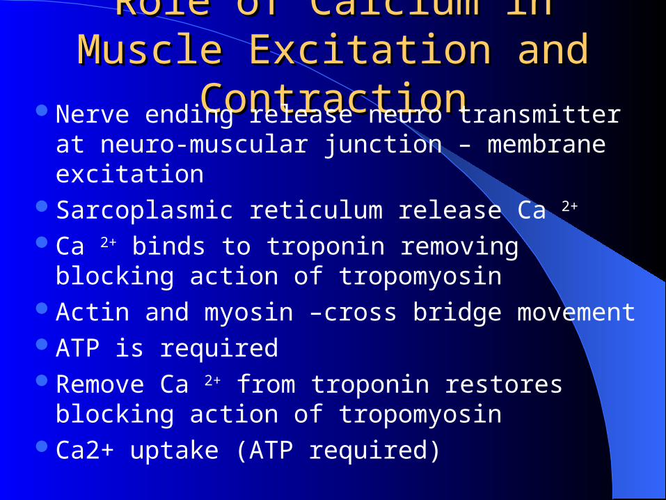
Role of Calcium in Muscle Role of Calcium in Muscle Excitation and ContractionExcitation and Contraction
Nerve ending release neuro transmitter at neuro-muscular junction – membrane excitation
Sarcoplasmic reticulum release Ca 2+
Ca 2+ binds to troponin removing blocking action of tropomyosin
Actin and myosin –cross bridge movementATP is requiredRemove Ca 2+ from troponin restores blocking
action of tropomyosinCa2+ uptake (ATP required)

Muscle DisordersMuscle Disorders1. Muscular dystrophy – progressive weaken of
the muscles.
2. Paralysis – loss of ability to produce voluntary movement. This is due to disease or injury of brain or spinal cord or nerve
3. Muscle atrophy – muscle shrinkage. Decrease in muscle size.
4. Muscle hypertrophy – increase in muscle size because of over work. e.g. heart frequently hypertrophy from over work.

Muscle StructureMuscle StructureFront ViewFront View Back ViewBack View
Reference: http://www.rrcc.cccoes.edu/academic/health/fitnesscenter/muscle.htm