the mode of action of sulfanilamide on streptococcus…
TRANSCRIPT

THE MODE OF ACTION OF SULFANILAMIDE ON STREPTOCOCCUS. II*
BY FREDERICK P. GAY, M.D., ADA R. CLARK, PH.D., JULIA A. STREET, AND DOROTHY W. MILES
(From the Department of Bacteriology, College of Physician, and Surgeon~, Columbia University, New York)
(Received for publication, January 14, 1939)
It is not surprising, in view of the truly amazing therapeutic results that may follow the use of sulfanilamide and related compounds in streptococcus and other infections, that considerable effort has been devoted to explaining the precise method by which this drug produces its results. The quest is not alone for the purpose of perfecting the drugs employed and their administration, but because in addition such investigations offer a new means of approach to the mechanism of action of chemotherapeutic drugs in general, concerning which we have had far too little information.
A bewildering series of facts are now available that bear on the mode of action of sulfanilamide on microorganisms, both in the test tube and in the animal body. The difficulty in interpreting these facts has been due to over-emphasis on the particular findings of individual investigators on the study of a single factor. Thus there are those who would explain sulfanilamide action as a direct effect upon the microorganism itself, whether acting in vivo or in vitro. In other words, there are those who would still consider the animal body as a test tube, much as Pasteur explained immunity in the early days of the development of ordered knowledge in this field. There are others, however, who from the beginning have taken cognizance of the importance of the cells of the body in the therapeutic results obtained by sulfanilamide.
The Direct Action of Sulfanilamide on Streptococcus
The effect of sulfanilamide on bacteria cannot be considered from the viewpoint of the action of this substance alone, but in the light of
* Aided by a grant from the Dr. Philip Hanson Hiss, Jr., Memorial Fund. 607
Dow
nloaded from http://rupress.org/jem
/article-pdf/69/5/607/1181565/607.pdf by guest on 06 February 2022

608 ACTION OF SULFANILAMIDE ON STREPTOCOCCUS. 11
all other environmental factors that enter into the experiment. In other words, sulfanilamide action depends on the balance of a series of factors that enter into the composition of the medium in which the two interacting substances, sulfanilamide and streptococcus, are tested. We are for the moment primarily considering the substances that enter into the reaction under the artificial conditions as observed in the test tube, leaving for later consideration the additional factors that intervene in the animal body.
Thus we find on analysis from our own experiments and from those of others, that the growth of streptococcus is influenced in the test tube
TABLE I
Factors That Influence the Growth of Streptococci Favoring
Good nutrient broth (Infusion broth with best peptone at right pH (7.5))
Glucose Peptone
Horse serum
Rabbit serum (Gay and Clark) Defibrinated rabbit blood (Gay and Clark) Defibrinated infant blood (Lyons and
Mangiaracine) Defibrinated horse blood (White and
Parker) Necrotic tissue (peptone ?) (Lockwood,
1938 a)
Inhibi t ing
Poor nutrient broth (Meat extract broth; peptone free broth; pH under 6.5 or over 8)
Glucose peptone water (Nitti a d.) Washing streptococci (thus removing pep-
tone) (Lockwood, 1938 b) Saline suspension of streptococci (Mellon
a d.) 40°C. (White and Parker) Human serum (Colebrook et al.) Deleucocyted human blood (Colebrook
et al., Finklestone-Sayliss et al. ) Whole defibrinated human blood (Cole-
brook et al., Lockwood, 1938 b) SULFANILAMIDE PtL~GOCYTES
by two sets of factors: those that favor the growth of the microorgan- ism, and those that inhibit it. It would be impossible to enumerate these growth factors in the precise order of their excellence in favoring or in inhibiting the growth of streptococcus, but they are listed in Table I, more or less in order of their significance.
When tested in any of the favoring media, as listed above, sulfanil- amide by general agreement produces bacteriostasis only. Further- more, the degree of bacteriostasis depends inversely on the sum total of adjuvant growth factors present; thus in a series of comparisons the ratio of growth in the presence of sulfanilamide broth, as compared with plain broth, was as 1-32,500 in 24 hours. The ratio was only
Dow
nloaded from http://rupress.org/jem
/article-pdf/69/5/607/1181565/607.pdf by guest on 06 February 2022
F. P. GAY, A. R. CLARK, .]'. A. STREET, D. W. MILES 609
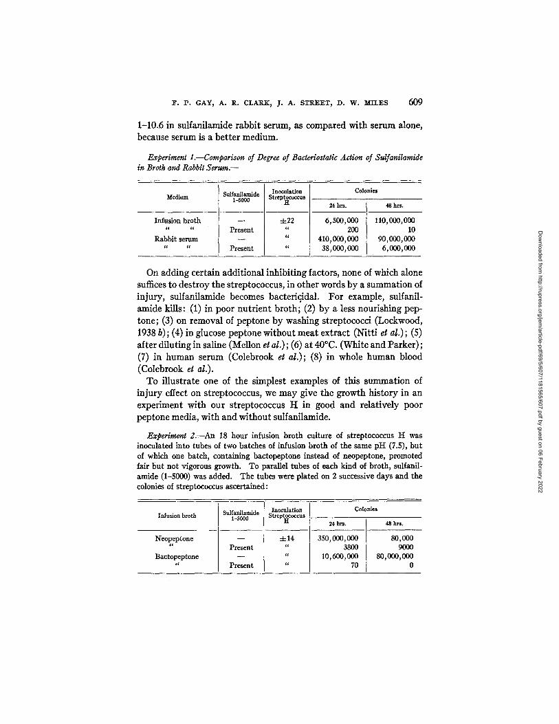
1-10.6 in sulfanilamide rabbit serum, as compared with serum alone, because serum is a better medium.
Experiment 1.--Comparison of Degree of Bacteriostatic Action of Sulfanilamide in Broth and Rabbit Serum.--
Medium
Infusion broth c~ c¢
Rabbit serum ~¢ cc
Sulfanilamide 1-5000
Present
Present
Inoculation StreP~iCocCUS
-4-22 cl
Colonies
24 h r s .
6,500,000 200
410,000,000 38,000,000
48 hrs.
110,000,000 10
90,000,000 6,000,000
On adding Certain additional inhibiting factors, none of which alone suffices to destroy the streptococcus, in other words by a summation of injury, sulfanilamide becomes bactericidal. For example, sulfanil- amide kills: (1) in poor nutrient broth; (2) by a less nourishing pep- tone; (3) on removal of peptone by washing streptococci (Lockwood, 1938 b); (4) in glucose peptone without meat extract (Nitti et at.); (5) after diluting in saline (Mellon et al.); (6) at 40°C. (White and Parker); (7) in human serum (Colebrook et al.); (8) in whole human blood (Colebrook e~ al.).
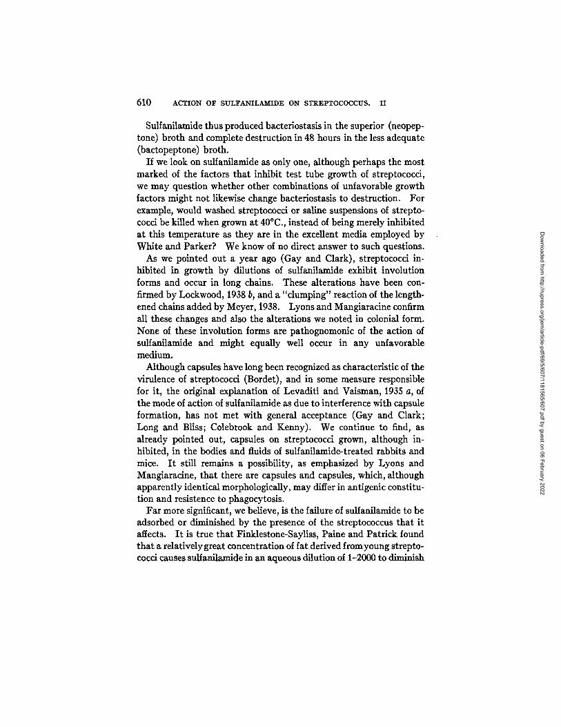
To illustrate one of the simplest examples of this summation of injury effect on streptococcus, we may give the growth history in an experiment with our streptococcus H in good and relatively poor peptone media, with and without sulfanilamide.
Experiment 2.--An 18 hour infusion broth culture of streptococcus H was inoculated into tubes of two batches of infusion broth of the same pH (7.5), but of which one batch, containing bactopeptone instead of neopeptone, promoted fair but not vigorous growth. To parallel tubes of each kind of broth, sulfanil- amide (1-5000) was added. The tubes were plated on 2 successive days and the colonies of streptococcus ascertained:
Infusion broth
Neopeptone c c
Bactopeptone
Sulfanilamide Inoculation 1-5000 Streph°c°ccus
- - :t:14 Present "
~c
Present "
Colonies
24 hrs. 48 hrs.
350,000,000 80,000 3800 9000
10,600,000 80,000,000 70 0
Dow
nloaded from http://rupress.org/jem
/article-pdf/69/5/607/1181565/607.pdf by guest on 06 February 2022
610 ACTION OF SULFANILAMIDE ON STREPTOCOCCUS. II
Sulfanilamide thus produced bactefiostasis in the superior (neopep- tone) broth and complete destruction in 48 hours in the less adequate (bactopeptone) broth.
If we look on sulfanilamide as only one, although perhaps the most marked of the factors that inhibit test tube growth of streptococci, we may question whether other combinations of unfavorable growth factors might not likewise change bacteriostasls to destruction. For example, would washed streptococci or saline suspensions of strepto- cocci be killed when grown at 40°C., instead of being merely inhibited at this temperature as they are in the excellent media employed by White and Parker? We know of no direct answer to such questions.
As we pointed out a year ago (Gay and Clark), streptococci in- hibited in growth by dilutions of sulfanilamide exhibit involution forms and occur in long chains. These alterations have been con- firmed by Lockwood, 1938 b, and a "dumping" reaction of the length- ened chains added by Meyer, 1938. Lyons and Mangiaracine confirm all these changes and also the alterations we noted in colonial form. None of these involution forms are pathognomonic of the action of sulfanilamide and might equally well occur in any unfavorable medium.
Although capsules have long been recognized as characteristic of the virulence of streptococci (Border), and in some measure responsible for it, the original explanation of Levaditi and Vaisman, 1935 a, of the mode of action of sulfanilamide as due to interference with capsule formation, has not met with general acceptance (Gay and Clark; Long and Bliss; Colebrook and Kenny). We continue to find, as already pointed out, capsules on streptococci grown, although in- hibited, in the bodies and fluids of sulfanilamide-treated rabbits and mice. It still remains a possibility, as emphasized by Lyons and Mangiaracine, that there are capsules and capsules, which, although apparently identical morphologically, may differ in antigenic constitu- tion and resistence to phagocytosis.
Far more significant, we believe, is the failure of sulfanilamide to be adsorbed or diminished by the presence of the streptococcus that it affects. I t is true that Finklestone-Sayliss, Paine and Patrick found that a relatively great concentration of fat derived fromyoung strepto- cocci causes sulfanilamide in an aqueous dilution of 1-2000 to diminish
Dow
nloaded from http://rupress.org/jem
/article-pdf/69/5/607/1181565/607.pdf by guest on 06 February 2022
F. P. GAY, A. R. CLARK, J. A. STREET, D. W. MILES 611
in the watery phase by nearly one-half in 2 hours. This may show that sulfanilamide is relatively more soluble in fat than in water, but under the conditions specified does not, we believe, show that sulfanil- amide in its operation on streptococcus is adsorbed by the fat in the microorganism. Biirgers, 1937, states that incubation of strepto- coccus with prontosil soluble renders the organisms more rapidly permeable by crystal violet, as shown by the rapidity of staining.
I t has apparently been proved by Yorke et al. and Pedlow and Reiner that arsenical compounds and acriflavine, both of which are highly destructive for trypanosomes both in test tube and body, are rapidly fixed on these cells whether they be alive or dead. Strangely enough we have been unable to find any extensive proof that bacterici- dal substances like optochin, gentian violet and acriflavine are actually fixed on the microorganisms they affect, and diminished thereby in the supernatant fluid. One of us (Gay and Beckwith) began experiments of this sort some years ago, and they have now been extended in con- nection with the present investigation as a check on the mode of action of sulfanilamide.
In brief we find: 10 cc. of gentian violet (1-10,000) or the same volume of acriflavine (1-10,000) is reduced one-half in color index on addition of 1 cc. of a concentrated (× 100) culture of streptococcus, in from 1 to 3 hours. Correspondingly, the bactericidal titer is also reduced one-half. In the presence of rabbit serum (50 per cent) acriflavine is more active than in broth; it is impossible to measure the color reduction in the presence of serum but the bactericidal titer is reduced one-half, as in broth.
Turning now to contact between sulfanilamide and streptococci (100 cc. of an 18 hour broth culture reduced by centrifugalization to 1 cc.), we have never been able to show that the drug, 10 cc. (1-10,000 to 1-100,000), was reduced in strength in from 2 to 5 hours at room temperature. The Marshall colorimetric test for sulfanilamide was employed. Every variation in the conditions of this test that occurred to us was tried: growth of the streptococcus in 20 per cent serum broth with subsequent addition to sulfanilamide broth; growth of the streptococcus in broth with sulfanilamide for 18 hours, or in serum sulfanilamide broth for 10 hours.
Biirgers' contention was based on indefinite staining results. We
Dow
nloaded from http://rupress.org/jem
/article-pdf/69/5/607/1181565/607.pdf by guest on 06 February 2022

612 ACTION OF SULFANILAMIDE ON STREPTOCOCCUS. II
have grown streptococci in broth or in sulfanilamide broth 1-10,000 for 18 hours and found that the sulfanilamide broth-grown organisms, in aliquot numbers, adsorbed as much but no more gentian violet than the plain broth-grown organisms. Observationally there was no difference in the rapidity or intensity of staining of the organisms in the two preparations. Furthermore, neither sulfanilamide-grown organisms nor organisms in the presence of sulfanilamide were found to be more susceptible to the action of gentian violet and acriflavine than normal organisms.
The E~ect of Sulfanilamide on the Virulence Factors of Streptococcus
Apart from growth inhibition and morphological changes wrought by sulfanilamide on the streptococcus, alteration and reduction in the virulence factors of streptococcus metabolism have been repeatedly claimed. Although the non-lethal effect of sulfanilamide on strepto- coccus may change temporarily the invasiveness of the organism it does not lead to any permanent impairment in its inherent virulence, as we pointed out and as has been confirmed by Lyons and Mangiara- cine. After 2 or 3 days growth in sulfanilamide broth, our strepto- coccus H (adapted to rabbits), when inoculated intrapleurally in rabbits (~.L.D. i 10 chains), or intraperitoneally in mice (~.L.D. 1000 chains), was just as virulent as the same organism grown in plain broth. In such experiments the scantier growth in sulfanilamide broth was adjusted to equal the number of organisms in the more luxuriant plain broth culture by dilution of the latter.
But the retention of potential Pathogenicity does not mean that one or more of the virulence factors may not be temporarily in- hibited in the presence of sulfanilamide. We have attempted to harmonize, for our own satisfaction, the somewhat discordant views that have been offered by numerous investigators on certain of the virulence factors, notably hemotoxin, leucocidin and fibrinolysin.
Hemotoxin Production.--It has been suggested as an hypothesis that the production of hemotoxin (hemolysin) may be inhibited when streptococci are in the presence of sulfanilamide or related compounds (]]Urgers, 1938; Meyer, 1938), and some experimental work has been presented by Osgood, by Huntington, and by King, Henschel and Green to support this hypothesis. As an alternative explanation,
Dow
nloaded from http://rupress.org/jem
/article-pdf/69/5/607/1181565/607.pdf by guest on 06 February 2022

F. P. GAY, A. R. CLARK, J. A. STREET, D. W. MILES 613
Levadi t i and Vaisman, 1935 b; Meyer , 1937; and Gross, Cooper and Lewis, 1938 a , believed tha t al though hemotoxin is formed i t m a y be inact iva ted by the drug. There is evidence against this al terna-
t i v e hypothesis, in the hands of Kemp; Gross, Cooper and Lewis, 1938 b; Hunt ing ton ; Osgood and Powell.
The following is a brief and general description of the methods used and the results obta ined in our invest igat ion of the effects of sulfanil- amide upon hemotoxin produced by Streptococcus kaemolyticus in vitro.
Organisms were grown either in broth and in broth plus sulfanilamide (con- centrations varying from 1-1000 to 1-10,000), or in rabbit serum and rabbit serum plus sulfanilamide (same concentrations) for periods of 27 to 72 hours. This incubation period permitted the sulfanilamide to exert its modifying effects upon the organisms, and one of these effects was, invariably, a bacteHostasis. Because of this bacteriostatic effect, a plate count of the number of organisms in the control and in the sulfanilamide tubes was made, so that in the experiments equal numbers of organisms could be obtained by dilution of the control. Plate counts in the nineteen experiments indicated that growth of a large inoculum (e.g. 0.2 cc. of an 18 hour culture) of Streptococcus kaemolyticus H, was always reduced in sulfanilamide media as compared with the control.
Organisms, then, which have been subjected for periods of 27 to 72 hours to the action of sulfanilamide were compared for hemotoxin product ion with their controls by means of the hemotoxin tes t de- scribed below:
Hemotoxin Test.--0.2 cc. (~ 1,000,000 streptococci), 0.1 cc. and 0.1 cc., of a 1-10 dilution, of the sulfanilamide-treated cultures were placed in separate Wasser- mann tubes. Amounts of untreated cultures diluted to correspond, in number of organisms, to the treated cultures were placed in other Wassermann tubes. To each tube 1 cc. of a 1 per cent suspension of washed rabbit red blood corpuscles was added, and the volume brought up to 2 cc. by addition of broth. The tubes were incubated at 37°C. and hemolysis noted at intervals by removing the tubes from the bath, centrifugalizing lightly to carry down the red cells, and comparing each tube in a comparator block with a series of standards. After each reading the tubes were shaken to resuspend the red cells, and immediately reincubated. The readings were continued until complete hemolysis was observed.
The results obta ined are as follows: In every case of exposure of the organisms to the drug (whether in bro th or in serum) the amoun t of hemolysis produced during the early hours of the tes t b y the
Dow
nloaded from http://rupress.org/jem
/article-pdf/69/5/607/1181565/607.pdf by guest on 06 February 2022

614 ACTION OF SULFANILAMIDE ON STREPTOCOCCUS. II
treated organisms was less than that produced by the controls. That this difference in hemolysis represents simply an inhibition in the ability to produce hemotoxin, or a delay only in its rate of production, is evident from the fact that in 15 of the 19 experiments at least one or more dilutions of culture of treated streptococci (6 to 7 hours) finally produced 100 per cent hemolysis of the red cells.
I t seemed possible that the results obtained to this point might be due to the fact that the control organisms were growing more rapidly during the course of the test than those organisms which had been previously exposed to the drug. To eliminate this possibility as a factor in the results, five experiments were performed in which (by means of frequent plate counts during the course of the test) a growth curve for the two cultures was obtained. The growth curves of the control and treated cultures were essentially parallel. In other words, the hemotoxin test was begun with equal numbers of untreated or sulfanilamide-treated organisms and as the test proceeded, both series grew out at the same rate. A given number of the untreated streptococci produced hemolysis more rapidly than an equal number of sulfanilamide-treated streptococci.
In view of these results, the conclusion was reached that the differ- ence in rate and amount of hemolysis between untreated controls and those exposed to the drug for 27 to 72 hours represents a definite though temporary alteration in the ability of the treated organisms to produce hemotoxin.
Experiments were also carried out to determine whether there was a decrease in the hemotoxin content of young streptococcus cultures in the presence of sulfanilamide.
Broth cultures were used in which the organisms were exposed to the action of the drug (concentrations of 1-1000 to 1-10,000) for periods varying from 3 to 15 hours. In some cases the media were enriched by the addition of 2.5 per cent rabbit serum, in order to facilitate the production of hemotoxin. The cultures were centrifugalized and the supernatant fluids placed in contact with rabbit red cells for 30 or 60 minutes. Varying dilutions of superuatant fluids of both sulfanil- amide broth and plain broth cultures were used, so that allowance might be made for the difference in the number of organisms in the two cultures.
Out of a series of tests, there was no indication that the sulfanilamide supernatant fluids contained less hemotoxin than the corresponding
Dow
nloaded from http://rupress.org/jem
/article-pdf/69/5/607/1181565/607.pdf by guest on 06 February 2022

F. P. GAY, A. R. CLARK, J. A. STREET~ D. W. MILES 615
control supernatant fluids. The results of these experiments indicate that culturing streptococci in the presence of 1-1000 to 1-10,000 sulfanilamide for periods of 3 to 15 hours fails to alter the amount of hemotoxin produced by such treated cultures, and that the presence of the drug during the production of the toxin does not serve to neu- tralize it.
To determine whether sulfanilamide has any "neutralizing" action on hemotoxin already formed in vitro by untreated streptococci, the following experiments were carried out. Both Berkefeld filtrates and centrifugalized supernatant fluid of a 6 hour broth culture were prepared. To varying amounts (0.4 cc. to 0.9 cc.) of each, sulfanil- amide in concentrations from 1-100 to 1-2000 was added. Appro- priate controls without sulfanilamide were prepared. After one-half hour 1 cc. of a 1 per cent rabbit blood cell suspension was added and the time necessary for complete hemolysis noted. The results indi- cated that sulfanilamide in concentrations varying from 1-1000 to 1-20,000 does not inhibit the hemolysis of a 1 per cent red cell sus- pension. In other words, the drug does not neutralize or inactivate preformed hemotoxin present in the filtrate or supernatant of an untreated streptococcus culture.
Another type of experiment was conducted in an effort to check the results of early experiments of Osgood, who reported evidence of neutralization of hemotoxin by sulfanilamide as shown in blood agar plates. 1 cc. of a 1-5,000,000 dilution of an 18 hour streptococcus culture was added to blood agar containing sulfanil- amide in concentrations varying from 1-5000 to 1-100,000, and pour plates made. Corresponding plates without drug were prepared. One series of plates was incubated aerobically and another anaerobically. The number of colonies on each plate was counted and the average diameter of the zone of hemolysis was determined by measuring 30 unselected colonies on each plate. The colony count showed that the sulfanilamide was not bacteriostatic under these experi- mental conditions. Measurements of the hemolytic zones indicated no decrease in the amount of hemolysis present in the sulfanilamide plates in either the aerobic or anaerobic series for any concentration of sulfanilamide used. From these results it was concluded that sulfanilamide in blood agar pour plates, incubated aerobically or anaerobically, does not neutralize the hemotoxin formed, or check the growth of streptococcus colonies previously unexposed to the drug.
The final conclusions from our experiments, then, would be that residence of streptococci in sulfanilamide broth or serum may inhibit,
Dow
nloaded from http://rupress.org/jem
/article-pdf/69/5/607/1181565/607.pdf by guest on 06 February 2022

616 ACTION O~ SULFANI~AKIDE ON BTI~PTOCOCCUS. II
although only temporarily, their ability to produce hemotoxin. However, when the unaffected streptococcus is grown in the presence of varying amounts of sulfanilamide for short periods of time, no diminution in the hemotoxin formed can be noted. No evidence was found that sulfanilamide neutralizes hemotoxin already formed. In short, the supposed action of sulfanilamide on streptococcus hemo- toxin can scarcely be used as explanatory of its therapeutic effect.
Leucocidin Production.--It has been suggested as an hypothesis without experimental proof that the therapeutic effect of sulfanilamide might be due to suppression of leucocidin formation, which property of streptococcus metabolism is known to be a factor in the virulence of this microorganism. The mere inhibition of growth might produce this result quantitatively (Mellon and Bambas; Bliss and Long). As corroborative evidence, leucocytes are relatively more normal in presence of sulfanilamide in vitro (Levaditi and Vaisman, 1935 b) or in marrow culture~ (Osgood) than when the drug is absent.
We have attacked this problem more directly in the following manner:
Streptococcus was grown in plain broth, as control, or in broth plus sulfanila- mide (from 1-1000 to 1-10#00). In ten different experiments, Berkefeld filtrates from such cultures or the supernatant fluids (at times heated to 56°C.) from centrifugalized cultures were used in varying amounts in order to equalize the difference in number of organisms in the two media. The Neisser-Wechsberg technique was employed (Gay and Oram).
In no instance was any difference in the leucocidin content, as measured by reduction of methylene blue, detected in the fluids of the sulfanilamide-grown cultures as compared with those of broth controls. Moreover, when direct observations were made on the appearance of the cells, in from 30 minutes to 2 hours, we could detect no difference in the degree of their injury in the two kinds of fltrates.
Fibrinolysin Production.--The species specific fibrinolysin formed by the hemolytic streptococcus is assuming an increasingly important significance in pathogenesis. Neither Huntington nor Kemp were able to demonstrate the inhibition of fibrinolysin in the presence of sulfanilamide experimentally.
Dow
nloaded from http://rupress.org/jem
/article-pdf/69/5/607/1181565/607.pdf by guest on 06 February 2022

F. P. GAY, A. R. CLARK, J'. A. STREET, D. W. MILES 617
In order to determine whether sulfanilamide interferes with the production or the activity of fibrinolysin, streptococcus was grown in broth plus sulfanilamide (1-10,000) or in broth without sulfanil- amide for 24, 48 or 72 hours. At these intervals the cultures were plated to determine the relative number of living organisms. The Berkefeld filtrates from these cultures, used in varying amounts to equalize the differences in number of organisms, were tested for fibrinolytic activity, by the technique of Tillett and Garner. In all cases the filtrates of sulfanilamide broth cultures lysed the human clots at the same rate as did the filtrates from the plain broth cultures.
The fibrinolytic activity of the broth suspensions of the centrifugal- ized organisms from the 48 hour sulfanilamide broth cultures was also compared with that of the suspensions of the broth-grown organ- isms, diluted in order to compare equivalent number of organisms. The degree of lysis was noted at intervals and as soon as lysis of a clot was complete the number of organisms was determined by plating the contents of the tube.
The intervals of time for complete lysis of the clots by the organisms from sulfanilamide broth cultures were always longer than required for the lysis by the organisms from the broth cultures. These results, then, would appear to indicate that fibrinolysin is produced more slowly by the sulfanilamide-grown organisms, but the difference may be easily accounted for by the smaller number of sulfanilamide organ- isms at the time of complete lysis. The number of sulfanilamide broth-grown organisms was only one-third to one-tenth the number of broth-grown organisms when lysis was complete. When the fibrinolytic test was carried out with the control broth-grown organ- isms suspended in broth plus sulfanilamide (1-10,000) they produced lysis as rapidly as those resuspended in plain broth and the number of living organisms at that time was approximately the same in the corresponding tubes.
Thus, there is no inhibition in fibrinolysin production which is not directly referable to a difference in number of viable organisms.
The Indirect Effect of Sulfanilamide on Streptococcus
Sulfanilamide and related compounds that produce bacteriostasis of streptococcus have been shown by one of us with Warren and
Dow
nloaded from http://rupress.org/jem
/article-pdf/69/5/607/1181565/607.pdf by guest on 06 February 2022

618 ACTION O~F SUL~?ANILAMIDE ON STREPTOCOCCUS. II
Stokinger, to increase the normal oxidation-reduction potential in the growth curve of the microorganism when grown aerobically. I t produces similar though less effect in broth alone. Other similar but non-bacteriostatic compounds (e.g., prontosil) have no such action. I t would seem that this change is in some way related to the effect of the drug on the streptococcus.
The suggestion of Main, Shinn and MeLlon, and Locke, Main and Mellon that sulfanilamide may act as an anticatalase, thereby allow- ing unhampered production of peroxide, with harmful effect on the streptococcus, may also be a factor of importance. This does not account, however, for the fact that repeated doses of sulfanilamide fail to convert bacteriostasis to a true lethal effect.
Turning to another aspect of the indirect action of sulfanilamide, there is, and has been since the beginning of studies on this drug, evidence of its action simply as an adjuvant to the natural defense mechanism of the body. From the earliest studies the probable significance of mobile cell intervention has been mentioned (Domagk, 1935; Levaditi and Vaisman, 1935 c; Long and Bliss; Btirgers, 1937), and in later publications this was verified by more direct observations of macrophage phagocytosis by Levaditi and by Domagk, 1937.
The reticulo-endothelial system would not seem directly concerned with the rapid results obtained in chemotherapy by sulfanilamide (Levaditi and Vaisman, 1935 c; Gross, Cooper and Peebles). In our previous publication we also found no histological change in the fixed tissues which would account for the macrophage accumulation that, in our minds, accounts for the second and ultimately destructive effect in vivo that follows bacteriostasis by the drug. Although pro- longed use of the drug does apparently lead to a reticulo-endothelial proliferation, in the hands of Davis et aL, this cannot, we believe, account for the immediate chemotherapeutic result. Sulfanilamide does not in itself attract leucocytes (Gay and Clark; Coman). There is no evidence that the microorganisms become drug-fast (Osgood). What the drug does do to the organisms is to render them temporarily impotent, and to reduce in slight degree their pathogenic properties. This inhibitory effect allows mobile cells, particularly the mononu- clear cells of the connective tissue, the opportunity to accumulate, to approach, and to phagocyte the disabled bacteria. This accumu-
Dow
nloaded from http://rupress.org/jem
/article-pdf/69/5/607/1181565/607.pdf by guest on 06 February 2022
IL P. GAY~ A. R. CLARK~ J. A. STREET, D. W. MILES 619
lation we have already demonstrated histologically in the pleural walls.
Our hypothesis as to the signal importance of host cells in sul- fanilamide cure has hitherto been based largely on these histological studies of the tissues surrounding an area in which cure under the influence of sulfanilamide was taking place. The work as reported seemed to show a direct relationship between the disappearance of the enfeebled streptococcus and local mononuclear cell mobilization. We turn now to evidence tending to show that phagocytosis is more active with sulfanilamide-treated cocci or in the body of sulfanilamide- treated animals. For this approach we find more corroboration by other writers. Finkelstein and Birkeland found that guinea pig blood leucocytes phagocyted in vitro more streptococci in the presence of sulfanilamide in certain concentrations. Biirgers, 1938; Osgood; and Mellon and Bambas, however, found no evidence of any such alleged opsonic action. We have used leucocytes both from rabbit and guinea pig exudates and completely failed to confirm the results of Finkelstein and Birkeland.
We have approached this question experimentally in three different ways:
1. Sterile exudates were obtained from the pleural cavities of rabbits which had been inoculated with an aleuronat-starch mixture 24 or 48 hours previously. Some of the rabbits had received subcutaneously 4 to 7 injections of 10 or 20 cc. each of sulfanilamide (2 per cent). The injections were given from 24 or 48 hours until 3 to 4 hours before removal of the exudate. The controls were un- treated. Streptococcus H (0.1 cc. or 0.2 cc. of an 18 hour broth culture) was added to the two kinds of pleural exudates (0.3 cc. to 0.8 cc.) and kept at 37°C., with constant agitation. Films were made at 1 and 2 hour intervals. At least 200 cells from each preparation were counted. In six experiments the amount of phagocytosis was practically the same in the exudates of the treated (average of phagocytic cells 4 per cent), as of the normal rabbits (average of phagocytic cells 3.5 per cent).
2. When immune serum was added to the exudates there was slightly more phagocytosis in the exudate of the sulfanilamide-treated rabbits (average phago- cytic cells 8.5 per cent) than in the normal exudate (average phagocytic ceils 6.5 per cent).
These experiments show clearly that no definite difference is observ- able in the in vitro phagocytosis by exudate cells from sulfanilamide-
Dow
nloaded from http://rupress.org/jem
/article-pdf/69/5/607/1181565/607.pdf by guest on 06 February 2022

620 ACTION O]~ SUL]~ANILAMIDE ON STREPTOCOCCUS. I I
treated rabbits, as compared with that of normal rabbits. The experiments, however, do not reproduce the actual conditions that obtain in therapeutic results in the body, in which latter the strepto- cocci have been subjected over a longer period of time to successive doses of sulfanilamide.
Lyons and Mangiaracine have found that adult or infant human leucocytes, which have no effect on normal streptococci, do phagocyte streptococci that have been repeatedly subcultured in sulfanilamide. Our experiments with rabbit leucocytes agree with these results:
3. Leucocytes were obtained from the pleural exudate of 24, 48 and 72 hour aleuronat-starch prepared rabbits in six different experiments. Cultures were grown 24, 48 and 72 hours, either in plain broth or in broth containing sulfanilam- ide 1-10,000. Mixtures of whole exudate or its leucocytes in serum were made with treated and untreated streptococci. counts made after ½ and 1 hour. Smears. were counted.
Whole exudate
Sulfanilamide broth streptococci Broth streptococci
Leucocytes + normal rabbit serum, 56°C. Sulfanilamide broth streptococci Broth streptococci
Leucocytes + rabbit immune serum Sulfanilamide broth streptococci Broth streptococci
Tubes were agitated at 37°C. and 200 cells or more of each preparation
Average of phagocytic cells #er cent
31.3 21
28.5 9
42.5 34.1
In all instances phagocytosis is greater when sulfanilamide-grown streptococci are put in contact with pleural exudate leucocytes, from the rabbit. This is true whether the phagocytic index is based on the percentage of all ceils that are phagocytic or on the average number of chains of streptococci in each phagocytic cell.
When we turn to observations on the degree of phagocytosis in the animal body under treatment with sulfanilamide, several sets of experiments are already available. Levaditi and Valsman, 1935 c, Bliss and Long, and Domagk, 1937, have all given evidence that the peritoneal exudate of sulfanilamide-treated mice shows greater phagocytosis of streptococci than the exudate of normal animals.
Dow
nloaded from http://rupress.org/jem
/article-pdf/69/5/607/1181565/607.pdf by guest on 06 February 2022
~. 1 ~. GAY, A. R. CLARK, J. A. STREET, D. W. MILES 62t
O u r own e x p e r i m e n t s a long th i s l ine, b o t h in mice a n d in r a b b i t s ,
a lso s eem u n e q u i v o c a l .
1. Injection of Sulfanilamide-Grown or Normal Streptococci Intraperitoneally in Mice.--Streptococci grown for 48 hours, either in plain broth or 1-10,000 sulfanilamide broth, were centrifugalized and, after conservation at 0 ° while counts were made, were suspended in fresh broth so as to give the same number of organisms per cc. 0.5 or 1 co. amounts of such suspensions were injected intra- peritoneaily in mice. The cavities had usually been prepared 2 to 3 hours before by broth injection. The exudates were examined in smears from ½ to 6 hours later. Mter 4 hours the organisms were so numerous that accurate counts were impossible. Both types of culture produce a fatal infection.
In the earlier stages ( ~ to 1 hour) the average number of phagocytic cells containing sulfanilamide streptococci was 33.3 per cent, whereas only 20.3 per cent of cells contained the normal broth organisms. Counts made at later intervals showed much less difference, indirectly indicating that the sulfanilamide-treated organisms had regained their virulence.
2. Phagocytosis of Streptococci in Peritoneal Cavities of Mice Treated with Sulfan- ilamide, and of Normal Mice.--Mice were given intraperitoneal injections of 1 co. (1-100) to 0.5 co. (1-10) of an 18 hour broth culture of streptococcus H. This is approximately 1000 ~r.l~.D. as this culture, adapted to rabbits, is relatively less virulent for mice. Cavities of a few were prepared 2 to 3 hours previously with broth. Half of the mice were treated subcutaneously with 0.5 co. of 2 per cent sulfanilamide and the others left as controls. The majority of the treated animals were given the drug 2 and 4 hours after infection, but a few received it simulta- neously or a few hours previously. Observations and counts were made on the exudates removed at hourly intervals for from 2 to 8 hours. Observations on 7 treated animals and 6 controls are included. 5 controls were dead before 24 hours, and 1 treated mouse died in 48 hours.
The average counts of phagocytic ceils on the entire range from 2 to 8 hours do not show any higher percentage of phagocytosis in the sulfanilamide-treated mice than in the controls. When the counts of the later periods only (6 to 8 hours) were averaged, there was a slight difference shown, 10.8 per cent for the sulfanilamide-treated mice, and 7 per cent for the control mice.
3. Pkagocytosis in Pleural Cavities of Rabbits Treated with Sulfanilamide as Compared with That in Cavities of Normal Rabbits.--Rabbit pleural cavities were prepared either 24 or 48 hours previously with aleuronat-starch, or 3 hours pre- viously with broth, and injected intrapleurally with concentrated fresh broth- grown cultures of streptococcus H (approximately 250 million). Half of these rabbits (total 4) were given injections of sulfanilamide subcutaneously; the first 1 to 3 hours before and a second simultaneously, or 1 hour after the injection of streptococcus. The exudates were examined 2 to 4 hours after infection. There was a control rabbit in each of the four experiments tried.
Dow
nloaded from http://rupress.org/jem
/article-pdf/69/5/607/1181565/607.pdf by guest on 06 February 2022

622 ACTION OF SULFANILAMIDE ON STREPTOCOCCUS. II
The average percentage of phagocytic cells in the sulfanilamide-treated rabbits was 14 per cent, as contrasted with 5 per cent phagocytic cells in the control rabbits.
I t is difficult in experiments of this type, and particularly with the strain we have employed, to obtain conditions of treatment by sul- fanilamide that will result in cure, and at the same time guarantee the presence of sufficient numbers of streptococci to afford good estimates of the degree of phagocytosis. In our mouse experiments, sulfanil- amide-grown streptococci are at first better phagocyted in vivo than are normal cocci. In the experiments with sulfanilamide-treated mice, although a curative dose is used, superior phagocytosis is not dear ly demonstrable.
Our experiments in the rabbit pleural cavity would seem clearly to indicate that even when enormous multiples of the lethal dose are employed (to give phagocytic pictures) phagocytosis is definitely superior in sulfanilamide-treated animals, as contrasted with untreated rabbits.
CONCLUSIONS
The precise mode of therapeutic action of sulfanilamide on strepto- coccus can be arrived at only by considering the sum total of factors that inhibit or favor the natural growth of the microorganism under the experimental conditions that obtain, whether in vivo or in vitro. Too sweeping conclusions have Mtherto been drawn from the study of a single variable factor, such as an unfavorable temperature or the absence or presence of peptone. We have attempted here to analyze the factors that have hitherto been recognized and some new ones, but particularly the relationship of these factors to one another.
The result obtained on adding sulfanilamide to the streptococcus in the test tube is usually bacteriostasis and not complete destruction of even small numbers of bacteria. This is on the condition that the suspending medium is a favorable one for the growth of the micro- organism; the more growth-promoting the medium is the less the bacteriostasis. If, on the other hand, the medium is too poor, or one that in itself inhibits growth, the addition of sulfanilamide may lead to sterilization of the culture.
Dow
nloaded from http://rupress.org/jem
/article-pdf/69/5/607/1181565/607.pdf by guest on 06 February 2022

F. P. GAY, A. R. CLARK, ~'. A. STREET, D. W. MILES 623
The cofiditions for growth of the streptococcus in the body of the rabbit or mouse, depend on the strain of bacteria used, but are on the whole favorable. Defence, however, in the form of phagocytosis by both polymorphonuclear and by mononuclear cells is attempted even in the susceptible animal. When sulfanilamide is used to treat such an animal, or when sulfanilamide-grown (inhibited) streptococci are employed, phagocytosis is pronounced, whether studied in the test tube or in the animal body. In the rabbit the delay by sulfanilamide and resultant increased phagocytosis by polymorphonuclears allows mononuclear cells to accumulate and recovery may result.
Sulfanilamide not only does not completely destroy the strepto- coccus but does not even impair its innate virulence. It acts upon the streptococcus not only by inhibiting growth but by a temporary inhibition of hemotoxin formation, but only under certain conditions. The drug does not neutralize hemotoxin already formed. No sig- nificant effect of sulfanilamide on the formation of leucocidin or fibrinolysin by streptococcus has been evident in our experiments.
Sulfanilamide differs in one important respect from other drugs that are destructive either in the test tube or actually in the body, for protozoa and bacteria. Protozoa fix or adsorb arsenicals and acri- flavine that kill them variably in vitro and in vivo. Streptococci fix both gentian violet and acriflavine, which dyes have marked destruc- tive action in the test tube but are less effective in vivo. Sulfanilamide is not diminished at all by contact in vitro with large masses of strepto- cocci, nor does the action of this drug render the microorganism more capable than untreated cocci to adsorb gentian violet or acriflavine, or to be destroyed by these highly bactericidal substances.
BIBLIOGRAPHY
Bliss, E. A., and Long, P. H., Y. Am. Med. Assn., 1937, 109, 1524. Bordet, ]., Ann. Inst. P~teur, 1897, 11, 177. Biirgers, Deutsch. n~ed. Woch., 1937, 63, 672;:1938, 64, 598. Colebrook, L., Buttle, G. A. H., and O'Meara, R. A. Q., Lancet, 1936, 231, 1323. Colebrook, L., and Kenny, M., Lancet, 1936, 230, 1279. Coman, D. R., Am. J. MeA. Sc., 1938, 196, 273. Davis, H. A., Harris, L. C., and Schmeisser, H. C., Arch. Path., 1938, 25, 750. Domagk, G., Deutsch. reed. Woch., 1935, 61, 250; Z. klin. Med., Berlin, 1937,
139., 775.
Dow
nloaded from http://rupress.org/jem
/article-pdf/69/5/607/1181565/607.pdf by guest on 06 February 2022
624 ACTION 01~ ~UL~ANILAMIDE ON STREPTO~O~CtY~. II
Finkelstein, R., and Birkeland, J. M., Science, 1938, 8I, 441. Finldestone-Sayliss, H., Paine, C. G., and Patrick, L. B., Lancet, 1937, o.33, 792. Gay, F. P., and Beckwith, T. D., Am. f . Hyg., 1922, 2, 467. Gay, F. P., and Clark, A. R., J. Exp. Med., 1937, 66, 535. Gay, F. P., and Oram, F., J. Immunol., 1933, 0.5, 501. Gross, P., Cooper, F., and Lewis, M., (a) Proc. Soc. Exp. Biol. and Meg., 1938,
38, 275; (b) J. Infect Dis., 1938, 63, 245. Gross, P., Cooper, F. B., and Peebles, M. L., Proc. Soc. Exp. Biol. and M~. ,
1937, 36, 311. Huntington, R. W., Proc. Soc. Exp. Biol. anti Meti., 1938, 38, 328. Kemp, H. A., Texas S~ate J. M~., 1938, 34, 208. King, J. T., Henschel, A. F., and Green, B. S., Proc. Soc. Exp. Biol. and Med.,
1938, 38, 810. Levaditi, C., Le mode d'action des d6rivfis benz6niques sulfur6s darts les infec-
tions microbiennes exp6rimentales, Monographies de l'Institut Alfred- Foumier, No. 5, Paris, 1937.
Levaditi, C., and Vaisman, A., (a) Compt. rencl. Soc. biol., 1935, 119, 946; (b) 1935, 19.0, 1077; (c) Presse m~ti., 1935, 43, 2097.
Locke, A., Main, E. R., and Mellon, R. R., Science, 1938, 88, 620. Lockwood, J. S., (a) Ann. Surg., 1938, 108, 801; (b) J. Immunol., 1938, 35, 155. Long, P. H., and Bliss, E. A., J. Am. Med. Assn., 1937, 108, 32. Lyons, C., and Mangiaracine, A., Ann. Surg., 1938, 108, 813. Main, E. R., Shinn, L. E., and Mellon, R. R., Proc. Soc. Exp. Biol. arut M~. ,
1938, 39, 272. Marshall, E. K., Emerson, K., and Cutting, W. C., o r. Am. M~. Assn., 1937,
108, 953. Mellon, R. R., and Bambas, L. L., Med. Rec., 1937, 146,247. Mellon, R. R., Gross, P., and Cooper, F. B., Sulphanilamide therapy of bacterial
infections, Springfield, C. C. Thomas, 1938, 234. Meyer, F., Quart. Bull. Sea View tIosp., 1938, 3, 380; 1937, 9., 148. Nitti, F., Bovet, D., and Depierre, F., Compt. rend. Soc. biol., 1937, 19.4, 16. Osgood, E. E., f . Am. M~. Assn., 1938, 110, 349. Osgood, E. E., and Powell, H. M., Proc. Soc. Exp. Biol. anti M~l., 1938, 39, 37. Pedlow, J. T., and Reiner, L., or. Pharraacol. and Exp. Th~rap., 1935, 85, 179. Tillett, W. S., and Garner, R. L., or. Exp. Med., 1933, 58, 485. Warren, J., Street, J. A., and Stokinger, H., Proc. Soc. Exp. Biol. and Med.,
1939, 40, 208. White, H. J., and Parker, J. M., or. Bact., 1938, 36, 481. Yorke, W., Murgatroyd, F., and Hawking, F., Ann. Trop. Med. anti Parasitol.,
1931, 9.5, 351.
Dow
nloaded from http://rupress.org/jem
/article-pdf/69/5/607/1181565/607.pdf by guest on 06 February 2022