the mites in the genus tropilaelaps are parasites of honey ... · the mites in the genus...
TRANSCRIPT

Description and importance of the disease: The mites in the genus Tropilaelaps are parasites of
honey bee brood. Feeding on bee larvae and pupae causes brood malformation, death of bees and
subsequent colony decline or absconding. Development requires about 1 week, and the mites are
dispersed on bees. There are at least four species in the genus Tropilaelaps. Each species tends to
be associated with a particular giant honey bee in Asia. Two species (T. clareae and
T. mercedesae) are damaging pests of Apis mellifera. The other two species (T. koenigerum and
T. thaii) appear to be harmless to A. mellifera.
Identification of the agent: Molecular and morphological methods are available for identifying
each species. An infestation by Tropilaelaps can be recognised either visually on bees or by
examining hive debris. Irregular brood pattern, dead or malformed immatures, bees with malformed
wings that crawl at the hive’s entrance, and especially the presence of fast-running, red-brown,
elongated mites on the combs, are diagnostic for the presence of T. clareae and/or T. mercedesae.
An early diagnosis can be made after opening brood cells and finding immature and adult mites
therein. The hive (colony) may be treated with various chemicals that cause the mites to drop off
combs and bees. Sticky boards on the bottom of the colony can be used to examine hive debris
and mites. Alternatively the “bump test” can be used for rapid screening. Definitive diagnosis at the
laboratory is based on morphological examination under a microscope. Confirmatory testing can be
done by conventional polymerase chain reaction and sequencing.
Serological tests: Serological tests are not applicable.
Requirements for vaccines: No vaccines are available.
Tropilaelaps spp. mites belong to the class Arachnida, subclass Acari, superorder Parasitiformes, order Mesostigmata and family Laelapidae (Anderson & Roberts, 2013). They should not be confused with the mite Varroa destructor, a parasite that is well-established in Europe. Tropilaelaps clareae occurs in Asia where it is a parasite of the native honey bee Apis dorsata breviligula. It is also a parasite of the introduced honey bee species A. mellifera in the Philippines and the native honey bee species A. dorsata binghami on Sulawesi Island in Indonesia. Tropilaelaps mercedesae, which was formerly mistaken for T. clareae, together with T. koenigerum, are parasites of the native A. dorsata dorsata in mainland Asia and Indonesia (except Sulawesi Island). Tropilaelaps mercedesae is also a parasite of the introduced A. mellifera in these and surrounding regions and, with another species, T. thaii, also parasitises A. laboriosa in mountainous Himalayan regions (Anderson & Morgan, 2007).
The colonising Tropilaelaps female (or females; as many as a dozen may occur within a single cell) lays from one to four eggs on a mature bee larva shortly before the brood cell is capped. The drone brood is preferred by Tropilaelaps and may be almost 100% parasitised (Burgett et al., 1983). The mite progeny, usually one male and several females feed on and seriously damage the bee brood. Development of the mite requires about 1 week. The adults, including the foundress female, emerge with the adult bee and search for new hosts (de Guzman et al., 2017).

The short life-cycle, as well as a very brief stay on adult bees, explains why populations of T. clareae increase faster than those of Varroa mites. When both T. clareae and Varroa destructor infest the same colony, the former may out-compete the Varroa mite (Burgett et al., 1983; Ritter & Schneider-Ritter, 1988). It has been reported that when both mite species are in the same cell, the reproduction of both mites declines (Rath et al., 1995).
Phoretic survival on bees is quite short (only 1–2 days) because Tropilaelaps cannot pierce the integument of adult bees. The phoretic time for Tropilaelaps spp. is important in understanding the life cycle, and recent research suggests the period can be as long as 5–10 days (Wilde, 2000a; 2000b). Gravid female mites will die within 2 days unless they deposit their eggs (Woyke, 1987).
Like Varroa, Tropilaelaps can act as a potential vector for honey bee viruses, such as deformed wing virus (DWV) (Forsgren et al., 2009). DWV has been reported to replicate in T. mercedesae, suggesting that the mite may act
as a biological vector of DWV (Dainat et al., 2009). The impact of the mite–virus complex is not fully understood.
Some data indicate that the major impact of Tropilaelaps infestation could be caused by the mite itself, reducing bee host immune responses (Khongphinitbunjong et al., 2015).
Infestation by Tropilaelaps causes the death of many bee larvae (up to 50%), resulting in an irregular brood
pattern and of which the cadavers that may partially protrude from the cells. Many malformed bees occur, with distorted abdomens, stubby wings and deformed or missing legs, probably resulting from DWV associated infection. Some of the affected bees crawl at the hive’s entrance (Atwal & Goyal, 1971). In addition, perforated cappings are seen, the result of sanitation activities by the worker bees, which evict the infested bee pupae or young adults. Some infested colonies abscond, carrying the mites to a new location.
The behavioral responses of honey bees to T. mercedesae depend on the Apis species. A. cerana and A. dorsata (the natural host of T. mercedesae) showed a higher behavioral resistance than A. mellifera (Khongphinitbunjong et al., 2012). In A. mellifera, T. mercedesae infestation significantly reduced honey bee lifespan and emergence weight; it also promoted DWV levels and associated clinical signs (Khongphinitbunjong et al., 2016) and could
cause severe damage for colonies.
Method
Purpose
Population freedom
from infestation
Individual animal freedom from
infestation prior to movement
Contribute to eradication
policies
Confirmation of clinical
cases
Prevalence of infestation
– surveillance
Immune status in individual animals
or populations post-vaccination
Agent identification
Morphology +++ +++ +++ +++ +++ n/a
Conventional PCR
++ ++ ++ ++ + n/a
Key: +++ = recommended method, validated for the purpose shown; ++ = suitable method but may need further validation; + = may be used in some situations, but cost, reliability, or other factors severely limits its application; – = not appropriate for this purpose; n/a = purpose not applicable. PCR = polymerase chain reaction
The first sign of an infestation by Tropilaelaps species is often the occurrence of red-brown, elongated mites on the combs or on adult bees (Figs. 2 and 3).
The body length depends on the species and varies between the male and the female. Tropilaelaps koenigerum
is the smallest member of the genus with a body length of < 0.7 mm for females and ~0.575 mm for males. Female T. mercedesae, T. clareae and T. thaii are much longer at ~0.95–0.99 mm, ~0.87–0.885 mm and ~0.89 mm respectively, while the body lengths of male T. mercedesae and T. clareae are slightly smaller than their respective females at 0.907–0.927 mm and 0.852–0.858 mm, respectively. Males of T. thaii have yet to be
discovered (Anderson & Roberts, 2013).

Tropilaelaps can easily be separated from the Varroa mite using a ×10 magnifying glass. The body of the Varroa mite is wider than it is long and it moves slowly, whereas the body of Tropilaelaps is elongated (Fig. 1), and it is a fast-moving mite.
Tropilaelaps should also not be confused with other honey bee ectoparasites such as Braula flies, or other Laelapidae mites living in debris of honey bee hives such as Mellitiphis alvearius (Cook & Bowman, 1983) (Fig. 4) or the Ameroseiidae mite Neocypholaelaps apicola (Delfinado-Baker & Baker, 1983; Kontschán et al., 2015).
1.5 mm
1 mm
0.4–0.5 mm
Approximately
0.6 to 1 mm
Varroa destructor Tropilaelaps spp.

Methods to collect mites include an ether or sugar roll (Ritter & Schneider-Ritter, 1988). Collect approximately 100–200 bees in a wide-mouthed jar with lid. Scrape the bees into the jar or use a modified vacuum to suck them in. Knock the bees to the bottom of the jar with a sharp blow; there should be about a 2.5–5 cm layer of bees on the bottom. Remove the lid and spray a 2-second burst with ether starter fluid. Alternatively, use enough 70% alcohol or soapy water to cover the bees; or add around 25 g powdered sugar (or flour). If using ether replace the lid and agitate or roll the jar for about 10 seconds; mites should stick to walls. If using soap or alcohol, agitate and then strain out the bees with a coarse hardware cloth or mesh strainer; mites will be in the liquid. If using sugar or other powder, put screening material (such as hardware cloth) on top of the jar and shake the mites on to white paper to count; repeat every 2 minutes. For a more accurate count, finish with an alcohol or soapy water wash to collect all the mites.
When monitoring honey bee colonies for the presence of Tropilaelaps (or Varroa), an examination of both drone and worker brood may provide an early indication of infestation. Mites can be observed inside capped bee brood by using a honey scratcher (with fork-like tines) to pull up capped pupae. The mites are clearly visible. The younger mite stages are whitish and may be almost motionless while feeding on their hosts’ bodies, as their mouthparts and front legs are fixed to the cuticle of the bee host (Ritter & Schneider-Ritter, 1988). The extent of parasitisation can be estimated by opening a predetermined number of brood cells; infestation rates are then calculated as per cent of capped brood containing live mites (Burgett & Kitprasert, 1990).
Another rapid and simple technique is the “bump test”. The method consists of firmly rapping a honey bee brood frame over a collecting pan. First, all adult bees are removed from one comb containing capped brood by shaking the frame over the colony. Once adult bees are cleared away, frames are firmly bumped over a white metal pan by hitting one end of the frame on the side of the pan, turning the frame, re-bumping the frame, and repeating the process once more for a total of four bumps. This process dislodges mites on the surface of the comb, which then can be counted (Pettis et al., 2013).
A precise diagnosis can be made using a sticky board covered with a mesh, such as fly screen, that prevents the bees from removing the dislodged mites. The mesh must be large enough for mites to pass through. Make a sticky board with poster board, cardboard or other white, stiff paper coated with petroleum jelly or other sticky substance (Koeniger et al., 2002; Ostiguy & Sammataro, 2000; Sammataro et al., 2000), or use a sheet of sticky shelf paper. Cut the paper to fit the bottom board of a hive. Cut a piece of hardware cloth or screen to fit on top of the sticky board. To keep the bees from cleaning off the board, fold under the outside edges of the screen to raise it off the board, and staple or tape in place. Leave the board in the colony for up to 3 days, collecting and examining the debris for mites. Acaricides are sometimes used to knock mites off bees and will appear on the sticky boards.

Rapid and reliable diagnosis is crucial to enable implementation of sanitary measures and to avoid spread in non-infested territories. In case of field suspicion, specimens suspected to belong to the genus Tropilaelaps should be
sent to official laboratories to confirm the diagnosis. Morphological identification should be carried out for primary diagnosis. This method is fast and cheap, not requiring sophisticated equipment. Confirmatory testing may be done using polymerase chain reaction for molecular identification of Tropilaelaps species.
The specimens to be identified are collected in honey bee hives, for example, in colonies, in batches of bees, on queen bees) or on bumble bees.
Suspect specimens should be killed before submission to the laboratory e.g. in 70% ethanol. Denatured ethanol should not be used where molecular methods are to be used because of possible inhibition of the polymerase chain reaction (PCR). Alternatively, samples can be stored overnight at –20°C to kill the specimens.
On arrival at the laboratory, packages should be opened in containment conditions. If the specimens are found to be alive on arrival, the submission should be placed at –80°C for approximately one hour before opening fully. This procedure immobilises the specimens, which can subsequently be stored in 70% ethanol.
The method is based on the visual examination of adult mites only, taking into account the morphological characteristics of the adult Tropilaelaps mite compared with those of other mite genera commonly found in bee colonies (particularly V. destructor). The visual examination described is not sufficient to differentiate amongst the four species of Tropilaelaps as they are morphologically very similar (Anderson & Morgan, 2007; Tangjingjai et al., 2003).
Classical entomological equipment and materials are required:
Stereomicroscope
Compound microscope (1000×)
Hot plate
Dishes: glass Petri dishes, porcelain ceramic dishes, watch glass or similar
Micro-dissecting needle holders equipped with minutien pins and with pins made of fishing line (with the extremity crushed in order to obtain a spoon-like shape)
Fine-tipped tweezers
Glass microscope slides (classic and concave) and cover slips
Hermetically sealed vials
Lactic acid
Mounting medium (e.g. Hoyer’s medium) and clear nail polish for the long-term conservation of microscopic slides
Ethanol 70% (avoid denatured ethanol).
All the specimens are placed in a dish and checked for homogeneity using a stereomicroscope. If the specimens are not homogeneous, then each type present is examined separately. Samples for examination should be selected from undamaged mites. Samples are taken using fine-tipped tweezers or needle holders and placed in a dish for further study.
Under the stereomicroscope, the mites are checked for the three primary identification criteria of Tropilaelaps spp. (see Table 2 below). If none of the three criteria are met, then further microscopic examination is not pursued.
For microscopic examination, the soft tissues must be cleared to reveal the morphological characteristics. Deposit a few drops of lactic acid on a microscope slide (using concave slides for larger specimens). Place the selected specimens on the slide in lactic acid with the needle holders (fishing line equipped) (or with extra-fine tweezers). Using two holders (minutien pin equipped), position the specimens so as to have a ventral view. Place a cover glass over the

microscope slide without crushing the mite, avoiding the formation of air bubbles. If possible, carefully press on the cover glass with a tweezer in order to spread open the legs, which are usually curled up beneath the body. Place the slide on a heating plate at approximately 50°C) and wait for the lactic acid to have effect (approximately 30 minutes). NB: the liquid should not boil on the slide as this would destroy the specimen.
Examine the slide(s) under the compound microscope at 100×, 200×, and then 400× magnification in order to observe fully the various diagnostic criteria as detailed in Table 2. Comparative observations should be carried out with reference slides if available. The depth of field viewed may need to vary according to the thickness of the mite’s body.
Specimens can be stored at room temperature in a hermetically sealed vial with 70% ethanol. Slides may be kept long term by mounting the mites in Hoyer’s medium, allowing to dry for 2 weeks at 50°C, then sealing the cover slip with clear nail varnish. For further information on storage and mounting of mites see Dietemann et al. (2013).
Tropilaelaps spp. are visible to the naked eye. It is approximately between 0.6 mm and 1.0 mm long and between 0.4 to 0.5 mm wide. Tropilaelaps is smaller than V. destructor (Figs. 1 and 4). If all the morphological characteristics of the adult mite are confirmed (criteria 1 to 9 in Table 2), the result is “positive” confirmation of the identification of Tropilaelaps genus. If one or more of the fundamental morphological characteristics (criteria 1 to 9) of Tropilaelaps spp. are not present the result is “negative” and the identification of Tropilaelaps genus is not confirmed. If the presence or absence of all nine criteria cannot be determined (e.g. due to a damaged sample), the result is “inconclusive” and molecular methods should be used for confirmation.
The following features should be examined:
a) The stigmata are tracheal openings;
b) The coxa is the first leg segment and connects the leg and the body;
c) The peritremes are tubular structures running on from stigmata. They could have a role in respiration;
d) The tritosternum is a bristle-like Y-shaped sensory organ located caudally to the gnathosoma (the gnathosoma is the body part of Acari that includes the mouthparts and oral aperture);
e) Reticulated means that it has broken eggshell or fish scale pattern.
Stereomicroscope Compound microscope
1. Tropilaelaps has four pairs of legs. The first pair is vertically aligned, resembling antennae (Fig. 5). Class Arachnida
X
2. The body is unsegmented, with a single visible region, due to the fusion of the prosoma (the equivalent of the cephalothorax) and the opisthosoma (or abdomen) into a single mass (Fig. 5). Subclass Acari
X
3. The body is longer than wide (as opposed to V. destructor) (Figs. 1 and 4). The ratio of length to width is greater than 1.3.
X
4. It has a pair of latero-ventral stigmata between coxa III and IV (Figs. 7). Order Parasitiforms
400×
5. Presence of elongated peritremes (Fig.7). Presence of a tritosternum (Fig.7) (optional criterion, difficult to observe). Suborder Mesostigmata
200×
6. Elongated epigynial plate, posteriorly rounded or sharp. Triangular-shaped ventrianal plate (Figs. 5 and 6). Laelapidae
100×
7. Elongated epigynial plate, at least twice as long as the ventrianal plate (Figs. 5 and 6).
100× or 200×

Stereomicroscope Compound microscope
8. Reticulated sternal plate (Fig. 7). 400×
9. Opisthosoma with coarse bristles, thick at the base, on the apical half of the ventral side (Figs. 5 and 6).
200×
Note: Criteria for distinguishing between males and females: the mobile digit of the male’s chelicerae is filiform (spermodactyls) (Fig. 8). The epigynial plate is shorter in the male than in the female (Fig. 8). (Anderson & Morgan, 2007)
200×
Four pairs of legs
Non-segmented brownish-red body
(fused prosoma and opisthosoma)
Palps
Elongated genital plate: rounded or sharp posteriorly and at least twice as long as the ventrianal plate
Opisthosoma with coarse bristles, thick
at the base, on the apical half of the
ventral side
Gnathosoma
Reticulated sternal plate
Ventrianal plate:
triangular-shaped
Magnification 400× Magnification 100×

Reticulated sternal plate
Sternal plate and epigynial plate (ventral view)
Tritosternum
Tritosternum
Stigmata
Elongated peritreme
T. clareae, female (ventral view)
T. clareae – Magnification 200×
T. clareae – Magnification 200×
T. clareae – Magnification 100×
Sternal plate
Ventrianal plate
Epigynial plate
Gnathosoma (ventral view)

The morphological identification of Tropilaelaps spp. is complicated because of their resemblance to
other mites that may be found in hives. PCR methods are increasingly used to confirm the suspicion of infestation. The conventional PCR method described below is based on the amplification of a partial sequence of the mitochondrial gene of Tropilaelaps spp. which encodes cytochrome oxidase I (COI) (Anderson & Morgan, 2007). The primers COI-TCF1 and COI-TCR2 amplify a fragment of 580 base pairs. The size of PCR products is determined by agarose gel electrophoresis in comparison with a DNA ladder (molecular weight marker). The primers are not specific to Tropilaelaps spp. and amplification of the COI gene from other parasites can occur, so it is necessary to sequence the DNA when a PCR product of the expected size is found.
The samples tested are typically about 10 adult mites, kept in > 95% non-denatured alcohol or kept dry. If stored in alcohol they should first be rinsed three times in a large volume of phosphate buffer (50 ml/tube) or simply dry for several minutes on tissue paper before the DNA extraction step. The mites are then transferred to a 1.5 ml microtube. This step is important to avoid inhibition. The mites are ground in 200 µl phosphate buffer using a disposable pellet pestle in the 1.5 ml microtube. Ground samples can be stored frozen at ≤ –16°C.
A conventional PCR detection and analysis system is required. Any suitable methods or kits can be applied for the extraction of DNA, amplification in a thermocycler, followed by electrophoresis on agarose gel. All equipment and materials should be validated for use in the individual laboratory. The usual measures are required to avoid DNA contamination, and procedures should follow the standards set out in Chapter 3.2 Biotechnology in the diagnosis of infectious diseases. Positive and negative controls must be used for all stages.
The primers developed by Anderson & Morgan (2007) are as follows:
Name Sequence
COI-TCF1 5’-CTATCCTCAATTATTGAAATAGGAAC-3’
COI-TCR2 5’-TAGCGGCTGTGAAATAGGCTCG-3’
The PCR reaction mixture should follow the manufacturer’s instructions for use and storage. Working stock solutions for the primers are prepared with nuclease-free low ethylene diamine
Genital plate longer for the female
FEMALE
Chelicerae with a spermodactyl for the male
MALE
tetra-acetic acid (EDTA) TE buffer at the concentration of 20 µM. The stock solutions are stored at –20°C.
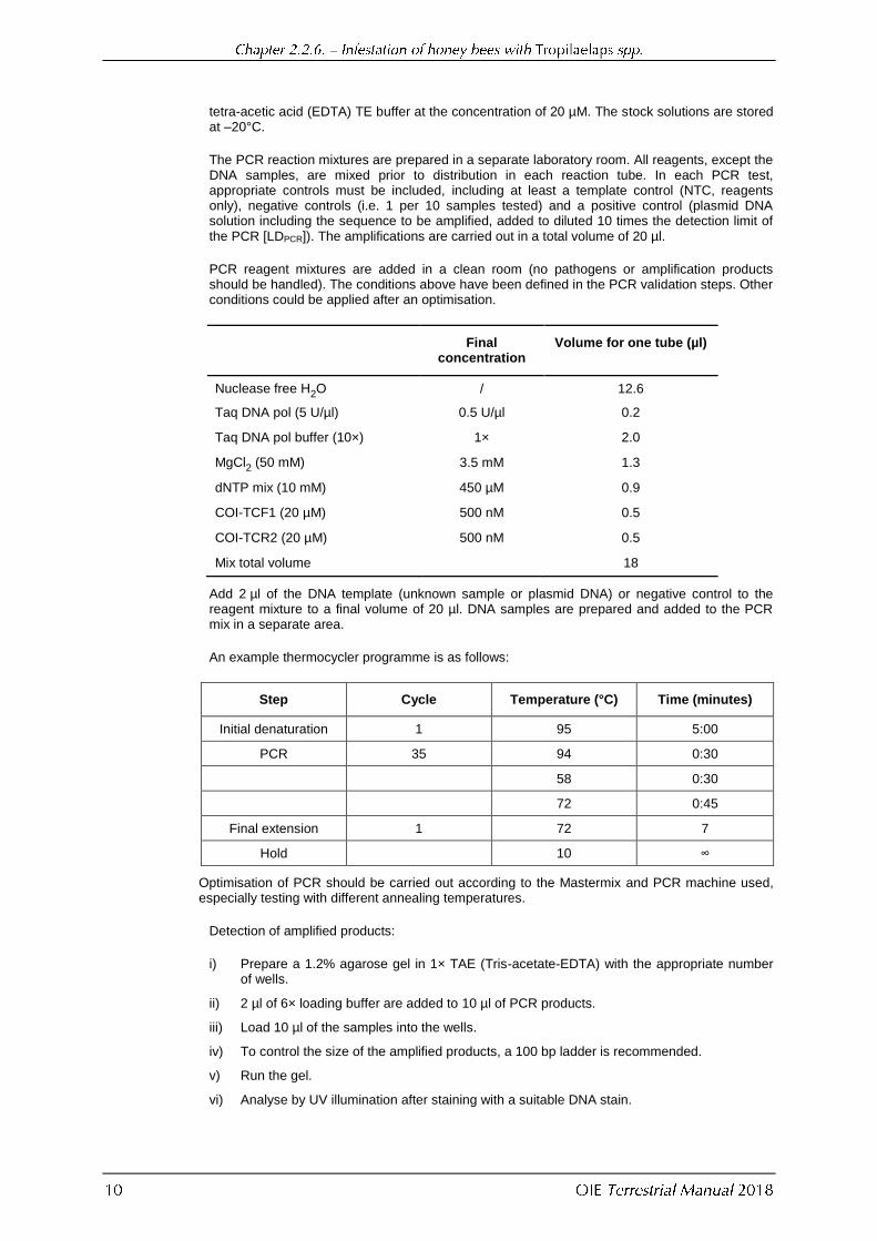
The PCR reaction mixtures are prepared in a separate laboratory room. All reagents, except the DNA samples, are mixed prior to distribution in each reaction tube. In each PCR test, appropriate controls must be included, including at least a template control (NTC, reagents only), negative controls (i.e. 1 per 10 samples tested) and a positive control (plasmid DNA solution including the sequence to be amplified, added to diluted 10 times the detection limit of the PCR [LDPCR]). The amplifications are carried out in a total volume of 20 µl.
PCR reagent mixtures are added in a clean room (no pathogens or amplification products should be handled). The conditions above have been defined in the PCR validation steps. Other conditions could be applied after an optimisation.
Final
concentration Volume for one tube (µl)
Nuclease free H2O / 12.6
Taq DNA pol (5 U/µl) 0.5 U/µl 0.2
Taq DNA pol buffer (10×) 1× 2.0
MgCl2 (50 mM) 3.5 mM 1.3
dNTP mix (10 mM) 450 µM 0.9
COI-TCF1 (20 µM) 500 nM 0.5
COI-TCR2 (20 µM) 500 nM 0.5
Mix total volume 18
Add 2 µl of the DNA template (unknown sample or plasmid DNA) or negative control to the reagent mixture to a final volume of 20 µl. DNA samples are prepared and added to the PCR mix in a separate area.
An example thermocycler programme is as follows:
Step Cycle Temperature (°C) Time (minutes)
Initial denaturation 1 95 5:00
PCR 35 94 0:30
58 0:30
72 0:45
Final extension 1 72 7
Hold 10 ∞
Optimisation of PCR should be carried out according to the Mastermix and PCR machine used, especially testing with different annealing temperatures.
Detection of amplified products:
i) Prepare a 1.2% agarose gel in 1× TAE (Tris-acetate-EDTA) with the appropriate number of wells.
ii) 2 µl of 6× loading buffer are added to 10 µl of PCR products.
iii) Load 10 µl of the samples into the wells.
iv) To control the size of the amplified products, a 100 bp ladder is recommended.
v) Run the gel.
vi) Analyse by UV illumination after staining with a suitable DNA stain.

The interpretation of the results is based on the presence or absence of the amplified product: the size of the expected PCR product is 580 bp including the two primers. However, the presence of a PCR product of the right size is not sufficient to identify the Tropilaelaps genus
and species. A sequencing step is required.
If a 580 bp band is detected the PCR product must be sequenced. The method is not described here, and can be outsourced. A panel of COI sequences available on Genbank (EF025423 to EF025468 and HQ533148 to HQ533159 [Luo et al., 2011]) is included in the analysis to construct the phylogenetic three and to identify the species of Tropilaelaps. An outgroup COI sequence from Varroa (EF025469, 253947435) is included.
Serological tests are not appropriate or relevant to bee colony infestations.
There are no vaccines available.
ANDERSON D.L. & MORGAN M.J. (2007). Genetic and morphological variation of bee-parasitic Tropilaelaps mites (Acari: Laelapidae): new and re-defined species. Exp. Appl. Acarol., 43, 1–24.
ANDERSON D.L. & ROBERTS J.M.K. (2013). Standard methods for Tropilaelaps mites research. In: The COLOSS BEEBOOK, Volume II: Standard methods for Apis mellifera pest and pathogen research, Dietemann V., Ellis J.D., Neumann P., eds. J. Apicultural Res., 52, http://dx.doi.org/10.3896/IBRA.1.52.4.21.
ATWAL A.S. & GOYAL N.P. (1971). Infestations of honeybee colonies with Tropilaelaps, and its control. J. Apic. Res., 10, 137–142.
BURGETT D.M. & KITPRASERT C. (1990). Evaluation of Apistan™ as a control for Tropilaelaps clareae (Acari: Laelapidae), an Asian honey bee brood mite parasite. Am. Bee J., 130, 51–53.
BURGETT M., AKRATANAKUL P. & MORSE R.A. (1983). Tropilaelaps clareae: a parasite of honeybees in south-east Asia. Bee World, 64, 25–28.
COOK V.A. & BOWMAN C.E. (1983). Mellitiphis alvearius, a little-known mite of the honeybee colony, found on New Zealand bees imported into England. Bee World, 64, 62–64.
DAINAT B., KEN T., BERTHOUD H. & NEUMANN P. (2009). The ectoparasitic mite Tropilaelaps mercedesae (Acari, Laelapidae) as a vector of honeybee viruses. Insectes Sociaux, 56, 40–43.
DE GUZMAN L.I., WILLIAMS G.R., KHONGPHINITBUNJONG K. & CHANTAWANNAKU P. (2017). Ecology, life history, and management of Tropilaelaps Mites. J. Econ. Entomol., 110, 319–332.
DELFINADO M.D. & BAKER E.W., (1961). Tropilaelaps, a new genus of mite from the Philippines (Laelapidae, Acarina). Fieldiana Zoology, 44, 53–56.
DELFINADO-BAKER M. & BAKER E., (1983). A new species of Neocypholaelaps (Acari: Ameroseiidae) from brood combs of the Indian honey bee. Apidologie, 14, 1–7.
DIETEMANN V., NAZZI F., MARTIN S.J., ANDERSON D.L., LOCKE B., DELAPLANE K.S., WAUQUIEZ Q., TANNAHILL C., FREY
E., ZIEGELMANN B., ROSENKRANZ P. & ELLIS J.D. (2013). Standard methods for varroa research. In: The COLOSS BEEBOOK, Volume II: Standard methods for Apis mellifera pest and pathogen research, Dietemann V., Ellis J.D., Neumann P., eds. J. Apicultural Res., 52, http://dx.doi.org/10.3896/IBRA.1.52.1.09.

FORSGREN E., DE MIRANDA J. R., ISAKSSON M., WEI S., & FRIES I. (2009). Deformed wing virus associated with Tropilaelaps mercedesae infesting European honey bees (Apis mellifera). Exp. Appl. Acarol., 47, 87–97.
KHONGPHINITBUNJONG K., DE GUZMAN L.I., BURGETT M.D., RINDERER T.E. & CHANTAWANNAKUL P. (2012). Behavioral responses underpinning resistance and susceptibility of honeybees to Tropilaelaps mercedesae. Apidologie, 43,
590–599.
KHONGPHINITBUNJONG K., DE GUZMAN, L. I., TARVER M. R., RINDERER T. E. & CHANTAWANNAKUL P. (2015). Interactions of Tropilaelaps mercedesae, honey bee viruses and immune response in Apis mellifera. J. Apic. Res., 54, 40–47.
KHONGPHINITBUNJONG K., NEUMANN P., CHANTAWANNAKUL P. & WILLIAMS G.R. (2016). The ectoparasitic mite Tropilaelaps mercedesae reduces western honey bee, Apis mellifera, longevity and emergence weight, and promotes Deformed wing virus infections. J. Invertebr. Pathol., 137, 38–42.
KOENIGER G., KOENIGER N., ANDERSON D.L., LEKPRAYOON C. & TINGEK S. (2002). Mites from debris and sealed brood cells of Apis dorsata colonies in Sabah, (Borneo) Malaysia, including a new haplotype of Varroa jacobsoni. Apidologie, 33, 15–24.
KONTSCHÁN J., TÓBIÁS I., BOZSIK, G. & SZOCS G. (2015). First record of Neocypholaelaps apicola from beehives in hungary (ACARI: Mesostigmata: Ameroseiidae): Re-description and DNA barcoding. Acta Zool. Acad. Sci. Hung., 61, 237–245.
LUO Q., ZHOU T., WANG Q., DAI P., WU Y. & SONG H. (2011). Identification of Tropilaelaps mites (Acari, Laelapidae) infesting Apis mellifera in China. Apidologie, 42, 485–498.
OSTIGUY N. & SAMMATARO D. (2000). A simplified technique for counting Varroa sticky boards. Apidologie, 31,
707–716.
PETTIS J.S., ROSE R., LICHTENBERG E.M., CHANTAWANNAKUL P., BUAWANGPONG N., SOMANA W. & VANENGELSDORP D. (2013). A rapid survey technique for tropilaelaps mite (Mesostigmata: Laelapidae) detection. J. Econ. Entomol., 106, 1535–1544.
RATH W., BOECKING O. & DRESCHER W. (1995). The phenomena of simultaneous infestation of Apis meliferea in Asia with the parasitic mites Varroa jacobsoni OUD, and Tropilaelaps clareae Delfinado and Barker. Am. Bee J., 135, 125–127.
RITTER W. & SCHNEIDER-RITTER U. (1988). Differences in biology and means of controlling Varroa jacobsoni and Tropilaelaps clareae, two novel parasitic mites of Apis mellifera. In: Africanized Honey Bees and Bee Mites,
Needham G.R., Page R.E. Jr., Delfinado-Baker M. & Bowman C.E., eds. Ellis Horwood, Chichester, UK, 387–395.
SAMMATARO D., GERSON U. & NEEDHAM G.R. (2000). Parasitic mites of honey bees: life history, implication s and impact. Ann. Rev. Entomol., 45, 519–548.
SMILEY R.L. (1991). Insect and Mite Pests in Food, an Illustrated Key, Gorham J.R., ed. Food and Drug Administration, United States Department of Agriculture, p. 6.
TANGJINGJAI W., VERAKALASA P., SITTIPRANEED S., KLINBUNGA S. & LEKPRAYOON C. (2003). Genetic differences between Tropilaelaps clareae and Tropilaelaps koenigerum in Thailand based on ITS and RAPD analyses. Apidologie, 34, 513–523.
WILDE J. (2000a). How long can Tropilaelaps clareae survive on adult honeybee workers? In: Proceedings of the Euroconference on Molecular Mechanisms of Disease Tolerance in Honeybees (MOMEDITO), held in Kralupy near Prague, Czech Republic, 17–19 October 2000. Bee Research Institute, Dol, Czech Republic, 217–221.
WILDE J. (2000b). Varroa destructor and Tropilaelaps clareae in Apis meliferea colonies in Nepal. In: Proceedings of the Euroconference on Molecular Mechanisms of Disease Tolerance in Honeybees (MOMEDITO), held in Kralupy near Prague, Czech Republic, 17–19 October 2000. Bee Research Institute, Dol, Czech Republic, 223–238.

WOYKE J. (1987). Length of stay of the parasitic mite Tropilaelaps clareae outside sealed honeybee brood cells as a basis for its effective control. J. Apic. Res., 26, 104–109.
*
* *
NB: There is an OIE Reference Laboratory for infestation of honey bees with Tropilaelaps spp.
(see Table in Part 4 of this Terrestrial Manual or consult the OIE Web site for the most up-to-date list: http://www.oie.int/en/our-scientific-expertise/reference-laboratories/list-of-laboratories/ ).
Please contact the OIE Reference Laboratory for any further information on diagnostic tests and reagents for infestation of honey bees with Tropilaelaps spp.
NB: FIRST ADOPTED IN 2004; MOST RECENT UPDATES ADOPTED IN 2018.