the metapleural gland of ants - university of texas at austin earlyonline.pdf · the metapleural...
TRANSCRIPT

Biol. Rev. (2010), pp. 000–000. 1doi: 10.1111/j.1469-185X.2010.00170.x
The metapleural gland of ants
Sze Huei Yek1,2∗ and Ulrich G. Mueller1
1 Section of Integrative Biology, University of Texas at Austin, 1 University Station C0930, 78712 Austin TX, USA2 Centre for Social Evolution, Department of Biology, University of Copenhagen, Universitetsparken 15, 2100 Copenhagen, Denmark
(Received 16 March 2010; revised 17 November 2010; accepted 24 November 2010)
ABSTRACT
The metapleural gland (MG) is a complex glandular structure unique to ants, suggesting a critical role in their originand ecological success. We synthesize the current understanding of the adaptive function, morphology, evolutionaryhistory, and chemical properties of the MG. Two functions of the MG, sanitation and chemical defence, have receivedthe strongest empirical support; two additional possible functions, recognition odour and territorial marking, are lesswell supported. The design of the MG is unusual for insects; glandular secretions are stored in a rigid, non-compressibleinvagination of the integument and the secretion is thought to ooze out passively through the non-closable openingof the MG or is groomed off by the legs and applied to target surfaces. MG loss has occurred repeatedly among theants, particularly in the subfamilies Formicinae and Myrmicinae, and the MG is more commonly absent in males thanin workers. MG chemistry has been characterized mostly in derived ant lineages with unique biologies (e.g. leafcutterants, fire ants), currently precluding any inferences about MG chemistry at the origin of the ants. A synthetic approachintegrating functional morphology, phylogenetic transitions and chemical ecology of the MGs of both the derived andthe unstudied early-branching (basal) ant lineages is needed to elucidate the evolutionary origin and diversification ofthe MG of ants.
Key words: antibiotic secretion, ants, chemical defence, evolution, metapleural gland, recognition odour, territoriality.
CONTENTS
I. Introduction . . . . . . . . . . . . . . . . . . . . . . . . . . . . . . . . . . . . . . . . . . . . . . . . . . . . . . . . . . . . . . . . . . . . . . . . . . . . . . . . . . . . . . . . . . . . . . . . 2II. Functions of the metapleural gland . . . . . . . . . . . . . . . . . . . . . . . . . . . . . . . . . . . . . . . . . . . . . . . . . . . . . . . . . . . . . . . . . . . . . . . . . 2
(1) Recognition odour . . . . . . . . . . . . . . . . . . . . . . . . . . . . . . . . . . . . . . . . . . . . . . . . . . . . . . . . . . . . . . . . . . . . . . . . . . . . . . . . . . . . . 3(2) Territory and nest-entrance marking . . . . . . . . . . . . . . . . . . . . . . . . . . . . . . . . . . . . . . . . . . . . . . . . . . . . . . . . . . . . . . . . . . 3(3) Antisepsis and hygiene . . . . . . . . . . . . . . . . . . . . . . . . . . . . . . . . . . . . . . . . . . . . . . . . . . . . . . . . . . . . . . . . . . . . . . . . . . . . . . . . . 5(4) Chemical defence . . . . . . . . . . . . . . . . . . . . . . . . . . . . . . . . . . . . . . . . . . . . . . . . . . . . . . . . . . . . . . . . . . . . . . . . . . . . . . . . . . . . . . 6(5) A pluralistic view of metapleural-gland function . . . . . . . . . . . . . . . . . . . . . . . . . . . . . . . . . . . . . . . . . . . . . . . . . . . . . . . 7
III. Morphology of the metapleural gland . . . . . . . . . . . . . . . . . . . . . . . . . . . . . . . . . . . . . . . . . . . . . . . . . . . . . . . . . . . . . . . . . . . . . . 7(1) One-chamber and two-chamber metapleural glands . . . . . . . . . . . . . . . . . . . . . . . . . . . . . . . . . . . . . . . . . . . . . . . . . . . 7(2) External morphology . . . . . . . . . . . . . . . . . . . . . . . . . . . . . . . . . . . . . . . . . . . . . . . . . . . . . . . . . . . . . . . . . . . . . . . . . . . . . . . . . . 8(3) Internal morphology . . . . . . . . . . . . . . . . . . . . . . . . . . . . . . . . . . . . . . . . . . . . . . . . . . . . . . . . . . . . . . . . . . . . . . . . . . . . . . . . . . . 10(4) Functional morphology . . . . . . . . . . . . . . . . . . . . . . . . . . . . . . . . . . . . . . . . . . . . . . . . . . . . . . . . . . . . . . . . . . . . . . . . . . . . . . . . 11
IV. Evolutionary losses and regains of the metapleural gland . . . . . . . . . . . . . . . . . . . . . . . . . . . . . . . . . . . . . . . . . . . . . . . . . . . 12(1) Evolutionary losses in social parasites . . . . . . . . . . . . . . . . . . . . . . . . . . . . . . . . . . . . . . . . . . . . . . . . . . . . . . . . . . . . . . . . . . 12(2) Evolutionary losses and regains in formicine ants . . . . . . . . . . . . . . . . . . . . . . . . . . . . . . . . . . . . . . . . . . . . . . . . . . . . . . 12(3) Metapleural gland of males . . . . . . . . . . . . . . . . . . . . . . . . . . . . . . . . . . . . . . . . . . . . . . . . . . . . . . . . . . . . . . . . . . . . . . . . . . . . 13
V. Chemistry of the metapleural gland . . . . . . . . . . . . . . . . . . . . . . . . . . . . . . . . . . . . . . . . . . . . . . . . . . . . . . . . . . . . . . . . . . . . . . . . 13(1) Metapleural gland chemistry of leafcutter ants . . . . . . . . . . . . . . . . . . . . . . . . . . . . . . . . . . . . . . . . . . . . . . . . . . . . . . . . . 13(2) Metapleural gland chemistry of fire ants . . . . . . . . . . . . . . . . . . . . . . . . . . . . . . . . . . . . . . . . . . . . . . . . . . . . . . . . . . . . . . . 14(3) Metapleural gland chemistry of Crematogaster (Physocrema) spp. . . . . . . . . . . . . . . . . . . . . . . . . . . . . . . . . . . . . . . . . . . 14(4) Proteinaceous components of metapleural gland secretions . . . . . . . . . . . . . . . . . . . . . . . . . . . . . . . . . . . . . . . . . . . . 14
* Address for correspondence: E-mail: [email protected]; [email protected]
Biological Reviews (2010) 000–000 © 2010 The Authors. Biological Reviews © 2010 Cambridge Philosophical Society

2 Sze Huei Yek and Ulrich G. Mueller
VI. Conclusions . . . . . . . . . . . . . . . . . . . . . . . . . . . . . . . . . . . . . . . . . . . . . . . . . . . . . . . . . . . . . . . . . . . . . . . . . . . . . . . . . . . . . . . . . . . . . . . . 14VII. Acknowledgements . . . . . . . . . . . . . . . . . . . . . . . . . . . . . . . . . . . . . . . . . . . . . . . . . . . . . . . . . . . . . . . . . . . . . . . . . . . . . . . . . . . . . . . . . 15
VIII. References . . . . . . . . . . . . . . . . . . . . . . . . . . . . . . . . . . . . . . . . . . . . . . . . . . . . . . . . . . . . . . . . . . . . . . . . . . . . . . . . . . . . . . . . . . . . . . . . . . 15IX. Supporting Information . . . . . . . . . . . . . . . . . . . . . . . . . . . . . . . . . . . . . . . . . . . . . . . . . . . . . . . . . . . . . . . . . . . . . . . . . . . . . . . . . . . . 18
I. INTRODUCTION
Social insects have evolved an astounding diversity ofexocrine glands to mediate social organisation and com-petitive interactions (Holldobler & Wilson, 1990; Jackson &Morgan, 1993; Billen & Morgan, 1998; Billen, 2009). Oneof these glands - the metapleural gland (MG) - is found onlyin ants (Formicidae) (Fig. 1). Because homologues of the MGare unknown from other insect lineages, its unique presencein ants suggests a critical role of this gland in their originand ecological success (Wilson, 1987; Holldobler & Wilson,1990; Ward, 2007).
The opening of the MG is generally a rather conspicuousexternal feature and therefore serves as an unambiguouscharacter for ant identification (Fig. 2). Other characteristicsused for ant identification [elbowed antennae, petiole(nodular waist), and eusociality] are each also shared withother insect lineages. Only the MG is diagnostic as a keysynapomorphy for the ant family Formicidae, includingfossilized lineages (Grimaldi, Agosti & Carpenter, 1997;Bolton, 2003; Engel & Grimaldi, 2005; Ward, 2007). Forexample, the presence of a MG in the Sphecomyrminaeplaces this extinct subfamily near the ancestor of extant antlineages, but the apparent absence of the MG in the ant-like, extinct lineage Armaniinae suggests a somewhat moredistant relationship (Ward, 2007).
The MG likely originated once in the ancestor of theFormicidae at least 120–140 million years ago (Brady et al.,2006). The gland is thought to be present ancestrally in allcastes (males, queens, and workers) (Wheeler, 1928; Tulloch,1935; Taylor, 2007), but was secondarily lost independentlyin diverse lineages (Brown, 1968; Holldobler & Engel-Siegel,1985), most notably within the subfamilies Formicinae andMyrmicinae. Absence of the MG occurs most frequentlyin males; when present in males, the MG is smaller thanin queens and workers (Holldobler & Engel-Siegel, 1985).To our knowledge, there exists no ant species where thegland is present only in the males. Likewise, the MG isgenerally more developed in queens than in workers. Antlineages differ with respect to the hypothesized primaryfunction of the MG (Table 1), gland architecture, and relativesize, ranging from reduced or absent MGs in many social-parasitic ants to hypertrophied MGs used in defence inthe Crematogaster subgenus Physocrema. A full understandingof MG evolution, therefore, requires integrated functional-morphological, chemical, behavioural, and phylogeneticanalyses. We review here the accumulated evidence thatmay contribute to such a synthetic analysis of the MG, thenconclude with the most promising areas for future researchon this ant-specific gland.
Fig. 1. Position of the metapleural gland (MG) in the posteriormesosoma of a typical ant (Linepithema humile). Modified fromPavan & Ronchetti (1955).
Myrmecologists were aware of the presence of theMG over 100 years ago (Meinert, 1861; Janet, 1898a, b;Wheeler, 1910). Early work focused on the morphology andhistology of the gland (see also Tulloch, 1936; Pavan &Ronchetti, 1955; Whelden, 1957a,b, 1960, 1963a,b; Tulloch,Shapiro & Hershenov, 1962), referring to the gland alsoas the metathoracic and sometimes erroneously, as themetasternal gland [Wheeler (1910) attributes this error toCarlo Emery, and indeed Emery (1900) refers to the MGas the ‘‘ghiandola metasternale’’; the gland actually residesin the lower plate of the metapleuron (Tulloch, 1935)]. Fornearly a century, the function of the MG remained enigmaticdespite a growing list of hypotheses derived mainly fromanatomical studies (Janet, 1898a,b; Tulloch, 1936; Gosswald,1953; Tulloch et al., 1962; Brown, 1968) (Table 1). UlrichMaschwitz’s seminal work nearly 40 years ago providedthe first experimental tests of these hypotheses (Maschwitz,Koob & Schildknecht, 1970; Maschwitz, 1974), rejectingsome widely accepted hypotheses (e.g. colony and speciesrecognition) and concluding that the main function of thegland is antibiotic secretion. Although antisepsis and hygieneare currently believed to be the primary functions of theMG in ants (e.g. Holldobler & Wilson, 1990; Schluns &Crozier, 2009), the accumulated evidence suggests diversefunctions of the MG among ant lineages and among ant castes(Table 1).
II. FUNCTIONS OF THE METAPLEURAL GLAND
Four hypotheses on MG function have been consideredseriously (Table 1), and these hypotheses are not mutuallyexclusive: colony or species recognition; territory or nest-entrance marking; antisepsis; and chemical defence. Only thelast two hypotheses have received uncontroversial empirical
Biological Reviews (2010) 000–000 © 2010 The Authors. Biological Reviews © 2010 Cambridge Philosophical Society

The metapleural gland of ants 3
Fig. 2. Scanning electron micrograph of the metapleural gland(MG) of Tapinoma erraticum. A brush composed of bristle-like hairspasses through the atrium of the MG ending at the openingof the atrium. Image courtesy of Roberto A. Keller, AmericanMuseum of Natural History.
support. Additional hypotheses, such as trail-pheromoneproduction or nutrient secretion for larvae, are now widelydismissed because of insufficient evidence, but some plausiblehypotheses remain untested (Table 1). In many ant species,the MG secretions may well serve more than one function.
(1) Recognition odour
Early researchers hypothesized that the MG producespheromones mediating nest-mate or species recognition(Janet, 1898a,b; Gosswald, 1953; Tulloch et al., 1962; Brown,1968). Although nest-mate and species recognition are fun-damentally different processes, Brown (1968) reasoned thatan individual lacking the MG may emit fewer recognitionodours, increasing its chances of acceptance by another antcolony. The recognition odour hypothesis could thereforeexplain the absence of the MG in ants whose life historiesrequire infiltration of other ant nests. For example, para-site queens must gain acceptance into a heterospecific hostcolony (Brandt et al., 2005; Nash & Boomsma, 2008), army-ant males (e.g. Dorylus or Eciton spp.) must enter conspecificcolonies to reach wingless queens that are surrounded bylarge numbers of workers (Rettenmeyer, 1963; Kronauer,2009), and slave-raider workers (e.g. Polyergus spp.) mustenter heterospecific Formica spp. nests to steal pupae as slaves(d’Ettorre & Errard, 1998).
The recognition odour hypothesis was weakened whenHolldobler & Engel-Siegel (1985) showed that the MG wasabsent in some ant lineages (e.g. the genus Oecophylla, mostCamponotus spp.) that are known to be aggressive and capa-ble of discrimination between nest-mates and non-nest-matecompetitors. Absence of the MG in these ants thereforeimplies that either the MG is not involved in recognition, orthat recognition is mediated by some other odour source inthese MG-less species. Moreover, Holldobler & Engel-Siegel
(1985) showed that the MG is absent in males of diverseant lineages, not only in army ant species where males enterconspecific nests for mating. The majority of male ants thatlack the MG or possess it in a reduced state do not enter het-erospecific nests and in fact mate in the open (males lackedthe MG in 15 out of 20 species examined by Holldobler& Engel-Siegel, 1985). The accumulated evidence suggeststhat absence of the MG evolved multiple times in male ants,apparently irrespective of their need to enter foreign coloniesfor mating (see Section IV), thus weakening the hypothe-sis that the MG mediates nest-mate or species recognitionin ants.
The recognition odour hypothesis was tested by Maschwitzet al. (1970) who conducted a series of behaviouralexperiments to assess the role of MG secretions in bothnest-mate and species recognition. When workers of Myrmica
laevinodis or Myrmica rubra were exposed to filter papercontaining MG secretions from either species, workersexplored the conspecific secretions to the same extent asthe heterospecific secretions. [The interpretation of theseexperiments is complicated by taxonomic ambiguity: Myrmica
laevinodis was later considered a subspecies of M. rubra,and the old M. rubra is now named M. ruginodis (Seifert,1988, 2007).] Maschwitz et al. (1970) further showed thatanaesthetized workers onto which MG secretions wereapplied from heterospecific workers were not attacked anddid not elicit noticeable alarm behaviours (e.g. open mandiblethreat) when they were returned awake to their natal nest.In addition, M. rubra workers that were treated with MGsecretions of heterospecific M. laevinodis were attacked wheninserted into a M. laevinodis nest. Lastly, workers of Formica
rufa and Formica polyctena from which the MGs were surgicallyremoved were attended non-aggressively by conspecifics,but were attacked by heterospecifics. The results indicatedthat MG secretions were insufficient to elicit acceptanceby a colony, thus contradicting the recognition odourhypothesis.
Subsequent to this work, the recognition odour hypothesishas been largely discarded and there have been no furtherattempts to test for contributions of MG odours to colonyrecognition. This may be unfortunate, as the gland’ssecretions may contribute to the odour bouquet of a colonyand thus modulate recognition in some ant species.
(2) Territory and nest-entrance marking
Tulloch et al. (1962) suggested that the MG secretes colony-specific pheromones used in territory marking. Territorialmarker pheromones in ants are typically deposited directlyonto the substratum through controlled use of an applicatorsuch as the sting or the legs (Morgan, 2009; Billen, 2009).Such pheromones are secreted by diverse glands, for examplethe poison, Dufour’s, pygidial, sternal, hindgut, rectal, tibial,and tarsal glands (Holldobler & Wilson, 1990). A commonfeature of all these secretions is that their applicationcan be controlled by the ants. The best-studied exampleis the African weaver ant Oecophylla longinoda that marks
Biological Reviews (2010) 000–000 © 2010 The Authors. Biological Reviews © 2010 Cambridge Philosophical Society

4 Sze Huei Yek and Ulrich G. Mueller
Table 1. Hypotheses on the function of the metapleural gland.
Hypotheses with empirical support for some ant lineages
Recognition odour Janet (1898a, b), Gosswald (1953), and Brown (1968) hypothesized that the MG produces pheromones that mediatenestmate or species recognition. Several ant lineages without MGs have life histories that require infiltration ofother ant nests (e.g. social parasites, army-ant males), which suggested to Gosswald (1953) and Brown (1968) thatan individual lacking the MG may emit fewer recognition odours, increasing its chances of acceptance during nestinfiltration. Maschwitz et al. (1970) failed to find behavioural-experimental support for the recognition odourhypothesis. Indirect support for this hypothesis therefore derives entirely from the absence in some (but not all)nest-infiltrating ant lineages, which can also be explained by other hypotheses (e.g. antisepsis).
Territory and nestentrancemarking
Tulloch et al. (1962) proposed that the MG secretes colony-specific pheromones used in territorial marking. The MGsecretions of several myrmicine species are thought to function as territorial or nest-entrance markers that regulateaggressive interactions between conspecific, neighbouring colonies (Jaffe & Puche, 1984; Jaffe et al., 1986;Cammaerts & Cammaerts, 1998, 2001), but the experimental evidence remains controversial. Territorial ornest-entrance marking by MG secretions has not been tested for ants outside the subfamily Myrmicinae.
Antisepsis andhygiene
Maschwitz et al. (1970) and Maschwitz (1974) hypothesized that the MG serves a general antibiotic function bysuppressing ant diseases and other detrimental microbes in the nest. The MG secretions of many ant speciesinhibit microbes, due to a combination of bacteriostatic acidity and possible non-specific antibiotics of MGsecretions. Although only derived ant lineages have been tested for antibiotic properties of MG secretions, it iswidely believed that the primary and original function of the MG is antisepsis.
Chemical defence Ant species in the Physocrema subgenus of Crematogaster have hypertrophied MGs that secrete deterrent chemicals.When threatened, these ants extrude a droplet of whitish and sticky liquid from the MG opening, which can beretracted back into the MG by the ants (Maschwitz, 1974). These MG secretions contain diverse phenoliccompounds that are toxic to invertebrate predators (Attygalle et al., 1989). Hypertrophy and phenolic secretions ofthe MG are best studied in the Asian Crematogaster (Physocrema) ants, but some Neotropical Crematogaster (C. acuata,montezumia) also appears to have enlarged MGs (Hosoishi & Ogata, 2009).
Hypotheses that remain inadequately testedMating pheromone Holldobler & Engel-Siegel (1985) speculated that the MG may produce mating pheromones in males as a secondary
function (in addition to a primary antiseptic function) and that these pheromones could evolve under sexualselection. Because the MG chemistry of male ants is completely unstudied, this hypothesis remains untested. Whilemating-pheromone production applies to males and females, this explanation can be ruled out for workers, whichdo not mate and are often sterile.
Trail pheromone Tulloch et al. (1962) suggested that the MG may secrete trail pheromones. However, the laterally oriented andelevated position of the MG on the ant body does not permit controlled application of the MG secretions to a trail,unlike the diverse trail-pheromone glands in ants (Morgan, 2008). Maschwitz et al. (1970) showed that much of theMG effluent is deposited inadvertently by ants on the ground, but that such depositions did not elicittrail-following behaviour in Myrmica spp.
Support ofantibiotic-producingbacteria
Poulsen et al. (2003) hypothesized that, in some specialized ant lineages such as leafcutter ants, a derived, secondaryfunction of the MG may be the support and sheltering of disease-suppressing mutualistic microbes. No microbeshave been found to date in any MG using either microscopic techniques (Stow & Beattie, 2008; various Australianants) or molecular screens (Mueller et al., 2008; Atta spp.).
Hypotheses disregarded because of lack of supporting evidenceFood production
for larvaeSmith (1857) believed that the MG secretes ‘‘saccharine fluids’’ to feed larvae. This hypothesis has subsequently been
disregarded because the elevated position on the metathorax and the known chemical composition of MGsecretions seem incompatible with larval feeding.
Food storagechamber
Donisthorpe (1941) suggests that, analogous to the crop of honeypot ants, the hypertrophied MG of Crematogasterdifformis could function as a pocket for the reception and storage of sugar liquid. This suggestion was based on theobservation that workers vigorously lick each other’s metathoraces (where the MGs are located). However,chemical analysis of the MG secretion of C. difformis identified a mixture of defensive phenolic compounds, but nosugary substances (Maschwitz, 1974; Attygalle et al., 1989).
Sound resonator Because the atrium of the MG is a rigid integumental invagination that is filled largely with air rather than secretion,Nachtwey (1961, 1963a, b) speculated that the air-filled atrium could function as a resonator in sound perceptionor sound production (e.g. stridulation). However, the volume of the air-filled atrium is too small to function as aneffective resonator for substrate-borne vibratory communication in ants (Flavio Roces, personal communication).
arboreal territories through direct application of colony-specific pheromones in the rectal fluid and increases therate of rectal fluid deposition in new territory (Holldobler& Wilson, 1978). The elevated position of the MG on antsprecludes direct application of MG secretions onto targets,but indirect application may occur through controlledspreading of MG secretions by leg movements, paralleling
the hypothesized application of antibiotic MG secretions tothe brood and garden in fungus-growing ants (Fernandez-Marín, Zimmermann & Wcislo, 2003; Fernandez-Marínet al., 2006, 2009). The use of territorial pheromones appearsto be a derived trait (Jaffe & Puche, 1984), because early-branching (basal) genera such as poneroid ants are thought torecognize their territory using visual cues and environmental
Biological Reviews (2010) 000–000 © 2010 The Authors. Biological Reviews © 2010 Cambridge Philosophical Society

The metapleural gland of ants 5
odour, but not via glandular territorial pheromones (Jaffe &Marcuse, 1983).
MG secretions appear to be used to mark nest entrances orterritories in some ant species, including Tetramorium caespitum,T. impurum (Cammaerts & Cammaerts, 2001), Pheidole pallidula
(Cammaerts & Cammaerts, 1998), Solenopsis geminata (Jaffe &Puche, 1984), and Pseudomyrmex triplarinus (Jaffe, Lopez &Aragort, 1986). These studies tested the ability of workers todifferentiate between non-native and native territories (Jaffe& Puche, 1984) and showed that workers are more aggressivewhen their own nest entrance had been marked by non-nativeants (Cammaerts & Cammaerts, 1998, 2001). Differentextracts (head, thorax, metathorax, legs, and metasoma) wereused to investigate the origin of these marker pheromones,but only extracts from the metathorax and the hind legsincreased aggressive tendencies (Cammaerts & Cammaerts,2001). Although these observations suggest that the MGcould contribute to territorial or nest-entrance marking,many details remain unclear. For example, the mechanismof application of the putative MG-derived, territorial markeronto the substratum is unknown for Solenopsis geminata (Jaffe &Puche, 1984), and the source of the marker can be narroweddown to the thoracic region but not specifically to the MG(Pseudomyrmex triplarinus, Jaffe et al., 1986; Tetramorium caespitum,
T. impurum, Cammaerts & Cammaerts, 2001). MG extractselicited territorial responses only in S. geminata, but not inS. invicta (Jaffe & Puche, 1984). Studies that manipulatethe flow of MG secretions (e.g. by experimentally sealingthe MG opening) seem most promising to test the role ofMG secretions in colony-specific marking of territories andnest entrances. Chemical analyses should also verify thehypothesized flow or application of MG secretions via thelegs onto the marked substratum.
(3) Antisepsis and hygiene
Maschwitz et al. (1970) first hypothesized that the MGsecretions have a general antiseptic function. Numerousstudies (see Appendix S1) have documented antibioticproperties of MG secretions against diverse microbes (yeast,bacteria, fungi). Although some studies failed to find evidenceof antibiotic activity (e.g. Diehl & Junqueira, 2001), it iswidely believed that the primary function of MG secretionsis sanitation by suppression of ant diseases or other microbesin the nest environment.
MG secretions are highly acidic (Maschwitz, 1974), andantimicrobial effects of the secretions may be largely due tothis acidity. For example, Atta sexdens have MG secretionswith a pH of 2.5, Myrmica laevinodis and M. rubra workerswith a pH of 3.0–3.5 (Maschwitz et al., 1970), Crematogaster
scutellaris, C. inflata, C. difformis and diverse ponerine workerswith a pH of 3–4 (Maschwitz, 1974), and Myrmecia gulosa
with a pH of 3.5 (Mackintosh et al., 1995). Aenictus fergusoni isthe only known ant with MG secretions that are not acidic(Maschwitz, 1974). Several studies on fungus-growing antshave shown that MG secretions lower the pH in the fungusgarden (Bot et al., 2002; Ortius-Lechner et al., 2000; Powell& Stradling, 1986; Papa & Papa, 1982; Maschwitz et al.,
1970), which would help suppress bacterial growth becausemany bacteria are inhibited at low pH levels. MG secre-tions of leafcutter ants contain a diversity of carboxylic acids(Schildknecht & Koob, 1971; do Nascimento et al., 1996;Ortius-Lechner et al., 2000, 2003), and these acids shouldlower the pH on the ant integument and possibly in the nestenvironment if the ants distribute sufficient amounts of MGsecretions.
Acidity is a property of many other ant secretions. Nearlyhalf of the known glandular secretions of ants contain car-boxylic acids (Hermann & Blum, 1981). Consequently, manyother ant glands secrete compounds that are weakly bac-teriostatic or, in the case of formic acid in the poisongland of formicine ants, significantly bactericidal. A bacteria-suppressing effect of a particular acidic glandular secretiontherefore may not be its primary function. For example,the primary function of formic acid in the poison gland offormicine ants is presumably defence, not antisepsis. Docu-mentation of an in vitro antibiotic effect of an acidic secretionis therefore insufficient to conclude antibiosis as the pri-mary function of the MG; this would require testing of thecontribution of acidity to antibiosis separate from the con-tributions of particular antibiotics, which is experimentallychallenging.
Powell & Stradling (1986) showed that removal of fungus-growing ant workers resulted in a pH increase in the ants’garden and eventual garden destruction by parasitic fungi.This could suggest that pH-lowering MG secretions areimportant in garden health, but other ant factors (e.g.grooming, secretion from other glands) that may also preventgarden deterioration cannot be ruled out in this particularexperiment. Although Powell & Stradling’s (1986) results areconsistent with a contribution of MG acidity to hygiene,further work is needed to establish the effect of acidity inde-pendent of the effects of specific antibiotics in MG secretions.
The mechanisms underlying the antibiotic action of MGsecretions are largely unknown, except for a membrane-destabilizing effect documented in an in vitro assay (Veal,Trimble & Beattie, 1992). Absorption of the active MGcompounds through the phospholipid membrane disruptsmembrane structure and function (Mackintosh et al., 1995),causing the cells to burst in both prokaryotes and eukaryotes.Mackintosh et al. (1995) discuss several possible mechanismsfor how the ants may protect themselves against thegeneralized antibiotic effects of their own MG secretions,such as a modified integument forming an effective barrieror secretions that become active and harmful only afterexposure to the external environment.
Brown (1968) reported that some ants groom the MGopening with their legs, and such grooming was confirmedin subsequent studies of dozens of species from six antsubfamilies (Farish, 1972; Basibuyuk & Quicke, 1999;Fernandez-Marín et al., 2003, 2006). MG grooming permitsactive dispersion of MG effluent, rather than passive anduncontrolled flow (Fernandez-Marín et al., 2006, 2009). Byincreasing the rate of MG grooming during microbialinfection, ants seem to be able to upregulate dispersion
Biological Reviews (2010) 000–000 © 2010 The Authors. Biological Reviews © 2010 Cambridge Philosophical Society

6 Sze Huei Yek and Ulrich G. Mueller
of MG secretions (Fernandez-Marín et al., 2006, 2009).Increased rates of MG grooming occur in defence againstboth virulent and more benign diseases, supporting theview that MG secretions have broad-spectrum antimicrobialproperties (Fernandez-Marín et al., 2009).
Preventive measures that serve as a first line of defenceshould be typical for eusocial lineages with perennialcolonies such as ants (Beattie et al., 1986; Boomsma, Schmid-Hempel & Hughes, 2005; Cremer, Armitage & Schmid-Hempel, 2007). MG secretions therefore may be designedto be more effective against early developmental stages ofpathogens (e.g. spores that have yet to germinate) than laterdevelopmental stages (e.g. growing hyphae). Empirical testshave confirmed this prediction for MG secretions (Vealet al., 1992; Beattie et al., 1986; Bot et al., 2002), and MGsecretions therefore appear to target fungal spores beforehyphal growth of a spreading infection becomes a seriousproblem.
If MG secretions serve primarily antiseptic functions, onemight expect that the caste that is most involved in pathogendefence (e.g. small nest workers in many species; gardeners inleafcutter ants) have larger MGs. This has been documentedfor some leafcutter ants where minima workers havedisproportionately large MGs compared to major workers(Wilson, 1980; Bot & Boomsma, 1996; Hughes et al., 2008).Moreover, small workers of the leafcutter A. octospinosus aremore effective at grooming than large workers. In infectionbioassays, small A. octospinosus workers are more resistantto pathogens than are larger workers (Hughes, Eilenberg& Boomsma, 2002; Poulsen, Hughes & Boomsma, 2006).In contrast to the above studies, Diehl & Junqueira (2001)found no survival differences among Atta sexdens piriventris sub-castes when challenged with the entomopathogen Beauveria
bassiana, and the disproportionately larger MGs of thesesmall workers apparently do not provide greater diseaseprotection.
Apart from caste-dependent factors, environmental factorsmay also modulate MG function. In Atta sexdens piriventris, MGsecretions vary throughout the year, with the highest levelsof MG secretion during warmer months (Diehl & Junqueira,2001). In Acromyrmex octospinosus, workers in field coloniesproduce greater quantities of MG secretions compared toworkers in laboratory colonies, but the MGs of these twotypes of workers do not show qualitative chemical differences(Ortius-Lechner et al., 2000). The observed quantitativedifferences could be due to differences in food availability,temperature, disease pressure, or any other spatially orseasonally varying factor. It is possible that both the betternutrition and low disease prevalence in the laboratorycolonies strengthen colony immunity, lowering secretoryflow in the MGs of laboratory workers.
Among the closest relatives to ants, bees and some vespoidwasps are important plant pollinators, whereas very fewants pollinate plants (see Rico-Gray & Oliveira, 2007, andreferences therein; de Vega et al., 2009). To explain thisdifference, Beattie et al. (1984) proposed that the antibioticMG secretions kill pollen or inhibit pollen germination,
rendering ants ineffective as plant pollinators unless theyhave reduced or absent MGs. Consistent with this hypothesis,reduction and absence of MGs is found in some pollinatingants, such as males of Myrmecia urens that pollinate theorchid Leporella fimbriata through pseudocopulation (Peakall,Beattie & James, 1987) and Camponotus compressus workersthat pollinate coconuts (Patel, 1937). However, reduction orabsence of the MG among ant pollinators is not the norm,and effective ant pollinators with well-developed MGs are notuncommon (e.g. Formica lasioides, F. oregonensis, Kincaid, 1963;Formica argentea, Hickman, 1974; Proformica longiseta, Gomez &Zamora, 1992). Moreover, contact of Crematogaster auberti andPheidole pallidula ants with functional MGs does not reducepollen viability in the ant-pollinated plant Cytinus hypocistis,whereas contact with the MG-less Camponotus pilicornis greatlyreduces pollen viability (de Vega et al., 2009). Based on thissmall survey of pollinating ants, it appears that possession of afunctional MG does not preclude an evolutionary transitionto ant pollination. The observed rarity of pollinating antspecies appears better explained by the fact that ant workersare flightless and cannot readily vector pollen among differentplants, thus limiting the distance that ants can dispersepollen compared to flying pollinators such as bees andwasps.
(4) Chemical defence
Crematogaster ants in the subgenus Physocrema havehypertrophied MGs (Janet, 1898b; Donisthorpe, 1941;Hosoishi & Ogata, 2008, 2009), and MG secretions ofCrematogaster difformis and Crematogaster inflata contain a mixtureof phenolic compounds that are noxious to predators(Attygalle et al., 1989; Jones et al., 2005). When C.(Physocrema)inflata workers are attacked, they expel from the MG openinga droplet of a whitish, viscous, sticky secretion which theycan retract back into the MG (Maschwitz, 1974; Buschinger& Maschwitz, 1984). These MG droplets deter hostile antsand vertebrate predators (Maschwitz, 1974; Ito et al., 2004),and volatile emissions from the droplets alarm nestmates(Maschwitz, 1974). The secretions repel ant predators byacting both as a chemical deterrent and as a glue (Maschwitz,1974). Despite extensive grooming, ants smeared with theMG secretions of C. inflata are unable to remove thesticky secretion (Maschwitz, 1974; Ito et al., 2004). Therepellent nature of MG secretions is best documented forCrematogaster (Physocrema) ants (Maschwitz, 1974; Attygalleet al., 1989).
Although both C. difformis and C. inflata use MG secretionsmainly in defence, the secretions serve additional functionsin these species. Volatile odours from the MG induce alarmbehaviour in nest-mates of C. inflata, but not in C. difformis
(Maschwitz, 1974) where the alarm pheromone is secretedby the mandibular gland. C. difformis is thought to use atwo-gland system for defence. The head, neck, appendages,and metasoma are protected by secretions from the Dufour’sgland, whereas the thorax and petiolar-postpetiolar regionare protected by secretions from the MG (Maschwitz, 1974;Jones et al., 2005).
Biological Reviews (2010) 000–000 © 2010 The Authors. Biological Reviews © 2010 Cambridge Philosophical Society
The metapleural gland of ants 7
(5) A pluralistic view of metapleural-gland function
To date, a disproportionate number of studies haveinvestigated antibiotic functions of MG secretions. Most ofthese studies focused on ant lineages with uniquely derivedbiologies (e.g. fungus-growers such as Atta and Acromyrmex
leafcutter ants, and solitarily foraging predators such asthe bulldog ant Myrmecia gulosa). Because of their derivedbiologies, the function(s) of the MG in these groups maybe uniquely modified and thus may not be representativefor ants at large. Further studies of a more representativesample across the ant phylogeny are needed, particularly ofthe early-branching (basal) ant lineages for which there iscurrently no information. Without such studies, it will bedifficult to elucidate the original function of the MG.
While the recent literature has favoured antibiosis asthe single, primary function of the MG, insect exocrineglands often serve several concurrent functions (Hermann& Blum, 1981; Holldobler & Wilson, 1990). Multiplefunctions of the MG should therefore not be ruled outa priori for ants. Sanitation and territorial marking are twofunctions of single glands hypothesized for a number of antspecies (Jaffe & Puche, 1984; Cammaerts & Cammaerts,1998), and pheromones used in communication can havestrong antibiotic properties (Cole, Blum & Roncadori,1975). MG secretions could have primary and secondaryfunctions that differ among castes or among life stages (nurseant versus forager), such as sex pheromone production inaddition to antibiotic production (Holldobler & Engel-Siegel,1985). Alternatively, MG function could be concentration-dependent [e.g., a low concentration of MG secretion couldpromote the growth of the symbiotic fungus in attine ants,whereas a high concentration may inhibit growth (Powell &Stradling, 1986)]. Lastly, of course, the different compoundsin the secretory mix could serve different functions. Evena single compound can simultaneously serve several roles.For example, indoleacetic acid in the MGs of leafcutter antsmay contribute to bacteriostasis by lowering the pH of thesecretion, but may also facilitate fungal growth in leafcuttergardens because of its auxin properties (Schildknecht et al.,1973). Because many ant species may possess such functionalcomplexity in MG secretion (e.g. between castes), the recentresearch emphasis on antibiotic effects may have precludedfruitful investigations into additional functions.
III. MORPHOLOGY OF THE METAPLEURALGLAND
Holldobler & Engel-Siegel’s (1985) comparative-morpholo-gical study of 59 ant species (24 genera, six subfamilies)still represents the landmark morphological work on theMG. Several studies expanded on this work by examiningadditional species and by providing detailed histologicaland ultrastructural analyses (e.g. Billen & van Boven, 1987;Fanfani & Valcurone Dazzini, 1991a,b; Schoeters & Billen,1992, 1993; Bot et al., 2001; Gusmao, Caetano & Nakano,
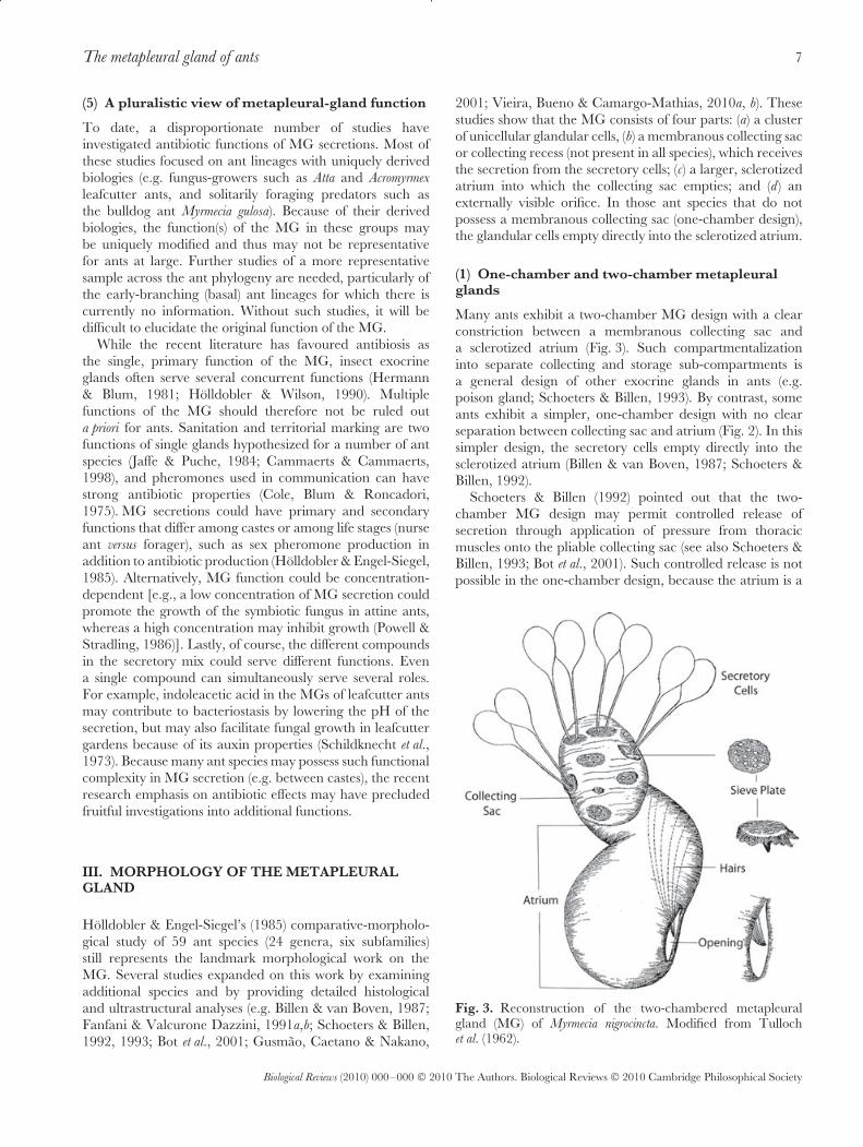
2001; Vieira, Bueno & Camargo-Mathias, 2010a, b). Thesestudies show that the MG consists of four parts: (a) a clusterof unicellular glandular cells, (b) a membranous collecting sacor collecting recess (not present in all species), which receivesthe secretion from the secretory cells; (c) a larger, sclerotizedatrium into which the collecting sac empties; and (d) anexternally visible orifice. In those ant species that do notpossess a membranous collecting sac (one-chamber design),the glandular cells empty directly into the sclerotized atrium.
(1) One-chamber and two-chamber metapleuralglands
Many ants exhibit a two-chamber MG design with a clearconstriction between a membranous collecting sac anda sclerotized atrium (Fig. 3). Such compartmentalizationinto separate collecting and storage sub-compartments isa general design of other exocrine glands in ants (e.g.poison gland; Schoeters & Billen, 1993). By contrast, someants exhibit a simpler, one-chamber design with no clearseparation between collecting sac and atrium (Fig. 2). In thissimpler design, the secretory cells empty directly into thesclerotized atrium (Billen & van Boven, 1987; Schoeters &Billen, 1992).
Schoeters & Billen (1992) pointed out that the two-chamber MG design may permit controlled release ofsecretion through application of pressure from thoracicmuscles onto the pliable collecting sac (see also Schoeters &Billen, 1993; Bot et al., 2001). Such controlled release is notpossible in the one-chamber design, because the atrium is a
Fig. 3. Reconstruction of the two-chambered metapleuralgland (MG) of Myrmecia nigrocincta. Modified from Tullochet al. (1962).
Biological Reviews (2010) 000–000 © 2010 The Authors. Biological Reviews © 2010 Cambridge Philosophical Society
8 Sze Huei Yek and Ulrich G. Mueller
highly sclerotized, rigid chamber, formed as an invaginationof the metapleural integument. The uncompartmentalized,simple MG design therefore could represent a more ancestralstate, perhaps evolutionarily derived from an integumentalpatch of cuticular glands that gradually became recessed andeventually invaginated to form a rigid chamber.
The one-chamber design has been described in poneroidants (Amblyopone pallipes, Whelden, 1957b; Diacamma spp.,Schoeters & Billen, 1992), doryline ants (nine species ofDorylus spp.; Billen & van Boven, 1987), and dolichoderineants (Dolichoderus quadripunctatus, Linepithema humile, Tapinoma
erraticum; Fanfani & Valcurone Dazzini, 1991b; RobertoKeller personal communication). By contrast, two-chamberdesigns have been described in the subfamilies Myrmeciinae(Myrmecia nigrocincta, M. pilosula, Tulloch et al., 1962;Holldobler & Engel-Siegel, 1985; Nothomyrmecia macrops,Holldobler & Engel-Siegel, 1985), Myrmicinae (Acromyrmex
octospinosus, Aphaenogaster rudis, Atta bisphaerica, A. laevigata,A. sexdens, Crematogaster striatula; Holldobler & Engel-Siegel, 1985; Fanfani & Valcurone Dazzini, 1991a;Schoeters & Billen, 1993; Bot et al., 2001), and Formicinae(Myrmecocystus mendax, Holldobler & Engel-Siegel, 1985).These phylogenetic patterns seem to support the view thatthe one-chamber design is ancestral among ants. However,only a comprehensive survey across the entire diversityof ant genera, including the early-branching (basal) antlineages, can hope to reconstruct evolutionary transitions inmorphological complexity of the MG. In fact, additionaldesigns that may not fit into a division between one-chamberand two-chamber designs appear to exist, as for examplethe MG designs with irregular membranous, fingerlikeexpansions in doryline and ecitonine army ants (Whelden,1963a,b; Roberto Keller, personal communication), in someectatommine ants (Whelden, 1960), and in some ponerineants (Roberto Keller, personal communication).
(2) External morphology
The external morphology of the MG varies greatly amongant lineages (Fig. 4). In many ants, the gland empties via anoval or slit-shaped opening, located either just above thearticulation of the hind coxa, or somewhat more dorsal-posteriorily below the propodeum in the recess between thearticulation of the hind coxa and the petiole. Slit-shaped oroval openings give the impression of a gland that is designedfor broad, brush-like release of the secretion, rather thanpoint application.
The MG opening in many ant lineages deviates fromthis general slit-like design of the opening, suggesting mod-ified functions. For example, the MG opening can be largeand round (Diacamma spp., Schoeters & Billen, 1992; somedolichoderines, Fanfani & Valcurone Dazzini, 1991b), suchthat the MG atrium appears as a relatively exposed andunprotected invagination of the metapleural integument,rather than as a sequestered receptacle designed for storageand controlled release of secretion. In lineages exhibitingarmy-ant-like morphology and behaviour (e.g. Leptanilla,dorylomorphs) and in Myrmecia spp., a carina-like lip flanks
the slit-like MG opening from above (Brady & Ward, 2005);this flange may protect the glandular atrium from accidentalcontamination, facilitate collection of the secretion by legs forsubsequent application, reduce evaporation, or serve otherfunctions. In ecitonine army ants, the flange extends anteri-orily as a ridge onto the mesosoma, perhaps to facilitate flowof the secretion in an anterior direction (Phil Ward, personalcommunication). Keller (2008) describes variations of thisflange in other ant lineages, noting phylogenetically infor-mative modifications of the structure and its carina-like exten-sions. Keller (2008) also describes a flap emanating from theventral margin of the MG orifice. This flap extends upward,sometimes covering the opening almost completely except fora narrow slit opening towards the posterior or above, ratherthan towards below. In addition to such morphological vari-ations in shape of the MG opening, the opening can beoriented in different directions (Fig. 4). For example, the MGopens towards the side and onto the coxa in some ants, buttowards the posterior and onto the propodeum in other ants(Keller, 2008; Phil Ward, personal communication). In sum,the metapleural gland opening and its associated structuresoffer a rich diversity of unexplored characters for phyloge-netic analyses, perhaps even allowing differentiation amongclosely related species. For example, queens of closely relatedAtta species can have MG openings that differ markedly insize, whereas the workers of the corresponding species do notshow this difference (Gusmao, Caetano & Nakano, 2001).
In leafcutter ants, Crematogaster (Physocrema) ants, andmany others, the glandular atrium is enlarged and formsa conspicuous bulge (bulla) above the MG opening. Suchhypertrophied glands occupy a significant volume of theposterior mesosoma. At the other extreme, the MG openingcan be small, indicated externally merely by a small porethat is easily overlooked. For example, the inquiline parasiteTeleutomyrmex schneideri was thought to have no metapleuralgland (Gosswald, 1953; Brown, 1968), but a small pore isactually present just above the hind coxa where a MG wouldbe found (S.H. Yek, C. Rabeling, & U.G. Mueller, personalobservations). Without dissection, it is unclear whether thispore is a vestigial feature or is associated with a functionalgland, as discussed by Brown (1968).
Whether large and round, narrowed to a slit, or partiallycovered by a lip, the MG atrium appears less protected fromcontamination with particles and chemicals than the typicalexocrine glands of ants (Billen & van Boven, 1987; Billen,2009). The reasons for the generally large opening of the MGare unclear. Rapid release of large quantities of secretion isone explanation for a large opening (e.g. in the Crematogaster
subgenus Physocrema). An alternative hypothesis may be thatthe large opening may allow simultaneous entry of air intothe atrium while releasing the secretion (Schoeters & Billen,1992). Indeed, the atrium always contains some air (Janet,1898a,b; Gosswald, 1953; Maschwitz et al., 1970; Poulsenet al., 2002b), depending presumably on a balance betweensecretory inflow and outflow. In a study by Poulsen et al.(2002b) on Acromyrmex octospinosus, for example, most gardenworkers of a laboratory colony had approximately one-third
Biological Reviews (2010) 000–000 © 2010 The Authors. Biological Reviews © 2010 Cambridge Philosophical Society

The metapleural gland of ants 9
Fig. 4. Morphological diversity of metapleural gland openings (arrows) of workers in a sample of early-branching (basal) andderived ant lineages. (A) Proceratium pergandei, (B) Amblyopone pallipes, (C) Paraponera clavata, (D) Daceton armigerum, (E) Anoplolepis custodiens,(F) Camponotus gigas. Images courtesy of Jeffrey Sossa-Calvo, Smithsonian Institution.
of the atrium volume filled with secretion, 5% of workershad a completely empty atrium, and only about 4% had acompletely filled atrium. Interestingly, atria are completelyfilled with air in starved workers, but re-accumulate secretionwhen the workers resume feeding (Maschwitz et al., 1970).In Acromyrmex octospinosus workers, food deprivation leadsrapidly to reduced secretory rates (Bot & Boomsma, 1996),and secretory rates seem to decline with age (Bot et al., 2001).The relative proportion of air and secretion in the atriumtherefore is variable during an ant’s life, and an atrium filledpartially with air is a normal condition. Perhaps the interfacebetween the secretion and the air in the metapleural atriumserves some unknown purpose.
Another variable feature among ant lineages is the areajust below the glandular opening, which is frequently smooth(Schoeters & Billen, 1993; Gusmao et al., 2001), even inspecies with a rugulose, pitted, or otherwise roughenedintegument. In Protanilla spp., a smooth trench extendsanteriorily from the MG opening (the metapleural trench;Bolton, 1990), perhaps to direct the flow of the MG secretiontowards the meso- and meta-coxae. In Atta spp. leafcutterants, a cuticular ridge just below the MG opening directsthe secretory flow directly onto the hind coxa; this designis thought to aid the spread of the secretion by coxalmovements (see Schoeters & Billen, 1993). Lastly, rows ofhairs are frequently arranged just outside the opening, buthairs can also originate inside the atrium (Meinert, 1861;Pavan & Ronchetti, 1955; Holldobler & Engel-Siegel, 1985;Billen & van Boven, 1987; Fanfani & Valcurone Dazzini,
1991a,b) (Fig. 2). By contrast, Atta bisphaerica, A. capiguara, andA. sexdens rubropilosa leafcutter ants are devoid of hairs at theorifice (Schoeters & Billen, 1993; Gusmao et al., 2001). Hairsat the MG opening may capture and hold the outflowingsecretion, prevent accidental entry of contaminant particlesinto the large opening of the gland, aid in the channelingand distribution of the secretion, or serve sensory functions.
In Tapinoma erraticum, Iridomyrmex purpureus, Linepithema
humile, Myrmecia pilosula, and M. nigrocincta, bristle-like hairsoriginate at the back of the atrium and project throughthe atrium towards the MG opening (Tulloch et al., 1962;Holldobler & Engel-Siegel, 1985; Fanfani & ValcuroneDazzini, 1991b; Keller, 2008; Figs 2 & 3). Such hairs areassumed to guide the secretion through the atrium to theMG opening, and Holldobler & Engel-Siegel (1985) thereforecall these hairs ‘‘dispenser bristles’’. A single row of hairs(Myrmecia nigrocincta; Tulloch et al., 1962) or two parallel rowsof hairs (Myrmica rubra; Tulloch, 1936) originate from thelateral, internal surface of the atrium, whereas a brush ofhairs originates at the back of the atrium in Tapinoma erraticum
(Fig. 2). The tips of these hairs converge at a common pointat the centre of the external opening, and in Lasius flavus
the converging hairs appear like a pointed, hollow brush(Wheeler, 1910). In such designs, the secretion may not flowalong the walls of the atrium, but could be suspended alonghairs in an otherwise air-filled atrium.
It is surprising that, despite the importance of the MGin ant identification, we are still lacking a comparativefunctional-morphological analysis of the structures associated
Biological Reviews (2010) 000–000 © 2010 The Authors. Biological Reviews © 2010 Cambridge Philosophical Society

10 Sze Huei Yek and Ulrich G. Mueller
with the MG opening. It is also surprising that the presenceor absence of the gland in males is rarely reported intaxonomic descriptions, whereas its presence/absence infemales and workers is generally noted. We are also lackinga comprehensive comparative analysis of MG chemistry toallow us to infer ancestral states and identify subsequenttransitions to derived states. Keller’s (2008) survey suggestsrich variation in MG morphology among ant lineages thatcould provide important clues for diverse MG functions whencombined with behavioural and ecological information (seealso Fig. 4). A comprehensive analysis should focus not onlyon the external and internal morphology of the MG, butalso on the structures that interact with the MG, such as thelegs, which may pick up and spread the secretion (Brown,1968; Fernandez-Marin et al., 2006, 2009). We are surprisedto find that studies on the MG have so far largely ignoredthe design of the body surfaces that are likely to facilitateremoval of the MG secretion.
(3) Internal morphology
The MG is divided into three parts, clusters of secretory(exocrine) cells, a collecting sac/recess into which the cellssecrete, and an atrium (Fig. 3). The glandular cells may benumerous enough to engulf the collecting sac, which thentakes the appearance of an internal lumen. If few secretorycells are present, they appear as a fan-shaped cluster attachedto a portion of the collecting sac. The average cluster is com-prised of 10–30 secretory cells, and several such tightlypacked clusters can be grouped into larger secretory struc-tures producing a cauliflower-like appearance (Schoeters &Billen, 1992, 1993; Gusmao et al., 2001; Bot et al., 2001).Each cell cluster empties into the collecting sac through itsown sieve-like plate (Billen & van Boven, 1987; Gusmaoet al., 2001; Fig. 3). The cell clusters and the collecting sacare generally located higher and more anteriorly than theatrium [but see Whelden’s (1963a, b) description of the MGof Eciton burchellii queens], perhaps to facilitate a passiveflow downwards to the glandular opening located at theinferior-posterior end of the atrium.
The intracellular structure of the secretory cells has beenstudied in very few ant species (Myrmecia nigrocincta, Tullochet al., 1962; Dorylus spp., Billen & van Boven, 1987; Atta
bisphaerica, A. laevigata, and A. sexdens, Schoeters & Billen,1993). The secretory cells show an end apparatus typical forinsect secretory cells, consisting of an intracellular collectingductule and a surrounding sheath of microvilli (Tulloch et al.,1962; Schoeters & Billen, 1992). Ultrastructural differencesbetween contiguous cells suggest asynchronous secretoryactivity (Schoeters & Billen, 1992). As in most ant glands,the secretory cell cytoplasm is rich in smooth endoplasmicreticulum and mitochondria (Billen & van Boven, 1987).
Each secretory cell of the MG possesses an intracellularductule that collects the secretion (see Billen, 1991 for adiagram of this kind of exocrine cell). The intracellularductule extends via a duct cell into the collecting sac, apliable membranous receptacle that can expand or contractwhen receiving or releasing the glandular secretion. Ductules
do not fuse (Billen & van Boven, 1987), and each secretory celltherefore empties individually into the collecting sac. As faras is known, no muscles attach to the collecting sac that coulddirectly control the release of the glandular secretion, nor arethere nerve endings terminating at the secretory tissue thatcould regulate glandular activity (Schoeters & Billen, 1993).However, Wheeler (1910, p. 38) includes a ganglion locatednext to the secretory cells in a drawing of the MG of Lasius
flavus. This may be a misidentification as no such ganglionhas been reported in any subsequent histological study.
From the collecting sac, the secretion flows into the scle-rotized atrium. The sclerotization of the atrium can beextensive, perhaps to protect the ant from the acidic ortoxic secretion it contains. In Diacamma spp., for example,the atrium wall is actually thicker than the exoskeleton(Schoeters & Billen, 1992). The atrium is positioned imme-diately below the integument and can appear externally as abulge (bulla) of the integument. An atrium is supplied by onlyone collecting sac (or one secretory area with sieve plates forone-chamber species). Many researchers have commentedon the similarity of the atrium wall to the integument (Janet,1898a, b; Whelden, 1957a, b; Billen & van Boven, 1987;Schoeters & Billen, 1992; Keller, 2008), suggesting that theatrium is a relatively unmodified integumental invagination.Because this structure usually is mostly filled with air, weprefer to use the term ‘atrium’, rather than ‘reservoir’ usedmore commonly in the literature.
Janet (1898a, b) and Schoeters & Billen (1993) describea narrow groove in the wall of the atrium of Myrmica
rubra and Atta sexdens. This groove appears to function asa gutter to guide the flow from the collecting sac to theMG opening. As described above, other ants such as Lasius
flavus, Myrmecia pilosula, M. nigrocincta, and Linepithema humile
have bristle-like hairs passing through the atrium that couldfunction to guide the secretory flow through the atrium(Wheeler, 1910; Tulloch et al., 1962; Holldobler & Engel-Siegel, 1985; Fanfani & Valcurone Dazzini, 1991b). Thehairs are sometimes arranged as a hollow brush (Wheeler,1910) (Fig. 2), perhaps drawing the secretion towards theMG opening by means of capillary forces, aiding in theevaporation of chemicals, or facilitating chemical interactionsbetween the secretion and air inside the atrium. We are notaware of a species which has both bristle-guides and a gutterin the MG atrium: these two features therefore may representalternative designs to channel the secretory flow through theatrium towards the opening.
The efflux from the atrium onto the body surface isthought to be passive (but see below), as the atrium is a rigidstructure that resists compression (Holldobler & Engel-Siegel,1985; Billen & van Boven, 1987). However, thoracic musclesadjacent to the pliable collecting sac may compress the sacand hence indirectly modulate the efflux from the atrium (Botet al., 2001). Schoeters & Billen (1992, 1993) discuss how theaction of specific metathoracic muscles could apply pressureto the glandular tissue and the collecting sac, causing thesecretion to be released into the atrium and towards the MGopening. In Crematogaster (Physocrema) difformis and C. inflata,
Biological Reviews (2010) 000–000 © 2010 The Authors. Biological Reviews © 2010 Cambridge Philosophical Society

The metapleural gland of ants 11
workers can expel a defensive secretory droplet, retain it atthe rounded, glandular orifice, then retract it back into theatrium (Buschinger & Maschwitz, 1984; Maschwitz, 1974).The neuromuscular mechanisms underlying this controlledexpulsion and retraction have not been studied.
The size of the secretory tissue differs among castes.Queens have about twice the number of glandular cellsthan the average worker (Holldobler & Engel-Siegel, 1985;Angus, Jones & Beattie, 1993; Appendix S2). For the handfulof species for which male glands have been examined, maleshave only about 20–50% of the number of glandular cellsas the corresponding workers (Appendix S2). We are notaware of any species where the gland is absent in workersbut present in sexual females, and we know only of Lasius
fuliginosus where the gland is pronounced in workers butabsent (or greatly reduced) in sexual females (Appendix S2).
Among ant species, larger ants tend to have larger glandswith more secretory cells, whereas cell number appearsto be independent of ant colony size (number of workers)(Angus et al., 1993). Among species in the fungus-growing anttribe Attini, relative MG bulla size (bulla size standardized bypronotum width) is not correlated with colony size (P = 0.07;Hughes et al., 2008). However, relative to body size, leafcut-ter ant species have significantly larger MGs compared tonon-leafcutter species, indicating that MG size increase waslinked to the origin of leafcutter fungiculture (Hughes et al.,2008). These patterns suggest that MG size evolution is notdriven by factors associated with colony size, but rather byfactors associated with body size and microbial interactions(see Section II.3).
The relationship between MG size and worker caste hasbeen studied in only a few species. Secretory-cell numberappears independent of worker size in Orectognathus versicolor
(minors, medias, and majors each have about 80 cellsper MG; Holldobler & Engel-Siegel, 1985; Appendix S2),but cell number increases with worker size in Acromyrmex
octospinosus leafcutter ants, where the largest workers haveabout 2–3 times the number of cells than the smallestworkers (Bot et al., 2001) (Appendix S2). The increase in cellnumber with leafcutter worker size is allometric, however,such that relative to body size, the smallest workers haveproportionately larger MGs compared to larger workers(Bot & Boomsma, 1996; Bot et al., 2001; Gusmao et al.,2001; Hughes et al., 2008). This greater investment into MGfunction by the smallest Acromyrmex octospinosus workers maybe linked to the garden- and nest-sanitation activities ofthis caste (see Section II.3). In Acromyrmex octospinosus, bullasize and number of secretory cells are highly correlated(r = 0.84), justifying measurement of the externally visiblebulla as a proxy for the size of the MG (Bot et al., 2001;Hughes et al., 2008).
(4) Functional morphology
The location of the MG opening low at the latero-posterior end of the mesosoma, often below the level of thepetiole insertion, could be a starting point for a functional-morphological analysis. No conspicuous homologous glands
are currently known from this area in the putative vespoidsister lineages of ants (Ward, 2007; Pilgrim, van Dohlen &Pitts, 2008), precluding comparative inferences about MGfunction at the origin of the ants.
The location of the MG opening in the centre of the ant isunusual. Only one other gland is known to empty in this area,the mesopleural gland, which is present only in dacetine andphalacromyrmecine ants (Bolton, 2003). By contrast, mostant glands empty at the mouth, near the tip of the metasoma,or on the legs; these locations permit easy contact with thesubstratum for point-application of a secretion. The centraland elevated position of the MG is less compatible withcertain hypothesized MG functions, such as the productionof a trail pheromone - known trail pheromones in ants aresecreted from structures that permit controlled applicationand that are close to the substratum, such as the tip of themetasoma or the legs (Holldobler & Wilson, 1990). How-ever, the central, elevated position of the MG is compatiblewith other functions, such as the production of recognitionpheromones, antibiotics, or defensive secretions to protect thevulnerable petiolar region. Other unusual features of the MGcompared to other exocrine glands may also inform hypothe-ses of MG function: (1) a large, non-closable opening; (2) afrequently slit-like opening, suggesting brushlike applicationrather than point application; (3) rigidity of the atrium, pre-cluding contraction and controlled glandular discharge; and(4) opening of the gland low near the coxae or the petiole.
For protection of body surfaces against diseases, a logicaldesign would be to have small glands distributed acrossthe entire integument (like the numerous wax-secretingglands that cover the insect body), rather than two centralgland openings from which secretions must be spread acrossthe body by laborious and time-consuming grooming. Thepresence of only two MG openings therefore suggests otherfunctions. The single, large opening of each MG could bebetter explained if the gland’s primary function is to supplysecretions that can be targeted by grooming to specificproblem sites, such as infected body parts, brood, or specificareas in the nest (Fernandez-Marín et al., 2006, 2009).However, the presumed uncontrolled oozing of secretionfrom the MG opening then remains unexplained (Maschwitzet al., 1970). To validate this widely accepted assumption,it will now be important to measure the uncontrolledefflux relative to the active spreading of MG secretionsby grooming. We note that the original study by Maschwitzet al. (1970) merely documented that MG secretions appliedto the surface of the mesosoma seem to flow passively ontothe substratum via the ants’ legs, but passive efflux of theviscous MG secretion through the MG opening has neverbeen quantified directly.
Expanding on reports of active spreading of MG secretionsby leg grooming motions (Brown, 1968; Maschwitz et al.,1970), Fernandez-Marín et al. (2006) recently confirmedthe so-called MG grooming in 26 ant species from fivesubfamilies. MG grooming involves a series of coordinatedfore leg and body movements. A worker ant partially extendsits legs to raise the body from the substratum, flexes the
Biological Reviews (2010) 000–000 © 2010 The Authors. Biological Reviews © 2010 Cambridge Philosophical Society

12 Sze Huei Yek and Ulrich G. Mueller
fore leg at the femoral-tibial joint to bring the posteriorsurface of the metatarsus in contact with the opening of theipsilateral MG, rubs the metatarsus over the opening, thenbrings the leg into contact with the lateral surface of theglossa (Fernandez-Marín et al., 2006). The occurrence of thisbehaviour across major ant lineages suggests that it aroseearly during ant evolution (see also Farish, 1972).
IV. EVOLUTIONARY LOSSES AND REGAINSOF THE METAPLEURAL GLAND
A survey of the early-branching (basal) ant lineages revealsthat the MG was ancestrally present in ant workers, females,and probably also males (Bolton, 2003), but that thegland was lost in many lineages during ant evolution. Weestimate that about 20% of ant species do not have a MG.Losses in workers have occurred sporadically (AppendixS2), for example during evolutionary transitions to socialparasitism. These parasitic lineages are not particularlyspeciose. Prominent ant lineages without a MG are foundin the Camponotini (Maschwitz, 1974; Holldobler & Engel-Siegel, 1985), including the genera Camponotus (about 1000described species) and Polyrhachis (about 500 species). Thisgroup comprises about 15% of described ant species diversityand is thought to have derived from a common ancestor withMGs (Ellen Schluns, unpublished data). Because the MG isabsent in the vast majority of species in the hyperdiversegenus Camponotus, the MG-less condition apparently did notprevent the radiation of this ecologically successful lineage.
(1) Evolutionary losses in social parasites
Gosswald (1953) first suggested that the MG may contributeto colony odour (‘‘Nestgeruch’’) in non-parasitic ants; glandloss would reduce odour levels for socially parasitic ants andthus improve the parasite’s chances of successfully entering ahost nest. Gosswald (1953) and Brown (1968) further notedthat MG loss appears to have occurred most frequently inworkerless inquiline ants (permanent social parasites), andless often in temporary social parasites which differ in theirtreatment of the host queen. Whereas an inquiline ant doesnot kill the host queen but coexists intimately with the host(the host workers rear inquiline reproductives), a temporarysocial parasite eventually kills the host queen, the parasiticstage is transitory, and the parasite workers ultimately takeover the colony as the host workers die out (Buschinger,2009). In a brief survey of parasitic ants, we confirmed MGloss in 67% of inquiline species, but only in 9% of temporarysocial parasites (Appendix S3). Because most inquiline socialparasites in our survey were myrmecine ants whereas mosttemporary social parasites were formicine ants (AppendixS3), an analysis of phylogenetically independent contrastsof a larger sample is needed to substantiate this intriguingassociation between inquilinism and MG loss.
Interestingly, two inquiline Acromyrmex species of compar-atively recent origin have retained small workers. These
socially parasitic workers have smaller MGs compared withtheir respective non-parasitic sister species, yet the respectiveparasitic queens have MGs of similar size to host queens(Sumner, Hughes & Boomsma, 2003; de Souza et al., 2006).Parasite queens are exposed to similar risks as non-parasitequeens during their mating flight and nest-searching phase,and undergo the additional risk of invasion of the hostcolony, whereas parasite workers never or rarely leave thehost colony. The convergent reduction in worker MGs inthese two recently evolved Acromyrmex inquilines supports theview that a transition to inquiline life results in selectionfor a reduction in MG size in workers, presumably becausemaintenance of such secretory structures is costly (Sumneret al., 2003; Poulsen et al., 2002b).
In sum, the many independently evolved socially parasiticants provide a rich testing ground for elucidating MG func-tion. The presence of a MG in some parasitic ants indicatesthat MG-derived odours do not preclude infiltration of hostcolonies. More importantly, evolutionary maintenance ofthe MG in workers of temporary social parasites but fre-quent reduction of the MG in inquilines (see Appendix S3)appears to contradict the recognition odour hypothesis, butis consistent with some other hypothesis (e.g. antibiosis).
(2) Evolutionary losses and regains in formicineants
Holldobler & Engel-Siegel (1985) noted two additionalattributes of MG-less ant lineages: nest-weaving and arboreallife. In nest-weavers, workers use larval silk to constructnests, for example, by weaving live leaves together intoa protective envelope. Nest-weaving occurs in formicinegenera such as Oecophylla, Polyrhachis, and several lineagesin the genus Camponotus (Johnson, Agapow & Crozier, 2003;Robson & Kohout, 2005). Under the most likely evolutionaryscenario, nest-weaving has been gained four times and lostonce in the subfamily Formicinae, and loss of the MG isassociated significantly with these four transitions to nest-weaving (Johnson et al., 2003).
Holldobler & Engel-Siegel (1985) suggested that arborealnesting could permit reduction or loss of the MG becausepathogen pressures may be less severe for arboreal ants thanground-nesting ants. However, many successful arboreal antlineages (e.g. pseudomyrmecines) have well-developed MGs,whereas some successful ground-dwelling lineages do not (e.g.Camponotus spp.) (see also discussion on MGs and arborealityin Orivel & Dejean, 1999). These two observations weakenthe arboreality hypothesis and indicate that arboreal nestingper se is not a major factor influencing MG loss. The analysisof species-specific factors that drive MG loss is complicatedby the fact that nest-weaving and arboreality are correlated(most nest weavers are also arboreal).
A particularly interesting case is the large formicine genusCamponotus which comprises nearly 1000 described speciesmost of which do not possess MGs. A few Camponotus specieshave unreduced MGs such as the South-East Asian giantrainforest ant Camponotus gigas (Holldobler & Engel-Siegel,1985). C. gigas nests in the soil at tree bases, under fallen
Biological Reviews (2010) 000–000 © 2010 The Authors. Biological Reviews © 2010 Cambridge Philosophical Society

The metapleural gland of ants 13
logs, or in tree cavities, but forages arboreally (Pfeiffer &Linsenmair, 1998, 2000, 2001). Other exceptions includeCamponotus sericeus (Dey & Coumar, 2008), which nests in theground, is strictly diurnal, and is widely distributed acrossAfrica, the Middle East, and India (Mody & Linsenmair,2003); Camponotus thadeus (Shattuck, 2005), a recentlydiscovered species that inhabits high-elevation rainforest inAustralia and appears to nest arboreally (Shattuck, 2005);and 10 species in the Camponotus subgenus Myrmonesites (BrianFisher, personal communication), all from Madagascar. Nonesting information is known from these Malagasy ants (BrianFisher, personal communication). These four exceptionalcamponotine lineages with MGs offer interesting test casesfor elucidating ecological factors in MG evolution.
(3) Metapleural gland of males
Holldobler & Engel-Siegel (1985) reported that the MGis absent in most males. By contrast, our survey indicatesthat the MG may be present in males of more lineagesthan currently realized (Appendix S2), including early-branching (basal) ant lineages such as the proceratiines,amblyoponines, and ponerines. This supports the ancestralpresence of the MG in ant males (Wheeler, 1928;Taylor, 2007), with multiple losses during their subsequentevolution. A comprehensive survey of males of the early-branching (basal) ant subfamilies (Martialinae, Leptanillinae,Proceratiinae, poneroids) is needed to confirm this pattern.Unfortunately, males are currently unknown for the mostearly-branching (basal) ant subfamily Martialinae (Rabeling,Brown & Verhaagh, 2008), males of the Leptanillinae andProceratiinae are difficult to collect, and the presence ofthe MG in leptanilline males can only be determined bycareful dissection (Baroni Urbani, 1977). Space constraintsto accommodate the large flight muscles in the mesosoma ofmales could drive MG loss. An alternative explanation forMG loss in males is that males are short-lived and thereforehave a reduced requirement for hygienic defences comparedto long-lived workers and queens (Maschwitz et al., 1970;Holldobler & Engel-Siegel, 1985).
V. CHEMISTRY OF THE METAPLEURAL GLAND
The chemical constituents of MG secretions have beenanalysed in only a handful of species, with most analysesfocused on ants with derived biologies such as leafcutter ants(43 compounds), fire ants (19 compounds), and Crematogaster
(Physocrema) spp. (16 compounds). MG chemistry differs signif-icantly among these three groups (Appendix S4). Carboxylicacids and fatty acids of various chain lengths dominate inleafcutter and fire ants, whereas phenolic compounds dom-inate in the Crematogaster (Physocrema) species. Proteinaceouscompounds comprise a significant fraction of MG secretionsin Atta spp. leafcutter ants (Maschwitz et al., 1970; do Nasci-mento et al., 1996), and it is presently unclear whether pro-teinaceous additions occur in the MG secretions of other ants.
(1) Metapleural gland chemistry of leafcutter ants
The most surprising insight emerging from a comparison ofleafcutter MG chemistry is how few chemicals are sharedamong attine species (Appendix S4). Only five chemicalswere consistently found from the MG secretions of severalleafcutter species (3-hydroxydecanoic acid, indoleaceticacid, phenylacetic acid, 3-hydroxydodecanoic acid, andheptadecanoic acid). The first three of these acids arecommonly reported as major constituents of MG secretionsin leafcutter ants, whereas 3-hydroxydodecanoic acid andheptadecanoic acid represent minor constituents.
3-hydroxydecanoic acid, also called myrmicacin, was thefirst compound identified from the MG of attine ants(Atta sexdens, Schildknecht & Koob, 1971). Myrmicacinwas later also discovered in three other attine species (Atta
cephalotes, Acromyrmex octospinosus, and Acromyrmex subterraneus)(do Nascimento et al., 1996; Ortius-Lechner et al., 2000).Due to its anti-microbial activity, myrmicacin was initiallyhypothesized to function as an ‘‘herbicide’’ in attine gardens(Schildknecht & Koob, 1971; Iizuka, Iwadare & Orito,1979). However, a specific herbicidal role of myrmicacinin attine gardens now appears unlikely for three reasons.First, myrmicacin is also found in non-attine ants that haveno need of ‘‘herbicides’’ in their nest (e.g. Messor barbarus
and Labidus coecus, Beattie et al., 1984). Second, myrmicacindisrupts molecular flow through cell walls, the function ofGolgi vesicles, and mitosis (see Nakamura, Miki-Hirosige& Iwanami, 1982 and references therein), suggesting anon-specialized, antibiotic effect typical for most organicacids. Third, even though myrmicacin seems to be a majorcompound in the MGs of Atta species, only small quantitiesof myrmicacin are found in Acromyrmex spp. leafcutter ants(Ortius-Lechner et al., 2000), suggesting that it has a minorrole in these close relatives of Atta leafcutter ants.
Indoleacetic acid (IAA) has been found in four species ofattine ants: Atta sexdens, Atta cephalotes, Acromyrmex octospinosus,and Acromyrmex subterraneus. The detection of IAA wassurprising because it is a well-known plant growth hormone(heteroauxin) (Schildknecht et al., 1973). IAA is the majorconstituent in Acromyrmex octospinosus (24–25% of total MGsecretion) but a minor one in Atta species (do Nascimentoet al., 1996). Despite its known auxin properties in plants,the functional role of IAA in leafcutter ants remains unclear.Initial studies claimed a negative influence on hyphal growthof the cultivated fungus (Schildknecht & Koob, 1971), whilelater studies found inhibition only at higher concentrationsbut growth enhancement at lower levels (Schildknecht et al.,1973). Other studies detected a small inhibitory effect ofIAA only when interacting synergistically with myrmicacin(Powell & Stradling, 1986).
Phenylacetic acid (PAA) is a major constituent of MGsecretions in Atta sexdens and Atta cephalotes where it comprises72% and 80% of the total secretion, respectively, butPAA is absent in Acromyrmex octospinosus. PAA also exhibitsheteroauxin activities (Wightman & Lighty, 1982), but isweaker than IAA. Interestingly, PAA occurs also in other antglands, such as the mandibular gland of the harvester ant
Biological Reviews (2010) 000–000 © 2010 The Authors. Biological Reviews © 2010 Cambridge Philosophical Society

14 Sze Huei Yek and Ulrich G. Mueller
Pogonomyrmex rugosus (Fales et al., 1992). The role of PAA as ananti-microbial agent is unclear. However, PAA undoubtedlycontributes to the acidity of the MG secretion and thereforealso to its general anti-microbial activity.
Although some closely related leafcutter species showqualitative similarities in MG chemistry (e.g. similar acidiccomposition in Atta sexdens and Atta cephalotes; AppendixS4), quantities of specific compounds vary substantiallyamong leafcutter species. This suggests conservation of acidicconstituents among closely related species, but possible mod-ulation of the quantity of these constituents depending onbehavioural roles (nurse ant versus forager) or environmentalcondition (healthy versus diseased colony). Consistent withthis view of behavioural and environmental modulation, dif-ferences in MG chemistry did not correlate with geneticdifferences among workers from the same Acromyrmex colony(Ortius-Lechner et al., 2003).
MG secretions between laboratory and field workershave only been compared in the leafcutter ant Acromyrmex
octospinosus (Ortius-Lechner et al., 2000). MG secretions werequalitatively similar between these workers, but fieldworkers carried on average three times more MG secretionthan laboratory workers (6070 ng versus 2099 ng). Thisquantitative difference could be due to several factors, such ashigher pathogen pressures in the field or the greater diversityof leaves harvested by field workers, which in turn couldinfluence microbial influx into a nest (van Bael et al., 2009).By contrast, laboratory colonies exist in a more hygienicenvironment, may be less challenged by pathogens, andmay therefore produce a reduced quantity of the potentiallymetabolically costly MG secretion (Poulsen et al., 2002a, b).
(2) Metapleural gland chemistry of fire ants
The MG secretions of fire ants (Solenopsis invicta and Solenopsis
geminata) contain a high percentage of fatty acids, along withmethyl-branched and unbranched alkanes (Appendix S4).Several of these chemicals are shared between these twospecies. Interestingly, Cabrera et al. (2004) detected low lev-els of cuticular hydrocarbons in MG secretions, but they didnot believe that the MG synthesizes cuticular hydrocarbons,and instead attributed hydrocarbon presence to accidentalcontamination entering through the large opening of theMG. Under this view, the MG opening allows for a two-way flow, permitting efflux of MG secretions but also entryof extrinsic chemicals derived from the ants (e.g. cuticularhydrocarbons) or derived from the nest environment. Thelikelihood of entry into the MG atrium presumably dependson a variety of interacting factors, such as the size and posi-tion of the MG opening, as well as the retention propertiesof the matrix in the MG atrium.
One hypothesized function of the MG of S. geminata is tosecrete territorial markers (see Section II.2; Jaffe & Puche,1984). A role in territorial marking was also suggested for S.
invicta because of similarities in MG fatty acid compositionto S. geminata (palmitic acid, linoleic acid, oleic acid, andstearic acid) and alkane composition (tricosane, tetracosane,and pentacosane). To our knowledge, no behavioural
experiments on the role of MG secretion in territorialityhave been carried out on S. invicta. Likewise, no anti-microbialassays are available for the MG secretions of any Solenopsis
ant, although the strong acidity of the MG secretions suggestsa possible anti-bacterial effect.
(3) Metapleural gland chemistry of Crematogaster(Physocrema) spp.
Species in the Physocrema subgenus of Crematogaster have aswollen propodeum accommodating hypertrophied MGs(Donisthorpe, 1941; Hosoishi & Ogata, 2008, 2009). Twospecies in this group (Crematogaster difformis and C. inflata)are known to exude repellent droplets of whitish liquidfrom their hypertrophied MGs (see Section II.4). The majorchemical components of the MGs are mainly alkylphenols(C. difformis) and alkylresorcinols (C. inflata), chemicals withknown anti-microbial and repellent properties that are foundalso in other biological sources (Attygalle et al., 1989; Joneset al., 2005 and references therein). Likewise, mellein (3,4-dihydro-8-hydroxy-3-methylisocoumarin), found in the MGsecretions of C. difformis, is known from diverse insects,including other species of ants, termites, and moths (Blumet al., 1982; Attygalle et al., 1989). Mellein occurs in other antglands such as the mandibular gland of Camponotus femoratus,an ant that builds ant-gardens (Seidel, Epstein & Davidson,1990). It is generally believed that the hypertrophied glandsof Crematogaster (Physocrema) spp. arose from glands with aprimary antimicrobial function (Maschwitz, 1974; Attygalleet al., 1989; Jones et al., 2005).
(4) Proteinaceous components of metapleural glandsecretions
The presence of proteinaceous compounds in MG secretionswas first noted by Maschwitz et al. (1970), consistent withlater ultrastructural analyses documenting a rich endoplas-mic reticulum in the secretory cells of the MG that implicatedsignificant peptide or protein synthesis (Schoeters & Billen,1993). Do Nascimento et al. (1996) found that MG secretionsof leafcutter ants consisted mainly of peptide components.Peptide identities remain unknown. Veal et al. (1992) heatedthe MG secretion of Myrmecia gulosa to 100◦C and treatedit with proteolytic enzymes, before testing for anti-microbialactivity. Anti-microbial activity did not differ before andafter heat and enzyme treatment, suggesting that the pep-tides in the MG secretions of M. gulosa do not contribute toits antibiotic effects.
VI. CONCLUSIONS
(1) Relative to other ant glands such as the well-researchedpoison gland or the diverse trail-pheromone glands(Morgan, 2009), current understanding of theevolution, comparative physiology, and chemicalecology of the MG is meagre. At present, theaccumulated information on the MG does not permit
Biological Reviews (2010) 000–000 © 2010 The Authors. Biological Reviews © 2010 Cambridge Philosophical Society

The metapleural gland of ants 15
a test of the hypothesis that the MG functioned as anadaptive innovation in the origin, diversification, andevolutionary success of ants (Wilson, 1987; Holldobler& Wilson, 1990; Ward, 2007).
(2) A disproportionate number of studies have focused ontesting antibiotic functions of MG secretions, largelydisregarding other plausible functions (Table 1). Mostempirical work to date focused on ant lineages withspecialized biologies, such as fungus-growing ants orbulldog ants. Other studies simply assumed a primaryfunction of antibiosis. Analyses of a representativephylogenetic sampling are needed to establish thegenerality of antibiotic properties of MG secretions,particularly of the early-branching (basal) ant lineages,where MGs remain completely unstudied.
(3) Clues for possible functions of the MG could begleaned from tracking the fate of MG secretions onants, in the nest, and in their territory. Manipulationof the flow of MG secretions in carefully designedbehavioural and microbial experiments could eluci-date the role of the MG in antibiosis and territorialmarking; such manipulation of secretory flow is pos-sible by experimentally closing the MG opening, aspioneered by Poulsen et al. (2002b). Such studies couldbe coupled with chemical analyses to verify the hypoth-esized passive flow of MG secretions via the hind legsonto the substratum, or its active spread via MGgrooming by the fore legs and application to specificsites (Fernandez-Marín et al., 2006, 2009).
(4) Current phylogenetic patterns suggest that early-branching (basal) ant lineages had a simple one-chamber MG design compared to more derivedlineages (two-chamber MG design), but only a com-prehensive comparative analysis capturing the truediversity in MG morphology will be able to confirmthis. Such comparative analyses should also includestructures that interact with the MG secretions, suchas the areas near the MG opening and the fore legsinvolved in MG grooming.
(5) MG secretions are strongly acidic and thus inherentlyantibacterial. This does not prove a primary antibioticfunction of the MG, just as the antimicrobial proper-ties of the highly acidic and defensive poison gland informicine ants does not indicate a primary antibioticfunction. Elucidation of the functions of MG secre-tion will require documentation of antibiotic effectsin situ, ideally separating general acidity effects fromspecific antibiotic effects of particular chemical com-pounds. In addition, the roles of the proteinaceousconstituents in MG secretions remain to be character-ized for any ant. Such comparative chemical-microbialecology will require the development of standardizedanalytical methods.
(6) All chemical studies to date investigated the MGsecretions of workers but not of male and female repro-ductives. Male MG morphology remains completelyunstudied for any ant. Comparative analyses of male
MG chemistry and functional morphology will informhypotheses of MG evolution, particularly because lossof the MG in males appears to have occurred morefrequently than in workers and queens. Comparativestudies of males with unique biologies (e.g. dimorphicmales with distinct life histories) may play a key role inthe understanding of MG function in males.
(7) An integrated approach drawing on functional mor-phology, phylogenetic history, comparative behaviour,and chemical ecology of a representative sampling ofant lineages, including the early-branching (basal) antlineages, is needed to elucidate the evolutionary ori-gin, diversification, transitions, and diverse adaptivefunctions of the MG of ants.
VII. ACKNOWLEDGEMENTS
We thank N. Biani, S. Brady, H. Ishak, M. Kweskin, B. Klein,J. Louton, N. Mehdiabadi, S. Mikheyev, C. Rabeling,F. Roces, E. Schluns, T. Schultz, R. Sen, J. Sossa-Calvo, M. Singer, S. Hosoishi, and U. Maschwitz forconstructive comments; and C. Rabeling, R. Keller, P. Ward,J. Boomsma, and B. Holldobler for insightful discussionduring the writing of the manuscript. U.G.M. was supportedby NSF awards DEB-0639879, DEB-0919519, and IOS-0920138.
VIII. REFERENCES
Angus, C. J., Jones, M. K. & Beattie, A. J. (1993). A possible explanation for sizedifferences in the metapleural glands of ants (Hymenoptera, Formicidae). Australian
Journal of Entomology 32, 73–77.Attygalle, A. B., Siegel, B., Vostrowsky, O., Bestmann, H. J. &
Maschwitz, U. (1989). Chemical composition and function of metapleural glandsecretion of the ant, Crematogaster deformis Smith (Hymenoptera: Myrmicinae). Journal
of Chemical Ecology 15, 317–328.Baroni Urbani, C. (1977). Materiali per una revisione della sottofamiglia Leptanillinae
Emery (Hymenoptera: Formicidae). Entomologica Basiliensia 2, 427–488.Basibuyuk, H. H. & Qjuicke, D. L. J. (1999). Grooming behaviours in the
Hymenoptera (Insecta): potential phylogenetic significance. Zoological Journal of the
Linnean Society 125, 349–382.Beattie, A. J., Turnbull, C., Hough, T., Jobson, S. & Knox, R. B. (1985). The
vulnerability of pollen and fungal spores to ant secretions: evidence and someevolutionary implications. American Journal of Botany 72, 606–614.
Beattie, A. J., Turnbull, C. L., Hough, T. & Knox, R. B. (1986). Antibioticproduction: a possible function for the metapleural glands of ants (Hymenoptera:Formicidae). Annals of the Entomological Society of America 79, 448–450.
Beattie, A. J., Turnbull, C., Knox, R. B. & Williams, E. G. (1984). Antinhibition of pollen function: a possible reason why ant pollination is rare. American
Journal of Botany 71, 421–426.Billen, J. (1991). Ultrastructural organization of the exocrine glands in ants. Ethology,
Ecology and Evolution 1, 67–73.Billen, J. (2009). Occurrence and structural organization of the exocrine glands in
the legs of ants. Arthropod Structure and Development 38, 2–15.Billen, J. & Morgan, E. D. (1998). Pheromone communication in social insects:
sources and secretions. In Pheromone Communication in Social Insects: Ants, Wasps,
Bees, and Termites (ed. R. K. Vander Meer, M. D. Breed, K. E. Espelie &M. L. Winston), pp. 3–33. Westview Press, Boulder, CO.
Billen, J. P. J. & Van Boven, J. K. A. (1987). The metapleural gland in OldWorld army ants: a morphological and ultrastructural description (Hymenoptera,Formicidae). Revue de Zoologie Africaine 101, 31–41.
Blum, M. S., Jones, T. H., Howard, D. F. & Overal, W. L. (1982). Biochemistry oftermite defenses: Coptotermes, Rhinotermes and Cornitermes species. Comparative Biochemistry
and Physiology Part B: Comparative Biochemistry 71, 731–733.
Biological Reviews (2010) 000–000 © 2010 The Authors. Biological Reviews © 2010 Cambridge Philosophical Society

16 Sze Huei Yek and Ulrich G. Mueller
Bolton, B. (1990). The higher classification of the ant subfamily Leptanillinae(Hymenoptera: Formicidae). Systematic Entomology 15, 267–282.
Bolton, B. (2003). Synopsis and classification of Formicidae. Memoirs of the American
Entomological Institute 71, 1–370.Bolton, B., Alpert, G., Ward, P. S. & Naskrecki, P. (2007). Bolton’s Catalogue of
Ants of the World 1758–2005. CD Catalogue, Harvard University Press.Boomsma, J. J., Schmid-Hempel, P. & Hughes, W. O. H. (2005). Life histories
and parasite pressure across the major groups of social insects. In Insect Evolutionary
Ecology (ed. M. D. E. Fellowes, G. J. Holloway & J. Rolff), pp. 139–175. CABIPublishing, Wallingford, UK.
Bot, A. N. M. & Boomsma, J. J. (1996). Variable metapleural gland size-allometriesin Acromyrmex leafcutter ants (Hymenoptera: Formicidae). Journal of the Kansas
Entomological Society 69, 375–383.Bot, A. N. M., Obermayer, M. L., Holldobler, B. & Boomsma, J. J. (2001).
Functional morphology of the metapleural gland in the leaf-cutting ant Acromyrmex
octospinosus. Insectes Sociaux 48, 63–66.Bot, A. N. M., Ortius-Lechner, D., Finster, K., Maile, R. & Boomsma, J. J.
(2002). Variable sensitivity of fungi and bacteria to compounds produced by themetapleural glands of leaf-cutting ants. Insectes Sociaux 49, 363–370.
Brady, S. G., Schultz, T. R., Fisher, B. L. & Ward, P. S. (2006). Evaluatingalternative hypotheses for the early evolution and diversification of ants. Proceedings
of the National Academy of Sciences USA 103, 18172–18177.Brady, S. G. & Ward, P. S. (2005). Morphological phylogeny of army ants and other
dorylomorphs (Hymenoptera: Formicidae). Systematic Entomology 30, 593–618.Brandt, M., Foitzik, S., Fischer-Blass, B. & Heinze, J. (2005). The coevolu-
tionary dynamics of obligate ant social parasite systems—between prudence andantagonism. Biological Reviews 80, 251–267.
Brown, W. L. (1968). An hypothesis concerning the function of the metapleuralglands in ants. The American Naturalist 102, 188–191.
Buschinger, A. (2009). Social parasitism among ants: a review (Hymenoptera:Formicidae). Myrmecological News 12, 219–235.
Buschinger, A. & Maschwitz, U. (1984). Defensive behavior and defensivemechanisms in ants. In Defensive Mechanisms in Social Insects (e.d. H. R. Hermann),pp. 95–150. Praeger Scientific, New York.
Cabrera, A., Williams, D., Hernandez, J. V., Caetano, F. H. & Jaffe, K. (2004).Metapleural- and postpharyngeal-gland secretions from workers of the ants Solenopsis
invicta and S. geminata. Chemistry and Biodiversity 1, 303–311.Cammaerts, M-C. & Cammaerts, R. (1998). Marking of nest entrance vicinity in
the ant Pheidole pallidula (Formicidae, Myrmicinae). Behavioural Processes 42, 19–31.Cammaerts, M-C. & Cammaerts, R. (2001). Marking of nest entrances and vicinity
in two related Tetramorium ant species (Hymenoptera: Formicidae). Journal of Insect
Behavior 14, 247–269.Cole, L. K., Blum, M. S. & Roncadori, R. W. (1975). Antifungal properties of the
insect alarm pheromones, citral, 2-heptanone, and 4-methyl-3-heptanone. Mycologia
67, 701–708.Cremer, S., Armitage, S. A. O. & Schmid-Hempel, P. (2007). Social immunity.
Current Biology 17, R693–R702.De Lima Mendonca, A., Da Silva, C. E., De Mesquita, F. L. T., Da Silva
Campos, R., Do Nascimento, R. R., De Azevedo Ximenes, E. C. P. &Sant’ana, A. E. G. (2009). Antimicrobial activities of components of the glandularsecretions of leaf cutting ants of the genus Atta. Antonie van Leeuwenhoek 95, 295–303.
De Souza, A. L. B, Soares, I. M. F., Cyrino, L. T. & Eduardo, S. J. (2006). Themetapleural gland in two subspecies of Acromyrmex subterraneus (Hymenoptera:Formicidae). Sociobiology 47, 19–25.
D’Ettorre, P. & Errard, C. (1998). Chemical disguise during colony founding inthe dulotic ant Polyergus rufescens Latr. (Hymenoptera, Formicidae). Insect Social Life 2,71–77.
De Vega, C., Arista, M., Ortiz, P. L., Herrera, C. M. & Talavera, S. (2009).The ant-pollination system of Cytinus hypocistis (Cytinaceae), a Mediterranean rootholoparasite. Annals of Botany 103, 1065–1075.
Dey, D. & Coumar, A. (2008). Report of a third Camponotus species with metapleuralgland from the world and first from India (Hymenoptera: Formicidae: Formicinae:Camponotini). Acta Entomologica Sinica 51, 234–236.
Diehl, E. & Junqueira, L. K. (2001). Seasonal variations of metapleural secretionin the leaf-cutting ant Atta sexdens piriventris Santschi (Myrmicinae: Attini), and lackof fungicide effect on Beauveria bassiana (Bals.) Vuillemin. Neotropical Entomology 30,517–522.
Do Nascimento, R. R., Schoeters, E., Morgan, E. D., Billen, J. & Stradling,D. J. (1996). Chemistry of metapleural gland secretions of three attine ants, Atta sexdens
rubropilosa, Atta cephalotes, and Acromyrmex octospinosus (Hymenoptera: Formicidae).Journal of Chemical Ecology 22, 987–1000.
Donisthorpe, H. (1941). Description of a new species of Crematogaster Lund, subgenusPhysocrema Forel, with a list of, and a key to, the known species of the subgenus. The
Entomologist 74, 225–227.Emery, C. (1900). Intorno al thorace delle formiche e particolarmente nei neutri.
Bollettino della Societa Entomologica Italiana 32, 103–119.
Engel, M. S. & Grimaldi, D. A. (2005). Primitive new ants in Cretaceous amberfrom Myanmar, New Jersey, and Canada (Hymenoptera: Formicidae). American
Museum Novitates 3485, 1–24.Fales, H. M., Jones, T. H., Jaouni, T., Blum, M. S. & Schmidt, J. O. (1992).
Phenylalkenals in ponerine (Leptogenys sp.) and myrmicine (Pogonomyrmex sp.). Journal
of Chemical Ecology 18, 847–854.Fanfani, A. & Valcurone Dazzini, M. (1991a). Le glandole metatoraciche di
Crematogaster striatula (Hymenoptera, Formicidae, Myrmicinae). Fragmenta Entomologica
23, 191–200.Fanfani, A. & Valcurone Dazzini, M. (1991b). Metapleural glands of some
Dolichoderinae ants. Ethology, Ecology and Evolution S1, 95–98.Farish, D. J. (1972). The evolutionary implications of qualitative variation in the
grooming behaviour of the Hymenoptera (Insecta). Animal Behaviour 20, 662–676.Fernandez-Marín, H., Zimmerman, J. K., Nash, D. R., Boomsma, J. J. &
Wcislo, W. T. (2009). Reduced biological control and enhanced chemical pestmanagement in the evolution of fungus farming in ants. Proceedings of The Royal Society
B: Biological Sciences 276, 2263–2269.Fernandez-Marín, H., Zimmerman, J. K., Rehner, S. A. & Wcislo, W. T.
(2006). Active use of the metapleural glands by ants in controlling fungal infection.Proceedings of The Royal Society B: Biological Sciences 273, 1689–1695.
Fernandez-Marín, H., Zimmerman, J. K. & Wcislo, W. T. (2003). Nest-foundingin Acromyrmex octospinosus (Hymenoptera, Formicidae, Attini): demography andputative prophylactic behaviors. Insectes Sociaux 50, 304–308.
Gomez, J. M. & Zamora, R. (1992). Pollination by ants: consequences of thequantitative effects on a mutualistic system. Oecologia 91, 410–418.
Gosswald, K. (1953). Histologische Untersuchungen an der arbeiterlosen AmeiseTeleutomyrmex schneideri Kutter (Hym. Formicidae). Mitteilungen der Schweizerischen
Entomologischen Gesellschaft 26, 81–128.Grimaldi, D., Agosti, D. & Carpenter, J. M. (1997). New and rediscovered
primitive ants (Hymenoptera: Formicidae) in Cretaceous amber from New Jersey,and their phylogenetic relationships. American Museum Novitates 3208, 1–43.
Gusmao, L. G. D., Caetano, F. H. & Nakano, O. (2001). Ultramorphology of themetapleural gland in three species of Atta (Hymenoptera, Formicidae). Iheringia. Serie
Zoologia 91, 33–36.Hermann, H. R. & Blum, M. S. (1981). Defensive mechanisms in the social
Hymenoptera. In Social Insects, Vol. II (ed. H. R. Hermann), pp. 77–197. AcademicPress, New York.
Hickman, J. C. (1974). Pollination by ants: a low energy system. Science 184,1290–1292.
Holldobler, B. & Engel-Siegel, H. (1985). On the metapleural gland of ants.Psyche 91, 201–224.
Holldobler, B. & Wilson, E. O. (1978). The multiple recruitment systems ofthe African weaver ant Oecophylla longinoda (Latreille) (Hymenoptera: Formicidae).Behavioral Ecology and Sociobiology 3, 19–60.
Holldobler, B. & Wilson, E. O. (1990). The Ants. The Belknap Press of HarvardUniversity Press, Cambridge, MA.
Hosoishi, S. & Ogata, K. (2008). The ant genus Crematogaster Lund, subgenusPhysocrema Forel, in the Indochinese Peninsula (Hymenoptera: Formicidae). Asian
Myrmecology 2, 1–10.Hosoishi, S. & Ogata, K. (2009). A taxonomic revision of the Asian endemic
subgenus Physocrema of the genus Crematogaster (Hymenoptera: Formicidae). Zootaxa
2062, 15–36.Huang, M. H. & Dornhaus, A. (2008). A meta-analysis of ant social parasitism:
host characteristics of different parasitism types and a test of Emery’s rule. Ecological
Entomology 33, 589–596.Hughes, W. O. H., Eilenberg, J. & Boomsma, J. J. (2002). Trade-offs in group
living: transmission and disease resistance in leaf-cutting ants. Proceedings of The Royal
Society B: Biological Sciences 269, 1811–1819.Hughes, W. O. H., Pagliarini, R., Madsen, H. B., Dijkstra, M. B. & Boomsma,
J. J. (2008). Antimicrobial defense shows an abrupt evolutionary transition in thefungus-growing ants. Evolution 62, 1252–1257.
Hull, D. A. & Beattie, A. J. (1988). Adverse effects on pollen exposed to Atta texana
and other North American ants: implications for ant pollination. Oecologia 75,153–155.
Iizuka, T., Iwadare, T. & Orito, K. (1979). Antibacterial activity of myrmicacinand related compounds on pathogenic bacteria in silkworm larvae, Streptococcus
faecalis AD-4. Journal of the Faculty of Agriculture, Hokkaido University 59, 262–266.Ito, F., Hashim, R., Yek, S. H., Kaufmann, E., Akino, T. & Billen, J. (2004).
Spectacular Batesian mimicry in ants. Naturwissenschaften 91, 481–484.Ito, F., Yamane, S., Eguchi, K., Noerdjito, W. A., Kahono, S., Tsuji, K.,
Ohkawara, K., Yamauchi, K., Nishida, T. & Nakamura, K. (2001). Antspecies diversity in the Bogor Botanic Garden, West Java, Indonesia, withdescriptions of two new species of the genus Leptanilla (Hymenoptera, Formicidae).Tropics 10, 379–404.
Jackson, B. D. & Morgan, E. D. (1993). Insect chemical communication:Pheromones and exocrine glands of ants. Chemoecology 4, 125–144.
Jaffe, K., Lopez, M. E. & Aragort, W. (1986). On the communication systems ofthe ants Pseudomyrmex termitarius and P. triplarinus. Insectes Sociaux 33, 105–117.
Biological Reviews (2010) 000–000 © 2010 The Authors. Biological Reviews © 2010 Cambridge Philosophical Society

The metapleural gland of ants 17
Jaffe, K. & Marcuse, M. (1983). Nestmate recognition and territorial behaviour inthe ant Odontomachus bauri emery (Formicidae: Ponerinae). Insectes Sociaux 30, 466–481.
Jaffe, K. & Puche, H. (1984). Colony-specific territorial marking with the metapleuralgland secretion in the ant Solenopsis geminata (Fabr). Journal of Insect Physiology 30,265–270.
Janet, C. H. (1898a). Sur une cavite du tegument servant, chez les Myrmicinae, aetaler, au contact de l’air, un produit de secretion. Comptes Rendus Hebdomadaires des
Seances de l’Academie des Sciences 126, 1168–1171.Janet, C. H. (1898b). Systeme glandulaire tegumentaire de la Myrmica rubra:
observations diverses sur les fourmis. In Etudes sur les Fourmis, les Guepes et les
Abeilles, Note 17 (C. Janet), pp. 1–28. Carre et Naud, Paris.Johnson, R. N., Agapow, P-M. & Crozier, R. H. (2003). A tree island approach to
inferring phylogeny in the ant subfamily Formicinae, with especial reference to theevolution of weaving. Molecular Phylogenetics and Evolution 29, 317–330.
Jones, T. H., Brunner, S. R., Edwards, A. A., Davidson, D. W. & Snelling,R. R. (2005). 6-Alkylsalicylic acids and 6-alkylresorcylic acids from ants in the genusCrematogaster from Brunei. Journal of Chemical Ecology 31, 407–417.
Keller, R. A. (2008). A phylogenetic analysis of morphology for the poneromorph ants
(Hymenoptera: Formicidae). PhD thesis, Cornell University.Kincaid, T. (1963). The ant-plant, Orthocarpus pusillus, Bentham. Transactions of the
American Microscopical Society 82, 101–105.Kronauer, D. J. C. (2009). Recent advances in army ant biology (Hymenoptera:
Formicidae). Myrmecological News 12, 51–65.Mackintosh, J. A., Trimble, J. E., Jones, M. K., Karuso, P. H., Beattie, A. J. &
Veal, D. A. (1995). Antimicrobial mode of action of secretions from the metapleuralgland of Myrmecia gulosa (Australian bull ant). Canadian Journal of Microbiology 41,136–144.
Maschwitz, U. (1974). Vergleichende Untersuchungen zur Funktion derAmeisenmetathorakaldruse. Oecologia 16, 303–310.
Maschwitz, U., Koob, K. & Schildknecht, H. (1970). Ein Beitrag zur Funktionder Metapleuraldruse der Ameisen. Journal of Insect Physiology 16, 387–404.
Meinert, F. (1861). Bidrag til de danske Myrers Naturhistorie. Det Kongelige Danske
Videnskabernes Selskabs Skrifter 5, 273–340.Mody, K. & Linsenmair, K. E. (2003). Finding its place in a competitive ant
community: leaf fidelity of Camponotus sericeus. Insectes Sociaux 50, 191–198.Morgan, E. D. (2008). Chemical sorcery for sociality: Exocrine secretions of ants
(Hymenoptera: Formicidae). Myrmecological News 11, 79–90.Morgan, E. D. (2009). Trail pheromones of ants. Physiological Entomology 34, 1–17.Mueller, U. G., Dash, D., Rabeling, C. & Rodrigues, A. (2008). Coevolution
between attine ants and actinomycete bacteria: a reevaluation. Evolution 62,2894–2912.
Nachtwey, R. (1961). Tonerzeugung durch schwingenden Membranen bei Ameisen(Plagiolepis, Leptothorax, Solenopsis). Insectes Sociaux 8, 369–381.
Nachtwey, R. (1963a). Das Phon-Organ der Ameisengattungen Solenopsis, Leptothorax,Iridomyrmex, und Lasius. Insectes Sociaux 10, 43–57.
Nachtwey, R. (1963b). Vergleichende Studien ueber das Phon-Organ derMyrmicinae, Dolichoderinae und Formicinae. Insectes Sociaux 10, 359–378.
Nakamura, S., Miki-Hirosige, H. & Iwanami, Y. (1982). Ultrastructural study ofCamellia japonica pollen treated with myrmicacin, an ant-origin inhibitor. American
Journal of Botany 69, 538–545.Nash, D. R. & Boomsma, J. J. (2008). Communication between hosts and social par-
asites. In Sociobiology of Communication: An Interdisciplinary Perspective (ed. P. D’Ettorre& D. P. Hughes), pp. 55–65. Oxford University Press, New York, USA.
Ogata, K., Terayama, M. & Masuko, K. (1995). The ant genus Leptanilla: discoveryof the worker-associated male of L. japonica, and a description of a new species fromTaiwan (Hymenoptera: Formicidae: Leptanillinae). Systematic Entomology 20, 27–34.
Orivel, J. & Dejean, A. (1999). L’adaptation a la vie arboricole chez les fourmis. L’
Annee Biologique 38, 131–148.Ortius-Lechner, D., Maile, R., Morgan, E. D. & Boomsma, J. J. (2000).
Metapleural gland secretion of the leaf-cutter ant Acromyrmex octospinosus: Newcompounds and their functional significance. Journal of Chemical Ecology 26,1667–1683.
Ortius-Lechner, D., Maile, R., Morgan, E. D., Petersen, H. C. & Boomsma,J. J. (2003). Lack of patriline-specific differences in chemical composition of themetapleural gland secretion in Acromyrmex octospinosus. Insectes Sociaux 50, 113–119.
Papa, F. & Papa, J. (1982). Etude de l’activite microbiologique dans les nidsd’Acromirmex octospinosus (Reich) en Guadalupe. Bulletin de la Societe de Pathologie Exotique et
de ses filiales 75, 404–414.Patel, J. S. (1937). The coconut - A monograph. Government Press, Madras, India.Pavan, M. & Ronchetti, G. (1955). Studi sulla morfologia esterna e anatomia
interna dell’operaia di Iridomyrmex humilis Mayr e ricerche chimiche e biologichesulla iridomirmecina. Atti della Societa Italiana di Scienze Naturali 94, 379–477.
Peakall, R., Beattie, A. J. & James, S. H. (1987). Pseudocopulation of an orchidby male ants: a test of two hypotheses accounting for the rarity of ant pollination.Oecologia 73, 522–524.
Petersen, B. (1968). Some novelties in presumed males of Leptanillinae (Hym.,Formicidae). Entomologiske Meddelelser 36, 577–598.
Pfeiffer, M. & Linsenmair, K. E. (1998). Polydomy and the organization of foragingin a colony of the Malaysian giant ant Camponotus gigas (Hym./Form.). Oecologia 117,579–590.
Pfeiffer, M. & Linsenmair, K. E. (2000). Contributions to the life history of theMalaysian giant ant Camponotus gigas (Hymenoptera, Formicidae). Insectes Sociaux 47,123–132.
Pfeiffer, M. & Linsenmair, K. E. (2001). Territoriality in the Malaysian giant antCamponotus gigas (Hymenoptera/Formicidae). Journal of Ethology 19, 75–85.
Pilgrim, E. M., Van Dohlen, C. D. & Pitts, J. P. (2008). Molecular phylogeneticsof Vespoidea indicate paraphyly of the superfamily and novel relationships of itscomponent families and subfamilies. Zoologica Scripta 37, 539–560.
Poulsen, M., Bot, A. N. M. & Boomsma, J. J. (2003). The effect of metapleuralgland secretion on the growth of a mutualistic bacterium on the cuticle of leaf-cuttingants. Naturwissenschaften 90, 406–409.
Poulsen, M., Bot, A. N. M., Currie, C. R. & Boomsma, J. J. (2002a). Mutualisticbacteria and a possible trade-off between alternative defence mechanisms inAcromyrmex leaf-cutting ants. Insectes Sociaux 49, 15–19.
Poulsen, M., Bot, A. N. M., Nielsen, M. G. & Boomsma, J. J. (2002b). Experi-mental evidence for the costs and hygienic significance of the antibiotic metapleuralgland secretion in leaf-cutting ants. Behavioral Ecology and Sociobiology 52, 151–157.
Poulsen, M., Hughes, W. O. H. & Boomsma, J. J. (2006). Differential resistanceand the importance of antibiotic production in Acromyrmex echinatior leaf-cutting antcastes towards the entomopathogenic fungus Aspergillus nomius. Insectes Sociaux 53,349–355.
Powell, R. J. & Stradling, D. J. (1986). Factors influencing the growth of Attamyces
bromatificus, a symbiont of attine ants. Transactions of the British Mycological Society 87,205–213.
Rabeling, C., Brown, J. M. & Verhaagh, M. (2008). Newly discovered sisterlineage sheds light on early ant evolution. Proceedings of the National Academy of Sciences
USA 105, 14913–14917.Rettenmeyer, C. W. (1963). Behavioral studies of army ants. University of Kansas
Science Bulletin 44, 281–465.Rico-Gray, V. & Oliveira, P. S. (2007). The Ecology and Evolution of Ant-Plant
Interactions. The University of Chicago Press, Chicago.Robson, S. K. A. & Kohout, R. J. (2005). Evolution of nest-weaving behaviour in
arboreal nesting ants of the genus Polyrhachis Fr. Smith (Hymenoptera: Formicidae).Australian Journal of Entomology 44, 164–169.
Schildknecht, H. (1976). Chemical ecology—a chapter of modern natural productschemistry. Angewandte Chemie 15, 214–222.
Schildknecht, H. & Koob, K. (1971). Myrmicacin, the first insect herbicide.Angewandte Chemie 10, 124–125.
Schildknecht, H., Reed, P. B., Reed, F. D. & Koob, K. (1973). Auxin activity inthe symbiosis of leaf-cutting ants and their fungus. Insect Biochemistry 3, 439–442.
Schluns, H. & Crozier, R. H. (2009). Molecular and chemical immune defenses inants (Hymenoptera: Formicidae). Myrmecological News 12, 237–249.
Schoeters, E. & Billen, J. (1992). Morphological and ultrastructural study of themetapleural gland in Diacamma (Hymenoptera, Formicidae). In Biology and Evolution
of Social Insects (ed. J. Billen), pp. 239–247. Leuven University Press, Leuven,Belgium.
Schoeters, E. & Billen, J. (1993). Anatomy and fine structure of the metapleuralgland in Atta (Hymenoptera, Formicidae). Belgian Journal of Zoology 123, 67–75.
Seidel, J. L., Epstein, W. W. & Davidson, D. W. (1990). Neotropical ant gardens:I. Chemical constituents. Journal of Chemical Ecology 16, 1791–1816.
Seifert, B. (1988). A taxonomic revision of the Myrmica species of Europe, AsiaMinor, and Caucasus (Hymenoptera, Formicidae). Abhandlungen und Berichte des
Naturkundemuseums Gorlitz 62, 1–75.Seifert, B. (2007). Die Ameisen Mittel- und Nordeuropas. Lutra–Verlags- und
Vertriebsgesellschaft, Tauer, Germany.Shattuck, S. (2005). Review of the Camponotus aureopilus species-group (Hymenoptera,
Formicidae), including a second Camponotus with a metapleural gland. Zootaxa 903,1–20.
Smith, F. (1857). Catalogue of the hymenopterous insects collected at Sarawak,Borneo; Mount Ophir, Malacca; and at Singapore, by A.R. Wallace. Journal of the
Proceedings of the Linnean Society of London, Zoology 2, 42–88.Stow, A. & Beattie, A. (2008). Chemical and genetic defenses against disease in
insect societies. Brain, Behavior, and Immunity 22, 1009–1013.Sumner, S., Hughes, W. O. H. & Boomsma, J. J. (2003). Evidence for differential
selection and potential adaptive evolution in the worker caste of an inquiline socialparasite. Behavioral Ecology and Sociobiology 54, 256–263.
Taylor, R. W. (2007). Bloody funny wasps! Speculations on the evolution ofeusociality in ants. In Advances in ant systematics (Hymenoptera: Formicidae): homage
to E.O. Wilson—50 years of contributions (eds. R. R. Snelling, B. L. Fisher, andP. S. Ward), pp. 580–609. Memoirs of the American Entomological Institute, 80.
Tulloch, G. S. (1935). Morphological studies of the thorax of the ant. Entomologica
Americana 15, 93–131.Tulloch, G. S. (1936). The metasternal glands of the ant, Myrmica rubra, with special
reference to the Golgi bodies and the intracellular canaliculi. Annals of the Entomological
Society of America 29, 81–84.
Biological Reviews (2010) 000–000 © 2010 The Authors. Biological Reviews © 2010 Cambridge Philosophical Society

18 Sze Huei Yek and Ulrich G. Mueller
Tulloch, G. S., Shapiro, J. E. & Hershenov, B. (1962). The ultrastructure of themetasternal glands of ants. Bulletin of the Brooklyn Entomological Society 57, 91–101.
Van Bael, S. A., Fernandez-Marín, H., Valencia, M. C., Rojas, E. I., Wcislo,W. T. & Herre, E. A. (2009). Two fungal symbioses collide: endophytic fungi arenot welcome in leaf-cutting ant gardens. Proceedings of The Royal Society B: Biological
Sciences 276, 2419–2426.Veal, D. A., Trimble, J. E. & Beattie, A. J. (1992). Antimicrobial properties of
secretions from the metapleural glands of Myrmecia gulosa (the Australian bull ant).Journal of Applied Microbiology 72, 188–194.
Vieira, A. S., Bueno, O. C. & Camargo-Mathias, M. I. (2010a). The functionalmorphology of the metapleural gland of the leaf-cutting ant Atta laevigata (Formicidae:Attini). Micron 41, 149–157.
Vieira, A. S., Bueno, O. C. & Camargo-Mathias, M. I. (2010b). Secretory profileof metapleural gland cells of the leaf-cutting ant Acromyrmex coronatus (Formicidae:Attini). Microscopy Research and Technique doi 10.1002/jemt.20876
Ward, P. S. (2007). Phylogeny, classification, and species-level taxonomy of ants(Hymenoptera: Formicidae). Zootaxa 1668, 549–563.
Wheeler, W. M. (1910). Ants: Their Structure, Development and Behavior. ColumbiaUniversity Press, New York.
Wheeler, W. M. (1928). Social Insects: Their Origin and Evolution. Harcourt, Brace andCompany, New York.
Whelden, R. M. (1957a). Notes on the anatomy of Rhytidoponera convexa Mayr(‘‘violacea’’ Forel) (Hymenoptera, Formicidae). Annals of the Entomological Society
of America 50, 271–282.Whelden, R. M. (1957b). Notes on the anatomy of the Formicidae I. Stigmatomma
pallipes (Haldeman). Journal of the New York Entomological Society 65, 1–21.Whelden, R. M. (1960). The anatomy of Rhytidoponera metallica F. Smith
(Hymenoptera: Formicidae). Annals of the Entomological Society of America 53, 793–808.Whelden, R. M. (1963a). Anatomy of the adult queen and workers of army ants
Eciton burchelli Westw. and E. hamatum Fabr. (Hymenoptera: Formicidae). Journal of
the New York Entomological Society 71, 14–30.Whelden, R. M. (1963b). The anatomy of the adult queen and workers of the army
ants Eciton burchelli Westwood and Eciton hamatum Fabricius (Continued). Journal of the
New York Entomological Society 71, 90–115.Wightman, F. & Lighty, D. L. (1982). Identification of phenylacetic acid as a natural
auxin in the shoots of higher plants. Physiologia Plantarum 55, 17–24.
Wilson, E. O. (1980). Caste and division of labor in leaf-cutter ants (Hymenoptera:Formicidae: Atta). Behavioral Ecology and Sociobiology 7, 143–156.
Wilson, E. O. (1987). Causes of ecological success: The case of the ants. Journal of
Animal Ecology 56, 1–9.
IX. SUPPORTING INFORMATION
Additional supporting information may be found in theonline version of this article.
Appendix S1. Antibiotic properties of metapleural glandsecretions.
Appendix S2. Presence or absence of the metapleuralgland in different castes (worker, soldier, queen, male) and,for species with metapleural glands, the number of secretorycells in one of the paired glands.
Appendix S3. Presence and absence of the metapleuralgland in reproductive females of inquiline social parasitesand temporary social parasites.
Appendix S4. Chemicals identified in metapleural glandsecretions of different ant species.
Please note: Wiley-Blackwell are not responsible for thecontent or functionality of any supporting materials suppliedby the authors. Any queries (other than missing material)should be directed to the corresponding author for thearticle.
Biological Reviews (2010) 000–000 © 2010 The Authors. Biological Reviews © 2010 Cambridge Philosophical Society