the management of periodontitis with hypercementosis: a
TRANSCRIPT

Med Clin Pract. 2020;3(S1):100097
w w w.e l sev ier .es /medic inac l in icapract ica
Original article
The management of periodontitis with hypercementosis: A case
report�
Sri Oktawati ∗, Patimah, Hasanuddin Thahir, Izat Wa Ode Anastasia, Sari Nuraini Puspita
Periodontology Department, Faculty of Dentistry, Hasanuddin University, Makassar, Indonesia
a r t i c l e i n f o
Article history:
Received 29 May 2019
Accepted 15 July 2019
Keywords:
Hypercementosis
Periodontitis
Regenerative treatment
a b s t r a c t
Objectives: The aim of this report was to describe the management of periodontitis with hypercementosis.
Methods: A 30-year-old male patient was referred from the Department of Conservation with complaints
of pain during root canal filling. The tooth was traumatized 3 years ago. An intraoral assessment showed
teeth #11 with grade 2 mobility, probing depth (mesial = 6 mm, distal: 7 mm, buccal: 5 mm, palatal:
5 mm), extrusion and discolorization. Radiography examination has shown lateral bone destruction and
hypercementosis. Regenerative treatment was carried out to resolve the case.
Results: The treatment was done using a bone graft combination with Platelet Rich Fibrin (PRF) and has
showed improvement after three months evaluation.
Conclusion: Periodontitis with hypercementosis is a rare case. Hence appropriate treatment planning
must be done to ensure a good prognosis. Combination of bone graft and platelet-rich fibrin (PRF) in the
regenerative periodontal procedure has shown better results after three months evaluation. However,
the right diagnosis, appropriate treatment planning, good multidisciplinary collaboration supported by
patient cooperation can guarantee the success of treatment.
© 2020 The Authors. Published by Elsevier Espana, S.L.U. This is an open access article under the CC
BY-NC-ND license (http://creativecommons.org/licenses/by-nc-nd/4.0/).
Introduction
Hypercementosis (cemental hyperplasia) is a nonneoplastic
deposition of excessive cementum which fuses with normal radicu-
lar cementum and can involve one tooth, several teeth or the entire
tooth. In general, the causes of hypercementosis are idiopathic, but
some conditions that are considered to be related to hypercemen-
tosis are tooth supra-eruption due to loss of antagonists, chronic
periapical lesions, traumatic occlusion and systemic diseases such
as Paget’s disease, toxic goiter, acromegaly, and gigantism.1,2
Periodontitis is an etiological factor for hypercementosis for-
mation that has never been reported in a review, but periodontal
tissues activities such as absorbing, directing and distributing the
force received by the tooth can move periodontal fibers and other
components of the extracellular matrix, and change the shape of
cementoblasts through the cytoskeleton. Cytoskeleton deforma-
tion can produce stimuli needed for cellular pressure, resulting in
� Peer-review under responsibility of the scientific committee of the International
Conference on Women and Societal Perspective on Quality of Life (WOSQUAL-2019).
Full-text and the content of it is under responsibility of authors of the article.∗ Corresponding author.
E-mail addresses: periounhas [email protected], [email protected]
(S. Oktawati).
increased mediator release and synthesis of cementum matrix on
the root surface.2,3
Periodontitis with hypercementosis reported in this case occur
in teeth that were traumatized three years ago, lead to necrotic
teeth and over time become supraposition, hence hypercementosis
that occurred in this case due to chronic periapical lesions.3
The ultimate goal of periodontal therapy are the elimination of
inflammatory process, prevention of periodontal disease progress
and regeneration of lost periodontal tissues.4 The treatment suc-
cess is associated with patient compliance. The patient prognosis
is very dependent on patient attitude, esthetic needs, willingness,
and ability to maintain oral hygiene.5
Based on clinical examination and radiography it was decided
to perform a regenerative periodontal treatment procedure using
a bone graft combined with Platelet Rich Fibrin. The objective
of this report was to describe the management of periodontitis
with hypercementosis using periodontal regenerative procedure
through a surgical approach using bone graft combined with
Platelet Rich Fibrin.3
Case report
The 30-year-old male patient was referred from the Depart-
ment of Conservation with complaints of mobile tooth and pain
https://doi.org/10.1016/j.mcpsp.2020.100097
2603-9249/© 2020 The Authors. Published by Elsevier Espana, S.L.U. This is an open access article under the CC BY-NC-ND license (http://creativecommons.org/licenses/by-
nc-nd/4.0/).
2 S. Oktawati et al. / Med Clin Pract. 2020;3(S1):100097
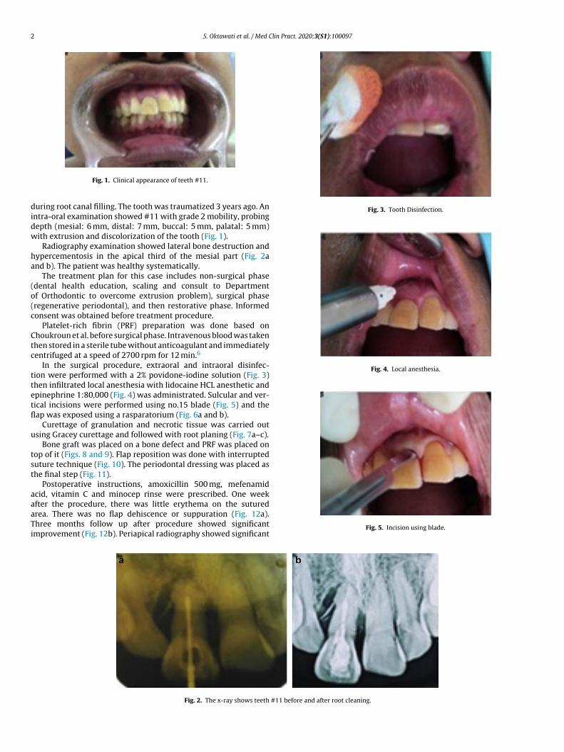
Fig. 1. Clinical appearance of teeth #11.
during root canal filling. The tooth was traumatized 3 years ago. An
intra-oral examination showed #11 with grade 2 mobility, probing
depth (mesial: 6 mm, distal: 7 mm, buccal: 5 mm, palatal: 5 mm)
with extrusion and discolorization of the tooth (Fig. 1).
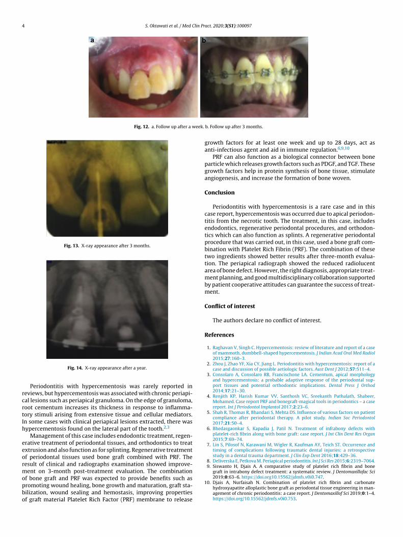
Radiography examination showed lateral bone destruction and
hypercementosis in the apical third of the mesial part (Fig. 2a
and b). The patient was healthy systematically.
The treatment plan for this case includes non-surgical phase
(dental health education, scaling and consult to Department
of Orthodontic to overcome extrusion problem), surgical phase
(regenerative periodontal), and then restorative phase. Informed
consent was obtained before treatment procedure.
Platelet-rich fibrin (PRF) preparation was done based on
Choukroun et al. before surgical phase. Intravenous blood was taken
then stored in a sterile tube without anticoagulant and immediately
centrifuged at a speed of 2700 rpm for 12 min.6
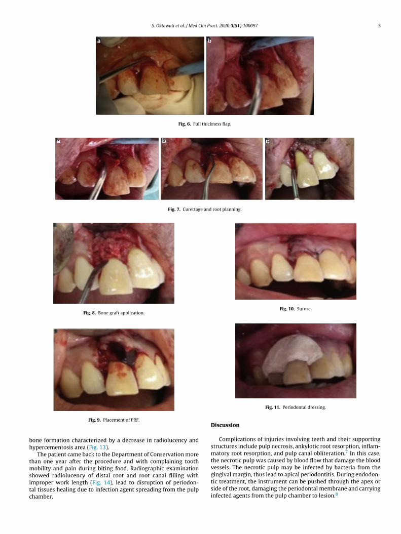
In the surgical procedure, extraoral and intraoral disinfec-
tion were performed with a 2% povidone-iodine solution (Fig. 3)
then infiltrated local anesthesia with lidocaine HCL anesthetic and
epinephrine 1:80,000 (Fig. 4) was administrated. Sulcular and ver-
tical incisions were performed using no.15 blade (Fig. 5) and the
flap was exposed using a rasparatorium (Fig. 6a and b).
Curettage of granulation and necrotic tissue was carried out
using Gracey curettage and followed with root planing (Fig. 7a–c).
Bone graft was placed on a bone defect and PRF was placed on
top of it (Figs. 8 and 9). Flap reposition was done with interrupted
suture technique (Fig. 10). The periodontal dressing was placed as
the final step (Fig. 11).
Postoperative instructions, amoxicillin 500 mg, mefenamid
acid, vitamin C and minocep rinse were prescribed. One week
after the procedure, there was little erythema on the sutured
area. There was no flap dehiscence or suppuration (Fig. 12a).
Three months follow up after procedure showed significant
improvement (Fig. 12b). Periapical radiography showed significant
Fig. 3. Tooth Disinfection.
Fig. 4. Local anesthesia.
Fig. 5. Incision using blade.
Fig. 2. The x-ray shows teeth #11 before and after root cleaning.
S. Oktawati et al. / Med Clin Pract. 2020;3(S1):100097 3
Fig. 6. Full thickness flap.
Fig. 7. Curettage and root planning.
Fig. 8. Bone graft application.
Fig. 9. Placement of PRF.
bone formation characterized by a decrease in radiolucency and
hypercementosis area (Fig. 13).
The patient came back to the Department of Conservation more
than one year after the procedure and with complaining tooth
mobility and pain during biting food. Radiographic examination
showed radiolucency of distal root and root canal filling with
improper work length (Fig. 14), lead to disruption of periodon-
tal tissues healing due to infection agent spreading from the pulp
chamber.
Fig. 10. Suture.
Fig. 11. Periodontal dressing.
Discussion
Complications of injuries involving teeth and their supporting
structures include pulp necrosis, ankylotic root resorption, inflam-
matory root resorption, and pulp canal obliteration.7 In this case,
the necrotic pulp was caused by blood flow that damage the blood
vessels. The necrotic pulp may be infected by bacteria from the
gingival margin, thus lead to apical periodontitis. During endodon-
tic treatment, the instrument can be pushed through the apex or
side of the root, damaging the periodontal membrane and carrying
infected agents from the pulp chamber to lesion.8
4 S. Oktawati et al. / Med Clin Pract. 2020;3(S1):100097
Fig. 12. a. Follow up after a week. b. Follow up after 3 months.
Fig. 13. X-ray appearance after 3 months.
Fig. 14. X-ray appearance after a year.
Periodontitis with hypercementosis was rarely reported in
reviews, but hypercementosis was associated with chronic periapi-
cal lesions such as periapical granuloma. On the edge of granuloma,
root cementum increases its thickness in response to inflamma-
tory stimuli arising from extensive tissue and cellular mediators.
In some cases with clinical periapical lesions extracted, there was
hypercementosis found on the lateral part of the tooth.2,3
Management of this case includes endodontic treatment, regen-
erative treatment of periodontal tissues, and orthodontics to treat
extrusion and also function as for splinting. Regenerative treatment
of periodontal tissues used bone graft combined with PRF. The
result of clinical and radiographs examination showed improve-
ment on 3-month post-treatment evaluation. The combination
of bone graft and PRF was expected to provide benefits such as
promoting wound healing, bone growth and maturation, graft sta-
bilization, wound sealing and hemostasis, improving properties
of graft material Platelet Rich Factor (PRF) membrane to release
growth factors for at least one week and up to 28 days, act as
anti-infectious agent and aid in immune regulation.6,9,10
PRF can also function as a biological connector between bone
particle which releases growth factors such as PDGF, and TGF. These
growth factors help in protein synthesis of bone tissue, stimulate
angiogenesis, and increase the formation of bone woven.
Conclusion
Periodontitis with hypercementosis is a rare case and in this
case report, hypercementosis was occurred due to apical periodon-
titis from the necrotic tooth. The treatment, in this case, includes
endodontics, regenerative periodontal procedures, and orthodon-
tics which can also function as splints. A regenerative periodontal
procedure that was carried out, in this case, used a bone graft com-
bination with Platelet Rich Fibrin (PRF). The combination of these
two ingredients showed better results after three-month evalua-
tion. The periapical radiograph showed the reduced radiolucent
area of bone defect. However, the right diagnosis, appropriate treat-
ment planning, and good multidisciplinary collaboration supported
by patient cooperative attitudes can guarantee the success of treat-
ment.
Conflict of interest
The authors declare no conflict of interest.
References
1. Raghavan V, Singh C. Hypercementosis: review of literature and report of a case
of mammoth, dumbbell-shaped hypercementosis. J Indian Acad Oral Med Radiol2015;27:160–3.
2. Zhou J, Zhao YF, Xia CY, Jiang L. Periodontitis with hypercementosis: report of a
case and discussion of possible aetiologic factors. Aust Dent J 2012;57:511–4.
3. Consolaro A, Consolaro RB, Francischone LA. Cementum, apical morphologyand hypercementosis: a probable adaptive response of the periodontal sup-
port tissues and potential orthodontic implications. Dental Press J Orthod2014;17:21–30.
4. Renjith KP, Harish Kumar VV, Santhosh VC, Sreekanth Puthalath, Shabeer,Mohamed. Case report PRF and bonegraft-magical tools in periodontics – a casereport. Int J Periodontol Implantol 2017;2:23–6.
5. Shah R, Thomas R, Bhandari S, Mehta DS. Influence of various factors on patientcompliance after periodontal therapy. A pilot study. Indian Soc Periodontol2017;21:50–4.
6. Bhedasgaonkar S, Kapadia J, Patil N. Treatment of infrabony defects with
platelet-rich fibrin along with bone graft: case report. J Int Clin Dent Res Organ
2015;7:69–74.
7. Lin S, Pilosof N, Karawani M, Wigler R, Kaufman AY, Teich ST. Occurrence and
timing of complications following traumatic dental injuries: a retrospective
study in a dental trauma department. J Clin Exp Dent 2016;18:429–36.8. Deliverska E, Petkova M. Periapical periodontitis. Int J Sci Res 2015;6:2319–7064.
9. Siswanto H, Djais A. A comparative study of platelet rich fibrin and bone
graft in intrabony defect treatment: a systematic review. J Dentomaxillofac Sci
2019;0:63–6. https://doi.org10.15562/jdmfs.v0i0.747.
10. Djais A, Nurfaisah N. Combination of platelet rich fibrin and carbonatehydroxyapatite alloplastic bone graft as periodontal tissue engineering in man-agement of chronic periodontitis: a case report. J Dentomaxillof Sci 2019;0:1–4.https://doi.org/10.15562/jdmfs.v0i0.753.