the long-term neurocompatibility of human fibrin sealant and equine collagen as biomatrices in...
TRANSCRIPT
ARTICLE IN PRESS
EXPERIMENTAL
ANDTOXICOLOGIC
PA THOLOGY
0940-2993/$ - se
doi:10.1016/j.et
�CorrespondE-mail addr
(A.H. Petter-Pu
Experimental and Toxicologic Pathology 58 (2007) 237–245
www.elsevier.de/etp
The long-term neurocompatibility of human fibrin sealant and
equine collagen as biomatrices in experimental spinal cord injury
Alexander H. Petter-Puchnera,�, Wolfgang Froetschera, Reinhild Krametter-Froetscherb,Dragan Lorinsonc, Heinz Redla, Martijn van Griensvena
aLudwig Boltzmann Institute of Experimental and Clinical Traumatology, Donaueschingenstrasse 13, 1200-Vienna, AustriabII. Medical University Clinic for Ruminants and Swine, Veterinary University of Vienna, AustriacClinic of Veterinary Surgery and Ophthalmology, Veterinary University of Vienna, Austria
Received 27 April 2006; accepted 30 July 2006
Abstract
Introduction: While fibrin sealant (FS) and equine collagen (EC) have been used as scaffold materials inexperimental spinal cord injury (SCI), questions concerning neurocompatibility still remain. In this study, we assessedpotential adverse effects, as well as functional and histological impact of FS and EC in subtotal hemisection of thethoracic spinal cord (SC) in rats.
Methods: 124 male rats were randomly assigned to four main groups (n ¼ 31): Sham (SH), Lesion only (L), fibrinsealant (GFS) and equine collagen group (GEC). SH animals received laminectomy only; all other animals underwentsubtotal lateral hemisection at T9. Treatment consisted of application of FS or EC into the lesion gap in GFS andGEC, which was left empty in L. GFS, GEC, L and SH were each further divided into 4 subgroups: One subgroup,consisting of 10 rats was subjected to behavioural and reflex testing before surgery and followed up on days 1,7, 14, 21,28 post op and then sacrificed. Haemalaun or cresyl violet (CV) was used to identify neutrophils in parasagittal cordsections which were obtained on day 1 (n ¼ 7). Sections stained for quantification of microglia/macrophages using ED-1 on day 3 (n ¼ 7), day 7 (n ¼ 7) and day 28 (n ¼ 7 out of 10). Additionally, neural filament (NF) staining was chosento detect axonal regeneration and the length of ingrowth into FS and EC, Luxol blue for myelination, Von Willebrandfactor for vascularisation, and glial fibrillary acidic protein (GFAP) staining for detection of astrocytes in glial scars onday 28.
Results: No adverse effects were observed in the treatment groups. Compared to L, GFS and GEC performedsignificantly better in the Basso, Beattie, Bresnahan (BBB) score and hopping responses. Proprioceptive placing wasmarkedly improved in FS and EC compared to L. Axonal regrowth was found in GFS and GEC – the regrowth in theGFS was accompanied by myelination and vascularisation. Glial scarring occurred in all groups. Discussion
Both biomatrices improved functional recovery compared to L and no adverse effects were perceived.r 2006 Elsevier GmbH. All rights reserved.
Keywords: Sprague-Dawley rat; Fibrin sealant; Collagen; Spinal cord injury; Biocompatibility
e front matter r 2006 Elsevier GmbH. All rights reserved.
p.2006.07.004
ing author. Fax: +43133110 460.
ess: [email protected]
chner).
Introduction
While continuous efforts in research provide betterinsights in pathomechanisms after spinal cord injury
ARTICLE IN PRESSA.H. Petter-Puchner et al. / Experimental and Toxicologic Pathology 58 (2007) 237–245238
(SCI), it remains a devastating condition with fewtherapeutical options in a young patient population(Bracken, 2002; Hulsebosch, 2002). Current strategiesfail to significantly enhance the regrowth of severedaxons in the adult human central nervous system, whichis impaired by different factors. Deleterious effects ofthe inflammatory response, e.g. neutrophil response(Taoka et al., 1998; Lee et al., 2000; Benveniste, 1992),specific neurite inhibitory reactions of the glial environ-ment (Schwab, 1990), excitotoxicity (Hermann et al.,2001) and apoptosis (Lee et al., 2000; Shuman et al.,1997) contribute to the aggravation of the initial damageand to the lack of substantial recovery after SCI.
According to literature, biomatrices could be espe-cially important in the treatment of transection orlaceration types of SCI, when a lesion gap has to bebridged – acting as carriers for therapeutic agents, suchas neurotrophic factors or stem cells (Iwakawa et al.,2001; Teng et al., 2002; Cheng et al., 2005). Fibrinsealant (FS) and equine collagen (EC) have beenproposed to possess desirable properties in the centralnervous system (Yoshii et al., 2003; Iwaya et al., 1999),where they are already applied by neurosurgeons ashemostatics or as sealants of leaks of the cerebrospinalfluid (CSF) (Novikova et al., 2003; Kataoka et al., 2004;Woerly et al., 2001; Cappabianca et al., 2006). FS hasalso proven its potential and benefit as neuroprotheticsin experimental neurotrauma (Ornelas et al., 2006a, b).Still, concerns were raised about safety and potentialimmunogenicity due to the allogenic character of bothcompounds. Attempts to replace bovine aprotinin (forinhibiting the degradation of the FS) in FS by TA haveproved to be detrimental in experimental settings(Schlag et al., 2002; Furtmuller et al., 2002). FScontaining TA evoked epileptic seizures, when it wasapplied directly to the spinal cord (SC). In order toprovide final evidence of the safety of TA-free FS andEC as scaffold materials for future local therapies (e.g.stem cell transplantation or deliverance of neurotro-phines) after SCI (Joosten et al., 1995; Murray, 2004),the present study was designed to elucidate neurocom-patibility and to rule out inflammatory or immunogenicresponses which could result in systemic adverse effectsand amplified secondary damage.
Materials and methods
Animal care
The experimental protocol and all animal procedureswere permitted by the Animal Care Committee of theCity of Vienna, Austria. The care and handling ofanimals was in accordance with national guidelines.Male Sprague-Dawley rats were obtained from the
’’Institut fuer Labortierkunde und – genetik der
Medizinischen Fakultaet der Universitaet Wien’’ (Him-berg, Austria). Animals were allowed to accommodatefor 1 week before surgery. Throughout the study period,pelleted rat chow and water were available ad libitum.During the first 48–72 h following surgery, rats receiveddaily subcutaneous injections of Ringer’s solution andantibiotics (50,000 IU procaine-penicillin-G, 50,000 IUdihydro-streptomycin sulfate; Omnamycin, Hoechst).Manual bladder expression was carried out if necessary.
Group assignment
124 male Sprague-Dawley rats (350–450 g) wererandomly assigned to one of the four main groupssham (SH), lesion only (L), fibrin sealant (GFS) andequine collagen (GEC) (n ¼ 31). These main groupswere all divided in three subgroups of 7 animals each tobe sacrificed on day 1, 3, 7 and one subgroup of 10animals to be sacrificed on day 28 (3� 7 animals plus 10animals ¼ 31 animals per main group). The group ofanimals surviving until day 28 were undergoing beha-vioural and reflex testing, as well as final histologicalexamination. 7 tissue samples of the 28 day subgroupwere randomly selected for histology to maintain equalgroup size for all observation periods.
SC surgery and biomatrix implantation
Anaesthesia was induced with 100mg/kg ketamine(Ketalars, Parke-Davis, Berlin, Germany) and 25mg/kg xylazine (Rompuns, Bayer, Leverkusen, Germany)intraperitoneally. Subsequently, 0.3mg/kg atropine(Atropinum sulfuricum; Nycomed, Vienna, Austria)was applied subcutaneously. Half of the initial keta-mine/xylazine and atropine dose was administered at30min intervals to maintain anaesthesia. Body tempera-ture was monitored rectally and maintained at 37–38 1Cusing a heating pad.
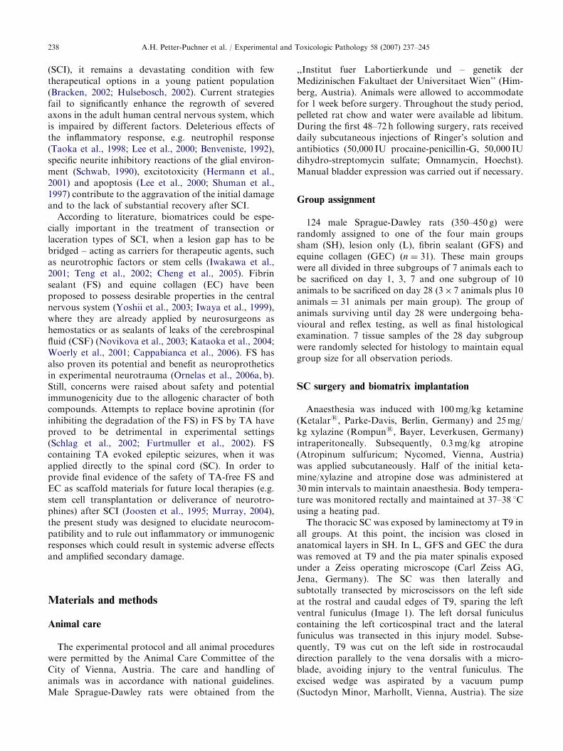
The thoracic SC was exposed by laminectomy at T9 inall groups. At this point, the incision was closed inanatomical layers in SH. In L, GFS and GEC the durawas removed at T9 and the pia mater spinalis exposedunder a Zeiss operating microscope (Carl Zeiss AG,Jena, Germany). The SC was then laterally andsubtotally transected by microscissors on the left sideat the rostral and caudal edges of T9, sparing the leftventral funiculus (Image 1). The left dorsal funiculuscontaining the left corticospinal tract and the lateralfuniculus was transected in this injury model. Subse-quently, T9 was cut on the left side in rostrocaudaldirection parallely to the vena dorsalis with a micro-blade, avoiding injury to the ventral funiculus. Theexcised wedge was aspirated by a vacuum pump(Suctodyn Minor, Marhollt, Vienna, Austria). The size

ARTICLE IN PRESS
T9
Right
Left
Image 1. Subtotal hemisection. This image serves as illustra-
tion of the surgical procedure. The left ventral funiculus was
removed on T9, whereas the contralateral side was spared in
toto, as well as the dorsal tracts and central vessels
ipsilaterally.
A.H. Petter-Puchner et al. / Experimental and Toxicologic Pathology 58 (2007) 237–245 239
of the defect was assessed with the operating microscopeand the lesion gap was then flushed with isotonicRinger solution (4 1C). This specific type of lesion waschosen to target two aims. Firstly, this lesion shouldcreate an adequate neurological deficit to detect treat-ment specific improvement of neurological deficits.Secondly, this model spares tracts providing motorfunction in order to avoid spastic sequelae and to beable to distinguish seizures as adverse effects oftreatment.
In GFS, the lesion gap was filled with 0.2ml of humantwo component FS (Tissucols, Baxter, Vienna, Austria)delivered by a proprietary double syringe fitted withan applicator needle (Duplojects, Baxter, Vienna,Austria). Of note, Tissucols is TA-free. In GEC, anEC sponge (TissuFleece Es, Baxter, Vienna, Austria)was fit into gap. TissuFleece Es collagen sponge andTissucols FS were provided by Baxter AG (Vienna,Austria). The dura was closed in all animals using apatch of Lyoduras (Braun, Melsungen, Germany). Theincision was closed in anatomical layers.
Behavioural function test
Motor function and reflexes were tested in all ratsby two veterinarians (W.F. and R.K.) prior to surgery,on the first day post-operative and then weekly ondays 7, 14, 21 in a blinded manner. Test protocolsrequired both observers to be present and actively scoreat the same time (Basso et al., 1995). In case ofdiscrepancies between ratings, the lower (worse) valuewas accepted.
Basso, Beattie, Bresnahan (BBB)-score
The function of both hindlimbs was scored with theBBB locomotor rating scale (Basso et al., 1995). Resultswere calculated as percentages relative to the maximumscore of 21, considered as 100%.
Reflex tests
Reflex tests were chosen to assess recovery and todetect adverse effects of treatment (e.g. increasedexcitability and susceptibility for seizures).
Tactile placing reflex
Proprioceptive integrity of rats was assessed qualita-tively, eliciting the tactile placing reflex of the hindlimbs.Normal reflex response comprised brisk and reliableflexing and extension movements at the edge of a tableresulting in a supporting posture. A normal tactileplacing response was considered present if more than30% of the stimuli induced a reflex.
Monopedal hopping reflex
The monopedal hopping reflex also served for testingof the proprioceptive integrity. Hindlimb hopping wastested in lateral and medial directions. The rat was heldgently over soft plastic padding by all limbs except theone being tested. Hindlimb hops were elicited mosteffectively by moving a rat over the surface with its bodyheld parallelly over the ground and the nose pointingslightly downwards. When a rat was moved it lifted thelimb and replaced it under the new centre of gravity.Three continuous, vigorous but graceful hops laterallyand three medially were demanded to achieve a fullscore of 1. Fewer hopping movements laterally and/ormedially, sluggish, ungraceful, larger movements weregraded 0 (Glassman, 1994).
Joint-bend placing
Proprioceptive (joint-bend) placing was qualitativelyassessed by using pressure to elicit limb displacement at theedge of a table. The rats were supported as above. Thestimulus consisted of a significant displacement of the limb(i.e. hip and knee extension and ankle plantar flexion) afterthe dorsum of the paw touched the edge of the platform. Anormal placing reflex consisted of responses in at least30% of the repetitions (Donatelle, 1977).
Foot orienting response and toe spreading
When rats without SCI are lifted and lowered by theproximal tail vertebrae, fanning of toes from the

ARTICLE IN PRESSA.H. Petter-Puchner et al. / Experimental and Toxicologic Pathology 58 (2007) 237–245240
hindlimb can be observed. Flaccid or retracted hin-dlimbs were scored 0. Lateral movement of thehindlimbs with toe spreading when the animal waslowered, or outward rotation of the foot when lifted,scored as 1 (Gruner et al., 1996). This procedure allowsqualitative testing of the reflex.
Histology
At the designated time points, rats were transcardiallyperfused in deep anaesthesia (as for surgery) with 100mlof heparinized Ringer’s solution followed by 100ml of4.5% paraformaldehyde in 0.1M phosphate buffer (pH7.4) at 4 1C. The middle thoracic SC was removed, post-fixed in the same solution, dehydrated and embedded inparaffin. Parasagittal serial sections (4–5 mm thick) werestained with hematoxylin and eosin (HE) for generalmorphology, cresyl violet (CV) and haemalaun on dayone post-operatively for identifying neutrophils, andluxol fast blue (LFB) at 28 days post-operatively forinvestigating the presence of myelin. Sections forcounting inflammatory cells at 3, 7 and 28 days post-operatively were viewed with a Polyvar microscope(Reichert, Vienna, Austria). The rostrocaudal extent ofthe lesion was observed and photographed on anAxioplans 2 microscope (Zeiss, Wetzlar, Germany) onday 1 and 28 post-operatively. Morphometrical mea-surement of the lesion (rostrocaudal extension) wasperformed with LUCIA morphometrics analysis soft-ware (Prague, CZ).
Immunohistochemistry
Parasagittal sections (4–5 mm thick) were cut fromparaffin-embedded specimens. Following blocking forendogenous peroxidase with H2O2 and non-specificantibody binding with bovine serum albumin (BSA,Serva, Heidelberg, Germany). Sections were incubatedat room temperature overnight with a range of primaryantibodies. The following primary antibodies were usedfor labelling specific proteins in cells:
�
Mouse monoclonal anti-rat ED-1 (ED-1, Serotec,Dusseldorf; Germany; diluted 1:100) for detection ofinflammation (macrophages/microglia) at all time-points of observation. � Polyclonal antibody (GFAP, Dako, Vienna, Austria;Code no. Z0334, diluted 1:100) for detection of glialfibrillary acidic protein (GFAP) on day 28 as markerfor astrocyte response.
� Rabbit monoclonal anti-human Von Willebrandfactor (vWF, Dako, Vienna, Austria; Code no.A0082, diluted 1:3000) to trace angiogenesis (en-dothelial cells) – on day 28.
� Mouse monoclonal anti-human neurofilament pro-tein (NF, Dako, Vienna, Austria; Code no. M0762,
diluted 1:100) was used to detect newly formed axons(axonal regrowth) entering the biomatrices or thelesion gap only. The parameter of interest was thelength of ingrowth on day 28.
All primary antibodies were diluted with 1% bovineserum albumin in phosphate-buffered saline solution.After incubation with the specific primary antibody,sections were washed and incubated with secondaryantibody (biotinylated multi-link swine anti-goat,mouse, rabbit, Dako Code no. E0453, diluted 1:100)followed by the Dako substrate for peroxidase stainingand visualisation with 3-amino-9-ethycarbazol (AEC).Absence of primary antibody served as a negativecontrol. Several sections, e.g. ED-1 sections, werecounter-stained with haemalaun (Mayer’s Protocol) todifferentiate between grey and white matter.
Counting neutrophils and macrophages/microglia
Cells in sagittal SC sections were counted as describedby Schnell et al. (1994). Neutrophils and macrophages/microglia were counted at 400� magnification. In thegrey matter all labelled cells within 40 squares of0.025mm2 each of a graticule were counted at threeregions of equal size rostrally or caudally from the lesionsite. The distance between the three regions was 0.2mm,and the regions were lying parallel to one another in thelongitudinal axis covering the grey matter starting at thelesion site (centre). Three sections of each SC wereseparately analysed and averaged. The mean value fromthe combined data of all 7 animals in each group wascalculated.
Axonal regrowth
Axonal regrowth was measured according to thelength of NF positive axons inside the biomatrices.
The length of ingrowth of NF-positive fibers into theFS (n ¼ 7) and EC (n ¼ 7) biomatrices was measured inmillimetres on day 28 post-operatively.
Extent of lesion
The rostrocaudal extent of the lesion was measuredon day 1 and day 28.
Statistics
For analysis of the locomotor (BBB) rating scaleresults the one-way ANOVA followed by Tukey’sMultiple Comparison test was employed. The w2-testwas used to compare results of reflex tests.
Differences in the rostrocaudal extent of the lesioncavities were analysed at day 1 and day 28 post-operatively with the unpaired t-test. Differences between

ARTICLE IN PRESSA.H. Petter-Puchner et al. / Experimental and Toxicologic Pathology 58 (2007) 237–245 241
groups in the length of NF-positive axonal regrowthinto the biomatrices were analysed by the Kruskal–Wallis-test.
Inflammatory cell counts between groups werecompared using one-way ANOVA followed by Tukey’sMultiple Comparison Test.
GraphPad software was used for data analysis (PrismV3.02, GraphPad-Software, San Diego, USA). In allcases p-values o0.05 were considered significant.
Results
Behavioural function tests
BBB scores
All FS treated rats (GFS, n ¼ 10 after 28 days)achieved a median BBB score of 10 for the left hindlimbafter the first day post-operatively, a BBB score of 15 bythe first week, 20 after the second, third and fourth weekpost-operatively. The median score of the EC-treatedrats (GEC, n ¼ 10 after 28 days) was 15 after the firstday post-operatively, plateaued at 15–16 after the firstweek post-operatively and then continuously improvedtill the end of the observation period similar to GFS,signifying full coordination of both hindlimbs. All ratsfrom group L (n ¼ 10) achieved a median BBB score of9 for the left hindlimb after the first day post-operatively, surpassed a score of 10 after the first weekpost-operatively and improved till the third week postoperatively to a value of 17–18. Recovery in L rats wasslow and significantly impaired in comparison with GFS(po0.05) and GEC (po0.01), it peaked in the thirdweek and deteriorated during the fourth week (Fig. 1).Motor function recovery was homogenous withingroups and unsuspicious for adverse effects related to
0
10
20
30
40
50
60
70
80
90
1 7 14
Post Lesion Time [days]
Median, Q1, Q3
L
EC
FS
p<0.01
% R
ange
Del
ta M
oto
r S
core
21 28
Fig. 1. BBB scores in groups L, GEC and GFS (n ¼ 10). The
scores of the left hindlimb (injury side) are shown. Each bar
was rescaled from 0% to 100% (minimum-maximum BBB
score). Recovery was most pronounced and EC group, while it
deteriorated in group L. Differences were significant between
treatment groups and lesion only on day 28 (po0.05) with a
trend during the whole observation period.
treatment regimens. The fast recovery in the BBBreflected the sparing of the left ventral funiculus (Bassoet al., 1995).
Contralateral hindlimb scores showed full recovery ofmotor function in all groups on the right side within the firstweek post-operatively and were not statistically different.
Reflex testing
Hopping response and toe spreading
Significant differences were detected between L andGFS and GEC in the second and third week post-operatively in hopping responses and toe spreading(both po0.05). In the treatment groups, significantimprovement in toe spreading was also found in thefourth week. Nine out of 10 animals in GFS and GEC,but only five out of 10 animals in L showed toespreading of the lesion-ipsilateral hindlimb after thefourth week post-operatively.
Proprioceptive placing
Significant differences in proprioceptive placing be-tween GFS, GEC and L were observed from the secondweek post-operatively until the end of the observationperiod (Fig. 2).
No differences in reflex responses occurred in theanimals right hindlimbs (data not shown). No seizuresoccurred during reflex testing.
Histology
All samples from L, GFS and GEC included thewhole lesion area at T9. SH-operated controls showedno evidence of intraparenchymal inflammation or tissuedestruction and had very few dural adhesions. Allanimals which underwent subtotal lateral transectionwere included in the study (no exclusion).
10
Median, Q1, Q3
LECFS
p<0.05
9
8
7
6
5
4
3
2
1
07 14
Post Lesion Time [days]
Num
ber
of
Rat
s
21 28
Fig. 2. Proprioceptive joint bend reflex of the left hindlimb in
groups L, GFS and GEC (n ¼ 10). The number of rats in
which a normal proprioceptive placing response could be
elicited was significantly increased in treatment groups
compared to controls after 28 days. This finding indicates
superior recovery of proprioceptive function due to treatment.

ARTICLE IN PRESS
3 days120
a
Mac
rop./
mic
rog. per
0.0
25 m
m2
100
80
60
40
20
00 0.2
Regions [mm]
0.4
Median, Q1, Q3LECFS
p<0.001
p<0.05
A.H. Petter-Puchner et al. / Experimental and Toxicologic Pathology 58 (2007) 237–245242
Recruitment of inflammatory cells
At days 3 (Fig. 3a), 7 and 28 the numbers anddistribution of macrophages/microglia in grey matterwere markedly different between groups, while parench-ymal infiltration by cells had increased by day 7 (Fig. 3b)and declined by day 28, especially in groups L and FS(Fig. 3c).
Slight differences in ‘‘monoclonal mouse anti-rat ED-1’’ stained for quantification of microglia/macrophageswere observed between groups on the first day post-operatively, but were not statistically significant. Nosigns of foreign body reaction, elicited by FS or EC,were detected.
b120
icro
g. per
0.0
25 m
m2
100
80
60
Median, Q1, Q3
LECFS
p<0.001
p<0.05
7 days
Astrogliosis (astrocytes)
A pronounced astrocyte response was present in L,GFS and GEC 28 days post-operatively. In GFS,GFAP-positive branches of thickened astrocytes pene-trated the biomatrix at the lesion site (Fig. 4).
c
Mac
rop./
m 40
20
00 0.2
Regions [mm]
0.4
120
. per
0.0
25 m
m2
100
80
Median, Q1, Q3
L
EC
FS
p<0.001
p<0.05
p<0.01
28 days
Angiogenesis (endothelial cells)
At day 28 post-operatively, vWF-positive endothelialcells were detected in the FS biomatrix, indicating neo-angiogenesis. The regenerated vessel walls were intenselystained and irregular but predominately orientatedalong the transverse axis of the SC (Fig. 5). vWF-positive endothelial cells were neither observed insidethe biomatrix of GEC, nor inside the scar tissue of L(only a few at the lesion site).
Mac
rop./
mic
rog 60
40
20
0
0 0.2
Regions [mm]
0.4
Fig. 3. (a) Recruitment of inflammatory cells in groups L,
GFS and GEC 3 days post operatively (n ¼ 7). Histology
showed a strong inflammatory response triggered by macro-
phages in all groups. This response was more pronounced in
treatment groups. Interestingly enough, these differences did
Axonal regrowth (neurofilament) and NF length inside the
biomatrices
NF was used to detect regrowing axons. No NF-positive fibers were noted within the lesion centre 28days after injury in L. In EC-treated rats, NF-positivefibers invaded the biomatrix longitudinally and trans-versely (Fig. 6). In FS biomatrix, numerous NF-positivefibers were observed. Axons were noted to grow forrelatively long distances (40.5mm) in both biomatricesand they surrounded the cystic cavities (Figs. 7 and 8).
not impair behavioural and reflex tests, performed at the same
timepoint. (b) Recruitment of inflammatory cells in groups L,
GFS and GEC 7 days post operatively (n ¼ 7). Macrophage/
microglia infiltration was not significantly attenuated com-
pared to day 3, but leveled between GFS and GEC in two out
of three sections. (c) Recruitment of inflammatory cells in
groups L, GFS and GEC 28 days post operatively (n ¼ 7). At
day 28 recruitment of macrophages and microglia had
Myelin (remyelination)
LFB staining at day 28 suggested that some remye-lination of ingrown axons had taken place inside thebiomatrix in GFS (Fig. 9). This could not be seen inGEC or L.
substantially decreased and no difference between L and
GFS could be detected. The situation in GEC was marked by
significantly higher levels of inflammatory cells in all sections.
Extent of lesionLesion size appeared homogenous in all groups onday 1 and day 28 and indicated good reproducibility ofthe lesion model.

ARTICLE IN PRESS
Fig. 4. GFAP-positive stained astrocytes. GFS, 28 days
postoperatively – GFAP-positive branches of thickened
astrocytes penetrated FS as indicated by arrow; magnification
� 640.
Fig. 5. FS Biomatrix vWF-positive stained. GFS (counter-
staining with haemalaun), 28 days postoperatively – the image
shows endothelial cells forming lumina (angiogenesis) in the
degraded FS; magnification � 640.
Fig. 6. NF-positive axonal regrowth in EC. GEC, 28 days
postoperatively – NF-positive axons appear as red lines,
compare the sparse and irregular distribution to situation in
FS (as shown in Fig. 6); magnification � 400.
Fig. 7. NF-positive axonal regrowth in FS. GFS, 28 days
postoperatively – FS matrix has been already degraded and a
dense, mostly longitudinal ingrowth of NF positive axons can
be observed; magnification � 400.
A.H. Petter-Puchner et al. / Experimental and Toxicologic Pathology 58 (2007) 237–245 243
Discussion
The results presented in this study demonstrate thatthe implantation of FS or EC in the SC is safe and yieldsimprovement of recovery in a rat model of subtotalspinal hemisection. No adverse effects, e.g. seizures or
immune reaction to the biomaterials were elicited byneither TA-free FS or by EC. Substantial benefits werefound in terms of motor function and proprioceptiverecovery, when compared to a control group. FS- andEC-treated animals showed no differences in the BBB inthe first, third and fourth week post-operatively, butwere superior to the L group by the end of theobservation period. The BBB score is highly sensitiveand reveals subtle qualities of hindlimb movement. Incontrast to the steady improvement in treatmentanimals, the deterioration of the BBB in the L group

ARTICLE IN PRESS
3
2
p<0.05
LECFS
Median,
Q1, Q3
1
NF
into
les
ion c
avit
y [
mm
]
0
Day 28
Post Lesion Time-Point
Fig. 8. NF length inside the biomatrices in groups L, GFS and
GEC after 28 days (n ¼ 7). Quantification of axonal regrowth
of NF-positive axons in FS and EC groups. No NF-positive
regrowth of axons was observed in the lesion gap of the L
group.
Fig. 9. Remyelination in FS. Remyelination of regenerated
axons was only found in GFS, 28 days post-operatively.
Colours were reversed in this image to better depict
remyelination as red structures in this luxol fast blue staining;
magnification � 640.
A.H. Petter-Puchner et al. / Experimental and Toxicologic Pathology 58 (2007) 237–245244
indirectly indicates positive effects of FS and EC onattenuating late aggravation of injury. The results ofbehavioural and reflex testing were confirmed byhistology, which confirmed the excellent compatibilityof FS and EC. In this context, the ingrowth of NF-stained neural fibers in both matrices was a promisingfeature. However, regenerating axons did not penetratethe scaffolds in their full length, a finding in accordancewith literature (Yoshii et al., 2003; Schnell and Schwab,1990; Bradbury et al., 2002). Regenerated axons did not
contribute to the significant improvement of functionalrecovery. Therefore, the reasons for the benefits oftreatment remain speculative. No attenuation of inflam-matory response could be observed in the treatmentgroups. The well-known haemostatic properties of FSand EC could serve as possible explanation (Dunn andGoa, 1999; Acheson et al., 2005). Although not directlyassessed, it seems likely that prevention of local haemor-rhages could have protected the spared neuronal tissuefrom secondary damage. A recent clinical trial based onmagnetic resonance imaging relates the size of haemor-rhages to the severeness of SCI (Boldin et al., 2006). Asuperior permissiveness of the ipsilateral funiculus andthe contra-lateral cord in GFS and GEC must beconsidered as the organic substrate for treatment effects.The fact, that extensive behavioural and reflex testing, aswell as elaborated histology could not deliver a conclusiveanswer to the precise mechanism which led to thebeneficial results, suggests further investigation withspecial focus on haemostasis, neuronal plasticity andapoptosis. Still, our results indicate that both hetero-logous biomatrices enhance neuroregenerative mechan-isms without provoking any adverse effects (Schnell et al.,1999; Rothwell and Hopkins, 1995). (Fig. 9)
Conclusion
Numerous attempts have focused on finding newstrategies leading to axonal regeneration in the adultinjured mammalian SC (McDonald and Sadowsky,2002). In context, we demonstrate that human TA-freeFS and EC provide useful biomatrices to enhanceaxonal regeneration in the subtotally transected SCwithout eliciting detectable adverse effects. Whereasregenerating axons successfully entered the biomatrices,they were unable to elongate into the milieu of the SCstumps. Using this experimental paradigm, we observedimproved functional recovery related to treatment.Further research is mandatory, because the precisemechanism on which these results are based, are notfully elucidated, yet. Aware of this limitation, weconclude that FS and EC are suitable materials to serveas scaffolds in experimental spinal cord injury.
References
Acheson EM, Kheirabadi BS, Deguzman R, Dick Jr EJ,
Holcomb JB. Comparison of hemorrhage control agents
applied to lethal extremity arterial hemorrhages in swine.
J Trauma 2005;59:865–74.
Basso DM, Beattie MS, Bresnahan JC. A sensitive and reliable
locomotor rating scale for open field testing in rats.
J Neurotrauma 1995;12:1–21.
Benveniste EN. Inflammatory cytokines within the central
nervous system: sources, function, and mechanism of
action. Am J Physiol 1992;263:C1–C16.

ARTICLE IN PRESSA.H. Petter-Puchner et al. / Experimental and Toxicologic Pathology 58 (2007) 237–245 245
Boldin C, Raith J, Fankhauser F, Haunschmid C, Schwantzer
G, Schweighofer F. Predicting neurologic recovery in
cervical spinal cord injury with postoperative MR imaging.
Spine 2006;31:554–9.
Bracken MB. Steroids for acute spinal cord injury, Cochrane
Database Syst. Review 2002;CD001046.
Bradbury EJ, Moon LD, Popat RJ, King VR, Bennett GS,
Patel PN, et al. Chondroitinase ABC promotes functional
recovery after spinal cord injury. Nature 2002;416:636–40.
Cappabianca P, Esposito F, Cavallo LM, Messina A, Solari
D, di Somma LG, et al. Use of equine collagen foil as dura
mater substitute in endoscopic endonasal transsphenoidal
surgery. Surg Neurol 2006;65:144–8.
Cheng H, Huang SS, Lin SM, Lin MJ, Chu YC, Chih CL, et
al. The neuroprotective effect of glial cell line-derived
neurotrophic factor in fibrin glue against chronic focal
cerebral ischemia in conscious rats. Brain Res 2005;
1033:28–33.
Donatelle JM. Growth of the corticospinal tract and the
development of placing reactions in the postnatal rat.
J Comp Neurol 1977;175:207–31.
Dunn CJ, Goa KL. Fibrin sealant: a review of its use in
surgery and endoscopy. Drugs 1999;58:863–86.
Furtmuller R, Schlag MG, Berger M, Hopf R, Huck S,
Sieghart W, et al. Tranexamic acid, a widely used
antifibrinolytic agent, causes convulsions by a gamma-
aminobutyric acid(A) receptor antagonistic effect. J Phar-
macol Exp Ther 2002;301:168–73.
Glassman RB. Behavioral effects of SI versus SII cortex
ablations on tactile orientation-localization and postural
reflexes of rats. Exp Neurol 1994;125:125–33.
Gruner JA, Yee AK, Blight AR. Histological and functional
evaluation of experimental spinal cord injury: evidence of a
stepwise response to graded compression. Brain Res
1996;729:90–101.
Hermann GE, Rogers RC, Bresnahan JC, Beattie MS. Tumor
necrosis factor-alpha induces cFOS and strongly potenti-
ates glutamate-mediated cell death in the rat spinal cord.
Neurobiol Dis 2001;8:590–9.
Hulsebosch CE. Recent advances in pathophysiology and
treatment of spinal cord injury. Adv Physiol Educ
2002;26:238–55.
Iwakawa M, Mizoi K, Tessler A, Itoh Y. Intraspinal implants
of fibrin glue containing glial cell line-derived neurotrophic
factor promote dorsal root regeneration into spinal cord.
Neurorehabil Neural Repair 2001;15:173–82.
Iwaya K, Mizoi K, Tessler A, Itoh Y. Neurotrophic agents in
fibrin glue mediate adult dorsal root regeneration into
spinal cord. Neurosurgery 1999;44:589–95.
Joosten EA, Bar PR, Gispen WH. Collagen implants and
cortico-spinal axonal growth after mid-thoracic spinal cord
lesion in the adult rat. J Neurosci Res 1995;41:481–90.
Kataoka K, Suzuki Y, Kitada M, Hashimoto T, Chou H, Bai
H, et al. Alginate enhances elongation of early regenerating
axons in spinal cord of young rats. Tissue Eng 2004;10:
493–504.
Lee YB, Yune TY, Baik SY, Shin YH, Du S, Rhim H, et al.
Role of tumor necrosis factor-alpha in neuronal and glial
apoptosis after spinal cord injury. Exp Neurol 2000;166:
190–5.
McDonald JW, Sadowsky C. Spinal-cord injury. Lancet
2002;359:417–25.
Murray M. Cellular transplants: steps toward restoration of
function in spinal injured animals. Prog Brain Res
2004;143:133–46.
Novikova LN, Novikov LN, Kellerth JO. Biopolymers and
biodegradable smart implants for tissue regeneration after
spinal cord injury. Curr Opin Neurol 2003;16:711–5.
Ornelas L, Padilla L, Di Silvio M, Schalch P, Esperante S,
Infante PL, et al. Fibrin glue: an alternative technique for
nerve coaptation – Part I. Wave amplitude, conduction
velocity, and plantar-length factors. J Reconstr Microsurg
2006a;22:119–22.
Ornelas L, Padilla L, Di Silvio M, Schalch P, Esperante S,
Infante RL, et al. Fibrin glue: an alternative technique for
nerve coaptation – Part II. Nerve regeneration and
histomorphometric assessment. J Reconstr Microsurg
2006b;22:123–8.
Rothwell NJ, Hopkins SJ. Cytokines and the nervous system
II: actions and mechanisms of action. Trends Neurosci
1995;18:130–6.
Schlag MG, Hopf R, Zifko U, Redl H. Epileptic seizures
following cortical application of fibrin sealants containing
tranexamic acid in rats. Acta Neurochir (Wien) 2002;
144:63–9.
Schnell L, Schwab ME. Axonal regeneration in the rat
spinal cord produced by an antibody against myelin-
associated neurite growth inhibitors. Nature 1990;343:
269–72.
Schnell L, Schneider R, Kolbeck R, Barde YA, Schwab ME.
Neurotrophin-3 enhances sprouting of corticospinal tract
during development and after adult spinal cord lesion.
Nature 1994;367:170–3.
Schnell L, Fearn S, Klassen H, Schwab ME, Perry VH. Acute
inflammatory responses to mechanical lesions in the CNS:
differences between brain and spinal cord. Eur J Neurosci
1999;11:3648–58.
Schwab ME. Myelin-associated inhibitors of neurite growth
and regeneration in the CNS. Trends Neurosci 1990;13:
452–6.
Shuman SL, Bresnahan JC, Beattie MS. Apoptosis of
microglia and oligodendrocytes after spinal cord contusion
in rats. J Neurosci Res 1997;50:798–808.
Taoka Y, Okajima K, Uchiba M, Murakami K, Harada N,
Johno M, et al. Activated protein C reduces the severity
of compression-induced spinal cord injury in rats by
inhibiting activation of leukocytes. J Neurosci 1998;18:
1393–8.
Teng YD, Lavik EB, Qu X, Park KI, Ourednik J, Zurakowski
D, et al. Functional recovery following traumatic spinal
cord injury mediated by a unique polymer scaffold seeded
with neural stem cells. Proc Natl Acad Sci USA 2002;
99:3024–9.
Woerly S, Doan VD, Evans-Martin F, Paramore CG, Peduzzi
JD. Spinal cord reconstruction using NeuroGel implants
and functional recovery after chronic injury. J Neurosci Res
2001;66:1187–97.
Yoshii S, Oka M, Shima M, Akagi M, Taniguchi A. Bridging a
spinal cord defect using collagen filament. Spine 2003;
28:2346–51.