the knee :’) · juxtapatellar hollow test : normally, when the knee is flexed, a hollow appears...
TRANSCRIPT
THE KNEE :’) Abdallah Al Jazzazi
ANATOMY
It is the largest and most complex joint in the body. The knee is a modified hinge joint combining two articulations, the tibiofemoral and the patellofemoral. The fibula is not part of the joint.
The tibiofemoral joint is inherently unstable, depends on ligaments and muscles for stability
The patellofemoral joint is so shaped that the patella moves in a shallow path (or track) between the femoral condyles; if this track is too shallow, the patella readily dislocates; if its line is abnormal, the patellar articular cartilage is subject to excessive wear.

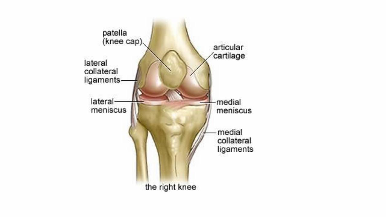
COMPONENTS
Articular bodies
Articular capsule
Bursae
Cartilage(hyaline)
Menisci (Fibrocartilage)
-Play a role in shock absorption, and may be cracked, or torn, when the knee is forcefully rotated and/or bent.
Ligaments
-Anterior & posterior cruciate ligaments
-Medial & lateral collateral ligaments
COMMON PRESENTING SYMPTOMS
Pain, with acute or gradual onset, is the most common knee symptom. Diffuse if degenerative, localized if due to injury or trauma(mechanical).
Swelling may be diffuse or localized. Important to know how fast swelling appeared.(immediate hemarthrosis, after a few hourstorn meniscus or articular cartilage)
Stiffness must be distinguished from inhibition of movement due to pain or simple weakness of the extensor apparatus.
Locking is different from stiffness. The knee, quite suddenly, cannot be straightened fully, although flexion is still possible. (due to a loose body). Pseudo-locking is when movement, usually flexion, is suddenly stopped by pain or the fear of impending pain.
Deformity is seldom a leading symptom.
Limp due to pain, deformity or instability
Loss of function manifests as a progressively decreasing walking distance, inability to run or difficulty walking up and down stairs
SIGNS TO LOOK FOR WHILE PATIENT IS UPRIGHT
Deformity (Valgus, Varus, fixed-flexion or hyperextension). Normal to have slight valgus(5 degrees in men, 7 in women), genu valgum or varum determined in relation to normal alignment. Make sure the deformity is in the knee, not in the lower end of the femur(previous fracture or tumor) or upper end of the tibia(malunited fracture or Paget’s).
Gait: In the stance phase note whether the knee extends fully, and see if there is any lateral or medial thrust signifying instability. In the swing phase note whether the knee moves freely or is held in one position.
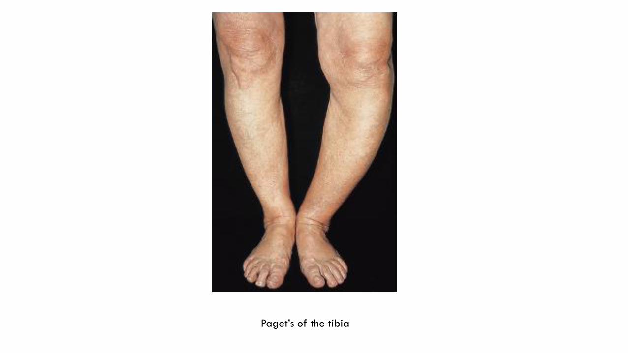
Paget’s of the tibia
SIGNS WITH PATIENT SITTING
General shape and symmetry of the two knees
With the knees at 90 degrees of flexion, the patellae should be facing straight forwards; note if they appear to be seated higher than usual (patella alta higher incidence of chondromalacia patellae.) or lower than usual (patella baja).
Observe how the patella moves with extention looking for tendency to subluxation
Patellar alignment: Q-angle(quadriceps angle) 14 degrees in men, 17 in women. If increased it is a risk factor for chondromalacia
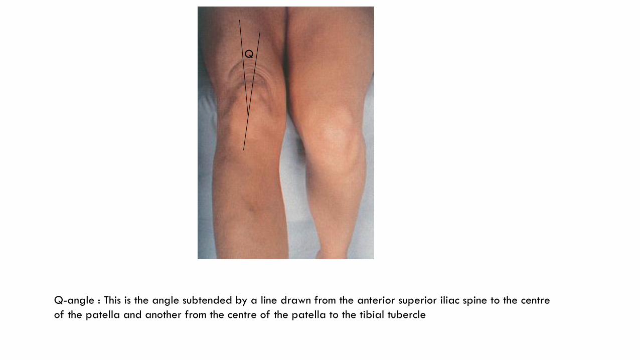
Q
Q-angle : This is the angle subtended by a line drawn from the anterior superior iliac spine to the centre
of the patella and another from the centre of the patella to the tibial tubercle

SIGNS WITH THE PATIENT LYING SUPINE
Look: Symmetry, valgus or varus, swellings, lumps, wasting of quads, bruising, old scars or sinuses
Feel: Temperature, soft tissues and bony outlines(looking for abnormal outlines and tenderness), synovial thickening (normally grasping the patella is easy, if the synovium is thickened your fingers will slip off)
Move: Passive and active extension, passive and active flexion, internal and external rotation(shouldn’t exceed 10 degrees), feel for crepitus throughout movement(signifies patellofemoral roughness)

1
2
3
4
5
6
1 quadriceps tendon; 2 edge of patella; 3 medial collateral ligament; 4 joint line; 5 lateral
collateral ligament; 6 patellar ligament

TESTS FOR INTRA-ARTICULAR FLUID Cross fluctuation : The left hand compresses and empties the suprapatellar pouch while the right hand straddles
the front of the joint below the patella; by squeezing with each hand alternately, a fluid impulse as synovial fluid moves between compartments is transmitted across the joint.
Patellar tap : The suprapatellar pouch is compressed with the left hand to squeeze any fluid from the pouch into
the joint. With the other hand the patella is then tapped sharply backwards onto the femoral condyles. In a positive test the patella can be felt striking the femur and bouncing off again (a type of ballottement).
Bulge test : After squeezing any fluid out of the suprapatellar pouch, the medial compartment is emptied by
pressing on the inner aspect of the joint; that hand is then lifted away and the lateral side is sharply compressed – a distinct ripple is seen on the flattened medial surface as fluid is shunted across. Useful if there is very little fluid!!!!!!
Juxtapatellar hollow test : Normally, when the knee is flexed, a hollow appears lateral to the patellar
ligament and disappears with further flexion; if there is excess fluid, the hollow fills and disappears at a lesser angle of flexion.
Comparison between knees is advised at all times when examining for an effusion.

a b
c d e
Testing for intra-articular fluid:
(a) The juxtapatellar hollow,
which disappears in flexion
if there is fluid in the knee.
(b) Patellar tap test.
(c,d,e) Doing the bulge test:
compress the suprapatellar
pouch (c), empty the medial
compartment (d), push fluid back
from the lateral compartment and
watch for the bulge on the medial
side (e).

TESTS FOR STABILITY Collateral ligaments The medial and lateral ligaments are tested by stressing the knee into valgus and varus: this is best done by tucking the patient’s foot under your arm and holding the extended knee firmly with one hand on each side of the joint; the leg is then angulated alternately towards abduction and adduction
Cruciate ligaments: With both knees flexed 90 degrees and the feet resting on the couch, the upper tibia is inspected from the side; if its upper end has dropped back, or can be gently pushed back, this indicates a tear of the posterior cruciate ligament (‘sag sign’). With the knee in the same position, the foot is anchored by the examiner sitting on it; then, using both hands, the upper end of the tibia is grasped firmly and rocked backwards and forwards to see if there is any anteroposterior glide (‘drawer test’). Excessive anterior movement (a positive anterior drawer sign)denotes anterior cruciate laxity and vice versa. More sensitive is the Lachman test, where the patient’s knee is flexed 20 degrees; with one hand grasping the lower thigh and the other the upper part of the leg, the joint surfaces are shifted backwards and forwards upon each other. Abnormal movement suggests an anterior cruciate ligament (ACL) injury. In both the drawer test and the Lachman test, note whether the endpoint of abnormal movement is ‘soft’ or ‘hard’.
TESTS FOR MENISCAL INJURIES
McMurray’s test: This classic test for a torn meniscus is seldom used now that the diagnosis can easily be made by MRI. However, advanced imaging is not always available and the clinical test has not been altogether discarded.
Thessaly test: This test is based on a dynamic reproduction of load transmission in the knee joint under normal or trauma conditions. With the affected knee flexed to 20 degrees and the foot placed flat on the ground, the patient takes his or her full weight on that leg while being supported (for balance) by the examiner. The patient is then instructed to twist his or her body to one side and then to the other three times (thus, with each turn, exerting a rotational force in the knee) while keeping the knee flexed at 20 degrees. Patients with meniscal tears experience medial or lateral joint line pain and may have a sense of locking. The test has shown a high diagnostic accuracy rate at the level of 95% in detecting meniscal tears, with a low number of false positive and negative recordings. A simple version of this test in the ‘squat test’ where a patient performs a deep squat and this reproduces a mechanical pain in the affected knee.
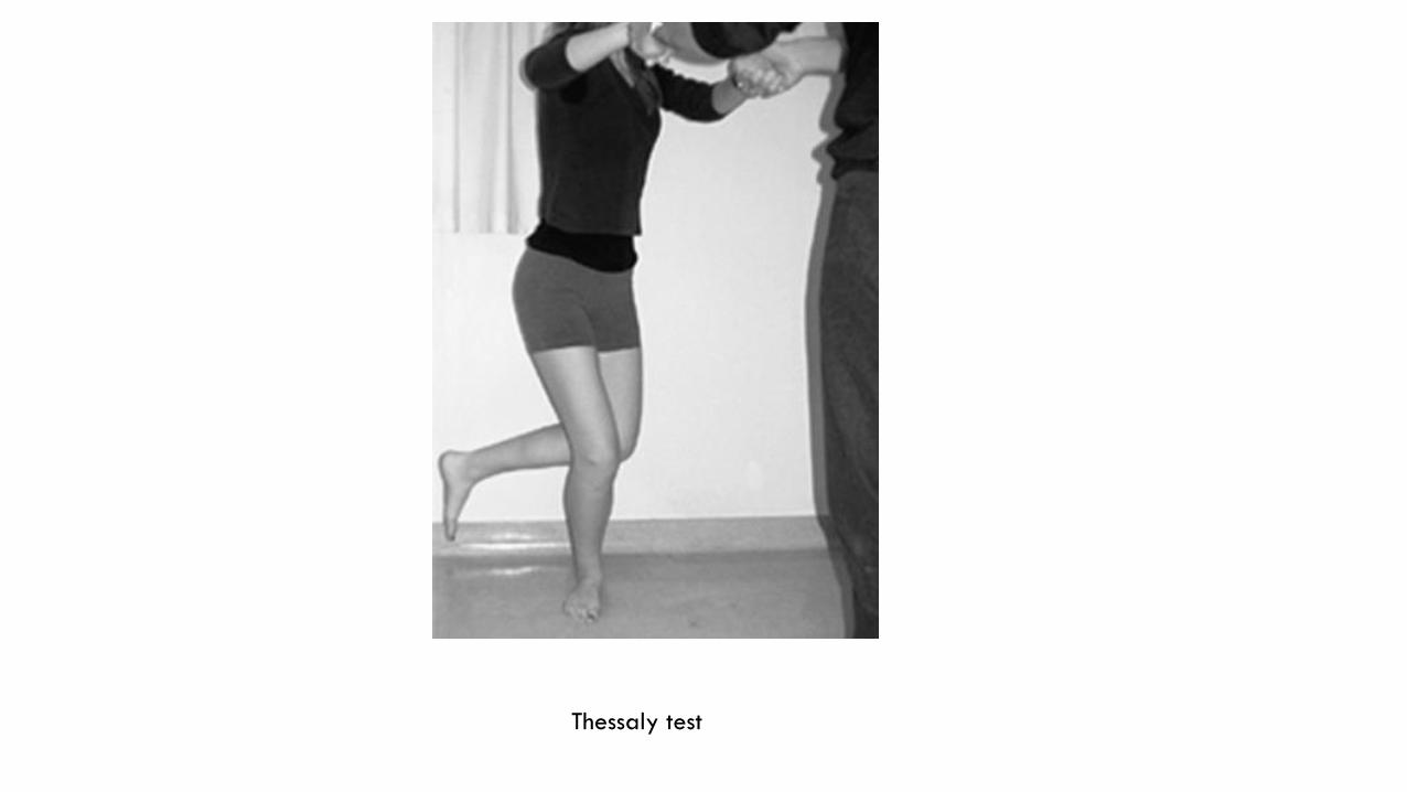
Thessaly test
SIGNS WITH PATIENT LYING PRONE
Scars or lumps in the popliteal fossa are noted. If there is a swelling, is it in the midline (most likely a bulging capsule) or to one side (possibly a bursa).
Apley’s test With the patient prone, the knee is flexed to 90 degrees and rotated while a compression force is applied; this, the grinding test, reproduces symptoms if a meniscus is torn. Rotation is then repeated while the leg is pulled upwards with the surgeon’s knee holding the thigh down; this, the distraction test, produces increased pain only if there is ligament damage.
IMAGING
X-ray: Anteroposterior and lateral views are routine; the anteroposterior view should always be taken with the patient standing. Both knees should be X-rayed, so as to compare the abnormal with the normal side. If available, previous X-ray imaging should always be used for comparison to determine progression of changes in the knee.
Ultrasound: guiding knee joint aspiration or synovial biopsy, and identifying tendon pathology in the knee
Bone scan: Helpful in showing ‘hot spots’ due to the spread of malignancy or loosening of components after joint replacement.
MRI: provides a reliable means of diagnosing injury or damage to the soft tissues of the knee, such as meniscal tears or cruciate ligament injuries, identifying the early stages of osteoarthritis. In addition, MRI scans are an essential part of the investigation of musculoskeletal tumors.
CT: for detailed understanding of 3D bone structure.
PET scans: Tumors
Arthroscopy: Not usually used now that MRI is readily available

DEFORMITIES
Bow legs in babies and knock knees in 4-year-olds are so common that they are considered to be normal stages of development, the child should be seen at intervals of 6 months to record progress. If by age 10 deformity is still present, surgery is advised.
Compensatory deformities : deformities of the proximal femur may give rise to complex compensatory deformities of the knees and legs once the child starts to walk.
PATHOLOGICAL DEFORMITIES IN CHILDREN
Disorders which cause distorted epiphyseal and/or physeal growth may give rise to bow leg or knock knee; these include some of the skeletal dysplasias and the various types of rickets, as well as injuries of the epiphyseal and physeal growth cartilage. A unilateral deformity is likely to be pathological. If angulation is severe, operative correction will be necessary, but it should be deferred until near the end of growth lest the deformity recur with further growth.
Blount’s disease: This is a progressive bow-leg deformity associated with abnormal growth of the posteromedial part of the proximal tibia. The children are usually overweight and start walking early; the condition is bilateral in 80% of cases. Children of Afro-Caribbean descent appear to be affected more frequently than others. In some cases there may be lateral subluxation of the tibia. Treatment is surgical.
Bow legs(genu varum) : Gauged from simple observation or measuring distance between knees with heels touching(should be < 6cm)

DEFORMITIES IN ADULTS
Angular deformities are common in adults (usually bow legs in men and knock knees in women). May follow childhood deformity, if so, usually causes no problems. However, if associated with early OA, patients may present with significant symptoms often as pronounced as those with more advanced joint damage. In the absence of overt osteoarthritis, if the patient complains of persistent severe pain and there are radiological signs of early joint damage (usually seen on MRI), an osteotomy can be performed.
Can be secondary to arthritis. OAvarus ; RAvalgus
Other causes: Ligament injuries, malunited fractues and Paget’s disease
GENU RECURVATUM (HYPEREXTENSION OF THE KNEE)
Congenital recurvatum: This may be due to abnormal intra-uterine posture; it usually recovers spontaneously. Could lead to true congenital dislocation of the knee(RARELY)
Lax ligaments: Prolonged traction, Chronic or recurring synovitis, rickets, poliomyelitis, Charcot’s disease
Other causes: Growth plate injuries and malunited fractures both can be surgically corrected
LESIONS OF MENISCI
The menisci have an important role in (1) improving articular congruency and increasing the stability of the knee; (2) controlling the complex rolling and gliding actions of the joint; and (3) distributing load during movement.
The medial meniscus is much less mobile than the lateral, and it cannot as easily accommodate to abnormal stresses. This may be why meniscal lesions are more common on the medial side than on the lateral. Even in the absence of injury, there is gradual degeneration and change in the material properties of the menisci with age, so splits and tears are more likely in later life. In younger people, meniscal tears are usually the result of trauma, with a specific injury identified in the history
TEARS OF MENISCI
More likely to tear across its length than its width. Tear usually follows rotational and shearing forces(eg when knee is flexed and twisted while taking weight) which is why its common in footballers. As one ages, relatively little force can cause a tear.
In some cases the split is vertical. If the separated fragment remains attached front and back, the lesion is called a bucket-handle tear. The torn portion can sometimes displace towards the centre of the joint and becomes jammed between femur and tibia. This causes a block to movement(locked knee).
Horizontal tears are usually degenerative and are more stable, if a loose piece is displaced it acts as an irritant causing effusion and mechanical symptoms.
Other patterns of tear can be identified: posterior or anterior horn tears and parrot beak tears where an oblique tear pattern creates a flap of meniscus that may be stable(unlikely to displace) or unstable (displaced or likely to displace).
All except the most peripheral part of the meniscus is avascular and spontaneous repair does not occur unless the tear is in the outer third, which is vascularized from the attached synovium and capsule.

TEARS CONTINUED
The patient is usually a young person who sustains a twisting injury to the knee on the sports field. Pain is often severe and further activity is avoided; occasionally the knee is ‘locked’ in partial flexion. Almost invariably, swelling appears some hours later, or perhaps the following day. With rest, the initial symptoms subside, only to recur periodically after trivial twists or strains. Sometimes the knee gives way spontaneously and this is again followed by pain and swelling.
Patients aged 40 or more may only complain of their knees giving away or locking.
Physical exam shows the joint flexed, an effusion may be present, and if prolonged the quads will be wasted. Tenderness is present on the joint line, medially. Extention is slightly limited.
TEARS CONTINUED
Investigations include: X-rays(usually normal), MRI
Differential diagnoses:
-Loose bodies
-Recurrent dislocation of the patella
-Fracture of the tibial spine
-Partial tear of the medial collateral ligament
-Torn ACL
TEARS CONTINUED
Treatment:
Conservative: If not locked and the MRI shows it to be repairable then proceed with arthroscopy. However, unstable tears present with infrequent symptoms that are not disabling so they are left alone.
Operative: Indications include a joint that cant be unlocked, if symptoms are recurrent and conservative therapy has failed. We can either repair, excise torn portion, excise part or all of the meniscus(total meniscectomy causes instability and secondary OA). Post-op pain and stiffness relieved by NSAIDs.
Locked knee: usually resolves spontaneously, if not, arthroscopy with removal of the fragment or repair.
MENISCAL DEGENERATION
Patients normally over 45, presents with signs and symptoms of a tear, without Hx of previous injury. MRI shows horizontal cleavage which is characteristic for degenerative changes.
Meniscectomy not helpful, only indicated if there are marked mechanical symptoms and recurrent sharp pain in the knee.

DISCOID LATERAL MENISCUS
In the foetus the meniscus is not semilunar but disc-like. If this disc-like shape persists postnatally symptoms can occur if the whole meniscus is unstable or more typically where a tear occurs.
Usually a young patient complaining that his knee keeps giving away and thuds loudly. Physical exam shows a clunk at 110 degrees of flexion and at 10 degrees while being straightened. Easily diagnosable by an MRI.
If only complaint is a clunk, no treatment is necessary. If pain affects quality of life then a part of it can be excised leaving a normally shaped meniscus.

MENISCAL CYSTS
Cysts of the menisci most often arise from horizontal cleavage tears. The multilocular cyst contains gelatinous fluid and is surrounded by thick fibrous tissue.
Lateral meniscus affected more commonly, patient complains of an ache or lumo on the side of the joint, symptoms are intermittent or worse after activity.
Physical exam shows a lump situated at or below the joint line usually anterior to the collateral ligament. It is seen most easily with the knee slightly flexed. Often firm. Medial cysts are larger and softer.
DD: Ganglion, Calcific deposits in the collateral ligament, prolapsed torn meniscus, tumour.
Tx: If asymptomatic, no treatment is needed. Otherwise treated operatively.

CHRONIC LIGAMENTOUS INSTABILITY
The knee depends heavily on ligaments for stability. Injuries are common in sportsmen, athletes and dancers. The patient with a torn or ruptured ligament may end up with chronic instability of the knee(a sense that its about to give away which it might).
Might be accompanied by pain and swelling. There may be a meniscal tear but meniscectomy makes matters worse.
DISLOCATION OF THE PATELLA
In 15–20% of cases (mostly children) the first episode is followed by recurrent dislocation or subluxation after minimal stress. This is due, in some measure, to disruption or stretching of the medially based ligamentous structures (e.g. the medial patellofemoral ligament – MPFL) which normally stabilize the extensor mechanism. However, in a significant proportion of cases there is no history of an acute strain and the initial episode is ‘spontaneous’. Predisposing factors are often present: (1) generalized ligamentous laxity;(2) underdevelopment of the lateral femoral condyle and flattening of the intercondylar groove; (3) maldevelopment of the patella, which may be too high or too small; (4) valgus deformity of the knee; (5) external tibial torsion or (6) a primary muscle defect.
Repeated dislocation damages the contiguous articular surfaces of the patella and femoral condyle; this may result in further flattening of the condyle, so facilitating further dislocations.
Dislocation almost always towards the lateral side
DISLOCATION CONT’D
Girls more commonly affected. Could be bilateral.
Dislocation happens the knee is being extended from a flexed position and the knee is stuck in flexion. There is acute pain and the patient may fall to the ground. If the patient is seen while it is displaced dx is obvious.
If he presents with it reduced, there might be marked swelling, tenderness and the apprehension test is positive.
The patient should be examined for previous predisposing conditions.
X-ray may show loose bodies, a lateral view may show a high-riding patella
MRI might show signs of previous patellofemoral soft tissue damage on the medial side of the knee and trochlear dysplasia.
DISLOCATION CONT’D
Tx:
Conservative: if dislocated it is pushed back into position, and then a knee brace is applied for 2-3 weeks. Quad strengthening exercises should be continued for 3 months.
Operative: Indications are if the patella cant be reduced, presence of a large, displaced osteochondral fragment, or if conservative therapy didn’t work and episodes are recurrent.
DISLOCATION CONT’D
Congenital dislocation is very very very rare and is hard to treat.
Habitual dislocation is where it displaces with flexion and reduces with extention. In longstanding cases it may be permanently displaced. Treatment requires an operation with quad lengthening.

PATELLOFEMORAL PAIN SYNDROME
This syndrome is common among active adolescents and young adults.
It is often (but not invariably) associated with softening and fibrillation of the articular surface of the patella – chondromalacia patellae. Some surgeons have tended to regard chondromalacia as the cause (rather than one of the effects) of the disorder. Against this are the facts that (1) chondromalacia is commonly found at arthroscopy in young adults who have no anterior knee pain; and (2) some patients with the typical clinical syndrome have no cartilage softening
If all other causes of anterior pain in the knee are excluded, this is the diagnosis.
It is believed that the basic disorder is probably mechanical overload of the patellofemoral joint.
PATELLOFEMORAL OVERLOAD SYNDROME CONT’D
A single injury may damage the articular surfaces(rare)
Much more common is repetitive overload due to malcongruence of the surfaces, malalignment of the lower extremity, muscular imbalance of the lower extremity, or overreactivity.
Damage to cartilage and bone aren’t necessarily of the same degree.
The patient is often a teenage girl, complains of persistent pain over the front of the knee. Symptoms are aggravated by activity or climbing stairs, or when standing up after prolonged sitting. The knee may give way and occasionally swells.
Often bilateral.
There is Hx of previous injury or recurrent instability

PATELLOFEMORAL PAIN SYNDROME CONT’D
Careful examination may reveal malalignment or tilting of the patellae. Other subtle signs include quadriceps wasting, fluid in the knee, tenderness under the edge of the patella and crepitus on moving the knee.
Patellofemoral pain is elicited by pressing the patella against the femur and asking the patient to contract the quadriceps – first with central pressure, then compressing the medial facet and then the lateral.
If, in addition, the apprehension test is positive, this suggests previous subluxation or dislocation.
Q-angle of more than 20 degrees is a predisposing factor. Also Patella alta.
The hip must be examined to make sure this isn’t referred pain.
PATELLOFEMORAL PAIN SYNDROME CONT’D
Investigations :
X-ray: Abnormal tilting or subluxation of the patella. Lateral view might show a high or small patella.
CT and MRI most accurate to show malposition of the patella
Arthroscopy can be used to exclude other causes.

CAUSES OF ANTERIOR KNEE PAIN 1 Referred from hip
2 Patellofemoral disorders
•Patellar instability
• Patellofemoral overload
• Osteochondral injury
• Patellofemoral osteoarthritis
3 Knee joint disorders
• Osteochondritis dissecans
• Loose body in the joint
• Synovial chondromatosis
• Plica syndrome
4 Periarticular disorders
• Patellar tendinitis
• Patellar ligament strain
• Bursitis
• Osgood–Schlatter disease
PATELLOFEMORAL PAIN SYNDROME CONT’D
Conservative Tx: Adjustment of stressful activities, deploying focused physiotherapy, combined with reassurance that most patients eventually recover without surgery. Exercises are directed specifically at strengthening the medial quadriceps so as to counterbalance the tendency to lateral tilting or subluxation of the patella.
Operative Tx: Considered only after 6 months of conservative therapy and there is evidence of a correctable abnormality.

شكرا جزيال على االصغاء