the journal of the lebanese dental association · 1 the journal of the lebanese dental association...
TRANSCRIPT
1
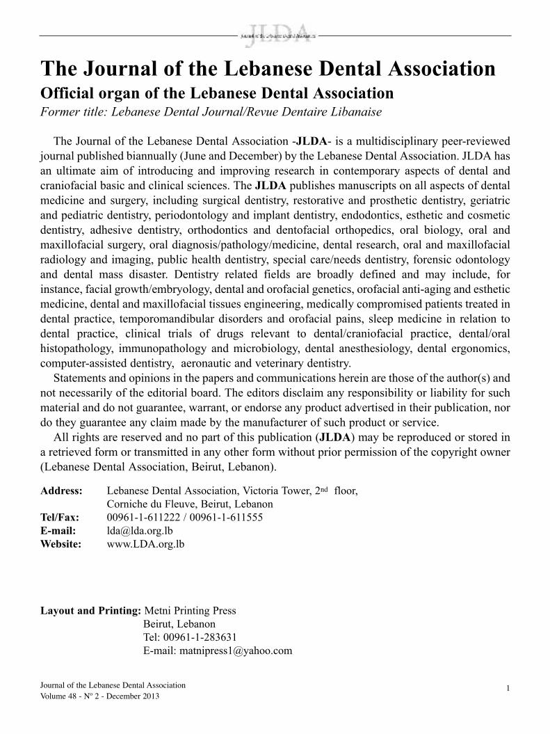
The Journal of the Lebanese Dental AssociationOfficial organ of the Lebanese Dental AssociationFormer title: Lebanese Dental Journal/Revue Dentaire Libanaise
The Journal of the Lebanese Dental Association -JLDA- is a multidisciplinary peer-reviewed
journal published biannually (June and December) by the Lebanese Dental Association. JLDA has
an ultimate aim of introducing and improving research in contemporary aspects of dental and
craniofacial basic and clinical sciences. The JLDA publishes manuscripts on all aspects of dental
medicine and surgery, including surgical dentistry, restorative and prosthetic dentistry, geriatric
and pediatric dentistry, periodontology and implant dentistry, endodontics, esthetic and cosmetic
dentistry, adhesive dentistry, orthodontics and dentofacial orthopedics, oral biology, oral and
maxillofacial surgery, oral diagnosis/pathology/medicine, dental research, oral and maxillofacial
radiology and imaging, public health dentistry, special care/needs dentistry, forensic odontology
and dental mass disaster. Dentistry related fields are broadly defined and may include, for
instance, facial growth/embryology, dental and orofacial genetics, orofacial anti-aging and esthetic
medicine, dental and maxillofacial tissues engineering, medically compromised patients treated in
dental practice, temporomandibular disorders and orofacial pains, sleep medicine in relation to
dental practice, clinical trials of drugs relevant to dental/craniofacial practice, dental/oral
histopathology, immunopathology and microbiology, dental anesthesiology, dental ergonomics,
computer-assisted dentistry, aeronautic and veterinary dentistry.
Statements and opinions in the papers and communications herein are those of the author(s) and
not necessarily of the editorial board. The editors disclaim any responsibility or liability for such
material and do not guarantee, warrant, or endorse any product advertised in their publication, nor
do they guarantee any claim made by the manufacturer of such product or service.
All rights are reserved and no part of this publication (JLDA) may be reproduced or stored in
a retrieved form or transmitted in any other form without prior permission of the copyright owner
(Lebanese Dental Association, Beirut, Lebanon).
Address: Lebanese Dental Association, Victoria Tower, 2nd floor,
Corniche du Fleuve, Beirut, Lebanon
Tel/Fax: 00961-1-611222 / 00961-1-611555
E-mail: [email protected]
Website: www.LDA.org.lb
Layout and Printing: Metni Printing Press
Beirut, Lebanon
Tel: 00961-1-283631
E-mail: [email protected]
Journal of the Lebanese Dental AssociationVolume 48 - Nº 2 - December 2013
ISSN 1810-9632
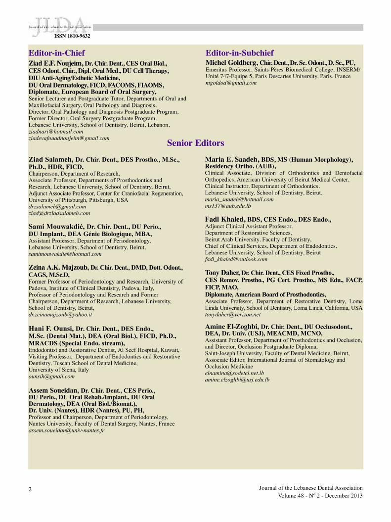
Editor-in-ChiefZiad E.F. Noujeim, Dr. Chir. Dent., CES Oral Biol., CES Odont. Chir., Dipl. Oral Med., DU Cell Therapy, DIU Anti-Aging/Esthetic Medicine, DU Oral Dermatology, FICD, FACOMS, FIAOMS, Diplomate, European Board of Oral Surgery,Senior Lecturer and Postgraduate Tutor, Departments of Oral andMaxillofacial Surgery, Oral Pathology and Diagnosis, Director, Oral Pathology and Diagnosis Postgraduate Program,Former Director, Oral Surgery Postgraduate Program, Lebanese University, School of Dentistry, Beirut, Lebanon, [email protected]@gmail.com
Ziad Salameh, Dr. Chir. Dent., DES Prostho., M.Sc.,Ph.D., HDR, FICD, Chairperson, Department of Research, Associate Professor, Departments of Prosthodontics andResearch, Lebanese University, School of Dentistry, Beirut,Adjunct Associate Professor, Center for Craniofacial Regeneration,University of Pittsburgh, Pittsburgh, [email protected]@drziadsalameh.com
Sami Mouwakdié, Dr. Chir. Dent., DU Perio., DU Implant., DEA Génie Biologique, MBA,Assistant Professor, Department of Periodontology, Lebanese University, School of Dentistry, Beirut, [email protected]
Zeina A.K. Majzoub, Dr. Chir. Dent., DMD, Dott. Odont.,CAGS, M.Sc.D, Former Professor of Periodontology and Research, University ofPadova, Institute of Clinical Dentistry, Padova, Italy, Professor of Periodontology and Research and FormerChairperson, Department of Research, Lebanese University,School of Dentistry, Beirut, [email protected]
Hani F. Ounsi, Dr. Chir. Dent., DES Endo., M.Sc. (Dental Mat.), DEA (Oral Biol.), FICD, Ph.D.,MRACDS (Special Endo. stream), Endodontist and Restorative Dentist, Al Seef Hospital, Kuwait,Visiting Professor, Department of Endodontics and RestorativeDentistry, Tuscan School of Dental Medicine, University of Siena, [email protected]
Assem Soueidan, Dr. Chir. Dent., CES Perio., DU Perio., DU Oral Rehab./Implant., DU OralDermatology, DEA (Oral Biol./Biomat.), Dr. Univ. (Nantes), HDR (Nantes), PU, PH, Professor and Chairperson, Department of Periodontology,Nantes University, Faculty of Dental Surgery, Nantes, [email protected]
Maria E. Saadeh, BDS, MS (Human Morphology),Residency Ortho. (AUB), Clinical Associate, Division of Orthodontics and DentofacialOrthopedics, American University of Beirut Medical Center, Clinical Instructor, Department of Orthodontics, Lebanese University, School of Dentistry, Beirut, [email protected]@aub.edu.lb
Fadl Khaled, BDS, CES Endo., DES Endo., Adjunct Clinical Assistant Professor, Department of Restorative Sciences, Beirut Arab University, Faculty of Dentistry, Chief of Clinical Services, Department of Endodontics, Lebanese University, School of Dentistry, [email protected]
Tony Daher, Dr. Chir. Dent., CES Fixed Prostho., CES Remov. Prostho., PG Cert. Prostho., MS Edu., FACP,FICP, MAO, Diplomate, American Board of Prosthodontics,Associate Professor, Department of Restorative Dentistry, LomaLinda University, School of Dentistry, Loma Linda, California, USA [email protected]
Amine El-Zoghbi, Dr. Chir. Dent., DU Occlusodont.,DEA, Dr. Univ. (USJ), MEACMD, MCNO, Assistant Professor, Department of Prosthodontics and Occlusion,and Director, Occlusion Postgraduate Diploma, Saint-Joseph University, Faculty of Dental Medicine, Beirut, Associate Editor, International Journal of Stomatology andOcclusion [email protected]@usj.edu.lb
2 Journal of the Lebanese Dental AssociationVolume 48 - Nº 2 - December 2013
Editor-in-SubchiefMichel Goldberg, Chir. Dent., Dr. Sc. Odont., D. Sc., PU,Emeritus Professor, Saints-Pères Biomedical College, INSERM/Unité 747-Equipe 5, Paris Descartes University, Paris, [email protected]
Senior Editors
Associate Editors
Rima Abdallah, BDS, CAGS, D.Sc. (Oral Biol.), Diplomate, American Board of Periodontology,Associate Clinical Professor, Periodontology and ImplantologyDivisions, Oral Surgical Sciences Department, Beirut Arab University, Faculty of Dentistry, Beirut, [email protected]
Marcel Noujeim, BDS, DU Oral Biol., DU OralRadiol., MS Oral/Max.Fac. Radiol., Diplomate, American Board of Oral/MaxillofacialRadiology,Associate Professor, Department of Comprehensive Dentistry,Director of Oral and Maxillofacial Radiology Postgraduate Program,University of Texas Health Science Center at San Antonio -UTHSCSA, USA [email protected]@uthscsa.edu
Roula Abiad, BDS, MS (Endo.), Dr. Dent. Sc., Assistant Dean, Assistant Professor, and Director of EndodonticDivision, Beirut Arab University, Faculty of Dentistry, Beirut, [email protected]@gmail.com
Charles Sfeir, Dr. Chir. Dent., Cert. Perio., MS, Ph.D.,Associate Professor and Director, Center for CraniofacialRegeneration,University of Pittsburgh, School of Dental Medicine, Pittsburgh, [email protected]
Zoubeida Yahfoufi Al Hage, DDS, Dr. Dentistry, Practice limited to Periodontology and Implant Dentistry,Beirut, [email protected]
Pascale Habre Hallage, Dr. Chir. Dent., CES Oral Biol., CES Fixed Prostho., DUICP, MSBM, DEA Neurosc., Dr. Biomed. Sc., Assistant Professor and Director of Postgraduate Program,Department of Fixed Prosthodontics and Occlusion,Saint-Joseph University, Faculty of Dental Medicine, Beirut, [email protected]@usj.edu.lb
Radhouane Dallel, Dr. Chir. Dent., Dr. Univ., HDR, PU, PH,Senior Scientist and Director, Neurobiology of Trigeminal Pain/Migraine Laboratory, NEURO-DOL, INSERM/UdA, U1107,Professor, Clermont 1 University, Faculty of Dental Surgery, Clermont-Ferrand, [email protected]@u-clermont1.fr
Tara Renton, BDS, M. Dent. Sc., FRACDS, FDSRCS(Engl.), Ph.D., ILTM, FHEA, Consultant Oral Surgeon, Professor and Head, Department of OralSurgery, King’s College London -KCL- Dental Institute, London, UK,[email protected]
Ahmed Feki, Dr. Chir. Dent., CES Oral Biol., CES
Odont. Chir., DU Head/Neck Morphol., DU Oral
Mucosal Dermatol., HDR, PU, PH, Professor and Chairperson, Department of Oral Surgery and OralMedicine, Louis Pasteur University, Faculty of Dental Surgery,Strasbourg, [email protected]
Editors EmeritiNadim Baba, DMD, MSD, FICD, FACP
Philippe Aramouni, DCD, DEA, CAGS, M.Sc.D., FICD
Michel Salameh, DCD, CES Pediat. Dent., MIADP, MIADH, MSFOP
Antoine Cassia, DCD, CES Biol. Bucc., CES Odont. Chir., DU Maxillofacial Prostho., DSO
Levon Naltchayan, DCD, CES Prostho.,
3Journal of the Lebanese Dental AssociationVolume 48 - Nº 2 - December 2013
Antoine Berbéri, BDS, MS, DU, CES Odont. Chir.,Dr. Univ., HDR, Diplomate, European Board ofOral Surgery,Associate Professor and Former Chairperson, Department of Oral and Maxillofacial Surgery, Lebanese University, School of Dentistry, Beirut,Scientist/Researcher, Doctoral School of Sciences and Technology,Lebanese University, Beirut,[email protected]
Georges Tawil, Dr. Chir. Dent., DDS, CES Odont.Chir., CES Perio., Dr.Sc.Odont., FICD, FACDProfessor of Periodontology, Saint-Joseph University,Faculty of Dental Medicine, Beirut, Lebanon, Editorial Consultant, International Journal of Oral andMaxillofacial Implants, Clinical Oral Implant [email protected]
Nadim Baba, DMD, MSD, FICD, FACPAssociate Professor, Department of Restorative Dentistry, LomaLinda University, School of Dentistry, Loma Linda, California, USAFormer Editor-in-Chief, JLDA,[email protected]
Jaime S. Brahim, BDS, MS, Dip. ABOM Surg.,Dip. ABO Med, FACOMS, FAAOMS, FIAOMSProfessor, Department of Oral and Maxillofacial Surgery,University of Maryland College of Dental Surgery, Baltimore,Maryland, [email protected]
Dina Debaybo, Dr. Chir. Dent., CAGS, M.Sc.D.,Dip. AB Pediat. Dent.Associate Professor of Pediatric Dentistry, European University College, Dubai, [email protected]
Nabil Tabbara, DMD, FAAFO, FAACPAdjunct Clinical Professor, University of Western Ontario, SchulichSchool of Medicine and Dentistry, London, Ontario, [email protected]
Mary Ann Jabra-Rizk, BS, Ph.DAssociate Professor, Department of Oncology and DiagnosticSciences, University of Maryland Dental School, Baltimore,Maryland, [email protected]
Karine Feghali, BDS, DU Perio., Ph.DPostdoctoral Fellow, Oral Ecology Research Group (GREB), LavalUniversity, Faculty of Dental Medicine, Quebec City, QC, [email protected]@yahoo.fr
Sukumaran Anil, BDS, MDS, Ph.D., FICD, FPFAProfessor and Consultant, Division of Periodontics, King SaudUniversity, College of Dentistry, Riyadh, KSA, [email protected]
Hani Adbul Salam, B.Sc., BDS, M.Sc., Ph.D Adjunct Professor and Director of Continuing Dental Educationfor the Middle East and North Africa, McGill University, Facultyof Dentistry, Montreal, [email protected]@[email protected]
Essam Osman, BDS, M.SD, Ph.D.Vice-President for Medical Sciences, Beirut Arab University,Dean, Faculty of Dentistry,Chairperson, Departments of Restorative Sciences and OralSurgical Sciences, and Director, Division of Dental Biomaterials, Professor of Dental Materials, Faculty of Dentistry, [email protected]@bau.edu.lb
Antoine Cassia, DCD, CES Odont. Chir., DU Maxillofacial Prostho., DSOAssociate Professor and Former Chairperson, Department of Oral Pathology and Diagnosis, Lebanese University, School of Dentistry, Beirut, Lebanon, Former Editor-in-Chief, JLDA,[email protected]
Ghassan Yared, DCD, DSO, FRCD (Can.), MRCDSOFormer Associate Professor, Department of Endodontics, andformer Director of Endodontics undergraduate program,University of Toronto, Faculty of Dentistry, Toronto, [email protected]@hotmail.com
André Assaf, BDS, CES Dent. Mat., CES FixedProstho., CES Remov. Prostho., DU Occlusodont.,DU Implant., DU Med. CommunicationAdjunct Clinical Associate Professor, Department of RestorativeSciences, Beirut Arab University, Faculty of Dentistry, BeirutPresident, Lebanese Society of Prosthodontics,[email protected]
Elias Khoury, DCD, DU Oral Impl., DU BoneAugmentation and Maxillo-Facial Reconstruction,DEA (Biol. Med. Eng.)Former Scientific Head, DU Réhabilitation Osseuse par Implantset Biomatériaux, Faculty of Dental Surgery, Nantes, France,Diplomate of the International Congress of Oral Implantologists,Member of the French Association of Implantology (AFI),Co-Designer of Implant Kontact (Biotech International),Private Practice, Paris, [email protected]
Georges Khoury, DCD, DU Maxillo-FacialImaging and Radiology, DU Oral Impl., DU BoneAugmentation and Maxillo-Facial Reconstruction,DEA (Biol. Med. Eng.)Faculty, Department of Oral Implantology, and Scientific Head,DU Reconstitution Osseuse Pré-Implantaire, Paris VII Faculty ofDental Surgery, Paris, France,Associate Fellow, American Academy of Implant Dentistry,[email protected]
Biostatistics and Epidemiology Consultants
M Fouad Ziadé, Ph.D (Biostat.), C. Stat., FRSS,MASA, MIEAAssociate Professor, Lebanese University, Faculty of PublicHealth, Beirut/Tripoli, [email protected] / [email protected]
Nada E. El-Osta, DCD, DES Prostho., MS (Biol. Med.Sc.), DIU Biostat., DU Forensic ScienceConsultant in Biostatitics / Epidemiology, Saint-Joseph University, Faculties of Medicine and DentalMedicine, Beirut, [email protected] / [email protected]
Editorial Advisory Board
4 Journal of the Lebanese Dental AssociationVolume 48 - Nº 2 - December 2013

C o n t e n t sC o n t e n t sVolume 48 - Nº 2 - December 2013
The LDA is a regular member of the FDI
8
10
12
14
20
28
36
53
55
65
68
77
83
85
Cited in the WHO EasternMediterranean Index Medicus
ISSN 1810-9632
Guest EditorialNayef E. Saadé and Suhayl J. Jabbur
Editorial CommentBarry S. Sessle
In Memorian: Emmanuel Tomb (1958-2005), Dentist, Scholar, Author, Polymath, and DentalEducator.
Ziad Noujeim
Trigeminal Autonomic Cephalalgias -TACs-: a review of neuroimaging studies.Norazah Abu Bakar, Tara Renton
Trigeminal N euralgia - TN: diagnosis and management challenges.Joe Faddoul, Sandra Kobaïter Maarrawi, Sharoni Weinberg, Patrick Tabet, Elie Samaha, Joseph Maarrawi
Current guidelines for the diagnosis and management of TemporoMandibular Disorders (TMDs).Charles McNeill
Anatomy and physiology of trigeminal networks conveying nociception: a review. Nadwa Chatila, Rahif Tawil, Céline Melin, Radhouane Dallel
Acupuncture and Trigeminal neuralgia pain.Nadim M. Jalbout
Low-Level-LASER Therapy - LLLT - for the treatment of oral myofascial pain. A systematic reviewof the last thirteen years.
Carolina Roldán-Barraza, Steffani Janko, Hans-Christoph Lauer
Low-Level LASER as supportive therapy of temporomandibular disorders -TMDs: a case report.Najib Abou Hamra, Elias Smaira
Medication Overuse Facial Pain - MOFP: a case series and proposed diagnostic criteria. M Sam Chong, Nadine Khawaja, Joan Hester, Tara Renton
An evaluation of the effects of Platelet Rich Plasma -PRP- compared to Sodium Hyaluronate -SH- inthe treatment of temporomandibular joint Osteoarthritis-OA.
Wissam Tohmé
Craniofacial Pain of Cardiac Origin (Angina / Acute Coronary Syndrome / Myocardial Infarction): a useful diagnostic tool for dentists.
Samar Khoury
Glossopharyngeal Neuralgia -GPN- secondary to ipsilateral compressive cerebellar mega-tonsil: a case report.Joe Faddoul, Sandra Kobaïter Maarrawi, Joe Abdel Hay, Elie Samaha, Joseph Maarrawi
FDI World Dental FederationM e m b e r
6 Journal of the Lebanese Dental AssociationVolume 48 - Nº 2 - December 2013
C o n t e n t sC o n t e n t sVolume 48 - Nº 2 - December 2013
88
92
101
105
112
113
115
118
121
126
Dental pulp pain: diagnostic implications.Khaldoun Rifaï, Georges Tehini
Practical pharmacological approach of orofacial pains: realities and clinical recommendations.Wissam Tohmé, Joelle Saroufim
The Mylohyoid Nerve -MHN-: clinical significance and implications for analgesia of mandibular teethin restorative and surgical dentistry.
Rita Bou Assaf, Wasfi Kanj
Pain in orthodontic treatment: etiology and management. Chimène Chalala, Ramzi V. Haddad
Undertreatment of pain: is it acceptable?Nadim M. Jalbout
Declaration of MontréalDeclaration that Access to Pain Management Is a Fundamental Human Right.
International Pain Summit (IPS) of the International Association for the Study of Pain (IASP), Montréal, Canada, October 2010
Efficacy of Arthrocentesis on pain and mouth opening in the treatment of Temporomandibular Joint-TMJ- internal derangements: a 2-year retrospective clinical evaluation.
Joseph B. Hobeiche
Botulinum Toxin injection for the management of orofacial pain conditions.Elie M. Ferneini
Proceedings of a special symposium on Orofacial pain: Orofacial Pain, a multidisciplinary approach.Lebanese Society for the Study of Pain (LSSP), in collaboration with Lebanese Society for the Treatment of Pain (STLD),
December 6, 2013, Campus of Medical Sciences, Saint-Joseph University, Beirut, Lebanon
JLDA Guide for contributors and authors.
7Journal of the Lebanese Dental AssociationVolume 48 - Nº 2 - December 2013
The LDA is a regular member of the FDI
Cited in the WHO EasternMediterranean Index Medicus
ISSN 1810-9632FDI World Dental Federation
M e m b e r
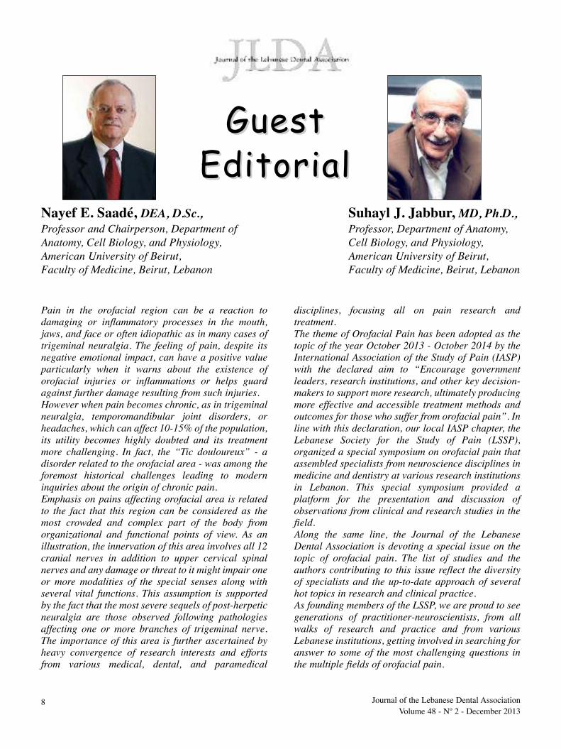
Nayef E. Saadé, DEA, D.Sc.,
Professor and Chairperson, Department of
Anatomy, Cell Biology, and Physiology,
American University of Beirut,
Faculty of Medicine, Beirut, Lebanon
Suhayl J. Jabbur, MD, Ph.D.,
Professor, Department of Anatomy,
Cell Biology, and Physiology,
American University of Beirut,
Faculty of Medicine, Beirut, Lebanon
Pain in the orofacial region can be a reaction todamaging or inflammatory processes in the mouth,jaws, and face or often idiopathic as in many cases oftrigeminal neuralgia. The feeling of pain, despite itsnegative emotional impact, can have a positive valueparticularly when it warns about the existence oforofacial injuries or inflammations or helps guardagainst further damage resulting from such injuries. However when pain becomes chronic, as in trigeminalneuralgia, temporomandibular joint disorders, orheadaches, which can affect 10-15% of the population,its utility becomes highly doubted and its treatmentmore challenging. In fact, the “Tic douloureux” - adisorder related to the orofacial area - was among theforemost historical challenges leading to moderninquiries about the origin of chronic pain.Emphasis on pains affecting orofacial area is relatedto the fact that this region can be considered as themost crowded and complex part of the body fromorganizational and functional points of view. As anillustration, the innervation of this area involves all 12cranial nerves in addition to upper cervical spinalnerves and any damage or threat to it might impair oneor more modalities of the special senses along withseveral vital functions. This assumption is supportedby the fact that the most severe sequels of post-herpeticneuralgia are those observed following pathologiesaffecting one or more branches of trigeminal nerve.The importance of this area is further ascertained byheavy convergence of research interests and effortsfrom various medical, dental, and paramedical
disciplines, focusing all on pain research andtreatment.The theme of Orofacial Pain has been adopted as thetopic of the year October 2013 - October 2014 by theInternational Association of the Study of Pain (IASP)with the declared aim to “Encourage governmentleaders, research institutions, and other key decision-makers to support more research, ultimately producingmore effective and accessible treatment methods andoutcomes for those who suffer from orofacial pain”. Inline with this declaration, our local IASP chapter, theLebanese Society for the Study of Pain (LSSP),organized a special symposium on orofacial pain thatassembled specialists from neuroscience disciplines inmedicine and dentistry at various research institutionsin Lebanon. This special symposium provided aplatform for the presentation and discussion ofobservations from clinical and research studies in thefield.Along the same line, the Journal of the LebaneseDental Association is devoting a special issue on thetopic of orofacial pain. The list of studies and theauthors contributing to this issue reflect the diversityof specialists and the up-to-date approach of severalhot topics in research and clinical practice.As founding members of the LSSP, we are proud to seegenerations of practitioner-neuroscientists, from allwalks of research and practice and from variousLebanese institutions, getting involved in searching foranswer to some of the most challenging questions inthe multiple fields of orofacial pain.
GuestGuest
EditorialEditorial
8 Journal of the Lebanese Dental AssociationVolume 48 - Nº 2 - December 2013
This JLDA special issue on orofacial pains is dedicated to Professors Nayef E. Saadé andSuhayl J. Jabbur for their unflagging support for neuroscience and pain research.
9Journal of the Lebanese Dental AssociationVolume 48 - Nº 2 - December 2013
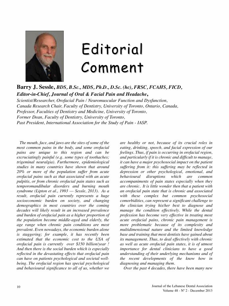
Barry J. Sessle, BDS, B.Sc., MDS, Ph.D., D.Sc. (hc), FRSC, FCAHS, FICD,
Editor-in-Chief, Journal of Oral & Facial Pain and Headache,Scientist/Researcher, Orofacial Pain / Neuromuscular Function and Dysfunction,
Canada Research Chair, Faculty of Dentistry, University of Toronto, Ontario, Canada,
Professor, Faculties of Dentistry and Medicine, University of Toronto,
Former Dean, Faculty of Dentistry, University of Toronto,
Past President, International Association for the Study of Pain - IASP.
The mouth, face, and jaws are the sites of some of themost common pains in the body, and some orofacialpains are unique to this region and can beexcruciatingly painful (e.g. some types of toothaches;trigeminal neuralgia). Furthermore, epidemiologicalstudies in many countries have shown that around20% or more of the population suffer from acuteorofacial pains such as that associated with an acutepulpitis, or from chronic orofacial pain states such astemporomandibular disorders and burning mouthsyndrome (Lipton et al., 1993 --- Sessle, 2013). As aresult, orofacial pain currently represents a hugesocioeconomic burden on society, and changingdemographics in most countries over the comingdecades will likely result in an increased prevalenceand burden of orofacial pain as a higher proportion ofthe population become middle-aged and elderly, theage range when chronic pain conditions are mostprevalent. Even nowadays, the economic burden aloneis staggering; for example, it has recently beenestimated that the economic cost to the USA oforofacial pain is currently over $150 billion/year !And then there is the social burden which is especiallyreflected in the devastating effects that orofacial paincan have on patients psychological and societal well-being. The orofacial region has special psychologicaland behavioural significance to all of us, whether we
are healthy or not, because of its crucial roles ineating, drinking, speech, and facial expression of ourfeelings. Thus, if pain is occurring in orofacial region,and particularly if it is chronic and difficult to manage,it can have a major psychosocial impact on the patientsuffering from it: this suffering may be reflected indepression or other psychological, emotional, andbehavioural disruptions which are commonaccompaniments of pain states especially when theyare chronic. It is little wonder then that a patient withan orofacial pain state that is chronic and associatedwith these complex but common psychosocialcomorbidities, can represent a significant challenge tothe clinician trying his/her best to diagnose andmanage the condition effectively. While the dentalprofession has become very effective in treating mostacute orofacial pains, chronic pain management ismore problematic because of its complexity andmultidimensional nature and the limited knowledgebase and training that most dentists have gained aboutits management. Thus, to deal effectively with chronicas well as acute orofacial pain states, it is of utmostimportance for dental clinicians to have a goodunderstanding of their underlying mechanisms and ofthe recent developments of the know how indiagnosing and managing them.
Over the past 4 decades, there have been many new
EditorialEditorial
CommentComment
10 Journal of the Lebanese Dental AssociationVolume 48 - Nº 2 - December 2013

or improved approaches for the diagnosis andmanagement of orofacial pain states, and severalpapers in this JLDA special issue outline many ofthese. We also know much more about theirmechanisms as a result of research studies in humansand animal models of orofacial pain, including thoseby Professors Suhayl Jabbur and Nayef Saadé who arequite appropriately being acknowledged and honouredin this JLDA special issue on orofacial pains. New insights have been gained on biological,molecular, and genetic processes underlying chronicas well as acute orofacial pains: these includediscoveries that tissue trauma can produce anincreased excitability of nociceptive sensory nervefibres that innervate orofacial tissues (“peripheralsensitisation”) and of nerve cells in the brain thatprocess or modulate pain-related signals that thesenerve cells receive from nociceptive fibre inputs(“central sensitisation”). Central sensitisation hasbeen shown to reflect a neuroplasticity of nociceptivepathways in the brain, emphasising that thesepathways are not “hard-wired” but can undergofunctional, even structural, neuroplastic changes as aresult of damage to orofacial tissues and alterations topain-modulatory pathways in patient’s brain: studiesof sensitisation phenomena have also revealed theinvolvement of several different chemical mediators aswell as interactions with modulating factors ofimmune, endocrine and cardiovascular systems thathave provided several novel targets for thedevelopment of new diagnostic or managementapproaches for pain. Furthermore, peripheralsensitisation and central sensitisation are nowrecognised as crucial elements in the development andmaintenance of persistent pain states and can accountfor increased pain sensitivity that can occur as a resultof an injury or inflammation of orofacial tissues.
Much more knowledge has also been gained onpsychological and behavioural factors that caninfluence pain expression: this new knowledge hasadded support to the current view held by most painexperts that the diagnosis and management of achronic pain patient need to be based on abiopsychosocial foundation given that pain, especiallywhen chronic, is complex and multidimensional. Thismeans that dentists need to be aware that they alonemay not have all the knowledge and clinical skillsrequired to provide comprehensive pain care for allchronic orofacial pain patients, and that consultationwith pain expert clinicians and other health
professionals such as psychologists and neurologistsmay be essential to provide optimal care.
To be sure, we still have much to learn more aboutorofacial pain, but with the new knowledge gained inrecent decades as a basis and the expected advancesover the coming years will assuredly result fromresearch approaches encompassing fields such asbioengineering, biomarkers, bioinformatics, imaging,immunology, molecular biology, genetics, andneuropsychology: these advances hold out promise fornew or improved clinical approaches that will helpalleviate the pain and suffering of many patientsexperiencing chronic pain or help reduce the risk ofpatients developing a chronic orofacial pain state.
Lipton JA, Ship JA, and Larach-Robinson D. Estimated
prevalence and distribution of reported orofacial pain in the
United States. J Am Dent Assoc 1993;124:115-121.
Sessle, B.J. (Ed.). Orofacial Pain: Recent Advances in
Assessment, Management, and Understanding of
Mechanisms. IASP Press, Washington, D.C., USA, Under
Press (expected in 2014), 509 pages.
11Journal of the Lebanese Dental AssociationVolume 48 - Nº 2 - December 2013
Emmanuel (Asaad) Tomb, a Lebanese French
dentist, earned the "Docteur en Chirurgie
Dentaire" degree in 1986, after which he pursued
postgraduate certificates in functional
myotherapy, a university diploma in dental
expertise (DU d'Expertise Bucco-Dentaire),
another diploma in the legal-juridical
compensation of body harm (DU d'études
relatives à la réparation juridique du dommage
corporel)and a Masters degree in Medical
Biology.
For many years, he worked, as Faculty, with
the Paris 7 University Faculty of Dental Surgery
(Garancière) where he was appointed Clinical
Associate(Attaché de Consultation) in the Facial
Pain and TMDs Clinic, and at the same time, he
had his own private practice in Vittel(France)and
collaborated, as Sworn Dental Expert, with the
Nancy Court of Appeal, in France. And later, he
also worked as Clinical Associate with the Pain's
Evaluation and Treatment Center at Laennec
Hospital in Paris, France.
Dr. Tomb's career was marked by a
combination of high academic achievement, both
in teaching under and post-graduate students, and
a highly regarded private and hospital consultant
practice where his opinion and expertise were
much in demand. He endeavored and published
extensively in the fields of head and neck pain,
and his topics of interest were migraine, cervical
pain, and TMDs. Indeed, and among his academic
exploits, he lectured with the International
Headache Society-IHS-in Amsterdam, the
International Academy of Legal Medicine -IALM-
in Dublin, and the European Federation of IASP
(International Association for the Study of Pain)
Chapters, in Barcelone. He was also invited
speaker in Beirut, Lebanon, where he addressed
to Lebanese and Arab
dentists, cervical and
cranio-facial pains in
seminars and workshops
organized by the Lebanese
Dental Association - LDA.
During his dental career,
Dr. Tomb earned an
international reputation in
the field of Head and Neck
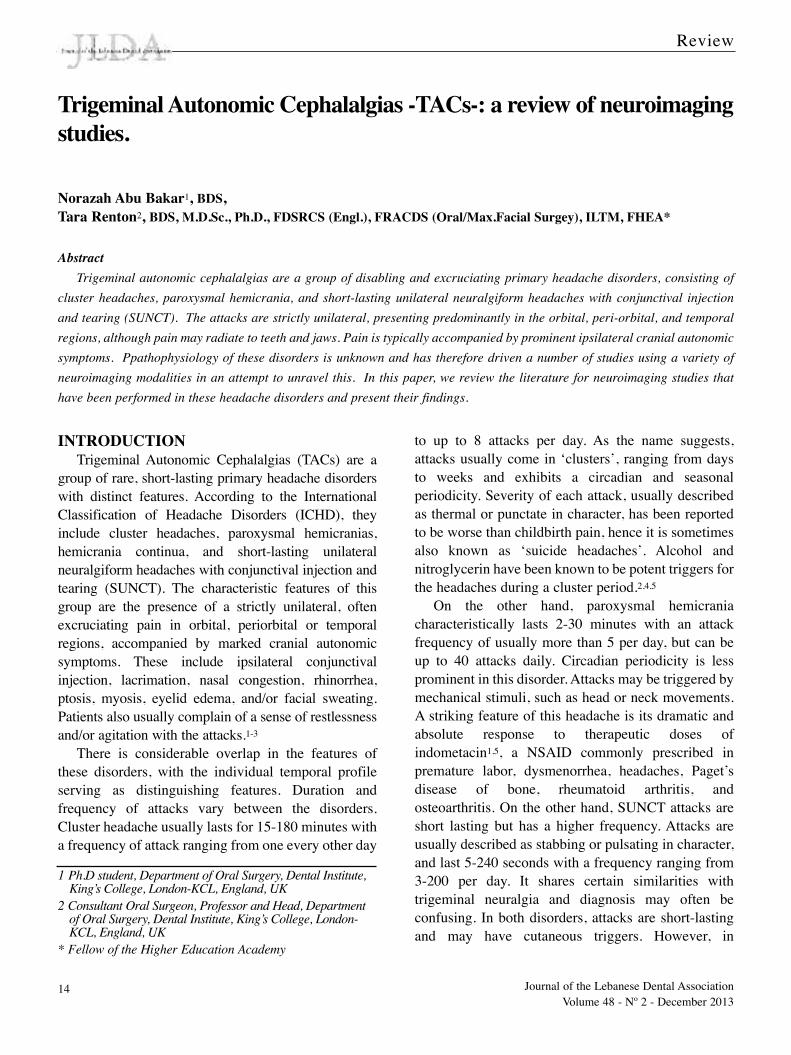
Pains, especially after he published (with Dr. Jean
Thomas, physician, and Dr. Elisabeth Thomas,
pharmacist and biologist) a textbook on Migraine:
this medical work was written in French and was
titled "LA MIGRAINE: LA COMPRENDRE ET
LA GUERIR DEFINITIVEMENT" (Migraine:
understand it and cure it for good) and Dr. Tomb is
often quoted in French dental and medical
literatures as the "VIRTUOSE DU
TRAITEMENT DE LA MALOCCLUSION
DENTAIRE".
Nine years after his tragic death in February
2005, Tomb's contributions to pain science are
now widely recognized in France and Europe.
On a personal standpoint, Dr. Tomb was
always polite, without any malice or prejudices.
He always had a ready and warm welcome and a
big smile for his patients and colleagues alike. He
seldom had a harsh word for anyone although he
had an imposing way of maintaining discipline in
his lectures and masterclasses. He was, indeed, a
delightful colleague and was highly respected and
appreciated by all his dental school and hospital
confrères and consœurs. He was one of this early
band of dental aspirants who obviously
succeeded, where many others failed. Against
many odds, he made it, making of himself a one of
a kind dental professional.
In Memorian
EMMANUEL TOMB (1958-2005), Dentist, scholar, author, polymath, and dental educator
12 Journal of the Lebanese Dental AssociationVolume 48 - Nº 2 - December 2013
Dr. Emmanuel (Asaad) Tomb was an
inspirational teacher for his students. His
dedication for his profession, his contributions to
the science of head and neck pain, and his impact
on his junior colleagues, made him a colorful
figure in French and European dentistry.
During his career, he shaped and influenced
countless number of general dentists in their
approach to head and neck pains. Emmanuel
made pain so indeniably interesting, attractive,
and relevant to everyday dentistry. His outspoken
language, honesty, sincerity, common-sense
approach, ability to understand and analyse, and
compelling logic, made him an outstanding and
superlative communicator.
I can say a lot and more about my old friend
"Asaad", and when he crosses my mind (and this
happens very often), i remember mostly his
passion for life, pride, straight talking, political
correctness, and razor-sharp logic. To me,
"Asaad" was an unforgettable fighter for his
happiness and the well-being of his patients. His
guidance and friendship deeply enriched the lives
of so many dentists. I consider him a pride for
Lebanon, France, and Dentistry.
Since 2005, he is sorely and sadly missed,
leaving a void that cannot be filled.
Dr. Tomb is survived by his daughter, Ambre,
who lives in France.
Ziad Noujeim,
Editor-in-Chief, JLDA
13Journal of the Lebanese Dental AssociationVolume 48 - Nº 2 - December 2013
Review
INTRODUCTION Trigeminal Autonomic Cephalalgias (TACs) are a
group of rare, short-lasting primary headache disorderswith distinct features. According to the InternationalClassification of Headache Disorders (ICHD), theyinclude cluster headaches, paroxysmal hemicranias,hemicrania continua, and short-lasting unilateralneuralgiform headaches with conjunctival injection andtearing (SUNCT). The characteristic features of thisgroup are the presence of a strictly unilateral, oftenexcruciating pain in orbital, periorbital or temporalregions, accompanied by marked cranial autonomicsymptoms. These include ipsilateral conjunctivalinjection, lacrimation, nasal congestion, rhinorrhea,ptosis, myosis, eyelid edema, and/or facial sweating.Patients also usually complain of a sense of restlessnessand/or agitation with the attacks.1-3
There is considerable overlap in the features ofthese disorders, with the individual temporal profileserving as distinguishing features. Duration andfrequency of attacks vary between the disorders.Cluster headache usually lasts for 15-180 minutes witha frequency of attack ranging from one every other day
to up to 8 attacks per day. As the name suggests,attacks usually come in ‘clusters’, ranging from daysto weeks and exhibits a circadian and seasonalperiodicity. Severity of each attack, usually describedas thermal or punctate in character, has been reportedto be worse than childbirth pain, hence it is sometimesalso known as ‘suicide headaches’. Alcohol andnitroglycerin have been known to be potent triggers forthe headaches during a cluster period.2,4,5
On the other hand, paroxysmal hemicraniacharacteristically lasts 2-30 minutes with an attackfrequency of usually more than 5 per day, but can beup to 40 attacks daily. Circadian periodicity is lessprominent in this disorder. Attacks may be triggered bymechanical stimuli, such as head or neck movements.A striking feature of this headache is its dramatic andabsolute response to therapeutic doses ofindometacin1,5, a NSAID commonly prescribed inpremature labor, dysmenorrhea, headaches, Paget’sdisease of bone, rheumatoid arthritis, andosteoarthritis. On the other hand, SUNCT attacks areshort lasting but has a higher frequency. Attacks areusually described as stabbing or pulsating in character,and last 5-240 seconds with a frequency ranging from3-200 per day. It shares certain similarities withtrigeminal neuralgia and diagnosis may often beconfusing. In both disorders, attacks are short-lastingand may have cutaneous triggers. However, in
Norazah Abu Bakar1, BDS, Tara Renton2, BDS, M.D.Sc., Ph.D., FDSRCS (Engl.), FRACDS (Oral/Max.Facial Surgey), ILTM, FHEA*
Trigeminal Autonomic Cephalalgias -TACs-: a review of neuroimagingstudies.
Abstract
Trigeminal autonomic cephalalgias are a group of disabling and excruciating primary headache disorders, consisting of
cluster headaches, paroxysmal hemicrania, and short-lasting unilateral neuralgiform headaches with conjunctival injection
and tearing (SUNCT). The attacks are strictly unilateral, presenting predominantly in the orbital, peri-orbital, and temporal
regions, although pain may radiate to teeth and jaws. Pain is typically accompanied by prominent ipsilateral cranial autonomic
symptoms. Ppathophysiology of these disorders is unknown and has therefore driven a number of studies using a variety of
neuroimaging modalities in an attempt to unravel this. In this paper, we review the literature for neuroimaging studies that
have been performed in these headache disorders and present their findings.
1 Ph.D student, Department of Oral Surgery, Dental Institute,King’s College, London-KCL, England, UK
2 Consultant Oral Surgeon, Professor and Head, Departmentof Oral Surgery, Dental Institute, King’s College, London-KCL, England, UK
* Fellow of the Higher Education Academy
14 Journal of the Lebanese Dental AssociationVolume 48 - Nº 2 - December 2013

SUNCT, cranial autonomic features (especiallyconjunctival injection and tearing) are prominentfeatures and pain is often predominantly in theophthalmic distribution of trigeminal nerve (V1).Moreover, patients with SUNCT are usually able totrigger an attack immediately following the previousone, thus do not have a refractory period.1,5
Hemicrania Continua (HC), described and coinedin 1984 by Sjaastad and Spierings, features continuoushead pain (continua) and unilaterality of head pain(hemicrania). Pain is moderate, rarely approaching ahigh intensity level, with nocturnal awakenings, butmost patients are able to work.
Several theories have been put forward in anattempt to explain the pathophysiology behind thisgroup of headaches. Cluster headache was initiallythought to be a vascular headache originating from aninflammation within cavernous sinus. Resultingvenous stasis causes pressure on trigeminal nerve andsimultaneously activates intersecting parasympatheticand sympathetic nerves, eliciting pain and autonomicsymptoms respectively.2-5 Moreover, vasoconstrictiveeffect of sumatriptan, a 5-hydroxytryptamine (5-HT)*agonist, in aborting these attacks, further supportedthis hypothesis.6,7 However, this theory could notexplain circadian rhythmicity of attacks. Hence, it wassuperseded by the hypothalamic theory.
Circadian and seasonal periodicity of clusterheadaches indicate a possible central involvement,with the human biological clock implicated as apotential site. This is situated in the suprachiasmaticnucleus within the hypothalamus, which is alsoresponsible for regulating hormonal activities. Thiscorrelated with the findings of a significant decrease inplasma testosterone levels in male cluster headachepatients. A reduced response to thyrotropin-releasinghormone further supported this hypothesis.Furthermore, a blunted nocturnal peak in melatonin**,a circadian system biomarker, has been found inpatients with cluster headache.4,6
This concept of a possible central involvement hasled to much of the neuroimaging studies in this groupof headaches, in an attempt to unravel thepathophysiological basis of these rare disorders. Muchof the work done in this field has concentrated oncluster headaches, with few studies on paroxysmalhemicrania and SUNCT. However, due to theirdistinctive clinical phenotype, this group of disordersare assumed to have the same pathophysiologicalbasis. This review aims to highlight the variousmethods used and the main findings of these studies.
NITROGLYCERIN: A RELIABLETRIGGER?
The episodic nature of cluster headache makes itdifficult to capture data on patients during spontaneousattacks, thus most neuroimaging studies to date havebeen performed on evoked attacks. The use ofnitroglycerin (a potent vasodilator prescribed in anginapectoris and chronic heart failure) as a triggering agenthas been studied by Ekbom8 who deduced that attacksare inducible whilst patients are in their cluster period,with sensitivity being highest in the middle of a boutand gradually reducing towards the end. The onset ofthe attack ranges from 30-50 minutes followingadministration of nitroglycerin, and it is preceded by afairly transient pulsation and pressure in temples andforehead. There is a refractory period of 6-8 hoursfollowing an attack and patients outside their clusterbout remain insensitive to provocation.
The first positron emission tomography (PET) studyon cluster headache was performed by Hsieh andassociates9 in 1996, using butanol as the tracer forregional cerebral blood flow (rCBF). They studied fourright-handed patients during their active cluster period,two with right-sided and two with left-sided attacks. Theheadaches were elicited within 18-35 minutes ofadministration of 1 mg sublingual nitroglycerin andsuccessfully terminated following subcutaneousadministration of sumatriptan (a synthetic drug of thetriptan class, prescribed in migraine headaches). A 100mm visual analogue scale (VAS) was used to enablepatients to rate their headache intensity. Each patientunderwent six scans: two at baseline (10 minutes apart),one following nitroglycerin administration, twofollowing onset of cluster headaches (10 minutes apart)
15Journal of the Lebanese Dental Association
* 5-hydroxytryptamine or serotonin is a monoamineneurotransmitter primarily found in GI tract, patelet, andCNS. It is popularly thought to be a contributor to feelingsof well-being and happiness.
**Melatonin (N-acetyl-5-methoxytryptamine) is a hormonethat entertains circadin rhythms of several biologicalfunctions. It also protects nuclear and mitochondrial DNAand has a pervasive and antioxidant roles.
Abu Bakar N, Renton T
and lastly following pain relief with sumatriptan.Authors reported that there was a preferential role of theright, non-dominant hemisphere, especially the anteriorcingulate cortex, in the affective-cognitive processing ofpain in these patients. The normal pain processingnetwork was activated but there were no changes seenin the brainstem or diencephalon. Furthermore, theyfound a marked increase in activity in the cavernoussinus region, which suggested its possible role as thecentral generator of cluster headaches. However, thishypothesis is challenged following further studies, asdiscussed later.
May and co-workers10 performed a similar study on17 cluster headache patients. None of them were in theiractive cluster period, whilst eight who were in remissionphase acted as controls. In this study, headaches wereprovoked by inhalation of 1.0-1.2 mg nitroglycerin,although one patient developed attacks spontaneously inthe scanner. Each patient underwent 12 or 13 consecutivescans with VAS ratings. All patients reported similarity ofthe triggered attacks to their usual headaches. Thecerebellum, bilateral anterior cingulate cortex, and insula,the contralateral posterior thalamus, ipsilateral basalganglia, and cerebellum were found to be activated inthese patients. However, unlike migraine, no brainstemactivation was reported during the attacks.11 A distinctivefinding from this study was activation in the ipsilateralhypothalamic grey, which was not observed in the controlgroup. This implies that this area is specifically activatedonly during a cluster headache attack, therefore providingsubstantial evidence of a possible hypothalamicinvolvement.12-13 An increase signal in the cavernoussinus region of patients who were in their active clusterperiod was seen. No differences were noticed betweenthe spontaneous and evoked attacks.
Sprenger and associates14 also presented anincidental case of a spontaneous cluster attack in apatient whilst undergoing PET scanning to study theeffects of deep brain stimulation. The areas activatedwere comparable to earlier studies done withnitroglycerin-induced cluster headaches. Hence,authors concluded it was unlikely that the use ofnitroglycerin to trigger the attacks confounded theimaging data.
THE CAVERNOUS SINUS THEORYCluster headache has long been coined a vascular
headache with the cavernous sinus being implicated asthe focal generator of symptoms. Early studies lookingat the cerebral blood flow of patients with clusterheadaches reported inconsistent results, with somereporting an increase, some a decrease whilst othersshowed no changes in cortical blood flow.15-17 Gaweland co-workers18 studied 119 cluster headache patientsusing Gallium single-photon emission computedtomography (SPECT). Patients in active cluster perioddisplayed a lesion on Gallium SPECT in the region ofcavernous sinus, which fades as the patient moves outof cluster. On the contrary, no definite pathology wasfound in the cavernous sinus region in a magneticresonance imaging (MRI) study of 14 cluster headachepatients.19 A repeat Gallium SPECT study done on 30cluster headache patients and 7 “migraineurs” showedthat marked activity within the parasellar region wasnot limited to cluster headaches only but was also seenin migraine20. Likewise, Schuh-Hofer and associates21
found no evidence for an inflammatory process in thecavernous sinus of six cluster headache patientsinvestigated using (99m)Tc-human serum albumin*and SPECT. These findings thus question the role ofcavernous sinus as the pathophysiological focus incluster headaches.
Despite consistent findings of significant activationwithin cavernous sinus region in PET studies,experimental pain studies have also reported similarfindings. A PET study22 performed observed theeffects of cranial pain elicited by capsaicin (an activecomponent of chili pepers prescribed, as analgesic, intopical ointments, nasal sprays, and dermal patches).Seven healthy subjects had a small amount ofcapsaicin injected to their forehead, in an attempt toelicit pain of the ophthalmic division of trigeminalnerve. Increased rCBF was observed bilaterally in theanterior insula, the ipsilateral anterior cingulate cortex,the contralateral thalamus, and bilaterally in thecerebellum as well as in the cavernous sinus.
Similar findings were reported by May andco-workers23 who performed a magneticresonance angiography (MRA) study in addition tothe H2(15)O PET study above. Four volunteers hadcapsaicin subcutaneously administered to the forehead
16
* (99m) Tc-human serum albumin is the most commonlyused radio-labeled colloid in Europe.
Volume 48 - Nº 2 - December 2013
Abu Bakar N, Renton T
to elicit pain. Patient who developed spontaneouscluster attack during the PET study was also included.A significant increase in blood flow was observed inipsilateral internal carotid artery in all subjects. Thefact that there is increased activity in cavernous sinusin experimental pain, during cluster attacks, and in“migraineurs” implies that this activation is notspecific to cluster headaches. Vascular changes seenare thus more likely to be an epiphenomenon inresponse to trigeminal pain, rather than an initiator ofattacks, hence dispelling the cavernous sinushypothesis. Moreover, no activation of thehypothalamus was seen in the experimental pain study,further reinforcing its specificity to cluster headaches.
THE HYPOTHALAMIC HYPOTHESISIn the wake of direct evidence found for a possible
hypothalamic involvement, other neuroimagingmodalities have been used to shed further light to thishypothesis. Morelli and associates24 performed the firstblood oxygen dependent level (BOLD) functionalmagnetic resonance imaging (fMRI) study on fourpatients with episodic cluster headaches. Patients hadregular recurrence of their attacks, thus their scanswere timed accordingly to allow spontaneous attacksto be captured. Significant activation in ipsilateralhypothalamic grey matter was observed.
May and co-workers25 performed a voxel-basedmorphometric analysis on MRI and PET scans of 25and 17 cluster headache patients respectively. Asignificant increase in grey matter density localised toinferior posterior hypothalamus was found bilaterallyin these patients compared to controls. No differencewas detected between patients with active headacheand in the headache-free state, indicating that thesechanges are permanent.
Taking this into account, Lodi and associates26
performed a proton magnetic resonance spectroscopy(1H-MRS) on 26 pain-free patients with clusterheadache. Biochemical levels of N- acetylaspartate(NAA), creatine-phosphocreatine (Cr) and choline(Cho) were assessed. Level of NAA (a neuronalbiomarker) was permanently reduced in thehypothalamus of these patients. Such abnormalities areusually identifiable in pathologies like stroke,degenerative disorders, and multiple sclerosis. Similar
findings were reported from another proton magneticresonance spectroscopy study of 47 episodic clusterheadache patients. In addition to a reduction inNAA/Cr, a change in the Cho/Cr levels was alsodetected. These neurochemical changes wereconsistent with increased grey matter density and ahypothalamic dysfunction in patients with clusterheadache, thus further strengthening the possiblecentral role of the hypothalamus in this disorder.27
PAROXYSMAL HEMICRANIA (PH)Seven patients with chronic paroxysmal hemicrania28
who were completely pain-free on oral indometacin,underwent 11 to 13 radioactive PET scans. Medicationwas stopped 24-48 hours prior to scanning sessions.Scans were randomised in two states: patient in pain andoff indometacin, and patient completely pain-free and offindometacin. All patients then underwent a furtherseparate scan being completely pain-free afteradministration of 100 mg intramuscular indometacin.Activations in contralateral posterior hypothalamus andventral midbrain, as well as the ipsilateral lentiformnucleus, anterior and posterior cingulate cortices,bilateral insulae, bilateral frontal cortices, contralateraltemporal cortex, contralateral postcentral gyrus,precuneus, and contralateral cerebellum were identifiedduring both the pain and interictal pain-free states.However, this was deactivated in the pain-free statefollowing indometacin administration. The consistenthypothalamic activation observed in this study and thosein cluster headaches highlight the relatedpathophysiological background of these syndromes andthe potential role of this region in initiating the attacks(the activated subcortical structures may play a pivotalrole in PH pathophysiology).
SHORT-LASTING UNILATERALNEURALGIFORM HEADACHES WITHCONJUNCTIVAL INJECTION ANDTEARING (SUNCT)
The first direct evidence for a hypothalamicdysfunction in SUNCT patients was reported by Mayand associates29 who performed a BOLD fMRI. Thepatient developed 6 consecutive spontaneous attacksin the scanner, lasting from 36 to 96 seconds, withinterattack intervals of 2 to 3.5 minutes. In contrast to
17Journal of the Lebanese Dental Association
Abu Bakar N, Renton T
the pain-free state, ipsilateral hypothalamic activationwas observed solely during attacks. This correspondedto the same area activated in cluster headaches.
Sprenger and co-workers30 performed fMRI on apatient with a two year history of SUNCT.Interestingly, a vascular contact was detected onipsilateral trigeminal nerve from structural MRI scans.Patient was able to self-trigger his attacks by touchinghis upper lip with the lower. Bilateral activation ofhypothalamus was reported, as well as activation ofthe other pain processing areas of the brain. Patientsubsequently had surgical decompression of hisipsilateral trigeminal nerve and was pain-freefollowing this intervention. In a separate BOLD fMRIstudy, Sprenger and other co-workers31 reportedsignificant activation in the ipsilateral hypothalamicgrey matter of a patient with atypical case of TAC.
Nine patients with primary SUNCT and one withsymptomatic SUNCT secondary to a brainstem lesionwere studied using BOLD fMRI scanning. Bilateralactivation of hypothalamus was observed in five of theprimary SUNCT cases, whilst two cases showedcontralateral activation. Meanwhile, two patients hadnegative activation ipsilateral to the pain. Moreover,there was no hypothalamic activation in the patientwith secondary SUNCT.32 Authors also investigatedtwo patients with SUNA (short lasting unilateralneuralgiform headache attacks with cranial autonomicsymptoms). This rare disorder is often thought to be asubset of SUNCT, due to its similar clinical phenotype,differing only in the amount of cranial autonomicinvolvement. In SUNA, patients may present witheither conjunctival injection or tearing or any of theother cranial autonomic symptoms.33
DEEP BRAIN STIMULATIONNeuroimaging studies have provided considerable
insight to the pivotal role of hypothalamus in thepathogenesis of TAC. This has brought aboutadvancements in treatment modalities, namely deepbrain stimulation (DBS), which has renderedintractable patients pain-free. PET studies performed
on 10 cluster headache patients with implantedhypothalamic DBS electrodes found that stimulationinduced activation and deactivation in cerebral areasnormally involved in pain processing network and inacute cluster headache attacks. In particular, activationwas reported in ipsilateral hypothalamic gray (the siteof the stimulator tip), ipsilateral thalamus,somatosensory cortex, praecuneus, anterior cingulatecortex, and the ipsilateral trigeminal nucleus andganglion. Deactivation was observed in the middletemporal gyrus, posterior cingulate cortex, andcontralateral anterior insula. There was no evidencefound for an antinociceptive effect or a pure inhibitionof hypothalamic activity as the mode of action of DBSin cluster headache, thus suggesting the possibility ofa yet unknown functional modulation of the neuronalpain-processing pathways.34
CONCLUSIONTACs are a group of primary headaches
characterized by unilaterality of pain, short duration ofsymptoms, and associated ipsilateral cranialautonomic symptoms (Horner* syndrome,lacrimation, nasal congestion): There have beennumerous studies using a multitude of neuroimagingmodalities to help unravel pathogenesis of TAC. Themajority have focused mainly on cluster headacheswith few studies reported on paroxysmal hemicraniaand SUNCT/SUNA. Despite our recent understandingof the hypothalamus as the primum movens structurein TAC, the exact pathways involved in the generationof resultant pain and cranial autonomic symptomshave yet to be identified. Exact mechanism explainingthe variation in attack frequency and duration betweencluster headaches, paroxysmal hemicrania, andSUNCT which are thought to share the samepathophysiological basis also remains a mystery. Thediscrepancy in hypothalamic activation visualised inthe SUNCT/SUNA patients in the above studies alsoposes the question of how the strictly unilateralsymptoms seen in these patients are exerted. Thus,although there has been much progress in ourunderstanding of the pathophysiology of thesedisorders, there still remains much to be answered.Hence, further studies in this field are warranted toshed light on these issues.
* Horner syndrome (or Horner-Bernard syndrome oroculosympathetic palsy)= a combination of signs/symptomscaused by disruption of a nerve pathway from brain to faceand eye, on one side of the body. Typically, it results in:decreased pupil size + drooping eyelid + decreasedsweating on the affected side of the face.
18 Volume 48 - Nº 2 - December 2013
Abu Bakar N, Renton T
REFERENCES1. Goadsby PJ, Lipton RB. A review of paroxysmal hemicranias,SUNCT syndrome and other short-lasting headaches withautonomic feature, including new cases. Brain 1997; 120: 193-209.
2. Balasubramaniam R, Klasser GD. Trigeminal autonomiccephalalgias. Part 1: cluster headache. Med Oral Pathol OralRadiol Endod 2007; 104: 345-358.
3. Matharu M, May A. Functional and structural neuroimaging intrigeminal autonomic cephalalgias. Current Pain and HeadacheReports 2008; 12: 132-137.
4. Goadsby PJ. Pathophysiology of cluster headache: a trigeminalautonomic cephalgia. The Lancet Neurology 2002; 1: 251-257.
5. Chong MS. Headache syndromes presenting with facial pain andautonomic features. In Zakrzewska JM, Harrison SD editors:Assessment and Management of Orofacial Pain. 1st edition,Elsevier; 2002; p209-245.
6. May A. Cluster headache: pathogenesis, diagnosis andmanagement. Lancet 2005; 366: 843-855.
7. Schoenen J. Cluster headaches- central or peripheral in origin?Lancet 1998; 352: 253-255.
8. Ekbom K. Nitroglycerin as a provocative agent in clusterheadache. Arch Neurol 1968; 19: 487-493.
9. Hsieh JC, Hannerz J, Ingvar M. Right-lateralised centralprocessing for pain of nitroglycerin-induced cluster headache. Pain1996; 67: 59-68.
10. May A, Bahra A, Büchel C, Frackowiak RSJ, Goadsby PJ.Hypothalmic activation in cluster headache attacks. Lancet 1998;352: 275-278.
11. Aurora SK. Pathophysiology of migraine and clusterheadaches. Seminars in Pain Medicine 2004; 2: 62-71.
12. Leone M, Proietti Cecchini A, Mea E, Curone M, Tullo V,Casucci G, Bonavita V, Bussone G. Functional neuroimaging andheadache pathophysiology: new findings and new prospects.Neurol Sci 2007; 28: 108-113.
13. Sánchez del Rio M, Alvarez Linera J. Functional neuroimagingof headaches. Lancet Neurol 2004; 3: 645-651.
14. Sprenger T, Boecker H, Tolle TR, Bussone G, May A, LeoneM. Specific hypothalamic activation during a spontaneous clusterheadache attack. Neurology 2004; 62: 516-517.
15. Norris JW, Hachinski VC, Cooper PW. Cerebral blood flowchanges in cluster headache. Acta Neurol Scandinav 1976; 54:371-374.
16. Nelson RF, du Boulay GH, Marshall J, Russell R, Symon L,Zilkha E. Cerebral blood flow studies in patients with clusterheadache. Headache 1980; 20: 184-189.
17. Krabbe A, Henriksen L, Olesen J. Tomographic determinationof cerebral blood flow during attacks of cluster headache.Cephalalgia 1984; 4: 17-23.
18. Gawel MJ, Krajewski A, Luo YM, Ichise M. The clusterdiathesis. Headache 1990; 30: 652-655.
19. Sjaastad O, Rinck P. Cluster headache: MRI studies of thecavernous sinus and the base of the brain. Headache 1990; 30: 350-351.
20. Sianard-Gainko J, Milet J, Ghuysen V, Schoenen J. Increasedparasellar activity on gallium SPECT is not specific for activecluster headache. Cephalalgia 1994; 14: 132-133.
21. Schuh-Hofer S, Richter M, Israel H, Geworski L, Villringer A,Munz DL, Arnold G. The use of radiolabelled human serumalbumin and SPECT/MRI co-registration to study inflammation inthe cavernous sinus of cluster headache patients. Cephalalgia2006; 26: 1115-1122.
22. May A, Kaube H, Büchel C, Eichten C, Rijntjes M, Jüptner M,Weiller C, Diener HC. Experimental cranial pain elicited bycapsaicin: a PET study. Pain 1998; 74: 61-66.
23. May A, Bahra A, Büchel C, Frackowiak RSJ, Goadsby PJ. PETand MRA findings in cluster headache and MRA in experimentalpain. Neurology 2000; 55: 1328-1335.
24. Morelli N, Pesaresi I, Caffario G, Maluccio MR, Gori S, DiSalle F, Murri L. Functional magnetic resonance imaging inepisodic cluster headache. J Headache Pain 2009; 10: 11-14.
25. May A, Ashburner J, Büchel C, McGonigle DJ, Friston KJ,Frackowiak RSJ, Goadsby PJ. Correlation between structural andfunctional changes in brain in an idiopathic headache syndrome.Nature Medicine 1999; 5: 836-838.
26. Lodi R, Pierangeli G, Tonon C, Cevoli S, Testa C, Bivona G,Magnifico F, Cortelli P, Montagna P, Barbiroli B. Study ofhypothalamic metabolism in cluster headache by proton MRspectroscopy. Neurology 2006; 66: 1264-1266.
27. Wang SJ, Lirng JF, Fuh JL, Chen JJ. Reduction inhypothalamic 1H-MRS metabolite ratios in patients with clusterheadache. J Neurol Neurosurg Psychiatry 2006 May; 77: 622-625.
28. Matharu MS, Cohen AS, Frackowiak RSJ, Goadsby PJ.Posterior hypothalamic activation in paroxysmal hemicrania. AnnNeurol 2006; 59: 535-545.
29. May A, Bahra A, Büchel C, Turner R, Goadsby PJ. Functionalmagnetic resonance imaging in spontaneous attacks of SUNCT:short-lasting neuralgiform headache with conjunctival injectionand tearing. Ann Neurol 1999; 46: 791-794.
30. Sprenger T, Valet M, Platzer S, Pfaffenrath V, Steude U, TolleTR. SUNCT: bilateral hypothalamic activation during headacheattacks and resolving of symptoms after trigeminal decompression.Pain 2005; 113: 422-426.
31. Sprenger T, Valet M, Hammes M, Erhard P, Berthele A, ConradB, Tolle TR. Hypothalamic activation in trigeminal autonomiccephalgia: functional imaging of an atypical case. Cephalalgia2004; 24: 753-757.
32. Cohen AS. Short-lasting unilateral neuralgiform headacheattacks with conjunctival injection and tearing. Cephalalgia 2007;27: 824-832.
33. Cohen AS, Matharu MS, Goadsby PJ. Short-lasting unilateralneuralgiform headache attacks with conjunctival injection andtearing (SUNCT) or cranial autonomic features (SUNA)- aprospective clinical study of SUNCT and SUNA. Brain 2006; 129:2746-2760.
34. May A, Leone M, Boecker H, Sprenger T, Juergens T, Bussone G,Tolle TR. Hypothalamic deep brain stimulation in positron emissiontomography. Journal of Neuroscience 2006; 26: 3589-3593.
Correspond with:Tara [email protected]
19Journal of the Lebanese Dental AssociationVolume 48 - Nº 2 - December 2013
Abu Bakar N, Renton T
Clinical Note
INTRODUCTIONPatients presenting with TN consult a variety of
clinicians, especially GPs, ENT specialists,maxillofacial surgeons, and dentists. The symptomscan occur spontaneously or may be triggered by localoral and maxillofacial causes such as touch, wind,chewing, and facial muscles movements. Therefore, agood diagnosis is essential to avoid unnecessarytherapeutic interventions. Once the diagnosis is madeand any symptomatic form of the condition rejected,management of TN is mainly medical. Only refractoryform of the disease or patient intolerance to medicaltreatment will be subject to neurosurgical intervention.
Finally, the frequent identification of a vascularloop in contact with the Trigeminal Nerve leading toneurovascular conflict reduces the diagnosis frequencyof “idiopathic” Trigeminal Neuralgia[5].
EPIDEMIOLOGYAge of presentation is usually between 50 and 70
years, with some patients presenting at an age olderthan 70 not being uncommon. A younger age of onsetshould push the investigations towards an underlying
tumoral or demyelinating pathology. Women to menratio is 3:2. Incidence is 5 to 6 new cases per 100,000per year[3,11,12]. Hereditary form of TN is rare.
ANATOMYThe name “trigeminal” (literally, three twins) refers
to the fact that 5th cranial nerve has 3 major divisions(ophtalmic, maxillary, and mandibular). Trigeminalnerve is a mixed (sensory and motor) cranial nerveresponsible for tactition (pressure), thermoception(temperature), and nociception (pain). Its nuclei are
Trigeminal N euralgia - TN: diagnosis and management challenges.
Joe Faddoul1,2, MD, Sandra Kobaïter Maarrawi1, B.Sc., M.Sc., Dr. Univ. (Neuroscience), Sharoni Weinberg1, B.Sc.,Patrick Tabet1, B.Sc., Elie Samaha1,2, MD, Joseph Maarrawi1,2, MD, M.Sc., Dr. Univ. (Neuroscience)
1. Laboratory of Neurosciences, Faculty of Medicine, Saint-Joseph University, Beirut, Lebanon
2. Department of Neurosurgery, Hôtel-Dieu de FranceHospital/Faculty of Medicine, Saint-Joseph University,Beirut
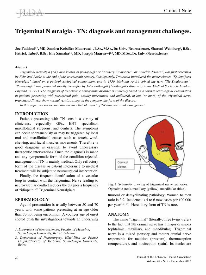
Fig. 1. Schematic drawing of trigeminal nerve territories:Ophtalmic (red), maxillary (yellow), mandibular (blue).
Abstract
Trigeminal Neuralgia (TN), also known as prosopalgia or “Fothergill’s disease”, or “suicide disease”, was first described
by Fehr and Locke at the end of the seventeenth century. Subsequently, Trousseau introduced the nomenclature "Epileptiform
Neuralgia” based on a pathophysiological connotation, and in 1756, Nicholas André coined the term "Tic Douloureux”.
"Prosopalgia" was presented shortly thereafter by John Fothergill (“Fothergill’s disease”) to the Medical Society in London,
England, in 1773. The diagnosis of this chronic neuropathic disorder is clinically based on a normal neurological examination
in patients presenting with paroxysmal pain, usually intermittent and unilateral, in one (or more) of the trigeminal nerve
branches. All tests show normal results, except in the symptomatic form of the disease.
In this paper, we review and discuss the clinical aspect of TN diagnosis and management.
20 Journal of the Lebanese Dental AssociationVolume 48 - Nº 2 - December 2013
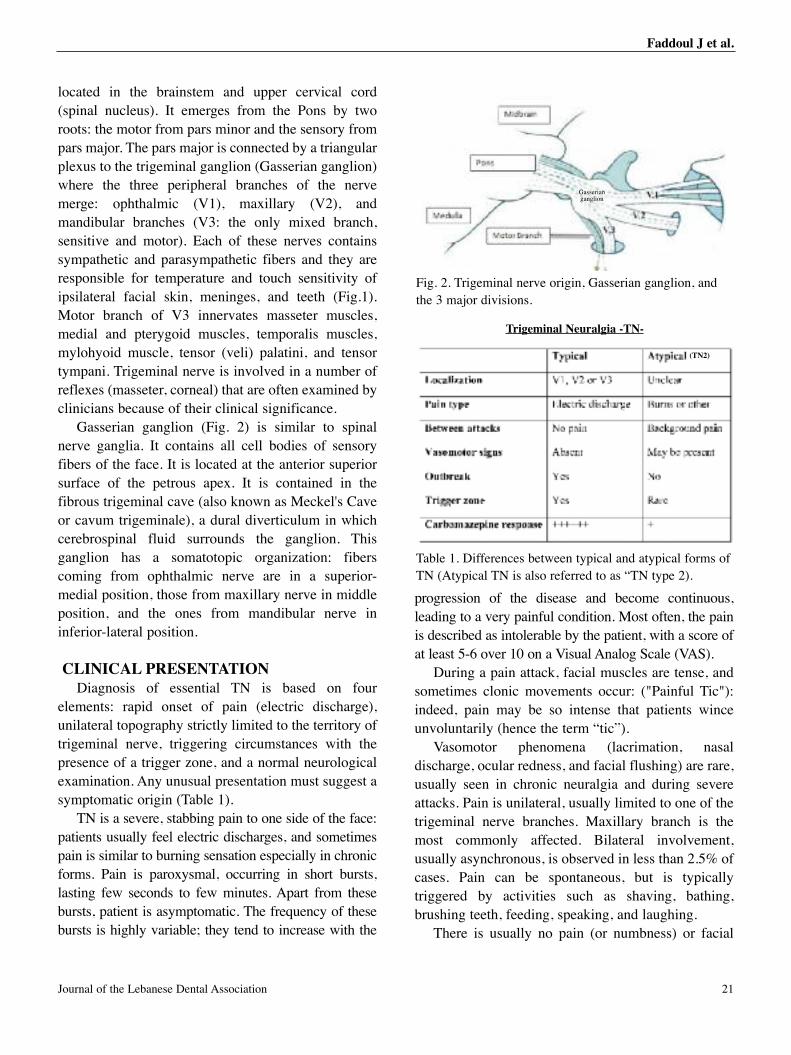
located in the brainstem and upper cervical cord(spinal nucleus). It emerges from the Pons by tworoots: the motor from pars minor and the sensory frompars major. The pars major is connected by a triangularplexus to the trigeminal ganglion (Gasserian ganglion)where the three peripheral branches of the nervemerge: ophthalmic (V1), maxillary (V2), andmandibular branches (V3: the only mixed branch,sensitive and motor). Each of these nerves containssympathetic and parasympathetic fibers and they areresponsible for temperature and touch sensitivity ofipsilateral facial skin, meninges, and teeth (Fig.1).Motor branch of V3 innervates masseter muscles,medial and pterygoid muscles, temporalis muscles,mylohyoid muscle, tensor (veli) palatini, and tensortympani. Trigeminal nerve is involved in a number ofreflexes (masseter, corneal) that are often examined byclinicians because of their clinical significance.
Gasserian ganglion (Fig. 2) is similar to spinalnerve ganglia. It contains all cell bodies of sensoryfibers of the face. It is located at the anterior superiorsurface of the petrous apex. It is contained in thefibrous trigeminal cave (also known as Meckel's Caveor cavum trigeminale), a dural diverticulum in whichcerebrospinal fluid surrounds the ganglion. Thisganglion has a somatotopic organization: fiberscoming from ophthalmic nerve are in a superior-medial position, those from maxillary nerve in middleposition, and the ones from mandibular nerve ininferior-lateral position.
CLINICAL PRESENTATIONDiagnosis of essential TN is based on four
elements: rapid onset of pain (electric discharge),unilateral topography strictly limited to the territory oftrigeminal nerve, triggering circumstances with thepresence of a trigger zone, and a normal neurologicalexamination. Any unusual presentation must suggest asymptomatic origin (Table 1).
TN is a severe, stabbing pain to one side of the face:patients usually feel electric discharges, and sometimespain is similar to burning sensation especially in chronicforms. Pain is paroxysmal, occurring in short bursts,lasting few seconds to few minutes. Apart from thesebursts, patient is asymptomatic. The frequency of thesebursts is highly variable; they tend to increase with the
progression of the disease and become continuous,leading to a very painful condition. Most often, the painis described as intolerable by the patient, with a score ofat least 5-6 over 10 on a Visual Analog Scale (VAS).
During a pain attack, facial muscles are tense, andsometimes clonic movements occur: ("Painful Tic"):indeed, pain may be so intense that patients winceunvoluntarily (hence the term “tic”).
Vasomotor phenomena (lacrimation, nasaldischarge, ocular redness, and facial flushing) are rare,usually seen in chronic neuralgia and during severeattacks. Pain is unilateral, usually limited to one of thetrigeminal nerve branches. Maxillary branch is themost commonly affected. Bilateral involvement,usually asynchronous, is observed in less than 2.5% ofcases. Pain can be spontaneous, but is typicallytriggered by activities such as shaving, bathing,brushing teeth, feeding, speaking, and laughing.
There is usually no pain (or numbness) or facial
Table 1. Differences between typical and atypical forms ofTN (Atypical TN is also referred to as “TN type 2).
Fig. 2. Trigeminal nerve origin, Gasserian ganglion, andthe 3 major divisions.
Trigeminal Neuralgia -TN-
Gasserian ganglion
(TN2)
Faddoul J et al.
21Journal of the Lebanese Dental Association
muscles dysfunction between attacks and although aflurry of attacks may last several weeks (or months),there are usually periods of months (sometimes years)that are pain-free.
Weight loss is sometimes observed when thepatient tries to avoid triggering his pain. There is oftena trigger point located in the painful trigeminalterritory (upper gum, lip, ala of nose). Usually, apainless mechanical stimulus with low intensity(sneezing) is enough to trigger the attack, whileelectric or noxious stimuli have no activity.
Neurological examination is normal, except for therare presence of a slight hypoesthesia in the painfularea after an attack or in chronic forms of the disease.Presence of a neurological deficit should suggest asymptomatic neuralgia.
Response to Carbamazepine (an anticonvulsant andmood-stabilizing drug prescribed in epilepsy andbipolar disorder), especially early in the disease, is akey criterion in the diagnosis of essential TN[16].Disease evolution is intermittent with spontaneousremission periods that can last several months. Theseperiods become shorter and less spaced with diseaseprogression. At the end, pain may become recurrentand chronic. Sometimes, symptom changes can beseen in chronic neuralgia: onset of a painfulbackground, absence of the trigger area, burning pain,vasomotor signs.. etc...
DIAGNOSISTN is generally a disease of middle age or later life,
and women are usually affected more than men. Mostpeople feel the pain in their jaws, cheeks, or lips onone side of the face, and pain is often so severe thatpatients are afraid to talk, eat, or move during attacks.In TN, investigation tests are negative. They areusually prescribed in order to rule out symptomaticforms of TN, especially in young patients, and whenclinical examination reveals associated neurologicalsigns. MRI, thin cut CT-Scan, or contrast enhanced CTcan reveal the cause of symptomatic neuralgia:demyelinating disease, Arnold-Chiari malformation (abrain malformation consisting of a downwarddisplacement of cerebellar tonsils through foramenmagnum), small posterior fossa tumor, or a skull baselesion compressing trigeminal nerve. The search for a
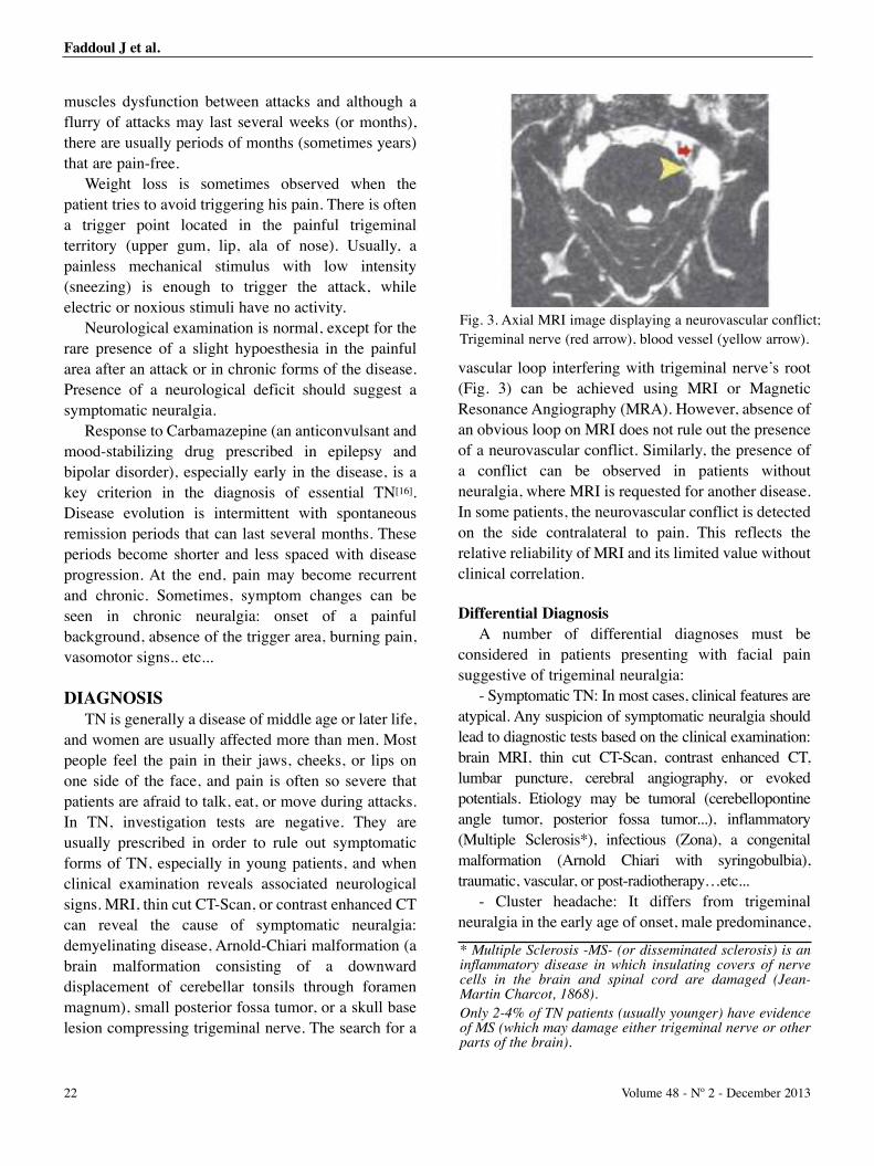
vascular loop interfering with trigeminal nerve’s root(Fig. 3) can be achieved using MRI or MagneticResonance Angiography (MRA). However, absence ofan obvious loop on MRI does not rule out the presenceof a neurovascular conflict. Similarly, the presence ofa conflict can be observed in patients withoutneuralgia, where MRI is requested for another disease.In some patients, the neurovascular conflict is detectedon the side contralateral to pain. This reflects therelative reliability of MRI and its limited value withoutclinical correlation.
Differential DiagnosisA number of differential diagnoses must be
considered in patients presenting with facial painsuggestive of trigeminal neuralgia:
- Symptomatic TN: In most cases, clinical features areatypical. Any suspicion of symptomatic neuralgia shouldlead to diagnostic tests based on the clinical examination:brain MRI, thin cut CT-Scan, contrast enhanced CT,lumbar puncture, cerebral angiography, or evokedpotentials. Etiology may be tumoral (cerebellopontineangle tumor, posterior fossa tumor...), inflammatory(Multiple Sclerosis*), infectious (Zona), a congenitalmalformation (Arnold Chiari with syringobulbia),traumatic, vascular, or post-radiotherapy…etc...
- Cluster headache: It differs from trigeminalneuralgia in the early age of onset, male predominance,
Fig. 3. Axial MRI image displaying a neurovascular conflict;Trigeminal nerve (red arrow), blood vessel (yellow arrow).
Faddoul J et al.
* Multiple Sclerosis -MS- (or disseminated sclerosis) is aninflammatory disease in which insulating covers of nervecells in the brain and spinal cord are damaged (Jean-Martin Charcot, 1868).
Only 2-4% of TN patients (usually younger) have evidenceof MS (which may damage either trigeminal nerve or otherparts of the brain).
22 Volume 48 - Nº 2 - December 2013
presence of sympathetic component, a dazzling retro-orbital pain following the carotid territory, and specificduration and frequency of attacks.
- Other pain origins: ENT (sinusitis), eyes(glaucoma), dental pain, temporo-mandibular joint,muscular (temporal myalgia), arterial inflammatorydisease (Horton’s disease), psychogenic, andpostherpetic neuralgia (after shingles) may causesimilar symptoms if trigeminal nerve is damaged.
Management The first line treatment of TN is pharmacological.
Surgical treatment is proposed only after failure ordecreased effectiveness of pharmacological treatmentover time or patient drug intolerance. Note that,historically, neurosurgical treatment was used as firstline therapy before the discovery of carbamazepine.
1- Pharmacological Treatment
Since its introduction in 1962 by Blom,Carbamazepine (Tegretol®) is the medical treatment ofchoice for TN: Dosage is gradually increased up to1000 mg per day, and rarely up to 1200 to 1800 mg perday. Therapeutic response is rapid and satisfactory in80% of patients, but over time, its effectiveness wearsoff in at least 50% of patients[5]. The immediate releaseform is preferable over the sustained-release form ofthe drug.
Response to treatment is an effective diagnostictest. In some cases, depletion of the analgesic effectover time may require a second or a third drug forcontrol of breakthrough episodes, and may lead to theneed of considering a neurosurgical treatment.However, there are no published studies directlycomparing monotherapy with polytherapy[19]. Side-effectsoccurring early in the treatment usually fade with timeand adverse reactions are rare[8].
Other drugs that are currently used as second-linetreatment or in combination with Carbamazepine areGabapentina (Neurontin®) or oxcarbazepine(Trileptal®). Compared to the pharmacokinetics ofolder antiepileptic drugs such as Carbamazepine, thesedrugs, introduced in the early 1990s, have longer half-lives, permitting a once or twice-daily dosing. Thisreduced dose decreases potential for drug interactions,general hepatic enzyme induction, and facilitation ofpolypharmacy[9].
Oxcarbazepine is chemically related to
Carbamazepine but follows a slightly different metabolicpathway which offers several clinical advantages. Unlikecarbamazepine, oxcarbazepine is not metabolized to anepoxide metabolite, believed to cause toxic effect, and itevokes a lesser decrease in white blood cell count.Aplastic anemia and agranulocytosis that occur withcarbamazepine may also occur but less frequently. Themost common side-effects are dizziness, headaches, andgastrointestinal disturbances; however the most seriousone is hyponatremia[7].
Little controlled data exists for the use ofOxcarbazepine, but trials on its efficacy has shownoutcomes to be similar to that of Carbamazepine. Thebetter tolerance of oxcarbazepine is considered to bean advantage, but due to the lack of control data and itshigher cost, carbamazepine is still prescribed as firstline treatment for TN[2].
Other medications have proved less effective, such asphenytoinb (Dilantin®, Di-HYDAN®), Clonazepamc
(Rivotril®, Klonopin®) and Baclofend (Lioresal®).WHO’s class I to III analgesics (including opiates) aregenerally unsatisfactory in the treatment of TrigeminalNeuralgia.
2- Neurosurgical Treatment
In the eighteenth century, Gasserian ganglionexcision was the first surgery proposed for thetreatment of trigeminal neuralgia. Thereafter, severalsurgical techniques have been described; each has itsadvantages and disadvantages. Currently, there arethree neurosurgical techniques for the treatment ofTNs refractory to medical treatment:
- Percutaneous techniques: PercutaneousRadiofrequency Trigeminal Gangliolysis, PercutaneousRetrogasserian Glycerol Rhizotomy, and PercutaneousBalloon Microcompression.
- Microvascular decompression.- Radiosurgery.
a: Gabapentin= anticonvulsant and analgesic drug,commonly prescribed in epilepsy and neuropathic painarising from diabetic neuropathy, and post-herpeticneuralgia, and central neuropathic pain.
b: Phenytoin= hydantoin - derivative anticonvulsantprescribed primarily in complex partial seizures andgeneralized tonic-clonic seizures.
c: Clonazepam= benzodiazepine drug, having anxiolytic,anticonvulsant, muscle relaxant, sedative, and hypnoticproperties.
d: Baclofen= derivative of GABA (Gamma-AminoButiricAcid), primarily used to treat spasticity, it is also used bycompounding pharmacies in topical pain creams as amuscle relaxant.
23Journal of the Lebanese Dental Association
Faddoul J et al.
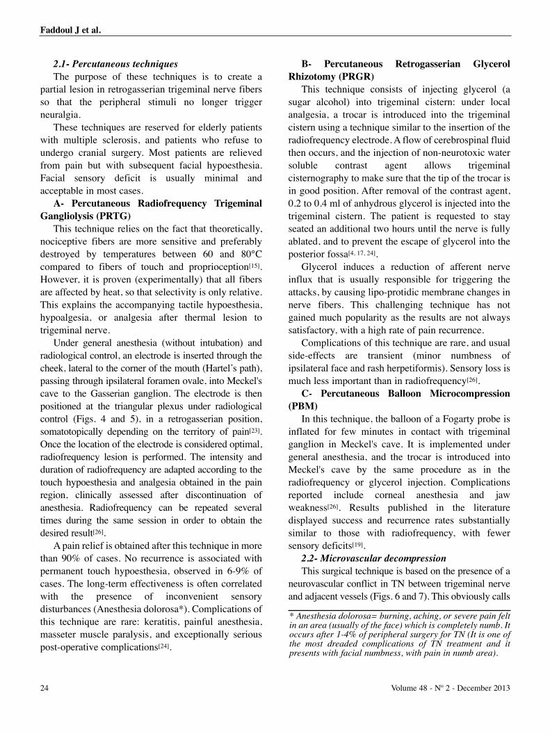
2.1- Percutaneous techniques
The purpose of these techniques is to create apartial lesion in retrogasserian trigeminal nerve fibersso that the peripheral stimuli no longer triggerneuralgia.
These techniques are reserved for elderly patientswith multiple sclerosis, and patients who refuse toundergo cranial surgery. Most patients are relievedfrom pain but with subsequent facial hypoesthesia.Facial sensory deficit is usually minimal andacceptable in most cases.
A- Percutaneous Radiofrequency TrigeminalGangliolysis (PRTG)
This technique relies on the fact that theoretically,nociceptive fibers are more sensitive and preferablydestroyed by temperatures between 60 and 80°Ccompared to fibers of touch and proprioception[15].However, it is proven (experimentally) that all fibersare affected by heat, so that selectivity is only relative.This explains the accompanying tactile hypoesthesia,hypoalgesia, or analgesia after thermal lesion totrigeminal nerve.
Under general anesthesia (without intubation) andradiological control, an electrode is inserted through thecheek, lateral to the corner of the mouth (Hartel’s path),passing through ipsilateral foramen ovale, into Meckel'scave to the Gasserian ganglion. The electrode is thenpositioned at the triangular plexus under radiologicalcontrol (Figs. 4 and 5), in a retrogasserian position,somatotopically depending on the territory of pain[23].Once the location of the electrode is considered optimal,radiofrequency lesion is performed. The intensity andduration of radiofrequency are adapted according to thetouch hypoesthesia and analgesia obtained in the painregion, clinically assessed after discontinuation ofanesthesia. Radiofrequency can be repeated severaltimes during the same session in order to obtain thedesired result[26].
A pain relief is obtained after this technique in morethan 90% of cases. No recurrence is associated withpermanent touch hypoesthesia, observed in 6-9% ofcases. The long-term effectiveness is often correlatedwith the presence of inconvenient sensorydisturbances (Anesthesia dolorosa*). Complications ofthis technique are rare: keratitis, painful anesthesia,masseter muscle paralysis, and exceptionally seriouspost-operative complications[24].
B- Percutaneous Retrogasserian GlycerolRhizotomy (PRGR)
This technique consists of injecting glycerol (asugar alcohol) into trigeminal cistern: under localanalgesia, a trocar is introduced into the trigeminalcistern using a technique similar to the insertion of theradiofrequency electrode. A flow of cerebrospinal fluidthen occurs, and the injection of non-neurotoxic watersoluble contrast agent allows trigeminalcisternography to make sure that the tip of the trocar isin good position. After removal of the contrast agent,0.2 to 0.4 ml of anhydrous glycerol is injected into thetrigeminal cistern. The patient is requested to stayseated an additional two hours until the nerve is fullyablated, and to prevent the escape of glycerol into theposterior fossa[4, 17, 24].
Glycerol induces a reduction of afferent nerveinflux that is usually responsible for triggering theattacks, by causing lipo-protidic membrane changes innerve fibers. This challenging technique has notgained much popularity as the results are not alwayssatisfactory, with a high rate of pain recurrence.
Complications of this technique are rare, and usualside-effects are transient (minor numbness ofipsilateral face and rash herpetiformis). Sensory loss ismuch less important than in radiofrequency[26].
C- Percutaneous Balloon Microcompression(PBM)
In this technique, the balloon of a Fogarty probe isinflated for few minutes in contact with trigeminalganglion in Meckel's cave. It is implemented undergeneral anesthesia, and the trocar is introduced intoMeckel's cave by the same procedure as in theradiofrequency or glycerol injection. Complicationsreported include corneal anesthesia and jawweakness[26]. Results published in the literaturedisplayed success and recurrence rates substantiallysimilar to those with radiofrequency, with fewersensory deficits[19].
2.2- Microvascular decompression
This surgical technique is based on the presence of aneurovascular conflict in TN between trigeminal nerveand adjacent vessels (Figs. 6 and 7). This obviously calls
* Anesthesia dolorosa= burning, aching, or severe pain feltin an area (usually of the face) which is completely numb. Itoccurs after 1-4% of peripheral surgery for TN (It is one ofthe most dreaded complications of TN treatment and itpresents with facial numbness, with pain in numb area).
24 Volume 48 - Nº 2 - December 2013
Faddoul J et al.
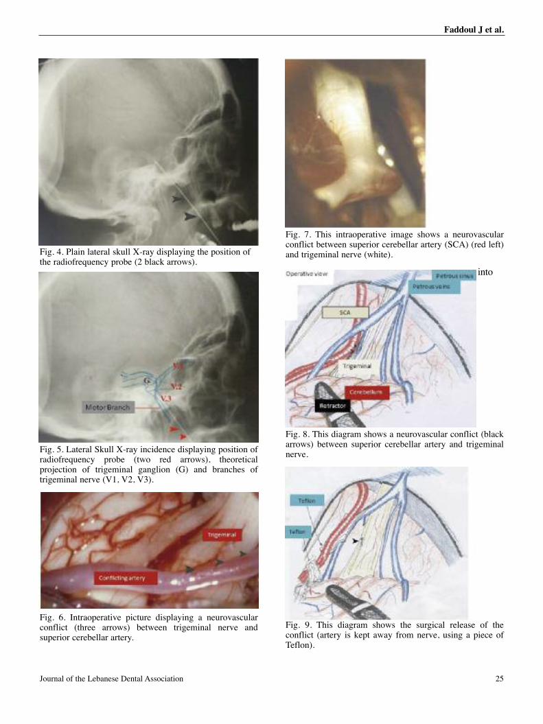
into
Fig. 4. Plain lateral skull X-ray displaying the position ofthe radiofrequency probe (2 black arrows).
Fig. 8. This diagram shows a neurovascular conflict (blackarrows) between superior cerebellar artery and trigeminalnerve.
Fig. 9. This diagram shows the surgical release of theconflict (artery is kept away from nerve, using a piece ofTeflon).
Fig. 5. Lateral Skull X-ray incidence displaying position ofradiofrequency probe (two red arrows), theoreticalprojection of trigeminal ganglion (G) and branches oftrigeminal nerve (V1, V2, V3).
Fig. 6. Intraoperative picture displaying a neurovascularconflict (three arrows) between trigeminal nerve andsuperior cerebellar artery.
Fig. 7. This intraoperative image shows a neurovascularconflict between superior cerebellar artery (SCA) (red left)and trigeminal nerve (white).
25Journal of the Lebanese Dental Association
Faddoul J et al.
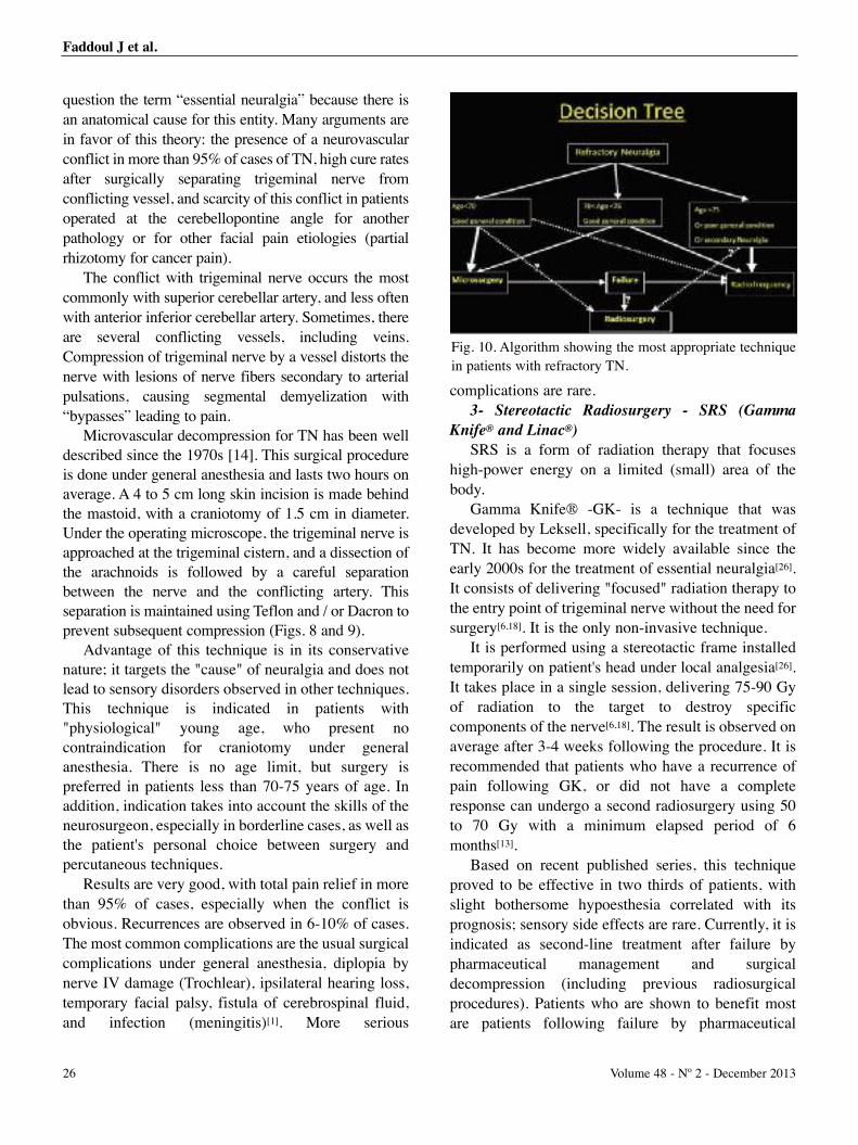
question the term “essential neuralgia” because there isan anatomical cause for this entity. Many arguments arein favor of this theory: the presence of a neurovascularconflict in more than 95% of cases of TN, high cure ratesafter surgically separating trigeminal nerve fromconflicting vessel, and scarcity of this conflict in patientsoperated at the cerebellopontine angle for anotherpathology or for other facial pain etiologies (partialrhizotomy for cancer pain).
The conflict with trigeminal nerve occurs the mostcommonly with superior cerebellar artery, and less oftenwith anterior inferior cerebellar artery. Sometimes, thereare several conflicting vessels, including veins.Compression of trigeminal nerve by a vessel distorts thenerve with lesions of nerve fibers secondary to arterialpulsations, causing segmental demyelization with“bypasses” leading to pain.
Microvascular decompression for TN has been welldescribed since the 1970s [14]. This surgical procedureis done under general anesthesia and lasts two hours onaverage. A 4 to 5 cm long skin incision is made behindthe mastoid, with a craniotomy of 1.5 cm in diameter.Under the operating microscope, the trigeminal nerve isapproached at the trigeminal cistern, and a dissection ofthe arachnoids is followed by a careful separationbetween the nerve and the conflicting artery. Thisseparation is maintained using Teflon and / or Dacron toprevent subsequent compression (Figs. 8 and 9).
Advantage of this technique is in its conservativenature; it targets the "cause" of neuralgia and does notlead to sensory disorders observed in other techniques.This technique is indicated in patients with"physiological" young age, who present nocontraindication for craniotomy under generalanesthesia. There is no age limit, but surgery ispreferred in patients less than 70-75 years of age. Inaddition, indication takes into account the skills of theneurosurgeon, especially in borderline cases, as well asthe patient's personal choice between surgery andpercutaneous techniques.
Results are very good, with total pain relief in morethan 95% of cases, especially when the conflict isobvious. Recurrences are observed in 6-10% of cases.The most common complications are the usual surgicalcomplications under general anesthesia, diplopia bynerve IV damage (Trochlear), ipsilateral hearing loss,temporary facial palsy, fistula of cerebrospinal fluid,and infection (meningitis)[1]. More serious
complications are rare.3- Stereotactic Radiosurgery - SRS (Gamma
Knife® and Linac®)
SRS is a form of radiation therapy that focuseshigh-power energy on a limited (small) area of thebody.
Gamma Knife® -GK- is a technique that wasdeveloped by Leksell, specifically for the treatment ofTN. It has become more widely available since theearly 2000s for the treatment of essential neuralgia[26].It consists of delivering "focused" radiation therapy tothe entry point of trigeminal nerve without the need forsurgery[6,18]. It is the only non-invasive technique.
It is performed using a stereotactic frame installedtemporarily on patient's head under local analgesia[26].It takes place in a single session, delivering 75-90 Gyof radiation to the target to destroy specificcomponents of the nerve[6,18]. The result is observed onaverage after 3-4 weeks following the procedure. It isrecommended that patients who have a recurrence ofpain following GK, or did not have a completeresponse can undergo a second radiosurgery using 50to 70 Gy with a minimum elapsed period of 6months[13].
Based on recent published series, this techniqueproved to be effective in two thirds of patients, withslight bothersome hypoesthesia correlated with itsprognosis; sensory side effects are rare. Currently, it isindicated as second-line treatment after failure bypharmaceutical management and surgicaldecompression (including previous radiosurgicalprocedures). Patients who are shown to benefit mostare patients following failure by pharmaceutical
Fig. 10. Algorithm showing the most appropriate techniquein patients with refractory TN.
26 Volume 48 - Nº 2 - December 2013
Faddoul J et al.
Journal of the Lebanese Dental AssociationVolume 48 - Nº 2 - December 2013
27
management who have not yet receivedpharmaceutical interventions [10, 13]. Randomizedcontrolled trials assessing more precisely itseffectiveness and potential complications are essentialto define its indications in the future.
Linac® is a newer stereotactic radiosurgerytechnique that uses a linear accelerator to create high-energy photons with a single source and a tightlyfocused beam. This technique rotates the radiationsource relative to the patient during exposure,permitting a prescribed dose to be delivered to thetarget area while minimizing the dose to surroundingtissues. Theoretically, the smaller penumbra of theLinac®-based SRS should have an advantage over theGamma Knife® for rates of success and reducedside-effects but no direct clinical comparisons havebeen completed[22].
4- Treatment choice
The treatment choice depends on several factors:patient's age, general condition, anestheticconsultation, desire of the patient and / or family,availability of neurosurgical techniques, and theneurosurgeon’s preferences based on his/herexperience. The algorithm displayed in figure 10provides a general idea for the choice of treatment.
REFERENCES1. Barker FG, Jannetta PJ, Bissonette DJ, Larkins MV, Jho HD.The long-term outcome of microvascular decompression fortrigeminal neuralgia. N Engl J Med. 1996;334: 1077-1083
2. Beydoun A. Safety and efficacy of oxcarbazepine: results ofrandomized, double-blind trials. Pharmacotherapy 2000;20(8 Pt2):152S-158S
3. Campbell GH, Lutsep HL. Trigeminal neuralgia. eMedicine2006; Available from: http://www.emedicine.com/neuro/TOPIC593.HTM.
4. Cappabianca P, Spaziante R, Graziussi G, Taglialatela G, PecaC, De Divitiis E. Percutaneous retrogasserian glycerol rhizolysisfor treatment of trigeminal neuralgia. Technique and results in 191patients. J Neurosurg Sci. Mar 1995;39(1):37-45.
5. Cheshire WP. Trigeminal neuralgia: for one nerve a multitude oftreatments. Expert. Rev. Neurother. 2007;7:1565-1579.
6. Deinsberger R, Tidstrand J. Linac radiosurgery as a tool in neurosurgery.Neurosurg Rev. 2005 April;28(2):79-88; discussion 89-90, 91.
7. Eisenberg E, River Y, Shifrin A, Krivov N. Antiepileptic drugsin the treatment of neuropthic pain. Drugs 2007;67(9): 1265-89.
8. Eller JL, Raslan AM, Burchiel KJ. Trigeminal neuralgia:definition and classification. Neurosurg Focus. 2005 May;18(5):E3.
9. Glauser. Oxcarbazepine in the treatment of epilepsy.Pharmacotherapy 2001;21: 904-19.
10. Gorgulho AA, De Salles AA. Impact of radiosurgery on the surgicaltreatment of trigeminal neuralgia. Surg Neurol 2006;66:350-356
11. Harrison’s Principles of Internal Medicine, McGraw Hill, 2007.
12. Hunt K, Patwardhan R. Trigeminal neuralgia: a modern-dayreview. Int Rev Neurobiol 2007;79:621-631.
13. IRSA*. Stereotactic radiosurgery for patients with intractabletypical trigeminal neuralgia which have failed medicalmanagement. Harrisburg (PA): IRSA; 2009 Jan. (Radiosurgerypractice guideline report no. 1-03).
14. Jannetta PJ. Arterial compression of the trigeminal nerve at the ponsin patients with trigeminal neuralgia. J. Neurosurg 1967;26:Suppl-62
15. Kanpolat Y, Savas A, Bekar A, Berk C. Percutaneous controlledradiofrequency trigeminal rhizotomy for the treatment of idiopathictrigeminal neuralgia: 25-year experience with 1,600 patients.Neurosurgery. 2001 March;48(3):524-32; discussion 532-4.
16. Kitt CA, Gruber K, Davis M, Woolf CJ, Levine JD. Trigeminalneuralgia: opportunities for research and treatment. Pain 2000;85:3-7.
17. Kondziolka D, Lunsford LD. Percutaneous RetrogasserianGlycerol Rhizotomy for Trigeminal Neuralgia: Technique andExpectations Neurosurg Focus. 2005 May 15;18(5):E7.
18. Kondziolka D, Perez B, Flickinger JC, Habeck M, LunsfordLD. Gamma knife radiosurgery for trigeminal neuralgia: resultsand expectations. Arch Neurol. 1998 December;55(12):1524-9.
19. Meglio M, Cioni B. Percutaneous procedures for trigeminalneuralgia: microcompression versus radiofrequencythermocoagulation. Personal experience. Pain 1989 July;38(1):9-16.
20. Nurmikko TJ, Eldridge PR. Trigeminal neuralgia–pathophysiology,diagnosis and current treatment. Br J Anaesth 2001;87:117-132.
21. Rose FC. Trigeminal neuralgia. Arch. Neurol. 1999;56:1163-1164.
22. Solberg TD, Goetsch SJ, Selch MT, Melega W, Lacan G,DeSalles AA. Functional stereotactic radiosurgery involving adedicated linear accelerator and gamma unit: a comparison study.J. Neurosurg. 2004;101 Suppl 3:373-380.
23. Sweet WH, Wepsic JF: Controllected thermocoagulation of thetrigeminal ganglion and rootlets for differential destruction of painfibers: Part 1 - Trigeminal neuralgia. J Neurosurg 1974;39:143-156.
24. Taha JM, Tew JM Jr. Treatment of trigeminal neuralgia bypercutaneous radiofrequency rhizotomy.Neurosurg Clin N Am.1997 January;8(1):31-9.
25. Tan LK, Robinson SN, Chatterjee S. Glycerol versus radiofrequencyrhizotomy - a comparison of their efficacy in the treatment of trigeminalneuralgia. Br J Neurosurg. 1995 April;9(2):165-9.
26. Tatli M, Satici O, Kanpolat Y, Sindou M. Various surgicalmodalities for trigeminal neuralgia: literature study of respectivelong-term outcomes. Acta Neurochir 2008;150:243-255.
Corresponding author: Joseph [email protected]@hotmail.com
* IRSA®= International RadioSurgery Association is anindependent organization (dedicated, since 1995, to provideeducational information and guidelines on stereotacticradiosurgery for brain tumors and brain disorders).
Faddoul J et al.
Current guidelines for the diagnosis and management ofTemporoMandibular Disorders (TMDs)
Charles McNeill*, DDS, FICD, FACD, FADIDiplomate/American Board of Orofacial Pain
Musculoskeletal conditions affecting the jaw(TMD) are the major cause of acute non-odontogenicpain and the most common cause of chronic pain in theorofacial region.
TMDs are rare in children prior to puberty with thepeak age around 35 to 45 years of age. Studiesreporting the rates of ongoing presence of TMD painrange from 9% to 15% in women and 3% to 10% inmen. Epidemiological studies reveal that females seektreatment more than males. Gender ratio variesbetween cross-sectional studies from anywhere from6:1 to 2:1 female to male.[1, 2] Jaw disorders are similarto other musculoskeletal disorders, but currently littleis known about the natural course of most jawdisorders and which signs and symptoms will progressto more serious conditions. As with othermusculoskeletal disorders, jaw symptoms often waxand wane. Even though they are not life threatening,they can significantly affect the quality of life. [3, 4]
They include masticatory muscle disorders (non-articular) and temporomandibular joint (articular)disorders. Internationally established painclassifications with operational diagnostic criteria forthe various TMD conditions serve as useful guides. [5]
The American Academy of Orofacial Pain’sclassification of temporomandibular disorders
includes a disparate group of nonarticular, masticatorymuscle conditions and articular conditions that oftenhave similar signs and symptoms.[6] Masticatorymuscle disorders include myalgia, myofascial pain,myositis, myospasm or trismus, contracture, andneoplasia.[7, 8]
• Myalgia is characterized by regional or local dull,aching muscle pain that increases during function.
• Myofascial pain is characterized by localizedtender sites or trigger points in the muscle, tendon, orfascia. Referral of pain to a distant site such as theteeth, ear, or head is present.
• Myositis is defined as a true inflammation ofmuscle usually due to direct trauma and/or infection.
• Myospasm or trismus is an acute muscle disordercharacterized by a sudden, involuntary, toniccontraction (fasciculation) of a muscle. Acute pain ispresent at rest as well as during function, and functionis significantly limited.
• Muscle contracture is a painless shortening of amuscle as a result of fibrosis or scarring of thesupporting tendons, ligaments, and/or muscle fibers.
• Muscle neoplasia is defined as a new, abnormal,or uncontrolled malignant or benign growth of tissuewithin the muscle.
Articular disorders include developmental oracquired disorders, articular disc disorders,inflammatory-immune disorders, infection,osteoarthritis, condylar dislocation, ankylosis, andfracture.[9-10]
• Developmental disorders of mandibular condyle
* Professor Emeritus of Oral and Maxillofacial Surgery andDirector, University of California San Francisco (UCSF)Center for Orofacial Pain, San Francisco, California, USA
Former President, American Academy of Orofacial Pain,Former Editor-in-Chief, Journal of Orofacial Pain,Founder, Craniomandibular Institute, USA.
Review
Journal of the Lebanese Dental AssociationVolume 48 - Nº 2 - December 2013
28
Abstract
TMDs are musculoskeletal disorders that can be detrimental to quality of life because of their chronicity and difficulty of
management: they are not psychological or social disorders.
Counseling and reversible treatment modalities are first choice options. TMDs assessment, diagnostic subgroups, and
management strategies are reviewed in this paper.
McNeill C
include agenesis, aplasia (faulty development),hypoplasia (incomplete or underdevelopment), andhyperplasia (non-neoplastic overdevelopment).Acquired disorders include benign (eg, osteoma,chondromas, synovial chondromatosis), malignant, ormetastatic neoplasms.
• The most common TMJ* disc disorder is discdisplacement with reduction, in which mandibularcondyle moves into a more normal position with thedisplaced disc during translation, usually creating ajoint sound at the time of the reduction (ie, clicking).Disc displacement without reduction (mandibularcondyle fails to re-establish an improved anatomicrelationship during movement), can be acute orchronic (over 3 months).
• Inflammation can occur in the synovium(synovitis) and/or capsule (capsulitis) as a result oflocal trauma, infection, or degeneration, or as a part ofa systemic polyarthritic or collagen disease(rheumatoid arthritis, lupus, Reiter’s syndrome).
• Osteoarthritis (OA), is a non-inflammatorydegenerative condition of the joint characterized bydeterioration and abrasion of articular tissue andconcomitant remodeling of the underlyingsubchondral bone. Initially OA is an activedegenerative process, but becomes stable within 18 to48 months (natural history).
• Mandibular condyle subluxation or dislocationoccurs when the condyle becomes positioned anteriorand superior to the crest of articular eminence, usuallyduring jaw opening, and is unable to return to a closedposition. This condition is referred to as subluxation ifthe patient is able to self-manipulate his/her mandibleback to a closed position. It is called open lock ordislocation if a health care provider has to reduce theanteriorly positioned mandibular condyle.
• Ankylosis can consist of either fibrous or bonyadhesions that restrict mandibular condyle movement.
• Fracture of mandibular condyle usually resultsfrom direct trauma to mandible but can be iatrogenicor secondary to a pathologic process.
ASSESSMENTIt becomes a daunting task to correctly identify all
of the possible sources of pain and/or dysfunction that
might be the cause, the effect or be coincidental to apatient’s complaints. Screening for jaw disorders(TMDs) is an essential part of all routine dentalexaminations. The need for the collection of additionalrecords and diagnostic tests is based on the magnitudeof the presenting complaints and the potential for theproblem progressing physically or psychosocially. Thediagnostic process is critical because an incorrect oromitted diagnosis is one of the most frequent causes oftreatment failure. There is not a gold standard, such asbiopsy with cancer, against which a diagnostic test canbe easily compared for accuracy and reliability.[11,12]
The best substitute is a comprehensive history,physical examination, and selective use of imaging forconditions affecting joint structures. (See Addendum:
AADR** TMD Policy Statement Revision).
• Comprehensive history parallels the traditionalmedical history and review of systems, and consists ofthe chief complaint(s), history of the presentillness(es), medical history, dental history, andpersonal history (social & family). It is important forthe clinician not to get lost in multiple complaints, andthus, the history of the present illness should include achronological history for each complaint.[3]
• Comprehensive physical examination consists ofa general inspection of the head and neck, including avisual inspection and palpation; a comprehensiveorthopedic evaluation of the TM joint, and cursoryevaluation of the cervical spine; a masticatory andcervical muscle evaluation; cursory evaluation of thecranial nerves; and an intra-oral evaluation. It isimportant to note that many clinical signs are notmeasured reliably from both intra- and inter-operatorstandpoints.[6]
• The first, second and third cervical nervesinnervate the angle of mandible, the region inferior toTMJ*, and parts of the ear, neck, and back of the head.Therefore, any irritation and/or dysfunction of thesenerves can be associated with facial and mandibularpain. Also, convergence of noxious input from theseupper cervical nerves with the trigeminal nerve canresult in referred pain to the orofacial region.[13]
• Imaging of the TM joint and orofacial structuresmay be necessary to rule out structural disorders. Theyare ordered primarily when the clinical examinationsuggests some form of joint pathology or when there is
* TMJ= Temporo-Mandibular Joint.
** AADR= American Association for Dental Research.
29Journal of the Lebanese Dental Association
McNeill C
a suspicion of some other serious non-musculoskeletalpathology such as an infection or tumor.[14]
• A panoramic screening radiograph can providehelpful screening evidence of possible pathoses in theTMJ or other orofacial structures.
• Cone Beam Computed Tomography (CBCT) isthe most accurate method for radiographicallyexamining patients with suspected TM jointdegenerative joint disease, bony abnormalities, i.e.,developmental anomalies, trauma, or hard tissueneoplastic conditions of the TM joint.(Radiation dosage is extremely low, approximating the
dosage between a panoramic radiograph and full-mouth
survey; approximately one/thirty-fifth of the dosage of a
medical CT scan.[15, 16])
• Magnetic resonance imaging (MRI) has diversecapabilities for examination of most cases of suspectedTM joint soft tissue disorders and pathology, i.e., discdisplacement, effusion, and tumors. However, for theroutine study of jaw disorder patients, MRI is rarelyindicated for non-surgical management primarilybecause the study does not usually change thetreatment approach.
• A variety of additional diagnostic studies areavailable for use in selected cases to assist inconfirming a physical diagnosis. Diagnostic tests mayinclude laboratory tests (blood chemistries) forsystemic arthritides, diagnostic injections for TM jointand masticatory muscle conditions, spray and stretchto determine if a soft-tissue trigger point is a source ofpain, physical therapy evaluation to determine theappropriateness of rehabilitation, to evaluate thecervical spine as a source of orofacial pain and toperform a fibromyalgia screening to determine thescope of pain.[17]
• There are a number of adjunctive diagnosticdevices marketed to diagnose jaw disorders (TMD),but recent published papers have questioned thesensitivity (percentage of correctly diagnosed patients)and reliability (percentage of correctly diagnosednormals) of these technical diagnostic “TMD” tests[18].Many of the devices, including electromyographytesting, jaw tracking, thermography, sonography andvibration analysis, lack research support and aresubject to great biologic variability[19-26]. The risk of an
incorrect diagnosis often based on over-interpretationof insignificant or normative physiological data canresult in mistreatment or over-treatment of the patientand are not supported by the American Academy ofOrofacial Pain, the Association of University-basedOrofacial Pain Clinics, and the International andAmerican Associations of Dental Research.
(There is insufficient evidence that vibration analysis of
temporomandibular joint can diagnosis disc displacement
with reduction more accurately than the use of stethoscope
and palpation. Jaw tracking instrumentation that provides
additional measurement data regarding mandibular
movements, does not justify treatment of the occlusion for
TMD patients. The “high-tech electronic devices” that
record jaw relationships and jaw movement are often
recording biologic variations with a misleading degree of
diagnostic specificity resulting in a high number of false
positive diagnoses, and unnecessary treatment. The
interpretation that these data is often interpreted as
supportive evidence that the occlusion needs to be altered
from a TMD management standpoint is not in agreement
with evidenced-based research.)
MANAGEMENT The majority of patients with jaw disorders achieve
good symptomatic relief with a medical model usingnoninvasive management[27,28]. Therefore, thescientific literature strongly supports that a specialeffort should be made to avoid aggressive, irreversibletherapy for most temporomandibular disorders. Thus,based on the complications, risks and potential pooroutcomes, surgery of the temporomandibular joint isonly suggested in carefully selected cases. Amultidisciplinary medical model that may include anycombination of patient education and self-care,cognitive behavioral intervention, pharmacotherapy,physical therapy, and/or occlusal appliance (orthotic)therapy is endorsed for the management of TMDpatients: Common goals are reduction of pain,reduction of adverse loading, improvement of mobilityand function, and restoration of daily living activities.The emphasis should be on conservative therapy thatfacilitates musculoskeletal system's natural healingcapacity[29,30]. Management requires the patient toassume responsibility for the physical and behavioralmanagement of his/her own problem. (See Addendum:
AADR TMD Policy Statement Revision). [31]
30 Volume 48 - Nº 2 - December 2013

It is strongly recommended that use of the termsphase I and phase II in the treatment of TMDs bediscontinued, as the terms imply that phase IIinevitably follows phase I. The scientific literaturedoes not support the need of two-phase treatmentbecause definitive occlusal therapy is not required foreffective treatment of TMD. There is no clear evidencethat natural occlusal morphologic variation is acommon cause of TMD[19, 32-35]. There is no evidenceof a higher incidence of TMD with any type ofmalocclusion, and significant proportions of thepopulation have occlusal discrepancies without anyTMD pain or dysfunction. There are manytestimonials and belief systems that claim thatocclusion is the primary etiologic factor for jawdisorders (TMD), but scientifically a direct correlationis largely unproven[12, 36]. Fortunately, in most areas ofmedicine and dentistry, when significant researchfindings contradict a long-believed concept orassumption, it will eventually lead to the abandonmentof that belief by both researchers and clinicians[37]. Butas Dr. Charles Greene recently stated, “unfortunately,the TMD field seems to be one of those areas wheresuch an orderly transition is very slow to make such atransition”[27]*.
• Patient Education and Self-CareWhen the jaw disorder is mild, patient education
and instructions in a self-care program may be all thatis required. Self-care instructions should be diagnosisspecific and goal oriented. These instructions typicallyinclude resting the masticatory system through softdiet, habit awareness, and modification. They mayinclude the use of moist heat, ice, self-massage, andgentle range of motion exercises.
• Cognitive Behavioral InterventionSimply making patients aware of their jaw habits is
often enough to improve jaw relaxation skills, butchanging persistent habits may require a structuredprogram with a clinician trained in behaviormodification strategies. Comprehensive stress
management and counseling programs using acombination of EMG biofeedback, progressiverelaxation, and self-directed changes in lifestyleappear to be more effective than any one behavioraltreatment procedure in isolation.[38] Patients withlong-standing pain or who have experienced multipletreatment failures require in-depth psychologicalevaluation and treatment by a mental healthprofessional such as a psychologist or psychiatrist.[39]
• PharmacotherapyThe indicated classes of pharmacologic agents
include analgesics, non-steroidal anti-inflammatorydrugs, corticosteroids, anxiolytics, muscle relaxants,and low-dose pain-dosing of antidepressants.[40] Thenon-opiate analgesics are effective for mild tomoderate acute pain, whereas the opioid narcoticsshould only be used short term for controlling acutesevere pain. Non-steroidal anti-inflammatory drugs(NSAIDs) are effective analgesics, and are prescribedfor painful articular inflammatory disorders. WhenNSAIDs are ineffective, oral or intra-articularinjections of corticosteroids should be considered. Thebenzodiazepines are most commonly prescribed fortheir anti-anxiety effects, but have secondary musclerelaxation affects. Muscle relaxants, with the possibleexception of Flexeril, derive their therapeutic actionfrom their sedative effect only, but can be useful formasticatory muscle pain. The tricyclic antidepressantswhen used in low dosages (10mg.-75mg.) have beenshown to work in the central nervous systemimproving the patient’s pain regulation. They areprescribed for chronic pain patients who haveneuropathic pain, persistent myofascial pain, and poorsleep. Early observations suggested that they, as wellas Flexeril (= cyclobenzaprine, a muscle relaxant),may also reduce sleep bruxism.
• Physical MedicinePhysical therapy is well recognized as an effective,
conservative approach for patients with jaw disorders.Physical therapy involves both a comprehensive head,neck and upper quarter orthopedic evaluation and arehabilitation program designed to restore optimalmasticatory and cervical functions. Treatment goalsinclude pain control, optimizing joint biomechanics
* Also see Dr. Charles S. Green’s article titled “Managingthe care of patients with TMDs: a new guideline for care”and a revision of AADR’s 1996 policy statement on TMDs,approved by AADR council in March 2010, and publishedin September 2010, in the Journal of the American DentalAssociation (JADA).
31Journal of the Lebanese Dental Association
McNeill C
hemorrhagic risk and interruption of the drug cannot be considered, the patient should be referred to a hospital, where the surgical procedure can be performed under medical supervision and where systemic measures can be taken if necessary.48,49,57
Phosphodiesterase Inhibitor Dipyridamole is one of the antiplatelet drugs that acts by
inhibiting phosphodiesterase, an enzyme involved in the breakdown of cyclic adenosine monophosphate (cAMP). The resulting increase in cAMP inhibits platelet activation and aggregation.6 However, the antiplatelet activity of dipyridamole is less than that of ASA and the ADP recep- tor blockers. Moreover, its action on phosphodiesterase is wholly reversible and ceases about 24 hours after the drug is discontinued.50 According to the European Stroke Prevention Study,51 dipyridamole seems as effective as ASA in secondary prevention of CVA and transient cerebral ischemia (TCI). The ASA–dipyridamole combination, however, proved twice as effective as each of these 2 drugs taken alone. This combination, marketed under the name Aggrenox (Boehringer Ingelheim, Vancouver, B.C.), is used for certain patients who have had TCI or CVA.
Few studies have measured the hemorrhagic complica- tions related to Aggrenox. Daily use of dipyridamole does not appear to increase blood loss significantly during surgi- cal procedures.52 However, cases of uncontrollable bleeding have been reported with Aggrenox, where bleeding could only be controlled by stopping the drug.53,54 The increased hemorrhagic risk associated with Aggrenox is probably attributable to the additive effects of ASA and dipyri- damole, which alter platelet function through independent mechanisms.
Because of the operative risk associated with Aggrenox, it is recommended that the patient undergo a medical consultation and stop taking it before surgery. The antiplatelet activity of the dipyridamole in Aggrenox is eliminated from the body within 24 hours.50 Dental proce- dures can then be performed, provided proper hemostasis is ensured, as described for patients taking ASA.
Conclusions All antiplatelet drugs used in the secondary prevention
of thromboembolic diseases can cause intraoperative and postoperative hemorrhagic complications. However, stop- ping these drugs before a procedure exposes the patient to vascular problems with the potential for significant morbid- ity. An algorithm for decision making (Fig. 2) has been developed that takes into account the pharmacological properties of antiplatelet drugs, their effects on hemostasis and the patient’s health. C
N
Schrör K. Antiplatelet drugs. A comparative review. D 1
advances in antiplatelet agents. C Med Chem 2
others. The Canadian American Ticlopidine Study (CATS) in t stroke. L
b
B
in systemic lupus erythematosus. A Rheum 1
Naeim F, Harwig S, Zighelboim J. Severe platelet
d
algorithmic approach. A Lab M 2
and range of motion, restoring functional musclestrength and endurance, and restoring the ability toperform activities of daily living such as talking,chewing, yawning, and singing. Management beginswith patient education and instructions in a structuredself-care program. The next goal is to reduce pain asefficiently as possible, so patients can begin exercisingtheir jaw, neck and upper quarter. Physical agents suchas moist heat, cold packs, transcutaneous electricalnerve stimulators (TENS), iontophoresis, ultrasound,and vapocoolants, may be used as well.[41]
• Orthotic TherapyOrthoses, also referred to as occlusal splints,
orthopedic appliances, night guards, bite plates, orbruxism appliances, are commonly used in themanagement of jaw disorders[29,32,42]. Patients whoreport that their symptoms are clearly related toexaggerated jaw activity during sleep (sleep bruxism)may benefit from an orthosis worn at bedtime althoughsome patients actually have increased symptoms withappliances worn at night[32]. Day-time bruxism andincreased jaw tenseness can be modified by the patientand typically do not require an orthosis. There is newevidence that the amount of time of tooth contactduring sleep is quite minimal (seconds to severalminutes) and is far less sustained than awakebruxism.[43] There is great debate about the design anduse of an orthosis for the greatest efficacy.[44-47] Recentoutcome studies report that orthotic therapy is no moreeffective than other treatment modalities especiallywhen two other treatments are utilized, especiallywhen emphasizing patient education[46,48].
Newer guidelines strongly suggest that orthosiswear only occur during sleep and not the morehistorical concept of full-time wear. Risks associatedwith full-time wear include irreversible changes inmandibular posture often requiring complex occlusaltreatment that would not otherwise be needed. Themost recent studies show the orthosis design may notbe a very critical factor as long as a template is placedbetween the teeth and that all the teeth on either themaxillary or mandibular arch are covered. Completecoverage of the teeth in the arch prevents tootheruption and/or overload of a limited number ofcovered teeth creating tooth sensitivity or increased
mobility such as with the use of a posterior tooth oranterior tooth contact only appliance. Anterior toothcontact only appliances (NTI) increase the loading inthe TM joint, they are contraindicated for joint relateddiagnoses[28].
• SurgeryTemporomandibular surgery is the indicated
treatment for a very small percentage of TMDpatients. The most appropriate patients for surgeryhave localized structural articular disorders rather thanglobal, diffuse pain conditions. Surgery is indicatedfor patients that require joint structural debridementand/or arthroplasty, such as for synovialchondromatosis, tumors, ankylosis, and displacedcondylar fractures. Surgical management may varyfrom closed surgical procedures (arthrocentesis andarthroscopy) or open surgical procedures (arthrotomy)to subcondylar osteotomies (condylotomy)[49,50].
CHRONIC PAIN WITH ASSOCIATEDCOMORBIDTIES
The above temporomandibular disorder treatmentmodalities were presented regarding acute TMDdisorders that are relatively short lived and not themore complex chronic TMD disorders that are usuallypresent for many months or years. When pain becomesmore persistent with associated neuroplasticitychanges including peripheral and/or central nervoussystem sensitization, management dramaticallychanges[51-54]. The effects of these complex changesassociated with an up-regulation in pain mechanismstypically result in allodynia (pain due to a stimuluswhich does not normally provoke pain) andhyperalgesia (an increased response to a stimuluswhich is normally painful). The chronic TMD painpatient develops an increase in anxiety, depression,and somatization (increased awareness of physicalsymptoms) psychometric levels that now require aninterdisciplinary treatment approach or the normalacute TMD treatments described above will fail[39,55].
Lastly, many chronic TMD pain patients will havea number of comorbid or concurrent pain conditions,including but not limited to the following: headache,neck and shoulder pain, back pain, global muscle pain(fibromyalgia), irritable bowel syndrome, abdominal
32 Volume 48 - Nº 2 - December 2013
McNeill C
pain, pelvic pain, and/or sleep depravation[56-59]. Thesevery difficult, complex patients cannot be treated inisolation or the treatment will be far less successful. Amultidisciplinary or interdisciplinary team of healthproviders is required in order to manage pain andreturn to the patient his/her reasonable functional dailyliving activities.
The management team can include pain specialists,anesthesiologists, neurologists and neurosurgeons,dentists and orofacial pain specialists, physicaltherapists, clinical psychologists, neuro-psychiatrists,nurses and other health care professionals trained tomanage chronic pain. It is essential that the dentistrecognize whom to treat and whom not to treat and whento refer to the appropriately trained health professionals.
ADDENDUM:
AADR TMD Policy Statement Revision
AADR* recognizes that temporomandibulardisorders (TMDs) encompass a group ofmusculoskeletal and neuromuscular conditions thatinvolve temporomandibular joints (TMJs), masticatorymuscles, and all associated tissues. Signs and symptomsassociated with these disorders are diverse, and mayinclude difficulties with chewing, speaking, and otherorofacial functions. They also are frequently associatedwith acute or persistent pain, and the patients oftensuffer from other painful disorders (comorbidities).Chronic forms of TMD pain may lead to absence fromor impairment of work or social interactions, resultingin an overall reduction in quality of life.
Based on the evidence from clinical trials as well asexperimental and epidemiologic studies:
1. It is recommended that differential diagnosis ofTMDs or related orofacial pain conditions should bebased primarily on information obtained from patient'shistory, clinical examination, and when indicated, TMJradiology or other imaging procedures. The choice ofadjunctive diagnostic procedures should be basedupon published, peer-reviewed data showingdiagnostic efficacy and safety. However, the consensusof recent scientific literature about currently availabletechnological diagnostic devices for TMDs is thatexcept for various imaging modalities, none of themshows the sensitivity and specificity required to
separate normal subjects from TMD patients or todistinguish among TMD subgroups. Currently,standard medical diagnostic or laboratory tests that areused for evaluating similar orthopedic,rheumatological and neurological disorders may alsobe utilized, when indicated, with TMD patients. Inaddition, various standardized and validatedpsychometric tests may be used to assess psychosocialdimensions of each patient’s TMD problem.
2. It is strongly recommended that, unless there arespecific and justifiable indications to the contrary,treatment of TMD patients initially should be based onthe use of conservative, reversible, and evidence-basedtherapeutic modalities. Studies of the natural history ofmany TMDs suggest that they tend to improve orresolve over time. While no specific therapies havebeen proven to be uniformly effective, many of theconservative modalities have proven to be at least aseffective in providing symptomatic relief as mostforms of invasive treatment. Because those modalitiesdo not produce irreversible changes, they presentmuch less risk of producing harm. Professionaltreatment should be augmented with a home careprogram, in which patients are taught about theirdisorder and how to manage their symptoms.
(adopted 1996, revised 2010) [31]
REFERENCES1. LeResche L, D.M., Epidemiology of orofacial pain: Prevalence,Incidence, and Risk Factors. Orofacial Pain: From Basic Science toClinical Management. BJ Sessle, JP Lund, R Dubner, (eds) 2008,Carol Stream, IL: Quintessence Publishing Co.
2. Lipton, J.A., J.A. Ship, and D. Larach-Robinson, Estimatedprevalence and distribution of reported orofacial pain in the UnitedStates. J Am Dent Assoc 1993;124(10): 115-21.
3. McNeill C. Management of Jaw Disorders (TMD).Temporomandibular Disorders and Orofacial Pain: SeparatingControversy from Consensus. Vol. Monograph 46, CraniofacialGrowht Series. 2009, University of Michigan, Ann Arbor (USA),Department of Orthodontics and Pediatric Dentistry and Center forHuman Growth and Development.
4. McNeill C. Diagnosis and Nonsurgical Management of OrofacialPain. Oral and Maxillary Surgery, ed. K.-E.k. I Anderson, MAPogrel. 2010, Oxford: Blackwell Publishing. 1175-1196.
5. McNeill C. What is pain and how do we classify orofacial pain?Orofacial Pain: From Basic Science to Clinical Management, ed.G.L. JP Lund, R Dubner, BJ Sessle (eds). 2001, Carol Stream, IL:Quintessence Publishing Co.
* AADR= American Association for Dental Research.
33Journal of the Lebanese Dental Association
McNeill C
6. Leeuw, R.d., ed. Orofacial Pain: Guidelines for Assessment,Diagnosis, and Management. American Academy of Orofacial Pain.2013, Quintessence Publishing Co.: Carol Stream, IL.
7. Mense, S., The pathogenesis of muscle pain. Curr Pain HeadacheReports 2003; 7(6): 419-25.
8. Stohler, C.S., Muscle-related temporomandibular disorders. JOrofac Pain 1999; 13(4): 273-84.
9. Milam, S.B., Pathogenesis of degenerative temporomandibularjoint arthritides. Odontology 2005; 93(1): 7-15.
10. de Leeuw R, B.G., Stegenga B, de Bont LGM, Clinical signs ofTMJ osteoathrosis and internal derangement 30 years afternonsurgical treatment. J Orofacial Pain 1994; 8: 18-24.
11. Dworkin, S.F., et al., Assessing clinical signs oftemporomandibular disorders: reliability of clinical examiners. JProsthet Dent 1990; 63(5): 574-9.
12. Ohrbach, R., et al., Preliminary development and validation ofthe Jaw Functional Limitation Scale. Community Dent OralEpidemiol 2008; 36(3): 228-36.
13. Sessle, B.J., et al., Convergence of cutaneous, tooth pulp, visceral,neck and muscle afferents onto nociceptive and non-nociceptiveneurones in trigeminal subnucleus caudalis (medullary dorsal horn) andits implications for referred pain. Pain 1986; 27(2): 219-35.
14. Hussain, A.M., et al., Role of different imaging modalities inassessment of temporomandibular joint erosions and osteophytes: asystematic review. Dentomaxillofac Radiol 2008; 37(2): 63-71.
15. Ludlow, J.B. and M. Ivanovic, Comparative dosimetry ofdental CBCT devices and 64-slice CT for oral and maxillofacialradiology. Oral Surg Oral Med Oral Pathol Oral Radiol Endod.2008;106(1):106-114.
16. Roberts, J.A., et al., Effective dose from cone beam CTexaminations in dentistry. Br J Radiol, 2009; 82: 35-40.
17. Sessle, B.J., The neural basis of temporomandibular joint andmasticatory muscle pain. J Orofac Pain 1999;13(4): 238-45.
18. C.S., G., The role of technology in TMD diagnosis. TMDs: AnEvidenced-based Approach to Diagnosis and Treatment, ed. C.G. DMLaskin, WL Hylander. 2006; Chicago: Quintessence Publishing Co.
19. Manfredini, D., et al., Dental occlusion, body posture andtemporomandibular disorders: where we are now and where we areheading for. J Oral Rehabil 2012; 39(6): 463-71.
20. Manfredini, D., et al., Surface electromyography of jaw musclesand kinesiographic recordings: diagnostic accuracy for myofascialpain. J Oral Rehabil 2011; 38(11): 791-9.
21. Manfredini, D., et al., Kinesiographic recordings of jawmovements are not accurate to detect magnetic resonance-diagnosedtemporomandibular joint (TMJ) effusion and disk displacement:findings from a validation study. Oral Surg Oral Med Oral PatholOral Radiol 2012; 114(4): 457-63.
22. Mohl, N.D., et al., Devices for the diagnosis and treatment oftemporomandibular disorders. Part II: Electromyography andsonography. J Prosthet Dent 1990; 63(3): 332-6.
23. Mohl, N.D., et al., Devices for the diagnosis and treatment oftemporomandibular disorders. Part I: Introduction, scientificevidence, and jaw tracking. J Prosthet Dent 1990; 63(2): 198-201.
24. Mohl, N.D., et al., Devices for the diagnosis and treatment oftemporomandibular disorders. Part III: Thermography, ultrasound,electrical stimulation, and electromyographic biofeedback. JProsthet Dent 1990; 63(4): 472-7.
25. Al-Saleh, M.A., et al., Electromyography in diagnosingtemporomandibular disorders. J Am Dent Assoc, 2012; 143(4): 351-62.
26. Health, C.A.f.D.a.T.i., Neuromuscular Occlusion for Diagnosisand Treatment of Temporomandibular Joint Disorders: A Review ofthe Clinical Evidence. Rapid Response Report: summary withCritical Appraisal 2013; 1-13.
27. Greene, C.S. and D.M. Laskin, Temporomandibular disorders:moving from a dentally based to a medically based model. J DentRes 2000; 79(10): 1736-9.
28. Magnusson, T., et al., Treatment effect on signs and symptoms oftemporomandibular disorders--comparison between stabilisationsplint and a new type of splint (NTI). A pilot study. Swed Dent J2004; 28(1): 11-20.
29. Fricton, J., et al., Systematic review and meta-analysis ofrandomized controlled trials evaluating intraoral orthopedicappliances for temporomandibular disorders. J Orofac Pain 2010;24(3): 237-54.
30. Stohler, C.S. and G.A. Zarb, On the management oftemporomandibular disorders: a plea for a low-tech, high-prudencetherapeutic approach. J Orofac Pain 1999; 13(4): 255-61.
31. Statement, N.T.A.C., Management of TemporomandibularDisorders. 1996; American Dental Association. 115-121.
32. Macedo, C.R., et al., Occlusal splints for treating sleep bruxism(tooth grinding). Cochrane Database Syst Rev 2007(4): CD005514.
33. McNamara, J.A., Jr. Seligman D.A., and Okeson J.P. Occlusion,Orthodontic treatment, and temporomandibular disorders: a review.J Orofac Pain 1995; 9(1): 73-90.
34. Seligman D.A. and Pullinger A.G. Analysis of occlusal variables,dental attrition, and age for distinguishing healthy controls fromfemale patients with intracapsular temporomandibular disorders. JProsthet Dent 2000; 83(1): 76-82.
35. Pullinger A.G., and Seligman D.A. Quantification and validation ofpredictive values of occlusal variables in temporomandibular disordersusing a multifactorial analysis. J Prosthet Dent 2000; 83(1): 66-75.
36. Forssell, H. and Kalso E. Application of principles of evidence-based medicine to occlusal treatment for temporomandibulardisorders: are there lessons to be learned? J Orofac Pain 2004;18(1): 9-22; discussion 23-32.
37. C.S., G., The role of technology in TMD diagnosis. An Evidence-based Approach to Diagnosis and Treatment, ed. C.G. DM Laskin, WlHylander. 2006; Chicago: Quintessence Publishing Co.
38. Dworkin, S.F. et al., A randomized clinical trial of a tailoredcomprehensive care treatment program for temporomandibulardisorders. J Orofac Pain 2002; 16(4): 259-76.
34 Volume 48 - Nº 2 - December 2013
McNeill C
39. Carlson, C.R., Psychological considerations for chronicorofacial pain. Oral Maxillofac Surg Clin North Am 2008; 20(2):185-95.
40. R.A., D., Pharmacologic approaches. TMDs: An Evidenced-based Approach in Diagnosis and Treatment, ed. G.S.G. D M Laskin,W L Hylander. 2006; Chicago: Quintessence Publishing Co.
41. Wright, E.F. and S.L. North, Management and treatment oftemporomandibular disorders: a clinical perspective. J Man ManipTher 2009; 17(4): 247-54.
42. Clark, G.T., et al., Sixty-eight years of experimental occlusalinterference studies: what have we learned? J Prosthet Dent 1999;82(6): 704-13.
43. Raphael, K.G., et al., Sleep bruxism and myofascialtemporomandibular disorders: a laboratory-basedpolysomnographic investigation. J Am Dent Assoc 2012; 143(11):1223-31.
44. Dao, T.T., et al., The efficacy of oral splints in the treatment ofmyofascial pain of the jaw muscles: a controlled clinical trial. Pain1994; 56(1): 85-94.
45. Schiffman, E.L., et al., Randomized effectiveness study of fourtherapeutic strategies for TMJ closed lock. J Dent Res 2007; 86(1):58-63.
46. Truelove, E., et al., The efficacy of traditional, low-cost andnonsplint therapies for temporomandibular disorder: a randomizedcontrolled trial. J Am Dent Assoc 2006. 137(8): 1099-107; quiz1169.
47. Wassell, R.W., N. Adams, and J. Kelly, The treatment oftemporomandibular disorders with stabilizing splints in generaldental practice: one-year follow-up. J Am Dent Assoc 2006;137(8): 1089-98; quiz 1168-9.
48. Alencar, F.G., Jr., Becker a., Evaluation of different occlusalsplints and counselling in the management of myofascial paindysfunction. Journal of Oral Rehabilitation, 2009; 36: 79-85.
49. Dolwick, M.F., Temporomandibular joint surgery for internalderangement. Dent Clin North Am 2007; 51(1): 195-208.
50. Reston, J.T. and C.M. Turkelson, Meta-analysis of surgicaltreatments for temporomandibular articular disorders. J OralMaxillofac Surg 2003; 61(1): 3-10; discussion 10-2.
51. Tsai, C.M., et al., Involvement of trigeminal subnucleuscaudalis (medullary dorsal horn) in craniofacial nociceptive reflexactivity. Pain 1999; 81(1-2): 115-28.
52. Mathews B, S.B., Peripheral Mechanisms of Orofacial Pain.2nd ed. Orofacial Pain: From Basic Science to Clinical Practice,ed. G.L. JP Lund, R Dubner, BJ Sessle (eds). 2008, Chicago:Quintessence Publishing Co.
53. Sessle, B.J., Iwata K, Central Nociceptive Pathways. 2nd ed.Orofacial Pain: From Basic Science to Clinical Practice, ed. G.L.JP Lund, R Dubner, BJ Sessle (eds). 2008, Chicago: QuintessencePublishing Co.
54. Maxiner, M.B., Pain Modulatory Systems. 2nd ed. OrofacialPain: From Basic Science to Clinical Practice, ed. G.J.L. J. Lund, RDubner,, B.J. Sessle. 2008, Chicago: Quintessence Publishing Co.
55. Stohler, C., Management of Persistent Orofacial Pain. 2nd ed.Orofacial Pain: From Basic Science to Clinical Practice, ed. G.J.L.J. Lund, R Dubner,, B.J. sessle. 2008, Chicago: QuintessencePublishing Co.
56. Ramero-Reyes M, Graff-Radford S. Is there hope for chronicpain and headache? Headache 2007 September;47(8):1262-1271.
57. Silberstein, S.D. and R.B. Lipton, Epidemiology of migraine.Neuroepidemiology 1993; 12(3): 179-94.
58. Grossi, M.L., et al., Irritable bowel syndrome patients versusresponding and nonresponding temporomandibular disorderpatients: a neuropsychologic profile comparative study. Int JProsthodont 2008; 21(3): 201-9.
Correspond with:Charles [email protected]/homepage
Journal of the Lebanese Dental AssociationVolume 48 - Nº 2 - December 2013
35
McNeill C
Review
INTRODUCTIONA number of acute and chronic pain syndromes
occur in the area innervated by the trigeminal nerve.Perhaps the main reasons to study the neurobiology oftrigeminal pain lie in the fact that trigeminal neuralgia,idiopathic orofacial pain, cluster headache,temporomandibular disorders and migraine arespecifically expressed in this region. Although little isknown about their pathophysiological mechanisms,these chronic pain syndromes are among the mostimpairing and difficult to treat, leading to non-negligible expenses for health care services, loss ofwork-hours, and decreased productivity. A betterunderstanding of the mechanisms of orofacialnociception will probably lead to the development ofnew treatment modalities for persistent orofacial pains.
Although modern neurobiological techniques haveprovided a better knowledge of nociceptive signalprocessing within the spinal cord, progress stillremains to be made in order to elucidate themechanisms underlying the processing of nociceptiveinformation within the brainstem trigeminal complex.
Anatomical and functional particularities of brainstemtrigeminal sensory complex may also provide a betterunderstanding of the processing of nociceptiveinformation within spinal dorsal horn neuronalnetworks.
The trigeminal system represents an interestingmodel for the general study of pain since thesomesthesic functions of oral cavity and the face arestrongly represented. For instance, density of receptorson tongue or lips is higher than anywhere else in thebody and orofacial thalamic and cortical maps arelarger than those of the hand in humans. The purposeof this paper is to review some aspects of anatomicaland functional organizations of trigeminal networksthat convey nociceptive information.
INNERVATION OF OROFACIAL REGIONOrofacial region is supplied by trigeminal nerve
and upper cervical nerves(1). Trigeminal nerve emergesfrom brainstem at pontine level as two roots, avoluminous sensory root and a small motor rootsupplying masticatory muscles, tensor tympani, tensor(veli) palatini, anterior belly of digastric muscle, andmylohyoid muscle. Sensory root consists of 3 branches(Figs. 1 and 2):
- The ophthalmic branch (V1) that innervates skinof forehead, upper eyelid, cornea, meninges, and mostof the nose. It is also responsible of innervation of acutaneous territory including the front of temporal
Anatomy and physiology of trigeminal networks conveyingnociception: a review.
Abstract
Trigeminal pain signaling system, like other sensory systems, employs specialized peripheral receptors to interact with its
environment, with the goal to integrate information arising simultaneously from different sources. The purpose of this paper is
to address some aspects of anatomical and functional organizations of trigeminal networks that convey nociceptive information.
Peripheral polymodality, redundancy of representations of orofacial structures, pain pathways of trigeminal system, sensory
transformation, and convergence are reviewed.
Nadwa Chatilaa, BDS, MS Pharmacol., Rahif Tawilb, BDS, DESS Oral Biol., DESS Prostho., DESS Oral Surg.,Céline Melinc, Dr. Chir. Dent., Dr. Univ., PH, MCU, Radhouane Dalleld, Dr. Chir. Dent., Dr. Univ., HDR, PU, PH
a. Doctoral student
b. Associate Clinical Instructor, Department of Prosthodontics,Lebanese University School of Dentistry, Beirut, Lebanon
c. Scientist and Researcher
d. Laboratory Director, Senior Scientist, and Researcher
a/c/d. Neurobiology of Trigeminal Pain and MigraineLaboratory, NEURO-DOL, INSERM/UdA, U1107,Faculty of Dental Surgery, Clermont 1 University,Clermont-Ferrand, France
36 Journal of the Lebanese Dental AssociationVolume 48 - Nº 2 - December 2013
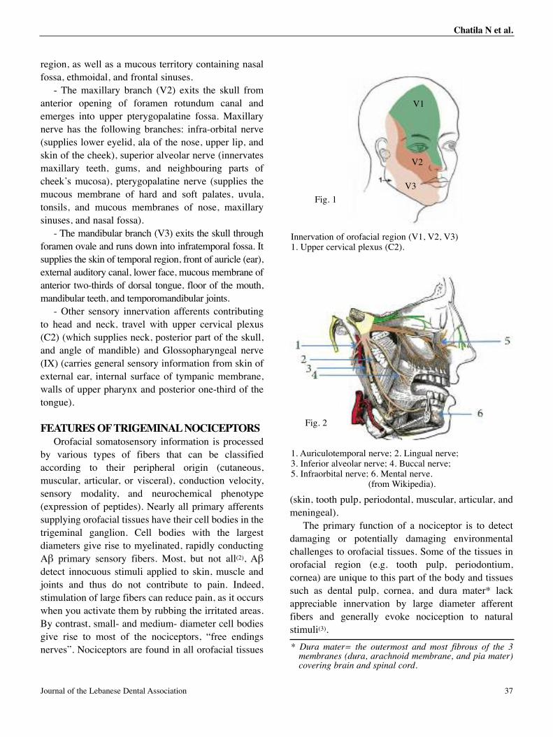
region, as well as a mucous territory containing nasalfossa, ethmoidal, and frontal sinuses.
- The maxillary branch (V2) exits the skull fromanterior opening of foramen rotundum canal andemerges into upper pterygopalatine fossa. Maxillarynerve has the following branches: infra-orbital nerve(supplies lower eyelid, ala of the nose, upper lip, andskin of the cheek), superior alveolar nerve (innervatesmaxillary teeth, gums, and neighbouring parts ofcheek’s mucosa), pterygopalatine nerve (supplies themucous membrane of hard and soft palates, uvula,tonsils, and mucous membranes of nose, maxillarysinuses, and nasal fossa).
- The mandibular branch (V3) exits the skull throughforamen ovale and runs down into infratemporal fossa. Itsupplies the skin of temporal region, front of auricle (ear),external auditory canal, lower face, mucous membrane ofanterior two-thirds of dorsal tongue, floor of the mouth,mandibular teeth, and temporomandibular joints.
- Other sensory innervation afferents contributingto head and neck, travel with upper cervical plexus(C2) (which supplies neck, posterior part of the skull,and angle of mandible) and Glossopharyngeal nerve(IX) (carries general sensory information from skin ofexternal ear, internal surface of tympanic membrane,walls of upper pharynx and posterior one-third of thetongue).
FEATURES OF TRIGEMINAL NOCICEPTORSOrofacial somatosensory information is processed
by various types of fibers that can be classifiedaccording to their peripheral origin (cutaneous,muscular, articular, or visceral), conduction velocity,sensory modality, and neurochemical phenotype(expression of peptides). Nearly all primary afferentssupplying orofacial tissues have their cell bodies in thetrigeminal ganglion. Cell bodies with the largestdiameters give rise to myelinated, rapidly conductingAβ primary sensory fibers. Most, but not all(2), Aβdetect innocuous stimuli applied to skin, muscle andjoints and thus do not contribute to pain. Indeed,stimulation of large fibers can reduce pain, as it occurswhen you activate them by rubbing the irritated areas.By contrast, small- and medium- diameter cell bodiesgive rise to most of the nociceptors, “free endingsnerves”. Nociceptors are found in all orofacial tissues
(skin, tooth pulp, periodontal, muscular, articular, andmeningeal).
The primary function of a nociceptor is to detectdamaging or potentially damaging environmentalchallenges to orofacial tissues. Some of the tissues inorofacial region (e.g. tooth pulp, periodontium,cornea) are unique to this part of the body and tissuessuch as dental pulp, cornea, and dura mater* lackappreciable innervation by large diameter afferentfibers and generally evoke nociception to naturalstimuli(3).
Innervation of orofacial region (V1, V2, V3)1. Upper cervical plexus (C2).
V1
V2
V3
1. Auriculotemporal nerve; 2. Lingual nerve; 3. Inferior alveolar nerve; 4. Buccal nerve; 5. Infraorbital nerve; 6. Mental nerve.
(from Wikipedia).
Fig. 2
Fig. 1
* Dura mater= the outermost and most fibrous of the 3membranes (dura, arachnoid membrane, and pia mater)covering brain and spinal cord.
37Journal of the Lebanese Dental Association
Chatila N et al.
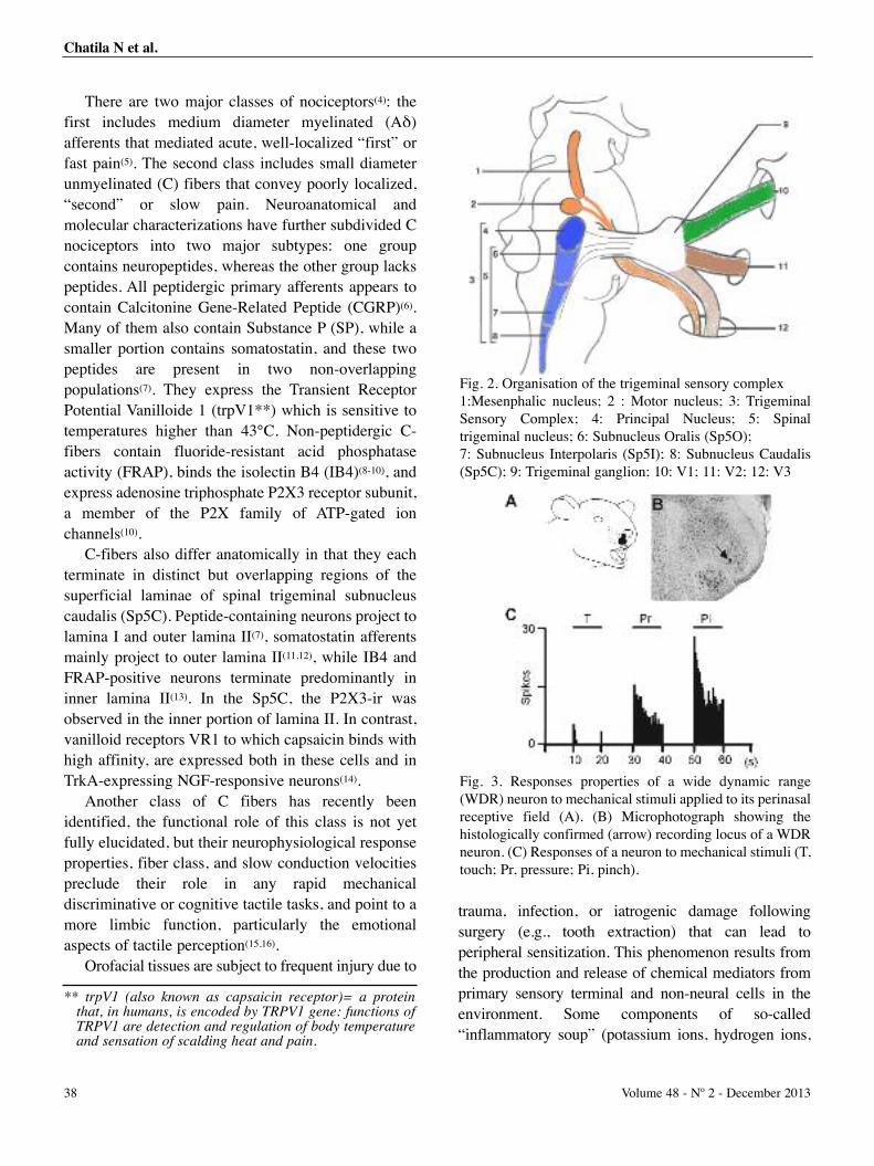
There are two major classes of nociceptors(4): thefirst includes medium diameter myelinated (Aδ)afferents that mediated acute, well-localized “first” orfast pain(5). The second class includes small diameterunmyelinated (C) fibers that convey poorly localized,“second” or slow pain. Neuroanatomical andmolecular characterizations have further subdivided Cnociceptors into two major subtypes: one groupcontains neuropeptides, whereas the other group lackspeptides. All peptidergic primary afferents appears tocontain Calcitonine Gene-Related Peptide (CGRP)(6).Many of them also contain Substance P (SP), while asmaller portion contains somatostatin, and these twopeptides are present in two non-overlappingpopulations(7). They express the Transient ReceptorPotential Vanilloide 1 (trpV1**) which is sensitive totemperatures higher than 43°C. Non-peptidergic C-fibers contain fluoride-resistant acid phosphataseactivity (FRAP), binds the isolectin B4 (IB4)(8-10), andexpress adenosine triphosphate P2X3 receptor subunit,a member of the P2X family of ATP-gated ionchannels(10).
C-fibers also differ anatomically in that they eachterminate in distinct but overlapping regions of thesuperficial laminae of spinal trigeminal subnucleuscaudalis (Sp5C). Peptide-containing neurons project tolamina I and outer lamina II(7), somatostatin afferentsmainly project to outer lamina II(11,12), while IB4 andFRAP-positive neurons terminate predominantly ininner lamina II(13). In the Sp5C, the P2X3-ir wasobserved in the inner portion of lamina II. In contrast,vanilloid receptors VR1 to which capsaicin binds withhigh affinity, are expressed both in these cells and inTrkA-expressing NGF-responsive neurons(14).
Another class of C fibers has recently beenidentified, the functional role of this class is not yetfully elucidated, but their neurophysiological responseproperties, fiber class, and slow conduction velocitiespreclude their role in any rapid mechanicaldiscriminative or cognitive tactile tasks, and point to amore limbic function, particularly the emotionalaspects of tactile perception(15,16).
Orofacial tissues are subject to frequent injury due to
trauma, infection, or iatrogenic damage followingsurgery (e.g., tooth extraction) that can lead toperipheral sensitization. This phenomenon results fromthe production and release of chemical mediators fromprimary sensory terminal and non-neural cells in theenvironment. Some components of so-called“inflammatory soup” (potassium ions, hydrogen ions,
Fig. 2. Organisation of the trigeminal sensory complex1:Mesenphalic nucleus; 2 : Motor nucleus; 3: TrigeminalSensory Complex; 4: Principal Nucleus; 5: Spinaltrigeminal nucleus; 6: Subnucleus Oralis (Sp5O); 7: Subnucleus Interpolaris (Sp5I); 8: Subnucleus Caudalis(Sp5C); 9: Trigeminal ganglion; 10: V1; 11: V2; 12: V3
Fig. 3. Responses properties of a wide dynamic range(WDR) neuron to mechanical stimuli applied to its perinasalreceptive field (A). (B) Microphotograph showing thehistologically confirmed (arrow) recording locus of a WDRneuron. (C) Responses of a neuron to mechanical stimuli (T,touch; Pr, pressure; Pi, pinch).
** trpV1 (also known as capsaicin receptor)= a proteinthat, in humans, is encoded by TRPV1 gene: functions ofTRPV1 are detection and regulation of body temperatureand sensation of scalding heat and pain.
38 Volume 48 - Nº 2 - December 2013
Chatila N et al.

ATP, histamine) can alter neuronal excitability directlyby interacting with ion channels on nociceptor surfacewhereas others (for example, bradykinin and NGF) bindto metabotropic receptors and mediate their effectsthrough second-messenger signaling cascades(17).Activation of nociceptors not only transmits afferentmessages to trigeminal sensory complex, but alsoinitiates the process of neurogenic inflammation. This isan efferent function of the nociceptor whereby releaseof neurotransmitters (notably substance P and CGRP)from peripheral terminal induces vasodilation andplasma extravasation, as well as activation of many non-neuronal cells, including mast cells and neutrophils.Moreover, electrical search stimuli have lead to thediscovery of very high threshold or insensitivecutaneous nociceptors that were primarily activated byinflammation processes(18). Many of these mechano-insensitive units respond to chemical stimulation andoften become responsive to mechanical and thermalstimulation, when sensitized. However, similar silentnociceptors are not yet described in the orofacialcutaneous tissues.
The ion channel TRPV1 is considered to be a molecularintegrator of different noxious stimuli such as heat, acids,and different endogenous pro-inflammatory substances. Itwas originally identified on the basis of its response tocapsaicin, the powerful irritant vanilloid found in hotpepper. Studies also reported that inflammation promotesTRPV1 activity by increasing TRPV1 expression, releaseof endovanilloids, and decrease of pH and by enhancedtemperature. Inhibition of TRPV1 was thereforehypothesized to reduce nociceptive activity, especially ininflammatory conditions(19). Recently, the effects of aTRPV1 antagonist (AZD1386) were investigated in aclinical model of acute inflammatory pain (the third molarextraction model). Administration of AZD1386 resulted ina rapid analgesia with a relatively short duration ofeffect(19).
Local anesthetics are commonly used to abolishtransmission of nociceptive information to centralnervous system (CNS) by blocking voltage-gatedsodium channels and thereby prevent the generation andpropagation of action potentials. However, theadministration of local anesthetics also producesnumbness from block of low-threshold pressure andtouch receptors, paralysis from block of motor axons,
and block of autonomic fibers. Selective block of painsignals might be possible either by targeting certainsodium channels that are present only in pain-sensingneurons(21) or by delivering sodium channel blockersselectively to nociceptive neurons. One strategy is todeliver a permanently charged sodium channel blockersuch as QX-314 (N-ethyl-lidocaine) by entering throughlarge-pore ion channels selectively expressed innociceptive neurons(22). The transient receptor potentialvanilloid 1 (TRPV1) channel is a primary nociceptivetransducer in nociceptive neurons, activated by noxiousheat (>43°C), capsaicin, protons andendocannabinoids(23). Chung and co-workers(24)
demonstrated that pore of TRPV1 channels, whenopened by the TRPV1 channel agonist capsaicin, mightbe large enough to deliver QX-314 or other largemolecule selectively into nociceptive neurons. Binshtokand associates(22) demonstrated that QX-314, a chargedmembrane-impermeant lidocaine derivative, can betargeted selectively into nociceptors when co-administrated with capsaicin, resulting in selectiveblocking of sodium channels and inhibition ofexcitability of nociceptors.
Fig. 4. Wind-up phenomenon. Consecutive single sweeprecordings showing the evolution (from bottom to top) ofthe responses of a wide dynamic range (WDR) neuronfollowing repetitive supramaximal percutaneous electricalstimulation applied to its receptive field.
39Journal of the Lebanese Dental Association
Chatila N et al.
Special features of trigeminal nerve and theirclinical significance
The peripheral trigeminal nervous system has somecharacteristics which could explain certain particularitiesof orofacial pain conditions: 1) a resistance in thedevelopment ectopic activity after nerve injury(26) and 2)a hypersensibility to estrogens(27).
Complex regional pain syndrome (CRPS)* is aneuropathic pain condition with sympatheticinvolvement, often resulting from trauma or bone fractureto distal limbs(28). The incidence of CRPS-like symptomsfollowing trauma to craniofacial tissues is relatively lowcompared to injury of other tissues(29). The exact reasonfor this observation is not certain. Spinal nerve injuryinduces sprouting of noradrenergic fibers into the dorsalroot ganglia(30), whereas comparable injury to trigeminalnerve produces no such sprouting into the trigeminalganglion(31,32). Furthermore, cervical sympathectomydoes not alter the development of ectopic discharge orincreased mechanical sensitivity after inferior alveolarnerve transection(33).
In injured segmental nerves, many primary afferentneurons spontaneously generate impulses and develophypersensitivity to a variety of physical, chemical andmetabolic stimuli. The resulting ectopic dischargecontributes to chronic paraesthesias and pain that oftenaccompany nerve injury in humans(34). However, whilemost amputees report chronic stump and/or phantompain(35), phantom and chronic neuropathic pains areextremely rare after tooth extractions and root canalextirpations, even when multiple teeth are involved(36,37).Using the teased fiber recording method, Tal and Devor(34)
have compared pathophysiological properties of afferentaxons injured in infraorbital nerve (ION) v/s sciatic nervein rats. Both myelinated and unmyelinated axons ending inION neuromas produced much less ongoing dischargethan those ending in sciatic nerve neuromas. Similarly,mechanosensitivity and acute injury discharge in IONneuromas were minimal. These differences may be relatedto the different spectrum of neuropathic symptomatologyassociated with trigeminal nerve injury.
Finally, trigeminal primary afferents seem particularlysensitive to estrogens. Estrogen can influence nociceptivesensory processing by regulating transcription of a varietyof proteins, and neurotransmitter receptors(38). Prevalenceof most chronic craniofacial pain conditions is higher inwomen than men(39). This is especially obvious for TMDs,migraine headache(39), and burning mouth syndrome(40).
Diogenes and associates(41) have demonstrated thatexpression of several genes in rat female trigeminalsensory neurons was regulated by estradiol. Prolactine(PRL) gene was found to be most upregulated (>40fold) in TG neurons of OVX rats treated with estradiolreplacement. Subsequent experiments showed thefollowing: (i) PRL is present in sensory neurons in areleasable pool; (ii) and PRL-induced sensitization ofcapsaicin-evoked responses in vitro and in vivo isunder control by estradiol; and (ii) PRL is able toincrease the phosphorylation levels of TRPV1 in TGneurons. These findings propose that PRL couldcontribute to the development of certain pain disordersmodulated by estrogen.
FEATURES OF TRIGEMINALNOCICEPTIVE NEURONS
Somatosensory inputs from orofacial area areconveyed by the trigeminal nerve to the first centralrelay located within the trigeminal brainstem sensorynuclear complex (Fig.2). This complex can besubdivided into principal sensory and spinal trigeminalnuclei. The latter consists of three regions, which, fromrostral to caudal, are the subnuclei oralis (Sp5O),interpolaris (Sp5I), and caudalis (Sp5C). The Sp5C,the largest subdivision of the spinal trigeminalnucleus, consists of an elongated laminated portionthat merges without clear boundaries with the cervicaldorsal horn, while the rostral Sp5C is displacedmedially by the caudal tip of Sp5I to form a distinctivetransition region. The ventral Sp5I/Sp5C transitionregion consists of rostral Sp5C with its fragmentedlaminar appearance, interstitial islands of neuronsembedded in the trigeminal spinal tract, and a ventralcrescent-shaped region of caudal Sp5I(42). Thedorsomedial aspect of the Sp5I/Sp5C transition regionis an important integrative region for peri-oral andintra-oral sensory input.
Sp5C is generally considered as the main relay of
** CRPS= 2 types recognized: Type 1 (corresponds to RSD:reflex sympathetic dystrophy) formerly termed Sudeck’satrophy - develops in distal extremities and causes poorlycontrollable pain, sensorimotor disorders, and serioustrophic alterations. Type 2 (formerly called causalgia)refers to cases where a definable nerve lesion is present.
40 Volume 48 - Nº 2 - December 2013
Chatila N et al.
orofacial nociceptive information to brain’s higherlevels. However, several observations point to theinvolvement of the rostral spinal trigeminal nucleus innociceptive mechanisms. For instance, a tractotomyperformed just above the level of the Sp5C in humansdoes not produce complete orofacial and dentalanalgesia whereas more rostral lesions can interferewith intraoral pain(43,44). Likewise, in cats, rats, andmonkeys, a tractotomy does not completely abolishnociceptive behavioral, reflex responses, or corticalactivity evoked by noxious orofacial stimulation(44-47).Moreover, neurons in both Sp5O and Sp5I respond tonoxious orofacial stimuli. Thus, spinal trigeminalsubnuclei, as a whole, are involved in the processing oforofacial nociceptive information. As there are few dataconcerning properties of Sp5I neurons, we will focusour discussion on the characteristics of Sp5O and Sp5Cneurons. However, it is possible that Sp5I shares severalfunctional properties with Sp5O and Sp5C.
Classification and distribution of trigeminalnociceptive neurons (Fig. 3)
Two main types of nociceptive neurons have beenidentified in the spinal trigeminal nucleus: the firsttype, termed nociceptive-specific units (NS), respondonly to noxious stimuli and are driven only fromsmall-diameter slow-conducting (Aδ and C)nociceptive afferents. The second type, termed widedynamic range (WDR) neurons (also labeledconvergent or multireceptive neurons), respond totactile as well as noxious stimuli and are driven byboth large-diameter Aβ afferents and small-diameterafferents (Aδ and C-fibers).
Electrophysiological studies(48) have documentedthat nociceptive neurons (WDR and NS) are located inthe three subdivisions of the spinal trigeminal nucleus.Nociceptive neurons did not appear to have anyparticular spatial grouping within the Sp5O and theSp5I but were intermingled with non-nociceptiveneurons(49-53). In contrast, Sp5C nociceptive neurons areconcentrated in the superficial (I/II) and deep (V/VI)laminae. Although there is no strict segregation of thesetwo populations, it has been shown in several speciesthat NS neurons are found mainly in the superficialdorsal horn (laminae I-II) whereas WDR neurons arelocated mainly in deep laminae (V-VI)(54-58).
Somatotopic organization and receptive fields oftrigeminal nociceptive neurons
A somatotopic organization could be observed in thespinal trigeminal nucleus. The dorsal part of eachsubnucleus contains neurons with a receptive field in theorofacial region supplied by mandibular branch oftrigeminal nerve. The ventral part contains neurons withan ophthalmic receptive field, and the area between thedorsal and ventral parts represents the maxillary region.Oral structures are usually represented medially(54,59-61).However, in the caudal Sp5C, the topographic pattern ofrepresentation of the face and mouth shifts, with theintraoral and perioral regions represented in the rostral partof the subnucleus and more lateral regions of the facemore caudally(62). Intraoral and perioral regions arerepresented in all the subdivisions of spinal trigeminalnucleus.
Spinal trigeminal subnuclei receive cutaneous,mucosal and/or tooth pulp afferent inputs. Moreover, theSp5C receives other types of peripheral inputs includingcornea, TMJ, jaw muscles, cerebrovasculature or dura(63).In addition, Sp5C can also be excited by other cranialnerve afferents (hypoglossal nerve or vagus) and by uppercervical nerve afferents(54,64). In contrast to the widespreadconvergence from cutaneous and non-cutaneous inputs toSp5C nociceptive neurons, Sp5O neurons respond chieflyto orofacial cutaneous/mucosa and/or tooth pulp stimuli.The narrow convergence of afferent inputs to the Sp5Osuggests that this subnucleus plays a relevant role in thespatial detection of pain from intraoral and perioralsuperficial tissues. In contrast, the larger convergence ofafferent inputs to the Sp5C suggests that it is involved inthe spread and referral of pain frequently observed inseveral orofacial pain conditions such as TMJ disorders,toothaches, and headaches(63).
PROPERTIES OF TRIGEMINALNOCICEPTIVE NEURONSResponses to electrical stimulation
The response of Sp5O and Sp5C WDR neurons toelectrical stimulation indicates that the whole spectrumof primary afferents can activate these neurons, rangingfrom large myelinated to unmyelinated fibers, while thetrigeminal NS neurons are activated by Aδ and C-fibers(54,60,65). The mean latencies of Aδ-fiber-evokedresponses in Sp5O are generally shorter than those
41Journal of the Lebanese Dental Association
Chatila N et al.
reported in the Sp5C. For instance, the mean latency totooth pulp stimulation in cat’s Sp5O is significantlyshorter than that for Sp5C neurons(66-68). These findingssuggest that Sp5O nociceptive neurons are involved inencoding the onset of nociceptive stimuli and that theiractivity allow to discriminate noxious from non-noxious stimuli. These phenomenon concern onlynociceptive responses elicited by stimulation of Aδ-fibers supplying intraoral or perioral tissues. However,mean latencies of C-fiber-evoked responses in Sp5Oare generally longer than those reported in Sp5C(69),suggesting that Sp5O WDR neurons receive C-fiberinputs indirectly, via Sp5C(70).
The Wind-up phenomenon (Fig.4), originallydescribed in dorsal horn, consists in a progressiveincrease in C-fiber-evoked responses followingrepeated percutaneous electrical stimulation(71,72). Thiswas also observed for the long latency responses ofSp5O(50) and Sp5C(55,73) neurons. Wind-up occurs inthe majority of WDR neurons located in the Sp5O anddeeper laminae of the Sp5C, while NS neurons locatedin the superficial laminae of the Sp5C frequentlyexhibit no or little windup(73,74). Recently, Coste andassociates(75) stated that wind-up enhances theencoding of short-duration orofacial nociceptivestimuli intensity and allows detection of subthresholdnociceptive input. As a consequence, wind-up endowstrigeminal WDR neurons with a greater ability toassess the intensity of nociceptive information. Wind-up thus may play a physiological role in trigeminalsensory processing, and enhanced wind-up mayunderlie the pathophysiology of several commonchronic headache and facial pain conditions.
Responses to chemical stimulation (Fig. 3)A substantial proportion of WDR and NS neurons in
Sp5C respond to algesic chemical stimuli delivered toafferents supplying oral mucosa(76-78). Most responsiveunits are located in superficial layers of dorsomedialSp5C although a few lie also in the deeper laminae.Majority of Sp5C nociceptive neurons respond to avariety of irritant chemicals, some of which act atspecific molecular receptors (e.g., capsaicin, histamine,serotonin, and nicotine), whereas others havenonspecific or unknown effects on nociceptor terminalmembranes (mustard oil, piperine, NaCl, ethanol).
Responses to the application of chemicals are notmarkedly affected by prior application of anotherchemical, except for capsaicin, piperine, and mustardoil, which often desensitize the tongue(76). Responses toall chemicals increase in a dose related manner,suggesting that the trigeminal nociceptive neurons arecapable to encode the intensity of chemical stimulation.In contrast, successive responses to repeatedapplications decrease significantly for nicotine,serotonin, capsaicin, and piperine(76).
Properties of deep tissue nociceptive neurons One important feature of neurons in the spinal
trigeminal nucleus is that almost all the neuronsresponsive to noxious stimulation of the deep tissuesare located predominantly in the Sp5C. These neuronshave widespread convergence from facial skin, tooth,oral mucosa, meninges, TMJ, and cranial muscles.Interestingly, TMJ, jaw muscles, and meninges areamong orofacial tissues that have a specificrepresentation at two distinct rostro-caudal levels inSp5I/Sp5C and in Sp5C/C1(3).
A considerable proportion (~50 %) of Sp5Cneurons can be also activated by noxious mechanicalor chemical (e.g., hypertonic saline, bradykinin,glutamate) stimulation of TMJ or masticatory musclesafferents(52,79,80). In addition, TMJ units can encode theconcentration of pro-inflammatory chemicals(80).Interestingly, persistent activation of TMJ tissuesproduced more robust and longer-lastinghyperexcitability of Sp5C neurons as compared withpersistent cutaneous stimulation(81). This maycontribute to mechanism of persistent pain associatedwith orofacial deep tissues painful conditions.
Most of joint and muscle neurons have deep andcutaneous receptive fields(52,53). A very small numberhas exclusively deep nociceptive fields(80). It is alsonoteworthy that most of the neurons showing afferentconvergence tend to have a larger cutaneous receptivefield than those receiving only cutaneous afferentinputs(80). The convergence of deep as well ascutaneous inputs onto the same neuron is considered tobe an integral mechanism underlying the poorlocalization, spread and referral of pain(63). Deepneurons could also play an important role in providingthe necessary signals onto segmental circuits to
42 Volume 48 - Nº 2 - December 2013
Chatila N et al.
suppress unwanted reflexes and facilitate reflexes thatenhance motor performance. Indeed, it has been shownthat injection of hypertonic saline into muscleproduces a predictable motor response(82). Thisresponse, which is characterized by reduction ofactivity in agonist muscles and increased activity inantagonist muscles, acts to limit overall movementpatterns and "protect" the injured muscle(52). Thisphenomenon has been termed the pain adaptationresponse(83).
ASCENDING PATHWAYSAscending projection neurons in the trigeminal
brainstem nuclear complex convey noxiousinformation to various suprasegmental structures.Multiple ascending pathways originate fromtrigeminal subnuclei.
Superficial laminae neurons of the Sp5C project toseveral areas of the central nervous system, which areimportant for processing signals relevant forhomeostasis*. They project to the ventrolateralmedulla(84) and the nucleus of the solitary tract(85), tworegions involved in cardio-respiratory regulation. Themost dense projections from superficial laminae ariseto the lateral parabrachial nucleus, an area that alsoparticipates in autonomic reactions(86-89). Superficiallaminae neurons of Sp5C also project to lateral andventrolateral columns of periaqueductal gray(90,91), aregion that contains different groups of neurons whichwhen activated, elicit anti-nociceptive and well-defined cardio-vascular and defensive reactions suchas decreases in blood pressure, hyporeactiveimmobility, avoidance behavior, and vocalization aswell as general emotional state of fear and anxiety(92).Sp5C-periaqueductal gray pathway could participatein feedback mechanisms involved in autonomic,aversive and anti-nociceptive responses to strongnociceptive stimulation. On the other hand, Sp5Cestablishes connections with the hypothalamus(93,94).The participation of the trigeminohypothalamicpathway in autonomic, neuroendocrine, and emotionalaspects of pain has been suggested.
Ascending projections from the deep laminae ofSp5C are similar to those from Sp5O. Majortermination sites include superior colliculus(95-98), zonaincerta(99-101), brainstem reticular areas(84,85), andanterior pretectal nucleus(102). These regions arenotably involved in motor reactions. Thus, deeplaminae of Sp5C and Sp5O may participate thesomatomotor integration.
Interestingly, both anatomical(103,104) andelectrophysiological(63,105-108) studies havedemonstrated that both superficial and deep laminae ofSp5C and Sp5O project to various thalamic structures,including ventral posterior medial nucleus (VPM) andposterior part of ventral medial nucleus (VMpo).Although these thalamic areas receive trigeminalprojections directly or indirectly via brainstemreticular formation and mesencephalon and preciselyencode different intensities of noxious stimuli,recordings in anaesthetized and awake monkeys haverevealed important differences between these areas. Agreat number of neurons in parafascicular nuclei andVMpo are modality specific, showing eithernociceptive or thermal responses. The receptive fieldsof VMpo cells in monkeys are restricted, whereasthose from parafascicular cells are often very large.Both their borders and the magnitude of their evokedresponses change with the monkey's behavioralstate(109). These features may indicate thatparafascicular cells are better suited to behavioralreactions, thus strongly implicating these regions inthe affective-emotional aspects of pain. Thissuggestion is supported by their cortical connectivityand by functional imaging studies. VMpo cells projectto the mid/anterior insular cortex, an area that isactivated by both innocuous and noxious thermalstimuli in humans and which has been implicated inthe affective components of pain on the basis of itsprojections to various limbic structures such as theamygdala and perirhinal cortex. Parafascicular cellsproject to area 24 of the cingulate cortex, the activityof which appears to be more selectively modulated bynoxious stimuli. In fact, this is a functionallyheterogeneous area constituted by adjacent zones,which have been implicated in attentional, motor andautonomic reactions, which might allow it to elicitvarious behavioral reactions(110).
* Homeostasis is the ability to maintain a constant internalenvironment in response to environmental changes.Nervous and endocrine systems control body homeostasis(homeostasis processes include temperature control, pHbalance, water and electrolyte balance, blood pressure,and respiration).
43Journal of the Lebanese Dental Association
Chatila N et al.

By contrast, in VPM, majority of neurons areWDR, have receptive fields, which are not modifiedby behavioral state and are smaller than those of spinalor trigeminal projecting neurons(109). This suggests thatVPM may subserve spatial discrimination. Theseregions project to the primary somatosensory cortex,and functional imaging studies have shown thatnoxious and innocuous stimuli similarly activate thecontralateral primary somatosensory cortex, indicatinga co-existence of pain and tactile representation in thisarea. Furthermore, single-unit recordings from acaudal ventral region of the thalamus in humansshowed neurons that could be activated by noxiousstimuli, and stimulation of this region induced thermaland/or painful sensations(111).
In conclusion, taken together, these data showthat the fine encoding properties or the fact thatthalamic neurons receive direct spinal and/ortrigeminal inputs alone cannot account for thesensory-discriminative aspects of pain, sinceencoding is shared by all (thalamic and also lowerCNS) regions implicated in pain processing.Moreover, the VPM, which are the best candidatesfor discrimination, can discharge with higherinstantaneous frequency to innocuous than tonoxious stimulation in awake monkeys. Thus it islikely that modulatory mechanisms and/or theconcomitant activity of multiple neuronalpopulations could determine the final painperception.
MODULATION OF TRIGEMINALNOCICEPTION
Orofacial somatosensory inputs activate strongmodulatory mechanisms implicating second orderneurons, local interneurons, and descendingmodulatory pathways. These segmental andsuprasegmental controls modulate nociceptive signalsat different levels at the central trigeminal networks.
Afferent-induced modulation of trigeminalnociception
Several lines of evidence show that stimulation ofperipheral afferents can modulate both neuronal andreflex responses at both metameric (segmental) orextra-metameric (plurisegmental) levels.
Modulation elicited by stimulation of non-nociceptive afferents
Activation of large diameter, myelinated peripheralfibers is able to inhibit firing of trigeminal neuronsinduced by noxious stimulation(112-115). Such inhibitoryeffects are metameric in nature, but analogous effectscan also be elicited by dorsal columns stimulation(116). Itwas proposed that such stimulation triggers segmentalinhibitory mechanisms via antidromic activation oflarge diameter peripheral fibers. Segmental inhibitionhas been proposed to explain the effectiveness oftherapeutic procedures that employ weak intensity, highfrequency percutaneous electrical nerve stimulation(117-
119), vibratory stimulation(120) and also spinal cordstimulation(121). These experimental data and theirclinical counterpart arise directly from the predictions ofthe gate control theory(122) and underlie the importantinteractions between large and small diameter fibers atthe level of the substantia gelatinosa.
Modulation elicited by stimulation of nociceptiveafferents
Activity of trigeminal nociceptive neurons can alsobe strongly inhibited by activation of nociceptors. Sucheffects are not somatotopically organized since they areelicited by stimulation of any part of the body. Theyhave been termed diffuse noxious inhibitory controls(DNIC)(123). The principal feature of DNIC is that theyinhibit the activity of all WDR neurons. Indeed, WDRneurons, whether recorded in the Sp5O(65,124,125) or inthe Sp5C(57,126) are under the influence of DNIC.Similar inhibitions elicited by remote noxious stimuliwere reported on the digastric reflex evoked by toothpulp stimulation in cats(127,128) and rats(128,129). However,comparing the strength of DNIC at trigeminal andlumbar levels shows that the inhibitions are morepronounced for trigeminal WDR neurons.
DNIC are subserved by a loop with the afferent andefferent pathways running within the ventrolateralquadrant and the dorsolateral funiculus of the spinalcord, respectively(130). Nevertheless, that sectioning thespinal cord suppresses DNIC suggests that thesecontrols also involve supraspinal areas(130). Thus aregion in the caudal medulla, the subnucleus reticularisdorsalis, was shown to be critically involved inDNIC(131). More recently, we demonstrated that the
44 Volume 48 - Nº 2 - December 2013
Chatila N et al.
spinoparabrachial(124) and the hypothalamicdopaminergic descending pathways(125) contribute tothe ascending and descending part, respectively, of theloop subserving DNIC.
Human studies conducted during the last yearshave demonstrated that several types of noxiousconditioning stimuli including ischemia, thermal,electrical, and chemical, reduce the perception oforofacial pain(132-137). Painful heterotopic conditioningstimuli also depress jaw(119,138) and blink reflexes(139),with stronger effects being observed with more intenseconditioning stimuli. Based on these data, Cadden andNewton(140) suggested that alterations in these controlsmay explain why jaw inhibitory reflexes are oftenabsent in patients with disorders such ascraniomandibular dysfunction or tensionheadache(141,142). Indeed, suppression of an inhibitoryaction on jaw closing motoneurons could, byincreasing the use of these muscles, be a predisposingfactor for myogenic pain experienced by suchpatients(119).
Descending modulation of trigeminal nociceptionElectrical or chemical stimulation of various
structures such as the cortex, periaqueductalgray(53,143,144), parabrachial area(145,146), anteriorpretectal nucleus(145), nucleus raphemagnus(112,143,147,148) and locus coeruleus(149) inhibit theresponse of Sp5O and Sp5C neurons to tooth pulpstimulation. Furthermore, electrical stimulation ofparabrachial area and nucleus raphe magnus modulateresponses of Sp5C neurons to noxious stimulation ofthe skin, deep tissues and cornea(143,144). Neuronalactivity in Sp5C evoked by trigeminovascularnociceptive afferents is also modulated by activationor inhibition of periaqueductal gray(150,151). However,stimulation of periaqueductal gray, nucleus raphemagnus, or locus coeruleus do not affect the inputoriginating from cold nociceptors in orofacialregions(152-155). More recently, it has been shown thatelectrical stimulation of secondary somatosensorycortex significantly reduced number of neuronsexpressing c-fos in the superficial layers of the Sp5Cin response to formalin injected into the orofacialregions(156). The stimulation of these structures canalso suppress reflex and behavioral responses to
noxious orofacial stimuli in awake, decerebrate, oranesthetized animals(156-159).
On the basis of anatomical and electrophysiologicaldata, it is generally accepted that inhibitory effectsinduced by electrical stimulation of periaqueductal gray,parabrachial area, nucleus raphe magnus, and locuscoeruleus, result from a direct action on trigeminalneurons(90,102,160-163). Antinociceptive action ofperiaqueductal gray is also mediated through nucleusraphe magnus since direct periaqueductal gray-nucleusraphe magnus connections have been clearlydemonstrated by anatomical investigations. Some ofthese projections to the spinal trigeminal nucleus havebeen shown to contain serotonin, norepinephrine,enkephalin, and cholecystokinin(60,161,163).
CONCLUSIONOur understanding of orofacial pain mechanisms
has lately grown impressively. Trigeminal painsystem, like other sensory systems, employsspecialized peripheral receptors to interact with theenvironment, with the goal to integrate informationarising simultaneously from different sources. Suchinformation is conveyed to central structures usingneural codes that change with regard to previoussensory experience and the interaction with othersensory systems. Pain signaling systems share someorganizational principles, such as convergence anddivergence, topographic projections, multiplicity ofrepresentation areas, and hierarchical organization.
This review illustrates several anatomical andfunctional particularities of trigeminal pain signalingsystem:
• Peripheral polymodality: Nociceptive neuronsare polymodal since they are able to transduce avariety of strong stimuli such as heat, pressure, andchemicals into signals that are perceived as pain.Molecular biology studies have recently identifiedspecific receptors and ions channels that allow us toperceive physical stimuli such as heat (TRPV1receptor), cold (Transient receptor potential cationchannel subfamily M member 8 (TRPM8)), andmechanical stimuli. These molecules are frequentlyexpressed on the same nociceptor.
• Redundancy: One relevant feature of thetrigeminal system is the redundancy of representations
45Journal of the Lebanese Dental Association
Chatila N et al.
of orofacial structures within the different brainstemnuclei. For instance, perioral cutaneous, oral mucosaand tooth pulp afferents project to all trigeminalsubnuclei, while the cornea, TMJ and meninges arerepresented in two regions of the Sp5C, the Sp5I/Sp5Cand Sp5C/upper cervical cord transition regions.
The trigeminal system also displays a functionalredundancy since a single sensory modality may beprocessed within different subnuclei. This is clearlyillustrated by dual integration of trigeminal nociceptiveinputs, since in addition to Sp5C, Sp5O participates alsoin the processing of nociceptive information fromorofacial regions. The functional significance of thisredundancy remains to be established.
• Multiplicity of pain pathways: Anatomical,electrophysiological, and more recently, functionalimaging studies, have shown that pain involves anumber of structures in the brain. Such data reveal amultiplicity of ascending nociceptive pathways arisingfrom laminae I and V of Sp5C as well as Sp5O.Several lines of evidence underline importantdifferences in anatomical and functional organizationof ascending nociceptive pathways. Such differencessuggest different roles in the processing of nociceptiveinformation. However, contribution of each of them totrigeminal pain processing remains obscure.
• Sensory transformation: Noxious inputsgenerated in peripheral tissues are conveyed throughseveral relay structures before reaching cortical areas.These intermediate structures are under the influenceof descending modulation arising from brainstem andcerebral cortex. It is obvious that noxious inputsundergo transformations at all central relay levels:such transformations probably provide the relevantcodes necessary for eliciting different pain sensationsand reactions.
• Convergence: there is a widespreadconvergence of inputs to trigeminal, thalamic, andcortical nociceptive neurons. In addition, a largefraction of such units respond to application of avariety of non-noxious and noxious stimuli, and to avariety of peripheral tissues. Such convergence is acommon feature of many neurons in the CNS andsuggests that individual cells can serve differentfunctions at different times, depending on theirpatterns of activity.
REFERENCES(1) Light AR. Trigeminal primary afferent receptors. In: PL G,editor. The initial processing of pain and its descending control:Spinal and trigeminal systems Sydney; 1992. p. 50-74.
(2) Djouhri L, Bleazard L, Lawson SN. Association of somaticaction potential shape with sensory receptive properties in guinea-pig dorsal root ganglion neurones. Journal of Physiology 1998 Dec15;513 ( Pt 3):857-72.
(3) Bereiter DA, Bereiter DF. Morphine and NMDA receptor antagonismreduce c-fos expression in spinal trigeminal nucleus produced by acuteinjury to the TMJ region. Pain 2000 Mar;85(1-2):65-77.
(4) Meyer, R.A., Ringkamp, M., Campbell, J.N., and Raja,S.N.Peripheral mechanisms of cutaneous nociception. In:M.Koltzenburg SBMa, editor. Wall and Melzack's Textbook ofPain. Philadelphia: Elsevier; 2008. p. 3-34.
(5) Basbaum AI, Bautista DM, Scherrer G, Julius D. Cellular andmolecular mechanisms of pain. Cell 2009 Oct 16;139(2):267-84.
(6) Ju G, Hokfelt T, Brodin E, Fahrenkrug J, Fischer JA, Frey P, etal. Primary sensory neurons of the rat showing calcitonin gene-related peptide immunoreactivity and their relation to substance P-, somatostatin-, galanin-, vasoactive intestinal polypeptide- andcholecystokinin-immunoreactive ganglion cells. Cell and tissueresearch 1987 Feb;247(2):417-31.
(7) Hokfelt T, Ljungdahl A, Terenius L, Elde R, Nilsson G.Immunohistochemical analysis of peptide pathways possiblyrelated to pain and analgesia: enkephalin and substance P.Proceedings of the National Academy of Sciences of the UnitedStates of America 1977 Jul;74(7):3081-5.
(8) Ambalavanar R, Morris R. The distribution of binding byisolectin I-B4 from Griffonia simplicifolia in the trigeminalganglion and brainstem trigeminal nuclei in the rat. Neuroscience1992;47(2):421-9.
(9) Silverman JD, Kruger L. Selective neuronal glycoconjugateexpression in sensory and autonomic ganglia: relation of lectinreactivity to peptide and enzyme markers. Journal ofNeurocytology 1990 Oct;19(5):789-801.
(10) Vulchanova L, Riedl MS, Shuster SJ, Stone LS, HargreavesKM, Buell G, et al. P2X3 is expressed by DRG neurons thatterminate in inner lamina II. European Journal of Neuroscience1998 Nov;10(11):3470-8.
(11) Alvarez FJ, Priestley JV. Ultrastructure of somatostatin-immunoreactive nerve terminals in laminae I and II of the rattrigeminal subnucleus caudalis. Neuroscience 1990;38(2):359-71.
(12) Ositelu DO, Morris R, Vaillant C. Innervation of facial skinbut not masticatory muscles or the tongue by trigeminal primaryafferents containing somatostatin in the rat. Neuroscience Letters1987 Aug 5;78(3):271-6.
(13) Sugimoto T, Fujiyoshi Y, Xiao C, He YF, Ichikawa H. Centralprojection of calcitonin gene-related peptide (CGRP)- andsubstance P (SP)-immunoreactive trigeminal primary neurons inthe rat. Journal of Comparative Neurology 1997 Feb17;378(3):425-42.
46 Volume 48 - Nº 2 - December 2013
Chatila N et al.
(14) Tominaga M, Caterina MJ, Malmberg AB, Rosen TA, GilbertH, Skinner K, et al. The cloned capsaicin receptor integratesmultiple pain-producing stimuli. Neuron 1998 Sep;21(3):531-43.
(15) Essick GK, James A, McGlone FP. Psychophysicalassessment of the affective components of non-painful touch.Neuroreport 1999 Jul 13;10(10):2083-7.
(16) McGlone F, Vallbo AB, Olausson H, Loken L, Wessberg J.Discriminative touch and emotional touch. Canadian Journal ofExperimental Psychology 2007 Sep;61(3):173-83.
(17) Woolf CJ, Salter MW. Neuronal plasticity: increasing the gainin pain. Science 2000 Jun 9;288(5472):1765-9.
(18) Meyer RA, Davis KD, Cohen RH, Treede RD, Campbell JN.Mechanically insensitive afferents (MIAs) in cutaneous nerves ofmonkey. Brain Research 1991 Oct 11;561(2):252-61.
(19) Quiding H, Jonzon B, Svensson O, Webster L, Reimfelt A,Karin A, et al. TRPV1 antagonistic analgesic effect: a randomizedstudy of AZD1386 in pain after third molar extraction. Pain 2013June;154(6):808-12.
(20) Xia R, Samad TA, Btesh J, Jiang LH, Kays I, Stjernborg L, et al.TRPV1 signaling: mechanistic understanding and therapeutic potential.Current Topics in Medicinal Chemistry 2011;11(17):2180-91.
(21) Cummins TR, Sheets PL, Waxman SG. The roles of sodiumchannels in nociception: Implications for mechanisms of pain. Pain2007 Oct;131(3):243-57.
(22) Binshtok AM, Bean BP, Woolf CJ. Inhibition of nociceptorsby TRPV1-mediated entry of impermeant sodium channelblockers. Nature 2007 Oct 4;449(7162):607-10.
(23) Caterina MJ, Schumacher MA, Tominaga M, Rosen TA, LevineJD, Julius D. The capsaicin receptor: a heat-activated ion channel inthe pain pathway. Nature 1997 Oct 23;389(6653):816-24.
(24) Chung MK, Güler AD, Caterina MJ. TRPV1 shows dynamicionic selectivity during agonist stimulation. Nature Neuroscience2008 May;11(5):555-64.
(25) Kym PR, Kort ME, Hutchins CW. Analgesic potential ofTRPV1 antagonists. Biochemical Pharmacology 2009 Aug1;78(3):211-6.
(26) Fried K, Bongenhielm U, Boissonade FM, Robinson PP.Nerve injury-induced pain in the trigeminal system. Neuroscientist2001 Apr;7(2):155-165.
(27) Hoffmann KD, Matthews MA. Comparison of sympatheticneurons in orofacial and upper extremity nerves: implications forcausalgia. Journal Oral and Maxillofacial Surg. 1990Jul;48(7):720-6; discussion 727.
(28) Wasner G, Backonja MM, Baron R. Traumatic neuralgias:complex regional pain syndromes (reflex sympathetic dystrophy andcausalgia): clinical characteristics, pathophysiological mechanismsand therapy. Neurologic Clinics 1998 Nov;16(4):851-68.
(29) Matthews B. Autonomic mechanisms in oral sensations.Proceedings of the Finnish Dental Society SuomenHammaslaakariseuran toimituksia 1989;85(4-5):365-73.
(30) McLachlan EM, Janig W, Devor M, Michaelis M. Peripheralnerve injury triggers noradrenergic sprouting within dorsal rootganglia. Nature 1993 June 10;363(6429):543-6.
(31) Benoliel R, Eliav E, Tal M. No sympathetic nerve sprouting inrat trigeminal ganglion following painful and non-painfulinfraorbital nerve neuropathy. Neuroscience Letters 2001 Jan19;297(3):151-4.
(32) Bongenhielm U, Boissonade FM, Westermark A, RobinsonPP, Fried K. Sympathetic nerve sprouting fails to occur in thetrigeminal ganglion after peripheral nerve injury in the rat. Pain1999 Sep;82(3):283-8.
(33) Bongenhielm U, Yates JM, Fried K, Robinson PP.Sympathectomy does not affect the early ectopic discharge frommyelinated fibres in ferret inferior alveolar nerve neuromas.Neuroscience Letters 1998 Apr 3;245(2):89-92.
(34) Tal M, Devor M. Ectopic discharge in injured nerves:comparison of trigeminal and somatic afferents. Brain Research1992 May 1;579(1):148-51.
(35) Sherman RA, Sherman CJ, Parker L. Chronic phantom andstump pain among American veterans: results of a survey. Pain1984 Jan;18(1):83-95.
(36) Pollman L. Phantom tooth phenomenon:painless and painfulsensation. In: Siegfried J, Zimmermann M, editors. Phantom andStump Pain Berlin: Springer; 1981. p. 77-80.
(37) Reisner H. Phantom tooth. In: Siegfried J, Zimmermann M,editors. Phantom and Stump Pain Berlin: Springer; 1981. p. 81-83.
(38) Sohrabji F, Miranda RC, Toran-Allerand CD. Estrogendifferentially regulates estrogen and nerve growth factor receptormRNAs in adult sensory neurons. Journal of Neuroscience. 1994Feb;14(2):459-71.
(39) Shinal RM, Fillingim RB. Overview of orofacial pain:epidemiology and gender differences in orofacial pain. DentalClinics of North America 2007 Jan;51(1):1-18.
(40) Thoppay JR, De Rossi SS, Ciarrocca KN. Burning mouthsyndrome. Dental Clinics of North America 2013 Jul;57(3):497-512.
(41) Diogenes A, Patwardhan AM, Jeske NA, Ruparel NB, GoffinV, Akopian AN, et al. Prolactin modulates TRPV1 in female rattrigeminal sensory neurons. Journal of Neuroscience. 2006 Aug2;26(31):8126-36.
(42) Phelan KD, Falls WM. The interstitial system of the spinaltrigeminal tract in the rat: anatomical evidence for morphologicaland functional heterogeneity. Somatosensory & Motor Research1989;6(4):367-99.
(43) Graham SH, Sharp FR, Dillon W. Intraoral sensation inpatients with brainstem lesions: role of the rostral spinal trigeminalnuclei in pons. Neurology 1988 Oct;38(10):1529-33.
(44) Young RF. Effect of trigeminal tractotomy on dental sensationin humans. Journal of Neurosurgery 1982 Jun;56(6):812-8.
47Journal of the Lebanese Dental Association
Chatila N et al.
(45) Denny-Brown D, Yanagisawa N. The function of thedescending root of the fifth nerve. Brain 1973 Dec;96(4):783-814.
(46) Dallel R, Clavelou P, Woda A. Effects of tractotomy onnociceptive reactions induced by tooth pulp stimulation in the rat.Experimental Neurology 1989 Oct;106(1):78-84.
(47) Broton JG, Rosenfeld JP. Effects of trigeminal tractotomy onfacial thermal nociception in the rat. Brain Research 1985 Apr29;333(1):63-72.
(48) Sessle BJ. Acute and chronic craniofacial pain: brainstemmechanisms of nociceptive transmission and neuroplasticity, andtheir clinical correlates. Crit Rev Oral Biol Med 2000;11(1):57-91.
(49) Dallel R, Raboisson P, Woda A, Sessle BJ. Properties ofnociceptive and non-nociceptive neurons in trigeminal subnucleusoralis of the rat. Brain Research. 1990;521(1-2):95-106.
(50) Dallel R, Duale C, Luccarini P, Molat JL. Stimulus-function,wind-up and modulation by diffuse noxious inhibitory controls ofresponses of convergent neurons of the spinal trigeminal nucleusoralis. . Eur J Neurosci 1999;11(1):31-40.
(51) Jacquin MF, Rhoades RW. Cell structure and responseproperties in the trigeminal subnucleus oralis. Somatosensory &Motor Research 1990;7(3):265-88.
(52) Ro JY, Capra NF. Evidence for subnucleus interpolaris incraniofacial muscle pain mechanisms demonstrated byintramuscular injections with hypertonic saline. Brain Research1999 Sep 18;842(1):166-83.
(53) Hayashi H, Sumino R, Sessle BJ. Functional organization oftrigeminal subnucleus interpolaris: nociceptive and innocuousafferent inputs, projections to thalamus, cerebellum, and spinalcord, and descending modulation from periaqueductal gray.Journal of Neurophysiology 1984 May;51(5):890-905.
(54) Hu JW. Response properties of nociceptive and non-nociceptive neurons in the rat's trigeminal subnucleus caudalis(medullary dorsal horn) related to cutaneous and deep craniofacialafferent stimulation and modulation by diffuse noxious inhibitorycontrols. Pain 1990 Jun;41(3):331-45.
(55) Price DD, Dubner R, Hu JW. Trigeminothalamic neurons innucleus caudalis responsive to tactile, thermal, and nociceptivestimulation of monkey's face. Journal of Neurophysiology 1976Sep;39(5):936-53.
(56) Burstein R, Yamamura H, Malick A, Strassman AM. Chemicalstimulation of the intracranial dura induces enhanced responses tofacial stimulation in brain stem trigeminal neurons. Journal ofNeurophysiology 1998 Feb;79(2):964-82.
(57) Raboisson P, Dallel R, Clavelou P, Sessle BJ, Woda A. Effectsof subcutaneous formalin on the activity of trigeminal brain stemnociceptive neurones in the rat. Journal of Neurophysiology 1995Feb;73(2):496-505.
(58) Maixner W, Dubner R, R. KD,Jr, Bushnell MC, Oliveras JL.Responses of monkey medullary dorsal horn neurons during thedetection of noxious heat stimuli. Journal of Neurophysiology1989 Aug;62(2):437-49.
(59) Darian-Smith I. The trigeminal system. In: Iggo A, editor.Handbook of sensory physiology.: Springer Verlag.; 1973. p. 271-314.
(60) McHaffie JG, Larson MA, Stein BE. Response properties ofnociceptive and low-threshold neurons in rat trigeminal parscaudalis. Journal of Comparative Neurology 1994 Sep15;347(3):409-25.
(61) Price DD, McHaffie JG, Larson MA. Spatial summation of heat-induced pain: influence of stimulus area and spatial separation ofstimuli on perceived pain sensation intensity and unpleasantness.Journal of Neurophysiology 1989 Dec;62(6):1270-9.
(62) Strassman AM, Vos BP. Somatotopic and laminar organizationof fos-like immunoreactivity in the medullary and upper cervicaldorsal horn induced by noxious facial stimulation in the rat.Journal of Comparative Neurology 1993 May 22;331(4):495-516.
(63) Sessle BJ, Hu JW, Amano N, Zhong G. Convergence ofcutaneous, tooth pulp, visceral, neck and muscle afferents ontonociceptive and non-nociceptive neurons in trigeminal subnucleuscaudalis (medullary dorsal horn) and its implications for referredpain. Pain 1986 Nov;27(2):219-35.
(64) Qin C, Chandler MJ, Miller KE, Foreman RD. Responses andafferent pathways of superficial and deeper c(1)-c(2) spinal cells tointrapericardial algogenic chemicals in rats. Journal ofNeurophysiology 2001 Apr;85(4):1522-32.
(65) Dallel R, Duale C, Luccarini P, Molat JL. Stimulus-function,wind-up and modulation by diffuse noxious inhibitory controls ofresponses of convergent neurons of the spinal trigeminal nucleusoralis. European Journal of Neuroscience 1999 Jan;11(1):31-40.
(66) Huopaniemi T, Jyvasjarvi E, Carlson S, Lindroos F,Pertovaara A. Response characteristics of tooth pulp-drivenpostsynaptic neurons in the spinal trigeminal subnucleus oralis ofthe cat. Acta Physiologica Scandinavica 1992 Feb;144(2):177-83.
(67) Hu JW, Sessle BJ. Trigeminal nociceptive and non-nociceptive neurones: brain stem intranuclear projections andmodulation by orofacial, periqueductal gray and nucleus raphemagnus stimuli. Brain Research 1979 Jul 20;170(3):547-52.
(68) Dong WK, Chudler EH, Kawakami Y. Tooth pulp-evokedpotentials in the trigeminal brainstem nuclear complex. BrainResearch 1990 Oct 8;529(1-2):131-42.
(69) Villanueva L, Le Bars D. The encoding of thermal stimuliapplied to the tail of the rat by lowering the excitability oftrigeminal convergent neurones. Brain Research 1985 Mar25;330(2):245-51.
(70) Dallel R, Duale C, Molat JL. Morphine administered in thesubstantia gelatinosa of the spinal trigeminal nucleus caudalisinhibits nociceptive activities in the spinal trigeminal nucleusoralis. Journal of Neuroscience 1998 May 15;18(10):3529-36.
(71) Mendell LM. Physiological properties of unmyelinated fiberprojection to the spinal cord. Experimental Neurology 1966Nov;16(3):316-32.
(72) Herrero JF, Laird JM, López-Garcia JA. Wind-up of spinalcord neurones and pain sensation: much ado about something?Progress in Neurobiology 2000 Jun;61(2):169-203.
48 Volume 48 - Nº 2 - December 2013
Chatila N et al.
(73) Luccarini P, Sessle BJ, Woda A. Superficial and deepconvergent nociceptive neurons are differentially affected by N-methyl-D-aspartate applied on the brainstem surface of the ratmedullary dorsal horn. Neuroscience 2001;107(2):311-6.
(74) Schouenborg J, Sjolund BH. Activity evoked by A- and C-afferent fibers in rat dorsal horn neurons and its relation to a flexionreflex. Journal of Neurophysiology 1983 Nov;50(5):1108-21.
(75) Coste J, Voisin DL, Luccarini P, Dallel R. A role for wind-upin trigeminal sensory processing: intensity coding of nociceptivestimuli in the rat. Cephalalgia 2008 Jun;28(6):631-9.
(76) Carstens E, Kuenzler N, Handwerker HO. Activation ofneurons in rat trigeminal subnucleus caudalis by different irritantchemicals applied to oral or ocular mucosa. Journal ofNeurophysiology 1998 Aug;80(2):465-92.
(77) Dessirier JM, Simons CT, Sudo M, Sudo S, Carstens E.Sensitization, desensitization and stimulus-induced recovery oftrigeminal neuronal responses to oral capsaicin and nicotine.Journal of Neurophysiology 2000 Oct;84(4):1851-62.
(78) Simons CT, Dessirier JM, Carstens MI, O'Mahony M,Carstens E. Neurobiological and psychophysical mechanismsunderlying the oral sensation produced by carbonated water.Journal of Neuroscience 1999 Sep 15;19(18):8134-44.
(79) Amano N, Hu JW, Sessle BJ. Responses of neurons in felinetrigeminal subnucleus caudalis (medullary dorsal horn) tocutaneous, intraoral, and muscle afferent stimuli. Journal ofNeurophysiology 1986 Feb;55(2):227-43.
(80) Takeshita S, Hirata H, Bereiter DA. Intensity coding by TMJ-responsive neurons in superficial laminae of caudal medullarydorsal horn of the rat. Journal of Neurophysiology 2001Nov;86(5):2393-404.
(81) Iwata K, Tashiro A, Tsuboi Y, Imai T, Sumino R, Morimoto T,et al. Medullary dorsal horn neuronal activity in rats with persistenttemporomandibular joint and perioral inflammation. Journal ofNeurophysiology 1999 Sep;82(3):1244-53.
(82) Stohler CS. Craniofacial pain and motor function:pathogenesis, clinical correlates, and implications. CriticalReviews Oral Biology Medicine 1999;10(4):504-18.
(83) Lund JP, Donga R, Widmer CG, Stohler CS. The pain-adaptation model: a discussion of the relationship between chronicmusculoskeletal pain and motor activity. Canadian Journal ofPhysiology and Pharmacology 1991 May;69(5):683-94.
(84) Esser MJ, Pronych SP, Allen GV. Trigeminal-reticularconnections: possible pathways for nociception-inducedcardiovascular reflex responses in the rat. Journal of ComparativeNeurology 1998 Feb 22;391(4):526-44.
(85) Villanueva L, de Pommery J, Menetrey D, Le Bars D. Spinalafferent projections to subnucleus reticularis dorsalis in the rat.Neuroscience Letters 1991 Dec 16;134(1):98-102.
(86) Allen GV, Barbrick B, Esser MJ. Trigeminal-parabrachialconnections: possible pathway for nociception-induced cardiovascularreflex responses. Brain Research 1996 Apr 9;715(1-2):125-35.
(87) Feil K, Herbert H. Topographic organization of spinal andtrigeminal somatosensory pathways to the rat parabrachial andKolliker-Fuse nuclei. Journal of Comparative Neurology 1995 Mar20;353(4):506-28.
(88) Cechetto DF, Standaert DG, Saper CB. Spinal and trigeminaldorsal horn projections to the parabrachial nucleus in the rat.Journal of Comparative Neurology 1985 Oct 8;240(2):153-60.
(89) Slugg RM, Light AR. Spinal cord and trigeminal projectionsto the pontine parabrachial region in the rat as demonstrated withPhaseolus vulgaris leucoagglutinin. Journal of ComparativeNeurology 1994 Jan 1;339(1):49-61.
(90) Li YQ, Takada M, Shinonaga Y, Mizuno N. Direct projectionsfrom the midbrain periaqueductal gray and the dorsal raphenucleus to the trigeminal sensory complex in the rat. Neuroscience1993 May;54(2):431-43.
(91) Clement CI, Keay KA, Podzebenko K, Gordon BD, BandlerR. Spinal sources of noxious visceral and noxious deep somaticafferent drive onto the ventrolateral periaqueductal gray of the rat.Journal of Comparative Neurology 2000 Sep 25;425(3):323-44.
(92) Keay KA, Bandler R. Parallel circuits mediating distinctemotional coping reactions to different types of stress.Neuroscience Biobehavioral Reviews 2001 Dec;25(7-8):669-78.
(93) Li JL, Kaneko T, Shigemoto R, Mizuno N. Distribution oftrigeminohypothalamic and spinohypothalamic tract neuronsdisplaying substance P receptor-like immunoreactivity in the rat.Journal of Comparative Neurology 1997 Feb 24;378(4):508-21.
(94) Malick A, Burstein R. Cells of origin of thetrigeminohypothalamic tract in the rat. Journal of ComparativeNeurology 1998 Oct 12;400(1):125-44.
(95) Huerta MF, Frankfurter A, Harting JK. Studies of the principalsensory and spinal trigeminal nuclei of the rat: projections to thesuperior colliculus, inferior olive, and cerebellum. Journal ofComparative Neurology 1983 Oct 20;220(2):147-67.
(96) Bruce LL, McHaffie JG, Stein BE. The organization oftrigeminotectal and trigeminothalamic neurons in rodents: adouble-labeling study with fluorescent dyes. Journal ofComparative Neurology 1987 Aug 15;262(3):315-30.
(97) Rhoades RW, Fish SE, Chiaia NL, Bennett-Clarke C, MooneyRD. Organization of the projections from the trigeminal brainstemcomplex to the superior colliculus in the rat and hamster:anterograde tracing with Phaseolus vulgaris leucoagglutinin andintra-axonal injection. Journal of Comparative Neurology 1989Nov 22;289(4):641-56.
(98) Ndiaye A, Pinganaud G, Buisseret-Delmas C, Buisseret P,Vanderwerf F. Organization of trigeminocollicular connections andtheir relations to the sensory innervation of the eyelids in the rat.Journal of Comparative Neurology 2002 Jul 8;448(4):373-87.
(99) Peschanski M. Trigeminal afferents to the diencephalon in therat. Neuroscience 1984 Jun;12(2):465-87.
49Journal of the Lebanese Dental Association
Chatila N et al.
(100) Roger M, Cadusseau J. Afferents to the zona incerta in therat: a combined retrograde and anterograde study. Journal ofComparative Neurology 1985 Nov 22;241(4):480-92.
(101) Iwata K, R. KD,Jr, Dubner R, Nahin RL. Diencephalicprojections from the superficial and deep laminae of the medullarydorsal horn in the rat. Journal of Comparative Neurology 1992 Jul15;321(3):404-20.
(102) Yoshida A, Chen K, Moritani M, Yabuta NH, Nagase Y,Takemura M, et al. Organization of the descending projectionsfrom the parabrachial nucleus to the trigeminal sensory nuclearcomplex and spinal dorsal horn in the rat. Journal of ComparativeNeurology 1997 Jun 23;383(1):94-111.
(103) Guy N, Chalus M, Dallel R, Voisin DL. Both oral and caudalparts of the spinal trigeminal nucleus project to the somatosensorythalamus in the rat. European Journal of Neuroscience 2005Feb;21(3):741-54.
(104) De Chazeron I, Raboisson P, Dallel R. Organization ofdiencephalic projections from the spinal trigeminal nucleus oralis:An anterograde tracing study in the rat. Neuroscience2004;127(4):921-928.
(105) Hutchison WD, Tsoukatos J, Dostrovsky JO. Quantitativeanalysis of orofacial thermoreceptive neurons in the superficialmedullary dorsal horn of the rat. Journal of Neurophysiology 1997Jun;77(6):3252-66.
(106) Azerad J, Woda A, Albe-Fessard D. Physiological Propertiesof neurons in different parts of the cat trigeminal sensory complex.Brain Research 1982 Aug 19;246(1):7-21.
(107) Cairns BE, McErlane SA, Fragoso MC, Jia WG, Soja PJ.Spontaneous discharge and peripherally evoked orofacialresponses of trigemino-thalamic tract neurons during wakefulnessand sleep. Journal of Neuroscience 1996 Dec 15;16(24):8149-59.
(108) Craig AD, Dostrovsky JO. Differential projections ofthermoreceptive and nociceptive lamina I trigeminothalamic andspinothalamic neurons in the cat. Journal of Neurophysiology 2001Aug;86(2):856-70.
(109) Buschnell M. Thamalic processing of sensory-discriminativeand affective-motivational dimensions of pain. In: Besson J-M,Guilbaud G, Ollat H, editors. Forebrain areas involved in painprocessing. Paris. 1995 (p. 63-77).
(110) Devinsky O, Morrell MJ, Vogt BA. Contributions of anteriorcingulate cortex to behaviour. Brain 1995 Feb;118 ( Pt 1):279-306.
(111) Lenz F, Dougherty P. Pain processing in the human thalamus.In: Steriade M, Jones E, editors. Elsevier Press; 1997. p. 617-651.
(112) Dickenson AH, Hellon RF, Woolf CJ. Tooth pulp input to thespinal trigeminal nucleus: a comparison of inhibitions followingsegmental and raphe magnus stimulation. Brain Research 1981 Jun9;214(1):73-87.
(113) Hu JW, Sessle BJ. Comparison of responses of cutaneousnociceptive and nonnociceptive brain stem neurons in trigeminalsubnucleus caudalis (medullary dorsal horn) and subnucleus oralisto natural and electrical stimulation of tooth pulp. Journal ofNeurophysiology 1984 Jul;52(1):39-53.
(114) Sessle BJ, Greenwood LF. Inputs to trigeminal brain stemneurones from facial, oral, tooth pulp and pharyngolaryngealtissues: I. Responses to innocuous and noxious stimuli. BrainResearch 1976 Nov 26;117(2):211-26.
(115) Young RF, Nord SG. Experimental modulation of medullarydental pulp units by mechanical stimulation of oro-facial fields.Experimental Neurology 1975 Dec;49(3):813-21.
(116) Atweh SF, Dajani BM, Saade NE, Jabbur SJ. Supraspinalinhibition of trigeminal input into subnucleus caudalis by dorsalcolumn stimulation. Brain Research 1985 Dec 2;348(2):401-4.
(117) Hansson P, Ekblom A. Afferent stimulation induced painrelief in acute oro-facial pain and its failure to induce sufficientpain reduction in dental and oral surgery. Pain 1984Nov;20(3):273-8.
(118) Maixner W, Fillingim R, Booker D, Sigurdsson A.Sensitivity of patients with painful temporomandibular disorders toexperimentally evoked pain. Pain 1995 Dec;63(3):341-51.
(119) Maillou P, Cadden SW. Effects of remote deep somaticnoxious stimuli on a jaw reflex in man. Archives of Oral Biology1997 Apr;42(4):323-7.
(120) Ottoson D, Ekblom A, Hansson P. Vibratory stimulation forthe relief of pain of dental origin. Pain 1981 Feb;10(1):37-45.
(121) Meyerson BA, Linderoth B. Mechanisms of spinal cordstimulation in neuropathic pain. Neurological Research 2000Apr;22(3):285-92.
(122) Melzack R, Wall PD. Pain mechanisms: a new theory.Science 1965 Nov 19;150(3699):971-9.
(123) Le Bars D, Dickenson A, Besson J-M. Diffuse noxiousinhibitory controls (DNIC). I.Effects on dorsal horn convergentneurones in the rat. Pain 1979;6:283-304.
(124) Lapirot O, Chebbi R, Monconduit L, Artola A, Dallel R,Luccarini P. NK1 receptor-expressing spinoparabrachial neuronstrigger diffuse noxious inhibitory controls through lateralparabrachial activation in the male rat. Pain 2009 Apr;142(3):245-54.
(125) Lapirot O, Melin C, Modolo A, Nicolas C, Messaoudi Y,Monconduit L, et al. Tonic and phasic descending dopaminergiccontrols of nociceptive transmission in the medullary dorsal horn.Pain 2011 Aug;152(8):1821-31.
(126) Dickenson AH, Brewer CM, Hayes NA. Effects of topicalbaclofen on C fibre-evoked neuronal activity in the rat dorsal horn.Neuroscience 1985 Feb;14(2):557-62.
(127) Cadden SW. The digastric reflex evoked by tooth-pulpstimulation in the cat and its modulation by stimuli applied to thelimbs. Brain Research 1985 Jun 10;336(1):33-43.
(128) Tal M, Sharav Y, Devor M. Modulation of the jaw-openingreflex by peripheral electrical stimulation. ExperimentalNeurology 1981 Dec;74(3):907-19.
(129) Kawakita K, Funakoshi M. Suppression of the jaw-openingreflex by conditioning a-delta fiber stimulation andelectroacupuncture in the rat. Experimental Neurology 1982Nov;78(2):461-5.
50 Volume 48 - Nº 2 - December 2013
Chatila N et al.
(130) Le Bars D. The whole body receptive field of dorsal hornmultireceptive neurones. Brain Res Rev 2002;40:29-44.
(131) Bouhassira D, Villanueva L, Bing Z, le Bars D. Involvementof the subnucleus reticularis dorsalis in diffuse noxious inhibitorycontrols in the rat. Brain Research 1992 Nov 13;595(2):353-7.
(132) Parsons CM, Goetzi FR. Effect of induce pain on painthreshold. Proceedings of the Society for Experimental Biologyand Medicine 1945 Dec;60:327-9.
(133) Talbot JD, Duncan GH, Bushnell MC, Boyer M. Diffusenoxious inhibitory controls (DNICs): psychophysical evidence inman for intersegmental suppression of noxious heat perception bycold pressor pain. Pain 1987 Aug;30(2):221-32.
(134) Talbot JD, Duncan GH, Bushnell MC. Effects of diffusenoxious inhibitory controls (DNICs) on the sensory-discriminativedimension of pain perception. Pain 1989 Feb;36(2):231-8.
(135) Pertovaara A, Kemppainen P, Johansson G, Karonen SL.Ischemic pain nonsegmentally produces a predominant reductionof pain and thermal sensitivity in man: a selective role forendogenous opioids. Brain Research 1982 Nov 11;251(1):83-92.
(136) Melzack R. Recent concepts of pain. Journal of Medicine1982;13(3):147-60.
(137) Sigurdsson A, Maixner W. Effects of experimental andclinical noxious counterirritants on pain perception. Pain 1994Jun;57(3):265-75.
(138) Cadden SW, van der Glas HW, Lobbezoo F, van der Bilt A.Effects of remote noxious stimulation on exteroceptive reflexes inhuman jaw-closing muscles. Brain Research 1996 Jul 8;726(1-2):189-97.
(139) Ellrich J, Treede RD. Characterization of blink reflexinterneurons by activation of diffuse noxious inhibitory controls inman. Brain Research 1998 Aug 24;803(1-2):161-8.
(140) Cadden SW, Newton JP. The effects of inhibitory controlstriggered by heterotopic noxious stimuli on a jaw reflex evoked byperioral stimuli in man. Archives of Oral Biology 1994June;39(6):473-80.
(141) De Laat A, van der Glas HW, Weytjens JL, van SteenbergheD. The masseteric post-stimulus electromyographic-complex inpeople with dysfunction of the mandibular joint. Archives of OralBiology 1985;30(2):177-180.
(142) Schoenen J, Jamart B, Gerard P, Lenarduzzi P, Delwaide PJ.Exteroceptive suppression of temporalis muscle activity in chronicheadache. Neurology 1987 Dec;37(12):1834-6.
(143) Sessle BJ, Hu JW, Dubner R, Lucier GE. Functionalproperties of neurons in cat trigeminal subnucleus caudalis(medullary dorsal horn). II. Modulation of responses to noxiousand nonnoxious stimuli by periaqueductal gray, nucleus raphemagnus, cerebral cortex, and afferent influences, and effect ofnaloxone. Journal of Neurophysiology 1981 Feb;45(2):193-207.
(144) Sessle BJ, Hu JW. Raphe-induced suppression of the jaw-opening reflex and single neurons in trigeminal subnucleus oralis,and influence of naloxone and subnucleus caudalis. Pain 1981Feb;10(1):19-36.
(145) Chiang CY, Chen IC, Dostrovsky JO, Sessle BJ. Anteriorpretectal nucleus-induced modulatory effects on trigeminalbrainstem somatosensory neurons. Neuroscience Letters 1992 Jan6;134(2):233-7.
(146) Meng ID, Hu JW, Bereiter DA. Parabrachial area andnucleus raphe magnus inhibition of corneal units in rostral andcaudal portions of trigeminal subnucleus caudalis in the rat. Pain2000 Sep;87(3):241-51.
(147) Chiang CY, Hu JW, Sessle BJ. Parabrachial area and nucleusraphe magnus-induced modulation of nociceptive andnonnociceptive trigeminal subnucleus caudalis neurons activatedby cutaneous or deep inputs. Journal of Neurophysiology 1994Jun;71(6):2430-45.
(148) Lovick TA, Wolstencroft JH. Inhibitory effects of nucleusraphe magnus on neuronal responses in the spinal trigeminalnucleus to nociceptive compared with non-nociceptive inputs. Pain1979 Oct;7(2):135-45.
(149) Matsutani K, Tsuruoka M, Shinya A, Furuya R, Kawawa T.Stimulation of the locus coeruleus suppresses trigeminalsensorimotor function in the rat. Brain Res Bull 2001;53(6):827-832.
(150) Knight YE, Goadsby PJ. The periaqueductal grey mattermodulates trigeminovascular input: a role in migraine?Neuroscience 2001;106(4):793-800.
(151) Knight YE, Bartsch T, Goadsby PJ. Trigeminalantinociception induced by bicuculline in the periaqueductal gray(PAG) is not affected by PAG P/Q-type calcium channel blockadein rat. Neuroscience Letters 2003 Jan 16;336(2):113-6.
(152) Dostrovsky JO. Raphe and periaqueductal gray inducedsuppression of non-nociceptive neuronal responses in the dorsalcolumn nuclei and trigeminal sub-nucleus caudalis. BrainResearch 1980 Oct 27;200(1):184-9.
(153) Dostrovsky JO, Hellon RF. The representation of facialtemperature in the caudal trigeminal nucleus of the cat. Journal ofPhysiology 1978 Apr;277:29-47.
(154) Davis KD, Dostrovsky JO. Responses of feline trigeminalspinal tract nucleus neurons to stimulation of the middle meningealartery and sagittal sinus. Journal of Neurophysiology 1988Feb;59(2):648-66.
(155) Dawson NJ, Dickenson AH, Hellon RF, Woolf CJ. Inhibitorycontrols on thermal neurones in the spinal trigeminal nucleus ofcats and rats. Brain Research 1981 Mar 30;209(2):440-5.
(156) Gojyo F, Sugiyo S, Kuroda R, Kawabata A, Varathan V,Shigenaga Y, et al. Effects of somatosensory cortical stimulationon expression of c-Fos in rat medullary dorsal horn in response toformalin-induced noxious stimulation. Journal of NeuroscienceResearch 2002 May 15;68(4):479-88.
(157) Mason P, Strassman A, Maciewicz R. Intracellular responsesof raphe magnus neurons during the jaw-opening reflex evoked bytooth pulp stimulation. Brain Research 1986 Aug 6;379(2):232-41.
(158) Oliveras JL, Redjemi F, Guilbaud G, Besson JM. Analgesiainduced by electrical stimulation of the inferior centralis nucleus ofthe raphe in the cat. Pain 1975 June;1(2):139-45.
51Journal of the Lebanese Dental Association
Chatila N et al.

(159) Chiang CY, Chen IC, Dostrovsky JO, Sessle BJ. Inhibitoryeffect of stimulation of the anterior pretectal nucleus on the jaw-opening reflex. Brain Research 1989 Sep 18;497(2):325-33.
(160) Dostrovsky JO, Sessle BJ, Hu JW. Presynaptic excitabilitychanges produced in brain stem endings of tooth pulp afferents byraphe and other central and peripheral influences. Brain Research1981 Aug 10;218(1-2):141-60.
(161) Beitz AJ, Clements JR, Ecklund LJ, Mullett MM. The nucleiof origin of brainstem enkephalin and cholecystokinin projectionsto the spinal trigeminal nucleus of the rat. Neuroscience 1987Feb;20(2):409-25.
(162) Simpson KL, Altman DW, Wang L, Kirifides ML, Lin RC,Waterhouse BD. Lateralization and functional organization of thelocus coeruleus projection to the trigeminal somatosensorypathway in rat. Journal of Comparative Neurology 1997 Aug18;385(1):135-47.
(163) Kirifides ML, Simpson KL, Lin RC, Waterhouse BD.Topographic organization and neurochemical identity of dorsalraphe neurons that project to the trigeminal somatosensorypathway in the rat. Journal of Comparative Neurology 2001 Jul2;435(3):325-40.
Corresponding author:Radhouane [email protected]@u-clermont1.fr Tel: 00 33 4 73 17 73 13Fax: 00 33 4 73 17 73 06www.u-clermont1.frwww.udapro.fr
Chatila N et al.
52 Journal of the Lebanese Dental AssociationVolume 48 - Nº 2 - December 2013
Clinical Report
Trigeminal neuralgia (TN), also known as “ticdouloureux” or “suicide disease”, is a neuropathicdisorder characterized by severe unilateral paroxysmalfacial pain originating from trigeminal nerve.
It has been described as among the most painfulconditions known to mankind and it is triggered bytactile irritation. Various drugs have been used to treatTN, among them anticonvulsants, centrally actingmuscle relaxants, antidepressants, local anestheticpatches, and opiates. However, none of these drugsare free of side-effects.
Other TN therapies include invasive surgery suchas microvascular decompression, percutaneousradiofrequency thermorhizotomy, and gamma kniferadiosurgery. However, effectiveness decreases withtime and the procedures are not free of complications.
Acupuncture is one of many therapeutic methodsthat are considered complementary to modernmedicine. It is successfully used in the treatment of awide range of neurological conditions such astrigeminal neuralgia, post-herpetic neuralgia,peripheral neuropathies, etc... This method oftreatment was practiced in China around 2000 BC butbecame an integral part of modern medicine onlylately. Acupuncture-induced analgesia is mediated byinhibition of pain transmission at spinal level andactivation of central pain modulating centers. Thisneural modulation of acupuncture is considered amode of peripheral sensory stimulation.1,2
In traditional Chinese medicine, acupuncture
needles are inserted in specific points at specificmeridians3 and stimulation of these needles - either byhand, electrical devices, or moxibustion - stimulatesupraspinal mechanisms (diffuse noxious inhibitorycontrol-DNIC), inhibiting neurons of dorsal horn ortrigeminal ganglion excited by pain.2 Release ofdynorphins, enkephalins, and other neuropeptides4
play a major role in pain control. Modernneuroimaging methods (functional MRI and SPECT*)showed an increase in cerebral blood flow and cerebraloxygen supply, also in activation of subcortical andcortical centers1 following acupuncture sessions.
In our clinic, we were successful in relieving atleast sixty percent of TN pains. Almost all thesepatients were refractory to conventional medicaltherapy and they were still on anticonvulsants with orwithout antidepressants (Table 1).
Acupuncture treatment sessions were implementedthree times per week (MWF), each session lastingtwenty minutes. The treatment was spread over aperiod of one month, followed by one or two sessionsper month depending on the case. In Table 1, local,distal, and ear acupuncture points were used.
Points stimulated were: GV 20, GB 14, ST 2,3,7,8,36, 44, LI 4, LIV 3, CV24, ear sympathetic, andshenmen.
Variations in choosing the points depended ontrigeminal branch involved.
Only one refractory nonresponsive case wasreferred to neurosurgery for invasive thermal ablationof the trigeminal branch nerve affected.
Over a period of fifteen years, eighty trigeminalneuralgia pain patients were referred to our clinic.
Acupuncture and Trigeminal Neuralgia pain.
Nadim M. Jalbout*, MBBCh, Diploma of Anesthesia, MIASP
Journal of the Lebanese Dental AssociationVolume 48 - Nº 2 - December 2013
* Associate Professor of Clinical Anesthesia, University ofBalamand, Faculty of Medicine, Beirut, Lebanon,
Consultant Anesthesiologist, Saint-George HospitalUniversity Medical Center - SGHUMC, Achrafieh, Beirut * SPECT= Single Photon Emission Computed Tomography
53
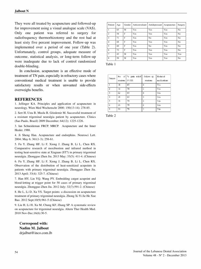
They were all treated by acupuncture and followed-upfor improvement using a visual analogue scale (VAS).Only one patient was referred to surgery forradiofrequency thermorhizotomy and the rest had atleast sixty five percent improvement. Follow-up wasimplemented over a period of one year (Table 2).Unfortunately, control groups, adequate measure ofoutcome, statistical analysis, or long-term follow-upwere inadequate due to lack of control randomizeddouble-blinding.
In conclusion, acupuncture is an effective mode oftreatment of TN pain, especially in refractory cases whereconventional medical treatment is unable to providesatisfactory results or when unwanted side-effectsoverweighs benefits.
REFERENCES1. Jellinger KA. Principles and application of acupuncture inneurology. Wien Med Wochenschr 2000; 150(13-14); 278-85.
2. Sert H, Usta B, Muslu B, Gözdemir M. Successful treatment ofa resistant trigeminal neuralgia patient by acupuncture. Clinics(Sao Paulo, Brazil) 2009 December; 64(12); 1225-1226.
3. Ian Schneideman FRCP, MRCP. Acupuncture and the InnerHealer, 1988.
4. Ji Sheng Han. Acupuncture and endorphins. Neurosci Lett.2004; May 6; 361(1-3); 258-61.
5. Fu Y, Zhang HF, Li F, Xiong J, Zhang B, Li L, Chen RX.Comparative research of moxibustion and infrared method intesting heat-sensitive state at Xiaguan (ST7) in primary trigeminalneuralgia. Zhongguo Zhen Jiu. 2013 May; 33(5): 411-4. (Chinese)
6. Fu Y, Zhang HF, Li F, Xiong J, Zhang B, Li L, Chen RX.Observation of the distribution of heat-sensitized acupoints inpatients with primary trigeminal neuralgia. Zhongguo Zhen Jiu.2013 April; 33(4): 325-7. (Chinese)
7. Han HY, Lin YQ, Wang PY. Embedding catgut acupoint andblood-letting at trigger point for 58 cases of primary trigeminalneuralgia. Zhongguo Zhen Jiu. 2012 July; 32(7):591-2. (Chinese)
8. He L, Li D, Xu YS. Target points: a discussion on acupuncturetreatment of primary trigeminal neuralgia. Zhong Xi Yi Jie He XueBao. 2012 Sept;10(9):961-5 (Chinese)
9. Liu H, Li H, Xu M, Chung KF, Zhang SP. A systematic reviewon acupuncture for trigeminal neuralgia. Altern Ther Health Med.2010 Nov-Dec;16(6):30-5.
Correspond with:Nadim M. Jalbout [email protected]
Jalbout N
Table 1
Table 2
Patient Age Gender Anticonvulsant Antidepressant Acupuncture Surgery
1 65 M Yes Yes Yes No
2 54 F Yes Yes Yes No
3 71 F Yes No Yes No
4 68 F Yes Yes Yes No
5 60 F Yes No Yes No
6 72 F Yes Yes Yes No
7 43 M Yes Yes Yes Yes
8 58 M Yes Yes Yes No
54 Journal of the Lebanese Dental AssociationVolume 48 - Nº 2 - December 2013
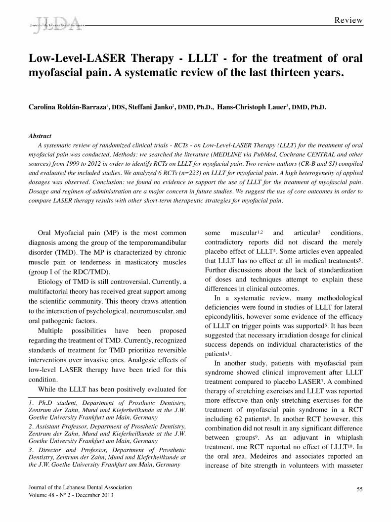
Review
Oral Myofacial pain (MP) is the most commondiagnosis among the group of the temporomandibulardisorder (TMD). The MP is characterized by chronicmuscle pain or tenderness in masticatory muscles(group I of the RDC/TMD).
Etiology of TMD is still controversial. Currently, amultifactorial theory has received great support amongthe scientific community. This theory draws attentionto the interaction of psychological, neuromuscular, andoral pathogenic factors.
Multiple possibilities have been proposedregarding the treatment of TMD. Currently, recognizedstandards of treatment for TMD prioritize reversibleinterventions over invasive ones. Analgesic effects oflow-level LASER therapy have been tried for thiscondition.
While the LLLT has been positively evaluated for
some muscular1,2 and articular3 conditions,contradictory reports did not discard the merelyplacebo effect of LLLT4. Some articles even appealedthat LLLT has no effect at all in medical treatments5.Further discussions about the lack of standardizationof doses and techniques attempt to explain thesedifferences in clinical outcomes.
In a systematic review, many methodologicaldeficiencies were found in studies of LLLT for lateralepicondylitis, however some evidence of the efficacyof LLLT on trigger points was supported6. It has beensuggested that necessary irradiation dosage for clinicalsuccess depends on individual characteristics of thepatients1.
In another study, patients with myofascial painsyndrome showed clinical improvement after LLLTtreatment compared to placebo LASER7. A combinedtherapy of stretching exercises and LLLT was reportedmore effective than only stretching exercises for thetreatment of myofascial pain syndrome in a RCTincluding 62 patients8. In another RCT however, thiscombination did not result in any significant differencebetween groups9. As an adjuvant in whiplashtreatment, one RCT reported no effect of LLLT10. Inthe oral area, Medeiros and associates reported anincrease of bite strength in volunteers with masseter
Low-Level-LASER Therapy - LLLT - for the treatment of oralmyofascial pain. A systematic review of the last thirteen years.
1. Ph.D student, Department of Prosthetic Dentistry,Zentrum der Zahn, Mund und Kieferheilkunde at the J.W.Goethe University Frankfurt am Main, Germany
2. Assistant Professor, Department of Prosthetic Dentistry,Zentrum der Zahn, Mund und Kieferheilkunde at the J.W.Goethe University Frankfurt am Main, Germany
3. Director and Professor, Department of ProstheticDentistry, Zentrum der Zahn, Mund und Kieferheilkunde atthe J.W. Goethe University Frankfurt am Main, Germany
Carolina Roldán-Barraza1, DDS, Steffani Janko2, DMD, Ph.D., Hans-Christoph Lauer3, DMD, Ph.D.
Abstract
A systematic review of randomized clinical trials - RCTs - on Low-Level-LASER Therapy (LLLT) for the treatment of oral
myofacial pain was conducted. Methods: we searched the literature (MEDLINE via PubMed, Cochrane CENTRAL and other
sources) from 1999 to 2012 in order to identify RCTs on LLLT for myofacial pain. Two review authors (CR-B and SJ) compiled
and evaluated the included studies. We analyzed 6 RCTs (n=223) on LLLT for myofacial pain. A high heterogeneity of applied
dosages was observed. Conclusion: we found no evidence to support the use of LLLT for the treatment of myofascial pain.
Dosage and regimen of administration are a major concern in future studies. We suggest the use of core outcomes in order to
compare LASER therapy results with other short-term therapeutic strategies for myofacial pain.
Journal of the Lebanese Dental AssociationVolume 48 - Nº 2 - December 2013
55
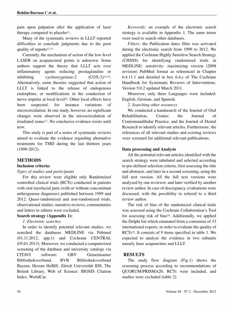
pain upon palpation after the application of lasertherapy compared to placebo11.
Many of the systematic reviews in LLLT reporteddifficulties to conclude judgments due to the poorquality of reports12,13
Currently, the mechanism of action of the low-levelLASER on acupunctural points is unknown. Someauthors support the theory that LLLT acts overinflammatory agents reducing prostaglandins orinhibiting cyclooxygenase-2 (COX-2)14,15.Alternatively, some theories suggested that action ofLLLT is linked to the release of endogenousendorphins, or modifications in the conduction ofnerve impulse at local level16. Other local effects havebeen suspected, for instance variations ofmicrocirculation. In one study, however, no significantchanges were observed in the microcirculation ofirradiated zones17. No conclusive evidence exists untilnow.
This study is part of a series of systematic reviewsaimed to evaluate the evidence regarding alternativetreatments for TMD during the last thirteen years(1999-2012).
METHODSInclusion criteria: Types of studies and participants
For this review were eligible only Randomizedcontrolled clinical trials (RCTs) conducted in patientswith oral myofacial pain (with or without concomitantarthrogenous diagnoses) published between 1999 and2012. Quasi-randomized and non-randomized trials,observational studies, narrative reviews, commentariesand letters to editors were excluded.Search strategy (Appendix 1):
1. Electronic searches
In order to identify potential relevant studies, wesearched the databases MEDLINE via Pubmed(01.11.2012, app.1) and Cochrane CENTRAL(05.01.2013). Moreover, we conducted a computerizedscreening of the database and university catalogs viaCITAVI software: GBV GemeinsamerBibliotheksverbund, BVB BibliotheksverbundBayern, Hessen HeBIS, Zürich Universität IDS, TheBritish Library, Web of Science: BIOSIS CitationIndex, WorldCat.
Keywords: an example of the electronic searchstrategy is available in Appendix 1. The same termswere used to search other databases.
Filters: the Publication dates filter was activatedduring the electronic search from 1999 to 2012. Weapplied the Cochrane Highly Sensitive Search Strategy(CHSSS) for identifying randomized trials inMEDLINE: sensitivity- maximizing version (2008revision) PubMed format as referenced in Chapter6.4.11.1 and detailed in box 6.4.c of The CochraneHandbook for Systematic Reviews of Interventions,Version 5.0.2 updated March 2011.
Moreover, only three Languages were included:English, German, and Spanish.
2. Searching other resources
We conducted a handsearch of the Journal of OralRehabilitation, Cranio: the Journal ofCraniomandibular Practice, and the Journal of DentalResearch to identify relevant articles. Furthermore, thereferences of all relevant studies and existing reviewswere screened for additional relevant publications.
Data processing and AnalysisAll the potential relevant articles identified with the
search strategy were tabulated and selected accordingto pre-defined selection criteria, first assessing the titleand abstracts, and later in a second screening, using thefull text version. All the full text versions wereanalyzed by one reviewer, and later verified by anotherreview author. In case of discrepancy, evaluations werediscussed, with the possibility to referral to a thirdreview author.
The risk of bias of the randomized clinical trialswas assessed using the Cochrane Collaboration’s Toolfor assessing risk of bias18. Additionally, we appliedthe Delphi list which emanated from a consensus of 33international experts, in order to evaluate the quality ofRCTs19. It consists of 9 items specified in table 1. Weexpected to analyze the evidence in two subunitsnamely laser acupuncture and LLLT.
RESULTS The study flow diagram (Fig.1) shows the
screening process according to recommendations ofQUORUM/PRISMA20. RCTs were included, andstudies were excluded (table 2).
Roldán-Barraza C et al.
56 Volume 48 - Nº 2 - December 2013
Included studies (Table 3)In spite of that, Carrasco and associates33 defined
myofascial pain syndrome as inclusion criterion, thiscondition was described as a unique localization oftrigger points in masseter and temporalis. Thisdescription was compatible with our definition.Kulekcioglu and co-workers35 distinguished in thesample 50% of myogenous TMD, and all other studiesincluded myofacial pain according to the RDC/TMD.
Five studies compared LASER acupuncture with aplacebo. Another study38 contrasted LASER therapyand splint with different evaluation times (30 min afterthe last session). And a last one34 evaluated LASERtherapy v/s MENS*. All these RCTs measuredoutcomes at short-term.
Application of LASER varied in dosage andcharacteristics of the irradiation. Every research teamindicated a regimen of two sessions weekly, apart fromKogawa and associates34 who ran through threesessions a week and the study of Kulekcioglu35 whichdid not specify the frequency.
Carrasco33 tried 8 sessions of three differentGaAlAs doses (25, 60, and 105 J/cm2 at 780nm, 50, 60and 70 mW); Kogawa34 applied 10 sessions GaAlAs(4J output, 830-904nm wavelength, and 100mWpower); Kulekcioglu35 used 15 sessions GaAs 3J/cm2
at 904nm 17mW; Moraes36 applied 8 sessions GaAlAs70J/cm2 at 808nm 100mW; Öz38 used 10 sessions3J/cm2 820nm 300mW; and Venezian37 programmed 8sessions GaAlAs780nm in two different doses(25J/cm2 50mW, and 60J/cm2 60mW).
LLLT was applied on trigger points or tender points(Carrasco-2009, Kulekcioglu-2003, Moraes-2012,Öz-2010, Venezian-2010).
Effects of interventionApparently, an important placebo effect influenced
the results of the studies on LLLT vs. Placebo. Nodifferences between laser acupuncture and placebowere found in the study by Carrasco33. Kulekcioglu35
did not observe significant differences between LLLTand placebo groups for neither pain intensity nor jointsounds; however they reported a positive improvementof jaw motion. These results were not differentbetween the subgroups of patients of myogenous and
arthrogenous TMD. Although EMG activity was not significantly
different between LASER and placebo interventions inthe study of Venezian37, the betterment in theexperimental group had longer effects.
Improvement of clinical parameters was reportedfor the laser group, and not for the placebo, howeverno analysis between groups was included in the studyby Moraes36.
In comparison to other treatment modalities,Kogawa34 found a significant greater reduction of painintensity for LLLT over MENS. Other outcomes werenot dissimilar between groups.
The only study38 evaluating clinical efficiency ofLLLT v/s splint therapy showed pain relief and jawmotion measurements enhancement in both groupssimilarly after treatment. However, these resultsrepresented only immediate changes in clinicaloutcomes.
Quality of evidence: Risk of bias (fig. 2, table 4, fig. 3)We registered a high heterogeneity in LASER
application between studies. Most of these trialspoorly described demographics of the participants andthe randomization methods.
Included RCTs were mainly at unclear risk of bias.Despite the relatively simple design of study, blindingwas not reported in all trials comparing LLLT withplacebo LLLT (not active laser).
Moreover, Carrasco (2009) and Venezian (2010)defined more than one placebo group, virtuallyidentical (they supposedly differ in doses, but the laserwas inactive).
The general positive accomplishment of the Delphilist was 61.91%. RCTs with score 6 or higher were57.14% of the included studies.
DISCUSSIONAll the included studies applied different dosages
from distinct characteristics of laser irradiation(wavelength and frequency). None but one of theincluded studies reported any advantage over placebocondition or a comparison group. Three studies foundsimilar results for LLLT and placebo. Two trials didnot conduct statistical analysis between these groups.Another study did not report differences between
*MENS= Microcurrent Electrical Neural Stimulation.
57Journal of the Lebanese Dental Association
Roldán-Barraza C et al.

LLLT and splint therapy at short-term. On the contrary,one study stated greater reduction of pain of LLLT incomparison with Microcurrent Electrical NeuralStimulation (MENS); nonetheless the therapeuticeffect of MENS is unknown.
In a recently published RCT39 (posterior to oursearch for literature), LLLT for treatment of myofacialpain was allegedly more efficient to produce painrelief and improvement of jaw opening whencompared to placebo. However, sample of the studywas limited and the time of follow-up was short.
LLLT to treat other chronic conditions remainscontroversial. In a systematic review40 of LLLT forlow-back pain, authors found small effects of thistherapy applied alone, and no effect when combinedwith exercises.
In another systematic review41 of LLLT for neckpain, authors avoided conclusive statements due tohigh heterogeneity and potential risk of bias of thestudies. As mentioned above, methodological deficitson laser therapy reports were frequent. There is a largerange of methodological topics which need to beimproved, for instance the report of randomizationmethods and the allocation concealment.
Lack of consensus about LASER dosage andapplication sites is notable. Dose controversies are acommon topic for LASER therapy, especially forLLLT42. Despite World Association for Laser Therapypublished recommendations about treatment doses(WALT standards)43, none of the included studiesapplied these recommendations. This fact makes theconduction of a meta-analysis of the published studiesdifficult. One systematic review of LLLT on jointsconcludes that appropriate energy dose of LASER canresult in clinical improvements44. Studies of LLLTincluded in our review did not overcome placebo, nonepre-established adequate ranges of energy doses.
We included only one study using LASERacupuncture. A comparison analysis between LASERtherapies was not possible. Moreover, dissimilarreports of the outcomes were not comparable. For thisreason it was not possible to conduct a meta-analysis.
We recognize several limitations in this review. Wedefined language and publication data as filters, a factthat reduced the extension of our findings. Mostimportantly, we did not use two parallel independent
reviewers to screen potential articles. The extractionand evaluation of studies was supervised by oursecond author (SJ) who had access to the entirearticles. As observed by the Cochrane Collaboration,this method may result in some errors duringextraction of data18. However, we did not need anyreferral to a third author, due to the fact that we fullyagreed on the evaluation process after discussion.
CONCLUSIONWe did not find evidence to support the use of
LLLT for the treatment of oral myofascial pain. Wesuggest for next investigations in oral myofascial painto adopt core outcomes associated to the generalresearch tendency on TMDs. Considering the short-term effect of LASER therapy, no psychologicalfactors are expected to be modified, but clinicalrelevant aspects should be included, namely painintensity, muscle tenderness upon palpation, andmouth opening. This may help to define an effectivedose for treatment, in case there is a real clinicalimpact of LLLT not attributable to placebo effect in thetreatment of TMDs.
58 Volume 48 - Nº 2 - December 2013
Roldán-Barraza C et al.
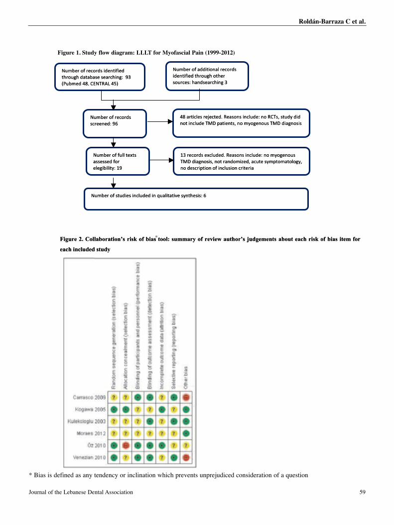
Figure 1. Study flow diagram: LLLT for Myofacial Pain (1999-2012)
!"#$%&'()'*++,-,(.*/'&%0(&+1',+%.-,),%+'-2&("32'(-2%&'1("&0%14'2*.+1%*&02,.3'5'
!"#$%&'()'&%0(&+1',+%.-,),%+'-2&("32'+*-*$*1%'1%*&02,.34''65'78"$#%+'9:;'<=!>?@A'9BC'''
9:'*&-,0/%1'&%D%0-%+E'?%*1(.1',.0/"+%4'.('?<>1;'1-"+F'+,+'.(-',.0/"+%'>GH'I*-,%.-1;'.('#F(3%.("1'>GH'+,*3.(1,1'
!"#$%&'()'&%0(&+1'10&%%.%+4'6J'7K6'&%I,-%+'1-"+FC'
L5'&%0(&+1'%M0/"+%+E'?%*1(.1',.0/"+%4'.('#F(3%.("1'>GH'+,*3.(1,1;'.(-'&*.+(#,N%+;'*0"-%'1F#I-(#*-(/(3F;'.('+%10&,I-,(.'()',.0/"1,(.'0&,-%&,*'
!"#$%&'()')"//'-%M-1'*11%11%+')(&'%/%3,$,/,-F4'L6'
!"#$%&'()'1-"+,%1',.0/"+%+',.'O"*/,-*-,P%'1F.-2%1,14'J'
dtu 1. SerguiF
'' ' '
' '
' ''' '
''
'' ' ' ' ' '
'''
'' ' ' ' '' '
'' '' '
' ' ' '
yo Mam: LLLT foragri dowy fld
'' ' '
' '
' ''' '
''
'' ' ' ' ' '
'''
'' ' ' ' '' '
'' '' '
' ' ' '
(1999nai Paliofac -2012)
'' ' '
' '
' ''' '
''
'' ' ' ' ' '
'''
'' ' ' ' '' '
'' '' '
' ' ' '
%0(&)'(%&$#!"* '*-*$'+32"&(-2
%+#$78" ='<;:'9
!"10&%
'' ' '
' '
' ''' '
''
'' ' ' ' ' '
'''
'' ' ' ' '' '
'' '' '
' ' ' '
!,+1(
-,),%+%.,+1'+&(34,.*1%'1%*&02 65C'B'9A@?>!=
1'+&%0(&)'(%&$#"4%+%%.& J'6 7K6'
FC+-"'1-%+,I
'' ' '
' '
' ''' '
''
'' ' ' ' ' '
'''
'' ' ' ' '' '
'' '' '
' ' ' '
1'+&%0(&*/'.,-,(+*+)'(%&$#!"%&-2(32"(&-2-,),%+%.+3,.021%*&+*.20%14&"( '5
9: % '+"0/.',1.(1%*'?E'&%D%0-%+%10/&-,'*1.-%,-I*>GH'"+%/.0,.(- %3(F#(.;'
'' ' '
' '
' ''' '
''
'' ' ' ' ' '
'''
'' ' ' ' '' '
'' '' '
' ' ' '
+,F'++-"'1;1><'?(%4'.1,1(.3*,+HG>1"(.%
'' ' '
' '
' ''' '
''
'' ' ' ' ' '
'''
'' ' ' ' '' '
'' '' '
' ' ' '
!*%
!
'' ' '
' '
' ''' '
''
'' ' ' ' ' '
'''
'' ' ' ' '' '
'' '' '
' ' ' '
-%M-1'//')")'(%&$#!"&)(*11%11%+
,/,-F4%/%3,$ L6
%++0/",.,%1'+1-")'(%&$#" */,-*-,P%'"O,.
'' ' '
' '
' ''' '
''
'' ' ' ' ' '
'''
'' ' ' ' '' '
'' '' '
' ' ' '
L5 +"0/.',1.(1%*'?E%++"0/'%M1&+'&%0("*0+;%*.+(#,N&.(-.(1,1;+,*3H'>G
*,&%-,&0(.',"1/.0,()(.',I-,&01+%.('
%1,1-21F.P%' 4'J
'' ' '
' '
' ''' '
''
'' ' ' ' ' '
'''
'' ' ' ' '' '
'' '' '
' ' ' '
%4+ 1"(3%.F('#('.;F(/(3(#*-#I-1F%"-
'' ' '
' '
' ''' '
''
'' ' ' ' ' '
'''
'' ' ' ' '' '
'' '' '
' ' ' '
'' ' '
' '
' ''' '
''
'' ' ' ' ' '
'''
'' ' ' ' '' '
'' '' '
' ' ' '
'' ' '
' '
' ''' '
''
'' ' ' ' ' '
'''
'' ' ' ' '' '
'' '' '
' ' ' '
'' ' '
' '
' ''' '
''
'' ' ' ' ' '
'''
'' ' ' ' '' '
'' '' '
' ' ' '
'' ' '
' '
' ''' '
''
'' ' ' ' ' '
'''
'' ' ' ' '' '
'' '' '
' ' ' '
'' ' '
' '
' ''' '
''
'' ' ' ' ' '
'''
'' ' ' ' '' '
'' '' '
' ' ' '
'' ' '
' '
' ''' '
''
'' ' ' ' ' '
'''
'' ' ' ' '' '
'' '' '
' ' ' '
'' ' '
' '
' ''' '
''
'' ' ' ' ' '
'''
'' ' ' ' '' '
'' '' '
' ' ' '
'' ' '
' '
' ''' '
''
'' ' ' ' ' '
'''
'' ' ' ' '' '
'' '' '
' ' ' '
'' ' '
' '
' ''' '
''
'' ' ' ' ' '
'''
'' ' ' ' '' '
'' '' '
' ' ' '
'' ' '
' '
' ''' '
''
'' ' ' ' ' '
'''
'' ' ' ' '' '
'' '' '
' ' ' '
'' ' '
' '
' ''' '
''
'' ' ' ' ' '
'''
'' ' ' ' '' '
'' '' '
' ' ' '
'' ' '
' '
' ''' '
''
'' ' ' ' ' '
'''
'' ' ' ' '' '
'' '' '
' ' ' '
'' ' '
' '
' ''' '
''
'' ' ' ' ' '
'''
'' ' ' ' '' '
'' '' '
' ' ' '
'' ' '
' '
' ''' '
''
'' ' ' ' ' '
'''
'' ' ' ' '' '
'' '' '
' ' ' '
'' ' '
' '
' ''' '
''
'' ' ' ' ' '
'''
'' ' ' ' '' '
'' '' '
' ' ' '
Figure 2. Collaboration’s risk of bias tool: summary of review author’s judgements about each risk of bias item for
each included study
olC2. erguiF
dedulcn ihace
us: toolasibof ksir’sonatiorabl
ydtu sd
ymmaru gdju’sorthauwevierof
asibof ksirhacet ouabtsnmege
form tei
59Journal of the Lebanese Dental Association
Figure 1. Study flow diagram: LLLT for Myofascial Pain (1999-2012)
* Bias is defined as any tendency or inclination which prevents unprejudiced consideration of a question
*
Roldán-Barraza C et al.
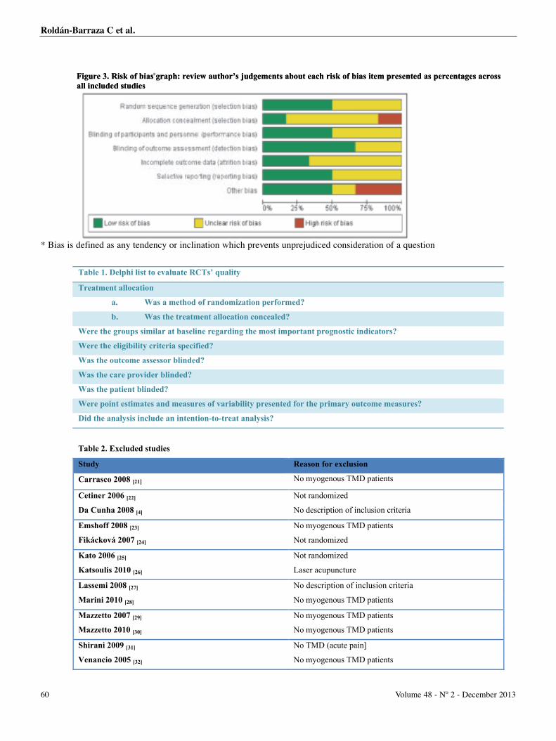
Figure 3. Risk of bias graph: review author’s judgements about each risk of bias item presented as percentages across all included studies
erguiF 3 ksiR. t sdedulcn ilal
hapgrasibof k ’sorthauwevier: seidtu
tsnmegedju bof ksirhacet ouab
tagenecrepasdteneserpm teiasib
sosracse
t sdedulcn ilal
seidtu
Table 1. Delphi list to evaluate RCTs’ quality
Treatment allocation
a. Was a method of randomization performed?
b. Was the treatment allocation concealed?
Were the groups similar at baseline regarding the most important prognostic indicators?
Were the eligibility criteria specified?
Was the outcome assessor blinded?
Was the care provider blinded?
Was the patient blinded?
Were point estimates and measures of variability presented for the primary outcome measures?
Did the analysis include an intention-to-treat analysis?
Table 2. Excluded studies
Study Reason for exclusion
Carrasco 2008 [21] No myogenous TMD patients
Cetiner 2006 [22] Not randomized
Da Cunha 2008 [4] No description of inclusion criteria
Emshoff 2008 [23] No myogenous TMD patients
Fikácková 2007 [24] Not randomized
Kato 2006 [25] Not randomized
Katsoulis 2010 [26] Laser acupuncture
Lassemi 2008 [27] No description of inclusion criteria
Marini 2010 [28] No myogenous TMD patients
Mazzetto 2007 [29] No myogenous TMD patients
Mazzetto 2010 [30] No myogenous TMD patients
Shirani 2009 [31] No TMD (acute pain]
Venancio 2005 [32] No myogenous TMD patients
* Bias is defined as any tendency or inclination which prevents unprejudiced consideration of a question
*
60 Volume 48 - Nº 2 - December 2013
Roldán-Barraza C et al.
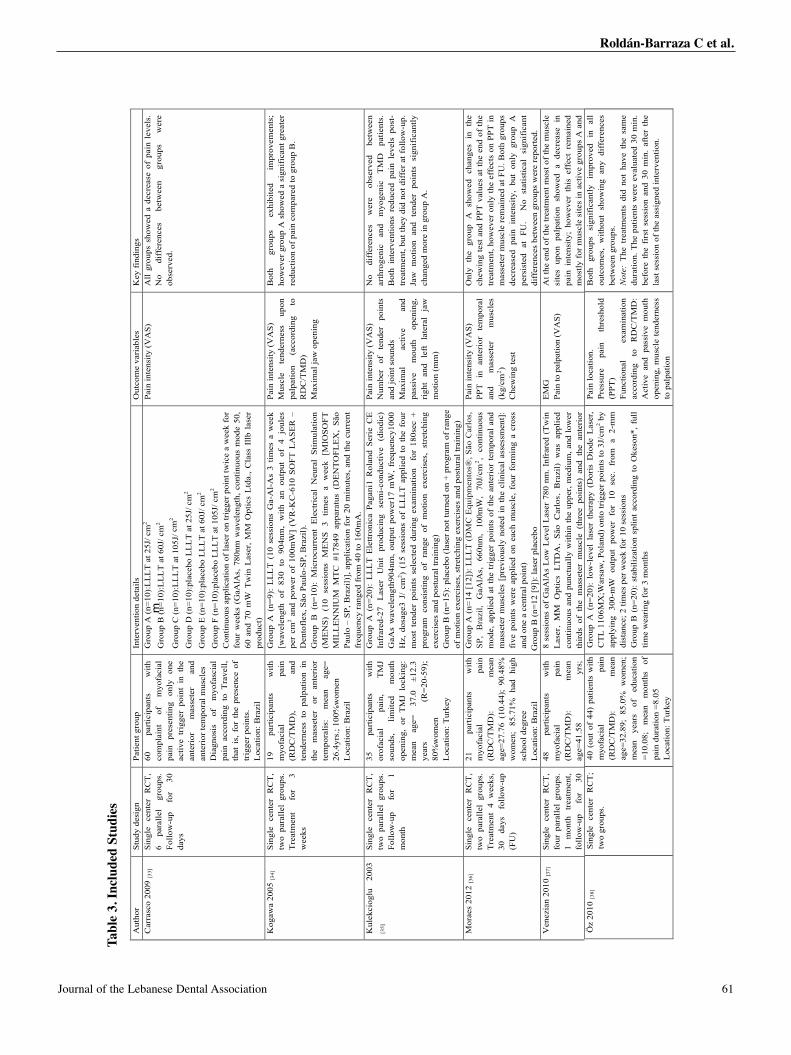
m
Tab
le 3
. Inc
lude
d st
udie
s A
utho
r St
udy
desi
gn
Patie
nt g
roup
In
terv
entio
n de
tails
O
utco
me
vari
able
s K
ey fi
ndin
gs
Car
rasc
o 20
09 [3
3]
Sing
le
cent
er
RC
T,
6 pa
ralle
l gr
oups
. Fo
llow
-up
for
30
days
60
part
icip
ants
w
ith
com
plai
nt
of
myo
faci
al
pain
pr
esen
ting
only
on
e ac
tive
trig
ger
poin
t in
the
an
teri
or
mas
sete
r an
d an
teri
or te
mpo
ral m
uscl
es
Dia
gnos
is
of
myo
fasc
ial
pain
acc
ordi
ng t
o T
rave
ll,
that
is,
for
the
pre
senc
e of
tr
igge
r poi
nts.
L
ocat
ion:
Bra
zil
Gro
up A
(n=1
0):L
LL
T a
t 25J
/ cm
2 G
roup
B (n
=10)
:LL
LT
at 6
0J/ c
m2
Gro
up C
(n=1
0):L
LL
T a
t 105
J/ c
m2
Gro
up D
(n=1
0):p
lace
bo L
LL
T a
t 25J
/ cm
2 G
roup
E (n
=10)
:pla
cebo
LL
LT
at 6
0J/ c
m2
Gro
up F
(n=1
0):p
lace
bo L
LL
T a
t 105
J/ c
m2
Con
tinuo
us a
pplic
atio
n of
lase
r on
trig
ger p
oint
twic
e a
wee
k fo
r fo
ur w
eeks
(G
aAlA
s, 7
80nm
wav
elen
gth,
con
tinuo
us m
ode
50,
60 a
nd 7
0 m
W T
win
Las
er,
MM
Opt
ics
Ltd
a.,
Cla
ss I
llb l
aser
pr
oduc
t)
Pain
inte
nsity
(VA
S)
All
grou
ps s
how
ed a
dec
reas
e of
pai
n le
vels
. N
o di
ffer
ence
s be
twee
n gr
oups
w
ere
obse
rved
.
Kog
awa
2005
[34]
Si
ngle
cen
ter
RC
T,
two
para
llel
grou
ps.
Tre
atm
ent
for
3 w
eeks
19
part
icip
ants
w
ith
myo
faci
al
pain
(R
DC
/TM
D),
and
tend
erne
ss t
o pa
lpat
ion
in
the
mas
sete
r or
an
teri
or
tem
pora
lis:
mea
n ag
e=
26.4
yrs.
; 100
%w
omen
L
ocat
ion:
Bra
zil
Gro
up A
(n=
9):
LL
LT
(10
ses
sion
s G
a-A
l-A
s 3
times
a w
eek
[wav
elen
gth
of 8
30 t
o 90
4nm
, w
ith a
n ou
tput
of
4 jo
ules
pe
r cm
2 and
pow
er o
f 10
0mW
] (V
R-K
C-6
10 S
OFT
LA
SER
–
Den
tofl
ex, S
ão P
aulo
-SP,
Bra
zil)
. G
roup
B (
n=10
): M
icro
curr
ent
Ele
ctri
cal
Neu
ral
Stim
ulat
ion
(ME
NS)
(1
0 se
ssio
ns
ME
NS
3 tim
es
a w
eek
[MIO
SOFT
M
ILL
EN
NIU
M M
TC
#17
849
appa
ratu
s (D
EN
TO
FLE
X,
São
Paul
o –
SP, B
razi
l)],
appl
icat
ion
for
20 m
inut
es, a
nd th
e cu
rren
t fr
eque
ncy
rang
ed fr
om 4
0 to
160
mA
.
Pain
inte
nsity
(VA
S)
Mus
cle
tend
erne
ss
upon
pa
lpat
ion
(acc
ordi
ng
to
RD
C/T
MD
) M
axim
al ja
w o
peni
ng
Bot
h gr
oups
ex
hibi
ted
impr
ovem
ents
; ho
wev
er g
roup
A s
how
ed a
sig
nifi
cant
gre
ater
re
duct
ion
of p
ain
com
pare
d to
gro
up B
.
Kul
ekci
oglu
20
03
[35]
Si
ngle
ce
nter
R
CT
, tw
o pa
ralle
l gr
oups
. Fo
llow
-up
for
1 m
onth
35
part
icip
ants
w
ith
orof
acia
l pa
in,
TM
J so
unds
, lim
ited
mou
th
open
ing,
or
TM
J lo
ckin
g:
mea
n ag
e=
37.0
±1
2.3
year
s (R
=20-
59);
80
%w
omen
L
ocat
ion:
Tur
key
Gro
up A
(n=
20):
LL
LT
Ele
ttron
ica
Paga
ni1
Rol
and
Seri
e C
E
Infr
ared
-27
Las
er
Uni
t pr
oduc
ing
sem
i-co
nduc
tive
(dio
dic)
G
aAs
wav
elen
gth9
04nm
, ou
tput
pow
er17
mW
, fr
eque
ncy1
000
Hz,
dos
age3
J/
cm2 )
(15
sess
ions
of
LL
LT
app
lied
to t
he f
our
mos
t te
nder
poi
nts
sele
cted
dur
ing
exam
inat
ion
for
180
sec
+ pr
ogra
m c
onsi
stin
g of
ran
ge o
f m
otio
n ex
erci
ses,
str
etch
ing
exer
cise
s an
d po
stur
al tr
aini
ng)
Gro
up B
(n=1
5): p
lace
bo (
lase
r not
turn
ed o
n +
prog
ram
of r
ange
of
mot
ion
exer
cise
s, s
tret
chin
g ex
erci
ses
and
post
ural
trai
ning
)
Pain
inte
nsity
(VA
S)
Num
ber
of
tend
er
poin
ts
and
join
t sou
nds
Max
imal
ac
tive
and
pass
ive
mou
th
open
ing,
ri
ght
and
left
la
tera
l ja
w
mot
ion
(mm
)
No
diff
eren
ces
wer
e ob
serv
ed
betw
een
arth
roge
nic
and
myo
geni
c T
MD
pa
tient
s.
Bot
h in
terv
entio
ns r
educ
ed p
ain
leve
ls p
ost-
trea
tmen
t, bu
t the
y di
d no
t dif
fer
at f
ollo
w-u
p.
Jaw
mot
ion
and
tend
er p
oint
s si
gnif
ican
tly
chan
ged
mor
e in
gro
up A
.
Mor
aes
2012
[36]
Si
ngle
ce
nter
R
CT
, tw
o pa
ralle
l gr
oups
. T
reat
men
t 4
wee
ks,
30
days
fo
llow
-up
(FU
)
21
part
icip
ants
w
ith
myo
faci
al
pain
(R
DC
/TM
D):
m
ean
age=
27.7
6 (1
0.44
); 9
0.48
%
wom
en;
85.7
1%
had
high
sc
hool
deg
ree
Loc
atio
n: B
razi
l
Gro
up A
(n=1
4 [1
2]):
LL
LT
(D
MC
Equ
ipm
ento
s®, S
ão C
arlo
s,
SP,
Bra
zil,!
GaA
lAs,
66
0nm
, 10
0mW
, 70
J/cm
2 , co
ntin
uous
m
ode,
app
lied
at t
he t
rigg
er p
oint
s of
the
ant
erio
r te
mpo
ral
and
mas
sete
r m
uscl
es [
prev
ious
ly n
oted
in
the
clin
ical
ass
essm
ent]
: fi
ve p
oint
s w
ere
appl
ied
on e
ach
mus
cle,
fou
r fo
rmin
g a
cros
s an
d on
e a
cent
ral p
oint
) G
roup
B (n
=12
[9])
: las
er p
lace
bo
Pain
inte
nsity
(VA
S)
PPT
in
an
teri
or
tem
pora
l an
d m
asse
ter
mus
cles
(k
g/cm
2 ) C
hew
ing
test
Onl
y th
e gr
oup
A s
how
ed c
hang
es i
n th
e ch
ewin
g te
st a
nd P
PT v
alue
s at
the
end
of th
e tr
eatm
ent,
how
ever
onl
y th
e ef
fect
s on
PPT
in
mas
sete
r m
uscl
e re
mai
ned
at F
U. B
oth
grou
ps
decr
ease
d pa
in i
nten
sity
, bu
t on
ly g
roup
A
pers
iste
d at
FU
.
No
stat
istic
al
sign
ific
ant
diff
eren
ces
betw
een
grou
ps w
ere
repo
rted
. V
enez
ian
2010
[37]
Sing
le
cent
er
RC
T,
four
par
alle
l gr
oups
. 1
mon
th
trea
tmen
t, fo
llow
-up
for
30
48
part
icip
ants
w
ith
myo
faci
al
pain
(R
DC
/TM
D):
m
ean
age=
41.5
8 yr
s;
8 se
ssio
ns o
f G
aAlA
s L
ow L
evel
Las
er 7
80 n
m. I
nfra
red
(Tw
in
Lase
r, M
M O
ptic
s LT
DA
, Sã
o C
arlo
s, B
razi
l) w
as a
pplie
d co
ntin
uous
and
pun
ctua
lly w
ithin
the
uppe
r, m
ediu
m, a
nd lo
wer
th
irds
of
the
mas
sete
r m
uscl
e (t
hree
poi
nts)
and
the
ant
erio
r
EM
G
Pain
to p
alpa
tion
(VA
S)
At t
he e
nd o
f the
trea
tmen
t mos
t of t
he m
uscl
e si
tes
upon
pal
patio
n sh
owed
a d
ecre
ase
in
pain
int
ensi
ty;
how
ever
thi
s ef
fect
rem
aine
d m
ostly
for m
uscl
e si
tes
in a
ctiv
e gr
oups
A a
nd
Öz
2010
[38]
Si
ngle
ce
nter
R
CT
; tw
o gr
oups
. 40
(ou
t of
44)
pat
ient
s w
ith
myo
faci
al
pain
(R
DC
/TM
D):
m
ean
age=
32.8
9; 8
5.0%
wom
en;
mea
n ye
ars
of
educ
atio
n =1
0.08
; m
ean
mon
ths
of
pain
dur
atio
n =8
.05
Loc
atio
n: T
urke
y
Gro
up A
(n=
20):
low
-lev
el l
aser
the
rapy
(D
oris
Dio
de L
aser
, C
TL
110
6MX
;War
saw
, Pol
and)
ont
o tr
igge
r poi
nts
to 3
J/cm
2 by
appl
ying
300
-mW
out
put
pow
er f
or
10 s
ec.
from
a 2
-mm
di
stan
ce; 2
tim
es p
er w
eek
for 1
0 se
ssio
ns
Gro
up B
(n=
20):
sta
biliz
atio
n sp
lint
acco
rdin
g to
Oke
son*
, ful
l tim
e w
eari
ng fo
r 3 m
onth
s
Pain
loca
tion.
Pr
essu
re
pain
th
resh
old
(PPT
) Fu
nctio
nal
exam
inat
ion
acco
rdin
g to
R
DC
/TM
D:
Act
ive
and
pass
ive
mou
th
open
ing,
mus
cle
tend
erne
ss
to p
alpa
tion
Bot
h gr
oups
si
gnif
ican
tly
impr
oved
in
al
l ou
tcom
es,
with
out
show
ing
any
diff
eren
ces
betw
een
grou
ps.
Not
e: T
he t
reat
men
ts d
id n
ot h
ave
the
sam
e du
ratio
n. T
he p
atie
nts
wer
e ev
alua
ted
30 m
in.
befo
re t
he f
irst
ses
sion
and
30
min
. af
ter
the
last
ses
sion
of t
he a
ssig
ned
inte
rven
tion.
Tabl
e 3.
Inc
lude
d St
udie
s
61Journal of the Lebanese Dental Association
Roldán-Barraza C et al.
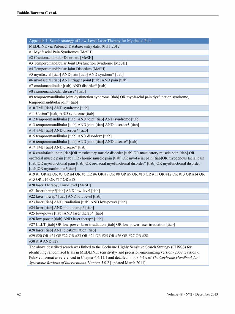
Appendix 1. Search strategy of Low-Level Laser Therapy for Myofacial Pain MEDLINE via Pubmed. Database entry date: 01.11.2012 #1 Myofascial Pain Syndromes [MeSH] #2 Craniomandibular Disorders [MeSH] #3 Temporomandibular Joint Dysfunction Syndrome [MeSH] #4 Temporomandibular Joint Disorders [MeSH] #5 myofascial [tiab] AND pain [tiab] AND syndrom* [tiab] #6 myofascial [tiab] AND trigger point [tiab] AND pain [tiab] #7 craniomandibular [tiab] AND disorder* [tiab] #8 craniomandibular disease* [tiab] #9 temporomandibular joint dysfunction syndrome [tiab] OR myofascial pain dysfunction syndrome, temporomandibular joint [tiab] #10 TMJ [tiab] AND syndrome [tiab] #11 Costen* [tiab] AND syndrome [tiab] #12 temporomandibular [tiab] AND joint [tiab] AND syndrome [tiab] #13 temporomandibular [tiab] AND joint [tiab] AND disorder* [tiab] #14 TMJ [tiab] AND disorder* [tiab] #15 temporomandibular [tiab] AND disorder* [tiab] #16 temporomandibular [tiab] AND joint [tiab] AND disease* [tiab] #17 TMJ [tiab] AND disease* [tiab] #18 craniofacial pain [tiab]OR masticatory muscle disorder [tiab] OR masticatory muscle pain [tiab] OR orofacial muscle pain [tiab] OR chronic muscle pain [tiab] OR myofacial pain [tiab]OR myogenous facial pain [tiab]OR myofunctional pain [tiab] OR orofacial myofunctional disorder* [tiab] OR myofunctional disorder [tiab]OR myoarthropat*[tiab] #19 #1 OR #2 OR #3 OR #4 OR #5 OR #6 OR #7 OR #8 OR #9 OR #10 OR #11 OR #12 OR #13 OR #14 OR #15 OR #16 OR #17 OR #18 #20 laser Therapy, Low-Level [MeSH] #21 laser therap*[tiab] AND low-level [tiab] #22 laser therap* [tiab] AND low level [tiab] #23 laser [tiab] AND irradiation [tiab] AND low-power [tiab] #24 laser [tiab] AND phototherap* [tiab] #25 low-power [tiab] AND laser therap* [tiab] #26 low power [tiab] AND laser therap* [tiab] #27 LLLT [tiab] OR low-power laser irradiation [tiab] OR low power laser irradiation [tiab] #28 laser [tiab] AND biostimulation [tiab] #29 #20 OR #21 OR#22 OR #23 OR #24 OR #25 OR #26 OR #27 OR #28 #30 #19 AND #29 The above described search was linked to the Cochrane Highly Sensitive Search Strategy (CHSSS) for identifying randomized trials in MEDLINE: sensitivity- and precision-maximizing version (2008 revision); PubMed format as referenced in Chapter 6.4.11.1 and detailed in box 6.4.c of The Cochrane Handbook for Systematic Reviews of Interventions, Version 5.0.2 [updated March 2011].
62 Volume 48 - Nº 2 - December 2013
Roldán-Barraza C et al.
REFERENCES1. Simunovic Z. Low level laser therapy with trigger pointstechnique: a clinical study on 243 patients. J Clin Laser Med Surg1996;14:163-167
2. Gür A, Karakoç M, Nas K, Cevik R, Saraç J, Demir E. Efficacyof low power laser therapy in fibromyalgia: a single-blind,placebo-controlled trial. Lasers Med Sci 2002;17:57-61
3. Bjordal JM, Couppé C, Chow RT, Tunér J, Ljunggren EA. Asystematic review of low level laser therapy with location-specificdoses for pain from chronic joint disorders. Aust J Physiother2003;49:107-116
4. da Cunha LA, Firoozmand LM, da Silva AP, Camargo SEA, EstevesSA, Oliveira W. Efficacy of low-level laser therapy in the treatment oftemporomandibular disorder. Int Dent J 2008;58:213-217
5. Gam AN, Thorsen H, Lønnberg F. The effect of low-level lasertherapy on musculoskeletal pain: a meta-analysis Pain 1993;52:63-66
6. Chang W, Wu J, Yang W, Jiang J. Therapeutic effects of low-level laser on lateral epicondylitis from differential interventions ofChinese-Western medicine: systematic review. Photomed LaserSurg 2010;28:327-336
7. Gur A, Sarac AJ, Cevik R, Altindag O, Sarac S. Efficacy of 904nm gallium arsenide low level laser therapy in the management ofchronic myofascial pain in the neck: a double-blind andrandomize-controlled trial. Lasers Surg Med 2004;35:229-235
8. Hakgüder A, Birtane M, Gürcan S, Kokino S, Turan FN.Efficacy of low level laser therapy in myofascial pain syndrome:an algometric and thermographic evaluation. Lasers Surg Med2003;33:339-343
9. Dundar U, Evcik D, Samli F, Pusak H, Kavuncu V. The effect ofgallium arsenide aluminum laser therapy in the management ofcervical myofascial pain syndrome: a double blind, placebo-controlled study. Clin Rheumatol 2007;26:930-934
10. Aigner N, Fialka C, Radda C, Vecsei V. Adjuvant laseracupuncture in the treatment of whiplash injuries: a prospective,randomized placebo-controlled trial. Wien Klin Wochenschr2006;118:95-99
11. Medeiros JS de, Vieira GF, Nishimura PY. Laser applicationeffects on the bite strength of the masseter muscle, as an orofacialpain treatment. Photomed Laser Surg 2005;23:373-376
12. Fargas-Babjak A. Acupuncture, transcutaneous electrical nervestimulation, and laser therapy in chronic pain. Clin J Pain2001;17:S105-13
13. Schüller BK, Neugebauer EAM. Evidenz zur Laserakupunkturbei orthopädischen Erkrankungen. Ein systematisches Review.Schmerz 2008;22:9-15
14. Sakurai Y, Yamaguchi M, Abiko Y. Inhibitory effect of low-level laser irradiation on LPS-stimulated prostaglandin E2production and cyclooxygenase-2 in human gingival fibroblastsEur J Oral Sci 2000;108:29-34
15. Shimizu N, Yamaguchi M, Goseki T, Shibata Y, Takiguchi H,Iwasawa T, Abiko Y. Inhibition of Prostaglandin E2 and Interleukin1- Production by Low-power Laser Irradiation in Stretched HumanPeriodontal Ligament Cells. J Dent Res 1995;74:1382-1388
16. Bjordal JM, Johnson MI, Iversen V, Aimbire F, Lopes-MartinsRAB. Low-Level Laser Therapy in Acute Pain: A SystematicReview of Possible Mechanisms of Action and Clinical Effects inRandomized Placebo-Controlled Trials. Photomed and Laser Surg2006;24:158-168
17. Tullberg M, Alstergren PJ, Ernberg MM. Effects of low-powerlaser exposure on masseter muscle pain and microcirculation. Pain2003;105:89-96
18. Cummings TM, White AR. Needling therapies in themanagement of myofascial trigger point pain: a systematic review.Arch Phys Med Rehabil 2001;82:986-992
19. Higgins JPT, Green S. Cochrane Handbook for SystematicReviews of Interventions. Version 5.1.0. http://www.cochrane-handbook.org/ 2011, accessed on 29.11.2013
20. Verhagen A, de Vet HCW, de Bie RA, Kessels AGH, Boers M,Bouter LM, Knipschild PG. The Delphi List: A Criteria List forQuality Assessment of Randomized Clinical Trials for ConductingSystematic Reviews Developed by Delphi Consensus. J ClinEpidemiol 1998;51:1235-1241
21. Carrasco TG, Mazzetto MO, Mazzetto RG, Mestriner W[JR.Low intensity laser therapy in temporomandibular disorder: aphase II double-blind study. Cranio 2008;26:274-281
22. Cetiner S, Kahraman SA, Yucetas S. Evaluation of low-levellaser therapy in the treatment of temporomandibular disorders.Photomed Laser Surg 2006;24:637-641
23. Emshoff R, Bosch R, Pumpel E, Schoning H, Strobl H. Low-level laser therapy for treatment of temporomandibular joint pain:a double-blind and placebo-controlled trial. Oral Surg Oral MedOral Pathol Oral Radiol Endod 2008;105:452-456
24. Fikácková H, Dostalova T, Navratil L, Klaschka J.Effectiveness of low-level laser therapy in temporomandibularjoint disorders: a placebo-controlled study. Photomed Laser Surg2007;25:297-303
25. Kato MT, Kogawa EM, Santos CN, Conti PCR. TENS andlow-level laser therapy in the management of temporomandibulardisorders. J Appl Oral Sci 2006;14:130-135
26. Katsoulis J, Ausfeld-Hafter B, Windecker-Getaz I, Katsoulis K,Blagojevic N, Mericske-Stern R. Laser acupuncture for myofascialpain of the masticatory muscles. A controlled pilot study. SchweizMonatsschr Zahnmed 2010;120:213-225
27. Lassemi E, Jafari S, Motamedi M, Navi F, Lasemi R. Low-level Laser Therapy in the management of TemporomandibularJoint Disorder. J Oral Laser Appl 2008;8:83-86
28. Marini I, Gatto MR, Bonetti GA. Effects of superpulsed low-level laser therapy on temporomandibular joint pain. Clin J Pain2010;26:611-616
Roldán-Barraza C et al.
63Journal of the Lebanese Dental Association
29. Mazzetto MO, Carrasco TG, Bidinelo EF, Andrade PizzoRCde, Mazzetto RG. Low intensity laser application intemporomandibular disorders: a phase I double-blind study. Cranio2007;25:186-192
30. Mazzetto MO, Hotta TH, Pizzo RCdeA. Measurements of jawmovements and TMJ pain intensity in patients treated withGaAlAs laser. Braz Dent J 2010;21:356-360
31. Shirani AM, Gutknecht N, Taghizadeh M, Mir M. Low-level lasertherapy and myofacial pain dysfunction syndrome: a randomizedcontrolled clinical trial. Lasers Med Sci 2009;24:715-720
32. Venancio RdeA, Camparis CM, Lizarelli RdeFZ. Low intensitylaser therapy in the treatment of temporomandibular disorders: adouble-blind study. J Oral Rehabil 2005;32:800-807
33. Carrasco TG, Guerisoli LDC, Guerisoli DMZ, Mazzetto MO.Evaluation of low intensity laser therapy in myofascial painsyndrome. Cranio 2009;27:243-247
34. Kogawa EM, Kato MT, Santos CN, Conti PCR. Evaluation ofthe efficacy of low-level laser therapy (LLLT) and themicroelectric neurostimulation (MENS) in the treatment ofmyogenic temporomandibular disorders: a randomized clinicaltrial. J Appl Oral Sci 2005;13:280-285
35. Kulekcioglu S, Sivrioglu K, Ozcan O, Parlak M. Effectivenessof low-level laser therapy in temporomandibular disorder. Scand JRheumatol 2003;32:114-118
36. Moraes Maia MLde, Ribeiro MAG, Maia LGM, Stuginski-Barbosa J, Costa YM, Porporatti AL, Conti PCR, Bonjardim LR.Evaluation of low-level laser therapy effectiveness on the pain andmasticatory performance of patients with myofascial pain. LasersMed Sci Nov 2012 DOI:10.1007/s10103-012-1228-7
37. Venezian GC, da Silva MAMR, Mazzetto RG, Mazzetto MO.Low level laser effects on pain to palpation and electromyographicactivity in TMD patients: a double-blind, randomized, placebo-controlled study. Cranio 2010;28:84-91
38. Öz S, Gokcen-Rohlig B, Saruhanoglu A, Tuncer EB.Management of myofascial pain: low-level laser therapy versusocclusal splints. Cranio 2010;21:1722-1728
39. Ahrari F, Madani AS, Ghafouri ZS, Tunér J. The efficacy oflow-level laser therapy for the treatment of myogenoustemporomandibular joint disorder. Lasers Med Sci 2013DOI:10.1007/s10103-012-1253-6
40. Yousefi-Nooraie R, Schonstein E, Heidari K, Rashidian A,Pennick V, Akbari-Kamrani M, Irani S, Shakiba B, Mortaz HejriSA, Mortaz Hejri SO, Jonaidi A. Low level laser therapy for non-specific low-back pain. Cohrane Database Syst Rev 2008;16DOI:10.1002/14651858.CD005107
41. Kadhim-Saleh A, Maganti H, Ghert M, Singh S, Farrokhyar F.Is low-level laser therapy in relieving neck pain effective?Systematic review and meta-analysis. Rheumatol Int 2013DOI:10.1007/s00296-013-2742-z
42. Chow R. Dose dilemmas in low-level laser therapy-the effectsof different paradigms and historical perspectives. Laser Ther2001;13:102-109
43. http://waltza.co.za/documentation-links/recommendations/dosage-recommendations/accessed on 20.11.2013
44. Jang H, Lee H. Meta-analysis of pain relief effects by laserradiation on joint areas. Photomed Laser Ther 2012;30:405-417
Correspond with: Carolina Roldá[email protected]
64 Journal of the Lebanese Dental AssociationVolume 48 - Nº 2 - December 2013
Roldán-Barraza C et al.
Case Report
INTRODUCTIONThe term “Temporomandibular disorder” (TMD) is
used to describe a large number of clinical signs andsymptoms that affect muscles of mastication,temporomandibular joint (TMJ), and associatedstructures1-3. Orofacial pain is the most commoncomplaint among individuals with TMD1,4. Othersymptoms include limited mandibular movements,joint noises,1,5,6 headache, dizziness, and ears ringing7,8,affecting the quality of life of patients.
Low-level LASER therapy (LLLT) is a particularform of LASER medicine that uses low-level (low-power) LASERs (or light-emitting diodes) to altercellular function. Other names for this therapy includesoft LASER, cold LASER, biostimulation LASER,and LASER acupuncture. It produces photo-biochemical reactions that result in pain relief.Therapeutic effects of LLLT include modulation ofinflammatory process and analgesia9-12.
LLLT may reduce pain related to inflammationby decreasing levels of prostaglandin E2,
interleukin 1-beta, tumor necrosis factor alpha,prostaglandine-endoperoxide synthase 2, oxydativestress, edema, and cellular influx of neutrophilgranulocytes (Karu, 2008 and Bjordal et al., 2003 and2006).
CASE REPORTA 26 year-old female complaining of orofacial
chronic pain (mainly in right and left masseters andtemporal muscles), was referred to our privatepractice. After history taking and clinical examination,patient was diagnosed with a temporomandibulardisorder condition combined with a mouth openinglimitation (< 30mm). CT Scan radiographs excluded adisc displacement of TMJs (Fig. 1).
A visual analog scale was used before and aftertherapy to record pain level upon palpation of the leftand right masseter, TMJs, lateral and medialpterygoids, and temporal muscles. This is a numericrating scale that runs from 0 (absence of pain) to 10(worst pain imaginable)1,2. The procedure wasexplained verbally to the patient and signed informedconsent was obtained for the use of LLLT assupportive therapy.
The treatment using a LLLT diode LASER(EzlaseTM, Biolase, USA) was performed twice per
Low-Level LASER as supportive therapy of temporomandibulardisorders -TMDs: a case report.
Abstract
Objective: The aim of this report was to evaluate the effectiveness of Low-Level LASER Therapy -LLLT- in the treatment of
a TMD.
Clinical Application: A patient complaining of TMD (myofascial pain, mainly in right and left masseter and temporal
muscles) and mouth opening limitation, was treated with LLLT, for a period of one month. A Visual Analogue Scale (VAS) was
used to evaluate pain level. An occlusal device therapy was used after treatment with LASER therapy.
Results: Patient was satisfied reporting a normal mouth opening and absence of pain in left and right temporal and masseter
muscles.
Conclusion: LLLT was proposed as supportive therapy in combination with conventional occlusal device. Treatment
protocol was effective in managing mouth opening limitation and TMJ muscles pain. Patient was put on a soft food diet during
the treatment period.
Najib Abou Hamra1, BDS, DESS Prostho., Elias Smaira2, Dr. Chir. Dent., DESS Prostho.
1. Clinical Instructor, Department of Prosthodontics,Lebanese University School of Dentistry, Beirut, Lebanon
2. Associate Clinical Instructor, Department ofProsthodontics, Lebanese University School of Dentistry,Beirut.
Journal of the Lebanese Dental AssociationVolume 48 - Nº 2 - December 2013
65
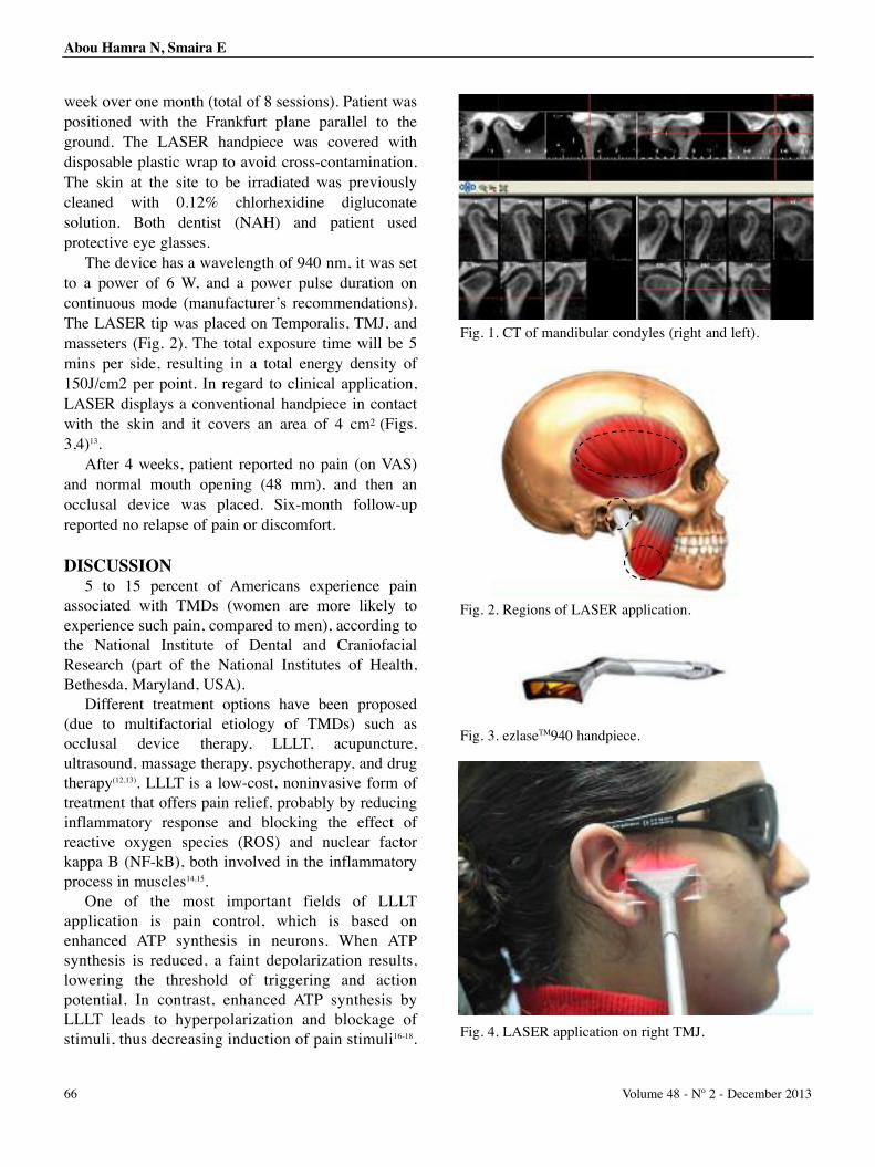
week over one month (total of 8 sessions). Patient waspositioned with the Frankfurt plane parallel to theground. The LASER handpiece was covered withdisposable plastic wrap to avoid cross-contamination.The skin at the site to be irradiated was previouslycleaned with 0.12% chlorhexidine digluconatesolution. Both dentist (NAH) and patient usedprotective eye glasses.
The device has a wavelength of 940 nm, it was setto a power of 6 W, and a power pulse duration oncontinuous mode (manufacturer’s recommendations).The LASER tip was placed on Temporalis, TMJ, andmasseters (Fig. 2). The total exposure time will be 5mins per side, resulting in a total energy density of150J/cm2 per point. In regard to clinical application,LASER displays a conventional handpiece in contactwith the skin and it covers an area of 4 cm2 (Figs.3,4)13.
After 4 weeks, patient reported no pain (on VAS)and normal mouth opening (48 mm), and then anocclusal device was placed. Six-month follow-upreported no relapse of pain or discomfort.
DISCUSSION5 to 15 percent of Americans experience pain
associated with TMDs (women are more likely toexperience such pain, compared to men), according tothe National Institute of Dental and CraniofacialResearch (part of the National Institutes of Health,Bethesda, Maryland, USA).
Different treatment options have been proposed(due to multifactorial etiology of TMDs) such asocclusal device therapy, LLLT, acupuncture,ultrasound, massage therapy, psychotherapy, and drugtherapy(12,13). LLLT is a low-cost, noninvasive form oftreatment that offers pain relief, probably by reducinginflammatory response and blocking the effect ofreactive oxygen species (ROS) and nuclear factorkappa B (NF-kB), both involved in the inflammatoryprocess in muscles14,15.
One of the most important fields of LLLTapplication is pain control, which is based onenhanced ATP synthesis in neurons. When ATPsynthesis is reduced, a faint depolarization results,lowering the threshold of triggering and actionpotential. In contrast, enhanced ATP synthesis byLLLT leads to hyperpolarization and blockage ofstimuli, thus decreasing induction of pain stimuli16-18.
Abou Hamra N, Smaira E
Fig. 1. CT of mandibular condyles (right and left).
Fig. 2. Regions of LASER application.
Fig. 3. ezlaseTM940 handpiece.
Fig. 4. LASER application on right TMJ.
66 Volume 48 - Nº 2 - December 2013
Patient amelioration was observed during amonth period according to VAS: before startingLLLT treatment sessions, pain on VAS was 8, afterfirst week (the first 2 sessions) it was decreased to 7,then respectively second, third and fourth week to 5,2 and 0. It is important to know that no drug wasprescribed during the treatment period.
CONCLUSIONThere is no consensus. yet, over ideal use of LLLT
but many reports suggest its relative effectiveness inrelieving short-term pain in rhumatoid arthritis(Brosseau et al., 2005), osteoarthritis (Jamtvedt et al.,2007), acute and chronic neck pain (Chow et al., 2009),and tendinopathy (Bjordal et al., 2008 and Tumilty et al.,2010). And within the limitation of this case report, theuse of LLLT to reduce muscle pain in TMD patientshowed a successful outcome. Further clinical studiesshould be designed to evaluate the effect of LLLT inTMD management and obtain a consensus regarding thebest LLLT application protocol for pain relief in TMDspatients.
Last, but not least, if high-power LASERs ablatetissues, low-power LASERs may stimulate them, henceencouraging cells for better functions.
REFERENCES1. de Godoy CH, Silva PF, de Araujo DS, Motta LJ, Biasotto-Gonzalez DA, Politti F, Mesquita-Ferrari RA, Fernandes KP,Albertini R, Bussadori SK. Evaluation of effect of low-level lasertherapy on adolescents with temporomandibular disorder: studyprotocol for a randomized controlled trial. Trials. 2013 Jul 22;14:229. doi: 10.1186/1745-6215-14-229.
2. Marini I, Gatto MR, Bonetti GA. Effects of superpulsed low-level laser therapy on temporomandibular joint pain. Clin J Pain.2010;14(Suppl 7):611–616.
3. Kato MT, Kogawa EM, Santos CN, Conti PCR. TENS and low-level laser therapy in the management of temporomandibulardisorders. J Appl Oral Sci. 2006;14(Suppl 2):130–135.
4. deFreitas RF, Ferreira MÂ, Barbosa GA, Calderon PS.Counselling and self-management therapies fortemporomandibular disorders: a systematic review. J Oral Rehabil.2013;11:864-74
5. Crider AB, Glaros AG. A meta-analysis of EMG biofeedbacktreatment of temporomandibular disorders. J Orofac Pain 1999;14:29–37.
6. Carrasco TG, Mazzetto MO, Mazzetto RG, Mestriner W. Lowintensity laser therapy in temporomandibular disorder: a phase IIdouble-blind study. J Craniomandibular Pract. 2008; 14(Suppl4):274–281.
7. Hilgenberg PB, Saldanha AD, Cunha CO, Rubo JH, Conti PC.Temporomandibular disorders, otologic symptoms and depressionlevels in tinnitus patients. J Oral Rehabil. 2012; 39:239-44.
8. Nilsson IM, List T, Drangsholt M. Headache and co-morbidpains associated with TMD pain in adolescents. J Dent Res. 2013;92:802-7.
9. Seada YI, Nofel R, SayedHM.Comparison between Trans-Cranial Electromagnetic Stimulation and Low-Level Laser onModulation of Trigeminal Neuralgia. J Phys Ther Sci. 2013;25:911-4.
10. Petrucci A, Sgolastra F, Gatto R, Mattei A, Monaco A.Effectiveness of low-level laser therapy in temporomandibulardisorders: a systematic review and meta-analysis. J Orofac Pain.2011; 14(Suppl 4):298–307.
11. Shirani AM, Gutknecht N, Taghizadeh M, Mir M. Low-level lasertherapy and myofacial pain dysfunction syndrome: a randomizedcontrolled clinical trial. Lasers Med Sci. 2009; 14:715–720.
12. Fikácková H, Dostálová T, Navrátil L, Klaschka J.Effectiveness of low-level laser therapy in temporomandibularjoint disorders: a placebo-controlled study. Photomed Laser Surg.2007;14(Suppl 4):297–303.
13. Venezian GC, da Silva MA, Mazzetto RG, Mazzetto MO. Lowlevel laser effects on pain to palpation and electromyographicactivity in TMD patients: a double-blind, randomized, placebo-controlled study. Cranio 2010 Apr;28(2):84-91
14. Luo L, Sun Z, Zhang L, Li X, Dong Y, Liu TC. Effects of low-level laser therapy on ROS homeostasis and expression of IGF-1and TGF-β1 in skeletal muscle during the repair process. LasersMed Sci. 2013; 28:725-34.
15. de Almeida P, Lopes-Martins RÁ, Tomazoni SS, Albuquerque-Pontes GM, Santos LA, Vanin AA, Frigo L, Vieira RP, Albertini R,de CarvalhoPde T, Leal-Junior EC. Low-level laser therapy andsodium diclofenac in acute inflammatory response induced byskeletal muscle trauma: effects in muscle morphology and mRNAgene expression of inflammatory markers. Photochem Photobiol.2013: 89:501-7.
16. Silveira PC, da Silva LA, Pinho CA, De Souza PS, RonsaniMM, SchefferDda L, Pinho RA. Effects of low-level laser therapy(GaAs) in an animal model of muscular damage induced bytrauma. Lasers Med Sci. 2013; 28:431-6.
17. Melchior Mde O, Venezian GC, Machado BC, Borges RF,Mazzetto MO. Does low intensity laser therapy reduce pain andchange orofacial myofunctional conditions? Cranio. 2013 Apr;31(2):133-9.
18. Núñez SC, Garcez AS, Suzuki SS, Ribeiro MS. Managementof mouth opening in patients with temporomandibular disordersthrough low-level laser therapy and transcutaneous electricalneural stimulation. Photomed Laser Surg 2006; 24:45-9.
Correspond with:Najib Abou [email protected]@hotmail.com
Abou Hamra N, Smaira E
Journal of the Lebanese Dental AssociationVolume 48 - Nº 2 - December 2013
67
Clinical Note
INTRODUCTIONOro-facial pain is pain in structures predominantly
innervated by the somatic portion of the trigeminalnerve namely the face, scalp, and mouth. It is oftendefined as pain which is experienced in the area belowthe meatal line, above the neck and up to the ear(1),thereby excluding headaches. Predominant oral causesof pain are dental with the majority being acute ratherthan chronic. There are a very wide range of causes forfacial pain and these have been divided into threebroad categories by Hapak(3) and associates:musculoligamentous, dentoalveolar, and neurologicaland vascular. This classification has been used in manyepidemiological studies.
The incidence of chronic orofacial pain is comparablewith other pain conditions in the body, accounting forbetween 20 to 25 % of chronic pain conditions(4). In thestudy of Locker & Grushka(6), pain or discomfort in the
jaws, oral mucosa, or face had been experienced by lessthan 10 % of the study population in the last four weeks.Bonica(7) estimated that five to seven million Americanssuffer from chronic pain in the face and mouth, whereasLipton and co-workers(4) estimated that between 25-45%are affected at some time of life. As mechanismsunderlying these pains begin to be identified, moreaccurate classifications may come to be used.
The commonly accepted classifications of facialpain have been published by The InternationalHeadache Society (IHS), International Association forthe Study of Pain (IASP), and the American Academyof Orofacial Pain (AAOP)(14,15). Woda andassociates(22) reported a recent cluster analysis in anattempt to streamline disorders that cause orofacialpain into aetiological categories: the neurovasculargroup included most ophthalmic division pain(headaches, giant cell arteritis) and alsotrigeminovascular headaches. The neuralgia groupincluded trigeminal neuralgia (typical and atypical;),post-herpetic neuralgia (post-shingles pain which is60% increased likelihood if the patient is over 50years) and post-traumatic neuralgia (includingiatrogenic nerve damage).
The idiopathic group included persistent idiopathicfacial pain (previously known atypical facial pain and
Medication Overuse Facial Pain - MOFP: a case series andproposed diagnostic criteria.
Abstract
Persistent idiopathic or chronic atypical facial pain (AFP) is a poorly defined syndrome. Patients with facial pain are often
given this diagnosis after other causes had been excluded, It is therefore likely to encompass a variety of conditions. We present
a case series of seven patients with facial pain of unknown cause, after clinical assessment and investigations. The common
factor in this group of patients was overuse of large doses of strong analgesics or triptans. Medication withdrawal was
associated with significant reduction in severity and frequency of facial pain. We propose the syndrome of Medication Overuse
Facial Pain as a cause of atypical facial pain in some patients. This condition appears to be analogous to Medication Overuse
Headache. Identification and medication withdrawal appear to be the key for managing these patients.
Conclusion: We have identified medication overuse as an important feature in some patients with atypical facial pain. A
thorough history of medication overuse should be specifically taken, as withdrawal can lead to pain relief.
M Sam Chong1, B.Sc. (Hons), MBBS, MD, FRCP,Nadine Khawaja2, BDS, MFDS, M. Surg. Dent., Joan Hester3, MBBS, FRCA, M.Sc., FFPMRCA, Tara Renton4, BDS, M.D.Sc., Ph.D., FDSRCS (Engl.), FRACDS (Oral/Max.Facial Surgery), ILTM, FHEA*
1. Consultant Neurologist, The National Hospital, QueensSquare and The Medway Hospital NHS Trust,Gillingham, Kent, UK
2. Senior Clinical Specialist Teacher, Department of OralSurgery, King’s College London - KCL Dental Institute,London, UK
3. Consultant in Pain Management, King’s CollegeHospital, London, UK
4. Consultant Oral Surgeon, Professor and Head,Department of Oral Surgery, King’s College LondonDental Institute, UK
* Fellow of the Higher Education Academy
68 Journal of the Lebanese Dental AssociationVolume 48 - Nº 2 - December 2013
atypical odontalgia). This is a particularly difficultgroup of patients to treat: they often present withwhole quadrants of teeth extraction and havingundergone extensive restorative dental treatment withno pain relief.
Chronic oro-facial pain conditions represent adiagnostic challenge for any dental or medicalpractitioner. Patients are frequently misdiagnosed orattribute their pain to a prior event such as a dentalprocedure, ENT (Ear, Nose and Throat) causes, orfacial trauma. Symptoms of depression and anxiety areprevalent in this population and compound thediagnostic conundrum. Treatment is less effective thanin other pain syndromes.
Persistent idiopathic facial pain (atypical facialpain)
One category of chronic oro-facial pain which isespecially difficult to manage is atypical facial pain(AFP). This term was used to denote a category ofpatients whose facial pain did not improve withneurosurgery. There is no precise definition of AFP inthe 1984 IASP classification. The 2002 IHSclassification used the term persistent idiopathic facialpain (PIFP) to denote a condition where “facial pain ispresent daily, persisting for most of the day, confinedat onset to one side of the face that is deep and poorlylocalised”. In these patients, there are no abnormalitieson physical examination or investigations”.
PIFP is reported to be more common in women.The area of pain does not follow any known neuralpathway and tends to be present for months withfrequent recurrences. Pain tends to be constantthroughout the day and is often absent during sleep.Psychological factors are common. PIFP may coexistwith other idiopathic pain syndromes in the mouth andface: atypical odontalgia, temporo-mandibular jointdisorders, and oral dysaesthesia (Woda and Pionchon,2001, Madland and Feinmann, 2002). AFP has beenreported to respond to a multidisciplinary approach;addressing psychological factors and prescribingantidepressant drugs.
Medication overuse headache (MOH) and Facialpain
Medication overuse is increasingly recognised as
an important cause of chronic headaches. MOH, alsoknown as rebound headache (L. Kudrow, 1982) mayevolve from migraine or tension type headache and iscommonly associated with excessive and frequent useof both opioid and non-opioid analgesics as well astriptans. It has been estimated that between 1-2% ofwomen in the developed countries have MOH.Medication withdrawal is the mainstay of treatment,although beneficial effects may not be apparent up to2 months after cessation. In addition, it has beenreported that usual medications used as prophylaxis forheadaches are ineffective until withdrawal from theoffending drug(s) have been successfully achieved.
Anatomical substrate for MOH is the ophthalmicdivision of trigeminal nerve. It is therefore likely thatstructures innervated by maxillary and mandibulardivisions may also give rise to chronic pain secondaryto medication overuse. We are not aware of any reportsin published literature on overuse of medication inpatients with orofacial pain, nor the effects ofmedication withdrawal in such patients.
We propose the concept of medication overusefacial pain (MOFP), a condition analogous tomedication overuse headache (MOH). In this clinicalstudy, we address a case series describing patients withoro-facial pain and excessive medication where painrelief was noticed following medication withdrawal.
METHODS Over a period of 12 months (2008), patients with
chronic facial pain with medication overuse wereidentified. Successful reduction in medication overuseassociated with pain relieve was achieved in 7 patientsand their details are outlined below. For all patients, athorough pain and medical history was taken togetherwith head and neck examination. All had undergoneprevious investigation with the majority having had anMRI scan of the head within 6 months of being seen inclinic. For those who did not have prior neuroimaging,this was performed. None of these patients hadabnormalities on the scan that provided a cause fortheir facial pain. Patients were asked to verbally reporttheir pain, at rest, on a scale ranging from 0 to 10 (0=no pain, 10= worst pain imaginable). Patients werethen instructed to completely stop any conventionalanalgesics that were being taken. In patients where
Chong MS et al.
69Journal of the Lebanese Dental Association
medication withdrawal alone fail to reduce facial pain,patients were then started on or asked to continue withanother form of medication; including tricyclicantidepressants (TCA), serotonin norepinephrine re-uptake inhibitors (SNRI), or anticonvulsants.
Patients were reviewed following cessation ofconventional medication at a minimum of 4 weeks(range 4-18 weeks). Another verbal pain reportassessment was attained and patients’ medication usewas again recorded at review to ascertain if patientshad returned to their old regimen. Patients’ generalcomments were also sought at review to clarify if anyproblems arose following cessation of the medication.Patients were then followed-up for a minimum of 3months after their attempted cessation of analgesicconsumption.
CASE 1 SH, 41 year old female
Initial presentation: Patient presented to neurology department 5
months after onset of facial pain. She was initiallytreated with a course of antibiotics for suspected“sinusitis” (not demonstrated on CT), with only a shortinitial improvement in symptoms which recurred whenantibiotics were discontinued. She sustainedspontaneous exacerbations of sharp stabbing painradiating from ear to jaw, superimposed on a constantburning cold pain “as if the face was frozen”. The painstarted on right cheek but spread bilaterally withinweeks. She said she was able to sleep at night but wokeevery morning with increased tactile sensitivity ofright cheek with stabbing pain in her teeth.Neurological examination was normal apart fromsubjective hyperalgesia of right face that did notcorrespond to any nerve distribution. A MRI scan ofthe head was reported to be normal. Medication regimen:
Solpadol® (codeine phosphate 30mg/paracetamol500mg) 4-6 tablets daily since June 2008 which gaveher some pain relieve but did not completely alleviateher symptoms.
After careful explanation, she discontinued painkillers, was prescribed imipramine, a tricyclicantidepressant (10mg at night) and applied hot/coldpacks to the face for pain relief.
On review: She reported a gradual improvement over 4 weeks
and was pain free 2 months after cessation ofSolpadol®. After a further 2 months, she discontinuedimipramine and remained pain free off all medicationsfor a total of 3 months before she was discharged.
CASE2 AM, 39 year old female
Initial presentation This patient presented to neurology outpatient
clinic 5 months after onset of facial pain. The paininitially presented as a chronic daily headache as resultof a whiplash injury to her neck. She then developedfacial pain, predominantly on right side. She describeddaily shooting/stabbing pain present from waking andlasting all day. About 2-3 times a week, pain wasperceived as bilateral. She had undergone 3 MRI scansof the head in last 2 years without an explanation forher facial pain. Medication regimen
In an effort to alleviate a chronic daily headache,the patient was taking increasing doses ofCodydramol® (Codeine tartrate 10mg/paracetamol500mg), with the dose on consultation being 6-8tablets/ day. Her family practitioner also prescribedamitriptyline, a tricyclic antidepressant (30mg atnight) with partial alleviation of her headache.
After assessment and investigations, she wasadvised to discontinue Codydramol® and started ontopiramate, an anticonvulsant drug. On review:
Within 2 weeks of discontinuing codydramol, shebecame pain free. After a further 10 days withoutheadaches or facial pain, she restarted takingcodydramol. When last assessed, she was taking 4tablets of codydramol a day and the facial pain wasevery other day. She was once again advised to stoptaking codydramol and the dose of topiramate wasincreased to 75mg twice a day. She was referred to thepain clinic to consider having an occipital nerve block.
CASE 3 KC, 54 year old female
Initial presentation: This patient was referred by otolaryngology
70 Volume 48 - Nº 2 - December 2013
Chong MS et al.
department with regards to a 2 year history of painaround the right eye, nose, and face. She described herpain as “deep and constant” with spontaneousexacerbations that last for over 2 hours. She alsosuffered from a long history of musculoskeletal backpain. MRI scan of the head and neck were reported tobe normal. Medication regimen:
In an effort to control facial and low back pain,patient was taking 2-8 tablets of Codydramol® every dayfor the past 2 years. She was also on venlafaxine, anantidepressant of the SNRI class (Serotonine-Norepinephrine reuptake inhibitor) (75mg a day). Afterclinical assessment, she was advised to discontinueCodydramol® but to remain on venlafaxine. On review:
Over the next 5 months, constant pain abated and thefrequency of exacerbations gradually reduced so that sheonly experienced 1 attack per week. She still consumed anaverage of 2 tablets of Codydramol® every week.
CASE 4GA, 34 year old female
Initial presentation: She presented with a 3 year history of right sided
facial pain. Her symptoms started around left earwhich rapidly spread to the right. She described aconstant dull aching background pain with shootingand stabling exacerbations, lasting up to 90 minutes.The pain can be triggered off by loud noises or tactilestimuli to the face. She had a right trigeminalrhizotomy in the past with temporary pain relieve. Shehas had 2 previous CT and 2 MRI scans of the head inthe last 3 years without any explanation for her facialpain. In particular, no vascular contact was seen alongthe course of the trigeminal nerves. Medication regimen:
Patient was taking a cocktail of medications:Methadone, a synthetic opioid (60mg), Carbamazepine,an anticonvulsant and mood-stabilizing drug (200mg),Venlafaxine (75mg), Sevredol® (morphine, 80mg),Topiramate, an anticonvulsant (100mg daily). Sheadmitted to consuming up to 30 tablets of Anadin®
(Aspirin, 325mg/Caffeine*, 15mg) a day.
Management: The patient was admitted for medication
withdrawal with a tapering course of prednisolone.She was discharged after one month with amitriptylinereplacing methadone and cessation of Anadin®. On review:
On 2 month follow-up, her continuous pain abatedand she described a “generally clearer head”. She stillexperienced episodic exacerbations.
At the sixth month follow-up, she managed to comeoff all medications and was pain free. She alsoannounced the pleasant surprise that she was 12 weekspregnant. She said that even the hyperacusis (= over-sensitivity to certain frequency and volume ranges ofsound) has improved greatly and no longer troubled her.
CASE 5 EC, 54 year old female
Initial presentation Four month history of pain in behind her eyes and
tightness in her cheeks as well as episodic migraineattacks with nausea and vomiting up to 4 times a week.Previous brain MRI scan had shown non-specificwhite matter changes only.Medication regimen:
She was taking Buprenorphine (a semi-syntheticmixed agonist-antagonist opioid receptor modulator),200mcg, Zolmitriptan (a selective serotonin receptoragonist used for acute treatment of migraine attacks)2.5mg up to 12 doses a month, Topiramate 50 mg,Imipramine 25mg, Melatonin (a hormone used to easeinsomnia), 2mg on presentation. Management:
The patient was advised to discontinue thebuprenorphine and to take zolmitriptan no more thantwice a weeek. On review
On 8 month follow-up, she described that althoughshe still experienced episodic migraines up to once aweek, her constant pain had subsided and thendisappeared.
CASE 6AH, 57 year old female
Initial presentation Eighteen month history of facial pain from left
* Caffeine is a stimulant drug. In humans, it acts as acentral nervous system stimulant, temporarily warding offdrowsiness and restoring alertness. It is the world’s mostwidely consumed psychoactive drug.
71Journal of the Lebanese Dental Association
Chong MS et al.
corner of mouth to cheek and temple. Each episodelasted approximately 2-3 hours. Prior to her neurologyconsultation, episodes occured up to 25 times permonth. She also suffered from migraine attacksconsisting of right forehead pain with nausea 8-12episodes a month. These episodes were distinct fromher facial pain. She would usually take 1-2 doses ofZolmitriptan which is effective in alleviating hermigraine but not facial pain. She was referred with adiagnosis of atypical trigeminal neuralgia. Medication regimen
Zolmitriptan 5mg (12-24 tablets per month) Management
Zolmitriptan was reduced to a maximum of 8tablets per month On Review
At 3 months following reduction in her medication,frequency of facial pain was reduced to less than oncea week. She was also started on Amitriptyline in aneffort to reduce frequency of migraine attacks.
CASE 7HM, 47 year old female
Initial presentationOver 20 year history of continuous facial pain with
exacerbations every 4 years. Diagnosed as chronicsinusitis. Regular sinus drainage with no alleviation.Latest bout started 1 year ago with daily constantthrobbing pain “as if face is in a clamp”. Pain severity8/10 on verbal analogue scale. Medication Regime
Codydramol® (dihydrocodeine tartrate 10mg/paracetamol 500mg), 6-8 tablets daily for the past 5 years.Management
Codydramol® was discontinued and she was startedon Topiramate, 25mg daily, and gradually increased to50mg twice a day.On Review
Complete pain relief within 6 weeks ofdiscontinuing Codydramol®. At three months follow-up, remained pain free. Long-term aim to reduce anddiscontinue Topiramate.
RESULTS The average duration of orofacial pain for this
group of patients at initial consultation is 16 months,
mean age was 46.57 year (range 34-57years). Allpatients were female.
Initial diagnoses in these patients included atypicaltrigeminal neuralgia, post-traumatic headache withfacial pain, and persistent idiopathic facial pain.
All of these patients have taken analgesics ortriptans in doses and frequencies known to beassociated with medication overuse headache (MOH).The most common medication were opioids withpatient GA consuming sevredol and methadoneequivalent to over 500 mg of morphine daily. Otherpatients have consumed lower doses of opioids butwere still taking medications at least 3 days a week.The majority (6/7) were taking opioids daily. Five outof the 7 patients were taking medication within therecommended daily dose, but at an excessivefrequency (on a daily basis). Four of these patientsexcessively consumed medication available OTC.Three out of the 7 patients were taking an excessivecombination of multiple drugs. Only one patient wastaking an excessive daily dose of triptan. All patientsexperienced reduction in facial pain when theysuccessfully reduced the dose and frequency ofanalgesic or triptan consumption. There was howevera high risk of relapse with patients AM and KCcontinuing to consume opioids in spite of initialimprovement in facial pain.
This case series demonstrates that a reduction inmedication consumption was associated withsignificant pain reduction in all patients and completepain resolution in three patients.
The majority of patients (5 out of 7) were thoughtto be on ineffective medication regimens, involvingOTC medications, at initial consultation. Theirmedication regimens were subsequently changed byintroducing an antidepressant medication and/ orcomplete cessation of the OTC. Only one patient waskept on the same medication regimen but was adviseda reduced frequency (case 6).
DISCUSSIONThis cohort* of patients demonstrates, for the first
time, that medication withdrawal may be appropriate
* Cohort is a group of people who share a commoncharacteristic or experience within a defined period. And acohort study (or panel study) is a form of longitudinal study(a type of observational study) used in medicine anddentistry.
72 Volume 48 - Nº 2 - December 2013
Chong MS et al.
for certain patients with chronic facial pain. Assessingmedications consumption and withdrawal whereappropriate is important in the management of patientswith facial pain where there is no obvious cause. Afailure to recognise and effectively deal withmedication overuse may result in ongoing or evenworsening symptoms. It may also lead to recognisedside-effects for the patient.
Is this facial pain associated with migraine?Migraine can be associated with facial pain. This
commonly occurs during an attack and many parts ofthe body (including limb pain) have been reported.Migraine alone cannot be the explanation for ourpatients with facial pain. Some of our patients have notreported prior migraine headaches. More importantly,many of our patients with MOFP had persistent painlasting months to years and managed to continue withtheir normal lives. One common feature of migraine isthat during an attack, normal activities are curtailed.Therefore, migraine alone cannot be the explanationfor facial pain experienced by this cohort of patients.Patients with migraine as well as MOFP (AH and EC)were able to differentiate between the 2 conditions.They have reported relieve of MOFP as distinct fromthe effects of medication reduction on their migraine.
What is the underlying diagnosis before patientsdevelop MOFP?
These patients were referred with initial diagnosesof atypical trigeminal neuralgia, chronic sinusitis, oratypical facial pain. In patients with AFP, sinusitis wasreported to be the most common extracranial“abnormality” seen on MRI scans (Ogutcen-Toller et.al., 2004). Whether this is a simple correlation or thatsinusitis is the underlying pathology is controversial.Some authors have insisted that sinusitis rarely causeschronic facial pain (Jones, 2009). Where scans wereperformed for reasons unrelated to facial pain, it is notuncommon to see evidence of a fluid level, thickeningof the mucosa, or polyps within sinuses. The evidencethat “chronic sinusitis” is the cause of facial pain issparse and in this cohort of patients, cannot be theexplanation for chronic pain.
Diagnosis of atypical trigeminal neuralgia (atypicalTN) however is more controversial. This term is
commonly used in patients with trigeminal neuralgiabut also constant pain between each bout of short“electric shock” sensation. Neurophysiological studiesdemonstrated enhancement of nociceptivetransmission within the brainstem in patients withatypical TN as distinct from those with typical TN.For this reason, pharmacotherapy and surgery may beless efficient. The diagnosis of atypical TN may becorrect in at least one of our patient (GA): she hadundergone trigeminal rhizotomy with transient benefit.However, it is also clear that medication overusecontributed significantly to her facial pain andwithdrawal of medications was associated with beingpain free for over 4 months at last follow-up. This mayprompt a re-evaluation of the diagnosis of atypical TNto exclude those with MOFP.
Triptans are also reported to be effective fortreating acute TN. It would be interesting to studywhether triptan overuse in patients with TN will alsolead to the development of MOFP. Although our seriesof patients are too small to draw firm conclusions, itappears that regardless of the initial diagnosis, MOFPcan evolve from any condition where there isexcessive opioid or triptan consumption. Therefore,analogous to MOH, MOFP may evolve from whateverthe initial cause of pain.
How long does MOFP take to develop?Patients described above have been taking
excessive amounts of analgesics and triptans formonths. It is unclear how long a susceptible individualhave to take medications before developing MOFP.The presumed period must be for many months. Onceagain, if MOFP is similar to MOH, someone will haveto take medications for over 3 months. It is alsounclear what characteristics predispose someone todevelop MOFP. Just taking analgesics alone isinsufficient (Anish Bahra paper on Rheumatologypatients). Cephalophobia (or fear of having headaches)appears to be a prominent trait in MOH. It is likely thatfear of facial pain drives the desire to take excessiveamounts of analgesics or triptan which lead to thedevelopment of MOFP. All patients described in thepresent case series report were also women. Thepresent sample is too small to make any definiteconclusion but it would appear that women who are in
73Journal of the Lebanese Dental Association
Chong MS et al.

fear of face pain are at risk of developing MOFP ifthey have access to strong opioid analgesics and in onecase, triptans.
How should patients with MOFP be managed?Once characteristics that are likely to lead to MOFP
are identified, strategies to prevent MOFP are obvious.Management of patients with undiagnosed andunexplained facial pain should not rely on justincreasing doses of ever stronger analgesics. It isimperative to spend time listening to the patient, takingan exhaustive history, thorough examination, and anawareness of Up to Date assessment methods.Evaluating these patients requires time and patienceand often a multidisciplinary team. Often thisinvestment of one on one assessment will itselfempower the patient to understand and cope withhis/her pain. This will help to overcome the demandfor ever stronger analgesics or more invasivetherapies. The use of antidepressant drugs (especiallytricyclics) in small doses as well as antiepileptics,early on, would appear to be a logical strategy. Thesemedications have been shown to be effective foralleviating neuropathic pain. There is also someevidence that both amitriptyline and gabapentinoidsmay “pre-empt” pain. This will help reduce the risk oftachyphylaxis and patients habituating to ever strongerdoses of opioids.
When MOFP has developed, management may bemore difficult. For patients with MOH, it has beenreported that one third of patients were not able todiscontinue their consumption of analgesics. Theanecdotal evidence in our clinical practice wouldsupport this. This case series described patients whohave successfully reduced or stop their excessiveanalgesic consumption, which led to a reduction infacial pain. However, there are a number of patientsinitially identified who may fulfil the criteria of MOFP(presumed MOFP) who were unable to reducefrequency and dosage of taking opioids. We have notsystematically followed them up but a number of themstill experience facial pain when seen in outpatientclinic. It is therefore imperative to early identify andrecognize the risk of MOFP in order to avoid thedevelopment of a condition where a substantial portionwill not improve.
How common is MOFP and why was it notdescribed previously?
It is simply impossible to estimate the prevalenceof MOFP. This is the first time we are aware that thiscondition has been described. Many of our patientswere referred from our ENT (otoloaryngology) ormaxillofacial surgery colleagues with a presumptivediagnosis of “chronic sinusitis”. Some patients havebeen seen in pain management clinics while othershave been under the care of neurologists. Increasingthe awareness of possible MOFP amongst thesecolleagues as well as those working in primary carewill hopefully reduce the incidence of this condition.Overuse and over-prescription of opioids can lead tosignificant problems, especially when pain appears sosevere and refractory to treatment. Consumption ofmaximum recommended dose of codeine ordihydrocodeine 60mg four times a day is equivalent of25-30mg of morphine daily. It can be even moreconfusing when opioids are administered parenterally.The fentanyl 12 patch* for example delivers theequivalent of 45mg of morphine a day. For this reason,prescribers of opioids should familiarise themselveswith the appropriate Up to Date guidelines.
The idea that medication overuse can lead tochronic headaches was recognised in the 1950’s and60’s (Katsarava and Jensen, 2007). The InternationalHeadache Society (IHS), in its first classification ofheadaches, helped defining and giving betterrecognition to this condition. It is possible that MOFPwas also described in the past but the possibleaetiological role of medication overuse was notrecognised. In our search of published literature, wedid find the case report of a man with intractable leftthen right facial pain and no known diagnosis presentfor years. This patient was prescribed opioids in everincreasing doses to the extent of havingintraventricular morphine 26mg on top of 400mginjected subcutaneously every day. He was admittedfor opioid withdrawal with the addition of clonidine, (asympatholic and centrally acting alpha 2 adrenergicagonist medication used to treat high blood pressure,anxiety disorders, migraine, and certain painconditions). Substantial reduction in opioidadministration was reported to be associated with
74 Volume 48 - Nº 2 - December 2013
Chong MS et al.
reduction in facial pain (Lorenz et. al., 2002). Thedescription of this man’s case is highly suggestive ofMOFP. Pain relief was ascribed to intraventricularclonidine although there is no evidence that this drugis effective for alleviating facial pain. Even the use ofclonidine orally as prophylaxis for migraine was notsupported by results from large placebo controlledstudies. It was more likely that opioid reduction ratherthan clonidine administration was responsible for painrelief in this man. It is likely that MOFP had existedbut was not recognised.
What is the underlying pathophysiology of MOFP?Pathogenesis of medication overuse headache
remains unclear. Clinical and preclinical studies havedemonstrated raised excitability of neurons in cerebralcortex and trigeminal system after medication overuse(Srikiatkhachorn et al., 2013 in: “J Chem Neuroanat”and “Headache”)40. Theories included:
- Cortical hyperexcitability enhancing the developmentof cortical spreading depression, while increasedexcitability of trigeminal neurons may facilitate the processof peripheral and central sensitization. These changes maybe secondary to the derangement of central, probablyserotonin (5-HT)-, and perhaps endocannabinoid-dependent or other, modulating systems. Increasedexpression of excitatory cortical 5-HT2A receptors mayincrease the susceptibility to developing cortical spreadingdepression, similar to migraine aura.
- A reduction of diffuse noxious inhibitory controlsmay facilitate the process of central sensitization,activate the nociceptive facilitating system, or promotesimilar molecular mechanisms to those involved inkindling. Low 5-HT levels also increase expression andrelease of calcitonin gene-related peptide fromtrigeminal ganglion and sensitize trigeminal nociceptors.
Thus, derangement of central modulation oftrigeminal system as a result of chronic medicationuse, may increase sensitivity to pain perception andfoster or reinforce medication overuse headache andpain (Srikiatkhachorn et al., 2013).
CONCLUSIONManagement of chronic orofacial pain is a
significant challenge. In many patients, pain lasts formonths and years. Current strategy for these patients is
to prescribe increasingly potent analgesics togetherwith invasive therapies in an effort to relieve pain. Thiscase series of patients illustrate the failure of such anapproach for treating some patients with chronic facialpain. The association of pain alleviation with analgesicwithdrawal would suggest that these patients havemedication overuse facial pain. This is a conditionanalogous to MOH and patients may respond to thesame treatment strategies. Further studies arewarranted to clarify how common this condition is andalso for a better management of these patients.
REFERENCES1. Madland G, Feinmann C. Chronic facial pain: amultidisciplinary problem. J Neurol Neurosurg Psychiatry. 2001Dec;71(6):716-9. Review
2. Okeson JP. The classification of orofacial pains. Oral MaxillofacSurg Clin North Am. 2008 May;20(2):133-44,
3. Hapak L, Gordon A, Locker D, Shandling M, Mock D,Tenenbaum HC. Differentiation between musculoligamentous,dentoalveolar, and neurologically based craniofacial pain with adiagnostic questionnaire. J Orofac Pain 1994;8(4):357-68.
4. Lipton JA, Ship JA, Larach-Robinson D. Estimated prevalenceand distribution of reported orofacial pain in the United States.JAm Dent Assoc. 1993 Oct;124(10):115-21.
5. Riley JL 3rd, Gilbert GH Orofacial pain symptoms: aninteraction between age and sex. Pain. 2001 Feb 15;90(3):245-56
6. Locker D, Grushka M. The impact of dental and facial pain.Journal Dental Research 1987; 66:1414-1417.
7. Bonica JJ. Pain: introduction. Res Publ Assoc Res Nerv MentDis. 1980;58:1-17.
8. Von Korff M, Simon G. The relationship between pain anddepression. Br J Psychiatry Suppl. 1996 Jun;(30):101-8.
9. Aggarwal VR, McBeth J, Zakrzewska JM, Macfarlane GJ.Unexplained orofacial pain - is an early diagnosis possible? BrDent J 2008 Aug 9;205(3):E6-1.
10. Dao TT, LeResche L. Gender differences in pain. J OrofacPain. 2000 Summer;14(3):169-84; discussion 184-95
11. Aggarwal VR, McBeth J, Lunt M, Zakrzewska JM, MacfarlaneGJ. Development and validation of classification criteria foridiopathic orofacial pain for use in population-based studies. JOrofac Pain 2007;21(3):203-15.
12. Benoliel R, Birman N, Eliav E, Sharav Y. The InternationalClassification of Headache Disorders: accurate diagnosis oforofacial pain? Cephalalgia 2008 Jul;28(7):752-62.
13. Zebenholzer K, Wober C, Vigl M, Wessely P, Wober-Bingol C.Facial pain and the second edition of the internationalclassification of headache disorders. Headache 2006Feb;46(2):259-63.
75Journal of the Lebanese Dental Association
Chong MS et al.
14. Türp JC, Hugger A, Nilges P, Hugger S, Siegert J, Busche E,Effenberger S, Schindler HJ. Recommendations for the standardizedevaluation and classification of painful temporomandibular disorders:an update. Schmerz 2006 Nov;20(6):481-9. (Article in German).
15. De Boever JA, Nilner M, Orthlieb JD, Steenks MH.Recommendations by the EACD for examination, diagnosis, andmanagement of patients with temporomandibular disorders andorofacial pain by the general dental practitioner. J Orofac Pain2008;22(3):268-78.
16. Aggarwal VR, McBeth J, Zakrzewska JM, Lunt M, MacfarlaneGJ. Are reports of mechanical dysfunction in chronic oro-facialpain related to somatisation? A population based study. Eur J Pain2008 May;12(4):501-7.
17. Bertoli E, de LR, Schmidt JE, Okeson JP, Carlson CR. Prevalenceand impact of post-traumatic stress disorder symptoms in patientswith masticatory muscle or temporomandibular joint pain:differences and similarities. J Orofac Pain 2007;21(2):107-19.17.
18. Dworkin SF, Burgess JA. Orofacial pain of psychogenic origin:current concepts and classification. J Am Dent Assoc. 1987Oct;115(4):565-71
19. Wong MC, McMillan AS, Zheng J, Lam CL. The consequencesof orofacial pain symptoms: a population-based study in HongKong. Community Dent Oral Epidemiol 2008 Oct;36(5):417-24.
20. John MT, Reissmann DR, Schierz O, Allen F. No significantretest effects in oral health-related quality of life assessment usingthe Oral Health Impact Profile. Acta Odontol Scand 2008Jun;66(3):135-8.
21. Wolf E, Birgerstam P, Nilner M, Petersson K. Nonspecificchronic orofacial pain: studying patient experiences andperspectives with a qualitative approach. J Orofac Pain2008;22(4):349-58
22. Woda A, Tubert-Jeannin S, Bouhassira D, Attal N, Fleiter B,Goulet JP, Gremeau-Richard C, Navez ML, Picard P, Pionchon P,Albuisson E. Towards a new taxonomy of idiopathic orofacialpain. Pain. 2005 Aug;116(3):396-406.
23. Renton T. An update on pain. Br Dent J. 2008 March 22;204(6): 335-8
24. Dieleman JP, Kerklaan J, Huygen FJ, Bouma PA, SturkenboomMC. Incidence rates and treatment of neuropathic pain conditionsin the general population. Pain 2008 Jul 31;137(3):681-8.
25. Hall GC, Carroll D, Parry D, McQuay HJ. Epidemiology andtreatment of neuropathic pain: The UK primary care perspective.Pain 2006 May;122(1-2):156-62.
26. (10) Schwaiger J, Kiechl S, Seppi K, Sawires M, Stockner H,Erlacher T, et al. Prevalence of primary headaches and cranialneuralgias in men and women aged 55-94 years (Bruneck Study).Cephalalgia 2009 Feb;29(2):179-87.
27. Dowson AJ, Lipscombe S, Sender J, Rees T, Watson D; Newguidelines for the management of migraine in primary care.MIPCA Migraine Guidelines Development Group. Migraine InPrimary Care Advisors. Curr Med Res Opin. 2002;18(7):414-39.
28. Gronseth G, Cruccu G, Alksne J, Argoff C, Brainin M, BurchielK, Nurmikko T, Zakrzewska JM. Practice parameter: thediagnostic evaluation and treatment of trigeminal neuralgia (anevidence-based review): report of the Quality StandardsSubcommittee of the American Academy of Neurology and theEuropean Federation of Neurological Societies.Neurology. 2008Oct 7;71(15):1183-90.
29. Cruccu G, Gronseth G, Alksne J, Argoff C, Brainin M, BurchielK, Nurmikko T, Zakrzewska JM; American Academy ofNeurology Society; European Federation of NeurologicalSociety.AAN-EFNS guidelines on trigeminal neuralgiamanagement Eur J Neurol. 2008 Oct;15(10):1013-28.
30. Argoff CE.. New analgesics for neuropathic pain: the lidocainepatch. Clin J Pain. 2000 Jun;16(2 Suppl):S62-6
31. Davies PS, Galer BS. Review of lidocaine patch 5% studies inthe treatment of postherpetic neuralgia. Drugs. 2004;64(9):937-47.
32. Argoff CE, Galer BS, Jensen MP, Oleka N, Gammaitoni AR.Effectiveness of the lidocaine patch 5% on pain qualities in threechronic pain states: assessment with the Neuropathic Pain Scale.Curr Med Res Opin. 2004;20 Suppl 2:S21-8.
33. Baliki MN, Geha PY, Jabakhanji R, Harden N, Schnitzer TJ,Apkarian AV. A preliminary fMRI study of analgesic treatment inchronic back pain and knee osteoarthritis. Mol Pain. 2008 Oct 25;4:47
34. Ferrari A, Coccia C, Sternieri E. Past, present, and futureprospects of medication-overuse headache classification.Headache. 2008 Jul;48(7):1096-102.
35. Obermann M, Katsarava Z. Management of medication-overuse headache. Expert Rev Neurother. 2007 Sep;7(9):1145-55
36. Katsarava Z, Holle D, Diener HC. Medication overuseheadache. Curr Neurol Neurosci Rep. 2009 Mar;9(2):115-9.
37. Bøe MG, Salvesen R, Mygland A. Chronic daily headache withmedication overuse: predictors of outcome 1 year after withdrawaltherapy. Eur J Neurol. 2009 Feb 19. [Epub ahead of print]
38. Radat F, Creac'h C, Guegan-Massardier E, Mick G, Guy N,Fabre N, Giraud P, Nachit-Ouinekh F, Lantéri-Minet M. Behavioraldependence in patients with medication overuse headache: a cross-sectional study in consulting patients using the DSM-IV criteria.Headache. 2008 Jul;48(7):1026-36. Epub 2007 Dec 11.
39. Zeeberg P, Olesen J, Jensen R.Medication overuse headacheand chronic migraine in a specialized headache centre: field-testingproposed new appendix criteria. Cephalalgia. 2009 Feb;29(2):214-20. Epub 2008.
40. Srikiatkhachorn A, le Grand SM, Supornsilpchai W, Storer RJ.Pathophysiology of Medication Overuse Headache-An Update.Headache. 2013 Oct 3. doi: 10.1111/head.12224. [Epub ahead of print]
Corresponding author:Tara [email protected]
Chong MS et al.
76 Journal of the Lebanese Dental AssociationVolume 48 - Nº 2 - December 2013
Tendons and ligaments connect muscles to bones,making it possible to perform all kinds of physicalactivities. Overuse or damage to tendons over a longperiod of time causes collagen fibers (that make up thetendons) to form small tears, a condition calledTendinosis(1). Ligaments are composed of collagenfibers and hold bones together, they stabilize joints andcontrol range of motion. Tendons and ligaments havepoor blood supply, and they do not easily heal fromdamage caused by sprains, strains, and repetitivemotion resulting in degenerative changes. At the levelof temporomandibular joint (Fig. 1), impairment of thejoint’s sheath which is composed of damaged tendonsand ligaments will result in joint’s dislocation(13), thusactivating an inflammatory and degenerative process(7).
Ostheoarthritis -OA- (known also as degenerativejoint disease) is the most common form of chronicmusculoskeletal disease which particularly affectsaging population.
Patients with OA of TMJ who did not respond toconservative medical therapy (splint therapy, selectedgrinding, or physiotherapy) can be treated by
arthrocentesis(2), which is associated with intra-articular injections of sodium hyaluronate (= thesodium salt of hyaluronic acid, a glycosaminoglycan -GAG - found in various connective, epithelial andneutral tissues) or corticosteroids. Results of bothsodium hyaluronate and corticosteroid proved to bevery effective in decreasing pain and improvingmandibular function(4,6).
Recent studies supported postoperative injectionsof autologous blood in the TMJ(8), in patients withchronic recurrent dislocation as a simple, safe, andcost-effective technique: this therapy accelerateshealing process after TMJ surgery (as substantiated byclinical and radiographic positive outcomes).Specifically, PRP enhances fibroblastic eventsinvolved in tissue healing including chemotaxis,proliferation of cells, proteosynthesis, reparation,extra-cellular matrix deposition, and remodeling oftissues(9).
Sodium Hyaluronate -SH- was first described in the1970s by Rydell and Balazs and Helfet in the treatmentof knee osteoarthritis. In this regard, Kopp andassociates first reported in 1985(17) and 1987(18) theoutcome of SH given after TMJ arthrocentesis.
PRP has been used in medicine and surgery sincethe 1970s (Sanchez-Gonzalez et al., 2012) and it is
Clinical Research
An evaluation of the effects of Platelet Rich Plasma -PRP-compared to Sodium Hyaluronate -SH- in the treatment oftemporomandibular joint OsteoArthritis -OA-
Abstract
The purpose of this study was to evaluate the efficacy and complications of (intra-articular) temporomandibular joint (TMJ)
injections of 40 patients with TMJ osteoarthritis. Subjects were randomly divided into two groups, and patients received either
one intra-articular injection of sodium hyaluronate (SH) or one intra-articular injection of platelet rich plasma (PRP). Effect
of treatment was evaluated 10 days, 20 days, and 2 months after the initial injection and it was based on pain intensity and
mandibular function. Both groups of patients had less pain intensity at the 20-days follow-up, and there was significantly less
pain intensity in the group of patients receiving PRP, compared to sodium hyaluronate’s one (P<0.05)) at the 2 months period.
In both groups, mandibular vertical opening was similar at the two month period.
In conclusion, this study confirms that TMJ injection with PRP or sodium hyaluronate may reduce pain and improve
function in patients with TMJ osteoarthritis. However, PRP injection was significantly more efficient in decreasing pain
intensity after several months.
Wissam Tohmé*, MD, DU (Pain Management)
* Assistant Professor of Anesthesia and Pain Managemant,University of Balamand Faculty of Medicine, Beirut,Lebanon, and Chairperson, Department of Anesthesiaand Pain Management, Saint-George Hospital UniversityMedical Center (SGHUMC), Ashrafieh, Beirut.
Journal of the Lebanese Dental AssociationVolume 48 - Nº 2 - December 2013
77
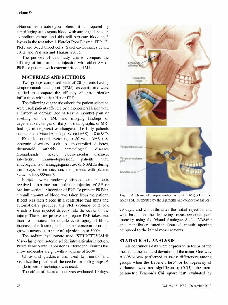
obtained from autologous blood: it is prepared bycentrifuging autologous blood with anticoagulant suchas sodium citrate, and this will separate blood in 3layers in the test tube: 1-Platelet Poor Plasma -PPP-, 2-PRP, and 3-red blood cells (Sanchez-Gonzalez et al.,2012, and Prakash and Thakur, 2011).
The purpose of this study was to compare theefficacy of intra-articular injection with either SH orPRP for patients with osteoarthritis of TMJ.
MATERIALS AND METHODSTwo groups composed each of 20 patients having
temporomandibular joint (TMJ) osteoarthritis werestudied to compare the efficacy of intra-articularinfiltration with either HA or PRP.
The following diagnostic criteria for patient selectionwere used: patients affected by a monolateral lesion witha history of chronic (for at least 4 months) pain orswelling of the TMJ and imaging findings ofdegenerative changes of the joint (radiographic or MRIfindings of degenerative changes). The forty patientsstudied had a Visual Analogue Score (VAS) of 8 to 9(17).
Exclusion criteria were: age > 80 years; VAS < 8;systemic disorders such as uncontrolled diabetes,rheumatoid arthritis, hematological diseases(coagulopathy), severe cardiovascular diseases,infections, immunodepression, patients withanticoagulants or antiaggregants, use of NSAIDs duringthe 5 days before injection, and patients with plateletvalues < 100,000/mm3.
Subjects were randomly divided, and patientsreceived either one intra-articular injection of SH orone intra-articular injection of PRP. To prepare PRP(14),a small amount of blood was taken from the patient.Blood was then placed in a centrifuge that spins andautomatically produces the PRP (volume of 2 cc),which is then injected directly into the center of theinjury. The entire process to prepare PRP takes lessthan 15 minutes. The double centrifuging of bloodincreased the histological platelets concentration andgrowth factors at the site of injection up to 500%.
The sodium hyaluronate used (STRUCTOVIAL®Viscoelastic and isotonic gel for intra-articular injection,Pierre Fabre Santé Laboratories, Boulogne, France) hasa low molecular weight with a volume of 2cc(16).
Ultrasound guidance was used to monitor andvisualize the position of the needle for both groups. Asingle injection technique was used.
The effect of the treatment was evaluated 10 days,
20 days, and 2 months after the initial injection andwas based on the following measurements: painintensity using the Visual Analogue Scale (VAS)(15)
and mandibular function (vertical mouth openingcompared to the initial measurement).
STATISTICAL ANALYSISAll continuous data were expressed in terms of the
mean and the standard deviation of the mean. One-wayANOVAa was performed to assess differences amonggroups when the Levene’s testb for homogeneity ofvariances was not significant (p<0.05); the non-parametric Pearson’s Chi square testc evaluated by
Fig. 1. Anatomy of tempomandibular joint (TMJ). (The discholds TMJ, supported by the ligaments and connective tissues).
Tohmé W
78 Volume 48 - Nº 2 - December 2013
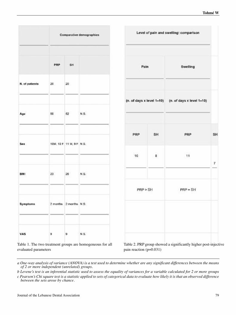
Table 2. PRP group showed a significantly higher post-injectivepain reaction (p=0.031)
Table 1. The two treatment groups are homogeneous for allevaluated parameters
Tohmé W
a One-way analysis of variance (ANOVA) is a test used to determine whether are any significant differences between the meansof 2 or more independent (unrelated) groups.
b Levene’s test is an inferential statistic used to assess the equality of variances for a variable calculated for 2 or more groups
c Pearson’s Chi square test is a statistic applied to sets of categorical data to evaluate how likely it is that an observed differencebetween the sets arose by chance.
79Journal of the Lebanese Dental Association
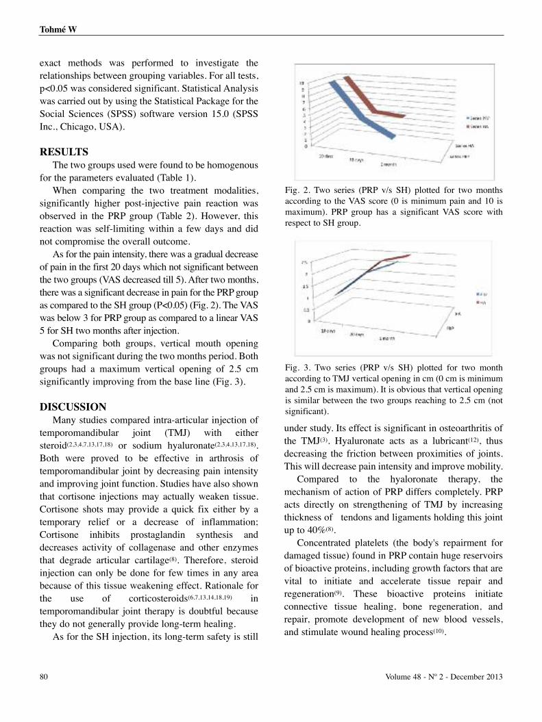
exact methods was performed to investigate therelationships between grouping variables. For all tests,p<0.05 was considered significant. Statistical Analysiswas carried out by using the Statistical Package for theSocial Sciences (SPSS) software version 15.0 (SPSSInc., Chicago, USA).
RESULTSThe two groups used were found to be homogenous
for the parameters evaluated (Table 1).When comparing the two treatment modalities,
significantly higher post-injective pain reaction wasobserved in the PRP group (Table 2). However, thisreaction was self-limiting within a few days and didnot compromise the overall outcome.
As for the pain intensity, there was a gradual decreaseof pain in the first 20 days which not significant betweenthe two groups (VAS decreased till 5). After two months,there was a significant decrease in pain for the PRP groupas compared to the SH group (P<0.05) (Fig. 2). The VASwas below 3 for PRP group as compared to a linear VAS5 for SH two months after injection.
Comparing both groups, vertical mouth openingwas not significant during the two months period. Bothgroups had a maximum vertical opening of 2.5 cmsignificantly improving from the base line (Fig. 3).
DISCUSSIONMany studies compared intra-articular injection of
temporomandibular joint (TMJ) with eithersteroid(2,3,4,7,13,17,18) or sodium hyaluronate(2,3,4,13,17,18).Both were proved to be effective in arthrosis oftemporomandibular joint by decreasing pain intensityand improving joint function. Studies have also shownthat cortisone injections may actually weaken tissue.Cortisone shots may provide a quick fix either by atemporary relief or a decrease of inflammation;Cortisone inhibits prostaglandin synthesis anddecreases activity of collagenase and other enzymesthat degrade articular cartilage(8). Therefore, steroidinjection can only be done for few times in any areabecause of this tissue weakening effect. Rationale forthe use of corticosteroids(6,7,13,14,18,19) intemporomandibular joint therapy is doubtful becausethey do not generally provide long-term healing.
As for the SH injection, its long-term safety is still
under study. Its effect is significant in osteoarthritis ofthe TMJ(3). Hyaluronate acts as a lubricant(12), thusdecreasing the friction between proximities of joints.This will decrease pain intensity and improve mobility.
Compared to the hyaloronate therapy, themechanism of action of PRP differs completely. PRPacts directly on strengthening of TMJ by increasingthickness of tendons and ligaments holding this jointup to 40%(8).
Concentrated platelets (the body's repairment fordamaged tissue) found in PRP contain huge reservoirsof bioactive proteins, including growth factors that arevital to initiate and accelerate tissue repair andregeneration(9). These bioactive proteins initiateconnective tissue healing, bone regeneration, andrepair, promote development of new blood vessels,and stimulate wound healing process(10).
Fig. 2. Two series (PRP v/s SH) plotted for two monthsaccording to the VAS score (0 is minimum pain and 10 ismaximum). PRP group has a significant VAS score withrespect to SH group.
Fig. 3. Two series (PRP v/s SH) plotted for two monthaccording to TMJ vertical opening in cm (0 cm is minimumand 2.5 cm is maximum). It is obvious that vertical openingis similar between the two groups reaching to 2.5 cm (notsignificant).
Tohmé W
80 Volume 48 - Nº 2 - December 2013

In this study, we compared the efficacy of the twoinjections (PRP v/s SH) in TMJ osteoarthritis, and thecompared results (Fig. 2) were effective regarding painscores and mandibular function, and according to ourresults, there was a great relation between the twogroups with respect to pain intensity for the first 20days; however, after a two month period, PRP grouphad an incremental increase in its regenerative effectand thus, an abrupt decrease in pain score with respectto SH group who has a steady linear effect and then anabrupt decrease. Gradual regenerative effects of PRPwill generally provide long-term healing and thus asignificant decrease in pain scores.
As for the mobility of the TMJ (Fig. 3), bothinjections had better results after 2 months. Thesimilarity between the two groups may probably be timedependent. Knowing the slow regenerative effect ofPRP may raise for us a four months period evaluationregarding the joint mobility the time needed for fullhistological activity.
Both groups did not show any complication duringthe 2 months period.
This study proved the efficacy and safety of clinicaluse of PRP in TMJ osteoarthritis.
CONCLUSIONIt is time to shift for a safer way to treat osteoarthritis
of TMJ? HA proved to be effective, but studies showedthat autologous PRP(19) may have an outstandingtherapeutic effect in treating TMJ osteoarthritis.
REFERENCES1. Akinbami BO. Evaluation of the mechanism and principles ofmanagement of temporomandibular joint dislocation. Systematicreview of literature and a proposed new classification oftemporomandibular joint dislocation. Head Face Med 2011 June15;7:102. Iannitti T, Lodi D, Palmieri B. Intra-Articular Injections for theTreatment of Osteoarthritis: focus on the clinical use of hyaluronicacid. Drugs RD. 2011;March 11(1):13-27.3. Ghosh P. Potential mechanism of action of intra-articularhyaluronan therapy in osteoarthritis: are the effects molecularweight dependent? Semin Arthritis Rheum 2002 Aug;32(1):10-37.4. Kotevoglu N, Lyibozkurt PC, Hiz O et al. A prospectiverandomised controlled clinical trial comparing the efficacy ofdifferent molecular weight hyaluronan solutions in the treatment ofknee osteoarthritis. Rheumatol Int 2006 Feb;26(4):325-330.5. Kon E, Mandelbaum B, Buda R et al. Platelet-rich plasma intra-articular injection versus hyaluronic acid viscosupplementation astreatments for cartilage pathology: from early degeneration toosteoarthritis. Arthroscopy 2011 Nov;27(11):1490-1501.6. Coombes BK, Bisset L, Vincenzino B. Efficacy and safety ofcorticosteroid injections and other injections for management oftendinopathy: a systematic review of randomised controlled trials.Lancet 2010 Nov 20;376(9754):1751-1767.7. Stoustrup P, Kristensen KD, Küseler A, et al. Reducedmandibular growth in experimental arthritis in thetemporomandibular joint treated with intra-articular corticosteroid.Europ J Orthod 2008 April;30(2):111-119.8. Yuan T, Zhang CQ, Wang JH. Augmenting tendon and ligamentrepair with platelet-rich plasma (PRP). Muscles ligaments tendons2013 Aug;3(3):139-149.9. Anitua E, Zalduendo MM, Alkhraisat MH, Orive G. Releasekinetics of platelet-derived and plasma-derived growth factorsfrom autologous plasma rich in growth factors. Ann Anat 2013Oct; 195(5):461-466.10. He L, Lin Y, Hu X et al. A comparative study of platelet-richfibrin (PRF) and platelet-rich plasma (PRP) on the effect ofproliferation and differentiation of rat osteoblasts in vitro. OralSurg Oral Med Oral Pathol Oral Radiol Endod. 2009 Nov;108(5):707-71311. Smith GN Jr, Mickler EA, Myers SL, Brandt KD. Effect ofintraarticular hyaluronan injection on synovial fluid hyaluronan inthe early stage of canine post-traumatic osteoarthritis. J Rheumatol2001 June;28(6):1341-6.12. Manfredini D, Guarda-Nardini L, Winocur E, et al. Researchdiagnostic criteria for temporomandibular disorders: a systematicreview of axis I epidemiologic findings. Oral Surg Oral Med OralPathol Oral Radiol Endod 2011 Oct;112(4):453-462.13. Møystad A, Mork-Knutsen BB, Biørland T. Injection of sodiumhyaluronate compared to a corticosteroid in the treatment ofpatients with temporomandibular joint osteoarthritis: a CTevaluation. Oral Surg Oral Med Oral Pathol Oral Radiol Endod2008 Feb;105(2):e53-60.
Tohmé W
81Journal of the Lebanese Dental Association
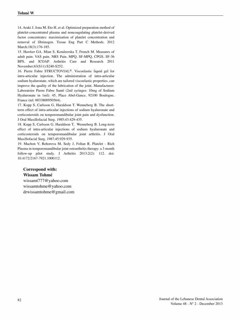
14. Araki J, Jona M, Eto H, et al. Optimized preparation method ofplatelet-concentrated plasma and noncoagulating platelet-derivedfactor concentrates: maximization of platelet concentration andremoval of fibrinogen. Tissue Eng Part C Methods. 2012March;18(3):176-185.15. Hawker GA, Mian S, Kendzerska T, French M. Measures ofadult pain: VAS pain, NRS Pain, MPQ, SF-MPQ, CPGS, SF-36BPS, and ICOAP. Arthritis Care and Research 2011November;63(S11):S240-S252.16. Pierre Fabre STRUCTOVIAL®. Viscoelastic liquid gel forintra-articular injection. The administration of intra-articularsodium hyaluronate, which are tailored viscoelastic properties, canimprove the quality of the lubrication of the joint. Manufacturer:Laboratoire Pierre Fabre Santé (2ml syringes: 10mg of SodiumHyaluronate in 1ml): 45, Place Abel-Gance, 92100 Boulogne,France (tel: 0033800950564).17. Kopp S, Carlsson G, Haraldson T, Wenneberg B. The short-term effect of intra-articular injections of sodium hyaluronate andcorticosteroids on temporomandibular joint pain and dysfunction.J Oral Maxillofacial Surg. 1985;43:429-435.18. Kopp S, Carlsson G, Haraldson T, Wenneberg B. Long-termeffect of intra-articular injections of sodium hyaluronate andcorticosteroids on temporomandibular joint arthritis. J OralMaxillofacial Surg. 1987;45:929-935.19. Machon V, Rehorova M, Sedy J, Foltan R. Platelet - RichPlasma in temporomandibular joint ostoarthritis therapy: a 3 monthfollow-up pilot study. J Arthritis 2013;2(2): 112. doi:10.4172/2167-7921.1000112.
Correspond with:Wissam Tohmé[email protected]@[email protected]
Tohmé W
82 Journal of the Lebanese Dental AssociationVolume 48 - Nº 2 - December 2013
Clinical Comment
INTRODUCTIONAngina pectoris, or angina for short, is the term
used when chest pain is thought to be attributable tomyocardial ischemia. In patients with myocardialischemia, chest pain is often, but not always, present.Myocardial ischemia can present itself with othersymptoms such as shortness of breath or the pain canbe located in other areas other than the chest.
PATHOPHYSIOLOGY OF MYOCARDIALISCHEMIA
Myocardial ischemia, and consequently angina,occur whenever myocardial oxygen demand exceedsoxygen supply.
MECHANISMS OF ANGINAThe mechanisms responsible for the sensation of
angina are complex and not entirely understood. Animportant feature is that myocardial ischemia reducesthe formation of adenosine triphosphate (ATP),resulting in a cascade of reactions leading to excretionof multiple chemical mediators. There is substantialevidence that primary mediator of angina is adenosine,via stimulation of the A1 adenosine receptor[2-5]. It isalso possible that venodilation, as a response toischemia, can activate these receptors. Nerve fiberstravel along sympathetic afferent pathways from heartand enter sympathetic ganglia in lower cervical andupper thoracic spinal cord (C7-T4). Impulses are thentransmitted via ascending spinothoracic pathways tomedial and lateral thalamus and ultimately activateseveral areas of cerebral cortex[1].
Angina is a discomfort that is referred to thecorresponding dermatomes that supply sympatheticafferent nerves to the same segments of the spinal cordas the heart (ie, C7-T4)[1]. Furthermore, stimulation of
sensory receptors in different myocardial regionsresults in the transmission via the same neuralpathway[5]. These characteristics account for twotypical features of angina: It is often a diffusediscomfort felt in the chest, neck, mandible, and downthe arm (typically the left); and most patientsexperience angina in the same distribution, regardlessof which area of the myocardium is ischemic[5].
CLINICAL FEATURES Initial presentation of myocardial ischemia with
angina may be one of a stable pattern or an acutecoronary syndrome/Myocardial infarction.
Most patients with myocardial ischemia willpresent with classic angina pectoris as the primaryclinical manifestation. However, in some patients,myocardial ischemia may be silent or atypical.• Quality
Angina is usually characterized more as adiscomfort rather than pain. Terms frequently used bypatients include squeezing, tightness, pressure,constriction, strangling, burning, heart burn, fullnessin the chest, band-like sensation, knot in the center ofthe chest, lump in throat, ache, heavy weight on chest(elephant sitting on chest), like a bra too tight, andtoothache (when there is radiation to mandible)[6]. Insome cases, patient cannot qualify the nature of thediscomfort, but places his or her hand fist in the centerof the chest, known as the “Levine’s sign1.”
It is generally not described as sharp, dull-aching,knife-like, stabbing, or pins and needles-like pain.• Location and radiation
As noted above, angina is a referred pain due toinvolvement of a neural reflex pathway via thoracicand cervical nerves. As a result, it is not felt in aspecific spot, but is usually a diffuse discomfort that
Craniofacial Pain of Cardiac Origin (Angina / Acute CoronarySyndrome / Myocardial Infarction): a useful diagnostic tool fordentists.Samar Khoury*, MD, FACC, Diplomate, American Board of Internal Medicine / American Board of Cardio-Vascular Diseases.
* Consultant, Cardiology Department, Genesis Health CareSystem, Good Samaritan Hospital, Zanesville, Ohio, USA
1. Levine’s sign is a clenched fist held over chest to expressischemic chest pain.
83Journal of the Lebanese Dental AssociationVolume 48 - Nº 2 - December 2013

may be difficult to localize.Angina is referred to the corresponding
dermatomes (C7-T4) that supply afferent nerves to thesame segments of the spinal cord as the heart. Thus,angina often radiates to other parts of the body,including upper abdomen (epigastric), shoulders, arms(upper and forearm), wrist, fingers, neck, throat,mandible and mandibular teeth* (but not maxilla), andrarely to the back (specifically the interscapularregion)[7,8]. Radiation to both arms is a strongerpredictor of acute myocardial infarction (AMI)**. Thelocation and radiation of angina are usually the sameeach time. Occasionally, location and radiation, but notquality, may be different after bypass surgery due todisruption of neural innervation of the heart.
In dental practice, it is important to be aware of anatypical presentation and location of angina,particularly when it presents as teeth or jaw pain. Earlyrecognition is crucial for appropriate diagnosis andearly treatment, and therefore better outcome/survival.I pulled out the following study[6] to support the above:
Craniofacial pain as the sole symptom of cardiacischemia: a prospective multicenter study. Kreiner M,
Okeson JP, Michelis V, Lujambio M, Isberg A. J Am
Dent Assoc. 2007;138(1):74-9.
“BACKGROUND: Craniofacial pain can be theonly symptom of cardiac ischemia. Failure torecognize its cardiac source can put the patient's life atrisk. Authors conducted a study to reveal theprevalence of the distribution of and sex differencesregarding craniofacial pain of cardiac origin.
METHODS: Authors prospectively selectedconsecutive patients (N = 186) who had had a verifiedcardiac ischemic episode. They studied location anddistribution of craniofacial and intraoral pain in detail.
RESULTS: Craniofacial pain was the onlycomplaint during the cardiac ischemic episode in 11patients (6 percent), three of them had AMI. Another60 patients (32 percent) reported craniofacial painconcomitant with pain in other regions. The mostcommon craniofacial pain locations were throat, leftmandible, right mandible, left temporomandibularjoint/ear region and teeth. Craniofacial pain was pre-ponderantly manifested in female subjects (P = .031)and was the dominating symptom in both sexes in theabsence of chest pain.
CONCLUSIONS: Craniofacial pain commonly isinduced by cardiac ischemia. This must be consideredin differential diagnosis of toothache and orofacialpain.
CLINICAL IMPLICATIONS: Because patientswho have AMI without chest pain run a higher risk ofexperiencing a missed diagnosis and death, thedentist's awareness of this symptomatology can becrucial for early diagnosis and timely treatment.
REFERRENCES:1. Foreman RD. Mechanisms of cardiac pain. Annu Rev Physiol1999; 61:143.
2. Sylvén C, Beermann B, Jonzon B, Brandt R. Angina pectoris-like pain provoked by intravenous adenosine in healthy volunteers.Br Med J (Clin Res Ed) 1986; 293:227.
3. Lagerqvist B, Sylvén C, Beermann B, et al. Intracoronaryadenosine causes angina pectoris like pain--an inquiry into thenature of visceral pain. Cardiovasc Res 1990; 24:609.
4. Gaspardone A, Crea F, Tomai F, et al. Muscular and cardiacadenosine-induced pain is mediated by A1 receptors. J Am CollCardiol 1995; 25:251.
5. Crea F, Gaspardone A, Kaski JC, et al. Relation betweenstimulation site of cardiac afferent nerves by adenosine anddistribution of cardiac pain: results of a study in patients withstable angina. J Am Coll Cardiol 1992; 20:1498.
6. Kreiner M, Okeson JP, Michelis V, et al. Craniofacial pain as thesole symptom of cardiac ischemia: a prospective multicenter study.J Am Dent Assoc 2007; 138:74-9.
7. Constant J. The clinical diagnosis of nonanginal chest pain: thedifferentiation of angina from nonanginal chest pain by history.Clin Cardiol 1983; 6:11.
8. Christie LG Jr, Conti CR. Systematic approach to evaluation ofangina-like chest pain: pathophysiology and clinical testing withemphasis on objective documentation of myocardial ischemia. AmHeart J 1981; 102:897.
Correspond with:Samar [email protected]
Khoury S
* toothache referred from heart disease.
** AMI or heart attack (sudden chest pain behind sternum,shortness of breath, sweating, nausea, vomiting, anxiety...).
84 Journal of the Lebanese Dental AssociationVolume 48 - Nº 2 - December 2013
Case Report
Glossopharyngeal Neuralgia -GPN- secondary to ipsilateralcompressive cerebellar mega-tonsil: a case report.
Joe Faddoul1, MD, Sandra Kobaïter Maarrawi1, B.Sc., M.Sc., Dr. Univ. (Neuroscience), Joe Abdel Hay1, MD,Elie Samaha1, MD, Joseph Maarrawi1,2, MD, Dr. Univ. (Neuroscience)
INTRODUCTIONGlossopharyngeal neuralgia (GPN) is a condition
in which there are repeated episodes of severe pain(lasting from few seconds to few minutes) in areasinnervated by the 9th cranial nerve (glosso-pharyngealnerve): nasopharynx, posterior one-third and base ofthe tongue, and tonsillar fossa. It is a pain syndromeclinically and pathophysiologically similar totrigeminal neuralgia (TN), but it is, by far, lesscommon. The treatment strategy is similar for bothentities: treatment initially consists of medicalmanagement with surgical options available forrefractory cases. Among accepted surgeries,microvascular decompression is generally an effectivetreatment for TN and GPN because it is a nonablativeprocedure that addresses the underlying etiology of aneuro-vascular conflict(1, 2). In contrast to TN, GPN ismost commonly caused by a secondary etiology, suchas infection or tumor(3).
CASE REPORTA 59-year-old woman reported a 3-year history of
left GPN characterized by paroxysmal pain attacks inleft pharyngeal region, originating from mandible,base of the tongue, and pharynx, and irradiating to leftear. These pain attacks, triggered by speech chewing,
first responded to Carbamazepine*, but were laterresistant to this drug. The patient reported no episodesof syncope and neurological examination was normal.Brain magnetic resonance imaging (MRI) displayed aleft cerebellar mega-tonsil (with a 3 mm herniationthrough foramen magnum) compressing brainstem andspinal cord (Figures 1 and 2). MRI failed to show aneurovascular conflict. Decompression of foramenmagnum, mainly on the left side, with enlargementduroplasty was performed. Arachnoids were keptintact and tonsillectomy was avoided as well (Fig. 3).
RESULTS Paroxysmal pain attacks disappeared immediately
after surgery. One year after surgical operation, patientwas totally free of symptoms. Control brain MRIdisplayed good decompression of occipito-cervicaljunction and absence of mass effect on brainstem(Figures 4 and 5).
DISCUSSIONGPN is an uncommon form of pain and represents
0.2 to 1.3% of the cases of facial pain(4). Incidence ofGPN is estimated to be 0.8/100.000 with a peak inpatients aged 70 to 79 years. Left side is mostlyaffected, with a bilateral occurence in 2% of cases.
1. Department of Neurosurgery, Hôtel-Dieu de FranceHospital / Faculty of Medicine, Saint-Joseph University,Beirut, Lebanon
2. Laboratory of Neurosciences / Faculty of Medicine,Saint-Joseph University, Beirut.
* Carbamazepine (Tegretol®)= an anticonvulsant and
mood-stabilizing drug used primarily in TN, epilepsy,
bipolar disorder, phantom limb syndrome, and complex
regional pain syndrome -CRPS-.
Abstract
Glossopharyngeal Neuralgia -GPN- is a pain syndrome caused by irritation of the ninth cranial nerve. It is clinically and
pathophysiologically comparable to trigeminal neuralgia -TN- with similar treatment options for both entities.
The clinical case we are addressing is a GPN secondary to an ipsilateral compressive cerebellar mega-tonsil, with no previous
reports of the same etiology in the medical literature. Decompression without tonsillectomy was sufficient for pain relief.
85Journal of the Lebanese Dental AssociationVolume 48 - Nº 2 - December 2013
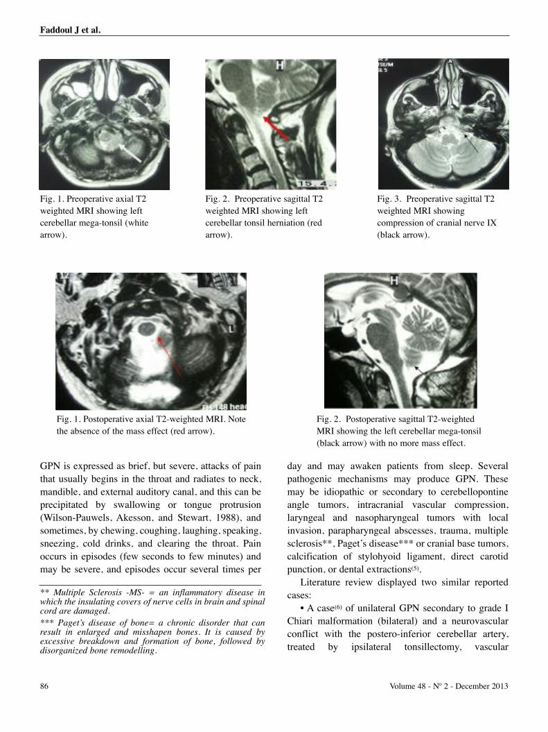
GPN is expressed as brief, but severe, attacks of painthat usually begins in the throat and radiates to neck,mandible, and external auditory canal, and this can beprecipitated by swallowing or tongue protrusion(Wilson-Pauwels, Akesson, and Stewart, 1988), andsometimes, by chewing, coughing, laughing, speaking,sneezing, cold drinks, and clearing the throat. Painoccurs in episodes (few seconds to few minutes) andmay be severe, and episodes occur several times per
day and may awaken patients from sleep. Severalpathogenic mechanisms may produce GPN. Thesemay be idiopathic or secondary to cerebellopontineangle tumors, intracranial vascular compression,laryngeal and nasopharyngeal tumors with localinvasion, parapharyngeal abscesses, trauma, multiplesclerosis**, Paget’s disease*** or cranial base tumors,calcification of stylohyoid ligament, direct carotidpunction, or dental extractions(5).
Literature review displayed two similar reportedcases:
• A case(6) of unilateral GPN secondary to grade IChiari malformation (bilateral) and a neurovascularconflict with the postero-inferior cerebellar artery,treated by ipsilateral tonsillectomy, vascular
Faddoul J et al.
! !
!
!
!
! !
Fig. 1. Preoperative axial T2weighted MRI showing leftcerebellar mega-tonsil (whitearrow).
! !
!
!
!
! !
Fig. 2. Preoperative sagittal T2weighted MRI showing leftcerebellar tonsil herniation (redarrow).
!
!
Fig. 1. Postoperative axial T2-weighted MRI. Notethe absence of the mass effect (red arrow).
!
!
Fig. 2. Postoperative sagittal T2-weightedMRI showing the left cerebellar mega-tonsil(black arrow) with no more mass effect.
Fig. 3. Preoperative sagittal T2weighted MRI showingcompression of cranial nerve IX(black arrow).
! !
!
!
!
! !
86 Volume 48 - Nº 2 - December 2013
** Multiple Sclerosis -MS- = an inflammatory disease inwhich the insulating covers of nerve cells in brain and spinalcord are damaged.
*** Paget’s disease of bone= a chronic disorder that canresult in enlarged and misshapen bones. It is caused byexcessive breakdown and formation of bone, followed bydisorganized bone remodelling.
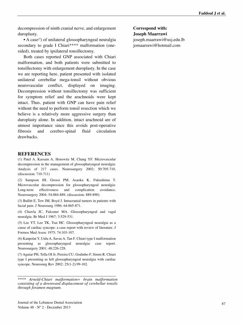
decompression of ninth cranial nerve, and enlargementduroplasty.
• A case(7) of unilateral glossopharyngeal neuralgiasecondary to grade I Chiari**** malformation (one-sided), treated by ipsilateral tonsillectomy.
Both cases reported GNP associated with Chiarimalformation, and both patients were submitted totonsillectomy with enlargement duroplasty. In the casewe are reporting here, patient presented with isolatedunilateral cerebellar mega-tonsil without obviousneurovascular conflict, displayed on imaging.Decompression without tonsillectomy was sufficientfor symptom relief and the arachnoids were keptintact. Thus, patient with GNP can have pain reliefwithout the need to perform tonsil resection which webelieve is a relatively more aggressive surgery thanduroplasty alone. In addition, intact arachnoid are ofutmost importance since this avoids post-operativefibrosis and cerebro-spinal fluid circulationdrawbacks.
REFERENCES(1) Patel A, Kassam A, Horowitz M, Chang YF. Microvasculardecompression in the management of glossopharyngeal neuralgia:Analysis of 217 cases. Neurosurgery 2002; 50:705-710,(discussion: 710-711)
(2) Sampson JH, Grossi PM, Asaoka K, Fukushima T.Microvascular decompression for glossopharyngeal neuralgia:Long-term effectiveness and complication avoidance.Neurosurgery 2004; 54:884-889, (discussion: 889-890).
(3) Bullitt E, Tew JM, Boyd J. Intracranial tumors in patients withfacial pain. J Neurosurg 1986; 64:865-871.
(4) Chawla JC, Falconer MA. Glossopharyngeal and vagalneuralgia. Br Med J 1967; 3:529-531.
(5) Lee YT, Lee TK, Tsai HC. Glossopharyngeal neuralgia as acause of cardiac syncope: a case report with review of literature. JFormos Med Assoc 1975; 74:103-107.
(6) Kanpolat Y, Unlu A, Savas A, Tan F. Chiari type I malformationpresenting as glossopharyngeal neuralgia: case report.Neurosurgery 2001; 48:226-228.
(7) Aguiar PH, Tella OI Jr, Pereira CU, Godinho F, Simm R. Chiaritype I presenting as left glossopharyngeal neuralgia with cardiacsyncope. Neurosurg Rev 2002; 25(1-2):99-102.
Correspond with:Joseph Maarrawi [email protected]@hotmail.com
Faddoul J et al.
**** Arnold-Chiari malformation= brain malformationconsisting of a downward displacement of cerebellar tonsilsthrough foramen magnum.
Journal of the Lebanese Dental AssociationVolume 48 - Nº 2 - December 2013
87

Review
Tooth pulp consists of a densely innervated andvascularized tissue surrounded by a hard mineralizedtissue. In normal healthy tooth, a hot or cold noxiousstimulus does not usually elicit pain because ofthermal insulating capacity (thermal diffusivity andconductivity) of the enamel. However under even mildinflammatory conditions, the low compliance of pulpchamber produces exaggerated pain.
Furthermore and unlike other tissues in humanbody, dentin responds by a painful sensation to anormal hot, cold, air-puff, or any type of stimulus1.This phenomenon suggests that teeth have a distinctivenociceptive mechanism by which they detect noxiousstimuli in inflammatory conditions or when dentin isexposed2.
Clinically, the type of dental pain should reflect thecondition of the tooth pulp which orientates the dentisttoward the treatment he should perform. Successfuldental treatment requires that the source of pain bedetected and understood. If the origin of pain is notfound, inappropriate dental care may result. Variabilityof pain experienced by patients presents a challenge interms of diagnostic methods. This review will helpclinicians to understand the basic neurophysiology andpathophysiology of pulpal pain in order to explain paincondition and to establish a correct diagnosis.
CLASSIFICATIONPulpal pain arises from vital or non-vital pulps.
Different attempts to classify pulpal pain according tothe histologic characteristics of the pulp have failed.The most popular classification is based on clinicalfeatures to determine the vitality of pulp and itsreparative capability3.
- Dentin sensitivity- Vital pulpal pain: reversible pulpitis- Vital pulpal pain: irreversible pulpitis- Non-vital pulpal pain
ETIOLOGY AND PATHOPHYSIOLOGYDental pain is the most common type of orofacial
pain(4) and the most common oral pain is due to toothpulp inflammation5. However, diagnosis of pain’sorigin is not always obvious. It could arise from non-dental sites and refer to adjacent teeth or in thecontrary originate from teeth and irradiate tosurrounding structures. The most appropriatetreatment is the removal of the cause rather thanaddressing symptoms. Pains of pulpal origin are eitherrelated to dentin hypersensitivity or to reversible orirreversible pulpitis.
Etiology of dentinal sensitivity is the activation ofnociceptors in dentin layer and exposure of dentinaltubules following enamel loss by mechanical erosionor attrition or by chemical agent such as acid erosion.Radicular dentin may be exposed as well by gingival
Dental pulp pain: diagnostic implications.
Abstract
Unlike other organs and tissues in human body, teeth display a distinctive and particular nociceptive mechanism. This has
lead researchers during the last three decades to further investigate neurophysiology and pathophysiology of dental pulp pain.
Clinically, the type of dental pain should reflect the condition of tooth pulp. Adequate and successful dental treatment
requires that the source of pain be detected and understood, however variability of pain experienced by patients may be
confusing for patients and misleading for dentists. This review addresses dental pain experienced by patients, it helps dental
clinicians to better understand dental pain conditions and establish an accurate diagnosis.
Khaldoun Rifai1, Dr. Chir. Dent., CES Oral Biol., CES Prostho., DSO, HDR, FICD, Georges Tehini2, Dr. Chir. Dent., CAGS, M.Sc., FICD
1 Professor, Department of Prosthodontics, LebaneseUniversity School of Dentistry, Beirut, Lebanon
2 Assistant Professor, Department of Prosthodontics,Lebanese University School of Dentistry, Beirut, Lebanon
Journal of the Lebanese Dental AssociationVolume 48 - Nº 2 - December 2013
88
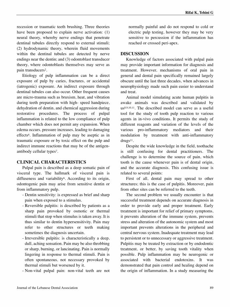
recession or traumatic teeth brushing. Three theorieshave been proposed to explain nerve activation: (1)neural theory, whereby nerve endings that penetratedentinal tubules directly respond to external stimuli;(2) hydrodynamic theory, wherein fluid movementswithin the dentinal tubules are detected by nerveendings near the dentin; and (3) odontoblast transducertheory, where odontoblasts themselves may serve aspain transducers2.
Etiology of pulp inflammation can be a directexposure of pulp by caries, fractures, or accidental(iatrogenic) exposure. An indirect exposure throughdentinal tubules can also occur. Other frequent causesare micro-trauma such as bruxism, heat, and vibrationduring teeth preparation with high- speed handpiece,dehydration of dentin, and chemical aggression duringrestorative procedures. The process of pulpalinflammation is related to the low compliance of pulpchamber which does not permit any expansion. Whenedema occurs, pressure increases, leading to damagingeffects6. Inflammation of pulp may be aseptic as intraumatic exposure or by toxic effect on the pulp andindirect immune reactions that may be of the antigen-antibody cellular types7.
CLINICAL CHARACTERISTICSPulpal pain is described as a deep somatic pain of
visceral type. The hallmark of visceral pain isdiffuseness and variability6. According to its origin,odontogenic pain may arise from sensitive dentin orfrom inflammatory pulp.
- Dentin sensitivity: is expressed as brief and sharppain when exposed to a stimulus.
- Reversible pulpitis: is described by patients as asharp pain provoked by osmotic or thermalstimuli that stop when stimulus is taken away. It isthus similar to dentin hypersensitivity. Pain mayrefer to other structures or teeth makingsometimes the diagnosis uncertain.
- Irreversible pulpitis: is characteristically a deep,dull, aching sensation. Pain may be also throbbingor sharp, burning, or lancinating. Pain is normallylingering in response to thermal stimuli. Pain isoften spontaneous, not necessary provoked bythermal stimuli but worsened by it.
- Non-vital pulpal pain: non-vital teeth are not
normally painful and do not respond to cold orelectric pulp testing, however they may be verysensitive to percussion if the inflammation hasreached or crossed peri-apex.
DISCUSSIONKnowledge of factors associated with pulpal pain
may provide important information for diagnosis andtreatment. However, mechanisms of oral pain ingeneral and dental pain specifically remained largelyobscure until the last three decades, when advances inneurophysiology made such pain easier to understandand treat.
Animal model simulating acute human pulpitis inawake animals was described and validated byus8,9,10,11. The described model can serve as a usefultool for the study of tooth pulp reaction to variousagents in in-vivo conditions. It permits the study ofdifferent reagents and variation of the levels of thevarious pro-inflammatory mediators and theirmodulation by treatment with anti-inflammatorydrugs11.
Despite the wide knowledge in the field, toothacheis still confusing for dental practitioners. Thechallenge is to determine the source of pain, whichtooth is the cause whenever pain is of dental origin,and the accurate diagnosis. This confusing issue isrelated to several points:
First of all, dental pain may spread to otherstructures; this is the case of pulpitis. Moreover, painfrom other sites can be referred to the teeth.
The second problem we usually encounter is thatsuccessful treatment depends on accurate diagnosis inorder to provide early and proper treatment. Earlytreatment is important for relief of primary symptoms,it prevents alteration of the immune system, preventsstress and alteration of the autonomic system and mostimportant prevents alterations in the peripheral andcentral nervous system. Inadequate treatment may leadto persistent or to unnecessary or aggressive treatment.Pulpitis may be treated by extraction or by endodontictreatment, or better, by saving tooth vitality whenpossible. Pulp inflammation may be neurogenic orassociated with bacterial endotoxins. It wasdemonstrated that pain control and healing depend onthe origin of inflammation. In a study measuring the
Rifai K, Tehini G
89Journal of the Lebanese Dental Association
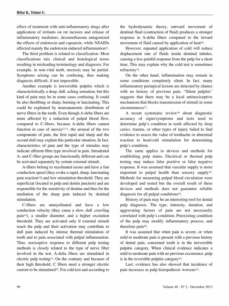
effect of treatment with anti-inflammatory drugs afterapplication of irritants on rat incisors and release ofinflammatory mediators, dexamethasone antagonizedthe effects of endotoxin and capsaicin, while NSAIDsaffected mainly the endotoxin-induced inflammation11.
The third problem is related to classification. Mostclassifications mix clinical and histological termsresulting in misleading terminology and diagnosis. Forexample, in non-vital teeth, necrosis may be partial.Symptoms arising can be confusing, thus makingdiagnosis difficult, if not impossible.
Another example is irreversible pulpitis which ischaracteristically a deep, dull, aching sensation, but thiskind of pain may be in some cases confusing. It couldbe also throbbing or sharp, burning or lancinating. Thiscould be explained by neuroanatomic distribution ofnerve fibers in the tooth. Even though A-delta fibers aremore affected by a reduction of pulpal blood flow,compared to C-fibers, because A-delta fibers cannotfunction in case of anoxia12-13, the arousal of the twocomponents of pain, the first rapid and sharp and thesecond dull may explain this particular situation. In fact,characteristics of pain and the type of stimulus mayindicate afferent fibre type involved in pain. IntradentalA- and C-fiber groups are functionally different and canbe activated separately by certain external stimuli.
A-fibers belong to myelinated axons and have a fastconduction speed (they evoke a rapid, sharp, lancinatingpain reaction14) and low stimulation threshold. They aresuperficial (located in pulp and dentin junction) and areresponsible for the sensitivity of dentine and thus for themediation of the sharp pain induced by dentinalstimulation.
C-fibers are unmyelinated and have a lowconduction velocity (they cause a slow, dull ,crawlingpain14), a smaller diameter, and a higher excitationthreshold. They are activated only if external stimulireach the pulp and their activation may contribute todull pain induced by intense thermal stimulation oftooth and to pain associated with pulpal inflammation.Thus, nociceptive response to different pulp testingmethods is closely related to the type of nerve fiberinvolved in the test. A-delta fibers are stimulated inelectric pulp testing14. On the contrary and because oftheir high threshold, C fibers need a stronger electriccurrent to be stimulated15. For cold test and according to
the hydrodynamic theory, outward movement ofdentinal fluid (contraction of fluid) produces a strongerresponse in A-delta fibers compared to the inwardmovement of fluid caused by application of heat16.
However, repeated application of cold will reducedisplacement rate of fluids inside dentinal tubules,causing a less painful response from the pulp for a shorttime. This may explain why the cold test is sometimesrefractory14.
On the other hand, inflammation may remain insome conditions completely silent. In fact, manyinflammatory periapical lesions are detected by chancewith no history of previous pain. “Silent pulpitis”suggests that there may be a local antinociceptivemechanism that blocks transmission of stimuli in somecircumstances17.
A recent systematic review18 about diagnosticaccuracy of signs/symptoms and tests used todetermine pulp’s condition in teeth affected by deepcaries, trauma, or other types of injury failed to findevidence to assess the value of toothache or abnormalreaction to heat/cold stimulation for determiningpulp’s condition.
The same applies to devices and methods forestablishing pulp status. Electrical or thermal pulptesting may induce false positive or false negativeresponse. It was assumed that vascular supply is moreimportant to pulpal health than sensory supply19.Methods for measuring pulpal blood circulation weredeveloped and tested but the overall result of thesedevices and methods does not guarantee reliablediagnosis for all pulpal conditions18.
History of pain may be an interesting tool for dentalpulp diagnosis. The type, intensity, duration, andaggravating factors of pain are not necessarilycorrelated with pulp’s condition. Preexisting conditionof the pulp may modify inflammatory process, andtherefore pain20.
It was assumed that when pain is severe, or whenmild to moderate pain is present with a previous historyof dental pain, concerned tooth is in the irreversiblepulpitis category. When clinical evidence indicates amild to moderate pain with no previous occurrence, pulpis in the reversible pulpitis category14.
Last, evidence has also showed that incidence ofpain increases as pulp histopathosis worsens14.
Rifai K, Tehini G
90 Volume 48 - Nº 2 - December 2013
CONCLUSIONDental pulp pain is a serious concern to both patient
and dentist. Variability of clinical characteristicsassociated with pulpal pain remains a challenge interms of diagnostic methods. Dental pain is a helpfultool to diagnose pulp condition or in contraryconfusing and thus may mislead the dentist in his/herdiagnostic search. Despite the huge amount ofpublications related to neurophysiology of tooth andpathophysiology of pain during the last three decades,several issues remain unsolved. According toBender14, 80% of patients with previous history ofpain, display histopathologic evidence of chronicpartial pulpitis with partial necrosis (endodontic orextraction indicated) and the other 20% manifest pulphistopathosis with slight inflammation to chronicpartial pulpitis without necrosis (a treatable category).
This review aims to provide dentists with the basicclinical knowledge of pulpal pain and discuss someconfusing clinical situations encountered during theirdaily practice. Further studies are warranted to furtherunderstand pulpal pain in order to implement newstrategies and methods of dental pulp diagnosis.
REFERENCES1- Cook SP, Vulchanova L, Hargreaves KM, Elde R, McCleskeyEW. Distincts ATP receptors on pain-sensing and sensing neurons.Nature 1997;387: 505-508.
2- Chung G, Jung SJ, Oh SB. Cellular and molecular mechanismsof dental nociception. J Dent Res 2013;92(11): 948-955.
3- Cohen S. Diagnostic procedures. In Cohen SC, Burns RC (eds.).Pathways of the pulp, 5th ed. St Louis: Mosby, 1990:20.
4- Lipton JA, Ship JA, Larach-Robinson D. Estimatedprevalence and distribution of reported orofacial pain in theUnited States. J Am Dent Assoc 1993;124:115–121.
5- Estrela C, Guedes OA, Silva JA, Leles CR, Estrela CR, PécoraJD. Diagnostic and clinical factors associated with pulpal andperiapical pain. Braz Dent J 2011;22:306–311.
6- Falace DE, Cailletau JG. The diagnosis of dental and orofacialpain. In Falace DE. Emergency dental care: Diagnosis nadManagement of Urgent Dental Problems. Baltimore: Williams andWilkins. 1995:3-4.
7- Naidorf I. Correlation of the of the inflammatory response withimmunological and clinical events. J Endod 1977;3:223.
8- Chidiac JJ, Rifai K, Hawwa NN, Massaad CA, Jurjus AR,Jabbur SJ, Saadé NE. Nociceptive behaviour induced by dentalapplication of irritants to rat incisors: a new model for toothinflammatory pain. Eur J Pain 2002;6(1):55-67.
9- Chidiac JJ, Hawwa N, Baliki M, Safieh-Garabedian B, Rifai K,Jabbur SJ, Saadé NE.A perfusion technique for the determinationof pro-inflammatory mediators induced by intradental applicationof irritants. J Pharmacol Toxicol Methods 2001 Nov-Dec;46(3):125-30.
10- Rifai K, Chidiac JJ, Hawwa N, Baliki M, Jabbur SJ, SaadéNE.Occlusion of dentinal tubules and selective block of pulpinnervation prevent the nociceptive behaviour induced in rats byintradental application of irritants. Arch Oral Biol 2004Jun;49(6):457-68.
11- Chidiac JJ, Al-Asmar B, Rifai K, Jabbur SJ, SaadéNE.Inflammatory mediators released following application ofirritants on the rat injured incisors. The effect of treatment withanti-inflammatory drugs. Cytokine 2009 May;46(2):194-200.
12- Torebjörk HE, Hallin RG. Perceptual changes accompanyingcontrolled preferential blocking of A and C fibers responses inintact human skin nerves. Exp Brain Res 1973;16:321–332.
13- Närhi MV. The characteristics of intradental sensory units andtheir responses to stimulation. J Dent Res 1985;64(SpecNo):564–571.
14- Bender IB. Pulpal pain diagnosis--a review. J Endod 2000Mar;26(3):175-9.
15- Närhi M, Virtanen A, Kuhta J, Huopaniemi T. Electricalstimulation of teeth with a pulp tester in the cat. Scand J Dent Res1979;87:32–38.
16- Närhi MV, Hirvonen TJ, Hakumäki MO. Responses ofintradental nerve fibres to stimulation of dentine and pulp. ActaPhysiol Scand 1982;115:173–178.
17- Holland GR. Management of dental pain. In Orofacial pain.From Basic Science to Clinical Management. Ed Lund JP, LavigneGJ, Dubner R and Sessle BJ. Quintessence Publishing Co, Inc.2001: 211-220.
18- Mejàre IA, Axelsson S, Davidson T, Frisk F, Hakeberg M,Kvist T, Norlund A, Petersson A, Portenier I, Sandberg H, TranaeusS, Bergenholtz G. Diagnosis of the condition of the dental pulp: asystematic review. Int Endod J 2012 Jul;45(7):597-613.2012.
19- Abd-Elmeguid A, Yu DC. Dental pulp neurophysiology: part 2.Current diagnostic tests to assess pulp vitality. J Can Dent Assoc2009 Mar;75:(2): 139-43.
20- Okesson JP. Differential diagnosis and managementconsiderations of intraoral pain disorders. In Okesson JP. Orofacialpain. Guidelines for assessment, diagnosis and management.Quintessence publishing Co, Inc, 1996:92.
Correspond with:Khaldoun [email protected]
Rifai K, Tehini G
Journal of the Lebanese Dental AssociationVolume 48 - Nº 2 - December 2013
91
Review
1. INTRODUCTION General dentist and dental specialist treat patients
with pain on a daily basis. Orofacial pain (perceived inthe face and/or oral cavity) is caused by diseases ordisorders of regional structures, dysfunction of thenervous system, or through referral from distantsources.
In addition to the diagnosis and treatment of acutedental pain and pathology, such as that which mayarise from trauma, infection, orofacial pain dentist hasthe responsibility to diagnose and treatnonodontogenic orofacial pain. Inadequate knowledgeof etiopathology of pain and neurobiologicalmechanisms underlying persistent pain can lead toinaccurate diagnoses and subsequent ineffective orharmful treatment. It’s also the responsibility oforofacial pain dentist to assess the need formultidisciplinary pain management.
Complexity of the spectrum of orofacial paindisorders is compounded by close proximity of
numerous anatomical structures, including eyes, nose,teeth, tongue, sinuses, ears, and temporo-mandibularjoints. Diagnostic approach categorizes orofacial paininto four groups based on underlying painmechanisms: musculoskeletal, neuropathic,neurovascular, and psychogenic pain.
2- CLASSIFICATION OF OROFACIAL PAINS There were many attempts to classify orofacial pain,
and the most updated classification was established bythe American Academy of Orofacial Pain and it wasbased on the IHS* classification (Table 1).
Even though it can occur in different anatomicallocations, management of chronic orofacial pain isusually applied in multidisciplinary pain clinicsregardless of the origin of pain. Interdisciplinarytherapies includes: education, counseling, medications,
Practical pharmacological approach of orofacial pains: realitiesand clinical recommendations.
Wissam Tohmé1, MD, DU (Pain Management), Joelle Saroufim2, MD
1. Assistant Professor of Anesthesia and Pain Management,University of Balamand, Faculty of Medicine, Beirut,Lebanon, and Chairperson, Department of Anesthesiaand Pain Management, Saint-George Hospital UniversityMedical Center (SGHUMC), Ashrafieh, Beirut.
2.Anesthesia 3rd year Resident, University of Balamand,Department of Anesthesia, Saint-George Hospital(SGHUMC), Ashrafieh, Beirut.,
* IHS= International Headache Society.
Table 1. Differential Diagnosis of Orofacial Pain
Abstract
An analgesic (or pain killer) is a drug used to achieve analgesia (relief from pain): this drug can work either on peripheral
or central nervous system. Analgesics are different from anesthetics which reversibly eliminate pain sensation.
When managing orofacial pains, analgesic choice is determined by the type of pain and its severity. The WHO “Pain
Ladder” (or analgesic ladder) is a 3-step “ladder” for cancer pain in adults. It described the guidelines for the use of drugs in
pain management: the general principle is to start with first step drugs(non-opioid), and then, to climb the ladder if pain is still
present, and medications usually range from household (over-the-counter) drugs (acetaminophen and NSAIDs), to powerful
opioids (morphine, buprenorphine, oxycodone).
For neuropathic orofacial pains, tricyclic antidepressants, class I antiarrythmics, and anticonvulsants remain the drugs of
choice.
92 Journal of the Lebanese Dental AssociationVolume 48 - Nº 2 - December 2013
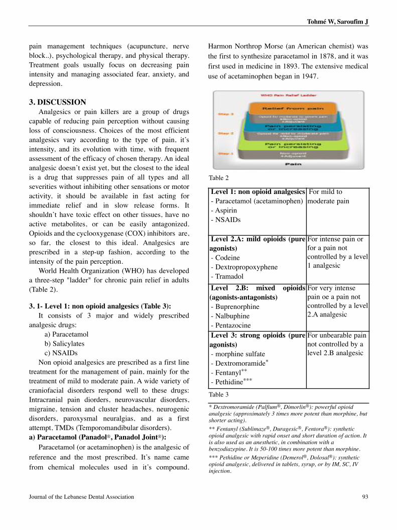
Journal of the Lebanese Dental Association 93
pain management techniques (acupuncture, nerveblock..), psychological therapy, and physical therapy.Treatment goals usually focus on decreasing painintensity and managing associated fear, anxiety, anddepression.
3. DISCUSSIONAnalgesics or pain killers are a group of drugs
capable of reducing pain perception without causingloss of consciousness. Choices of the most efficientanalgesics vary according to the type of pain, it’sintensity, and its evolution with time, with frequentassessment of the efficacy of chosen therapy. An idealanalgesic doesn’t exist yet, but the closest to the idealis a drug that suppresses pain of all types and allseverities without inhibiting other sensations or motoractivity, it should be available in fast acting forimmediate relief and in slow release forms. Itshouldn’t have toxic effect on other tissues, have noactive metabolites, or can be easily antagonized.Opioids and the cyclooxygenase (COX) inhibitors are,so far, the closest to this ideal. Analgesics areprescribed in a step-up fashion, according to theintensity of the pain perception.
World Health Organization (WHO) has developeda three-step "ladder" for chronic pain relief in adults(Table 2).
3. 1- Level 1: non opioid analgesics (Table 3): It consists of 3 major and widely prescribed
analgesic drugs:a) Paracetamolb) Salicylatesc) NSAIDs
Non opioid analgesics are prescribed as a first linetreatment for the management of pain, mainly for thetreatment of mild to moderate pain. A wide variety ofcraniofacial disorders respond well to these drugs:Intracranial pain diorders, neurovascular disorders,migraine, tension and cluster headaches, neurogenicdisorders, paroxysmal neuralgias, and as a firstattempt, TMDs (Temporomandibular disorders).a) Paracetamol (Panadol®, Panadol Joint®):
Paracetamol (or acetaminophen) is the analgesic ofreference and the most prescribed. It’s name camefrom chemical molecules used in it’s compound.
Harmon Northrop Morse (an American chemist) wasthe first to synthesize paracetamol in 1878, and it wasfirst used in medicine in 1893. The extensive medicaluse of acetaminophen began in 1947.
Tohmé W, Saroufim J
Level 1: non opioid analgesics- Paracetamol (acetaminophen)- Aspirin- NSAIDs
For mild tomoderate pain
Level 2.A: mild opioids (pureagonists)- Codeine- Dextropropoxyphene- Tramadol
For intense pain orfor a pain notcontrolled by a level1 analgesic
Level 2.B: mixed opioids(agonists-antagonists)- Buprenorphine- Nalbuphine- Pentazocine
For very intensepain oe a pain notcontrolled by a level2.A analgesic
Level 3: strong opioids (pureagonists)- morphine sulfate- Dextromoramide*
- Fentanyl**
- Pethidine***
For unbearable painnot controlled by alevel 2.B analgesic
* Dextromoramide (Palfium®, Dimorlin®): powerful opioidanalgesic (approximately 3 times more potent than morphine, butshorter acting).
** Fentanyl (Sublimaze®, Duragesic®, Fentora®): syntheticopioid analgesic with rapid onset and short duration of action. Itis also used as an anesthetic, in combination with abenzodiazepine. It is 50-100 times more potent than morphine.
*** Pethidine or Meperidine (Demerol®, Dolosal®): syntheticopioid analgesic, delivered in tablets, syrup, or by IM, SC, IVinjection.
Table 2
Table 3
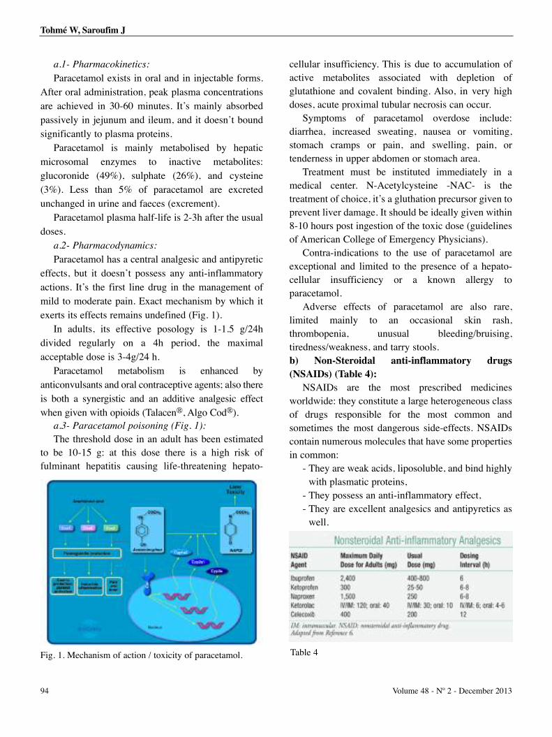
a.1- Pharmacokinetics:
Paracetamol exists in oral and in injectable forms.After oral administration, peak plasma concentrationsare achieved in 30-60 minutes. It’s mainly absorbedpassively in jejunum and ileum, and it doesn’t boundsignificantly to plasma proteins.
Paracetamol is mainly metabolised by hepaticmicrosomal enzymes to inactive metabolites:glucoronide (49%), sulphate (26%), and cysteine(3%). Less than 5% of paracetamol are excretedunchanged in urine and faeces (excrement).
Paracetamol plasma half-life is 2-3h after the usualdoses.
a.2- Pharmacodynamics:
Paracetamol has a central analgesic and antipyreticeffects, but it doesn’t possess any anti-inflammatoryactions. It’s the first line drug in the management ofmild to moderate pain. Exact mechanism by which itexerts its effects remains undefined (Fig. 1).
In adults, its effective posology is 1-1.5 g/24hdivided regularly on a 4h period, the maximalacceptable dose is 3-4g/24 h.
Paracetamol metabolism is enhanced byanticonvulsants and oral contraceptive agents; also thereis both a synergistic and an additive analgesic effectwhen given with opioids (Talacen®, Algo Cod®).
a.3- Paracetamol poisoning (Fig. 1):
The threshold dose in an adult has been estimatedto be 10-15 g: at this dose there is a high risk offulminant hepatitis causing life-threatening hepato-
cellular insufficiency. This is due to accumulation ofactive metabolites associated with depletion ofglutathione and covalent binding. Also, in very highdoses, acute proximal tubular necrosis can occur.
Symptoms of paracetamol overdose include:diarrhea, increased sweating, nausea or vomiting,stomach cramps or pain, and swelling, pain, ortenderness in upper abdomen or stomach area.
Treatment must be instituted immediately in amedical center. N-Acetylcysteine -NAC- is thetreatment of choice, it’s a gluthation precursor given toprevent liver damage. It should be ideally given within8-10 hours post ingestion of the toxic dose (guidelinesof American College of Emergency Physicians).
Contra-indications to the use of paracetamol areexceptional and limited to the presence of a hepato-cellular insufficiency or a known allergy toparacetamol.
Adverse effects of paracetamol are also rare,limited mainly to an occasional skin rash,thrombopenia, unusual bleeding/bruising,tiredness/weakness, and tarry stools.b) Non-Steroidal anti-inflammatory drugs(NSAIDs) (Table 4):
NSAIDs are the most prescribed medicinesworldwide: they constitute a large heterogeneous classof drugs responsible for the most common andsometimes the most dangerous side-effects. NSAIDscontain numerous molecules that have some propertiesin common:
- They are weak acids, liposoluble, and bind highlywith plasmatic proteins,
- They possess an anti-inflammatory effect, - They are excellent analgesics and antipyretics as
well.
94 Volume 48 - Nº 2 - December 2013
Tohmé W, Saroufim J
Fig. 1. Mechanism of action / toxicity of paracetamol. Table 4
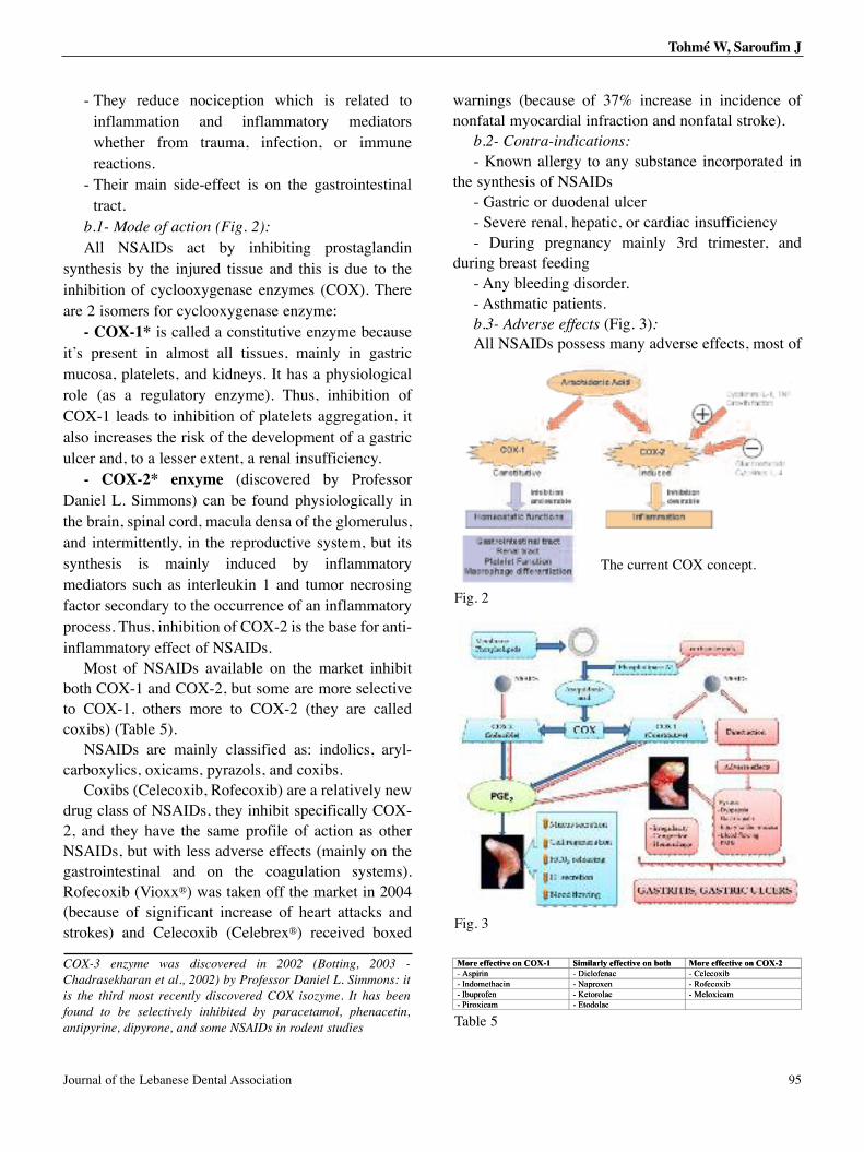
Journal of the Lebanese Dental Association 95
- They reduce nociception which is related toinflammation and inflammatory mediatorswhether from trauma, infection, or immunereactions.
- Their main side-effect is on the gastrointestinaltract.
b.1- Mode of action (Fig. 2):
All NSAIDs act by inhibiting prostaglandinsynthesis by the injured tissue and this is due to theinhibition of cyclooxygenase enzymes (COX). Thereare 2 isomers for cyclooxygenase enzyme:
- COX-1* is called a constitutive enzyme becauseit’s present in almost all tissues, mainly in gastricmucosa, platelets, and kidneys. It has a physiologicalrole (as a regulatory enzyme). Thus, inhibition ofCOX-1 leads to inhibition of platelets aggregation, italso increases the risk of the development of a gastriculcer and, to a lesser extent, a renal insufficiency.
- COX-2* enxyme (discovered by ProfessorDaniel L. Simmons) can be found physiologically inthe brain, spinal cord, macula densa of the glomerulus,and intermittently, in the reproductive system, but itssynthesis is mainly induced by inflammatorymediators such as interleukin 1 and tumor necrosingfactor secondary to the occurrence of an inflammatoryprocess. Thus, inhibition of COX-2 is the base for anti-inflammatory effect of NSAIDs.
Most of NSAIDs available on the market inhibitboth COX-1 and COX-2, but some are more selectiveto COX-1, others more to COX-2 (they are calledcoxibs) (Table 5).
NSAIDs are mainly classified as: indolics, aryl-carboxylics, oxicams, pyrazols, and coxibs.
Coxibs (Celecoxib, Rofecoxib) are a relatively newdrug class of NSAIDs, they inhibit specifically COX-2, and they have the same profile of action as otherNSAIDs, but with less adverse effects (mainly on thegastrointestinal and on the coagulation systems).Rofecoxib (Vioxx®) was taken off the market in 2004(because of significant increase of heart attacks andstrokes) and Celecoxib (Celebrex®) received boxed
warnings (because of 37% increase in incidence ofnonfatal myocardial infraction and nonfatal stroke).
b.2- Contra-indications:
- Known allergy to any substance incorporated inthe synthesis of NSAIDs
- Gastric or duodenal ulcer- Severe renal, hepatic, or cardiac insufficiency- During pregnancy mainly 3rd trimester, and
during breast feeding- Any bleeding disorder.- Asthmatic patients.b.3- Adverse effects (Fig. 3):All NSAIDs possess many adverse effects, most of
More effective on COX-1 Similarly effective on both More effective on COX-2 - Aspirin - Diclofenac - Celecoxib - Indomethacin - Naproxen - Rofecoxib - Ibuprofen - Ketorolac - Meloxicam - Piroxicam - Etodolac
cffe eeorM- nripis A- hte Indom- ne Ibuprof fe- roxii P cam
XO C onvetic -1 miS- D
nicha - Nn - Km - E
oth b onveticffey elarlmicnaeoffelci D
proa N xencaorolte K
caodolt
C onveticffe eeorM- boxicel Ce- boxiceRoffe- macoxile M
XO -2
Fig. 2
Table 5
Tohmé W, Saroufim J
The current COX concept.
Fig. 3
COX-3 enzyme was discovered in 2002 (Botting, 2003 -
Chadrasekharan et al., 2002) by Professor Daniel L. Simmons: it
is the third most recently discovered COX isozyme. It has been
found to be selectively inhibited by paracetamol, phenacetin,
antipyrine, dipyrone, and some NSAIDs in rodent studies
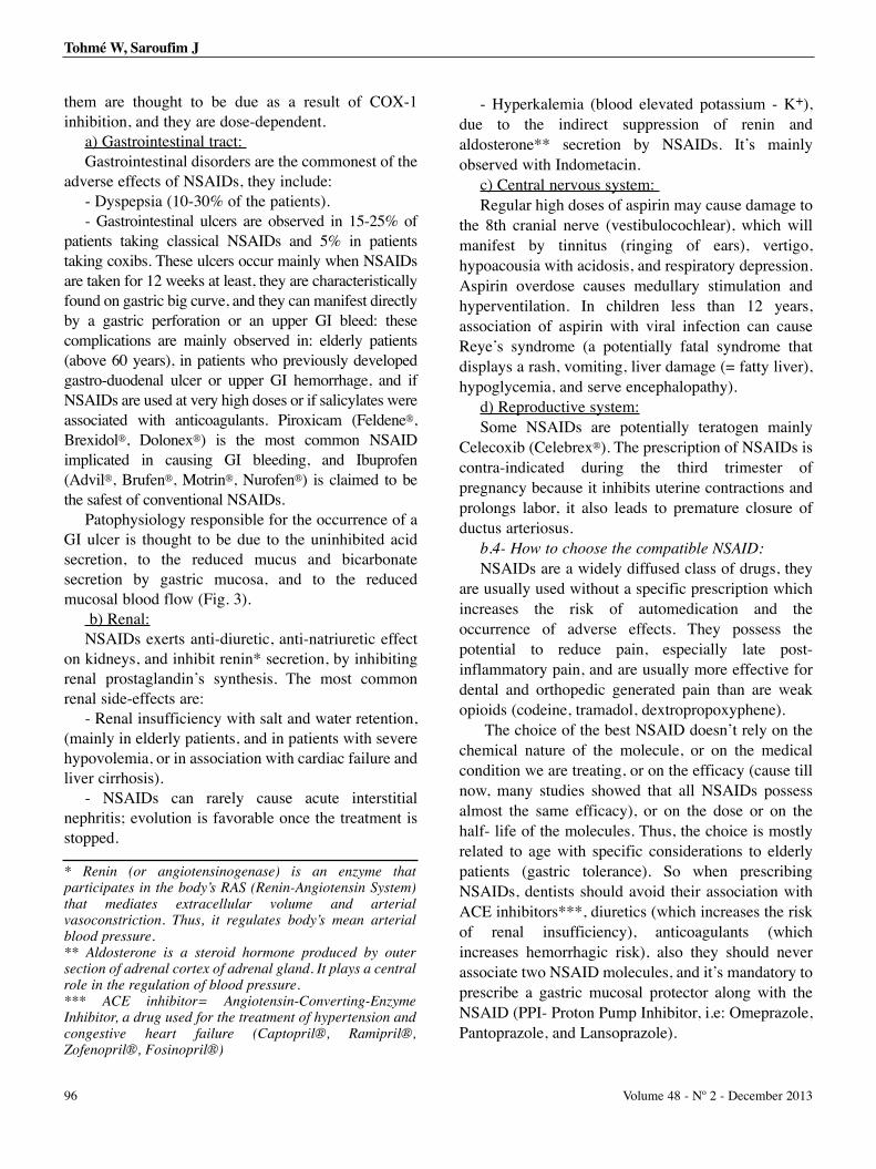
them are thought to be due as a result of COX-1inhibition, and they are dose-dependent.
a) Gastrointestinal tract: Gastrointestinal disorders are the commonest of the
adverse effects of NSAIDs, they include:- Dyspepsia (10-30% of the patients).- Gastrointestinal ulcers are observed in 15-25% of
patients taking classical NSAIDs and 5% in patientstaking coxibs. These ulcers occur mainly when NSAIDsare taken for 12 weeks at least, they are characteristicallyfound on gastric big curve, and they can manifest directlyby a gastric perforation or an upper GI bleed: thesecomplications are mainly observed in: elderly patients(above 60 years), in patients who previously developedgastro-duodenal ulcer or upper GI hemorrhage, and ifNSAIDs are used at very high doses or if salicylates wereassociated with anticoagulants. Piroxicam (Feldene®,Brexidol®, Dolonex®) is the most common NSAIDimplicated in causing GI bleeding, and Ibuprofen(Advil®, Brufen®, Motrin®, Nurofen®) is claimed to bethe safest of conventional NSAIDs.
Patophysiology responsible for the occurrence of aGI ulcer is thought to be due to the uninhibited acidsecretion, to the reduced mucus and bicarbonatesecretion by gastric mucosa, and to the reducedmucosal blood flow (Fig. 3).
b) Renal:NSAIDs exerts anti-diuretic, anti-natriuretic effect
on kidneys, and inhibit renin* secretion, by inhibitingrenal prostaglandin’s synthesis. The most commonrenal side-effects are:
- Renal insufficiency with salt and water retention,(mainly in elderly patients, and in patients with severehypovolemia, or in association with cardiac failure andliver cirrhosis).
- NSAIDs can rarely cause acute interstitialnephritis; evolution is favorable once the treatment isstopped.
- Hyperkalemia (blood elevated potassium - K+),due to the indirect suppression of renin andaldosterone** secretion by NSAIDs. It’s mainlyobserved with Indometacin.
c) Central nervous system: Regular high doses of aspirin may cause damage to
the 8th cranial nerve (vestibulocochlear), which willmanifest by tinnitus (ringing of ears), vertigo,hypoacousia with acidosis, and respiratory depression.Aspirin overdose causes medullary stimulation andhyperventilation. In children less than 12 years,association of aspirin with viral infection can causeReye’s syndrome (a potentially fatal syndrome thatdisplays a rash, vomiting, liver damage (= fatty liver),hypoglycemia, and serve encephalopathy).
d) Reproductive system:Some NSAIDs are potentially teratogen mainly
Celecoxib (Celebrex®). The prescription of NSAIDs iscontra-indicated during the third trimester ofpregnancy because it inhibits uterine contractions andprolongs labor, it also leads to premature closure ofductus arteriosus.
b.4- How to choose the compatible NSAID:
NSAIDs are a widely diffused class of drugs, theyare usually used without a specific prescription whichincreases the risk of automedication and theoccurrence of adverse effects. They possess thepotential to reduce pain, especially late post-inflammatory pain, and are usually more effective fordental and orthopedic generated pain than are weakopioids (codeine, tramadol, dextropropoxyphene).
The choice of the best NSAID doesn’t rely on thechemical nature of the molecule, or on the medicalcondition we are treating, or on the efficacy (cause tillnow, many studies showed that all NSAIDs possessalmost the same efficacy), or on the dose or on thehalf- life of the molecules. Thus, the choice is mostlyrelated to age with specific considerations to elderlypatients (gastric tolerance). So when prescribingNSAIDs, dentists should avoid their association withACE inhibitors***, diuretics (which increases the riskof renal insufficiency), anticoagulants (whichincreases hemorrhagic risk), also they should neverassociate two NSAID molecules, and it’s mandatory toprescribe a gastric mucosal protector along with theNSAID (PPI- Proton Pump Inhibitor, i.e: Omeprazole,Pantoprazole, and Lansoprazole).
Tohmé W, Saroufim J
96 Volume 48 - Nº 2 - December 2013
* Renin (or angiotensinogenase) is an enzyme thatparticipates in the body’s RAS (Renin-Angiotensin System)that mediates extracellular volume and arterialvasoconstriction. Thus, it regulates body’s mean arterialblood pressure.** Aldosterone is a steroid hormone produced by outersection of adrenal cortex of adrenal gland. It plays a centralrole in the regulation of blood pressure.*** ACE inhibitor= Angiotensin-Converting-EnzymeInhibitor, a drug used for the treatment of hypertension andcongestive heart failure (Captopril®, Ramipril®,Zofenopril®, Fosinopril®)
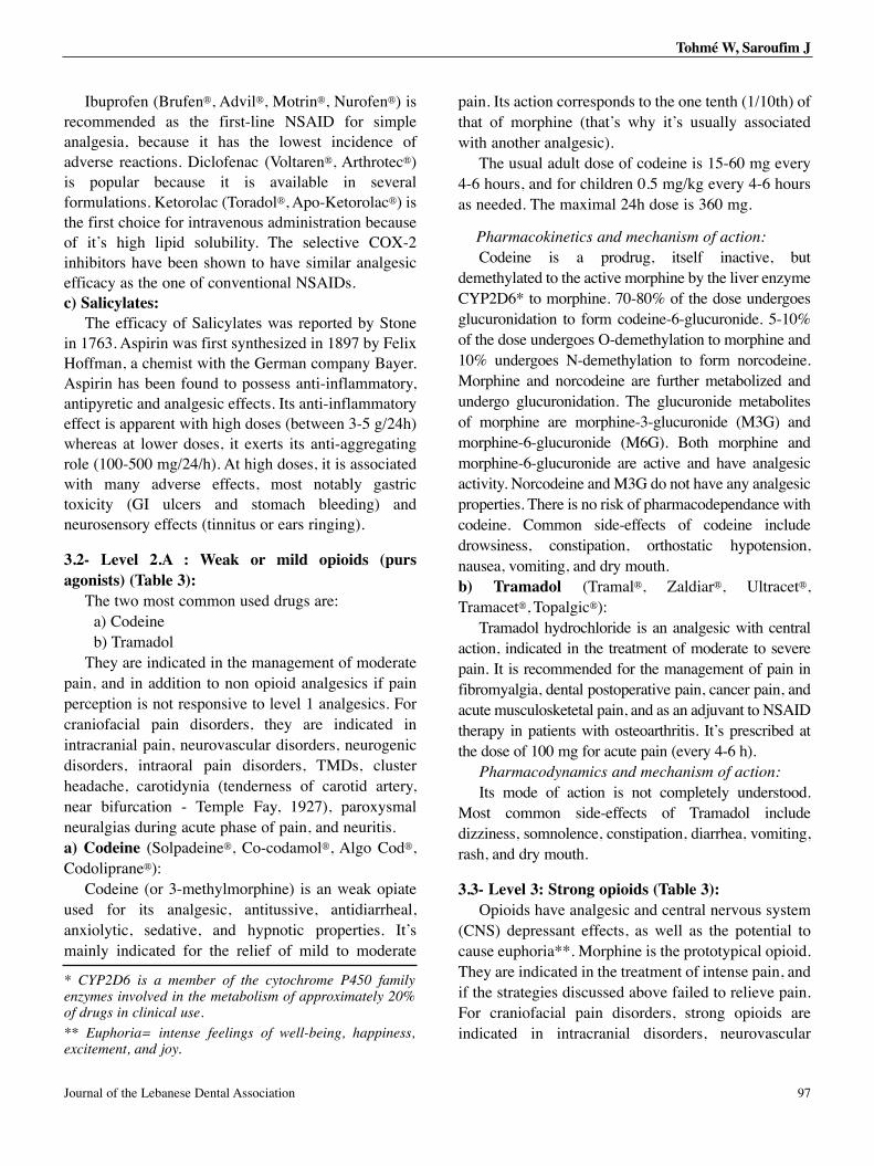
Ibuprofen (Brufen®, Advil®, Motrin®, Nurofen®) isrecommended as the first-line NSAID for simpleanalgesia, because it has the lowest incidence ofadverse reactions. Diclofenac (Voltaren®, Arthrotec®)is popular because it is available in severalformulations. Ketorolac (Toradol®, Apo-Ketorolac®) isthe first choice for intravenous administration becauseof it’s high lipid solubility. The selective COX-2inhibitors have been shown to have similar analgesicefficacy as the one of conventional NSAIDs.c) Salicylates:
The efficacy of Salicylates was reported by Stonein 1763. Aspirin was first synthesized in 1897 by FelixHoffman, a chemist with the German company Bayer.Aspirin has been found to possess anti-inflammatory,antipyretic and analgesic effects. Its anti-inflammatoryeffect is apparent with high doses (between 3-5 g/24h)whereas at lower doses, it exerts its anti-aggregatingrole (100-500 mg/24/h). At high doses, it is associatedwith many adverse effects, most notably gastrictoxicity (GI ulcers and stomach bleeding) andneurosensory effects (tinnitus or ears ringing).
3.2- Level 2.A : Weak or mild opioids (pursagonists) (Table 3):
The two most common used drugs are:a) Codeineb) Tramadol
They are indicated in the management of moderatepain, and in addition to non opioid analgesics if painperception is not responsive to level 1 analgesics. Forcraniofacial pain disorders, they are indicated inintracranial pain, neurovascular disorders, neurogenicdisorders, intraoral pain disorders, TMDs, clusterheadache, carotidynia (tenderness of carotid artery,near bifurcation - Temple Fay, 1927), paroxysmalneuralgias during acute phase of pain, and neuritis.a) Codeine (Solpadeine®, Co-codamol®, Algo Cod®,Codoliprane®):
Codeine (or 3-methylmorphine) is an weak opiateused for its analgesic, antitussive, antidiarrheal,anxiolytic, sedative, and hypnotic properties. It’smainly indicated for the relief of mild to moderate
pain. Its action corresponds to the one tenth (1/10th) ofthat of morphine (that’s why it’s usually associatedwith another analgesic).
The usual adult dose of codeine is 15-60 mg every4-6 hours, and for children 0.5 mg/kg every 4-6 hoursas needed. The maximal 24h dose is 360 mg.
Pharmacokinetics and mechanism of action:
Codeine is a prodrug, itself inactive, butdemethylated to the active morphine by the liver enzymeCYP2D6* to morphine. 70-80% of the dose undergoesglucuronidation to form codeine-6-glucuronide. 5-10%of the dose undergoes O-demethylation to morphine and10% undergoes N-demethylation to form norcodeine.Morphine and norcodeine are further metabolized andundergo glucuronidation. The glucuronide metabolitesof morphine are morphine-3-glucuronide (M3G) andmorphine-6-glucuronide (M6G). Both morphine andmorphine-6-glucuronide are active and have analgesicactivity. Norcodeine and M3G do not have any analgesicproperties. There is no risk of pharmacodependance withcodeine. Common side-effects of codeine includedrowsiness, constipation, orthostatic hypotension,nausea, vomiting, and dry mouth.b) Tramadol (Tramal®, Zaldiar®, Ultracet®,Tramacet®, Topalgic®):
Tramadol hydrochloride is an analgesic with centralaction, indicated in the treatment of moderate to severepain. It is recommended for the management of pain infibromyalgia, dental postoperative pain, cancer pain, andacute musculosketetal pain, and as an adjuvant to NSAIDtherapy in patients with osteoarthritis. It’s prescribed atthe dose of 100 mg for acute pain (every 4-6 h).
Pharmacodynamics and mechanism of action:
Its mode of action is not completely understood.Most common side-effects of Tramadol includedizziness, somnolence, constipation, diarrhea, vomiting,rash, and dry mouth.
3.3- Level 3: Strong opioids (Table 3):Opioids have analgesic and central nervous system
(CNS) depressant effects, as well as the potential tocause euphoria**. Morphine is the prototypical opioid.They are indicated in the treatment of intense pain, andif the strategies discussed above failed to relieve pain.For craniofacial pain disorders, strong opioids areindicated in intracranial disorders, neurovascular
Tohmé W, Saroufim J
97Journal of the Lebanese Dental Association
* CYP2D6 is a member of the cytochrome P450 familyenzymes involved in the metabolism of approximately 20%of drugs in clinical use.
** Euphoria= intense feelings of well-being, happiness,excitement, and joy.
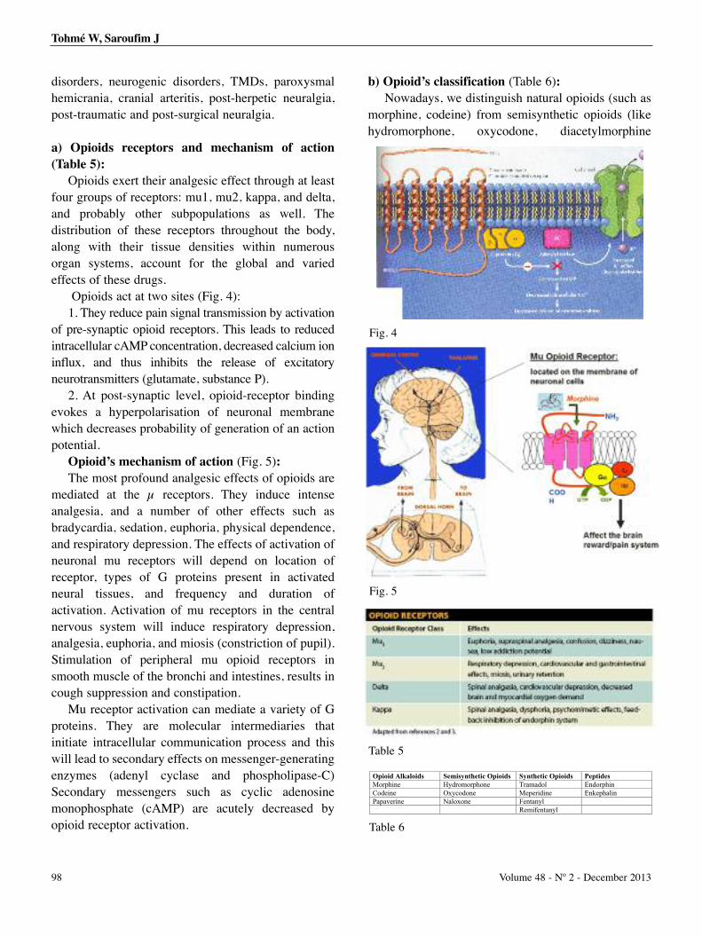
disorders, neurogenic disorders, TMDs, paroxysmalhemicrania, cranial arteritis, post-herpetic neuralgia,post-traumatic and post-surgical neuralgia.
a) Opioids receptors and mechanism of action(Table 5):
Opioids exert their analgesic effect through at leastfour groups of receptors: mu1, mu2, kappa, and delta,and probably other subpopulations as well. Thedistribution of these receptors throughout the body,along with their tissue densities within numerousorgan systems, account for the global and variedeffects of these drugs.
Opioids act at two sites (Fig. 4):1. They reduce pain signal transmission by activation
of pre-synaptic opioid receptors. This leads to reducedintracellular cAMP concentration, decreased calcium ioninflux, and thus inhibits the release of excitatoryneurotransmitters (glutamate, substance P).
2. At post-synaptic level, opioid-receptor bindingevokes a hyperpolarisation of neuronal membranewhich decreases probability of generation of an actionpotential.
Opioid’s mechanism of action (Fig. 5):The most profound analgesic effects of opioids are
mediated at the µ receptors. They induce intenseanalgesia, and a number of other effects such asbradycardia, sedation, euphoria, physical dependence,and respiratory depression. The effects of activation ofneuronal mu receptors will depend on location ofreceptor, types of G proteins present in activatedneural tissues, and frequency and duration ofactivation. Activation of mu receptors in the centralnervous system will induce respiratory depression,analgesia, euphoria, and miosis (constriction of pupil).Stimulation of peripheral mu opioid receptors insmooth muscle of the bronchi and intestines, results incough suppression and constipation.
Mu receptor activation can mediate a variety of Gproteins. They are molecular intermediaries thatinitiate intracellular communication process and thiswill lead to secondary effects on messenger-generatingenzymes (adenyl cyclase and phospholipase-C)Secondary messengers such as cyclic adenosinemonophosphate (cAMP) are acutely decreased byopioid receptor activation.
b) Opioid’s classification (Table 6): Nowadays, we distinguish natural opioids (such as
morphine, codeine) from semisynthetic opioids (likehydromorphone, oxycodone, diacetylmorphine
Tohmé W, Saroufim J
98 Volume 48 - Nº 2 - December 2013
Table 5
Fig. 4
Fig. 5
Table 6
Opioid Alkaloids Semisynthetic Opioids Synthetic Opioids Peptides Morphine Hydromorphone Tramadol Endorphin Codeine Oxycodone Meperidine Enkephalin Papaverine Naloxone Fentanyl Remifentanyl
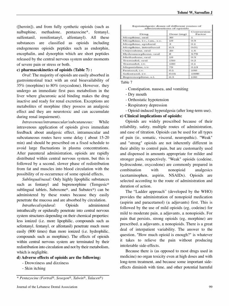
([heroin]), and from fully synthetic opioids (such asnalbuphine, methadone, pentazocine*, fentanyl,sulfentanil, remifentanyl, alfentanyl). All thesesubstances are classified as opioids includingendogeneous opioids peptides such as endorphin,encephalin, and dynorphin which are short peptidesreleased by the central nervous system under momentsof severe pain or stress or both.c) pharmacokinetics of opioids (Table 7) :
Oral: The majority of opioids are easily absorbed ingastrointestinal tract with an oral bioavailability of35% (morphine) to 80% (oxycodone). However, theyundergo an immediate first pass metabolism in theliver where glucuronic acid binding makes the druginactive and ready for renal excretion. Exceptions aremetabolites of morphine (they possess an analgesiceffect and they are neurotoxic and can accumulateduring renal impairment).
Intravenous/intramuscular/subcutaneous: Whileintravenous application of opioids gives immediatefeedback about analgesic effect, intramuscular andsubcutaneous routes have some delay ( about 15-20min) and should be prescribed on a fixed schedule toavoid large fluctuations in plasma concentrations.After parenteral administration, opioids are rapidlydistributed within central nervous system, but this isfollowed by a second, slower phase of redistributionfrom fat and muscles into blood circulation with thepossibility of re-occurrence of some opioid effects,
Sublingual/nasal: Only highly lipophilic substancessuch as fentanyl and buprenorphine (Temgesic®
sublingual tablets, Suboxone®, and Subutex®) can beadministered by these routes because they easilypenetrate the mucosa and are absorbed by circulation.
Intrathecal/epidural: Opioids administeredintrathecally or epidurally penetrate into central nervoussystem structures depending on their chemical properties:less ionized (i.e. more lipophilic, compounds such assefentanyl, fentanyl, or alfentanil) penetrate much moreeasily (800 times) than more ionized (i.e. hydrophilic,compounds such as morphine). The effects of opioidswithin central nervous system are terminated by theirredistribution into circulation and not by their metabolism,which is negligible. d) Adverse effects of opioids are the following:
- Drowziness and dizziness- Skin itching
- Constipation, nausea, and vomiting- Dry mouth- Orthostatic hypotension- Respiratory depression- Opioid-induced hyperalgesia (after long-term use).
e) Clinical implications of opioids: Opioids are widely prescribed because of their
reliability, safety, multiple routes of administration,and ease of titration. Opioids can be used for all typesof pain (ie, somatic, visceral, neuropathic). "Weak"and "strong" opioids are not inherently different intheir ability to control pain, but are customarily usedand dispensed in amounts appropriate for milder andstronger pain, respectively. "Weak" opioids (codeine,hydrocodone, oxycodone) are commonly prepared incombination with nonopioid analgesics(acetaminophen, aspirin, NSAIDs). Opioids areselected according to the route of administration andduration of action.
The “Ladder approach” (developed by the WHO)provides the administration of nonopioid medication(aspirin and paracetamol) (± adjuvants) first. This isfollowed by the use of mild opioids (eg, codeine) formild to moderate pain, ± adjuvants, ± nonopioids. Forpain that persists, strong opioids (eg, morphine) areprescribed, ± adjuvants, ± nonopioids. There is a greatdeal of interpatient variability. The answer to thequestion, "How much opioid is enough?" is whateverit takes to relieve the pain without producingintolerable side-effects.
Because there is (as opposed to most drugs used inmedicine) no organ toxicity even at high doses and withlong-term treatment, and because some important side-effects diminish with time, and other potential harmful
Tohmé W, Saroufim J
99Journal of the Lebanese Dental Association
Table 7
* Pentazocine (Fortral®, Sosegon®, Talwin®, Talacen®)
side-effects can be avoided with correct use, opioids willprobably remain the mainstay of pain management formost of patients for many years to come.
In summary, we addressed a practicalpharmacological approach of orofacial pains, and ineveryday practice, it seems obvious that combinationof two molecules from different pharmacologicalclasses gives the best results in the reduction of painperception. For example, in dentistry, the mostprescribed model is the combination of a weak opioidagonist such as codeine with a NSAID for themanagement of most orofacial pains.
4- CONCLUSIONOrofacial disorders consist of a wide variety of
debilitating chronic reluctant diseases. The most commonis temporo-mandibular joint (TMJ) and masticatorymuscle disorders and surveys showed that approximately7% (or 13 millions) Americans suffer from facial or jawpain. These disorders are difficult to diagnose due to theproximity of multiple anatomical structures, and they arehard to manage because of the need of a strict dailyregimen with the combination of appropriatemedications. Management of patients with chronic pain isa true challenge, it often requires a multidisciplinaryapproach that includes dentist, physician, physicaltherapist, and psychologist / psychiatrist.
In orofacial neuropathic pain, conventionalanalgesics are inefficient and there is often benefit fromclass I antiarrhythmics (interfere with the Na+ channel =mexiletine), tricyclic antidepressants -TCAs-(amitriptyline, nortriptyline, desipramine, protriptyline,etc...), and anticonvulsants (Clonazepam,carbamazepine, oxcarbazepine, sodium valproate,phenytoin, gabapentin, pregabalin, etc...) that are notconsidered and classified as analgesics.
Drug therapy for chronic pain often involvessimultaneous use of more than one drug. This takesadvantage of the different mechanisms of action ofdifferent drugs. It may also allow the use of smallerdoses and may reduce adverse effects. The mostcommon example of this in dentistry is thecombination of an opioid such as codeine with aNSAID.
REFERENCES 1. Textbook of Anaesthesia. Alan R.Aitkenhead, David J.Rowbotham,Graham Smith, Churchill Livingstone, 4th edition, 2004.
2. Neurology. A.Behin, S.Blond, E.Broussolle, Masson 2007.
3. Rhumatology, Xavier Chevalier, Philippe Goupille, Jean Sibilia,Cofer, Masson, 2002.
4. Dayer P, Desmeules J, Collart L: [Pharmacology of tramadol]Drugs. 1997;53 Suppl 2:18-24.
5. Harati Y, Gooch C, Swenson M, Edelman S, Greene D, RaskinP, Donofrio P, Cornblath D, Sachdeo R, Siu CO, Kamin M.Double-blind randomized trial of tramadol for the treatment of thepain of diabetic neuropathy. Neurology. 1998 Jun;50(6):1842-6.
6. Harati Y, Gooch C, Swenson M, Edelman SV, Greene D, RaskinP, Donofrio P, Cornblath D, Olson WH, Kamin M. Maintenance ofthe long-term effectiveness of tramadol in treatment of the pain ofdiabetic neuropathy. J Diabetes Complications. 2000 Mar-Apr;14(2):65-70.
7. Fields HL, Liebeskind JC. Pharmacological Approaches to theTreatment of Chronic pain: New concepts and Critical Issues.Seattle: IASP press, 1994.
8. James R.Fricton, DDS, MS. Temporo-mandibular joint andmuscle disorders. Department of Diagnostic and Surgical Sciences,University of Minnesota School of Dentistry, June 2004.
9. Rosenbaum, R. Orofacial Pain: Guidelines for Assessment,Diagnosis, and management: A systematic Review of Temporo-Mandibular Disorders. Diagnosis and Treatment. Chicago:Quintessence, 2004.
10. Singer E, Dionne R. A controlled evaluation of ibuprofen anddiazepam for chronic orofacial muscle pain. Journal of OrofacialPain 1997; 11 (2): 139-146.
Correspond with:Wissam Tohmé[email protected]@[email protected]
Tohmé W, Saroufim J
100 Journal of the Lebanese Dental AssociationVolume 48 - Nº 2 - December 2013
Clinical Comment
101
INTRODUCTIONMandibular division of trigeminal nerve (V3)
consists of motor and sensory fibers. Motor divisionsupplies muscles of mastication: masseter, temporal,pterygoid, mylohyoid, and anterior belly of digastric.Sensory portion of trigeminal supplies touch-pain-temperature to the face7-9.
Immediately after exiting the skull throughforamen ovale, mandibular nerve divides into anteriorand posterior branches. Anterior branches arepredominantly devoted to masticatory muscles. Incontrast, posterior branches innervate structuresinvolved in mastication, salivation, speech, and tastesensations – namely the lingual nerve, inferior alveolarnerve (IAN) and auriculotemporal nerve (ATN).Inferior alveolar nerve, and just before entering themandibular foramen, gives off a small mylohyoid
branch, the mylohyoid nerve -MHN-, that piercessphenomandibular ligament and enters a shallowgroove, the mylohyoid groove (MHG) on medialsurface of mandible9 (Fig. 1).
MHN, or nerve to mylohyoid, is a branch of theIAN, which arises above mandibular foramen. Itoriginates at variable distances superior to mandibularforamen. Bennett and Townsend (2001) reported theaverage branching distance to be 13.4 mm whileWilson and co-workers (1984) reported an averagebranching distance of 14.7mm5. The nerve then passesdownward and anteriorly within the mylohyoid grooveon the medial surface of mandible (Fig. 2). The nervecourses anteriorly and parallel to mylohyoid muscle,giving branches that provide motor fibers toinnervation to mylohyoid muscle on its infero-lateralsuperficial surface, also it supplies anterior belly ofdigastric muscle on its supero-medial deep surface2,10.MHN is primarily motor in nature, but it contains asensory component to anterior and posteriormandibular teeth. It also supplies the chin and tip ofthe tongue in some individuals2, 12.
The Mylohyoid Nerve -MHN: clinical significance andimplications for analgesia of mandibular teeth in restorative andsurgical dentistry.
Abstract
Inferior alveolar nerve block (IANB) is widely used in dental clinical practice for permanent mandibular teeth analgesia
and impacted wisdom teeth surgery. Causes of dental analgesic failure should always be identified in order to ensure successful
painfree dental treatment. Factors implicated in mandibular teeth analgesia failure include bifid inferior alveolar nerve,
double/accessory mental foramen, exaggerated bone density, cross innervation of incisors, tissue inflammation, inactive
anesthetic solution, and mylohyoid nerve accessory innervation. Mylohyoid Nerve-MHN-,a motor nerve to mylohyoid and
digastric anterior belly muscles , is often implicated as a secondary( = additional) sensitive innervation of mandibular
permanent teeth. It may escape analgesia in IANBs, and alternative local analgesia (MHN block) should be implemented to
ensure total pain control of mandibular teeth.
Rita Bou Assaf1, BDS, DESS Oral Surg., DESS Endo., Wasfi Kanj2, BDS, DESS Oral Surg.
1 Associate Clinical Instructor, Department of Oral and
Maxillo-Facial Surgery, Lebanese University School of
Dentistry, Beirut, Lebanon.
2 Associate Clinical Instructor, Department of Oral and
Maxillo-Facial Surgery, Lebanese University School of
Dentistry, Beirut.
Journal of the Lebanese Dental AssociationVolume 48 - Nº 2 - December 2013
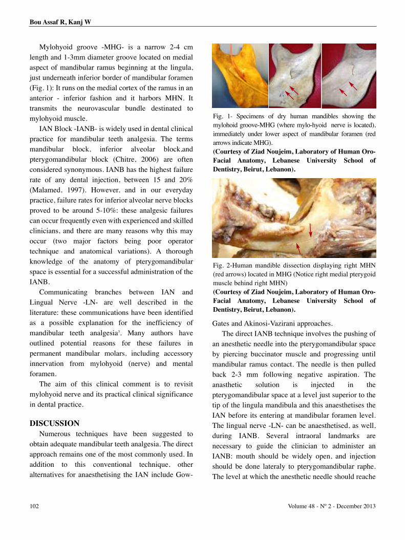
Mylohyoid groove -MHG- is a narrow 2-4 cmlength and 1-3mm diameter groove located on medialaspect of mandibular ramus beginning at the lingula,just underneath inferior border of mandibular foramen(Fig. 1): It runs on the medial cortex of the ramus in ananterior - inferior fashion and it harbors MHN. Ittransmits the neurovascular bundle destinated tomylohyoid muscle.
IAN Block -IANB- is widely used in dental clinicalpractice for mandibular teeth analgesia. The termsmandibular block, inferior alveolar block,andpterygomandibular block (Chitre, 2006) are oftenconsidered synonymous. IANB has the highest failurerate of any dental injection, between 15 and 20%(Malamed, 1997). However, and in our everydaypractice, failure rates for inferior alveolar nerve blocksproved to be around 5-10%: these analgesic failurescan occur frequently even with experienced and skilledclinicians, and there are many reasons why this mayoccur (two major factors being poor operatortechnique and anatomical variations). A thoroughknowledge of the anatomy of pterygomandibularspace is essential for a successful administration of theIANB.
Communicating branches between IAN andLingual Nerve -LN- are well described in theliterature: these communications have been identifiedas a possible explanation for the inefficiency ofmandibular teeth analgesia3. Many authors haveoutlined potential reasons for these failures inpermanent mandibular molars, including accessoryinnervation from mylohyoid (nerve) and mentalforamen.
The aim of this clinical comment is to revisitmylohyoid nerve and its practical clinical significancein dental practice.
DISCUSSIONNumerous techniques have been suggested to
obtain adequate mandibular teeth analgesia. The directapproach remains one of the most commonly used. Inaddition to this conventional technique, otheralternatives for anaesthetising the IAN include Gow-
Gates and Akinosi-Vazirani approaches.The direct IANB technique involves the pushing of
an anesthetic needle into the pterygomandibular spaceby piercing buccinator muscle and progressing untilmandibular ramus contact. The needle is then pulledback 2-3 mm following negative aspiration. Theanasthetic solution is injected in thepterygomandibular space at a level just superior to thetip of the lingula mandibula and this anaesthetises theIAN before its entering at mandibular foramen level.The lingual nerve -LN- can be anaesthetised, as well,during IANB. Several intraoral landmarks arenecessary to guide the clinician to administer anIANB: mouth should be widely open, and injectionshould be done lateraly to pterygomandibular raphe.The level at which the anesthetic needle should reache
102
Fig. 1- Specimens of dry human mandibles showing themylohoid groove-MHG (where mylo-hyoid nerve is located),immediately under lower aspect of mandibular foramen (redarrows indicate MHG).(Courtesy of Ziad Noujeim, Laboratory of Human Oro-Facial Anatomy, Lebanese University School ofDentistry, Beirut, Lebanon).
Fig. 2-Human mandible dissection displaying right MHN(red arrows) located in MHG (Notice right medial pterygoidmuscle behind right MHN) (Courtesy of Ziad Noujeim, Laboratory of Human Oro-Facial Anatomy, Lebanese University School ofDentistry, Beirut, Lebanon).
Bou Assaf R, Kanj W
Volume 48 - Nº 2 - December 2013
Journal of the Lebanese Dental Association 103
the bone is indicated indicated by the maximumconcavity of anterior border of mandibular ramus(coronoid notch).
Several factors are incriminated in locoregionalanalgesia failure of mandibular teeth, such as a bifidinferior alveolar nerve, bone density, inactivity ofanesthetic product in presence of tissue inflammation,incorrect technique, MHN accessory innervation,inactive anesthetic solution, and cross innervation ofincisors. Since the nerve to the mylohyoid has beenshown to carry dental sensory fibers, a block of thisnerve may be necessary to provide complete localanalgesia of mandibular teeth. Administering MHNblock in the lingual mucosa near the second premolartooth, has been recommended (Roda and Blanton,1994; Blanton and Jeske, 2003), and MHN may beblocked on the lingual aspect of the mandible betweenthe first and second premolars (Sutton, 1974). Thisblock may be administered on lingual mandible, aswell, behind the distal root of the first molar (Sillanpääet al., 1988). The analgesic efficacy of the nerve to themylohyoid may be due to the thin lingual mandibularcortex. Clarck and associates15 found a higher successrate in posterior mandibular teeth analgesia bycombining inferior alveolar nerve block and nerve tothe mylohyoid block.
CONCLUSIONMHN is the only muscular branch of V3 posterior
division (Woodburne and Burkel, 1988) and its role inmandibular teeth sensation remains a controversy.However, many authors reported the accessoryinnervation to pulps of anterior and posteriormandibular teeth (Frommer et al., 1972 --- Rood, 1976--- Madeira et al., 1978 --- Wilson et al., 1984 ---Sillanpää et al., 1988 --- Clark et al., 1999).
Failure to achieve complete pulpal analgesia ofmandibular teeth after an apparent successful IANblock remains a serious clinical problem in restorativeand surgical dentistry. Consequently, it is advocated toblock the MHN, in combination with IAN block, inorder to possibly enhance pulpal analgesia ofmandibular teeth.
REFERENCES1- López AB, Diago MP. Failure of locoregional anesthesia in
dental practice. Review of the literature. Oral Patol Oral Cir Bucal
2006; 11:E510-3.
2- Thotakura B, Rajendran SS, Vaithianathan G, Subramaniam A.
Variations in the posterior division branches of the mandibular
nerve in human cadavers. Singapore Med J 2013; 54(3): 149-15.
3- Potu BK, Pulakunta T, Ray B et al. Unusual communication
between the lingual nerve and mylohyoid nerves in a South Indian
male cadaver: its clinical significance. Romanian Journal of
Morphology and Embryology 2009; 50(1):145–146
4- Buch HA. Clinical Anatomy of Inferior Alveolar Nerve Block
Anesthesia. Clinical Anatomy 2011; 24:515–517.
5- Buch HA, Agnihotri RG. A Recurrent Variant Branch of the
Inferior Alveolar Nerve: Is It Unique? Clinical Anatomy 2012;
25:437-443.
6- H. Kenneth Walker, Cranial Nerve V: The Trigeminal Nerve.
p.318-321.
7- Khoury JN, Mihailidis S, Gabriel M, Townsend G. Applied
anatomy of the pterygomandibular space: improving the success of
inferior alveolar nerve blocks. Australian Dental Journal 2011; 56:
112-121
8- Narayana K, Narayan P, Ashwin K, Prabhu LV. Incidence, types
and clinical implications of a non-metrical variant - mylohyoid
bridging in human mandibles. Folia Morphol. (Warsz) 2007
Feb;66(1):20-24.
9- Lin K, Uzbelger Feldman D, Barbe MF. Transverse Cervical
Nerve: Implications for Dental Anesthesia. Clinical Anatomy
2013; 26:688–692.
10- Oth O, Louryan S, Van Sint Jan S, Rooze M, Glineur R.
Impact of the mandibular divergence on the position of the inferior
alveolar nerve and mylohyoid nerve: a computed tomography
study and its relevance to bilateral sagittal split osteotomy. Surg
Radiol Anat 2013; 35: 241–247.
11- Stein P, Brueckner J, Milliner M. Sensory Innervation of
Mandibular Teeth by the Nerve to the Mylohyoid: Implications in
Local Anesthesia: Clinical Anatomy 2007; 20:591–595.
12- Shane Tubbs R, Loukas M, Shoja MM et al. The nerve to the
mylohyoid as a donor for facial nerve reanimation procedures: a
cadaveric feasibility study. J Neurosurg 2007; 106: 677–679.
13- Madhyastha S, Prabhu LV, Saralaya VV et al. Dual
innervations of mylohyoid muscle: a case report. Romanian
Journal of Morphology and Embryology 2009; 50(2):305–306.
14- Shantharam V, Manjunath KY, Shastri D. Bony Bridging of the
Mylohyoid Groove. Anatomica Karnataka 2011; Vol-5(3): 45-49.
Bou Assaf R, Kanj W
15- Clarck S, Reader A, Beck M, Meyers WJ. Anesthetic efficacy
of the mylohyoid nerve block and combination inferior alveolar
nerve block / mylohyoid nerve block. Oral Surg Oral Med Oral
Pathol Oral Radiol Endod 1999 May;87:557-563.
16- Frommer J, Mele FA, Monroe CW. The possible role of themylohyoid nerve in mandibular posterior tooth sensation. JAmerican Dent Assoc 1972 July;85(1):113-117
17- Rood JP. The analgesia and innervation of mandibular teeth.Brit Dent J 1976 April 6;140(7):237-239.
18- Madeira MC, Percinoto C, das Graças M. Clinical significanceof supplementary innervation of the lower incisor teeth: Adissection study of the mylohyoid nerve. Oral Surgery 1978Nov;46(5):608-614.
19- Sillanpää M, Vuori V, Lehtinen R. The mylohyoid nerve andmandibular anesthesia. Int J Oral Maxillofac. Surg 1988June;17(3):206-207.
20- Thotakura B, Rajendran SS, Gnanasundaram V, SubramaniamA. Variations in the posterior division branches of the mandibularnerve in human cadavers. Singapore Med J 2013 March;54(3):149-151.
Correspond with:Rita Bou [email protected]
Bou Assaf R, Kanj W
104 Journal of the Lebanese Dental AssociationVolume 48 - Nº 2 - December 2013
Review
BACKGROUNDPain and orthodontic treatment can be perceived as
the two faces for one coin, and this can be a reason todiscourage orthodontic treatment for some futurepatients or reduce compliance for others undergoingtreatment.1-3 Orthodontic treatment requires applyingforces on teeth, thereby generating differentperceptions of pain in 91 to 95 % of patients, rangingfrom discomfort to severe pain.4-8
Surveys were performed to analyse the experience oforthodontic pain among patients and it was rated as a keydeterrent to orthodontic therapy and a major reason fordiscontinuing treatment.9-11 One survey rated pain as thegreatest dislike during treatment and the fourth amongmajor fears prior to orthodontic treatment.12
Different factors play a role in determining theintensity of pain such as age, gender, psychologicalstatus, types of appliances, and mechanical employedtechniques.13
This disagreable sensation can be experiencedduring different procedures like separator placement,banding, bonding, as well as archwire activation anddebonding. The main concern of orthodontists is to
control, as much as possible, “this painful experience”to patients, which might reflect positively on theneeded compliance and perseverance of those patientsfor a successful achievable result. The aim of thisreview is to highlight possible causes and managementof discomfort and pain during and after orthodontictreatment.
MECHANISM OF ORTHODONTIC PAINChanges to teeth environment are crucial for
orthodontic tooth movement. These changes includevascular alterations, recruitment of inflammatorymediators, and alveolar socket remodeling, and theassumption is that sources of that inflammatory painduring orthodontic treatment are from surroundingperiodontal ligament. Collagen fibers of that latter aredisrupted, some undergo compression, others tension,and necrosis will result, and the process of healing willstart by formation of a blood clot and granulationtissue.14 Granulation tissue will be later colonized byvascular and nervous components15 and formation ofnew connective tissue will initiate.16-18
Several studies stated changes in circulation, tissuemetabolism, and pulp vitality affected or compromisedby applied orthodontic forces.19-22 Thus another probablecause for orthodontic pain is the involvement ofintradental nociceptive nerves where periodontalinflammatory reactions occur. These latter will spread tothe pulp due to the release of some inflammatory
Chimène Chalala1, BDS, DESS Ortho., Resid. Ortho. (AUB), Ramzi V. Haddad2, BDS, Resid. Ortho. (AUB), MS(Human Morphol.)
Pain in orthodontic treatment: etiology and management.
Abstract
Orthodontic treatment is considered, by most patients, as a painful procedure. Studies have shown this reaction to have a
major negative effect on orthodontic treatment and was reported to be an important reason for discontinuing treatment. Pain
can be experienced during different orthodontic procedures like separator placement, banding, bonding, as well as archwire
activation and debonding. This review attempts to highlight published literature regarding pain during different stages of
orthodontic treatment, with an overview of current management strategies used to alleviate orthodontic pain.
1. Associate Chief of Clinical Services, Department ofOrthodontics, Lebanese University School of Dentistry,Beirut, Lebanon, and Clinical Associate, Division ofOrthodontics and Dentofacial Orthopedics, AmericanUniversity of Beirut Medical Center - AUBMC, Beirut
2. Assistant Professor and Clinical Director, Division ofOrthodontics and Dentofacial Orthopedics, AUBMC,Beirut, Lebanon.
105Journal of the Lebanese Dental AssociationVolume 48 - Nº 2 - December 2013
mediators23 (substance P-SPa-, histamineb, enkephalinc,dopamined, serotonine, prostaglandinsf-PGs, andleukotrienesg) causing hyperalgesiah. The branchingaxons which innervate pulp and periodontal ligament,will undergo neurogenic inflammation resulting in painsensation.24-26
In most of cases, no considerable damage to theteeth and their periodontium was noticed duringorthodontic tooth movement. In general, pulpal side-effects are scarce but some may take place as alteredpulpal respiration rate, internal root resorption, pulpalobliteration by secondary dentin, and pulpal necrosis.27
Pulp ischaemia28 is the transient factor that causes painand discomfort during first days after orthodonticappliance adjustment. It results also from excessiveforces applied with the overzealous wearing ofintermaxillary elastics or excessively activatedremovable appliances.29 This ischaemic pulpal painwill settle usually within a week if appropriate forcesand mechanics are implemented. Death or necrosis ofpulp was seldom reported during orthodontictreatment.30 Besides, tooth sensitivity increases afterlight premature contacts ensuing from abnormalloading of teeth31 which is common during the firstphases of orthodontic treatment. These clinicalsymptoms will disappear progressively afterelimination of interferences.
It is the responsibility of the orthodontist torecognize, as early as possible, any pulpal syndromesarising during orthodontic treatment and to treat themappropriately without delay.32 Furthermore, teeth withsevere periodontal injury are more vulnerable to pulpalreactions during orthodontic treatment, namely duringintrusion and extrusion movements. 33
REPORTED ORTHODONTIC CAUSES OFPAIN
Optimal forces in orthodontics are those forces ableto move teeth most rapidly, with minimum discomfortto the patient and least damage to the teeth and theirsurrounding tissues.
Several authors advocated the use of light forceswhen moving teeth. It is thought that those forcesproduce more physiological effect, induce less pain,and minimize side-effects on different involved oraltissues.34-38 Impingement on adjacent tissues by largeforces was shown to cause greater periodontalcompression, and thus, more pain.39
Other authors defended the use of heavy forces,especially for canines, justifying that higher forces perunit area increased the rate of biological response.40-41
On the other hand, some reports did not correlatelevel of applied force to discomfort experienced bypatients after studying the relationship between initialtooth positions, applied force levels, and experiencedpain and where no statistically significant correlationamong the three parameters was found.42
SEPARATORSOrthodontic separators are most often used by
orthodontists to create spaces mesial and distal to theteeth, precisely posterior teeth, in order to fit theproper size of bands. They can be circular rubberbands or brass wires, spring type steel separators, andlatex elastics. Placement of separators is known as apainful experience for almost all patients. Discomfortassociated with orthodontic separation starts usually
Chalala C, Haddad RV
106
a. SP= a neuropeptide that functions as a neurotransmitter and as a neuromodulator.
b. Histamine= a neurotransmitter (produced by basophils and mast cells) involved in the inflammatory response.
c. Enkephaline= a pentapeptide involved in regulating nociception in the body.
d. Dopamine= functions as a neurotransmitter in the brain (the brain includes several distinct dopamine systems).
e. Serotonin (5-HT)= a monoamine neurotransmitter primarily found in GI tract, platelets, and CNS. It has various functions,including regulation of mood, appetite and sleep, cognitive functions (memory, learning...), and it is thought to contribute tofeelings of well-being and happiness.
f. PGs= a group of hormone-like lipid compounds that have a variety of strong physiological effects (regulating contractionand relaxation of vascular smooth muscle tissue, sensitization of spinal neurons to pain, inducing labor, regulatinginflammation, etc...)
g. Leukotrienes= a family of eicosanoid inflammatory mediators produced in leucocytes, but also found in other immunecells. They use lipid signaling to convey information to either the cell producing them or neighboring cells, in order toregulate immune responses. Leukotriene production is usually accompanied by histamine and PGs, which also act asinflammatory mediators.
h. Hyperalgesia= an increased sensitivity to pain, which may be caused by damage to nociceptors or peripheral nerves(primary hyperalgesia is pain sensitivity occuring directly in damaged tissues, and secondary hyperalgesia is pain sensitivityoccuring secondary in undamaged tissues).
Volume 48 - Nº 2 - December 2013
four hours after placement of separators and the levelof pain increases during the next following 24hours.13,43-46 An association was detected between painintensity and crevicular fluid mediator levels: at 1hour, an association was perceived betweenprostaglandin E2 (PGE2) levels and pain intensitywhile at day 1, pain intensity was associated toInterleukin-1ß (IL-1beta) levels.4
Intensity of pain and amount of separation were notsimilar with different types of separators. Both amountof separation and discomfort were found less withKessling separators compared to elastomeric and brasswire separators.47
ARCHWIRE PLACEMENT AND ACTIVATIONOne of major patients concern is the pain they are
going to experience after the first appointment whenplacing the first orthodontic wire. Pain associated withinitial archwire placement is experienced by majorityof patients 4 hours after archwire placement, whichwill peak at 24 hours, and then decline.6,13,48,49
Orthodontic pain can vary, thereby it can beexacerbated in evenings and at nights, and usually lastsfor 2 or 3 days and will gradually decrease in itsintensity by fifth or sixth day. For some patients, it canbe worse than pain experienced from tooth extraction.5
Some authors assessed orthodontic tooth pain atdifferent time levels: after bracket placement(baseline), 1 hour after placement of initial archwires,1 day after archwire placement, 1 week after archwireplacement, and 1 month after archwire placement.Orthodontic patients experienced significant pain anddiscomfort 1 day after initial archwire placement.50
Studying of orthodontic pain was pushed to a level tocompare various types of archwires in pain perception.No difference in intensity or duration of pain betweendifferent archwires was found.49,51
Other reports confirmed the prevalence of that pain athigher intensity in mandible than in maxilla51, and inanterior teeth than in posterior ones. This was explainedby differences in root surface area, increasedinvolvement of anterior teeth during alignment, andmore frequent involvement of anterior teeth in biting.6,13,49
ORTHOPEDIC SUTURAL EXPANSIONWhen craniofacial orthopaedics are indicated, high
forces are usually applied. These forces are absorbed
and transmitted to the craniofacial complex where aseries of reactions, characterized by development ofinternal stress, will develop.52,53
One of the most adopted orthopedic forces inorthodontics is maxillary expansion, where heavyforces generated by a screw incorporated in anappliance will be applied on palate to split midpalatalsuture. In an experimental expansion in rats, traumatictears, exudates, death of fibroblasts, disruption ofcollagen fibers, and acute inflammation wereobserved. Inflammatory process resulted in a painfulsensation, which is often expressed in the wholecraniofacial region.54
Human reports in the literature confirmed thatpainful experience occurs after sagittal palatalexpansion in children, and this occurs during initialphase and diminishes with time. 55-57
EXTRA-ORAL APPLIANCES (HEAD GEARAND FACEMASK)
Patients often experience discomfort after 24 hoursof headgear wear and pain decreases significantly after3 days of wear.58 Another study59 showed that 28 % ofpatients reported pain as the factor that prevented themfrom wearing their headgear. Pain caused by the use ofprotraction headgear (Facemask) was not of amuscular origin but part of the acute inflammatoryreaction occurring in sutural regions.60
DEBONDINGRemoval of braces is the moment that most of the
patients wait for, but it is not accomplished withoutpain. Williams and Bishara61 evaluated patient’sdiscomfort at orthodontic bracket debonding andconcluded that tooth mobility and force applicationwere the two important influencing factors. Theyrecommended that applying intrusive forces whiledebonding produce less pain in comparison withforces applied in other directions. They also suggestedapplying finger pressure or asking the patient to bite ona cotton roll to decrease pain. In another report,Rinchuse62 suggested the use of an occlusal rim wax tominimize pain.
PERCEPTION OF PAINPsychological state is an important factor in pain
perception following activation of an orthodontic
Chalala C, Haddad RV
Journal of the Lebanese Dental Association 107
appliance. It has been reported that this pain is notdirectly related to the magnitude of force exerted butdepends heavily on the psychological well-being ofthe individual.63-66
Orthodontic pain may also be related to gender. It isbelieved that females may be more sensitive to pain, whilemales can tolerate it better.65 Other reports showedcontradictory results where there was no differencesbetween males and females in reporting pain sensation.13
The effect of age on pain perception was alsoinvestigated during orthodontic treatment. Severalstudies reported that adult subjects perceive more painthan young patients.6,42 A study conducted during allphases of treatment such as separator placement,bonding, archwire placement, and activations, foundthat adolescents reported a higher level of pain thanpre-adolescents and adults.64
CLASSIFICATION OF LOW-LEVELORTHODONTIC PAIN
Burstone67 classified orthodontic pain in two ways: A- Based on degree of pain perceived in response
to amount of force application and it can be dividedinto three levels:
1. First level: patient is not aware of pain unless theorthodontist manipulates teeth to be moved bythe appliance, e.g. using instruments such as aband pusher or force gauge.
2. Second level: pain or discomfort caused duringclenching or heavy biting, usually occurs withinthe first week of appliance placement. Patientwill be able to masticate food with this type ofpain.
3. Third level: if this type of pain is perceived,patient might be unable to masticate food ofnormal consistency.
B- Based on time of onset, and it can be dividedinto two time frames:
1. Immediate: which is associated with suddenplacement of heavy forces on tooth, e.g. hardfigure of eight tie between central incisors toclose a midline diastema.
2. Delayed: produced by variety of force valuesfrom light to heavy and representinghyperalgesia of periodontal membrane. This typeof pain response decreases with time (i.e. painreaction might start as third degree but becomessecond or a first degree with time).
MANAGEMENT OF ORTHODONTIC PAINChewing
Chewing gum or plastic wafer during first fewhours of appliance activation are suggested to reducepain as it will reduce ischemia and inflammation inperiodontal ligament.68 This will also temporarilydisplace teeth sufficiently and stimulation of vascularand lymphatic circulation would prevent the build-upof metabolic products, which are known to stimulatepain receptors.69
White70 found that approximately 63% of patientsreported less discomfort after chewing Aspergum® (achewing gum containing 227mg of aspirin), afterorthodontic activation. Hwang and co-workers71
obtained the same results in 56 % of their patients afterchewing Thera-Bite Wafers.Low-Level LASER Therapy-LLLT
Low-level LASER (a form of LASER medicinethat uses low-level/low-power LASERs or light-emitting diodes to alter cellular function) has beenclaimed to minimize orthodontic pain. The mechanismof LASER analgesia has been attributed to its anti-inflammatory effects.72 Despite few reports thatshowed success in the reduction of orthodonticpain,73,74 efficacy of low-level LASER therapy inreducing orthodontic pain was deemed discouragingand this type of phototherapy did not show immediatepain relief in orthodontic patients.75
Pain killers: anti- inflammatory versus pureanalgesic drugs
Analgesics (pain killers) remain the main treatmentmodality to reduce orthodontic pain. A variety of drugsare suggested by practitioners but some are moreefficient than others. Most of prescribed medicationsare painkillers and they can be classified into anti-inflammatory or pure analgesic drugs. Although all ofthem influence pain level perceived by patients, theireffect is dissimilar on the rate of tooth movement.Nonsteroidal anti- inflammatory drugs (NSAIDs) areknown to inhibit prostaglandins secretion, namelyprostaglandine E2, an important inflammatorymediator essential for orthodontic tooth movement:PGE2 is responsible for initiation of osteoclasticactivity essential for tooth movement, therefore adelay in orthodontic treatment is expected.
Ibuprofen, naproxen sodium, acetaminophen
Chalala C, Haddad RV
108 Volume 48 - Nº 2 - December 2013
(paracetamol), piroxicam, and aspirin are the mostprescribed medications for pain alleviation.Acetaminophen is defined as a mild analgesic and nota nonsteroidal anti-inflammatory drug as all previousdrugs already cited because it exhibits only weak anti-inflammatory activity. In a meta-analysis76 comparinganalgesic effect between ibuprofen andacetaminophen, there was no statistically significantdifference between both drugs at any time point butibuprofen had decreased pain at 2 hours and 6 hoursafter separators placement compared to placebo andnot at 24 hours when pain peaks.
Preoperative medication was studied77 with a goalof considerably decreasing perception of pain due toseparators. No significant difference in pain wasnoticed after placement of separators betweenacetaminophen (650 mg) and ibuprofen (400 mg)taken 1 hour before separator placement, indicating aneffective result of both analgesics. However, 20 mg ofpiroxicam administered 1 hour prior to separatorplacement were found more efficient in decreasinglevels of pain than 400 mg of ibuprofen or placebo.78
This significant decrease in pain was perceived at 2hours, 6 hours, during nighttime, at 24 hours, and onthe second and third days after separator placement.
Pre and postoperative medications were alsoadvocated for a better control of pain resulting fromseparation of teeth. Positive results were reported withthe use of 400 mg of ibuprofen 1 hour prior and 3 and7 hours after separators placement.79-82
Differentiation between anti-inflammatoryanalgesic and purely analgesic drugs and the priorityof the first ones in relieving pain after separatorsplacement is of utmost significance in term of toothmovement. Anti-inflammatory analgesics inhibitprostaglandins secretion slowing down orthodontictooth movement. Hence, it is important to limit asmuch as possible any interference with theinflammation response which is a normal tissuereaction in orthodontic tooth movement.
If bands are not used on molars but bondable tubes (noneed for separators), patients will experience pain for thefirst time after insertion of the first wire. Studies reportedthat acetaminophen is the analgesic of choice in treatingorthodontic pain without affecting orthodontic toothmovement.83,84,86,87 Aspirin and ibuprofen had the same
reduced effect on tooth movement while acetaminophendid not affect orthodontic tooth movement in rats andexhibited a statistically significant difference with the 2above mentioned drugs83. In terms of pain, ibuprofeninduces lower pain level during the first day after bondingcompared to acetaminophen.87
CONCLUSIONOrthodontic pain is a major concern for parents,
patients, and orthodontists. Studies have shown that it isthe main negative factor to orthodontic treatment and animportant reason for discontinuing treatment. It isimportant to inform patients about expected pain duringdifferent phases of treatment. When prescribing theanalgesic of choice, appropriate drug should be selected.Thus, appropriate mechanotherapy and adequate painmanagement techniques should be well comprehended.
REFERENCES1. Bos A, Hoogstraten J, Prahl-Andersen B. Towards acomprehensive model for the study of compliance in orthodontics.Eur J Orthod 2005;27:296– 301.
2. Oliver RG, Knapman Y. Attitudes to orthodontic treatment. Br JOrthod 1985;19:47–54.
3. Blechman AM. Pain-free and mobility-free orthodontics? Am JOrthod Dentofacial Orthop 1998;113:379–83.
4. Bergius M, Berggren U, Kiliaridis S. Experience of pain duringan orthodontic procedure. Eur J Oral Sci 2002;110:92–8.
5. Jones M, Chan C. The pain and discomfort experienced duringorthodontic treatment: a randomized controlled clinical trial of twoinitial aligning arch wires. Am J Orthod Dentofacial Orthop2005;128: 435–41.
6. Scheurer PA, Firestone A, Burgin WB. Perception of pain as aresult of orthodontic treatment with fixed appliances. Eur J Orthod1996;18:349– 57.
7. Kvam E, Bondevik O, Gjerdet NR. Traumatic ulcers and pain inadults during orthodontic treatment. Community Dent OralEpidemiol 1989;17:154–7.
8. Lew KK. Attitudes and perceptions of adults towardsorthodontic treatment in an Asian community. Community DentOral Epidemiol 1993;21:31–5.
9. Oliver RG, Knapman Y. Attitudes to orthodontic treatment. Br JOrthod 1985;19:47–54.
10. Haynes S. Discontinuation of orthodontic treatment relative topatient age. J Dent 1974 Jul;2(4):138-42.
11. Kluemper GT, Hiser DG, Rayens MK, Jay MJ. Efficacy of awax containing benzocaine in the relief of oral mucosal paincaused by orthodontic appliances. Am J Orthod DentofacialOrthop 2002;122(4):359-65.
Chalala C, Haddad RV
Journal of the Lebanese Dental Association 109

12. O'Connor PJ. Patients’ perceptions before, during, and afterorthodontic treatment. J Clin Orthod 2000;34(10):591-2.
13. Ngan P, Kess B, Wilson S. Perception of discomfort by patientsundergoing orthodontic treatment. Am J Orthod DentofacialOrthop 1989;96(1):47-53.
14. Sismanidou C, Hilliges M, Lindskog S. Healing of the rootsurface-associated periodontium: an immunohistochemical study oforthodontic root resorption in man. Eur J Orthod 1996;18:435-44.
15. Parlange LM, Sims MR. A T.E.M. Stereological analysis ofblood vessels and nerves in marmoset periodontal ligamentfollowing endodontics and magnetic incisor extrusion. Eur JOrthod 1993;15(1):33-44.
16. Melcher AH. Repair of the wounds in the periodontium of therat. Influence of the periodontal ligament on osteogenesis. ArchOral Biol 1970;15(12):1183-1204.
17. Melcher AH. On the repair potential of periodontal tissues. JPeriodontol 1976;47: 256-260.
18. Wikesjö UM, Nilvéus RE, Selvig KA. Significance of earlyhealing events on periodontal repair: a review. J Periodontol1992;63(3):158-65.
19. Butcher EO, Taylor AC. The vascularity of the incisor pulp ofthe monkey and its alteration by tooth retraction. J Dent Res1952;31(2):239-47.
20. Stenvik A, Mjör IA. Pulp and dentine reactions to experimentaltooth intrusion. A histologic study of the initial changes. Am JOrthod 1970;57(4):370-85.
21. Biesterfeld RC, Taintor JF, Marsh CL. The significance ofalterations of pulpal respiration. A review of literature. J OralPathol. 1979 June;8(3):129-39.
22. Hamersky PA, Weimer AD, Taintor JF. The effect oforthodontic force application on the pulpal tissue respiration rate inthe human premolar. Am J Orthod 1980;77(4):368-78.
23. Ahlberg KF. Functional studies on experimentally inducedinflammations of the dental pulp. Tandlakartidningen 1978;70(13-14):837-8 (article in Swedish).
24. Byers MR. Dental sensory receptors. Int Rev Neurobiol1984;25:39-94.
25. Byers MR. Terminal arborization of individual sensory axonsin dentin and pulp of rat molars. Brain Res 1985;345(1):181-5.
26. Yamaguchi M, Kasai K. Inflammation in periodontal tissues inresponse to mechanical forces. Arch Immunol Ther Exp (Warsz)2005;53(5):388-98.
27. Barwick PJ, Ramsay DS. Effect of brief intrusive force onhuman pulpal blood flow. Am J Orthod Dentofac Orthop1996;110(3):273-9.
28. Hamilton RS, Gutmann JL. Endodontic-orthodonticrelationships: A review of integrated treatment planningchallenges. Int Endod J 1999; 32:343-60.
29. Gurkeerat S. Textbook of Orthodontics (2007). 2nd edition,Jaypee Brothers Medical Publishers, New Delhi, 319.
30. Rotstein I, Engel G. Conservative management of a combinedendodontic-orthodontic lesion. Endodont Dent Traumatol1991;7:266–269.
31. Ikeda T, Nakano M, Bando E, Suzuki A. The effect of lightpremature occlusal contact on tooth pain threshold in humans. JOral Rehabil 1998 Aug;25(8):589-95.
32. Jacobs SG. The treatment of traumatized permanent anteriorteeth: Case report and literature review. Part I-Management ofintruded incisors. Aust Orthod J 1995;13:213-8.
33. Bauss O, Röhling J, Sadat-Khonsari R, Kiliaridis S. Influenceof orthodontic intrusion on pulpal vitality of previouslytraumatized maxillary permanent incisors. Am J OrthodDentofacial Orthop 2008;134(1):12-7.
34. Reitan K. Some factors determining the evaluation of forces inorthodontics. Am J Orthod 1959;29:105-113.
35. Reitan K. Tissue behavior during orthodontic tooth movement.Am J Orthod 1960;46: 881-900.
36. Reitan K. Effects of force magnitude and direction of toothmovement on different alveolar bone types. Am J Orthod1964;34:244-255.
37. Reitan K. Initial tissue behavior during apical root resorption.Angle Orthod 1974;44:68-82.
38. Storey E, Smith R. Force in orthodontics and its relation totooth movement. Aust Dent J 1952;56:11-18.
39. Gianelly AA, Goldman HM. Biologic basis of orthodontics(1971). Lea & Febiger, Philadelphia, 164-165.
40. Hixon EH, Atikian H, Callow GE, McDonald HW, Tacy RJ.Optimal force, differential force, and anchorage. Am J Orthod1969;55(5):437-57.
41. Hixon EH, Aasen TO, Clark RA, Klosterman R, Miller SS,Odom WM. On force and tooth movement. Am J Orthod1970;57(5):476-8.
42. Jones ML, Richmond S. Initial tooth movement: force applicationand pain--a relationship? Am J Orthod 1985;88(2):111-6.
43. Ngan P, Wilson S, Shanfeld J, Amini H. The effect of ibuprofenon the level of discomfort in patients undergoing orthodontictreatment. Am J Orthod Dentofacial Orthop 1994;106(1):88-95.
44. Bondemark L, Fredriksson K, Ilros S. Separation effect andperception of pain and discomfort from two types of orthodonticseparators. World J Orthod 2004;5(2):172-6.
45. Utomi IL, Odukoya OO. Pain and discomfort associated withorthodontic separator placement in patients attending the LagosUniversity Teaching Hospital, Lagos, Nigeria. OdontostomatolTrop 2013;36(141):5-13.
46. Giannopoulou C, Dudic A, Kiliaridis S. Pain discomfort andcrevicular fluid changes induced by orthodontic elastic separatorsin children. J Pain. 2006 May;7(5):367-76.
47. Sandhu GPS, Naik CR. Separation effect and perception ofpain discomfort from three types of orthodontic separators. J IndOrthod Soc 2013;47(1):6-9.
48. Jones M. An investigation into initial discomfort caused byplacement of an archwire. Eur J Orthod 1984;6:48-54.
49. Erdinç AM, Dinçer B. Perception of pain during orthodontictreatment with fixed appliances. Eur J Orthod 2004;26(1):79-85.
50. Leavitt AH, King GJ, Ramsay DS, Jackson DL. A longitudinalevaluation of pulpal pain during orthodontic tooth movement.Orthod Craniofac Res 2002;5(1):29-37.
51. Fernandes LM, Ogaard B, Skoglund L. Pain and discomfortexperienced after placement of a conventional or a superelasticNiTi aligning archwire. J Orofac Orthop1998;59(6):331-9.
Chalala C, Haddad RV
110 Volume 48 - Nº 2 - December 2013
52. Mao JJ. Mechanobiology of craniofacial sutures. J Dent Res2002;81(12):810-6.
53. Mao JJ, Wang X, Kopher RA. Biomechanics of craniofacialsutures: orthopedic implications. Angle Orthod 2003;73(2):128-35.
54. Ten Cate AR, Freeman E, Dickinson JB. Sutural development:structure and its response to rapid expansion. Am J Orthod1977;71(6):622-36.
55. Handelman CS. Nonsurgical rapid maxillary alveolarexpansion in adults: a clinical evaluation. Angle Orthod1997;67:291-305.
56. Needleman HL, Hoang CD, Allred E, Hertzberg J, Berde C.Reports of pain by children undergoing rapid palatal expansion.Pediatr Dent 2000;22(3):221-6.
57. Schuster G, Borel-Scherf I, Schopf PM. J Orofac Orthop.Frequency of and complications in the use of RPE appliances--results of a survey in the Federal State of Hesse, Germany. J OrofacOrthop 2005;66(2):148-61.
58. Cureton SL. Headgear and pain. J Clin Orthod 1994;28(9):525-30.
59. Egolf RJ, BeGole EA, Upshaw HS. Factors associated withorthodontic patient compliance with intraoral elastic and headgearwear. Am J Orthod Dentofacial Orthop 1990;97(4):336-48.
60. Ngan PW, Yiu C, Hagg U, Wei SH, Bowley J. Masticatorymuscle pain before, during, and after treatment with orthopedicprotraction headgear: a pilot study. Angle Orthod 1997;67(6):433-7.
61. Williams OL, Bishara SE. Patient discomfort levels at the timeof debonding: a pilot study. Am J Orthod Dentofacial Orthop1992;101(4):313-7.
62. Rinchuse DJ. Pain-free debonding with occlusal rim wax. JClin Orthod 1994;28(10):587-8.
63. Dubner R. Neurophysiology of pain. Dent Clin North Am1978;22(1):11-30.
64. Brown DF, Moerenhout RG. The pain experience andpsychological adjustment to orthodontic treatment ofpreadolescents, adolescents, and adults. Am J Orthod DentofacialOrthop 1991;100(4):349-56.
65. Bergius M, Kiliaridis S, Berggren U. Pain in orthodontics. Areview and discussion of the literature. J Orofac Orthop2000;61(2):125-37.
66. Sergl HG, Klages U, Zentner A. Pain and discomfort duringorthodontic treatment: causative factors and effects on compliance.Am J Orthod Dentofacial Orthop 1998;114(6):684-91.
67. Burstone CJ. The biomechanics of tooth movement. In: KrausBS, Riedel RA (eds). Vistas in orthodontics (1962). Lea & Febiger,Philadelphia. 197–213.
68. Furstman L, Bernick S. Clinical considerations of theperiodontium. Am J Orthod 1972;61(2):138-55.
69. Proffit WR. Contemporary orthodontics. 3rd edition. MosbyCompany, St Louis. 2000.
70. White LW. Pain and cooperation in orthodontic treatment. JClin Orthod 1984;18(8):572-5.
71. Hwang JY, Tee CH, Huang AT, Taft L. Effectiveness of thera-bite wafers in reducing pain. J Clin Orthod 1994;28(5):291-2.
72. Harris DM. Biomolecular mechanisms for laser biostimulation.J Clin Laser Med Surg 1991;277-280.
73. Artés-Ribas M, Arnabat-Dominguez J, Puigdollers A.Analgesic effect of a low-level laser therapy (830 nm) in earlyorthodontic treatment. Lasers Med Sci 2013;28(1):335-41.
74. Eslamian L, Borzabadi-Farahani A, Hassanzadeh-Azhiri A,Badiee MR, Fekrazad R. The effect of 810-nm low-level lasertherapy on pain caused by orthodontic elastomeric separators.Lasers Med Sci 2014;[Epub].
75. Lim HM, Lew KK, Tay DK. A clinical investigation of the efficacyof low level laser therapy in reducing orthodontic postadjustment pain.Am J Orthod Dentofacial Orthop 1995;108(6):614-22.
76. Angelopoulou MV, Vlachou V, Halazonetis DJ. Pharmacologicalmanagement of pain during orthodontic treatment: a meta-analysis.Orthod Craniofac Res 2012; 15(2):71-83.
77. Bird SE, Williams K, Kula K. Preoperative acetaminophen vsibuprofen for control of pain after orthodontic separator placement.Am J Orthod Dentofacial Orthop 2007;132(4):504-10.
78. Kohli SS, Kohli VS. Effectiveness of piroxicam and ibuprofenpremedication on orthodontic patients' pain experiences. AngleOrthod 2011;81(6):1097-102.
79. Patel S, McGorray SP, Yezierski R, Fillingim R, Logan H,Wheeler TT. Effects of analgesics on orthodontic pain. Am JOrthod Dentofacial Orthop 2011;139(1):53-8.
80. Bernhardt MK, Southard KA, Batterson KD, Logan HL, BakerKA, Jakobsen JR. The effect of preemptive and/or postoperativeibuprofen therapy for orthodontic pain. Am J Orthod DentofacialOrthop 2001;120(1):20-7.
81. Minor V, Marris CK, McGorray SP, Yezierski R, Fillingim R,Logan H, Wheeler TT. Effects of preoperative ibuprofen on painafter separator placement. Am J Orthod Dentofacial Orthop2009;136(4):510-7.
82. Bradley RL, Ellis PE, Thomas P, Bellis H, Ireland AJ, SandyJR. A randomized clinical trial comparing the efficacy of ibuprofenand paracetamol in the control of orthodontic pain. Am J OrthodDentofacial Orthop 2007; 132(4):511-7.
83. Arias OR, Marquez-Orozco MC. Aspirin, acetaminophen, andibuprofen: their effects on orthodontic tooth movement. Am JOrthod Dentofacial Orthop 2006; 130(3):364-70.
84. Karthi M, Anbuslevan GJ, Senthilkumar KP, Tamizharsi S,Raja S, Prabhakar K. NSAIDs in orthodontic tooth movement. JPharm Bioallied Sci 2012;4:304-6.
85. Ousehal L, Lakhdar A, Elquars F. Comparison of the effect ofparacetamol and ibuprofen on orthodontic pain. Int Orthod2009;7(2):193-206.
86. Salmassian R, Oesterle LJ, Shellhart WC, Newman SM.Comparison of the efficacy of ibuprofen and acetaminophen incontrolling pain after orthodontic tooth movement. Am J OrthodDentofacial Orthop 2009;135(4):516-21.
87. Tunçer Z, Polat-Ozsoy O, Demirbilek M, Bostanoglu E. Effectsof various analgesics on the level of prostaglandin E2 duringorthodontic tooth movement. Eur J Orthod 2013; Jul 23. [Epub].
Correspond with:Chimène [email protected]
Chalala C, Haddad RV
Journal of the Lebanese Dental AssociationVolume 48 - Nº 2 - December 2013
111
Health Human Rights
Awareness of pain and methods of treatment hasbecome an up to date issue for both the personexperiencing pain and the physicians responsible totreat this pain. This knowledge is readily available oninternet, patient education brochures of pain societiesand World Health Organization (WHO) laws.
Despite major advances in pain research anddifferent modalities of treatment and pain relief drugs,millions of patients still suffer from undertreatment.
The aim of this introduction is not to explain andenumerate the different ways of pain treatment butrather to stress the issue of human right to pain relief.Accordingly, it is not acceptable anymore to tell thepatient that he/she will be having pain either acute,postoperative, or chronic. Pain treatment has become ahuman right and the International Covenant onEconomic, Social and Cultural Rights (ICESCR1)emphasizes the right “of everyone to the enjoyment ofthe highest attainable standard of physical and mentalhealth”. However, difficulty remains in theimplementation of this right in different countries thatsigned this treaty, and the sanctions enforced on thosewho signed but do not comply. The major difficultystill hovers over the poor (developing) countries whereresources are limited and medical aid lacks the strictnecessary means to provide treatment facilities. Add tothis, the strict laws for opioid analgesic prescriptions,lack of skilled personnel, and migration of physiciansand other health care professionals to urban areaswhere better income provides better living.
Thus, the main taskforce to solve the issue ofimproper pain management should focus on paineducation, providing facilities and resources for paintreatment and easier access to strong analgesics and
opioids. This can be attained only by establishing painclinics with group practice of physicians and healthcare providers interested in the topic and backed up byresearch in a university set up. WHOrecommendations and each country’s rules andregulations for release of opioid,and also other stronganalgesics should be taken into consideration.
Lebanon is a leader country in its interest inprograms for pain relief, treatment, and study. TheLebanese Ministry of Public Health has givenpermission for the establishment of a subspecialty inpain management**. There are two pain societies inLebanon: the Lebanese Society for the Study of Painreleased by the National Council for ScientificResearch and the Lebanese Society for the treatment ofPain under the patronage of the Lebanese Order ofPhysicians (=Lebanese Medical Association).
At this stage, i cannot but stress the importance ofthe International Association for the Study of Pain -IASP- on pain research and treatment modalities and iwould like to express my gratitude to IASP efforts thatlead to the October 2010 Declaration of Montréal.
REFERENCES1. Alston P. International covenant on economic,social and culturalrights.In: UNITAR and UN Center for human rights. Manual onhuman rights reporting. United Nations, Geneva, Switzerland, 1997.
2. International Association for the Study of Pain. Declaration ofMontréal. October 2010
Correspond with:Nadim M. Jalbout [email protected]
Undertreatment of pain: is it acceptable?
Nadim M. Jalbout*, MBBCh, Diploma of Anesthesia, MIASP
* Associate Professor of Clinical Anesthesia, University ofBalamand, Faculty of Medicine, Beirut, Lebanon,
Consultant Anesthesiologist, Saint-George HospitalUniversity Medical Center - SGHUMC, Achrafieh, Beirut
** Pain management (or Pain Medicine or Algiatry) is abranch of Medicine employing an interdisciplinaryapproach for easing the suffering of pain patients. Painmanagement team includes anesthesiologists,neurologists and neurosurgeons, dentists, clinicalpsychologists, psychiatrists, occupational therapists,physiotherapists, and nurses.
112 Journal of the Lebanese Dental AssociationVolume 48 - Nº 2 - December 2013
We, as delegates to the International Pain Summit (IPS)of the International Association for the Study of Pain (IASP)(comprising IASP representatives from Chapters in 64countries plus members in 130 countries, as well asmembers of the community), have given in-depth attentionto the unrelieved pain in the world,
Finding that pain management is inadequate in most ofthe world because:
* There is inadequate access to treatment for acute paincaused by trauma, disease, and terminal illness andfailure to recognize that chronic pain is a seriouschronic health problem requiring access tomanagement akin to other chronic diseases such asdiabetes or chronic heart disease.
* There are major deficits in knowledge of health careprofessionals regarding the mechanisms andmanagement of pain.
* Chronic pain with or without diagnosis is highlystigmatized.
* Most countries have no national policy at all or veryinadequate policies regarding the management of painas a health problem, including an inadequate level ofresearch and education.
* Pain Medicine is not recognized as a distinct specialtywith a unique body of knowledge and defined scope ofpractice founded on research and comprehensivetraining programs.
* The World Health Organization (WHO) estimates that5 billion people live in countries with low or no accessto controlled medicines and have no or insufficientaccess to treatment for moderate to severe pain.
* There are severe restrictions on the availability ofopioids and other essential medications, critical to themanagement of pain.
And, recognizing the intrinsic dignity of all persons andthat withholding of pain treatment is profoundly wrong,leading to unnecessary suffering which is harmful; wedeclare that the following human rights must be recognizedthroughout the world:
Article 1. The right of all people to have access to pain
management without discrimination (Footnotes 1-4).
Article 2. The right of people in pain to
acknowledgment of their pain and to be informed about
how it can be assessed and managed (Footnote 5).
Article 3. The right of all people with pain to have
access to appropriate assessment and treatment of the
pain by adequately trained health care professionals
(Footnotes 6-8).
In order to assure these rights, we recognize thefollowing obligations:
* The obligation of governments and all health careinstitutions, within the scope of the legal limits of theirauthority and taking into account the health careresources reasonably available, to establish laws,policies, and systems that will help to promote, andwill certainly not inhibit, the access of people in painto fully adequate pain management. Failure toestablish such laws, policies, and systems is unethicaland a breach of the human rights of people harmed asa result.
* The obligation of all health care professionals in atreatment relationship with a patient, within the scopeof the legal limits of their professional practice andtaking into account the treatment resources reasonablyavailable, to offer to a patient in pain the managementthat would be offered by a reasonably careful andcompetent health care professional in that field ofpractice. Failure to offer such management is a breachof the patient's human rights.
Note: This Declaration has been prepared having due
regard to current general circumstances and modes of
health care delivery in the developed and developing world.
Nevertheless, it is the responsibility of: governments, of
those involved at every level of health care administration,
and of health professionals to update the modes of
implementation of the Articles of this Declaration as new
frameworks for pain management are developed.
113
“DECLARATION OF MONTRéAL”Declaration that Access to Pain Management
is a Fundamental Human Right
International Pain Summit (IPS) of the International Association for the Study of Pain (IASP)Montréal, Canada, October 2010
Journal of the Lebanese Dental AssociationVolume 48 - Nº 2 - December 2013
Health Human Rights

FOOTNOTESThis includes, but is not limited to, discrimination on
the basis of age, sex, gender, medical diagnosis, race orethnicity, religion, culture, marital, civil or socioeconomicstatus, sexual orientation, and political or other opinion.
International Covenant on Economic, Social andCultural Rights (ICESCR) (1966). The State parties of theICESCR recognize "the right of everyone to the highestattainable standard of physical and mental health" (Art. 12),creating the "conditions which would assure to all medicalservice and medical attention in the event of sickness."
Universal Declaration of Human Rights (1948): Rightsto Health (Article 25); Convention on the Rights of a Child(Article 24); Convention on the Elimination of All Forms ofDiscrimination Against Women (Article 12); Convention onthe Elimination of All Forms of Racial Discrimination(Article 5(e) (iv)).
The Committee on Economic, Social and CulturalRights. General Comment No.14, 22nd Session, April-May2000 E/C 12/2000/4. "Core obligations" of all signatorynations included an obligation to ensure access to healthfacilities, goods, and services without discrimination, toprovide essential drugs as defined by WHO, and to adoptand implement a national health strategy.
Committee on Economic, Social and Cultural Rights.General Comment No.14, 22nd Session, April-May 2000,E/C 12/2000/4, para. 12. General Comment No. 14 statedthat health accessibility "includes the right to seek, receiveand impart information and ideas concerning health issues."
Appropriate assessment includes recording the results ofassessment (e.g., pain as the "5th vital sign," can focusattention on unrelieved pain, triggering appropriatetreatment interventions and adjustments). Appropriatetreatment includes access to pain medications, includingopioids and other essential medications for pain, and best-practice interdisciplinary and integrativenonpharmacological therapies, with access to professionalsskilled in the safe and effective use of these medicines andtreatments and supported by health policies, legalframeworks, and procedures to assure such access andprevent inappropriate use. Given the lack of adequatelytrained health professionals, this will require providingeducational programs regarding pain assessment andtreatment in all of the health care professions and programswithin the community for community care workersdelivering pain care. It also includes establishment ofprograms in pain medicine for the education of specialistphysicians in pain medicine and palliative medicine.Accreditation policies to assure appropriate standards oftraining and care should also be established.
Failure to provide access to pain management violatesthe United Nations 1961 Single Convention on NarcoticDrugs declaring the medical use of narcotic drugs
indispensable for the relief of pain and mandating adequateprovision of narcotic drugs for medical use.
The UN Universal Declaration of Human Rights (1948)(Article 5) states: "No one shall be subjected to torture or tocruel, inhuman or degrading treatment…" Comment:Deliberately ignoring a patient's need for pain managementor failing to call for specialized help if unable to achievepain relief may represent a violation of Article 5.
The UN Special Rapporteur on the Right to Health and theUN Special Rapporteur on the question of torture and othercruel, inhuman, and degrading treatment stated: "The failure toensure access to controlled medicines for the relief of pain andsuffering threatens fundamental rights to health and toprotection against cruel, inhuman and degrading treatment."
REFERENCESANZCA. Statement on patients' rights to pain management.ANZCA PS 45; 2001. Available at: www.anzca.edu.au.
Brennan F, Carr DB, Cousins MJ. Pain management: afundamental human right. Anesth Analg 2007;105:205-21.
Cousins MJ, Brennan F, Carr DB. Pain relief: a universal humanright. Pain 2004:112:1-4.
FEDELAT. Proclamation of pain treatment and the application ofpalliative care as human rights, May 22, 2008.
IAHPC. Joint declaration and statement of commitment onpalliative care and pain treatment as human rights. Available at:www.hospicecare.com.
Scholten W, Nygren-Krug H, Zucker HA. The World HealthOrganization paves the way for action to free people from theshackles of pain. Anesth Analg 2007;105:1-4.
Somerville M. Death of pain: pain, suffering, and ethics. InGebhart GF, Hammond DL, Jensen TS, editors. Proceedings of the7th World Congress on Pain. Progress in Pain Research andManagement, Vol. 2. Seattle: IASP Press; 1994. p. 41-58.
Correspond with:IASP - International Association for the Study ofPain [email protected]: +1.202.524.5300Fax: +1.202.524.5301Post: IASP Secretariat
1510 H Street NW, Suite 600Washington, DC 20005-1020USA
www.iasp-pain.org
114 Journal of the Lebanese Dental AssociationVolume 48 - Nº 2 - December 2013
Clinical Research
INTRODUCTIONTemporomandibular joints (TMJ) disorders are an
ever increasingly encountered clinical condition.Internal derangement, defined as an abnormalrelationship of the articular disc to the mandibularcondyle, is cited as one of the most commondisorders(1). TMJ internal derangement featuresdisplacement of the intra-articular disc with resultingclicking and popping sounds.
In the past three decades, arthroscopic surgery ofthe TMJ has been introduced as an alternativeminimally invasive procedure(2,3,22,23). Later, TMJarthrocentesis** was first described by Nitzan et al.(4)
as a “relatively easy, minimally invasive, and highlyefficient procedure” and is currently widely used in thetreatment of various internal derangements as well asfor diagnostic purposes(5). Nowadays, it is the firstsurgical procedure used for a TMJ displaced disc. It isa minimally invasive procedure which reduces pain,joint sounds, and improves mouth opening. It works onthe principle that it could loosen adherent disc, removeinflammatory content and pain-mediators allowing
nutrient perfusion and thereby free sliding movementof the disc(6,7).
Several studies reported a high success rate forarthrocentesis in treating patients with pain andlimitation of mouth opening resulting from closed lockof the TMJ(8-11).
The aim of this retrospective clinical study was toevaluate the efficacy of arthrocentesis on pain andmouth opening in the treatment of TMJ internalderangements.
MATERIALS AND METHODSTwenty-four patients (8 men and 16 women) were
included in this study. A review of patient’s medicalcharts showed that they all had internal TMJderangement and were treated with arthrocentesis. Thepatients were all treated between February 2008 andJune 2011 at a specialized private clinic in Beirut(Lebanon). Inclusion criteria were the following:
• TMJ pain with mouth opening and/or chewingdifficulty, and/or a positive MRI diagnosis of TMJ discdisplacement without reduction (DD w/oR),
• a history of clicking with subsequent sudden onsetof limited mouth opening with no clicking,
Efficacy of Arthrocentesis on pain and mouth opening in thetreatment of Temporomandibular Joint -TMJ- internalderangements: a 2-year retrospective clinical evaluation.
Joseph B. Hobeiche*, Dr. Chir. Dent., DU Occlusion, DU Prostho., MBA (Public Health), DEA, Dr. Univ.,MCNO (France), MAACD
* Assistant Professor, Department of Fixed Prosthodontics
and Occlusion, Faculty of Dental Medicine, Saint-Joseph
University, Beirut, Lebanon** also known as “joint aspiration”.
Abstract
Objectives: The objective of this study was to evaluate the efficacy of arthrocentesis on pain and mouth opening in the
treatment of TMJ internal derangement (disc displacement without reduction).
Methods: Twenty-four patients were included in this study, knowing that they have TMJ derangement and treatment with
TMJ arthrocentesis. The study was carried out in a dental specialized private clinic in Beirut, Lebanon, between February 2009
and June 2011.
Results: Twenty patients were selected. Arthrocentesis showed a significant reduction in pain and an increase in maximal
mouth opening at the follow-up (p<0.05).
Conclusion: Within the limitations of this study, arthrocentesis proved to be an effective method in reducing pain and
increasing maximum mouth opening (MMO) in patients with TMJ internal derangement.
115Journal of the Lebanese Dental AssociationVolume 48 - Nº 2 - December 2013

• No history of TMJ surgery, bilateral jointinvolvement, or serious systemic disease.
Patients with limited mouth opening caused byonly masticatory muscle spasm, prior TMJ surgery,bilateral joint involvement, and serious systemicdiseases were excluded from the study.
For a retrospective analysis, a questionnaireincluded dichotomous responses regarding thepresence of symptoms and a visual analogue scale forpain(5). Patients were asked to make an appointmentfor clinical examination. The same clinician (JBH)assessed the following parameters: age, gender,diagnosis, TMJ pain [using a visual analogue scale(VAS; 0= no pain and 10= worse unbearable pain)],and maximal mouth opening (MMO). Preoperativeand postoperative measurements of these parameterswere taken. Treatment plan began with an occlusaltherapy as described by Nassif and co-workers(12)
using an anterior deprogrammer device, laterconverted to a centric relation occlusal device andselective occlusal equilibration, if required.
Arthrocentesis was performed under localanalgesia with 3% carbocaine and IV sedation. Theprocedure consisted in inserting two 18-gauge needlesin the upper TMJ space and flushing of this space with250ml of lactated Ringer’s* solution (Fig. 1). Uponcompletion of the procedure, one needle waswithdrawn and 5ml of sodium hyaluronate wasinjected in the upper joint space.
Non-steroidal anti-inflammatory drugs wereprescribed along with muscle relaxants for 2 weeksand patients were advised to perform range of motionexercises on daily basis.
Follow-up of the patients ranged between 18 to 24months.
The statistical analysis was done using paired t testfor pain and Maximum Mouth Opening (MMO).Statistical significance was set to P=0.05.
RESULTSFour patients were excluded from the study for
failure to attend recall sessions. Eight men and 16women patients (ages ranging between 18-47 years)remained in the study. The duration of chiefcomplaints before consultation ranged between 1-2
years for all subjects. Chief complaint for mostpatients was pain (80%). A significant reduction inpain (P<0.05) as well as significant increase inmaximal mouth opening (P<0.05) was noticed.
DISCUSSIONThe management of temporomandibular disorders
has always been a therapeutic challenge, mainly whenpain is a major chief complaint(13). The results of thepresent study showed a significant reduction in painand significantly higher MMO in patients with TMJinternal derangement. These results are in accordancewith the results reported by Fridrich and associates(14).
Success rates of arthrocentesis has been reported, inseveral studies(13,16,17), to vary from 70% to 90% from6 months to 3 years follow-up in patients with TMJclosed lock.
Frost and Kendell(18) reported the effects ofarthrocentesis in patients presenting acute closed lock,chronic closed lock, and chronic displaced disc withreduction, the results found were respectivelyexcellent, good, and intermediate.
Studies(5,6,7,8,9,10,11,14,15,16,17,18,19,20,21) showed thatarthrocentesis for the treatment of TMJ internalderangements offers favorable long-term stable resultswith regards to increasing maximal mouth openingand reducing TMJ pain and dysfunction, This is inaccordance with the findings of our present study.Dimitroulis and co-workers(9) reported that treatmentefficacy of arthrocentesis was the same compared withsuccessful conventional non-surgical treatment andarthroscopic surgery as well.
Fig. 1: Flushing (“lavage”) of TMJ (courtesy of ClaudeLévy, MD, Paris, France).
Hobeiche J
* Ringer’s is a sterile solution of sodium chloride (6g/L),sodium lactate (3.1g/L), potassium chloride (0.3g/L), andcalcium chloride (0.2g/L). Lactated Ringer’s has anosmolarity of 273 mOsmol/L and a pH of 6.5.
116 Volume 48 - Nº 2 - December 2013

CONCLUSIONArthrocentesis (Greek: arthros, joint + kentesis,
puncture) is a clinical procedure (using a syringe) thataims to remove tissue breakdown products and reduceinflammation.
The mechanism of arthrocentesis in improving theclinical symptoms is still unclear: release of negativepressure on the disc, release of adhesions, andreduction in surface friction and viscosity of thesynovial fluids are all suggested as possible reasons forTMJ improvement after arthrocentesis(5). In particular,pain decrease after arthrocentesis was related to thereduction of inflammatory components and painmediators in situ, allowing normal TMJ movements(18).
Our 2-year retrospective study suggests that TMJarthrocentesis is a simple and safe procedure for patientswith TMJ internal derangements: indeed, arthrocentesisimproves mouth opening and decreases pain.
Future prospective clinical studies are warranted tovalidate the results of our study in the management ofTMJ internal derangement.
REFERENCES1- Kuruvilla VE, Prasad K. Arthrocentesis in TMJ InternalDerangement: A Prospective Study. J Maxillofac Oral Surg.2012;11:53-6.
2- Sanders B. Arthroscopic surgery of the temporomandibularjoint: treatment of internal derangement with persistent closedlock. Oral Surg Oral Med Oral Pathol 1986; 62:361-72.
3-Kurita K, Goss AN, Ogi N, Toyama M. Correlation betweenpreoperative mouth opening and surgical outcome afterarthroscopic lysis and lavage in patients with disc displacementwithout reduction. J Oral Maxillofac Surg. 1998;56:1394-97.
4- Nitzan DW, Dolwick MF, Martinez GA. Temporomandibularjoint arthrocentesis: a simplified treatment for severe, limitedmouth opening. J Oral Maxillofac Surg. 1991; 49:1163-7.
5- Hobeich JB, Salameh ZA, Ismail E, Sadig WM, Hokayem NE,Almas K. Arthroscopy versus arthrocentesis. A retrospective studyof disc displacement management without reduction. Saudi Med J.2007 Oct;28(10):1541-4
6- Brennan PA, Ilankovan V. Arthrocentesis for temporomandibularjoint pain dysfunction syndrome. J Oral Maxillofac Surg2006;64:949–951.
7- Kaneyama K, Segami N, Nishmura M. The ideal lavage volumefor removing bradykinin, interleukin-6, and protein from thetemporomandibular joint by arthrocentesis. J Oral Maxillofac Surg2004;62:657–661.
8-Goudot P, Jaquinet AR, Hugonnet S, Haefliger W, Richter M.Improvement of pain and function after arthroscopy andarthrocentesis of the temporomandibular joint: a comparativestudy. J Cranio-Maxillofac Surg. 2000; 28:39-43.
9-Dimitroulis G, Dolwick MF, Martinez A. Temporomandibularjoint arthrocentesis and lavage for the treatment of closed lock: afollow-up study. Br J Oral Maxillofac Surg. 1995; 33: 23-26.
10- Carvajal W, Laskin DM. Long term evaluation ofarthrocentesis for the treatment of internal derangements of thetemporomandibular joint. J Oral Maxillofac Surg 2000;58:852-57.
11- Nitzan DW, Price A. The use of arthrocentesis for the treatmentof osteoarthritic temporomandibular joints. J Oral Maxillofac Surg.2001;59:1154–1159.
12- Nassif NJ, Al-Ghamdi KS. Managing bruxism andtemporomandibular disorders using a centric relation occlusaldevice. Compen Contin Educ Dent 1999;20:1071- 86.
13- Kaplan AS, Assael LA. Temporomandibular disorders:diagnosis and treatment. 1. Philadelphia: W B Saunder’s &Company; 1991. pp. 143–160.
14- Fridrich KL, Wise JM, Zeitler DL. Prospective comparison ofarthroscopy and arthrocentesis for temporomandibular jointdisorders. J Oral Maxillofac Surg. 1996;54:816-15- Carvajal W,Laskin DM. Long-term evaluation of arthrocentesis for thetreatment of internal derangements of the temporomandibularjoint. J Oral Maxillofac Surg 2000;58:852-57.
16- Hosaka H, Murakami K, Goto K, Iizuka T. Outcome ofarthrocentesis for temporomandibular joint with closed lock at 3years follow-up. Oral Surg Oral Med Oral Pathol Oral RadiolEndod 1996; 82:501-4.
17-Nitzan DW, Samson B, Better H. Long-term outcome ofarthrocentesis for sudden-onset, persistent, severe closed lock of thetemporomandibular joint. J Oral Maxillofac Surg 1997;55:151-7.
18- Frost DE, Kendell BD. The use of arthrocentesis for treatment oftemporo-mandibular disorders. J Oral Maxillofac Surg 1999; 57: 583-7.
19- Alpaslan C, Dolwick MF, Heft MW: Five year retrospectiveevaluation of temporomandibular joint arthrocentesis Int J OralMaxillofac Surg 2003; 32:263-67
20- Neeli AS, Umarani M, Kotrashetti SM, Baliga S. Arthrocentesisfor the treatment of internal derangement of the temporomandibularjoint. J Maxillofac Oral Surg 2010 Dec;9(4):350-354.
21- Ahmed N, Sidebottom A, O'Connor M, Kerr HL. Prospectiveoutcome assessment of the therapeutic benefits of arthroscopy andarthrocentesis of the temporomandibular joint. Br J OralMaxillofac Surg 2012;50:745-8
22- Hobeiche J, Salameh Z, Tashkandi E, Almas K. Arthroscopy vsopen-joint surgery for the management of internal derangement ofthe temporo-mandibular joint: a retrospective study comparingfemale subjects from two centers. J Contemp Dent Pract 2008March 1;9(3):48-55.
23- Claude Lévy, Bernard Meyer, Kathlyn Marsot-Dupuch, GérardVincent, Jean-Francois Doubrère. Pathologies temporo-mandibulaires. La bibliothèque orthodontique. Editions SID -1998; p15-p 47-58.
Correspond with:Joseph Hobeiche [email protected]
Hobeiche J
Journal of the Lebanese Dental AssociationVolume 48 - Nº 2 - December 2013
117
Clinical Note
INTRODUCTIONBotulinum toxin -BTX- is a protein and neurotoxin
produced by Clostridium botulinum (a Gram+, rod-shaped, anaerobic spore-forming motile bacteriumwhich produces 8 toxin serotypes: A, B, C, D, E, F, G,and H). Its mode of action is to block the release ofacetylcholine from cholinergic nerve endings, causingmuscle paralysis. Its effects are temporary and variabledepending on the dose and frequency ofadministration. Botox’s first medical use was to treatstrabismus in 1980. In the early 2000, cosmetic effectsof this neuromodulator were described. In 2002, theUS Food and Drug Administration (FDA) approvedthe first cosmetic indication for Botox® (BotulinumToxin type A)(1,2). Currently, its cosmetic use includeson-label as well as off-label indications to treat facialwrinkles in upper and lower thirds of the face(2).
Recently, the therapeutic indications of Botox®
have expanded to include a wide range of medical anddental conditions. This has been aided by a greaterunderstanding of its underlying physiology as well asimproved efficacy and safety. This clinical noteexamines the various indications of Botox in treatingnon-cosmetic and painful conditions of orofacialregion. Headache is a major indication for head andneck therapeutic use of Botox®(3). Other head and neckindications include: blepharospasm (excessiveblinking), strabismus (squints), cervical dystonia(spasmodic torticollis), and chronic migraine.Orofacial indications include: masticatory myalgia,trigeminal neuralgia, temporomandibular jointdisorders, and bruxism.
Proper knowledge and clinical experience arerequired to increase patient safety. Botulinum toxinhas a great safety profile. However, complications canoccur. Common adverse events include inflammation,swelling, infection, pain at injection site, erythema,bruising, tenderness, bleeding, and redness. Headacheand blepharoptosis can occur with Botox injections(4)
as well. Additionally, local weakness may occur and itis usually related to improper dosage or injectiontechnique(5). Contraindications for Botox include aknown hypersensitivity to the toxin, pregnant femalepatients, infection at the injection site, allergy to milkprotein, and neuromuscular disorders(6,7).
Botulinum Toxin injection for the management of orofacial painconditions.
Elie M. Ferneini*, B.Sc., MS (Health Sciences), DMD, MD, MHS, MBA/HCM, FACS, FACD
* Assistant Clinical Professor, Oral and MaxillofacialSurgery Division, Department of Craniofacial Sciences,University of Connecticut School of Dental Medicine,Farmington, Connecticut, USA
Clinical Instructor, Department of Community Medicineand Health Care, University of Connecticut School ofMedicine, Farmington, Connecticut, USA
Clinical Instructor, Department of Surgery, Division ofGeneral Surgery, University of Connecticut School ofMedicine, Farmington, Connecticut, USA
Private Practice, Greater Waterbury OMS, Waterbury,Connecticut, USA
Medical Director, Beau Visage Medical Spa, Cheshire,Connecticut, USA
Abstract
Botulinum toxin (Botox®) is an exotoxin produced from Clostridium botulinum. Its mode of action is to block the release of
acetylcholine from the cholinergic nerve end plates, leading to inactivity of the innervated muscles. Botox® is best known for
its beneficial role in facial rejuvenation. Recent literature has highlighted its application in multiple non-cosmetic medical head
and neck conditions. This clinical note will review the current indications pertaining to the use of Botox® in managing different
orofacial pain conditions.
118 Journal of the Lebanese Dental AssociationVolume 48 - Nº 2 - December 2013
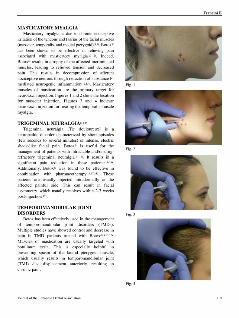
MASTICATORY MYALGIAMasticatory myalgia is due to chronic nociceptive
irritation of the tendons and fascias of the facial muscles(masseter, temporalis, and medial pterygoid)(8,9). Botox®
has been shown to be effective in relieving painassociated with masticatory myalgia(10-12). Indeed,Botox® results in atrophy of the affected incriminatedmuscles, leading to relieved tension and decreasedpain. This results in decompression of afferentnociceptive neurons through reduction of substance P-mediated neurogenic inflammation(12,13). Masticatorymuscles of mastication are the primary target forneurotoxin injection. Figures 1 and 2 show the locationfor masseter injection. Figures 3 and 4 indicateneurotoxin injection for treating the temporalis musclemyalgia.
TRIGEMINAL NEURALGIA(13,15)
Trigeminal neuralgia (Tic douloureux) is aneuropathic disorder characterized by short episodes(few seconds to several minutes) of intense, electricshock-like facial pain. Botox® is useful for themanagement of patients with intractable and/or drug-refractory trigeminal neuralgia(14-16). It results in asignificant pain reduction in these patients(14-16).Additionally, Botox® was found to be effective incombination with pharmacotherapy(14,17,18). Thesepatients are usually injected intradermally at theaffected painful side. This can result in facialasymmetry, which usually resolves within 2-3 weekspost-injection(19).
TEMPOROMANDIBULAR JOINTDISORDERS
Botox has been effectively used in the managementof temporomandibular joint disorders (TMDs).Multiple studies have showed control and decrease inpain in TMD patients treated with Botox®(8,10,12).Muscles of mastication are usually targeted withbotulinum toxin. This is especially helpful inpreventing spasm of the lateral pterygoid muscle,which usually results in temporomandibular joint(TMJ) disc displacement anteriorly, resulting inchronic pain.
Fig. 1
Fig. 2
Fig. 3
Fig. 4
Ferneini E
Journal of the Lebanese Dental Association 119
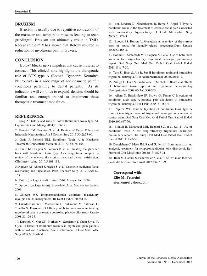
BRUXISMBruxism is usually due to repetitive contraction of
the masseter and temporalis muscles leading to teethgrinding(20). Bruxism can ultimately result in TMD.Recent studies(9,10) has shown that Botox® resulted inreduction of myofascial pain in bruxers.
CONCLUSIONBotox® blocks nerve impulses that cause muscles to
contract. This clinical note highlights the therapeuticrole of BTX type A (Botox®, Dysport®, Xeomin®,Neuronox®) in a wide range of non-cosmetic painfulconditions pertaining to dental patients. As itsindications will continue to expand, dentists should befamiliar and enough trained to implement thesetherapeutic treatment modalities.
REFERENCES:1. Lang A History and uses of botox (botulinum toxin type A).Lippincotts Case Manag 2004;9:109-12.
2. Ferneini EM, Boynton T, et al. Review of Facial Fillers andInjectable Neurotoxins. Am J Cosmet Surg 2013;30(2):53-60.
3. Gady J, Ferneini EM. Botulinum Toxin A & HeadacheTreatment. Connecticut Medicine 2013;77(3):165-166.
4. Boulle KD, Fagien S, Sommer B, et. al. Treating the glabellarlines with botulinum toxin type A-hemagglutinin complex: areview of the science, the clinical data, and patient satisfaction.Clin Interv Aging. 2010;5:101-118.
5. Nguyen AT, Ahmad J, Fagien S, et al. Cosmetic medicine: facialresurfacing and injectables. Plast Reconstr Surg. 2012;129:142-153.
6. Botox (package insert). Irvine, Calif: Allergan Inc; 2009.
7. Dysport (package insert). Scottsdale, Ariz: Medicis Aesthetics;2009.
8. Solberg WK Temporomandibular disorders: masticatorymyalgia and its management. Br Dent J 1986;160:351-6.
9. Guarda-Nardini L, Manfredini D, Salamone M, Salmaso L,Tonello S, Ferronato G Efficacy of botulinum toxin in treatingmyofascial pain in bruxers: a controlled placebo pilot study. Cranio2008;26:126-35.
10. Kurtoglu C, Gur OH, Kurkcu M, Sertdemir Y, Guler-Uysal F,Uysal H Effect of botulinum toxin-A in myofascial pain patientswith or without functional disc displacement. J Oral MaxillofacSurg 2008;66:1644-51.
11. von Lindern JJ, Niederhagen B, Berge S, Appel T Type Abotulinum toxin in the treatment of chronic facial pain associatedwith masticatory hyperactivity. J Oral Maxillofac Surg2003;61:774-8.
12. Bhogal PS, Hutton A, Monaghan A. A review of the currentuses of botox for dentally-related procedures.Dent Update2006;33:165-8.
13. Bohluli B, Motamedi MH, Bagheri SC, et al. Use of botulinumtoxin A for drug-refractory trigeminal neuralgia: preliminaryreport. Oral Surg Oral Med Oral Pathol Oral Radiol Endod2011;111:47-50.
14. Turk U, Ilhan S, Alp R, Sur H Botulinum toxin and intractabletrigeminal neuralgia. Clin Neuropharmacol 2005;28:161-2.
15. Zuniga C, Diaz S, Piedimonte F, Micheli F. Beneficial effectsof botulinum toxin type A in trigeminal neuralgia.ArqNeuropsiquiatr 2008;66(3A):500-503.
16. Allam N, Brasil-Neto JP, Brown G, Tomaz C Injections ofbotulinum toxin type A produce pain alleviation in intractabletrigeminal neuralgia. Clin J Pain 2005;21:182-4.
17. Ngeow WC, Nair R Injection of botulinum toxin type A(botox) into trigger zone of trigeminal neuralgia as a means tocontrol pain. Oral Surg Oral Med Oral Pathol Oral Radiol Endod2010;109:e47-50.
18. Bohluli B, Motamedi MH, Bagheri SC, et al. (2011) Use ofbotulinum toxin A for drug-refractory trigeminal neuralgia:preliminary report. Oral Surg Oral Med Oral Pathol Oral RadiolEndod 2011;111:47-50.
19. Denglehem C, Maes JM, Raoul G, Ferri J [Botulinum toxin A:analgesic treatment for temporomandibular joint disorders]. RevStomatol Chir Maxillofac 2012;113(1):27-31.
20. Behr M, Hahnel S, Faltermeier A, et al. The two main theorieson dental bruxism. Ann Anat 2012;194:216-9.
Correspond with:Elie M. Ferneini [email protected]
Ferneini E
120 Journal of the Lebanese Dental AssociationVolume 48 - Nº 2 - December 2013
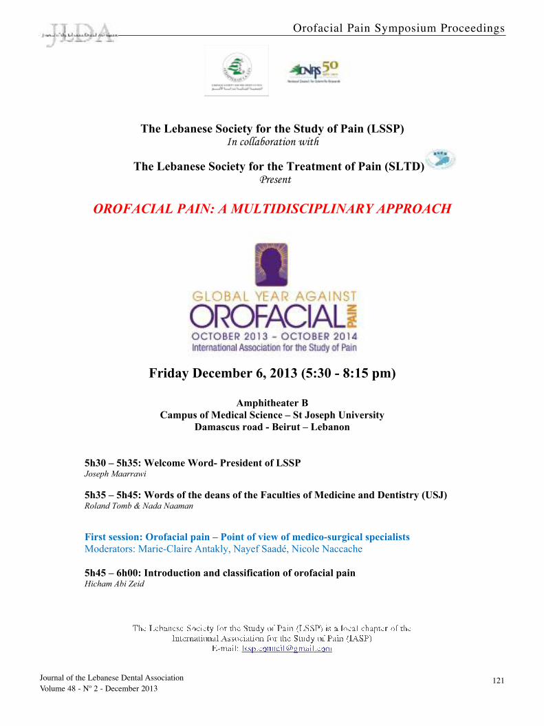
The Lebanese Society for the Study of Pain (LSSP) In collaboration with
The Lebanese Society for the Treatment of Pain (SLTD) Present
OROFACIAL PAIN: A MULTIDISCIPLINARY APPROACH
Friday December 6, 2013 (5:30 - 8:15 pm)
Amphitheater B
Campus of Medical Science – St Joseph University
Damascus road - Beirut – Lebanon
5h30 – 5h35: Welcome Word- President of LSSP Joseph Maarrawi
5h35 – 5h45: Words of the deans of the Faculties of Medicine and Dentistry (USJ) Roland Tomb & Nada Naaman
First session: Orofacial pain – Point of view of medico-surgical specialists
Moderators: Marie-Claire Antakly, Nayef Saadé, Nicole Naccache 5h45 – 6h00: Introduction and classification of orofacial pain Hicham Abi Zeid
Orofacial Pain Symposium Proceedings
Journal of the Lebanese Dental AssociationVolume 48 - Nº 2 - December 2013
121
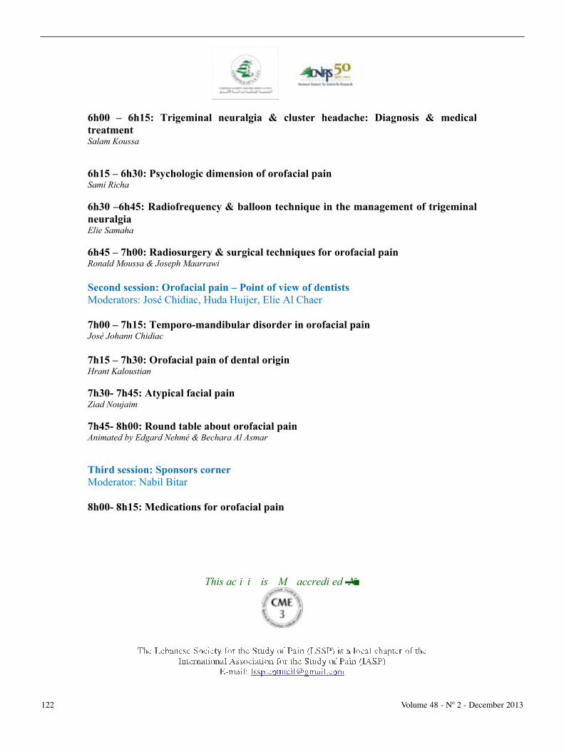
6h00 – 6h15: Trigeminal neuralgia & cluster headache: Diagnosis & medical
treatment Salam Koussa
6h15 – 6h30: Psychologic dimension of orofacial pain Sami Richa
6h30 –6h45: Radiofrequency & balloon technique in the management of trigeminal
neuralgia Elie Samaha
6h45 – 7h00: Radiosurgery & surgical techniques for orofacial pain Ronald Moussa & Joseph Maarrawi
Second session: Orofacial pain – Point of view of dentists
Moderators: José Chidiac, Huda Huijer, Elie Al Chaer 7h00 – 7h15: Temporo-mandibular disorder in orofacial pain José Johann Chidiac
7h15 – 7h30: Orofacial pain of dental origin Hrant Kaloustian
7h30- 7h45: Atypical facial pain Ziad Noujaim
7h45- 8h00: Round table about orofacial pain Animated by Edgard Nehmé & Bechara Al Asmar
Third session: Sponsors corner
Moderator: Nabil Bitar 8h00- 8h15: Medications for orofacial pain
This meeting is fully sponsored by Novartis and will be followed by a diner offered by the sponsor. Please confirm your attendance to the diner by sending an e-mail to:
[email protected] or by calling 03015835
This activity is CME accredited (3)
122 Volume 48 - Nº 2 - December 2013
ABSTRACTS
1- Introduction and classification of orofacial pain
Hicham Abi Zeid
Head and face contain a large number of anatomical structures: the brain, eyes, nose, sinuses, teeth…. This density complicates diagnosis and treatment, added is the prominent psychological significance attributed to this region. Management of orofacial pain thus demands the services of clinicians from various specialties: dentists, pain specialists, neurologists, psychiatrists… Complex referral patterns to adjacent structures are common in orofacial pain, and indeed, one man’s headache is another man’s facial pain. Thus, patient with orofacial pain may go from one specialist to the next in order to get help. Chronic orofacial pain disorders are classified into three major clinical groups: Musculoskeletal conditions or temporomandibular disorders, orofacial neuropathies and neurovascular orofacial pain. Except for musculoskeletal pain, most are unique to the trigeminal system.
2- Trigeminal neuralgia & cluster headache: Diagnosis & medical treatment Salam Koussa
Trigeminal autonomic cephalgias (TAC) are primary headaches with a common clinical phenotype consisting of trigeminal pain with autonomic signs, which may include lacrimation, rhinorrhoea and miosis. Cluster headache is the archetypal TAC, with severe pain and major autonomic activation. Neuroimaging, and careful physical and neurological evaluation, should be considered in all patients with TAC or TAC-like syndromes, particularly in those with atypical presentation. Trigeminal neuralgia (TN), also known as tic douloureux, is a distinct facial pain syndrome that may become recurrent and chronic. It is characterized by unilateral pain following the distribution of cranial nerve V. International Headache Society criteria for TN are reviewed and atypical presentations are discussed.
3- Psychologic dimension of orofacial pain
Sami Richa Somatoform disorders represent a part of psychiatric disorders with no justifying organic lesion. Among these disorders, chronic pain can occur anywhere in the body and especially in the orofacial area. Management is based in combined medical and psychotherapeutic support.
4- Radiofrequency & balloon technique in the management of trigeminal neuralgia
Elie Samaha
Trigeminal neuralgia is the most common painful orofacial pain. It is often mistaken for a pain of dental origin. Pain relief was marked by the discovery of Carbamazepine. 75% of diagnosed
Journal of the Lebanese Dental Association 123
patients become resistant to oral medication; consequently, a more invasive therapy is needed. Percutaneous procedures (Injection of Glycerol, radiofrequency and balloon compression technique) are the mini-invasive way with immediate relieving effect. Adverse effects and recurrence rate of these techniques are relatively rare. Among all aggressive therapeutic approaches, the simplest techniques are percutaneous one, to be considered in the management of trigeminal neuralgia refractory to medical treatment.
5- Radiosurgery & surgical techniques for orofacial pain
Ronald Moussa & Joseph Maarrawi
Radiosurgery (Gamma Knife or LINAC) is a recently used technique for the treatment of essential neuralgia. It consists of delivering "focused" radiation therapy to the entry point of the trigeminal nerve without the need for surgery. It is performed using a stereotactic frame installed temporarily on the patient's head under local anesthesia. It takes place in a single session, the result is observed 3-4 weeks on average after the procedure. There are currently no sufficient data to judge its long-term effectiveness. Based on recent published series, this technique is effective in two thirds of patients, with slight hypoesthesia correlated positively with its prognosis. Sensory side effects are rare. Currently, it is indicated as second-line treatment after failure of surgical decompression. Randomized controlled trials assessing more precisely its effectiveness and potential complications are essential to define its indications in the future.
Microvascular decompression is a surgical technique based on the presence of a neurovascular conflict between the trigeminal nerve and adjacent vessels in trigeminal neuralgia. The compression of the trigeminal nerve by a vessel distorts the nerve with lesions of nerve fibers secondary to arterial pulsations, causing segmental demyelization with “bypasses” leading to pain. Microvascular decompression has been well described since the 1970s for patients with refractory pain. This surgical procedure is done under general anesthesia and consists of approaching the trigeminal nerve at the trigeminal cistern through a keyhole, with dissection of the arachnoids followed by a careful separation between the nerve and the conflicting artery. The advantage of this technique is its conservative aspect; it targets the "cause" of neuralgia and does not lead to sensory disorders observed with other less invasive techniques. This technique is indicated in patients with "physiological" young age, who present no contraindication for craniotomy under general anesthesia. The results are very good, with total pain relief in more than 95% of cases, especially when the conflict is obvious. Recurrences are observed in 6-10% of cases. Serious complications are rare.
6- Temporo-mandibular disorder in orofacial pain
José Johann Chidiac
Temporomandibular disorders (TMD) have a multifactorial etiology. They are therefore difficult to diagnose and treat. This presentation is about what is related to TMD and what is not: Starting from the relationship between the ear, the neck, the migraines and TMD, in acute and chronic situations. A guide to medical practitioners and to medical personnel is described as well.
124 Volume 48 - Nº 2 - December 2013
7- Orofacial pain of dental origin
Hrant Kaloustian
Oral, facial and cervico-cephalic pain is often intricate. This presentation will focus specifically on the pain of dental origin by pulpal and periodontal inflammation that can cause this tangle.
8- Atypical facial pain
Ziad Noujaim
In 1982, Joseph Marbach reported probably the first description of neuropathic oro-facial pain by John Hunter over 200 years ago. A problem for dental practitioners in diagnosing neuropathic pain has been the use of various terms ascribed by different investigators over the years .Originally, the condition was described as "Atypical Odontalgia"-AO-in response to its "atypical" nature ,and this term was subsequently listed in the Taxonomy of Chronic Pain Syndromes by the IASP, in 1994. Other terms have included "Idiopathic Odontalgia", "Neurovascular Odontalgia", "Phantom Tooth Pain"-PTP, and "Neuropathic Oro-Facial Pain". Patients affected with "Atypical Facial Pain"-AFP- may present to the endodontist with a persistent severe pain ,yet there are no clearly identifiable clinical or radiographic abnormalities: this specific neuropathic pain was first described as a painful and unusual condition that occurs in the dento-alveolar structures and oral mucosa (Rees and Harris,1979),with patients reporting pain that was moderate to severe in intensity, and with a pattern of referral that may cross the anatomical midline of maxilla and/or mandible. In clinical practice, AFP presents as a syndrome featuring oral paresthesia or persistent pain following procedures such pulp extirpation, routine inferior alveolar nerve block, oral peripheral nerve trauma, teeth extractions, apicoectomy, periodontal surgery, and exenteration of maxillary sinus contents. The purpose of our short presentation is to revisit this unusual, unclear, and troublesome pain syndrome and address the current state of knowledge that will enable dentists to understand, diagnose and manage this unique kind of pain. It is essential for dentists and endodontists to gain and apply the knowledge of neuroplasticity of the nervous system associated with oro-facial neuropathic pain, and this will allow them to adopt a crucial role for patients sustaining neuropathic oro-facial pain, in order to achieve a successful outcome.
Journal of the Lebanese Dental Association 125
GUIDE FOR CONTRIBUTORS AND AUTHORSISSN 1810-9632
REVIEWING AND EDITING PROCESS.
All submitted manuscripts will be subject to the peer-
refereeing process and submission of a manuscript
implies that the work accomplished by author(s) has
not been published (or submitted for publication)
previously in its present form, in English or in any
other language. Submission and peer-review of all
manuscripts are conducted exclusively online and all
correspondence, including the Editor-in-Chief final
decision and request(s) for revisions, are electronically
conducted (by e-mail).
Manuscripts will be reviewed by the Editor-in-Chief,
one (or two) editorial consultant(s) and, at least, two
specialized reviewers, and in case of conflict of
opinion, an additional reviewer or special consultant
will be asked to act as adjudicator. Reviews are double-
blind (for authors and reviewers) and manuscripts
accepted for publication will be edited in consultation
with author(s), to ensure conciseness, clarity and
stylistic consistency. Studies that draw conclusions
from statistical evidence will have the statistical
analysis reviewed by biostatistics and epidemiology
consultants.
Manuscripts will kindly be addressed to:
JLDA Editor-in-Chief, Ziad Noujeim,
at the following electronic mails:
and
GUIDELINES AND MANUSCRIPT FORMAT.
The JLDA follows recommendations of the
International of Medical Journals Editors («Vancouver»
style ) in regard to preparation of manuscripts and
authorship (Ann Intern Med 1997; 126: 36-47 and update
October 2007). Submitted manuscripts not complying
with the guide for authors will kindly be returned to
authors for revision and resubmission.
Manuscripts should be submitted exclusively in
English according to the Oxford English Dictionary or
Websters.
1. Title page: the following data should be provided on
title page, in the order given below:
- title should be informative and concise (avoid
formulae and abbreviations)
- regarding author(s) names and affiliations, present
author(s) (academic or private) affiliation(s) where
actual work was implemented, below the names.
- indicate (at the end of the manuscript) the name and
e-mail address of the corresponding author (handles
correspondence).
- Indicate your manuscript type (case report, review,
mini-review, research study, technical note, short
communication, pilot study, letter to the editor,
…etc…)
2. Abstract: submit a factual/concise abstract (not
exceeding 300 words) that should be typed on a
separate page following the title page: abstract should
be structured as follows: a) statement of the problem, b)
purpose of the study, c) materials and methods(if any),
d) results (if any), e) conclusion.
3. Main text:
* clinical reports/case reports/technical notes:
should describe author(s) methods for meeting patient
treatment challenge and be no longer than 5-6 pages
(A4).
* review article /mini review:
may concern either clinical or basic scientific topics.
Standard length of a review article is 5, 000-7, 000
words, and mini review 2, 000-3, 000/
* research studies:
should follow IMRAD format (introduction, objective
of the study, working hypothesis-if any, material and
methods-including statistical method(s) used, results
and discussion).
* letters to the editor:
should be up to 800 words in length : they usually reply
to a material published in JLDA in order to make
constructive comments/criticism or introduce a point of
view. They should be accompanied by 5 to 10
references, but no illustrations. These letters do not
carry an abstract.
* abbreviations:
define them if they are not standard and ensure
consistency throughout the manuscript.
* subdivision of the manuscript:
divide manuscript into defined and numbered sections.
Subsections should be numbered 1.1 (then 1.1.1, 1.1.2,
1.1.3, …), 1.2 (then 1.2.1, 1.2.2, 1.2.3, …) etc…
Provide a brief heading to each subsection and make
sure that each heading appears on each subsection and
make sure that each heading appears on its own
separate line. Note that abstract shouldn't be included in
section numbering.
126 Journal of the Lebanese Dental AssociationVolume 48 - Nº 2 - December 2013
GUIDE FOR CONTRIBUTORS AND AUTHORSISSN 1810-9632
* conflict of interest:
at the end of the text and under a subheading «Conflict
of interest statement», authors should disclose any
financial /personal relationship(s) with other
organizations/associations/people/that could
inappropriately influence (bias) their work.
Examples of potential conflicts include employement,
consultancies, honoraria, patent
registrations/applications, paid expert testimony, stock
ownership, grants or others sources of funding.
All sources of funding should be declared as an
acknowledgement at the end of the text and authors
should declare if sponsor(s) have a role in the study
design, analysis, collection and interpretation of data.
And if the study sponsor(s) are not involved in the
study, authors should so state.
* acknowledgements:
persons/professionals who provided technical help,
writing assistance, consultancy, support… should be
listed in an acknowledgements section, at the end of the
manuscript.
* figures:
should be numbered consecutively in accordance with
their appearance in the text and figure legends listed on
a separate page.
High-resolution graphics should be provided separate
from the main text file.
* tables :
should be numbered consecutively in accordance with
their appearance in the text. Kindly place footnotes to
tables below the table body and indicate them with
superscript lowercase letters. Avoid vertical rules.
* references :
references to dental/medical/biomedical… literature
should be indicated by superscript Arabic numerals in
square parentheses and if a reference is cited more than
once in the text, the same number must be used each
time. Reference style follows the «Vancouver» format
stated in the «Uniform requirements for manuscripts
submitted to biomedical journals» ( updated October
2007). Titles of journals are to be abbreviated
according to Index Medicus style.
* illustrations:
- Number the illustrations according to their sequence
in the text.
- Produce images that fit the desired size of printed
version.
- Use uniform lettering and sizing of your original
artwork
- Convert images to TIFF format:
1- colour or greyscale photographs: use a minimum of
300 dpi
2- bitmapped line drawings: use a minimum of 1, 000
dpi
3- combinations bitmapped line/colour or greyscale:
use a minimum of 500 dpi.
Kindly:
- do not supply files too low in resolution (GIF, BMP,
PICT, WPG)
- do not submit graphics that are disproportionately
large for the content.
For colour illustrations, please make sure that
artwork files are in TIFF format and with the
appropriate resolution.
COPYRIGHT RELEASE
The following statement must accompany any
submitted manuscript before it can be reviewed for
publication, and this statement must be signed by ALL
authors:
«The undersigned author(s) transfer(s) all copyright
ownership of the manuscript (title of manuscript to be
stated) to the editorial board of the Journal of the
Lebanese Dental Association, if the manuscript is
published in the JLDA. The undersigned author(s)
warrant(s) the manuscript is original, is not under
consideration for publication by any other printed
journal/electronic medium and has not been published
previously.
The author(s) confirm that he/she/they have reviewed
and approved the final version of the manuscript».
PERMISSION AND WAIVER
- Permission of author(s) and publisher (LDA) must be
obtained for the direct use of material (text, photos,
drawings) under copyright that is not the property of
the author(s).
- Waivers must be obtained for photographs showing
persons. When such waivers are not supplied, faces
will be masked to prevent identification.
127Journal of the Lebanese Dental AssociationVolume 48 - Nº 2 - December 2013