the j biological c vol. 278, no. 4, issue of january 24 ... · formed with ecori-linearized parg7.8...
TRANSCRIPT

Functional Analysis of a Divergent System II Protein, Ccs1, Involvedin c-Type Cytochrome Biogenesis*□S
Received for publication, August 23, 2002, and in revised form, October 16, 2002Published, JBC Papers in Press, November 9, 2002, DOI 10.1074/jbc.M208652200
Beth Welty Dreyfuss, Patrice P. Hamel, Stacie S. Nakamoto, and Sabeeha Merchant‡
From the Department of Chemistry and Biochemistry, UCLA, Los Angeles, California 90095-1569
The Ccs1 gene, encoding a highly divergent novel com-ponent of a system II type c-type cytochrome biogenesispathway, is encoded by the previously defined CCS1locus in Chlamydomonas reinhardtii. phoA and lacZ�bacterial topological reporters were used to deduce atopological model of the Synechocystis sp. 6803 Ccs1 ho-mologue, CcsB. CcsB, and therefore by analogy Ccs1,possesses a large soluble lumenal domain at its C termi-nus that is tethered in the thylakoid membrane by threeclosely spaced transmembrane domains in the N-termi-nal portion of the protein. Molecular analysis of ccs1alleles reveals that the entire C-terminal soluble domainis essential for Ccs1 function and that a stromal loopappears to be important in vivo, at least for maintenanceof Ccs1. Site-directed mutational analysis reveals that asingle histidine (His274) within the last transmembranedomain, preceding the large lumenal domain, is re-quired for c-type cytochrome assembly, whereas an in-variant cysteine residue (Cys199) is shown to be non-essential. Ccs1 is proposed to interact with other Ccscomponents based on its reduced accumulation in ccs2,ccs3, ccs4, and ccsA strains.
Universal to all energy-transducing membrane systems isthe presence of c-type cytochromes on the p-side of the mem-brane, which corresponds to the plastid lumen, the mitochon-drial intermembrane space, and the bacterial periplasm. Theirdistinguishing feature is the covalent attachment of the hemeprosthetic group through thioether linkage(s) between one or,in most cases, both of the cysteine residues lying in theCXXC(H/K) motif of the apocytochrome and the vinyl groups ofheme. Genetic approaches to identify c-type cytochrome-spe-cific assembly factors led to the conclusion that at least threedistinct systems (I, II, and III) evolved for the conversion ofthese molecules to their holoforms (for review see Refs. 1–5).System I, also referred to as the Ccm1 pathway, is known from
extensive studies in �- and �-proteobacterial models such asRhodobacter capsulatus (6–9), Bradyrhizobium japonicum(10–12), Paracoccus denitrificans (13–16), and Escherichia coli(17, 18). 9 to 12 genes, whose products are dedicated to theassembly of all c-type cytochromes, define the Ccm pathway(for review see Refs. 1, 19, and 20). The Ccm2 proteins includesubunits for a putative ABC-type transporter (6, 7, 10, 14,21–23), components of a cytochrome biogenesis-specific thiolmetabolism sub-pathway (8, 19, 24–29), a putative cyt c/hemelyase (30), a unique periplasmic heme chaperone (18, 31) andits accompanying heme delivery component (32–35). In con-trast, system III, which was discovered through extensive ge-netic analysis in fungi and seems to be restricted to the mito-chondria of vertebrates and invertebrates, is a minimal systemwith a single component, the so-called cytochrome c and c1
heme lyases (CCHL and CC1HL) (36–39). The system IIICCHLs display no sequence similarity to system I and systemII components; the similarity between individual CCHLs isitself limited to the occurrence in their N-terminal domains ofbetween one to three CPV motifs that are believed to be in-volved in an interaction with heme (40, 41).
System II, the subject of this paper, operates in plastids,cyanobacteria, and some bacteria (42). Genetic studies in thegreen alga Chlamydomonas reinhardtii have assigned up to sixloci, plastid ccsA and nuclear CCS1 to CCS5 (43–45), to thematuration of chloroplast c-type cytochromes, membrane-bound cyt f and soluble cyt c6. Two of these have been identifiedmolecularly. CcsA, encoded by the ccsA locus (46), is a multiplemembrane-spanning protein and contains the tryptophan-richmotif with the “WWD” signature first noted in CcmC and CcmFof system I (4, 21, 46), and Ccs1, which also displays charac-teristic features of a membrane protein (47). Ccs1 lacks anydomains or structural features that might speak to a specificchemical function, and it appears to be unique to system II(20, 47).
Genetic studies in Bacillus subtilis and Arabidopsis thali-ana, and functional genomics in Bordetella pertussis, revealedtwo additional components required for c-type cytochrome bio-genesis in system II (48–50). One is a membrane-anchoredthioredoxin-like protein with its thiol-reducing active site onthe p-side of the membrane, called ResA/HCF164/CcsX, respec-tively (49–51), and the other is CcdA, discovered originally inB. subtilis (48, 52). CcdA corresponds to the central portion ofDipZ/DsbD, which functions in transmembrane thiol redox me-tabolism in E. coli and other bacteria (53). In B. pertussis,DipZ/DsbD is believed to function together with CcsX to pro-vide the reductant to reduce apocytochromes, either directly orindirectly, on the p-side of the membrane before attachment ofheme to the cysteinyl thiols (49).
* This work was supported by National Institutes of Health GrantGM48350, NRSA Grant GM17483 from the National Institutes ofHealth (to B. W. D.), American Heart Association Post-doctoral Fellow-ship 0120100Y (to P. H.), and a United States Public Health ServiceNRSA Award GM07185 from the National Institutes of Health (toS. S. N.). The costs of publication of this article were defrayed in part bythe payment of page charges. This article must therefore be herebymarked “advertisement” in accordance with 18 U.S.C. Section 1734solely to indicate this fact.
□S The on-line version of this article (available at http://www.jbc.org)contains Experimental Procedures, Figs. 1 and 2, and Table I.
‡ To whom correspondence should be addressed: Dept. of Chemistryand Biochemistry, UCLA, Box 951569, Los Angeles, CA 90095-1569.Tel.: 310-825-8300; Fax: 310-206-1035; E-mail: [email protected].
1 The abbreviations used are: Ccm, cytochrome c maturation; ccs,cytochrome c synthesis; Chl, chlorophyll; cyt, cytochrome; Me2SO, di-methyl sulfoxide; RT, reverse transcription.
2 For consistency, the ccm nomenclature for the system I genes will beemployed throughout.
THE JOURNAL OF BIOLOGICAL CHEMISTRY Vol. 278, No. 4, Issue of January 24, pp. 2604–2613, 2003© 2003 by The American Society for Biochemistry and Molecular Biology, Inc. Printed in U.S.A.
This paper is available on line at http://www.jbc.org2604

For CcsA, CcdA, or CcsX, conserved sequence motifs suggestfunctional domains, but this has not been the case for Ccs1,which is a highly divergent protein. Its apparently essentialfunction in popular bacterial model systems has hindered mu-tational analysis (54). Nevertheless, the fact that the cyto-chrome assembly pathways operate on multiple, divergentapoprotein substrates (two in C. reinhardtii and up to sevenin B. pertussis (42)) may be suggestive of a direct interactionbetween Ccs1 and the apocytochrome. The limited sequencerelationship among Ccs1 proteins would therefore be a con-sequence of co-evolution with the highly divergent apocyto-chromes. In this work, we undertake molecular and func-tional analyses of Ccs1. First, we confirm through molecularcomplementation that mutants previously assigned to theCCS1 locus are the result of lesions in the Ccs1 gene. Molec-ular characterization of each ccs1 allele reveals that a stro-mal loop appears to be functionally important, at least for thestability of Ccs1 in vivo. Second, we suggest that Ccs1 functionstogether not only with CcsA, as has been shown in an accom-panying paper (55), but also with multiple other Ccs compo-nents to form a “CCS complex.” Third, we undertake membranetopological analysis and site-directed mutagenesis to generatea functional model for Ccs1. We find that a single histidineresidue, located within the final transmembrane domain, pre-ceding the large soluble domain is necessary for c-type cyto-chrome assembly in chloroplasts.
EXPERIMENTAL PROCEDURES
Strains and Culture Conditions—C. reinhardtii wild-type strain CC-125 (MT�) and mutant strains ccsA-B6 (CC-2695/CC-2934), ccs1-ac206(CC-939/CC-1112), ccs1-2 (CC-3422/3423), ccs1-3 (CC-3424/CC-3425),ccs1-4 (CC-3426), abf3 (now ccs1-5::NIT1), ccs2-1 to ccs2-5 (CC-3428 toCC-3437), ccs3-F18 (CC-3092/CC-3093), ccs4-F2D8 (CC-3910/CC-3720),and ccs5-1::ARG7 (CC-3717/CC-3718), described previously (4, 43, 45,47, 56, 57), can be obtained from the Chlamydomonas Genetics Center(Duke University, Durham, NC). Arginine-auxotrophic strain arg7cw15Aused for insertional mutagenesis was obtained from Prof. J.-D. Rochaix,University of Geneva, Switzerland. Wild-type strains were grown at22–25 °C in TAP medium (58) under cool fluorescent lights (15–125�mol m�2 s�1) with agitation (225 rpm). Mutant strains were grownunder the same conditions, except that the illumination was alwaysreduced (15–25 �mol m�2 s�1). The C. reinhardtii mutant y-1 (yellow-in-the-dark; CC-735) strain was grown in TAP medium at 22 °C,wrapped in aluminum foil when necessary to prevent exposure to light.To de-green light-exposed green cells, one-half of the culture was di-luted every day into fresh TAP medium. In 10 days green cells werede-greened to a Chl concentration of 0.029 �g/ml. To re-green the cells(to a Chl concentration of 3.6 �g/ml), the flasks were unwrapped andexposed to light (100–125 �mol m�2 s�1) at 22 °C.
Insertional Mutagenesis and Identification of ccs Mutants—ccs1-6::ARG7 strain was generated by insertional mutagenesis as de-scribed previously (45). Briefly, arg7cw15A-recipient cells were trans-formed with EcoRI-linearized pARG7.8�3 by the glass bead transfor-mation method (59, 60). Arginine prototrophic colonies were screenedbased on their variable fluorescence to identify candidate mutantsblocked on the reducing side of Photosystem II (61). Candidate ccsmutants were screened for accumulation of holocyt f and holocyt c6 byheme staining and immunoblot analysis as described below. Theccs1-6::ARG7 strain was deposited into the Chlamydomonas GeneticsCenter (CC-3715/CC-3716).
Complementation of ccs1 Strains—ccs1 strains grown in TAP me-dium (3–7 � 106 cells/ml) were collected by centrifugation, 1,500 � g for5 min, and used directly for transformation (ccs1-6::ARG7) after resus-pension in TAP medium (2 � 108 cells/ml) or (for strains ccs1-ac206 andccs1-2 through 4) were resuspended in autolysin (prepared according toRef. 62) at 2 � 108 cells/ml and incubated for 30–45 min to digest awaythe cell wall, after which autolysin was diluted by addition of 40 ml ofTAP medium. The cells were recovered by centrifugation at 1,500 � gfor 5 min. For glass bead transformation (60), 0.3 ml of cells werevortexed for 15 s in the presence of 0.3 mg of acid-washed glass beadand DNA. 1 �g of SalI-linearized wild-type pCcs1-2 DNA (simply re-ferred to as pCcs1 for remainder of paper (47)) was used for comple-mentation transformations. Co-transformation experiments included
the addition of 1 �g of EcoRI-linearized pSP109 encoding the ble marker(63). 1 �g of EcoRI-linearized pTZ18U was used in control transforma-tion reactions to assess the frequency of reversion. Vortexed cells werediluted in 10 ml of TAP and transferred to 50-ml flasks for recoveryovernight in a shaking incubator. Cells were harvested by centrifuga-tion and resuspended in 1 ml of minimal medium without acetate (58).0.5 ml of cells were plated on minimal agar plates and incubated at50–125 �mol m�2 s�1 until photosynthetic colonies appeared (about 1–2weeks). The remaining 0.5 ml of cells were plated onto TAP � zeomycin(ZeocinTM, Invitrogen) (10 �g/ml) agar plates and incubated at 50–125�mol m�2 s�1 until zeomycin-resistant transformants appeared (about2–3 weeks). For the “empty vector” control transformations, the entiremix was plated onto minimal agar plates for selection for phototrophicgrowth.
The presence of introduced Ccs1 sequences was confirmed by ampli-fication of the integrated pCcs1 DNA using a gene-specific primer(CCS1–7; see Table I of the Supplemental Material for all primers) andthe Universal M13 �20 primer specific for the vector. 7 �l of genomicDNA, prepared as described previously (64), was amplified using TaqDNA polymerase in the presence of 5% Me2SO. Amplification conditionswere 94 °C for 5 min prior to addition of polymerase, 25 cycles of 94 °Cfor 1 min, 52 °C for 45 s, 72 °C for 1 min, with a final 5-min extensionat 72 °C.
Protein Preparation and Analysis—Cytochromes were detected afterfreeze-thaw fractionation and analysis of electrophoretically separatedsupernatant and pellet fractions by immunodecoration or by hemestaining as described previously (43, 47, 65). Enriched thylakoid mem-brane fractions were prepared from sonicated cell lysates and analyzedimmediately by denaturing PAGE according to Ref. 43. To increaseefficiency of transfer of electrophoretically separated enriched thyla-koid membrane proteins, 0.01% SDS was added to the transfer buffer(25 mM Tris, 192 mM glycine, 20% methanol). Enriched thylakoid mem-brane proteins were transferred at 50 V, 4 °C for �2 h to 0.2-�mpolyvinylidene difluoride membranes (Immobilon PSQ, Millipore Corp.,Bedford, MA). Polyclonal antisera raised against C. reinhardtii cyto-chrome c6 (1:1000), cytochrome f fusion protein (1:1000) (66), and Trx-Ccs1 fusion protein (1:100) (see Experimental Procedures in the Sup-plemental Material) were used for detection of cyt c6, cyt f, and Ccs1,respectively. Bound antibodies were detected chromagenically usingalkaline phosphatase-conjugated secondary antibodies.
Southern Blot Analysis—3 �g of genomic DNA (see the ExperimentalProcedures for isolation in the Supplemental Material) was digestedwith restriction enzymes and analyzed by Southern blot hybridization.For strains ccs1-ac206 and ccs1-1, the probe was prepared using Gen-esis non-radioactive nucleic acid labeling kit (Roche Molecular Bio-chemicals), hybridized, and detected chromogenically following themanufacturer’s procedure. For strains ccs1-3, ccs1-4, and the inser-tional mutant, ccs1-6::ARG7, the probe was prepared and detected asdescribed previously (67, 68).
Sequencing of ccs1 Alleles and CC125—Genomic DNA from ccs1-ac206, ccs1-2, ccs1-3, ccs1-4, and CC125 strains representing �4 kbcontaining Ccs1 encoding DNA was sequenced. Sequences representingboth DNA strands were obtained for the entire region from CC125. Forthe mutant alleles, the entire gene except intron 7 was sequenced.CCS1 was amplified from genomic DNA in five fragments (Fig. 1B) byusing primers sets A � CCS1–1 � CCS1–10, B � CCS1–2 � CCS1–9,C � CCS1–3 � CCS1–12, D � CCS1–4 � CCS1–11, and intron 7 �CCS1–17 � CCS1–20 using either Taq DNA polymerase or ExpandTM
DNA polymerase (Roche Molecular Biochemicals). Amplification reac-tions (25 �l) contained 0.2 mM dNTPs, 0.64 pmol of each primer, 1.5 mM
MgCl2, 5% Me2SO in addition to the manufacturer’s recommendedcomponents. Reactions were preheated at 94 °C for 2 min, prior to theaddition of the polymerase, followed by 30 cycles as follows: 94 °C for30 s; 56 °C for 45 s; 72 °C for 1 min; with a final 7-min extension at72 °C. Amplification products were gel-purified by the freeze-squeezemethod (69), sequenced directly by dye termination cycle sequencingusing 3� dye-labeled dideoxynucleotide triphosphates according to themanufacturer’s instruction, and run on an ABI PRISMTM DNA Se-quencer (PerkinElmer Life Sciences). Sequences were compiled andcompared using ABI PRISMTM AutoAssembler program (PerkinElmerLife Sciences). Mutations were confirmed by sequencing multiple inde-pendent amplification products.
RNA Preparation and Analysis—The procedure for RNA isolationhas been described previously (70). The abundance of Ccs1 mRNA wasestimated by amplification of cDNA under conditions that were suitablefor quantitative estimation of transcript abundance relative to Cpx1transcript abundance. Total RNA was treated with RQ1-DNase (Pro-mega, Madison, WI), phenol/chloroform-extracted, and ethanol-precip-
Functional Analysis of Plastid Ccs1 2605
itated as preparation for template for reverse transcription. Five �g oftreated RNA was used as template for Moloney murine leukemia virus-reverse transcriptase according to the manufacturer’s suggested proce-dure (Invitrogen) using pdN6 random primers (Amersham Biosciences)(1.5 �l/20-�l reaction). Control reactions were set up with same inputRNA but without the addition of reverse transcriptase (�RT). 1.5 �l ofproduct was amplified directly in reactions (25 �l) containing 1.5 mM
MgCl2, 0.2 mM dNTPs, 0.64 pmol of each primer, 5% Me2SO, and 1.25units of Taq polymerase (Fisher) with other components as specified bythe manufacturer of the enzyme. Ccs1 transcripts or Cpx1 transcriptswere amplified with primer sets CCS1–5 and CCS1–6 (Fig. 1B) orCPX1–1 and CPX1–2, respectively (64). Amplifications conditions areas follows: 94 °C for 2 min; 30 cycles at 94 °C for 30 s, 50 °C for 30 s,72 °C for 45 s and a final extension of 72 °C for 7 min (on a GeneAmpPCR system 2400; PerkinElmer Life Sciences). The yield of both prod-ucts was dependent on the amount of input RNA (0.05–10 �g). Theamount of pdN6 primer was determined to be saturating for synthesis ofthe cDNA, and the subsequent amplification reaction was in the expo-nential stage up to 35 cycles. The presence of the H274A mutation wasconfirmed by SacII digestion of the PCR product.
Generation and Analysis of CcsB-phoA and CcsB-lacZ TopologicalReporters—Eight CcsB-PhoA translational fusions were generated byPCR amplification of various segments of the Synechocystis ccsB gene(slr2087) with Pfu polymerase. The pccsB:phoA plasmids expressingtranslational fusions of CcsB to PhoA with fusions at positions 23, 67,134, 225, 288, 349, 410, and 458 of the CcsB polypeptide were con-structed as described in the Experimental Procedures of the Supple-mental Material. CcsB-encoding PCR products were cloned intopRGK200 (8), in-frame with the downstream phoA gene encoding alka-line phosphatase to yield the series of ccsB:phoA fusion plasmids. Thereciprocal ccsB:lacZ� fusions for all junctions were generated from theseries of pccsB:phoA plasmids by replacing a 2.6-kb SalI-PstI fragmentincluding the entire phoA gene with a 0.7-kb PCR-amplified lacZ seg-ment corresponding to the � fragment of �-galactosidase in-frame withthe upstream CcsB moiety. Alkaline phosphatase and �-galactosidaseactivities of each CcsB-PhoA and CcsB-LacZ� fusion were measured fortwo different clones in three independent assays as described in theaccompanying paper (55).
Generation of Site-directed Mutant Strains—Cys199, His274, andAsp348 were each mutagenized to alanine by overlap extension PCR (71)using Pfu polymerase (Stratagene, La Jolla, CA) and complementarymutagenic primer C199A-1 and C199A-2, H274A-1 and H274A-2, andD348A-1 and D348A-2 (see details in Experimental Procedures of theSupplemental Material). All mutagenized fragments were subcloned
into pCcs1 and sequenced to verify introduction of the desired mutationand absence of non-target mutations. In addition to the desired muta-tion, mutagenic primers also contained silent mutations, which wereused to distinguish mutagenized DNA from wild-type DNA. For comple-mentation experiments, 1 �g of SalI-linearized mutant Ccs1 plasmidDNA was transformed into ccs1-6::ARG7 or ccs1-4 as described above.Co-transformants of pCcs1-H274A were generated by transformation ofccs1-4arg7 strain after autolysin treatment with 1 �g of SalI-linearizedpCcs1-H274A DNA and 1 �g of pARG7 as described above. Arginineprototrophs were selected by plating on TAP agar plates (�arginine).The presence of the mutated Ccs1 gene was confirmed by amplificationof the region containing the mutation followed by diagnostic restrictionenzyme digestion of the amplification product.
RESULTS
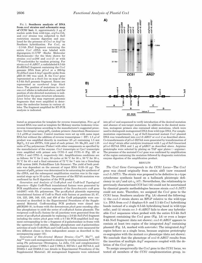
The Ccs1 Gene Corresponds to the CCS1 Locus—The Ccs1gene was cloned originally from strain abf3 (now renamedccs1-5::NIT1). The strain was proposed to be defective in c-typecytochrome synthesis based on a hallmark pleiotropic defi-ciency in cyt f and cyt c6 (47). Nevertheless, the relationship topreviously characterized CCS loci (44) could not be ascertainedby classical genetic methodologies because strain ccs1-5::NIT1could not mate. Therefore, we assigned the Ccs1 gene to theCCS1 locus. Southern analysis (Fig. 1A) showed the following:(i) the ccs1-3 strain shows an RFLP relative to the wild-type(i.e. DNA from ccs1-3 displays 6.0- and 3.1-kb Ccs1 hybridizingbands instead of a single 6.5-kb hybridizing band in wild-typecells), and (ii) strain ccs 1–6::ARG7 fails to show any hybridiz-able Ccs1 sequences when probed with the entire 6.5-kb SalIfragment containing the Ccs1 gene (Fig. 1A) or even a larger8-kb NotI fragment (data not shown). ccs1-6::ARG7 appears tocontain at least two copies of the integrated Arg7 containingplasmid (Fig. 1A, marked with asterisks). The integrated Arg7copies behave as a single locus, because arginine prototrophyco-segregates with the mutant ccs phenotype (data not shown).We conclude that the phenotype of ccs1-6::ARG7 results fromthe insertion of multiple Arg7 sequences coupled with the de-letion of the Ccs1 gene.
To assign unequivocally the Ccs1 gene to the CCS1 locus, wetested all members of the CCS1 complementation group, in-
FIG. 1. Southern analysis of DNAfrom ccs1 strains and schematic mapof CCS1 loci. A, approximately 3 �g ofnucleic acids from wild-type, arg7cw15A,and ccs1 strains was subjected to SalIrestriction enzyme digestion and ana-lyzed for the presence of Ccs1 or Arg7 bySouthern hybridization. For Ccs1, an�2.3-kb XhoI fragment containing theentire Ccs1 cDNA was labeled withdigoxigenin-11-UTP (Roche MolecularBiochemicals) (for the blots shown forstrains ccs1-ac206 and ccs1-2) or with32P-nucleotides by random priming. Forstrain ccs1-6::ARG7, a radiolabeled 6.5-kbEcoRI/SalI fragment containing the Ccs1genomic DNA from pCcs1 or a 200-bpNcoI/SacI exon 8 Arg7-specific probe frompKS-18 (86) was used. B, the Ccs1 generepresented as a thick line on map of the6.5-kb SalI genomic fragment. Exons arerepresented as numbered large blackboxes. The position of mutations in vari-ous ccs1 alleles is indicated above, and theposition of site-directed mutations is indi-cated below the gene structure schematic.Lines below the map represent genomicfragments that were amplified to deter-mine the molecular lesions in various al-leles. The fragment amplified for RT-PCRanalysis is indicated.
Functional Analysis of Plastid Ccs12606

cluding four UV-generated mutants (ccs1-ac206, ccs1-2 toccs1-4) plus ccs1-6::ARG7 by molecular complementation forrestoration of phototrophic growth. For all alleles, transforma-tion of the mutants with a plasmid containing 6.5 kb of genomicCcs1 DNA yielded phototrophic colonies on minimal medium(Table I). No phototrophic colonies were observed when thestains were transformed with empty vector DNA. When indi-vidual phototrophic colonies were tested for the presence of theintegrated plasmid copy of Ccs1, all were found to be positiverelative to the untransformed recipient (representative exam-ples in Fig. 2A). Strain ccs1-2 could not be tested directly forrescue with pCcs1 because it carries a leaky allele and displaysappreciable growth on minimal medium. Therefore, we intro-duced pCcs1 by co-transformation with the dominant blemarker, conferring resistance to zeomycin. Co-transformants ofinterest were identified among the zeomycin-resistant coloniesby specific amplification of the introduced copy of Ccs1. Theseco-transformants, ccs1-2 (pCcs1), displayed wild-type phototro-phic growth and fluorescence rise and decay kinetics (notshown). Several rescued colonies from each transformationwere tested by immunoblot and heme stain analysis and foundconsistently to accumulate wild-type (or near wild-type) levelsof cyt f (representative transformant shown in Fig. 2B). Occa-sionally, slight variability in holocyt f abundance was noted ina particular strain, but this was attributed to positional effectsresulting from unique integration of pCcs1 in individual trans-formants. Because selection for phototrophic growth relies onlyon restoration of cyt f function, complemented transformantswere also tested for cyt c6 accumulation. As expected, copper-deficient transformants accumulated holocyt c6 to approxi-mately wild-type levels, confirming that the transformantswere rescued for Ccs function.
Ccs1 Accumulation during Cytochrome Biogenesis—To mon-itor Ccs1 abundance, we raised antibodies against the putativeC-terminal lumenal domain of Ccs1 (see below for topologicalmodel). The antiserum recognized a protein of �60 kDa (Fig. 3).The signal is quite weak and is detected only when freshlyprepared membranes were analyzed. When the membraneswere purified on gradients or when they were stored (evenfrozen at �80 °C), the signal became weaker and was difficultto visualize over the noise. Therefore, for immunoblot analysis,we used a rapid method for preparing a thylakoid membrane-enriched fraction, which was solubilized directly and immedi-ately used for electrophoretic separation.
Previously, we found that coprogen oxidase, a tetrapyrrolebiosynthetic enzyme, was induced in copper deficiency, and weattributed this to an increased demand for heme synthesiswhen cyt c6 was induced (72). Therefore, we wondered whetherCcs1 accumulation might similarly be affected by copper nutri-tional status. However, we noted that cells adapted to eithercopper-replete or copper-deficient conditions accumulate thesame amount of Ccs1 (Fig. 3A). On the other hand, the abun-
dance of Ccs1 did increase on a per cell basis as the culturegrew from log phase to stationary phase (Fig. 3B). At low celldensity, we also observed a faster migrating band (marked with
TABLE IComplementation of ccs1 alleles by transformation with wild-type Ccs1 gene
ccs1 strains were transformed with either pCcs1 or vector DNA (pTZ18U) and selected for restoration of photosynthetic growth on minimalmedium.
StrainpCcs1 pTZ18U
No. prototrophsa,b No. transformations No. prototrophsa No. transformations
ccs1-ac206 39c 2 0c 1ccs1-3 112 5 0 4ccs1-4 �300–600b 4 0 4ccs1-6�ARG7 99 3 0 2
a Average number of colonies. 6 � 107 cells were used per transformation experiment.b The number of transformants varied depending on the batch of autolysin used to digest cell walls prior to glass bead transformation. For strain
ccs1-4, the number of transformants obtained was very high, and the number of colonies was estimated.c Prototrophs appeared as large well defined colonies rising up from lawn of very slowly growing cells. Lawn of slowing growing cells remained visible
in plasmid control transformation experiment, but no prototrophic colonies were visible, even after 3 weeks of growth at 50–125 �mol m�2 s�1.
FIG. 2. Complementation of ccs1 strains by transformationwith wild-type genomic Ccs1 DNA. A, genomic DNA from ccs1strains and representative complemented transformants was amplified.The introduced pCcs1 DNA was distinguished by using a Ccs1-specificprimer (CCS1–7) and a plasmid specific primer (Universal). pCcs1 wasamplified in parallel to generate a product standard. B, extracts wereprepared from copper-deficient cultures of each strain. Proteins fromthe insoluble membrane fraction (equivalent to 5 �g of chlorophyll)were separated in a 12% polyacrylamide gel under denaturing condi-tions, and the immunoblots were probed with antiserum againstChlamydomonas cyt f. Total soluble proteins (equivalent to 5 �g ofchlorophyll) were separated in 15% polyacrylamide gels under nonde-naturing conditions, and immunoblots were probed with antiseraagainst Chlamydomonas cyt c6. Heme-containing proteins immobilizedon the membranes were detected by chemiluminescence. Because cyt c6expression is strictly regulated by copper availability, the extent ofholocytochrome c6 accumulation reflects minor differences in cellularcopper nutritional status (87).
Functional Analysis of Plastid Ccs1 2607

faint arrow) whose appearance correlated with cell density, i.e.more predominant in cultures in early exponential growth thanstationary phase cultures (Fig. 3B). We know that the fastermigrating band is Ccs1-specific because it is absent in immu-noblots of ccs1 null mutants (see Fig. 9). At present, it isunclear if the faster migrating molecule is a physiologicallyrelevant species or simply represents a degradation productgenerated during sample preparation.
The y-1 strain (deficient in the light-independent protochlo-rophyllide reductase, see Ref. 73) has been used as a modelsystem to study thylakoid membrane biogenesis by light-initi-ated greening of de-greened cells (74–76). In contrast to chlo-rophyll proteins, cytochromes do accumulate in dark grown andnon-green plastids (77), but their abundance increases alongwith other components of the thylakoid membrane as the de-greened cells re-assemble their photosynthetic apparatus (78,
79). We hypothesized that Ccs1 would be present in non-greenplastids. As expected, both Ccs1 and cyt f are present in darkgrown y-1 cells. The abundance of both proteins increased inparallel to each other and with the synthesis of chlorophyll(Fig. 4). Nevertheless, although the accumulation of cyt f re-quires Ccs1 function, the accumulation of Ccs1 is independentof cyt f. For instance, a petA deletion mutant (FIEB1, 80)accumulates Ccs1 to wild-type levels (data not shown).
Functional Analysis and Topology—With the objective of
FIG. 3. Abundance of Ccs1 under various growth conditions.Enriched thylakoid membrane fractions (corresponding to 50 �g of Chlfor each lane) from wild-type cultures were tested for the accumulationof Ccs1 by immunoblot analysis. Wild-type cells were grown in thepresence or absence of copper (A) and to different cell densities (B).Solubilized proteins were separated by electrophoresis on SDS-contain-ing polyacrylamide (10%) gels, transferred to polyvinylidene difluoridemembranes, incubated overnight with anti-Ccs1 antisera, and detectedchromogenically using an alkaline phosphatase-conjugated secondaryantibody. Arrows indicate position of Ccs1-specific species. The anti-Ccs1 antiserum specifically recognizes a protein of �60 kDa that ispresent in wild-type samples and absent in ccs1 mutants (see Fig. 9).
FIG. 4. Ccs1 and cytochrome f accumulation during greening.A, Ccs1, cyt f, and CF1 (as loading control) were monitored during thegreening of y-1 (yellow in the dark) mutant cells. Enriched thylakoidmembranes fractions were prepared after the indicated number ofhours of light exposure. Protein samples corresponding to 5.25 � 106
cells per lane were separated electrophoretically on an 8.75% SDS-containing gel for immunoblot analysis. B, the parallel accumulation ofChl was determined by organic extraction of Chl and spectrophotomet-ric quantitation.
FIG. 5. Topological arrangement of Chlamydomonas Ccs1 in the thylakoid membrane. The predicted topology is based on phoA and lacZfusion analysis of Synechocystis CcsB and the alignment of C. reinhardtii Ccs1 and Synechocystis CcsB sequences (see Supplemental Material Figs.1 and 2). The equivalent positions of Synechocystis fusion constructs are indicated by numbered arrows (Syn1 through Syn8). This topology isconsistent with the topological prediction based on the “positive-inside” rule (number of basic residues in loop 1: C. reinhardtii � �3, Synecho-cystis � �1. Number of basic residues in loop 2: C. reinhardtii � �12, Synechocystis � �14 (88)). The point mutations in ccs1 alleles are indicatedby open arrows at the position of the mutations. Residues altered by site-directed mutagenesis are shown as follows: C199A and D348A mutations,which did not result in a discernible phenotype are indicated by circles. The H274A mutation, which did result in a ccs phenotype, is indicated bya square. The relative position of the insertion of pNIT1 in the insertional mutant ccs1-5::NIT1 and the breakpoint in the ccs1-3 allele is indicatedby an * near the C terminus of protein. Regions within soluble lumenal domain displaying limited blocks of sequence conservation are indicatedwith bold lines. Region II containing an invariant signature motif VNxP, where x is a polar and generally positively charged residue (51 identifiablesequences, data base search May, 2002), is highlighted in black, whereas regions I and III showing less sequence conservation are highlighted ingray (see Supplemental Material Figs. 1 and 2 for alignment of Ccs1 sequences).
Functional Analysis of Plastid Ccs12608

deducing a functional model of Ccs1, we assembled a multiplealignment of all Ccs1-like sequences (see Supplemental Mate-rial Figs. 1 and 2). Based on these alignments, we predicted atopological arrangement of Ccs1 within the thylakoid mem-brane, and we also identified invariant residues (Fig. 5) (81).The topological predictions indicated that Ccs1 could containthree transmembrane segments in the N-terminal region of theprotein followed by a large hydrophilic lumenal loop, followedby a fourth transmembrane span with a weak prediction ratingat the C terminus (see hydropathy profiles in Ref. 47). To testthe topology predictions, we used a cyanobacterial homologueof Ccs1, Synechocystis CcsB, in phoA and lacZ� fusions. Therelationship between Synechocystis CcsB and chloroplast Ccs1is obvious, and the model of the cyanobacterial protein shouldbe extendable to the chloroplast situation. Analysis of the fu-sion constructs confirmed three membrane spans domains clus-tered at the N terminus of the Ccs1 homologue (Table II). PhoAfusions on the p-side of the membrane, Syn2 and Syn4, showhigher alkaline phosphatase activity compared with fusionsSyn1 and Syn3 on the n-side. Conversely, Syn1 and Syn3fusions are more active on the n-side as �-galactosidase fusions(Table II). Fusion constructs Syn 4-8 (where Syn8 is at the veryC terminus of the protein) all show high alkaline phosphataseactivity and conversely low �-galactosidase activity, which isconsistent with the p-side location of the entire C-terminaldomain. On this basis, we discount the weak prediction of afourth transmembrane segment and favor the topology dia-grammed in Fig. 5.
An Essential Histidine—Multiple alignment of Ccs1-like se-quences at the outset of these experiments revealed very fewresidues in Ccs1 that are absolutely conserved (see Ref. 47) andhence might be catalytically significant. Because biochemicalanalysis of ccs1 mutants suggested that Ccs1 participates interminal steps of cytochrome synthesis involving attachment of
heme to the apoprotein within the thylakoid lumen (43, 44), weconsidered that Ccs1 might be involved in substrate binding,either heme or apoprotein. Alignment of Ccs1 homologues high-lighted three invariant residues, cysteine 199, histidine 274,and aspartic acid 348, with interesting functional groups andpotential for interaction with heme. These three residues werechosen for site-directed mutagenesis and were changed to theneutral amino acid alanine. The corresponding alanine encod-ing mutated versions of Ccs1 were then tested for their abilityto rescue strain ccs1-6::ARG7 for photosynthetic growth onminimal medium. Plasmids carrying the C199A and D348Amutations could complement ccs1-6::ARG7. Numerous photo-synthetic colonies appeared after transformation (Table III) atfrequencies comparable with wild-type (Table II). Representa-tive C199A and D348A transformants were analyzed for theaccumulation of holocyt f and holocyt c6 (Fig. 6). As expectedfrom their ability to grow on minimal medium, C199A andD348A transformants were fully capable of synthesizing holo-cyt f, and under copper-deficient growth conditions, bothC199A and D348A transformants were able to synthesize ho-locyt c6. Therefore, we conclude that cysteine 199 and asparticacid 348 are not required for Ccs1 function under laboratorytest conditions.
On the other hand, H274A failed to rescue eitherccs1-6::ARG7 or ccs1-4 (which rescues at high frequency) (TableIII), suggesting that histidine 274 is essential for Ccs1 function.To confirm the role of histidine 274, pH274A was introduced byco-transformation of ccs1-4arg7 with pArg7. Thirty-four H274Aco-transformants were identified by specific amplificationamong 81 arginine prototrophs (two transformation experi-ments). Thirty of the 34 H274A co-transformants failed to show
FIG. 6. Accumulation of c-type cytochromes in C199A andD348A mutants. Protein samples from cells transformed with plasmidcarrying wild-type, C199A-encoding, or D348A-encoding copies of Ccs1were prepared as described previously in Fig. 1 and analyzed by immu-noblot or heme staining. For cyt f, membrane fractions were separatedon a 10% SDS-containing polyacrylamide gel. An equivalent of 10 �g ofChl per sample was loaded. For cyt c6, soluble protein samples wereseparated on a 15% native gel. An equivalent of 10 �g of Chl per samplewas loaded.
TABLE IIIComplementation of ccs1 alleles by transformation with site-directed
mutations in the Ccs1 geneSite-directed mutations in Ccs1 were tested for their ability to com-
plement ccs1 strains and restore photosynthetic growth when selectedon minimal medium.
Mutation Strain No. prototrophsa No. transformations
C199A ccs1-6�ARG7 150 5H274A ccs1-6�ARG7 0 3H274A ccs1-4 0 1D348A ccs1-6�ARG7 79 5
a Average number of colonies resulting from 6 � 107 cells per trans-formation experiment.
TABLE IITopology analysis of Synechocystis sp. PCC 6803 CcsB by phoA and lacZ fusion analysis
Alkaline phosphatase and �-galactosidase activities of CcsB fusion proteins expressed in E. coli were measured as described under “Experi-mental Procedures.” At least two representatives of each CcsB fusion were tested for activity. The value is indicated as the mean � S.D. of threeindependent measurements for the two representatives. n-side and p-side correspond to the negative and positive side of the membrane,respectively.
Fusionconstruct
Position inCcsBa
Position inCcs1b phoA activity lacZ activity Topology
Miller units units
vector 23 (�3) 10 (�2)Syn 1 23 119 85 (�3) 144 (�38) n-SideSyn 2 67 164 1368 (�124) 10 (�3) p-SideSyn 3 134 231 65 (�7) 58 (�7) n-SideSyn 4 225 323 1031 (�125) 9 (�2) p-SideSyn 5 288 388 978 (�121) 12 (�2) p-SideSyn 6 349 492 613 (�45) 12 (�4) p-SideSyn 7 416 559 530 (�44) 13 (�2) p-SideSyn 8 458 613 420 (�68) 18 (�1.6) p-Side
a Position of actual fusion with CcsB from Synechocystis sp. PCC 6803.b Equivalent position of CcsB fusion to Ccs1 from C. reinhardtii based on multiple alignment (See Supplemental Material Figs. 1 and 2).
Functional Analysis of Plastid Ccs1 2609

photosynthetic growth on minimal medium and were unable toaccumulate cyt f (see Fig. 7, H274A lanes 1, 2, and 4 forrepresentative examples). Four of the 34 H274A co-transfor-mants displayed limited and spotty growth on minimal me-dium and accumulated �5% of wild-type levels of cyt f, consist-ent with their limited photosynthetic capacity (see Fig. 7,H274A lane 3 for a representative). Four of the H274A co-transformants were analyzed in more detail and confirmed byRT-PCR amplification followed by diagnostic restriction diges-tion of the product to express the H274A mutated version of theCcs1 mRNA (data not shown). All four confirmed H274A mu-tants accumulated very low levels of Ccs1 (�2–5% of wild type)(Fig. 7). Three of the four H274A mutants (1, 2, and 4) failed toaccumulate either holocyt f or holocyt c6, although very lowlevels of an anti-cyt f immunoreactive species still accumulatedin these transformants. We concluded that the immunoreactivespecies is the apoprotein form because it is �0.7 kDa smallerthan native holocyt f (corresponding to loss of the heme group)and also the band does not stain for heme. The H274A trans-formant 3 that displays very limited growth on minimal me-dium appears to accumulate both the apoprotein and holopro-tein forms of cyt f based on the observation of a doublet in Fig.7. Only the upper band shows heme staining, confirming itsidentity as holocyt f. Interestingly, transformant 3 does notshow any accumulation of either apo or holo form of cyt c6. Weconclude that the His274 residue is important for Ccs1 function.
Molecular Analysis of ccs1 Alleles—The existing collection ofccs1 alleles also provided an opportunity to distinguish func-tional domains in Ccs1. With this in mind, the mutations ineach UV-generated ccs1 allele were identified by sequencingthe Ccs1 genomic DNA from each strain (Fig. 1B) (GenBankTM
accession numbers AY095299–AY095304). Ccs1 was se-quenced also from the corresponding wild-type strain, CC125(GenBankTM accession number AY095298) (44). The mutationsin the ccs1 alleles are summarized in Table IV. In ccs1-ac206,the conserved guanine nucleotide at the 3� splice site junctionof intron 4 and exon 5 is mutated to A (Table IV). Becauseintron 4 is 108 nucleotides in length, failure to splice intron 4from the ccs1-ac206 mRNA would result in an mRNA encodingan additional 36 amino acids within the predicted stromal loopof Ccs1 (see Fig. 5). The longer species was not detected byRT-PCR; however, immunoblot analysis did reveal a slowermigrating species, which could be a translation product fromthe unspliced longer transcript (discussed below). The ccs1-2phenotype results from a missense mutation wherein non-con-served glycine 260 is changed to asparagine, ccs1-4 results froma nonsense mutation at codon 144, and the ccs1-3 phenotypeappears to be the result of a rearrangement within intron 9,which would be expected to destroy the very C-terminal 77
FIG. 7. Mutation to invariant His274 results in c-type cyto-chrome-deficient phenotype. Enriched thylakoid membrane frac-tions were examined for the accumulation of Ccs1 and cyt f (as de-scribed previously). Soluble extracts from copper-deficient cultureswere examined for the accumulation of cyt c6. The presence holocyto-chromes in the samples was examined by heme staining (as described inFig. 1). Exposures of heme staining representing equivalent intensitiesfor wild-type samples are shown. Dilution series of wild-type, ccs1-4arg7 (recipient strain for transformation), and four independent rep-resentative H274A site-directed mutant transformants are shown.
FIG. 8. Expression of Ccs1 in ccs1 strains and other ccs mu-tants. C. reinhardtii total RNA was isolated from wild-type or ccsmutants, digested with RQ1 DNase, and used as template for reversedtranscription with random pN6 primers. The cDNA corresponding toCcs1 transcripts were detected by amplification with CCS1–5 andCCS1–6 primers to yield a 345-bp product. A cDNA corresponding toCpx1 transcripts was amplified in parallel as an internal control usingCPX1–1 and CPX1–2 primers resulting in a 634-bp product. Lanesmarked -RT show the result of amplification reaction on the same RNApreparations but without reversed transcription. Plasmid DNA contain-ing either Ccs1 cDNA or Cpx1 cDNA were used as templates for am-plification with the same primers to generate a standard product (lanemarked plasmid). A, Ccs1 transcript abundance in various ccs1 strains.B, Ccs1 transcript abundance in other ccs mutants.
TABLE IVSummary of ccs1 mutations
Strain Mutation Predicted result on translation
ccs1-ac206 G1442/GTG3 a1442/GTG Failure to splice intron 4, potential read-through adding 36 aminoacids between Gln250 and Val251
ccs1-2a G1470G14713 AA Gly2603 N(AAC)ccs1-3 Rearrangement within intron 9 Truncation after 536 residuesccs1-4 Cys6113 Ala Gln1443 Stop (TAA)
a Silent mutation at Thr9523 Cys (YUAU3 YUAC) also observed in ccs1-2 allele.
Functional Analysis of Plastid Ccs12610

amino acids of the protein. The re-arrangement was verified byamplification (Fig. 1B) and was consistent with Southern anal-ysis (Fig. 1A). Specifically, fragments A–C could be amplifiedfrom ccs1-3 genomic DNA, and sequence analysis confirmed thewild-type sequence. Only the 5� portion of fragment D could beamplified using a primer annealing within exon 9. Primersannealing downstream of exon 9 used in conjunction with theupstream CCS1–12 primer consistently failed to produce am-plification products from ccs1-3 DNA, whereas wild-type DNAyielded a product. On this basis, we placed the breakpoint forthe genomic rearrangement in ccs1-3 within intron 9 �6.0 kbfrom the 5� SalI site. We noted that exon 10 could be amplifiedfrom ccs1-3 to yield a product of the expected size, suggestingthat the rearrangement event may be an inversion within thegene.
To assess the effect of each mutation on Ccs1 expression, weassayed for the presence of Ccs1 transcripts by a quantitativePCR-based method (Fig. 8). Reverse-transcribed cDNA wasused as the template for amplification using primers that hy-bridized to exon 4 and exon 6. The amounts of Ccs1 cDNAswere normalized against the amounts of Cpx1 cDNAs, becauseCpx1 is expressed constitutively under these conditions, andthe level of expression is unaffected in mutants. A product withthe size expected for the mature Ccs1 message was amplifiedfrom all strains (except ccs1-6::ARG7 in which Ccs1 has beencompletely deleted). Amplification products corresponding totemplates derived from unspliced messages containing intron 4and/or intron 5 were never observed from any RNA prepara-tion. Strains ccs1-2 and ccs1-3 accumulate Ccs1 transcripts towild-type levels (Fig. 8A) as do ccs2, ccs3, ccs4, and ccs5 alleles(Fig. 8B), but ccs1-ac206 (mutation at conserved splice site G),ccs1-4 (early nonsense), and ccs1-5::NIT1 (insert in exon 10)accumulate only about 25% of wild-type levels of Ccs1 tran-scripts. Surprisingly, the ccs1-ac206 mRNA that accumulatesseems to be spliced correctly, despite the mutation in intron 4.A product representing mRNA containing the unspliced intron4 was never observed in the ccs1-ac206 RNA population underthe conditions used, even when intron 4-specific primers wereused to target such a species (data not shown). The longerprotein product (see below) implicates the existence of intron4-containing mRNA in a translatable pool, but we concludethat the species must be short lived and hence not well repre-sented in the mRNA pool. The decreased abundance of ccs1-4mRNA is not surprising because non-sense-mediated mRNAdecay (82) has been observed previously in Chlamydomonas(65). Previously, we could not detect Ccs1 mRNA inccs1-5::NIT1 by RNA blot analysis (47). In this work, a smallsteady state amount is implicated by the RT-PCR results,which indicates that Ccs1 is still being transcribed in the in-sertional mutant.
The abundance of Ccs1 in the ccs mutants relative to wild-type cells was examined by immunoblot analysis of thylakoidmembranes. The sensitivity was limited to detection of �2–5%of wild-type levels of Ccs1 (Fig. 9A). Nevertheless, very lowamounts of Ccs1 could clearly be seen in membranes from themissense ccs1-2 strain. We suggest that the mutation mustde-stabilize the protein, indicating the structural importance ofthe stromal loop. Membranes from ccs1-ac206 contain a slowermigrating form of Ccs1 in addition to very low levels of awild-type sized Ccs1. The abundance of both forms is highlyvariable between sample preparations. The larger form is mostlikely the result of translational readthrough of the unsplicedintron 4 in ccs1-ac206, which would add 36 amino acids to Ccs1,and the estimated size of the slower migrating form is consist-ent with a 4-kDa increase. Ccs1 was not detected in any otherccs1 strain, which is expected from the nature of their molec-
ular lesions. Interestingly, when we examined mutants atother CCS loci, we noted that strains ccsA, ccs2, ccs3, and ccs4each accumulated only 10–15% of wild-type levels of Ccs1 (Fig.9B) even though Ccs1 mRNA accumulates to normal levels.The decreased accumulation of Ccs1 in these strains appears tobe either translationally or post-translationally controlled (see“Discussion”). On the other hand, ccs5–1::ARG7, a leaky ccsstrain that accumulates �5–10% of wild-type levels of cyt f andcyt c6 (45), accumulates wild-type amounts of Ccs1. Perhapsthis suggests significant functional differences in the site ofaction of Ccs5 versus CcsA, Ccs1 through Ccs4.
DISCUSSION
Topology and Functional Importance of Domains withinCcs1—Topology studies of Synechocystis sp. PCC 6803 CcsB(cyanobacterial homologue of plastid Ccs1) suggest that Ccs1/CcsB is anchored in the thylakoid membrane by three trans-membrane domains within the N-terminal half of the protein,which places the C-terminal half as an extramembrane lume-nal domain (Fig. 5). This topology differs slightly from themodel for B. pertussis, CcsB (49), in which an additional trans-membrane domain at the very C terminus of the protein wasproposed, based on positive (albeit low) alkaline phosphataseactivity for the full-length C-terminal phoA fusion. In our work,we have used both phoA and lacZ� fusion analysis tostrengthen the model that plastid/cyanobacterial type Ccs1/CcsB has only three transmembrane domains, and the entireC-terminal domain resides on the lumen side of the thylakoidmembrane.
The topological model was a prerequisite for deriving insightfrom molecular analysis of the ccs1 alleles and for building afunctional model based on mutational analysis of conservedresidues. Our first conclusion is that the stromal loop in Ccs1 isimportant (Fig. 5). The expected outcome of failure to spliceintron 4 in strain ccs1-ac206 is the insertion of 36 codons in theregion of the mRNA corresponding to the stromal loop, andindeed, a protein of larger size was observed in ccs1-ac206 (Fig.9A). Strain ccs1-ac206 also appears to produce normal sizedCcs1 at very low levels, probably from a pool of normally splicedintron 4 (see “Results”). However, holocytochromes do not ac-cumulate (Fig. 2), which is consistent with previous studies
FIG. 9. Abundance of Ccs1 in various ccs1 strains and other ccsmutants. Enriched thylakoid membrane fractions (corresponding to 50�g of chlorophyll) from wild-type or ccs strains were tested for thepresence of Ccs1 by immunoblot analysis as described in Fig. 3. A,detection of Ccs1 in various ccs1 strains. B, detection of Ccs1 in other ccsmutants.
Functional Analysis of Plastid Ccs1 2611

that showed by pulse-chase analysis that ccs1-ac206 is com-pletely incapable of holocytochrome formation (43). We con-clude that the insertion of additional amino acids in the stro-mal loop renders the larger form non-functional and that thislarger form must exert a dominant-negative effect on the lesserabundant normal Ccs1 population, suggesting Ccs1 associa-tions with other Ccs components in vivo. Blue native-PAGEindicates that Ccs1 is found in an �200-kDa CcsA-dependentCcs complex in the thylakoid membrane (see accompanyingpaper (55)). This size is more than adequate to accommodatetwo subunits of Ccs1 and/or the products of other CCS loci. Thedecreased abundance of Ccs1 in ccs2, ccs3, and ccs4 mutants(Fig. 9B) also argues in favor of Ccs1 interactions with addi-tional Ccs components.
The importance of the stromal loop is underscored by molec-ular analysis of strain ccs1-2 in which mutation of a singleresidue in the stromal loop, due to a conversion of a non-conserved glycine to an asparagine, results in dramaticallyreduced Ccs1 accumulation (�2% of wild-type levels). The al-tered residue lies a mere two amino acids away from a highlyconserved pair of residues, 262KG263 in the Chlamydomonasprotein. The mutation must destabilize Ccs1 in the membrane,perhaps by affecting interactions with partner subunits. TheG260N mutant form of Ccs1 is functional to the extent that itaccumulates in the membrane (at least for assembly of holocytf); between 1–5% of wild-type levels of holocyt f can be observedin thylakoid membranes from ccs1-2 (44), and the strain growsto a limited extent on copper-replete minimal media. However,in copper-deficient medium, ccs1-2 cannot synthesize holocytc6, and this is clearly evident in pulse-chase experiments (44).The separate effect of the ccs1-2 mutation on cyt c6 versus cyt faccumulation is interesting because it shows that the role ofCcs1 in the assembly of cyt f can be separated from its role incyt c6 assembly (see also discussion of site-directed mutants,below). Because the mutation occurs within a stromal loop ofCcs1 whereas biochemical evidence clearly places the apocyto-chrome substrates and site of cytochrome maturation withinthe lumen, we think it unlikely that the mutation contributesdirectly to altered interaction with apocytochromes. It is morelikely that apocyt f is a kinetically favored substrate in the Ccsassembly pathway. A correlation between apoprotein abun-dance and the molecular lesions in Ccs1 was not observed inthe Chlamydomonas mutants. Indeed, the four potentially nullmutants (ccs1-3 through ccs1-6) all accumulate apocyt f and insome cases to appreciable levels (see apocyt f band observed inccs1 strains in Fig. 2). The definitive evidence for the proposedchaperone function of Ccs1 therefore requires further study.
Molecular analyses of ccs1-3, ccs1-5::NIT1, and a site-di-rected C-terminal deletion construct, �542–613, highlight thefunctional importance of the very C-terminal region of the largelumenal domain. Strains ccs1-3 and ccs1-5 result from lesionswithin intron 9. Both strains do accumulate Ccs1 message andretain the potential to encode at least 536 of the 613 aminoacids of Ccs1. However, both strains fail to synthesize eitherholocytochrome c6 or f as confirmed by radiolabeling experi-ments (47), and the C-terminal deletion construct (�542–613)fails to rescue ccs1-4 (data not shown). In all three cases, thetruncation in Ccs1 occurs after the third region of sequenceconservation (Fig. 5, and see Supplemental Material Figs. 1and 2), yet the protein is non-functional. In the B. pertussisstudy (49) as well, only the full-length fusion construct was ableto complement the ccsB mutant. These results emphasize thefunctional importance of the entire C-terminal domain andargue for the same topological placement of the C terminus onthe p-side of the membrane where cytochrome maturationoccurs.
A Non-essential Cysteine and Identification of a FunctionallyImportant Histidine—Site-directed mutagenesis identified ahistidinyl residue, His274, as important for Ccs1 function in thematuration of c-type cytochromes. Two other residues, cysteine199 and aspartic acid 348 that were absolutely conserved at thestart of these studies, were shown by mutagenesis to be non-essential for Ccs1 function. Analysis of 51 Ccs1-like proteins inthe data bases (as of May, 2002) revealed that the cysteineresidue is totally invariant, but the aspartic acid residue is notconserved. It remains possible that the invariant cysteine res-idue is important for Ccs1 function but may not be absolutelyessential; therefore, the mutation does not present a visiblephenotype under the conditions examined. The conserved cys-teine is an attractive candidate for participation in the thiore-duction pathway involved in cytochrome biogenesis (1, 19).Interestingly, an additional N-terminal domain containing fourclustered cysteines, two within a thioredoxin motif, has beenidentified in the Ccs1 homologue of B. subtilis, ResB (83). Thiscysteine-rich region is present in all Bacillus ResB proteinscurrently identified and in Geobacillus stearothermophilus butis lacking in all other identified Ccs1 homologues (data basesearch May, 2002). The function of this domain remains spec-ulative. Identification and multiple alignments of additionalCcs1 homologues have identified few other conserved residueswith attractive functional potential. However, an invariantsignature sequence, VNXP, located within the large stromalloop (see Fig. 5) that had not been aligned previously (see Ref.47) has been highlighted. Whether this sequence element couldbe involved in an interaction with heme is of future researchinterest.
The H274A mutated version of Ccs1 is clearly non-func-tional. The mutated constructs failed to complement ccs1 mu-tants regardless of the particular allele used for the transfor-mation experiments, and even when a period of recovery inacetate-supplemented medium was allowed after transforma-tion. Detailed analysis of four strains carrying H274A con-structs showed that three of the four strains were completelydevoid of holocytochromes in the plastid. The mutations mustaffect a catalytic function of Ccs1 because the mutated proteindoes accumulate. A fourth transformant was capable of limitedholocyt f formation but failed to form holocyt c6, a phenotypethat is similar to that of strain ccs1-2. We cannot explain whyone transformant makes a small amount of holocyt f. The fourH274A transformants appear to contain comparable amountsof Ccs1, but it is possible that the subtle variations in Ccs1abundance are not revealed by immunoblot analysis, and itmay be that small differences in expression of mutant Ccs1genes (resulting from independent integration events in eachtransformant and accompanying variations in transgene ex-pression (84)) contributes to variations in the severity of thephenotype. We note that all four H274A transformants accu-mulate very low levels of apocyt f but no apocyt c6. Perhapsapocyt c6 is more susceptible to proteolysis relative to apocyt fwhich may be sheltered within the membrane or within the cytb6f complex. The presence of a small steady state pool of apocytf but not apocyt c6 may contribute to the preferential synthesisof holocyt f in the histidine transformant discussed above andin the ccs1-2 allele.
In Wolinella succinogenes, a protein designated NrfI is re-sponsible for heme attachment to the unique CXXCK motif ofthe pentaheme cytochrome c catalytic subunit, NrfA, of nitritereductase (85). Even though pairwise BLAST analysis (Gen-BankTM) does not reveal significant sequence similarity be-tween any NrfI and any Ccs1, topology predictions based onmultiple alignments of NrfI homologues predicts a similartransmembrane structure for the N-terminal two-thirds of NrfI
Functional Analysis of Plastid Ccs12612
as for Ccs1. Therefore, NrfI and its homologues may be struc-turally analogous to a Ccs1/CcsA fusion with three tightlyspaced transmembrane domains at the N terminus followed bya large extramembrane domain on the p-side, followed by aC-terminal CcsA-related portion containing multiple mem-brane-spanning segments and a conserved WWD domain.Alignment of NrfI homologues identifies a conserved histidinein the third transmembrane domain, preceding the large solu-ble domain and positioned toward the n-side of the membranespan. We wonder if this histidine is analogous to the essentialhistidine identified in Ccs1 in this work and indicates a com-mon ancestral origin between NrfI and the CcsA/Ccs1 compo-nents in system II.
Acknowledgments—We acknowledge the past and present membersof the Merchant laboratory for support and intellectual input during thecourse of this study; J. Leichman and S. Gabilly for technical assistance;Dr. J. Quinn for help during the analysis of Ccs1 expression; and Dr.J. M. Moseley for generation of the ccs1-4arg7 strain and for other helpduring the course of the project. We thank Prof. J.-D. Rochaix, Univer-sity of Geneva, for hosting B. W. D in his laboratory during which periodthe ccs1-6::ARG7 strain was identified.
REFERENCES
1. Thony-Meyer, L. (1997) Microbiol. Mol. Biol. Rev. 61, 337–3762. Kranz, R., Lill, R., Goldman, B., Bonnard, G., and Merchant, S. (1998) Mol.
Microbiol. 29, 383–3963. Page, M. D., Sambongi, Y., and Ferguson, S. J. (1998) Trends Biochem. Sci. 23,
103–1084. Xie, Z., and Merchant, S. (1998) Biochim. Biophys. Acta 1365, 309–3185. Nakamoto, S. S., Hamel, P., and Merchant, S. (2000) Biochimie (Paris) 82,
603–6146. Beckman, D. L., Trawick, D. R., and Kranz, R. G. (1992) Genes Dev. 6, 268–2837. Goldman, B. S., Beckman, D. L., Bali, A., Monika, E. M., Gabbert, K. K., and
Kranz, R. G. (1997) J. Mol. Biol. 268, 724–7388. Monika, E. M., Goldman, B. S., Beckman, D. L., and Kranz, R. G. (1997) J. Mol.
Biol. 271, 679–6929. Deshmukh, M., Brasseur, G., and Daldal, F. (2000) Mol. Microbiol. 35,
123–13810. Ramseier, T. M., Winteler, H. V., and Hennecke, H. (1991) J. Biol. Chem. 266,
7793–780311. Ritz, D., Bott, M., and Hennecke, H. (1993) Mol. Microbiol. 9, 729–74012. Ritz, D., Thony-Meyer, L., and Hennecke, H. (1995) Mol. Gen. Genet. 247,
27–3813. Page, M. D., and Ferguson, S. J. (1995) Mol. Microbiol. 15, 307–31814. Page, M. D., Pearce, D. A., Norris, H. A., and Ferguson, S. J. (1997) Microbi-
ology 143, 563–57615. Page, M. D., and Ferguson, S. J. (1997) Mol. Microbiol. 24, 977–99016. Page, M. D., and Ferguson, S. J. (1999) Microbiology 145, 3047–305717. Thony-Meyer, L., Fischer, F., Kunzler, P., Ritz, D., and Hennecke, H. (1995) J.
Bacteriol. 17, 4321–432618. Schulz, H., Hennecke, H., and Thony-Meyer, L. (1998) Science 281, 1197–120019. Fabianek, R. A., Hennecke, H., and Thony-Meyer, L. (2000) FEMS Microbiol.
Rev. 24, 303–31620. Thony-Meyer, L. (2000) Biochim. Biophys. Acta 1459, 316–32421. Kranz, R. G. (1989) J. Bacteriol. 171, 456–46422. Goldman, B. S., Beckman, D. L., Monika, E. M., and Kranz, R. G. (1998) Proc.
Natl. Acad. Sci. U. S. A. 95, 5003–500823. Goldman, B. S., and Kranz, R. G. (2001) Res. Microbiol. 152, 323–32924. Beckman, D. L., and Kranz, R. G. (1993) Proc. Natl. Acad. Sci. U. S. A. 90,
2179–218325. Vargas, C., Wu, G., Davies, A. E., and Downie, J. A. (1994) J. Bacteriol. 176,
4117–412326. Fabianek, R. A., Huber-Wunderlich, M., Glockshuber, R., Kunzler, P.,
Hennecke, H., and Thony-Meyer, L. (1997) J. Biol. Chem. 272, 4467–447327. Page, M. D., Saunders, N. F., and Ferguson, S. J. (1997) Microbiology 143,
3111–312228. Fabianek, R. A., Hofer, T., and Thony-Meyer, L. (1999) Arch. Microbiol. 171,
92–10029. Reid, E., Cole, J., and Eaves, D. J. (2001) Biochem. J. 355, 51–5830. Ren, Q., Ahuja, U., and Thony-Meyer, L. (2002) J. Biol. Chem. 277, 7657–766331. Reid, E., Eaves, D. J., and Cole, J. A. (1998) FEMS Microbiol. Lett. 166,
369–37532. Gaballa, A., Baysse, C., Koedam, N., Muyldermans, S., and Cornelis, P. (1998)
Mol. Microbiol. 30, 547–55533. Schulz, H., Fabianek, R. A., Pellicioli, E. C., Hennecke, H., and Thony-Meyer,
L. (1999) Proc. Natl. Acad. Sci. U. S. A. 96, 6462–646734. Ren, Q., and Thony-Meyer, L. (2001) J. Biol. Chem. 276, 32591–3259635. Schulz, H., Pellicioli, E. C., and Thony-Meyer, L. (2000) Mol. Microbiol. 37,
1379–138836. Dumont, M. E., Ernst, J. F., Hampsey, D. M., and Sherman, F. (1987) EMBO
J. 6, 235–24137. Drygas, M. E., Lambowitz, A. M., and Nargang, F. E. (1989) J. Biol. Chem.
264, 17897–1790638. Zollner, A., Rodel, G., and Haid, A. (1992) Eur. J. Biochem. 207, 1093–110039. Cervera, A. M., Gozalbo, D., McCreath, K. J., Gow, N. A., Martinez, J. P., and
Casanova, M. (1998) Mol. Microbiol. 30, 67–8140. Steiner, H., Zollner, A., Haid, A., Neupert, W., and Lill, R. (1995) J. Biol.
Chem. 270, 22842–2284941. Zhang, L., and Guarente, L. (1995) EMBO J. 14, 313–32042. Kranz, R. G., Beckett, C. S., and Goldman, B. S. (2002) Res. Microbiol. 153, 1–643. Howe, G., and Merchant, S. (1992) EMBO J. 11, 2789–280144. Xie, Z., Culler, D., Dreyfuss, B. W., Kuras, R., Wollman, F. A., Girard-Bascou,
J., and Merchant, S. (1998) Genetics 148, 681–69245. Dreyfuss, B. W., and Merchant, S. (1998) in Photosynthesis: Mechanisms and
Effects (Garab, G., ed) Vol. IV, pp. 3139–3142, Kluwer Academic Publish-ers, The Netherlands
46. Xie, Z., and Merchant, S. (1996) J. Biol. Chem. 271, 4632–463947. Inoue, K., Dreyfuss, B. W., Kindle, K. L., Stern, D. B., Merchant, S., and
Sodeinde, O. A. (1997) J. Biol. Chem. 272, 31747–3175448. Schiott, T., Throne-Holst, M., and Hederstedt, L. (1997) J. Bacteriol. 179,
4523–452949. Beckett, C. S., Loughman, J. A., Karberg, K. A., Donato, G. M., Goldman,
W. E., and Kranz, R. G. (2000) Mol. Microbiol. 38, 465–48150. Lennartz, K., Plucken, H., Seidler, A., Westhoff, P., Bechtold, N., and
Meierhoff, K. (2001) Plant Cell 13, 2539–255151. Sun, G., Sharkova, E., Chesnut, R., Birkey, S., Duggan, M. F., Sorokin, A.,
Pujic, P., Ehrlich, S. D., and Hulett, F. M. (1996) J. Bacteriol. 178,1374–1385
52. Schiott, T., von Wachenfeldt, C., and Hederstedt, L. (1997) J. Bacteriol. 179,1962–1973
53. Katzen, F., Deshmukh, M., Daldal, F., and Beckwith, J. (2002) EMBO J. 21,3960–3969
54. Tichy, M., and Vermaas, W. (1999) J. Biol. Chem. 274, 32396–3240155. Hamel, P., Dreyfuss, B. W., Xie, Z., Gabilly, S., and Merchant, S. (2002) J. Biol.
Chem. 277, 2593–260356. Gorman, D. S., and Levine, R. P. (1966) Plant Physiol. 41, 1648–165657. Lemaire, C., Girard-Bascou, J., Wollman, F.-A., and Bennoun, P. (1986) Bio-
chim. Biophys. Acta 851, 229–23858. Harris, E. H. (1989) The Chlamydomonas Sourcebook: A Comprehensive Guide
to Biology and Laboratory Use, Academic Press, San Diego59. Gumpel, N. J., Rochaix, J. D., and Purton, S. (1994) Curr. Genet. 26, 438–44260. Kindle, K. L. (1990) Proc. Natl. Acad. Sci. U. S. A. 87, 1228–123261. Fenton, J. M., and Crofts, A. R. (1990) Photosynth. Res. 26, 59–6662. Funke, R., Kovar, J., and Weeks, D. P. (1997) Plant Physiol. 114, 237–24463. Lumbreras, V., Stevens, D. R., and Purton, S. (1998) Plant J. 14, 441–44764. Quinn, J. M., Nakamoto, S. S., and Merchant, S. (1999) J. Biol. Chem. 274,
14444–1445465. Li, H. H., Quinn, J., Culler, D., Girard-Bascou, J., and Merchant, S. (1996)
J. Biol. Chem. 271, 31283–3128966. Chen, X., Kindle, K. L., and Stern, D. B. (1995) Plant Cell 7, 1295–130567. Church, G., and Gilbert, W. (1984) Proc. Natl. Acad. Sci. U. S. A. 81,
1991–199568. Merchant, S., Hill, K., Kim, J. H., Thompson, J., Zaitlin, D., and Bogorad, L.
(1990) J. Biol. Chem. 265, 12372–1237969. Tautz, D., and Renz, M. (1983) Anal. Biochem. 132, 14–1970. Merchant, S., and Bogorad, L. (1986) Mol. Cell. Biol. 6, 462–46971. Ho, S., Hunt, D., Horton, R., Pullen, J., and Pease, L. (1989) Gene (Amst.) 77,
51–5972. Hill, K. L., and Merchant, S. (1995) EMBO J. 14, 857–86573. Cahoon, A. B., and Timko, M. P. (2000) Plant Cell 12, 559–56874. Ohad, I. (1974) Methods Enzymol. 32, 865–87175. Malnoe, P., Mayfield, S. P., and Rochaix, J. D. (1988) J. Cell Biol. 106,
609–61676. Schuster, G., Neuchushtai, R., Ferreira, P. C., Thornber, J. P., and Ohad, I.
(1988) Eur. J. Biochem. 177, 411–41677. Anderson, C. M., and Gray, J. C. (1991) Plant Physiol. 96, 584–58778. Schuldiner, S., and Ohad, I. (1969) Biochim. Biophys. Acta 180, 165–17779. de Heij, H. T., Jochemsen, A. G., Willemsen, P. T., and Groot, G. S. (1984) Eur.
J. Biochem. 138, 161–16880. Kuras, R., and Wollman, F.-A. (1994) EMBO J. 13, 1019–102781. Rost, B., Casadio, R., Fariselli, P., and Sander, C. (1995) Protein Sci. 4,
521–53382. Maquat, L. E. (2002) Science 295, 2221–222283. Le Brun, N. E., Bengtsson, J., and Hederstedt, L. (2000) Mol. Microbiol. 36,
638–65084. Harris, E. H. (2001) Annu. Rev. Plant Physiol. Plant Mol. Biol. 52, 363–40685. Pisa, R., Stein, T., Eichler, R., Gross, R., and Simon, J. (2002) Mol. Microbiol.
43, 763–77086. Auchincloss, A. H., Loroch, A. I., and Rochaix, J. D. (1999) Mol. Gen. Genet.
261, 21–3087. Quinn, J. M., and Merchant, S. (1998) Methods Enzymol. 297, 263–27988. von Heijne, G. (1992) J. Mol. Biol. 225, 487–494
Functional Analysis of Plastid Ccs1 2613

SUPPLEMENTAL EXPERIMENTAL PROCEDURES Generation of Ccs1 antiserum The 1625-base pair cDNA fragment corresponding to amino
acid residues 303-506 was amplified from the Ccs1 cDNA clone (Inoue et al., 1997, J. Biol.
Chem. 272, 31747) using primers TRX5’ and TRX3’, cloned in-frame into the tryptophan-
inducible expression vector pTrxFus (Invitrogen Corp., San Diego, CA), and introduced into E.
coli strain GI274. Cells were grown in RM medium (42 mM Na2PO4, 17 mM KH2PO4, 8.5 mM
NaCl, 18 mM NH4Cl, 1 mM MgCl, 20 µg/ml casamino acids) with 100 µg/mL ampicillin.
Expression was maximized by optimization of growth conditions. The fusion protein localized
to inclusion bodies, which were visible under 100X magnification. Cutures were centrifuged at
9,820 x g for 10 min at 4°C and the resulting pellet was resuspended in (1/10 culture volume)
TGE (25 mM TrisHCl (pH 8.0), 10 mM EDTA, 50 mM glucose). Cells were lysed by sonication
(3 x 30 sec; probe sonicator at 35% - 0.6 relative output) and freeze-thaw cycles. Inclusion
bodies were prepared by washing in TGE with 1% Triton X-100 (1/50 culture volume), and
centrifuging at 7,740 x g for 10 min at 4 °C. An enriched inclusion body sample was solubilized
in 7M urea, 50mM Tris (pH 8.3), 20mM NaCl, 2mM DTT at 4 °C overnight followed by
dialysis. Polyclonal antibodies were raised in Elite rabbits by Covance Research Products Inc.
(Denver, PA) by popliteal lymph node injection (200 µg antigen), followed by intranodal boosts
(2 x 250 µg antigen, 1 x 200 µg antigen). The antiserum recognizes a protein of approximately
60kDa that is present in wild-type samples and absent in ccs1 mutants.
Preparation of nucleic acid For isolation of total genomic DNA, cells were harvested by
centrifugation (4,400 x g, 5 min), resuspended in lysis buffer (10 mM Tris (pH8.0), 10 mM
EDTA, 10 mM NaCl; 0.5% SDS; 200 µg/ml proteinase K) and incubated at 50 °C for 2 hours.

Cell lysate was extracted with phenol/CHCl3. The aqueous phase was treated with RNaseA (20
µg/ml) at 37 °C for 30 min and then extracted once more with phenol/CHCl3. CsCl was
dissolved in the aqueous phase to yield ≈ 83% solution and ethidium bromide was added from a
10 mg/ml stock to 0.2 µg/ml final concentration. The CsCl gradients were centrifuged in an
NVT65 rotor (Beckman) at 65,000 rpm, 15 °C, overnight. Genomic DNA bands were removed
from gradients, dialyzed against three successive solutions of 50 mM Tris (pH8.0), 10 mM
EDTA, 0.5 M NaCl; followed by 50 mM Tris (pH 8.0), 10 mM EDTA, and finally 10 mM Tris
(pH 8.0), 10 mM EDTA. Nucleic acids were precipitated in ethanol (70%) and resuspended in
10 mM Tris (pH 8.0), 10 mM EDTA.
Constructions of CcsB-PhoA and CcsB-LacZ fusions Whole genomic DNA from
Synechocystis sp. PCC 6803 strain (a generous gift from X. Zhou and P. Chitnis) was used as a
template along with F-ccsBSac as a forward primer and a fusion specific R-ccsB oligonucleotide
as a reverse primer (see primers in Supplemental Table II). F-Ccs1Sac was engineered with a
SacI site upstream of the first ATG codon in the ccsB ORF (Tichy and Vermass, 1999, J. Biol.
Chem. 274, 32396) and R-ccsB primers were designed with a SalI site at the desired CcsB-PhoA
fusion junction. PCR products were digested with SacI and SalI and cloned into SacI/SalI
digested pRGK200 (Monika et al., 1997, J. Mol. Biol. 271, 679), in frame with the downstream
phoA gene encoding alkaline phosphatase. The fusions at positions 288, 349, 416, 458 of the
CcsB polypeptide were engineered in plasmid pccsB-225:phoA. PCR fragments were amplified
from Synechocystis genomic DNA using F-ccsBNde as a forward primer and the R-ccsB reverse
primer corresponding to the desired site of the translational fusion. Primer F-ccsBNde spans a
NdeI site that is unique in ccsB ORF and corresponds to position 199-200 of the CcsB
polypeptide. The NdeI/SalI fragment from pccsB-225:phoA was replaced by the NdeI/SalI

digested PCR products. The resulting plasmids express translational fusions of CcsB to PhoA at
positions 288, 349, 416 and 458.
Generation of site-directed mutant strains Complementary mutagenic primer C199A-1 and
C199A-2, H274A-1 and H274A-2, and D348A-1 and D348A-2 and gene specific primers
flanking restriction sites utilized subsequently for subcloning were used to amplify fragments
from pCcs1 in all first round amplifications. Products from the first round of amplification were
gel purified using the QiaEx II gel extraction protocol (QIAGEN Inc., Valencia, CA) and served
as templates for the second round. For C199A, primers CCS1-13 and C199A-2 as well as CCS1-
14 and C199A-1 were used in the first round of amplification followed by a second round of
amplification using CCS1-13 and CCS1-14 to amplify a 528-base pair fragment containing BsaI
and Eco47III sites for subcloning. For H274A, primers CCS1-5 and H274A-2 as well as CCS1-8
and H274A-1 were used in the first round of amplification followed by a second round of
amplification using primers CCS1-5 and CCS1-8 to amplify a 503-base pair fragment containing
BsiWI and MscI sites for subcloning. For both C199A and H274A, the PCR conditions were 30
cycles of 95 °C for 45 seconds, 55 °C for 45 seconds, 72 °C for 1.5 minutes. For D348A, primers
D348A-1 and CCS1-18 as well as D348A-2 and H274A-1 were used in the first round of
amplification followed by a second amplification using primers CCS1-18 and H274A-1 to
amplify a 1.6 kb fragment containing MscI and SgrAI sites for subcloning. PCR conditions were
30 cycles of 95 °C for 45 seconds, 58 °C for 45 seconds, 72 °C for 2 minutes.

Supplemental Fig. 1, Dreyfuss et al.
Supplemental Figure 1.
* 20 * 40 * 60Chlamydomonas : MQPYASVSGRCLSRPDALHVIPFGRPLQAIAGRRFVRCFAKGGQPGDKKKLNVTDKLRLG : 60Arabidopsis : ..MIVTLNPKILHFS...KIHPFSRPSSYLCRTRNVSLITN....CKLQKPQDGNQRSSS : 51Porphyra : ............................................................ : -Synechocystis : ............................................................ : -Bordetella : ............................................................ : -Geobacter : ................................LFFPDIFVYN.......RLCPGRGSGID : 21Acidithiobacillus : ............................................................ : -Bacillus : ..........................MKQVKCECGHINPVGTVLCESCGRALQETQPPLA : 34Mycobacterium : ............................................................ : -Aquifex : ...MKRVLEFLGGFGGLVFGFVFFVGMSILGLFHLEEHPPLYWAGFFASVFLFVLSFLLN : 57
↓↓↓↓ * 80 * 100 * 120Chlamydomonas : NTPPTLDVLKAPRPTDAPSAIDDAPSTSGLGLGGGVASPRTLVQSNAVQVAWRRLMKELS : 120Arabidopsis : NRNLTKTISLSDSAPPVTEETGDGIVKGGGNGGGGGGDGRGGLG..FLKILPRKVLSVLS : 109Porphyra : .....MQINLKFKK..................................KDVRWYLLRLFS : 21Synechocystis : .....MTIANPSPSN...............................FFQQLGRQCLKTLA : 24Bordetella : .....MNATSTAPSSRHT.............................LRSLAGDVFELLG : 26Geobacter : DHNPKHCGSVTLTTSD................................RGFLQALWDFFC : 49Acidithiobacillus : ............................................................ : -Bacillus : DMRYDGSARRSQTYN.................................KTIIDKIWNFFS : 61Mycobacterium : ......................................................MWRSLT : 6Aquifex : LINWVKALIKDYKKHG.................................SVLAFVYDFLA : 84
↓↓↓↓ * 140 * 160 * 180Chlamydomonas : SLPRAIAIMALIAVLSGLGTFIPQNKSIEY..YLVNYPDGAEKVLGFLTGDLILTLQLDH : 178Arabidopsis : NLPLAITEMFTIAALMALGTVIEQGETPDF..YFQKYPE.DNPVLGFFTWRWISTLGLDH : 166Porphyra : NLQFSIILLLVIASFSVIGTIIEQNKDLDF..YQAHYSV.SGEHFIILNWKNIELFGLNH : 78Synechocystis : DLRLAIALLLLIAVFSISGTVIEQGESLSF..YQQNYPE.DPALFGFLSWQVILQLGLNQ : 81Bordetella : SMRFAVSLLMFICIASIVGTVLAQNRPSN......VYVD....QFGPFWFEVFDKFSIWH : 76Geobacter : SLKLAIFLLILLAATSIIGTIIPQQNPLPP..EYIAAIG....GTGSMKFKVYSTLGFFD : 103Acidithiobacillus : .MRLAVSLLVLLAIASVIGTVLNQQQP......YEDYVL....KFGSFWFAVFRDVGLYN : 49Bacillus : SVKVGIWLIVITLAASAFGTIFPQEAYLPPGAQADTYYK...EQYGTFG.QLYYLLGFHH : 117Mycobacterium : SMGTALVLLFLLALAAIPGALLPQRGLNAA..KVDDYLA....AH.PLIGPWLDELQAFD : 59Aquifex : SLKLAIFIMLVLGILSMLGSTYIKQN..Q...SFEWYLD....QFGYDVGIWIWKLWLND : 135 ∗∗∗∗∗∗∗∗∗∗∗∗∗∗∗∗∗∗∗∗∗∗∗∗∗∗∗∗∗∗∗∗∗∗∗∗∗∗∗∗∗∗∗∗∗∗∗∗∗∗∗∗∗∗∗∗∗∗∗∗∗∗∗∗∗∗∗∗∗∗∗∗∗∗∗∗∗∗∗∗
TM1
* 200 * 220 * 240Chlamydomonas : IYTADYFYLSMGLLAASLAACTYTRQWPAVKVAQRWRFLTQ........PKSLLKQGRTE : 230Arabidopsis : MYSAPIFLGMLVLLAASLMACTYTTQIPLVKVARRWSFMKS........DEAIKKQEFAD : 218Porphyra : VYTTWWFLTLLFIFSLSLLVCSLSRQIPSLQNARRWCFYKN........PNQFKKFTGSQ : 130Synechocystis : VYRTWWFLGLLILFGSSLTACTFNRQFPALKAARSWQFYHQ........PRQFKKLALSF : 133Bordetella : VYNSWWFLLIMTFLVVSTSVCLIRNTLKMLREARSFREHVR...ASSLRAFPHRVQTEVA : 133Geobacter : MYHSWWFILLLYLFTVNIVACSIKRLRVWKTISEPTLVMDEGFERTLTLTHDFKKEGDAA : 163Acidithiobacillus : VYRTNWYLAIVGFLVLSTSTCLIRNTPRMLREMREPDLAVG..SGYDPRGMVNNTEMFSP : 107Bacillus : LYGSWWYLLLIASIGISLVICSLDRVIPLYRALKNQGVRRS....PAFLRRQRLFSETVT : 173Mycobacterium : VFSSFWFTAIYVLLFVSLVGCLAPRTIEHARSLRATPVAAP.....RN.LARLPKHAHAR : 113Aquifex : VFHSWYYILFIVLLAVNLIFCSIKRLPRVWKQAFSKERILK.......LDEHAEKHLKPI : 188 ∗∗∗∗∗∗∗∗∗∗∗∗∗∗∗∗∗∗∗∗∗∗∗∗∗∗∗∗∗∗∗∗∗∗∗∗∗∗∗∗∗∗∗∗∗∗∗∗∗∗∗∗∗∗∗∗∗∗∗∗∗∗∗∗∗∗∗∗∗∗∗∗∗∗∗∗∗∗∗∗
TM2 ↓↓↓↓ * 260 * 280 * 300Chlamydomonas : VLPNARVSDLGAILLQRGYQVFVKDGS....LYGFKGLAGKLGPIGVHAALLLCLFGTAW : 286Arabidopsis : TLPRASIQDLGMILMGDGFEVFMKGPS....LYAFKGLAGRFAPIGVHIAMLLIMVGGTL : 274Porphyra : EIQKTTLHLVASCLQKFNYHIFQQGNS....IYCYKGLLGRLAPIFVHASIILLLIGSVL : 186Synechocystis : SLPDGDINKIESLLRDRGYKIFQEGDS....VYARKGLMGKVGPIIVHGAMLIILGGAIW : 189Bordetella : QPVSETAQGVTGLLGQFGYAVRERRDGDGIMLAAKKGSANRLGYIFAHSAMVIICIGGLL : 193Geobacter : DLNEKMKAFLKSEFAEPVVTERDGEFH....LFAQKSPYSRLGVYVVHLSIVIIFIGALL : 219Acidithiobacillus : LAIQSASNMVVAVMRGRGYRPKLHESNGGVVVTGRKGRYNRLGYILTHAAIIVFCAAALY : 167Bacillus : VLNGESKEKIVTLLKKKHYRIREKEGS....ILAEKGRFSRWGPYVNHIGLIIFLIGAML : 229Mycobacterium : LAGEPAALAATITGRLRGWRSITRQQGDSVEVSAEKGYLREFGNLVFHFALLGLLVAVAV : 173Aquifex : TVKIPDKDKVLKFLLKKGFKVFVEEEGNKLYVFAEKGRFSRLGVYITHIALLVIMAGALI : 248 ∗∗∗∗∗∗∗∗∗∗∗∗∗∗∗∗∗∗∗∗∗∗∗∗∗∗∗∗∗∗∗∗∗∗∗∗∗∗∗∗∗∗∗∗∗∗∗∗∗∗∗∗∗∗∗∗∗∗∗∗∗∗∗∗∗∗∗∗∗∗∗∗∗∗∗∗∗∗∗∗
TM3

Supplemental Fig. 1, Dreyfuss et al.
↓↓↓↓ * 320 * 340 * 360Chlamydomonas : SGFGTLKGNVMCPEGQD...FQVASFLQPSSPIASMPASASN.................. : 325Arabidopsis : SATGSFRGSVTVPQGLN...FVMGDVLAPIG.FFSIPTDAFNT................. : 313Porphyra : GLVSGFSAQEMVPSGEL...FRLQNIIASG..QFSYIPQDFS.................. : 223Synechocystis : GALTGFFAQHMIPSGET...FQVSNIIEKGPLADSQIPKDWG.................. : 228Bordetella : DSEAPVRLQVLFDGKQP...IAINGEMARADIPDSALLSVNNPSYRANLWVPEGSNASLA : 250Geobacter : GSFFGYKAYVNIVEGS.....GASTVMSRKG.....VPIDLGF................. : 252Acidithiobacillus : NADIPVKLDMLTGAVRP....ENNFHIPLSEVSKKAWLSDNNPAYRGTVTVPEGQSTQVV : 223Bacillus : RFVPGMYVDETLWVREGETAAIPGTDGKYYLKNNQFSVETYNS.............KTEK : 276Mycobacterium : GKLFGYEGNVIVIADGGPG.FCSASPAAFDSFRAGNTVDGTCLHP..............I : 218Aquifex : DAIVGVRGSLIVAEGDTNDVMLVGAEQKPYKLPFAVHLIDFRI................. : 291 ∗∗∗∗∗∗∗∗
* 380 * 400 * 420Chlamydomonas : VIHVN...............KFTIDYRPDGSVAQFYSDLSLLDPAQGGKEMMR....KTI : 366Arabidopsis : EVHVN...............RFTMDYYDSGEVSQFHSDLSLRDLN..GKEVLR....KTI : 352Porphyra : .ARVN...............NFIIEYNQDNSISQFFSDISILDTE..GTELKH....STI : 261Synechocystis : .IKVN...............RFWINYTENGAIDQFYSDLSVVNNQ..GEELDR....QTI : 266Bordetella : TIAVNDRSLVQPLPFSLRLKKFTVDYYSTGMPSRFLSLVEVTDPET..GEMFD....QTI : 304Geobacter : TVKCE...............DFSVSFYDTGAPKEFKSLLTVIDGG..KVVIDK....RPV : 291Acidithiobacillus : YELVGNGYLVQPLPFRIMLKRFHVAYYSTGMPKDFISNIVLYNNE..GKVLKE....ANV : 277Bacillus : KVFADAIDRVGD.......GRVAKNFQTDAVLYKREGKIVYGEKPK.LEKVTE....EDI : 324Mycobacterium : CVRVN...............NFQAHYLPSGQATSFAADIDYQADPATADLIANSWRPYRL : 263Aquifex : KTYAEE..............NPNVDKRFAQAVSSYESDIEIINGG...KVEAK....GTV : 330 1-1-1-1-1 2
1 ↓↓↓↓ * 440 * 460 * 480Chlamydomonas : SVNDPFRFNGVTMYQTDWSLSAVTLRVL..................GQDAPLARAAQAAE : 408Arabidopsis : SVNDPLRYGGVTVYQTDWSFSALQV.....................TKDGE......... : 382Porphyra : HVNSPLQFKGLTIYQTDWDIIAIRIK.....................INNTD........ : 292Synechocystis : SVNHPLRHRGVTFYQTNWGIAGVKVQ.....................LNNSP........ : 297Bordetella : EVNEPLRYKGVTVYQSSFDDGGSSVSLTGYPLVGAGDTPFKVDGVVGQTSEIGARIGDQP : 364Geobacter : IVNEPLTYKGITFYQSSYGPADEGGLYH..................LTVRERKGGAP... : 330Acidithiobacillus : RVNHPLTYHGVQIFQASFVDGGSLLKMKR.................YMFNDPGAGAVDEQ : 320Bacillus : RVNQPLRFDSFSVYQVDYKENQLDQMVFQ.................LIDKKTKKSFG... : 364Mycobacterium : QVNHPLRVGGDRVYLQGHGYAPTFTVT...................FPDGQTRTSTVQWR : 304Aquifex : KVNEPFDFGRYRLFQATYGILDGTSGMG..................VIVVDRKKAHE... : 369 -22-22-2-2--222--2
2 * 500 * 520 * 540Chlamydomonas : AQAAASTSGPTSSASSTSDALPQQRTAFNLPMASLEGKPGVAGRLWATFLPL........ : 460Arabidopsis : ........GP.................FNLAMAPIKING..DKKLYGTFLPV........ : 407Porphyra : ..........................TLQIPLKKVVLPN..N.KIWVGLIAQ........ : 315Synechocystis : ..........................VLQLPMAPLQTAN..GGQLWGAYIPT........ : 321Bordetella : KSMKIEFTALRVINVEDFSGGQPGAQNLSLREHVASVAGSAAGKKNENLRNVGPRIDYKL : 424Geobacter : .........................VRLSLSMGERKVLP..DGSAVQLLEAT........ : 355Acidithiobacillus : ARVGQSIKLPGTTYMLKLKG.......FSLDNVVPADAIESRPGAAHKHINLGPSFTYIA : 373Bacillus : ..........................SLKINLLDPDSVYDLGNGYKVEIASYLP...DFY : 395Mycobacterium : PDNPQTLLSAGVVRIDPPAGSYPNPDERRKHQIAIQGLLAPTEQLDGTLLSSR....FPA : 360Aquifex : ........DPEKAVIG..........TFEIKTGQVVEFKDMLISIDRVVLNV........ : 403
* 560 * 580 * 600Chlamydomonas : .AEPGQDGSAPKGISILARD.................................PQ.SVVF : 485Arabidopsis : .GDTN..APNVKGISMLARD.................................LQ.SIVV : 430Porphyra : .DQDN.......QLALVLQD.................................LQKQATL : 334Synechocystis : .KTDFS.....EGAALLVKD.................................LQGTMII : 342Bordetella : IDDAGQAHEFQNYMLPVVLDGVPVFLSAVRNNPADDFQYLRIPADDQGSLAEFMRLRAAL : 484Geobacter : .DEIG.......RFIPQFRG.................................PAVRVAV : 374Acidithiobacillus : QSTSASSAEFKTYMQPITRDGQSYFVQGVRTAFGTPYQYLFIPTGPNGSIGLFMKYLSAL : 433Bacillus : FNQDGE....PSTKTKIPNN...............................PAFVFNIIT : 420Mycobacterium : LNAPAVAIDIYRGDTGLDSG...............RPQ.............SLFTLDHRL : 392Aquifex : .HDPNN...RNELAPAVVLK.................................VMLNREL : 426
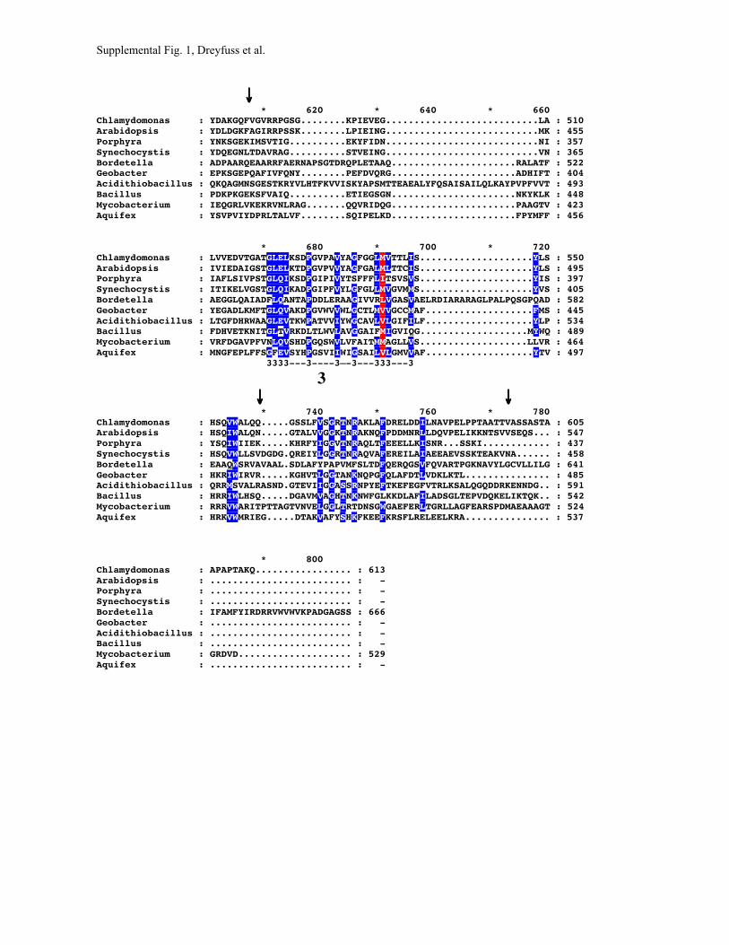
Supplemental Fig. 1, Dreyfuss et al.
↓↓↓↓ * 620 * 640 * 660Chlamydomonas : YDAKGQFVGVRRPGSG........KPIEVEG...........................LA : 510Arabidopsis : YDLDGKFAGIRRPSSK........LPIEING...........................MK : 455Porphyra : YNKSGEKIMSVTIG..........EKYFIDN...........................NI : 357Synechocystis : YDQEGNLTDAVRAG..........STVEING...........................VN : 365Bordetella : ADPAARQEAARRFAERNAPSGTDRQPLETAAQ......................RALATF : 522Geobacter : EPKSGEPQAFIVFQNY........PEFDVQRG......................ADHIFT : 404Acidithiobacillus : QKQAGMNSGESTKRYVLHTFKVVISKYAPSMTTEAEALYFQSAISAILQLKAYPVPFVVT : 493Bacillus : PDKPKGEKSFVAIQ..........ETIEGSGN......................NKYKLK : 448Mycobacterium : IEQGRLVKEKRVNLRAG.......QQVRIDQG......................PAAGTV : 423Aquifex : YSVPVIYDPRLTALVF........SQIPELKD......................FPYMFF : 456
* 680 * 700 * 720Chlamydomonas : LVVEDVTGATGLELKSDPGVPAVYAGFGGLMVTTLIS....................YLS : 550Arabidopsis : IVIEDAIGSTGLELKTDPGVPVVYAGFGALMLTTCIS....................YLS : 495Porphyra : IAFLSIVPSTGLQIKSDPGIPIVYTSFFFLITSVSVS....................YIS : 397Synechocystis : ITIKELVGSTGLQIKADPGIPFVYLGFGLLMVGVMMS....................YVS : 405Bordetella : AEGGLQAIADFLQANTAPDDLERAAGIVVRLVGASVAELRDIARARAGLPALPQSGPQAD : 582Geobacter : YEGADLKMFTGLQVAKDPGVWVVWLGCTLMVVGCCMAF...................FMS : 445Acidithiobacillus : LTGFDHRWAAGLEVTKWPATVVIYWGCAVLVLGIFILF...................YLP : 534Bacillus : FDHVETKNITGLTVRKDLTLWVLAVGGAIFMIGVIQG...................MYWQ : 489Mycobacterium : VRFDGAVPFVNLQVSHDPGQSWVLVFAITMMAGLLVS...................LLVR : 464Aquifex : MNGFEPLFFSGFEVSYHPGSVIIWIGSAILVLGMVVAF...................YTV : 497 3333---3----3—-3---333---3
3 ↓↓↓↓ ↓↓↓↓ * 740 * 760 * 780Chlamydomonas : HSQVWALQQ.....GSSLFVSGRTNRAKLAFDRELDDILNAVPELPPTAATTVASSASTA : 605Arabidopsis : HSQIWALQN.....GTALVVGGKTNRAKNQFPDDMNRLLDQVPELIKKNTSVVSEQS... : 547Porphyra : YSQIWIIEK.....KHRFYIGGVTNRAQLTFEEELLKISNR...SSKI............ : 437Synechocystis : HSQVWLLSVDGDG.QREIYLGGRTNRAQVAFEREILAIAEEAEVSSKTEAKVNA...... : 458Bordetella : EAAQWSRVAVAAL.SDLAFYPAPVMFSLTDFQERQGSVFQVARTPGKNAVYLGCVLLILG : 641Geobacter : HKRIWIRVR.....KGHVTLGGTANKNQPGFQLAFDTLVDKLKTL............... : 485Acidithiobacillus : QRRMSVALRASND.GTEVIIGGASSRNPYEFTKEFEGFVTRLKSALQGQDDRKENNDG.. : 591Bacillus : HRRIWLHSQ.....DGAVMVAGHTNKNWFGLKKDLAFILADSGLTEPVDQKELIKTQK.. : 542Mycobacterium : RRRVWARITPTTAGTVNVELGGLTRTDNSGWGAEFERLTGRLLAGFEARSPDMAEAAAGT : 524Aquifex : HRKVWMRIEG.....DTAKVAFYSHKFKEEFKRSFLRELEELKRA............... : 537
* 800Chlamydomonas : APAPTAKQ................. : 613Arabidopsis : ......................... : -Porphyra : ......................... : -Synechocystis : ......................... : -Bordetella : IFAMFYIRDRRVWVWVKPADGAGSS : 666Geobacter : ......................... : -Acidithiobacillus : ......................... : -Bacillus : ......................... : -Mycobacterium : GRDVD.................... : 529Aquifex : ......................... : -

Supplemental Fig. 2, Dreyfuss et al.
Supplemental Figure 2.
* 20 * 40 * 60Chlamydomonas : MQPYASVSGRCLSRPDALHVIPFGRPLQAIAGRRFVRCFAKGGQPGDKKKLNVTDKLRLG : 60Arabidopsis : --MIVTLNPKILHFS---KIHPFSRPSSYLCRTRNVSLITNCKLQKPQDGNQRSSSNRNL : 55Oyrza : ------------------------------------------------------------ : -Porphyra : ------------------------------------------------------------ : -Odontella : ------------------------------------------------------------ : -Guillardia : ------------------------------------------------------------ : -Cyanidium : ------------------------------------------------------------ : -Synechocystis : ------------------------------------------------------------ : -Synechococcus : ------------------------------------------------------------ : -Anabaena : ------------------------------------------------------------ : -Nostoc : ------------------------------------------------------------ : -Prochlorococcus : ------------------------------------------------------------ : -
↓↓↓↓ * 80 * 100 * 120Chlamydomonas : NTPPTLDVLKAPRPTDAPSAIDDAPSTSGLGLGGGVASPRTLVQSNAVQVAWRRLMKELS : 120Arabidopsis : TKTISLSDSAPPVTEETGDGIVKGGGNGGGGGGDGRGGLG------FLKILPRKVLSVLS : 109Oyrza : --------------------------------------------------LTKRTLSLLS : 10Porphyra : ---MQINLKFKK------------------------------------KDVRWYLLRLFS : 21Odontella : --------------------------------------------------MKQSVLRFLA : 10Guillardia : ------------------------------------------------MNYIIKFLNKLN : 12Cyanidium : --MVSITLKTN--------------------------------------RIFRSCLNLAT : 20Synechocystis : ---MTIANPSP------------------------SNFFQ--------QLGRQC-LKTLA : 24Synechococcus : ---MTVSDSSP-N-------------------SPWHSFPR--------KVWRTG-LKWIA : 28Anabaena : ---MTTDNSAPTA-------------------SPWWSLPG--------KFLRREFLPVLT : 30Nostoc : ---MTLEDSASKE-------------------LKWWAIPG--------QFLRQELLPVLT : 30Prochlorococcus : ------------------------------------------------MVIFKKFILKIS : 12
↓↓↓↓ * 140 * 160 * 180Chlamydomonas : SLPRAIAIMALIAVLSGLGTFIPQNKSIEYYLVNYPDGAEKVLGFLTGDLILTLQLDHIY : 180Arabidopsis : NLPLAITEMFTIAALMALGTVIEQGETPDFYFQKYPE-DNPVLGFFTWRWISTLGLDHMY : 168Oyrza : NLPLAISEMFAIAALMALGTVIDQGEAPSYYFEKFPE-DNPVFGFITWRWILTPGFDHMF : 69Porphyra : NLQFSIILLLVIASFSVIGTIIEQNKDLDFYQAHYSV-SGEHFIILNWKNIELFGLNHVY : 80Odontella : DLRFAISILLIIASCSVIGTVIEQDQSIEIYKLNYPL-TNRIFGFLSWDIILKFGLDHVY : 69Guillardia : NLTVAIIILLAIALASALGTVIEQNKNTDFYLKNYPL-TKPLFNFVTSDLILKFGLDHVY : 71Cyanidium : NLKFSITLFIIICIVSAIGTIIPQDKPKEFYMNTYSL----KVGMPLWKIIQLLSLEKIF : 76Synechocystis : DLRLAIALLLLIAVFSISGTVIEQGESLSFYQQNYPE-DPALFGFLSWQVILQLGLNQVY : 83Synechococcus : DLRVAIALLLLISVFSILGTVIEQGSTIQFYQENYPE-DPALLGFLSWKVLLGLGLDHVY : 87Anabaena : DLRLAIALLLIIALFSISGTVIEQGQSPAFYQSNYPE-HPALFGFLTWKVIQVVGLDHVY : 89Nostoc : NLRLAIALLLLIAIFSSTGTVIEQGQSPAFYQANYPE-HPALFGFLTWKVIQVVGLDHVY : 89Prochlorococcus : SLRFAILLIIFIAISSGVGTFIPQGNDQQEYIDFYNE--TPILGFINGSQVIRLQLDHIY : 70 ∗∗∗∗∗∗∗∗∗∗∗∗∗∗∗∗∗∗∗∗∗∗∗∗∗∗∗∗∗∗∗∗∗∗∗∗∗∗∗∗∗∗∗∗∗∗∗∗∗∗∗∗∗∗∗∗∗∗∗∗∗∗∗∗∗∗∗∗∗∗∗∗∗∗∗∗∗∗∗∗
TM1 ↓↓↓↓ * 200 * 220 * 240Chlamydomonas : TADYFYLSMGLLAASLAACTYTRQWPAVKVAQRWRFLTQPKSLLKQGRTE----VLPNAR : 236Arabidopsis : SAPIFLGMLVLLAASLMACTYTTQIPLVKVARRWSFMKSDEAIKKQEFAD----TLPRAS : 224Oyrza : SSPVFLGLLALLAASLMACTYTTQIPIVKVARRWSFMHSAGSIRKQEFAE----SLPRAS : 125Porphyra : TTWWFLTLLFIFSLSLLVCSLSRQIPSLQNARRWCFYKNPNQFKKFTGSQ----EIQKTT : 136Odontella : KTWWFLGFIALFGLSLFTCTILQQFPSLKIARRCQFFRTTQQFGLLKLSR----NLGNLS : 125Guillardia : TSWWFIFLIILLLLSLTLCTITRQLPALKLARLWQFYTNFNTKAKFQIRF----KTNSSS : 127Cyanidium : YSNFYLILLLCLSFSLFFCSLKSQFPYLRTSRIIKLNNNNPPTSPLHESK-----K--IK : 129Synechocystis : RTWWFLGLLILFGSSLTACTFNRQFPALKAARSWQFYHQPRQFKKLALSF----SLPDGD : 139Synechococcus : TTWWYLVLLLAFGVSLIACTFRRQLPALKTARNWNYYSQARQFNKLALST----ELDHGS : 143Anabaena : RTWWFLSLLVLFGTSLTACTFTRQLPALKTAQRWKYYEEPRQFQKLALSA----ELDAGS : 145Nostoc : RTWWFLALLILFGTSLTACSFTRQLPALKAAQRWKYYEEPRQFQKLALSA----ELDNGS : 145Prochlorococcus : TSNWFLFSLILLCISLAACSFRRQIPSLKAALKWTDYKDEKKFYKLELITNYEIIQDADH : 130 ∗∗∗∗∗∗∗∗∗∗∗∗∗∗∗∗∗∗∗∗
TM2

* 260 * 280 * 300 Chlamydomonas : VSDLGAILLQRGYQVFVKDGS---LYGFKGLAGKLGPIGVHAALLLCLFGTAWSGFGTLK : 293 Arabidopsis : IQDLGMILMGDGFEVFMKGPS---LYAFKGLAGRFAPIGVHIAMLLIMVGGTLSATGSFR : 281 Oyrza : IQDLGVILMGYGYEVFTKGPS---LYAFKGLAGRFAPIGVHIAMIFIMAGATLSATGSFK : 182 Porphyra : LHLVASCLQKFNYHIFQQGNS---IYCYKGLLGRLAPIFVHASIILLLIGSVLGLVSGFS : 193 Odontella : LSQLLFRIKKNQYSIFQ--QK-NIVYCYKGLIGRIAPIIVHFSMILILIGAIFGALNGFK : 182 Guillardia : LTKLTYYLEEKNYKIKHFNHF---VYAYKGIFGRVSPIIVHFSLVIVLIGSMLSTTQGRT : 184 Cyanidium : YNNIASKNDSSCVQLVSQGYK---IYTFDKNLDKAGPLLIHLSLILILLGSAIHAFNDFI : 186 Synechocystis : INKIESLLRDRGYKIFQEGDS---VYARKGLMGKVGPIIVHGAMLIILGGAIWGALTGFF : 196 Synechococcus : LQSLKPQLEKKRYKIFQDGEK---LYARKGIVGRIGPIIVHIGMIVTLVGSIWGAFGGFM : 200 Anabaena : VNSLSQILQNRRYKIFQ--EKDDILYARKGIVGRIGPIIVHIGIVTILLGSIWGAMTGFI : 203 Nostoc : LNSLSQLLQKRRYKIFPDREKENILYARKGIVGRIGPIIVHIGIVAILLGGIWGAMTGFM : 205 Prochlorococcus : ILKADSLLRKKGWNISKFENR---LSARKGLEGKLGPIIVHIGLIILLIGSAYGNFSSQS : 187 ∗∗∗∗∗∗∗∗∗∗∗∗∗∗∗∗∗∗∗∗∗∗∗∗∗∗∗∗∗∗∗∗∗∗∗∗∗∗∗∗∗∗∗∗
TM3 ↓↓ * 320 * 340 * 360 Chlamydomonas : GNVMCPEGQDFQVASFLQPSSPIASMPASASNVIHVNKFTIDYRPDGSVAQFYSDLSLLD : 353 Arabidopsis : GSVTVPQGLNFVMGDVLAPIGFFSIPTDAFNTEVHVNRFTMDYYDSGEVSQFHSDLSLRD : 341 Oyrza : GSVDVPQGLNFVIGDVMKPKGVLSFVPDVFNTEVHVNRFYMEYYDSGEVSQFYSDLSLFD : 242 Porphyra : AQEMVPSGELFRLQNIIA-SGQFSY--IPQDFSARVNNFIIEYNQDNSISQFFSDISILD : 250 Odontella : AQELIPKTEAFHVQNILS-NGQLTR--IPKVVS-RVNDFWITYTKQATVAQFYSDVSILN : 238 Guillardia : QEAFIVVN---QEKPVLD------------TYEAYVNDFKIAYNSQGLIDQFYSDLILET : 229 Cyanidium : AQEMIPIYEVSHIQNVIS-SGRISK--IPQTISLKASAFTVEHENEKVVKQFITNLAMLN : 243 Synechocystis : AQHMIPSGETFQVSNIIE-KGPLADSQIPKDWGIKVNRFWINYTENGAIDQFYSDLSVVN : 255 Synechococcus : AQEMIPSGVNFKVNNVFK-AGIFSE--SDRPWSVNVNRFWIDYTPTGDIDQFYSDLSVVD : 257 Anabaena : AQEMVPSGETFQVKNIID-AGPLAAGQFPQDWSVRVNRFWIDYTPKGGIDQFYSDMSVLD : 262 Nostoc : AQEMVASGDTFQVTNIVD-AGPLAA-QVSKDWSVRVNRFWIDYTPSGGIDQFYSDMSVLN : 263 Prochlorococcus : KEQYLRLGESLDLINEST----------NSRVKIKLKNFFIERESDGKPKQFISNLEFFS : 237 1-11-111-1-1
1 ↓↓ * 380 * 400 * 420 Chlamydomonas : PAQGGKEMMRKTISVNDPFRFNGVTMYQTDWSLSAVTLRVLGQDAPLARAAQAAEAQAAA : 413 Arabidopsis : LN--GKEVLRKTISVNDPLRYGGVTVYQTDWSFSALQVTKDGE----------------- : 382 Oyrza : LD--GKEVMRKTIKVNDPLRYGGVTIYQTDWGFSALQVKKNGE----------------- : 283 Porphyra : TE--GTELKHSTIHVNSPLQFKGLTIYQTDWDIIAIRIKINNT----------------- : 291 Odontella : TE--GNEIVRKTIFVNSPIKYNNIDFYQTDWNVIGLRVQNDTS----------------- : 279 Guillardia : RQ---ASKIQKTIYVNEPLNYSNITIYQTDWNIDNLVICIDNQ----------------- : 269 Cyanidium : SK--GEVLKQGLVSVNHPLVYKQVYIFQMDWKLFGIRVNYGKN----------------- : 284 Synechocystis : NQ--GEELDRQTISVNHPLRHRGVTFYQTNWGIAGVKVQLNNS----------------- : 296 Synechococcus : GE--GQELERKTISVNHPLRYDGITFYQTSWSIGGVQVQLNNS----------------- : 298 Anabaena : NQ--GQEVDHKKIFVNQPLRYHGVTFYQTDWGISGVRVRLNKS----------------- : 303 Nostoc : KQ--GEEVDHKKIFVNEPLRYRGITFYQTDWGIAGVRVQFNNS----------------- : 304 Prochlorococcus : KQ--QNLNEIKTTQVNQPIRFKGLTIYQADWSVSNIVMEIDSV----------------- : 278 2-22-22-2—-22-22222-2—2-2
2 * 440 * 460 * 480 Chlamydomonas : STSGPTSSASSTSDALPQQRTAFNLPMASLEGKPGVAGRLWATFLPLAEPGQDGSAPKGI : 473 Arabidopsis : ---GP-----------------FNLAMAPIKING--DKKLYGTFLPVGDTN--APNVKGI : 418 Oyrza : ---GP-----------------FNLAMAPLKLNG--DKKLFGTLLPLENSG--SSNVKGI : 319 Porphyra : --------------------DTLQIPLKKVVLPN---NKIWVGLIAQD-------QDNQL : 321 Odontella : ----S----------------ILQYPLINLTNAG--N-KVWVTWIPSD-----SEFKKGL : 311 Guillardia : --------N------------YYSIPLQFIELPNGSESKYWINRLDLFG--------QSV : 301 Cyanidium : --------------------KIYEFPVQKIEANN---EQQWSCTIPAK-------NQSKL : 314 Synechocystis : ----P----------------VLQLPMAPLQTAN--GGQLWGAYIPTK-----TDFSEGA : 329 Synechococcus : ----P----------------IFQLPAAQIPTEN--GAKLWGSWVPIK-----PDMSAGV : 331 Anabaena : ----P----------------IFQLPMALLNTNG--QGRIWGTWIPTK-----PDLSEGV : 336 Nostoc : ----P----------------IFQLPMALLNTKG--QGRIWGTWVPTK-----PDLSEGV : 337 Prochlorococcus : ---------------------LYQLQLKPIPEIG---DQIWGLLIELG-----RENKKNY : 309

↓↓ * 500 * 520 * 540 Chlamydomonas : SILARDPQ-SVVFYDAKGQFVGVRRPGSGKPIEVEGLALVVEDVTGATGLELKSDPGVPA : 532 Arabidopsis : SMLARDLQ-SIVVYDLDGKFAGIRRPSSKLPIEINGMKIVIEDAIGSTGLELKTDPGVPV : 477 Oyrza : SMLARDLQ-SIVLYDQEGKFVGVRRPSSKLPIEIDGNEIVIEDAIGSTGLDLKTDPGIPI : 378 Porphyra : ALVLQDLQKQATLYNKSGEKIMSVTIGEK--YFIDNNIIAFLSIVPSTGLQIKSDPGIPI : 379 Odontella : TILVDNLQGYCSIYDESGIFVGNLELNET--FNLN-MPITLVDILSSTGLQIKTDPGIPF : 368 Guillardia : FCVVNDLTGIVYLYNQNKDLICISSLGEF--ITLNGHTITFNKLVASTGLQFKLDSFIPL : 359 Cyanidium : ILIFKNMSDEFYVYDNNQNFLKIGKINTP--QLIRNTYFTVISKISGTGLQIKKDSSINI : 372 Synechocystis : ALLVKDLQGTMIIYDQEGNLTDAVRAGST--VEINGVNITIKELVGSTGLQIKADPGIPF : 387 Synechococcus : SILMQDLQGSAIVYNEQGELVGAVRVGDR--LDVGDISLKLVDLVGSTGLQIKADPGVPV : 389 Anabaena : SLLAKDLQGMVLIYDAQGKLVDTVRAGMS--TQVNGVTLKVLDVVGSTGLQIKADPGIPI : 394 Nostoc : SLLAKDLQGMVLIYDPNGKLVDTVRAGMS--TQVNGVKLKILDVIGSTGLQIKADPGIPI : 395 Prochlorococcus : LLTIDNENGPLKVSNIEDFSENFVYLNNN-PIEINSSKLSLKKIIPSSGLIIKNDPSIPF : 368 3-3333333-33-33-
3 ↓↓ * 560 * 580 * 600 Chlamydomonas : VYAGFGGLMVTTLISYLSHSQVWALQQ-GS---SLFVSGRTNRAKLAFDRELDDILNAVP : 588 Arabidopsis : VYAGFGALMLTTCISYLSHSQIWALQN-GT---ALVVGGKTNRAKNQFPDDMNRLLDQVP : 533 Oyrza : VYAGFGALMLTTCISYLSHSQVCAY------------G--T--SYDYF------------ : 410 Porphyra : VYTSFFFLITSVSVSYISYSQIWIIEKKHR----FYIGGVTNRAQLTFEEELLKISNRSS : 435 Odontella : NLYWIPIFNV----KYIN------------------------------------------ : 382 Guillardia : VYLGFLLLMISTLLSYISYSQVWLVKN-GS---TTYIFGSTNRAKFAFIKQLTEIANQC- : 414 Cyanidium : VYTGFLLLIIGLVINHKGSKKRT------------------------------------- : 395 Synechocystis : VYLGFGLLMVGVMMSYVSHSQVWLLSVDGDGQREIYLGGRTNRAQVAFEREILAIAEEAE : 447 Synechococcus : VYTGFLLVMLGVVMSYVSYSQVWALAA-GD---RFYLGGKTNRAQVAFERELLEIINTLE : 445 Anabaena : VYTGFGILMLGVVMSYFSHSQIWALQK-GD---RLYVGGKTNRAQVAFEQEVLDILERLN : 450 Nostoc : VYSGFGLLMLGVVMSYFSHSQIWALQK-GD---LLYVGGKTNRAQVAFEQEVLEILDRLS : 451 Prochlorococcus : IYLSFTLIIVGTILSLIPTNQIWILFNENTN--KLFIGGLSNRNLLGFKKEFLKLSDEIK : 426
↓↓ * 620 Chlamydomonas : ELPPTAATTVASSASTAAPAPTAKQ : 613 Arabidopsis : ELIKKNTSVVSEQS----------- : 547 Oyrza : ------------------------- : - Porphyra : KI----------------------- : 437 Odontella : ------------------------- : - Guillardia : ------------------------- : - Cyanidium : ------------------------- : - Synechocystis : ---VSSKTEAKVNA----------- : 458 Synechococcus : TSHSQATPENTLTSIEQ-------- : 462 Anabaena : ---SQSATVINQQS----------- : 461 Nostoc : ---SEPKIEEKETAIEV-------- : 465 Prochlorococcus : NN----------------------- : 428

Supplemental Figure. 1. Multiple alignment of Ccs1 from phyllogenetic representative organisms . Ccs1 homologs from green algae Chlamydomonas reinhardtii (T09105), eudicot plant Arabidopsis thaliana (NC_003070), plastid genome of red algae Porphyra purpurea (NC_000925), cyanobacterial Synechocystis sp. PCC 6803 (NC_000911), beta-proteobacterium Bordetella pertussis (Sanger 520, complete genome), delta-proteobacterium Geobacter sulfurreducens (TIGR_35554, preliminary sequence data was obtained from The Institute for Genomic research website at http://www.tigr.org, ORF predicted to initiate at UUG start codon), gamma-proteobacterium Acidithiobacillus ferrooxidans (AAC68692), Bacillus group of Firmicutes bacterium Bacillus subtilis (NC_000964), Actinobactium group of Firmicutes bacterium Mycobacterium tuberculosis CDC1551(NP_334959), and Aquificales bacterium Aquiflex aeolicus (NC_000918) were aligned using CLUSTALW algorithm (Blosum62 scoring matrix) in Bioedit software (http://www.mbio.ncsu.edu/BioEdit/bioedit.html). The alignment was edited in the GeneDoc multiple alignment editor software (http://www.psc.edu/biomed/genedoc/). Amino-acids conserved in all sequences are shaded red and those conserved in the majority of the sequences are shaded blue. Location of transmembrane (TM) domains predicted by PHD algorithm (http://www.embl-heidelberg.de/predictprotein/predictprotein.html) are underlined (∗). Sites of phoA and LacZ fusions constructs are marked with arrows above sequences. Functionally essential histidine (H274 in C. reinhardtii) is highlighted in green. Three blocks of conserved sequences in the predicted soluble lumenal domain are marked below the sequences as with 1, 2 and 3 respectively.

Supplemental Figure. 2. Multiple alignment of Ccs1 from representative oxygenic photosynthetic organisms. Ccs1 homologs from the nuclear genome of green algae Chlamydomonas reinhardtii (T09105), eudicot plant Arabidopsis thaliana (NC_003070), monocot Oryza sativa (hypothetical protein), plastid genome of red algae Porphyra purpurea (NC_000925), brown algae Odontella sinensis (NC_001713), cryptonomad alga Guillardia theta (NC_000926), and from cyanobacterial genomes Synechocystis sp. PCC 6803 (NC_000911), Synechococcus sp. PCC 7002(AF052290), Anaebena (Nostoc ) sp. PCC 7120 (NC_0037272), Nostoc punctiforme, Prochlorococcus marinus MED4 (see draft genomes for the sequences of the later two at http://www.jgi.doe.gov/JGI_microbia l/html/index.html) were aligned using CLUSTALW algorithm (Blosum62 scoring matrix) in Bioedit software (http://www.mbio.ncsu.edu/BioEdit/bioedit.html). The alignment was edited in the GeneDoc multiple alignment editor software (http://www.psc.edu/biomed/genedoc/). Amino-acids conserved in all sequences are shaded red and those conserved in the majority of the sequences are shaded blue. Location of transmembrane (TM) domains predicted by PHD algorithm (http://www.embl-heidelberg.de/predictprotein/predictprotein.html) are underlined (∗). Sites of phoA and LacZ fusions constructs are marked with arrows above sequences. Functionally essential histidine (H274 in C. reinhardtii) is highlighted in green. Three blocks of conserved sequences in the predicted soluble in the predicted soluble lumenal domain are marked below the sequences as with 1, 2 and 3 respectively. Mature portion of Oryza Ccs1 protein was predicted based on BLAST result of translated genomic fragment OSM132311(courtesy of Monsanto rice-research.org queried against Arabidopsis Ccs1. Junction of splice site was predicted using NetPlantGene (http://www.cbs.dtu.dk/services/NetPGene/). N-terminal transit peptide region could not be predicted due to low homology and lack of predicted splice sites

Supplemental Table I. Primers used for amplification of C. reinhardtii Ccs1
Primer Position in genomic sequencea Sequence (5' –3')b 1 –206 to –190 CACATACGACCCCAAGC 2 +2169 to +2154 GCAGGTACCGTCATGC 3 +2852 to +2868 CACAGGGCGTCACCATG 4 +4357 to +4341 CCTTCCCAAGTTCCACG 5 +1240 to +1255 CAGCCGAAGTCGCTGC 6 +1892 to +1873 GTGAACTTGTTGACGTGGAT 7 +3938 to +3953 GGCGAAGCAGTAGGG 8 +1741 to +1724 CATAGCAAGCACTCGGAG 9 +916 to +932 TGACCCTACAGCTGGAC 10 +956 to +938 GAAATAGTCAGCGGTGTAG 11 +3364 to +3381 CAGTCGGTGGTGTTCTAC 12 +3443 to +3426 TCCACCTCGATTGGCTTG 13 +728 to +747 GTACCGCAACCAGGCACCTA 14 +1255 to +1240 GCAGCGACTTCGGCTG 15 +323 to +343 CAAGAAGAAGCTCAACGTGAC 16 +1406 to +1389 TCAGCAGCGACGGACTGA 17 +1977 to +1995 CATTCCTGCGATAGTGTTG 18 +3173 to +3157 CCTGCAGCCAGCACTAC 20 +3237 to +3218 AGCTCAAATTGTCAGTACAC C199A-1 +981 to +1013 GCCTCCCTGGCcgCggcCACCTACACCCGCCAG C199A-2 +1013 to +981 CTGGCGGGTGTAGGTGgccGcgGCCAGGGAGGC H274A-1 +1499 to +1530 GGCCCATCGGCGTGgcCGCGGCGCTGCTGCTG H274A-2 +1530 to +1499 CAGCAGCAGCGCCGCGgcCACGCCGATGGGCC D348A-1 +2042 to +2071 CCGCAGTTCTACagcGctTTGTCGCTGCTT D348A-2 +2071 to +2042 AAGCAGCGACAAagCgctGTAGAACTGCGG TRX5'c +1801 to +1818 TAGGTACCAGACTTCCAGGTGGTGGCTTC TRX3'c +3441 to +3426 TAGGATCCCACCTCGATTGGCTTG a Numbering of the genomic DNA sequence defined as position +1 representing the start of
translation.
b Lowercase, bold nucleotides indicate mutations
c Primers Trx5' and Trx3' designed with KpnI and BamHI restriction sites at the 5' ends of the
primers, respectively, to facilitate cloning in-frame into the thioredoxin fusion expression vector
pTrxFus.

Supplemental Table II. Primers used for amplification of Synechocystis sp. PCC6803 Ccs1 for topology analysis
Primer Sequence (5' –3') R-ccsB23 ACGCGTCGACAGGGTTTTGAGGCACTGC R-ccsB67 ACGCGTCGACCCAAATAGAGCTGGATCTTC R-ccsB134 ACGCGTCGAAAAACTTAAAGCTAG R-ccsB225 ACGCGTCGACTTGGGAATTTGACTGTCG R-ccsB288 ACGCGTCGACCCCGCAATGCCCCAGTTAG R-ccsB349 ACGCGTCGACAAATTGCCCTCTTGGTC R-ccsB416 ACGCGTCGACCCATCAACGCTCAATAAC R-ccsB458 ACGCGTCGACGCATTAACTTTAGCTTCTG F-ccsBSac TGTGAGCTCTTTCATGACCATTGC F-ccsBNde TTTGCCCAACATATGATTCCCAGTGGT LacPstI GTTGCTGCAGTTTGGAACAAG