the interaction of the gammaherpesvirus 68 orf73 …molecularvirology.org/pdf/ottinger et al...
TRANSCRIPT
JOURNAL OF VIROLOGY, May 2009, p. 4423–4434 Vol. 83, No. 90022-538X/09/$08.00�0 doi:10.1128/JVI.02274-08Copyright © 2009, American Society for Microbiology. All Rights Reserved.
The Interaction of the Gammaherpesvirus 68 orf73 Protein with CellularBET Proteins Affects the Activation of Cell Cycle Promoters�
Matthias Ottinger,1,2§* Daniel Pliquet,1§ Thomas Christalla,1 Ronald Frank,3James P. Stewart,4 and Thomas F. Schulz1*
Institut fur Virologie, Medizinische Hochschule Hannover, Hannover, Germany1; Department of Pathology, Harvard Medical School,Boston, Massachusetts2; Helmholz Zentrum fur Infektionsforschung, Abteilung Chemische Biologie, Braunschweig, Germany3; and
School of Infection and Host Defence, The University of Liverpool, Liverpool, United Kingdom4
Received 29 October 2008/Accepted 16 February 2009
Infection of mice with murine gammaherpesvirus 68 (MHV-68) provides a valuable animal model forgamma-2 herpesvirus (rhadinovirus) infection and pathogenesis. The MHV-68 orf73 protein has been shownto be required for the establishment of viral latency in vivo. This study describes a novel transcriptionalactivation function of the MHV-68 orf73 protein and identifies the cellular bromodomain containing BETproteins Brd2/RING3, Brd3/ORFX, and BRD4 as interaction partners for the MHV-68 orf73 protein. BETprotein members are known to interact with acetylated histones, and Brd2 and Brd4 have been implicated infundamental cellular processes, including cell cycle regulation and transcriptional regulation. Using MHV-68orf73 peptide array assays, we identified Brd2 and Brd4 interaction sites in the orf73 protein. Mutation of onebinding site led to a loss of the interaction with Brd2/4 but not the retinoblastoma protein Rb, to impairedchromatin association, and to a decreased ability to activate the BET-responsive cyclin D1, D2, and Epromoters. The results therefore pinpoint the binding site for Brd2/4 in a rhadinoviral orf73 protein andsuggest that the recruitment of a member of the BET protein family allows the MHV-68 orf73 protein toactivate the promoters of G1/S cyclins. These findings point to parallels between the transcriptional activatorfunctions of rhadinoviral orf73 proteins and papillomavirus E2 proteins.
Many gammaherpesviruses promote lymphocyte prolifera-tion and can persist in the lymphocyte compartment as well asin a number of other cell types, including endothelial andepithelial cells and fibroblasts (16, 17, 19, 56, 59). To ensureepisome maintenance in these dividing cells, gammaherpesvi-ruses express proteins that facilitate the replication of latentviral episomes and ensure the segregation of these viral ge-nomes into progeny cells during cell division. The Epstein-Barrvirus (EBV) EBNA-1 protein fulfills these essential functions(49, 62), as do the proteins encoded by open reading frame 73(ORF73) of the gamma-2 herpesviruses (rhadinoviruses)Kaposi’s sarcoma-associated herpesvirus (KSHV), herpesvirussaimiri (HVS), and murine gammaherpesvirus 68 (MHV-68)(4, 20, 28, 29, 43). The orf73 protein of KSHV, latency asso-ciated nuclear antigen 1 (LANA-1), is well characterized andhas multiple functions. LANA-1 mediates replication and epi-some maintenance, acts as a transcriptional repressor and ac-tivator, and deregulates the cell division cycle (3, 24, 50). Someof these functions are mediated via the interaction of LANA-1with a number of cellular proteins, including p53 (23), theretinoblastoma protein (48), MeCP2 (35), mSin3 (36), andmultiple members of the BET (bromodomain and extra ter-
minal domain) family of proteins (45, 47, 65). The MHV-68orf73 protein, the KSHV LANA-1 homolog, is critical for theestablishment and maintenance of a latent infection in mice(20, 43). MHV-68 orf73 is expressed in latently infected cells aswell as during lytic infection (2, 15, 19, 51). The moleculardetails of how the MHV-68 protein functions are still largelyunexplored.
BET proteins interact via their bromodomains with acety-lated histones (13, 30, 33) and are highly conserved, with mem-bers in plants, yeast, Drosophila melanogaster, and up to mam-mals (18). An additional characteristic feature of BET proteinsis the highly conserved extra terminal (ET) domain (see Fig.1), which serves as a protein-protein interaction module inBrd2, Brd3, and Brd4 with KSHV LANA-1 (45, 47, 65) andbetween the yeast (Saccharomyces cerevisiae) BET proteinBdf1 and the TAF7 subunit of the general transcription factorTFIID (39). Mammalian BET proteins are encoded by fourgenes, Brd2, Brd3, Brd4, and Brd6, out of which Brd2, alsocalled RING3, and Brd4 are the best characterized. Brd2 is atranscriptional regulator that plays a role in cell cycle regulation(11, 12, 33, 55). Brd2 overexpression in B lymphocytes in vivo inmice has been shown to induce lymphoma (26). Brd4 interactswith pTEFb (transcriptional elongation factor b) and therebypromotes RNA polymerase II (Pol II)-dependent transcrip-tional elongation (7, 32, 61). Brd4 overexpression and deple-tion both result in deregulated cell cycle progression (14, 38,45). Recently, Brd4 has been shown to play an important rolein the G1/S transition through its ability to stimulate transcrip-tion of G1/S-specific genes, among them, cyclin D1 and cyclinD2 (42). Furthermore, the C-terminal domain of the longisoform of Brd4 (Brd4L) (amino acids [aa] 1 to 1362) serves as
* Corresponding author. Mailing address for Thomas F. Schulz:Institut fur Virologie, Medizinische Hochschule Hannover, Carl-Neu-berg Str. 1, 30625 Hannover, Germany. Phone: 49-511-532-6736. Fax:49-511-532-8736. E-mail: [email protected]. Mailingaddress for Matthias Ottinger: Department of Pathology, HarvardMedial School, 77 Ave. Louis Pasteur, NRB 952, Boston, MA 02115.Phone: (617) 432-0743. Fax: (617) 432-2218. E-mail: [email protected].
§ These authors contributed equally.� Published ahead of print on 25 February 2009.
4423
at University of Liverpool Library on July 14, 2009
jvi.asm.org
Dow
nloaded from
a chromatin tether for different papillomaviruses via its inter-action with their E2 proteins (1, 6, 63, 64). Brd4 is critical forpapillomavirus E2 transcriptional activation (31, 40, 54) andmay play a role in its transcriptional repression function (53,60). The same C-terminal domain of Brd4L has recently beenshown to interact with the transcriptional elongation factorpTEF and to inhibit human immunodeficiency virus type 1(HIV-1) Tat-mediated, pTEF-dependent transcription (7). Wehave shown previously that KSHV LANA-1 interacts withBrd2 and Brd4 via their conserved ET domains (45, 57, 58).This interaction contributes to the association of LANA-1 withcellular heterochromatin and modulates the transcriptional ac-tivator role of the short isoform of Brd4 (Brd4S) (aa 1 to 722),which lacks the pTEFb interaction domain and by itself acti-vates the cyclin E promoter (7, 45, 57, 58).
In this study we show that Brd2, Brd3, and Brd4 also interactwith the MHV-68 orf73 protein. By identifying and mutating abinding site for Brd4 and Brd2 in the MHV-68 orf73 protein,we show that the orf73/BET interaction is crucial for the abilityto activate the cyclin D1, D2, and E promoters. The resultspinpoint the binding site for two BET proteins in a rhadino-viral orf73 protein and indicate that, similarly to papillomavi-rus E2, a rhadinoviral orf73 protein utilizes a member of theBET protein family to exert its transcriptional activation func-tion.
MATERIALS AND METHODS
Cell culture methods. Murine 3T3 fibroblasts as well as the human embryonickidney epithelial cell line HEK 293T were cultured at subconfluence in Dulbec-co’s modified Eagle’s medium (Gibco) supplemented with 10% heat-inactivatedfetal bovine serum, 50 IU/ml penicillin and 50 �g/ml streptomycin, and 100 �g/mlglutamine. The KSHV- and EBV-negative human Burkitt’s lymphoma cell lineBJAB was maintained in RPMI 1640 with 10% bovine growth serum plus 50IU/ml penicillin and 50 �g/ml streptomycin. Spodoptera frugiperda SF9 insectcells were cultured in spinner flask cultures as previously described (45).
DNA constructs and baculoviruses. The full-length MHV-68 orf73 clone wasgenerated by PCR using DNA from MHV-68-infected cells and the forwardprimer ORF73 START (ATGCGGCCGCCGCCGCCACCATGCCCACATCCCCACCGAC, NotI site and orf73 start codon underlined), which has an opti-mized Kozak sequence and binds at genomic bp 104868 (GenBank accessionnumber U97553), and the reverse primer ORF73 STOP (GCGGATCCTTAAGCGTAGTCTGGAACGTCGTATGGGTAAGCGTAGTCTGGAACGTCGTATGGGTATGTCTGAGACCCTTGTCCCTG, BamH1 site underlined), whichintroduces a double hemagglutinin (HA) tag and binds at genomic bp 103927.The PCR product was inserted into pVR1255 to obtain an MHV-68 orf73full-length expression construct with a double HA tag at the 3� end/C terminus(pVR1255 orf73). This and all other generated constructs were sequenced. AllMHV-68 orf73 constructs were derived from this plasmid.
The MHV-68 orf73 constructs with internal mutations were generated using asite-directed mutagenesis kit (Stratagene), pVR1255 orf73 as the template, andappropriate primers. To introduce larger mutations, e.g., the 225-7A mutation(described below), up to three successive rounds of site-directed mutagenesiswere performed using a template with intermediate mutations. Details can beobtained from the authors upon request. Constructs were sequence verified. Thevector EGFPC1 was purchased from Clontech. The enhanced green fluorescentprotein (EGFP)-Brd2/RING3 full-length (aa 2 to 801) and EGFP-Brd4S full-length (aa 2 to 722) vectors were described previously (45, 58). The vectormyc-Rb was a kind gift from S. Mittnacht, ICR, London, United Kingdom.
For prokaryotic expression, the constructs glutathione S-transferase (GST)–Brd2/RING3 (aa 601 to 801), GST-Brd4S (aa 607 to 722), and GST-Brd3/ORFX(aa 569 to 726) (see Fig. 1C and D) were described previously (45).
All luciferase-based reporter constructs were in the pGL2basic (pGL2b; Pro-mega) backbone. The murine cyclin D1 promoter region, encompassing 7.9 kbp(44), and the murine cyclin D2 promoter region, encompassing 2.3 kbp (8), werekindly provided by M. Eilers. Further, the human cyclin E promoter was used asthe reporter plasmid (25).
The Brd2/RING3 baculovirus was described previously (47). The baculovirusfor the expression of the Brd4S full-length protein (aa 2 to 722) with an amino-terminal myc epitope and a carboxy-terminal hexahistidine tag was generatedfrom pENTR1A-Brd4 by in vitro recombination with BaculoDirect C-terminallinear DNA (Invitrogen). pENTR1A-Brd4 was generated by PCR with the Brd4S
full-length template (aa 2 to 722) in pcDNA3 (45) and oligonucleotides Brd4S
BAC F (AGAGGATCCATTATGGAGCAGAAGCTGATCTCCGAGGAGGACCTGTCTGCGGAGAGCGGC) and Brd4S BAC R (AGACTCGAGGCAGGACCTGTTTCGG).
Transient transfections and luciferase-based reporter assays. For transfec-tions, cells were grown to subconfluence in six-well plates (Greiner). HEK 293Tand 3T3 cells were transfected with FuGENE6 (Roche) (FuGENE-DNA ratio of3 �l:1 �g). For luciferase reporter assays, cells were transfected with 50 ngluciferase reporter plasmid per well together with 500, 1,000, or 2,500 ng of orf73expression plasmid or empty vector. At 48 h posttransfection, cells were washedonce in cold phosphate-buffered saline (PBS) and then lysed and scraped on icein reporter lysis buffer (Promega). Lysates were spun at high speed for 1 min topellet the debris. Lysates were analyzed for luciferase activity using a luciferaseassay system (Promega).
Electroporation of B cells. BJAB cells were electroporated following publishedprocedures, with some modifications (45). Briefly, 1.3 � 107 BJAB cells wereresuspended in 400 �l medium without antibiotics. Endotoxin-free DNA (20 �g)was added to the cells, and cells were electroporated as previously described (45).
Protein analyses by immunoblotting. Cell lysates were subjected to sodiumdodecyl sulfate-polyacrylamide gel electrophoresis (SDS-PAGE). Proteins weretransferred to nitrocellulose membranes, and immunodetection of HA-taggedMHV-68 orf73 proteins was performed with a rat anti-HA monoclonal antibody(12CA5; Roche). For immunoprecipitation experiments, HA-tagged orf73 pro-teins were detected with a rat monoclonal anti-HA antibody (3F10; Roche).KSHV LANA-1 was detected with serum from KSHV-positive patients. Brd2/RING3 was detected with a polyclonal rabbit anti-RING3 antibody (47). Anti-green fluorescent protein (GFP) immunoblotting was performed with a mono-clonal anti-GFP antibody (JL-8; BD Biosciences). myc-tagged Rb was detectedwith a monoclonal anti-c-myc antibody (9E10; Biomol). Cellular actin was de-tected with a panactin mouse monoclonal antibody (MAB 1501; Chemicon/Millipore). Endogenous Brd4 was detected with an affinity-purified rabbit poly-clonal antiserum raised against the amino-terminal aa 1 to 470 of Brd4 (54).Species-specific horseradish peroxidase-conjugated secondary antibodies wereused.
Expression and purification of recombinant proteins from Escherichia coli andinsect cells. GST and GST fusion proteins were expressed as previously de-scribed (47). For the purification of GST and GST fusion proteins, glutathioneG Sepharose resin was used as previously established (45). Brd4S and Brd2/RING3 were expressed in SF9 insect cells and purified using a Ni� affinitypurification protocol as previously established (45).
GST fusion protein binding assays. GST pull-down assays using GST-RING3,GST-Brd4S, and GST-ORFX (see Fig. 1C and D) with MHV-68 orf73 proteinwere performed as previously described (45). Briefly, GST proteins were boundto glutathione G Sepharose, washed, and then incubated with lysates from 293Tcells transfected with empty vector, pVR1255 MHV-68 orf73, or pcDNA3 KSHVLANA.
Salt extraction of MHV-68 orf73 proteins from nuclear preparations. Nuclearpreparations from 293T cells that transiently expressed MHV-68 orf73 proteinswere subjected to protein extraction using increasing KCl concentrations similarto a previously published protocol for KSHV LANA (57). Transfected 293T cellswere lysed for 30 min in a low-ionic-strength buffer (5% glycerol, 1% NP-40, 0.2mM EDTA, 10 mM Tris-HCl [pH 7.9], leupeptin [50 �M], benzamidine [200�M], aprotinin [100 U/ml], pepstatin A [1 �M], phenylmethylsulfonyl fluoride [1mM]). Nuclear material was separated from the lysates by centrifugation. Next,the pellet was incubated with the same buffer plus 50 mM KCl, followed bycentrifugation. Supernatants were collected and the pellets subjected to incuba-tion with the same buffer with 200 mM KCl. The procedure was repeated with300 mM KCl. Supernatants were subjected to SDS-PAGE and immunoblotting.
Co-IP assays. Coimmunoprecipitation (Co-IP) experiments with lysates fromtransfected 293T cells were performed similarly to previously described proce-dures (58). Briefly, lysates of 293T cells coexpressing EGFP or EGFP-BETfusion proteins together with MHV-68 orf73-HA proteins were incubated with amonoclonal anti-HA antibody coupled to Sepharose A resin. After intensivewashing, SDS-PAGE and immunoblotting with mouse anti-GFP and rat anti-HAantibodies were performed. For myc-Rb immunoprecipitations, an anti-mycmonoclonal antibody was used. The Co-IP experiments for endogenous Brd4with ectopically expressed HA-tagged MHV-68 orf73 proteins in BJAB cellswere performed similarly to a previously published procedure (45). Briefly, 1.3 �
4424 OTTINGER ET AL. J. VIROL.
at University of Liverpool Library on July 14, 2009
jvi.asm.org
Dow
nloaded from
107 BJAB cells were electroporated with 4 �g of EGFPC1 (to assess electropo-ration efficiency) together with 16 �g of pVR1255HA (mock), pVR1255 orf73wt, or pVR1255 orf73 228-4A. Seventy-two hours later, 3.5 � 107 cells persample were harvested and lysed in 3.1 ml Tris-buffered saline (20 mM Tris-HCl[pH 7.4], 150 mM NaCl, 1 mM EDTA) plus 1% Triton X-100 (TBS-T). Aftercentrifugation at 16,000 � g for 1 min, 1 ml of supernatants was incubatedovernight at 4°C with anti-HA, anti-Brd4, and as a negative control, rabbitimmunoglobulin G (IgG) coupled to Sepharose A resin. After extensive washing,proteins were separated by 4 to 12% bis-Tris PAGE, blotted, and detected withspecific antibodies as indicated in the figures.
Peptide array assays. MHV-68 orf73 peptides were chemically synthesized oncellulose membranes with the Spot technique performed according to describedprocedures (21, 22). The entire MHV-68 orf73 was represented as overlappingpeptides of 15 aa in length each shifted by 3 aa. Hence, each peptide wasidentical with the previous peptide in 12 out of 15 residues. Each of the followingsteps was carried out with 10 ml of solution on a rocking platform at roomtemperature unless mentioned otherwise. Washing steps lasted 10 min. To de-termine unspecific binding by the first and secondary antibodies, the peptidemembrane was moistened with ethanol, washed three times with Tris-bufferedsaline (50 mM Tris-HCl [pH 7.0], 137 mM NaCl, 2.7 mM KCl) plus 0.05% Tween20 (TBS-T) and blocked overnight with blocking buffer (20% [vol/vol] Genosysbuffer [Sigma] and 5% [wt/vol] Saccharose in TBS-T [pH 8.0]) at 4°C. Themembrane was then washed once with TBS-T (pH 7.0) and incubated withanti-Brd2/RING3 polyclonal rabbit antiserum diluted 1:100 (or anti-myc mono-clonal antibody [9E10; Biomol] at a 10-�g/ml final concentration to detectBrd4S) in blocking buffer for 2 h, washed three times in TBS-T, and incubatedwith a secondary alkaline phosphatase (AP)-conjugated antibody (goat anti-rabbit AP [Harlan Serolab] or rabbit anti-mouse AP [Dako]). The AP conjugateswere diluted 1:500 in blocking buffer and incubated with the membrane for 90min. Next, the filter was washed twice with TBS-T, PBS, and citric-acid-basedsaline (137 mM NaCl, 2.7 mM KCl, 50 mM citric acid-1-hydrate [pH 7.0]) each.Then, the membrane was incubated in staining solution (5 mM MgCl2, 2.4 mgBCIP [5-bromo-4-chloro-3-indolylphosphate], 3 mg methyl-thiazoletetrazoline in10 ml citric-acid-based saline) for 30 min under slow agitation. After a wash withPBS, the image of signals was documented by scanning the membrane (HPScanjet 5370C) and signals were quantified with Phoretix array 1.0 software. Thequantification resulted in a series of dimensionless values, representing grayvalues of each peptide signal. These values were later used for subtraction fromthe values obtained from the actual protein-peptide interaction experiment. Toremove bound antibodies, the membrane was next stripped as follows (all stepswere carried out with 15 ml of solution). The membrane was washed twice withwater, once with dimethylformamide and then with dimethylformamide in asonication bath at room temperature until the dye signals vanished completely;three times with buffer A (8 M urea, 1% [wt/vol] SDS, 0.5% [vol/vol] �-mercap-toethanol [pH 7.0]) for 5 min on a rocking platform and 5 min in the sonicationbath at 40°C; three times with buffer B (10% acetic acid, 50% ethanol, 40%water); and three times with ethanol. After the membrane was stripped, theblocking step was repeated as described above, followed by a washing step withTBS-T and incubation with SF9-expressed and purified Brd2/RING3 or Brd4S
protein at a concentration of 1 �g recombinant protein per ml blocking buffer for3.5 h. Next, the membrane was washed three times with TBS-T, incubated withthe respective primary and secondary antibodies, and stained as described above.
RESULTS
The MHV-68 orf73 protein interacts with cellular BET pro-teins via their highly conserved ET domain. Work done by ourgroup has demonstrated previously that the KSHV LANA-1protein interacts with several members of the BET proteinfamily (45, 47, 58). Initially identified in a yeast two-hybridscreen, Brd2 interacts with KSHV LANA-1 via the carboxy-terminal domains of both proteins (47). Further, we recentlydemonstrated that LANA-1 interacts with two other BET fam-ily members, Brd3/ORFX and Brd4 (45).
In this study, we investigated whether the MHV-68 orf73protein, the LANA-1 homolog, might also target cellular BETproteins. Mammalian BET proteins are encoded by four genes,Brd2, Brd3, Brd4, and Brd6 (18). Brd4 encodes two alterna-tively spliced isoforms, Brd4S and Brd4L (Fig. 1A) (18). Two
bromodomains, histone interaction modules, and the less well-characterized ET domain, a protein-protein interaction do-main, are characteristic for BET protein members and arehighly conserved between the paralogous proteins. A compar-ison of the 64-aa-long ET domains of the human and murineorthologous proteins reveals 100% sequence identity for Brd2,Brd3, and Brd4 and 95.3% identity in the case of Brd6 (Fig.1B). The ET domain is sufficient for the interaction withKSHV LANA-1. We hypothesized that the MHV-68 orf73protein might also interact with BET proteins via their ETdomain. To test this, GST fusion proteins encompassing theET domains of Brd2, Brd4, and Brd3 (Fig. 1C) were bacteriallyexpressed, bound to glutathione Sepharose resin, and then incu-bated with lysates from cells that expressed an HA epitope-taggedversion of the MHV-68 orf73 protein. The MHV-68 orf73 pro-tein bound to GST-Brd2, GST-Brd4, and GST-Brd3 but not toGST (Fig. 1D). Furthermore, the binding of MHV-68 orf73 toBET proteins was confirmed by Co-IP experiments. To do this,MHV-68 orf73 was coexpressed with EGFP-tagged, full-lengthBrd2 or Brd4 protein (see Fig. 4; also, data not shown) orEGFP-tagged BET protein fragments similar to the GST fu-sion proteins depicted in Fig. 1C. Immunoprecipitation with ananti-GFP antibody revealed binding of MHV-68 orf73 to theseBET proteins but not to EGFP alone (data not shown).
Mapping of the Brd2/RING3 binding site in the MHV-68orf73 protein. To map the linear Brd2/RING3 interaction sitein the MHV-68 orf73 protein, 101 peptides representing thecomplete orf73 protein were synthesized, each peptide over-lapping with the neighboring ones in 12 out of 15 positions.The 110-kDa protein Brd2/RING3 was expressed in SF9 insectcells, Ni� affinity purified via its hexahistidine tag (Fig. 2A andB), and used to probe the MHV-68 orf73 peptide array (Fig.2D). Signals for individual peptides were quantified and back-ground signals (binding of antibodies) (Fig. 2C) subtractedfrom the signals obtained after BRD2 incubation (Fig. 2D). Asshown in Fig. 2D to F, four peptides, 3W, 3X, 3Y, and 4A,exhibited elevated Brd2 binding. These four peptides all con-tained the amino acids QAKKLK, corresponding to aa 226 to231 in the MHV-68 orf73 protein (Fig. 2F). The aa 226 to 231(QAKKLK) were considered the footprint for direct Brd2binding.
Mapping of the Brd4 binding site in the MHV-68 orf73protein. The interaction sites for Brd4 were mapped in a man-ner similar to that described above, using a newly synthesizedMHV-68 orf73 peptide array. A Brd4 baculovirus for the ex-pression of myc epitope- and hexahistidine-tagged Brd4S wasgenerated, and the recombinant protein, with a molecular massof approximately 110 kDa, was Ni� affinity purified similarly toBrd2 (Fig. 3A and B). Probing the peptide array with thepurified Brd4S protein led to the identification of two regionsin the orf73 protein, represented by peptides 1D to 1G and 3X,3Y, 4A, and 4B (Fig. 3F). Interestingly, peptides 3X, 3Y, and4A contained the QAKKLK motif (aa 226 to 231), alreadyidentified as a Brd2 interaction site (Fig. 2F), suggesting iden-tity or substantial overlap of the Brd2 and Brd4 binding sites inthe MHV-68 orf73 protein. In contrast, the additional interac-tion site found for Brd4 (GCKRRC, aa 19 to 24; peptides 1Dto 1G) did not react with Brd2 (Fig. 2).
BET binding site mutants and their interactions with Brd2,Brd4, and pRb. By use of site-directed mutagenesis, a number
VOL. 83, 2009 MHV-68 orf73 INTERACTS WITH BET PROTEINS 4425
at University of Liverpool Library on July 14, 2009
jvi.asm.org
Dow
nloaded from

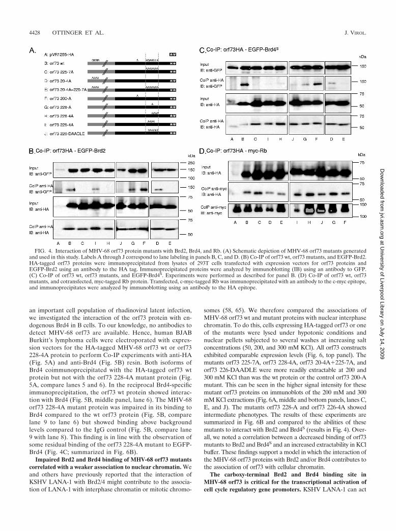
of MHV-68 orf73 mutant proteins with different mutations intheir Brd2 and Brd4 binding sites and a carboxy-terminal dou-ble HA epitope tag were generated (for a schematic depiction,see Fig. 4A). In the mutant orf73 225-7A, 7 alanines replacedaa 225 to 231 (KQAKKLK in the wild-type [wt] protein), whichhad been identified as a site of Brd2 as well as Brd4 interaction.In the mutant orf73 20-4A, the wt amino acids CKRR (aa 20 to
FIG. 1. The MHV-68 orf73 protein interacts with the carboxy ter-mini of the cellular BET proteins Brd2/RING3, Brd4S, and Brd3/ORFX. (A) Schematic depiction of the human BET proteins. BR1,bromodomain 1; BR2, bromodomain 2. (B) High degree of sequenceconservation among human and murine ET domains. The human BETproteins are encoded by four genes, Brd2, Brd3, Brd4, and Brd6, andare characterized by two bromodomains and an ET domain. The BETprotein sizes range from 722 to 1,362 aa for Brd4S and Brd4L, respec-tively. (B) ClustalW protein sequence alignment of human (hu) andmurine (mu) BET protein 64-aa-long ET domains. ET domains of huBrd2 (aa 640 to 703), mu Brd2 (aa 638 to 701), hu Brd3 (aa 570 to 633),mu Brd3 (aa 571 to 634), hu Brd4 (aa 608 to 671), mu Brd4 (aa 609 to672), hu Brd6 (aa 508 to 571), and mu Brd6 (aa 504 to 567) werealigned. Boxed residues differ from the consensus sequence. The align-ment shows 100% identity of the human and murine orthologous Brd2ET domains, of human and murine Brd3 ET domains, and of humanand murine Brd4 ET domains. The Brd6 ET domains of mice andhumans are 95.3% identical. ET domain identity between paralogousBET proteins is also high, ranging from 84.4% to 92.2%. (C) Sche-matic of the GST-BET fusion proteins. Labels A, B, C, and D corre-spond to lane labeling in panel D. (D) The full-length MHV-68orf73-HA protein interacted with GST-BET fusion proteins containingthe ET domains of Brd2/RING3, Brd4, and Brd3/ORFX in GST pull-down assays. Bacterially expressed GST fusion proteins were bound toglutathione Sepharose resin and subsequently incubated with lysatesfrom 293T cells transfected with MHV-68 orf73-HA expression plas-mid. Ten percent of the input lysates was loaded for comparison. Top,anti-HA immunoblot (IB) used to detect the orf73 protein; bottom, anti-GSTimmunoblot used to verify expression of the GST-BET fusion proteins.
FIG. 2. A peptide array assay identifies a binding site for purifiedBrd2 in the MHV-68 orf73 protein. (A) Schematic depiction of thebaculovirus-expressed Brd2/RING3 protein that was purified and usedfor studies of direct interaction by peptide array assays, with resultsshown in panel D. BR1, bromodomain 1; BR2, bromodomain 2.(B) The purity of Brd2/RING3 was determined by SDS-PAGE fol-lowed by Coomassie staining. The identity of Brd2/RING2 was con-firmed by anti-RING3 immunoblotting (IB). (C) Peptides representingthe complete MHV-68 orf73 protein with each peptide overlappingwith the previous one in 12 out of 15 residues were first incubated withSF9 lysate from untransfected SF9 insect cells and then probed with aprimary rabbit anti-RING3 polyclonal antibody followed by incubationwith a secondary AP-conjugated antibody to determine backgroundstaining. The nomenclature of peptides starts with 1A in the top leftcorner. (D) After the membrane was stripped, the same peptide arraywas incubated with affinity-purified Brd2/RING3 protein (see legendfor panel B). Bound protein was detected as described for panel C. Thepeptide with the strongest signal intensity is marked with an asterisk.(E) Quantification of signal intensities shown in panel D minus back-ground intensities shown in panel C, determined using Phoretix array1.0 quantification software. (F) Sequences of the four peptides, 3W,3X, 3Y, and 4A, that bound RING3, all containing the amino acidsQAKKLK, corresponding to positions 226 to 231 in the MHV-68 orf73protein.
4426 OTTINGER ET AL. J. VIROL.
at University of Liverpool Library on July 14, 2009
jvi.asm.org
Dow
nloaded from
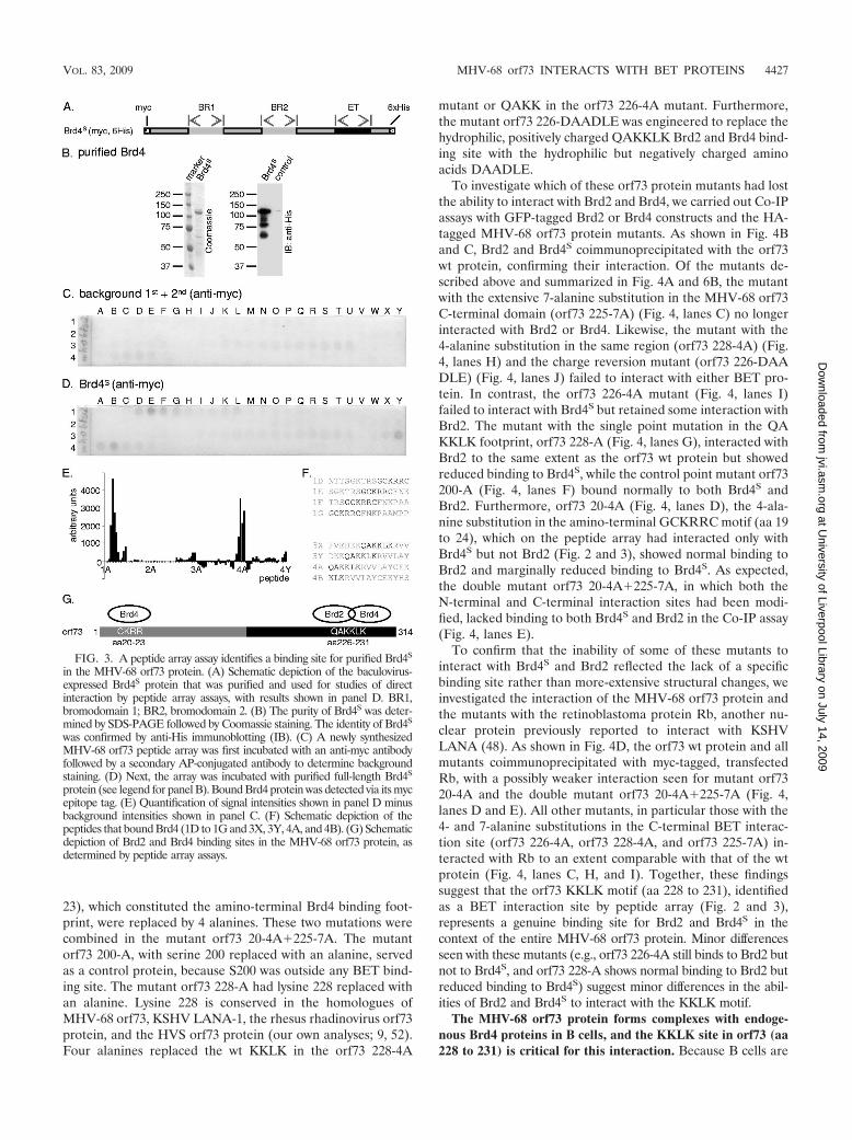
23), which constituted the amino-terminal Brd4 binding foot-print, were replaced by 4 alanines. These two mutations werecombined in the mutant orf73 20-4A�225-7A. The mutantorf73 200-A, with serine 200 replaced with an alanine, servedas a control protein, because S200 was outside any BET bind-ing site. The mutant orf73 228-A had lysine 228 replaced withan alanine. Lysine 228 is conserved in the homologues ofMHV-68 orf73, KSHV LANA-1, the rhesus rhadinovirus orf73protein, and the HVS orf73 protein (our own analyses; 9, 52).Four alanines replaced the wt KKLK in the orf73 228-4A
mutant or QAKK in the orf73 226-4A mutant. Furthermore,the mutant orf73 226-DAADLE was engineered to replace thehydrophilic, positively charged QAKKLK Brd2 and Brd4 bind-ing site with the hydrophilic but negatively charged aminoacids DAADLE.
To investigate which of these orf73 protein mutants had lostthe ability to interact with Brd2 and Brd4, we carried out Co-IPassays with GFP-tagged Brd2 or Brd4 constructs and the HA-tagged MHV-68 orf73 protein mutants. As shown in Fig. 4Band C, Brd2 and Brd4S coimmunoprecipitated with the orf73wt protein, confirming their interaction. Of the mutants de-scribed above and summarized in Fig. 4A and 6B, the mutantwith the extensive 7-alanine substitution in the MHV-68 orf73C-terminal domain (orf73 225-7A) (Fig. 4, lanes C) no longerinteracted with Brd2 or Brd4. Likewise, the mutant with the4-alanine substitution in the same region (orf73 228-4A) (Fig.4, lanes H) and the charge reversion mutant (orf73 226-DAADLE) (Fig. 4, lanes J) failed to interact with either BET pro-tein. In contrast, the orf73 226-4A mutant (Fig. 4, lanes I)failed to interact with Brd4S but retained some interaction withBrd2. The mutant with the single point mutation in the QAKKLK footprint, orf73 228-A (Fig. 4, lanes G), interacted withBrd2 to the same extent as the orf73 wt protein but showedreduced binding to Brd4S, while the control point mutant orf73200-A (Fig. 4, lanes F) bound normally to both Brd4S andBrd2. Furthermore, orf73 20-4A (Fig. 4, lanes D), the 4-ala-nine substitution in the amino-terminal GCKRRC motif (aa 19to 24), which on the peptide array had interacted only withBrd4S but not Brd2 (Fig. 2 and 3), showed normal binding toBrd2 and marginally reduced binding to Brd4S. As expected,the double mutant orf73 20-4A�225-7A, in which both theN-terminal and C-terminal interaction sites had been modi-fied, lacked binding to both Brd4S and Brd2 in the Co-IP assay(Fig. 4, lanes E).
To confirm that the inability of some of these mutants tointeract with Brd4S and Brd2 reflected the lack of a specificbinding site rather than more-extensive structural changes, weinvestigated the interaction of the MHV-68 orf73 protein andthe mutants with the retinoblastoma protein Rb, another nu-clear protein previously reported to interact with KSHVLANA (48). As shown in Fig. 4D, the orf73 wt protein and allmutants coimmunoprecipitated with myc-tagged, transfectedRb, with a possibly weaker interaction seen for mutant orf7320-4A and the double mutant orf73 20-4A�225-7A (Fig. 4,lanes D and E). All other mutants, in particular those with the4- and 7-alanine substitutions in the C-terminal BET interac-tion site (orf73 226-4A, orf73 228-4A, and orf73 225-7A) in-teracted with Rb to an extent comparable with that of the wtprotein (Fig. 4, lanes C, H, and I). Together, these findingssuggest that the orf73 KKLK motif (aa 228 to 231), identifiedas a BET interaction site by peptide array (Fig. 2 and 3),represents a genuine binding site for Brd2 and Brd4S in thecontext of the entire MHV-68 orf73 protein. Minor differencesseen with these mutants (e.g., orf73 226-4A still binds to Brd2 butnot to Brd4S, and orf73 228-A shows normal binding to Brd2 butreduced binding to Brd4S) suggest minor differences in the abil-ities of Brd2 and Brd4S to interact with the KKLK motif.
The MHV-68 orf73 protein forms complexes with endoge-nous Brd4 proteins in B cells, and the KKLK site in orf73 (aa228 to 231) is critical for this interaction. Because B cells are
FIG. 3. A peptide array assay identifies a binding site for purified Brd4S
in the MHV-68 orf73 protein. (A) Schematic depiction of the baculovirus-expressed Brd4S protein that was purified and used for studies of directinteraction by peptide array assays, with results shown in panel D. BR1,bromodomain 1; BR2, bromodomain 2. (B) The purity of Brd4S was deter-mined by SDS-PAGE followed by Coomassie staining. The identity of Brd4S
was confirmed by anti-His immunoblotting (IB). (C) A newly synthesizedMHV-68 orf73 peptide array was first incubated with an anti-myc antibodyfollowed by a secondary AP-conjugated antibody to determine backgroundstaining. (D) Next, the array was incubated with purified full-length Brd4S
protein (see legend for panel B). Bound Brd4 protein was detected via its mycepitope tag. (E) Quantification of signal intensities shown in panel D minusbackground intensities shown in panel C. (F) Schematic depiction of thepeptides that bound Brd4 (1D to 1G and 3X, 3Y, 4A, and 4B). (G) Schematicdepiction of Brd2 and Brd4 binding sites in the MHV-68 orf73 protein, asdetermined by peptide array assays.
VOL. 83, 2009 MHV-68 orf73 INTERACTS WITH BET PROTEINS 4427
at University of Liverpool Library on July 14, 2009
jvi.asm.org
Dow
nloaded from
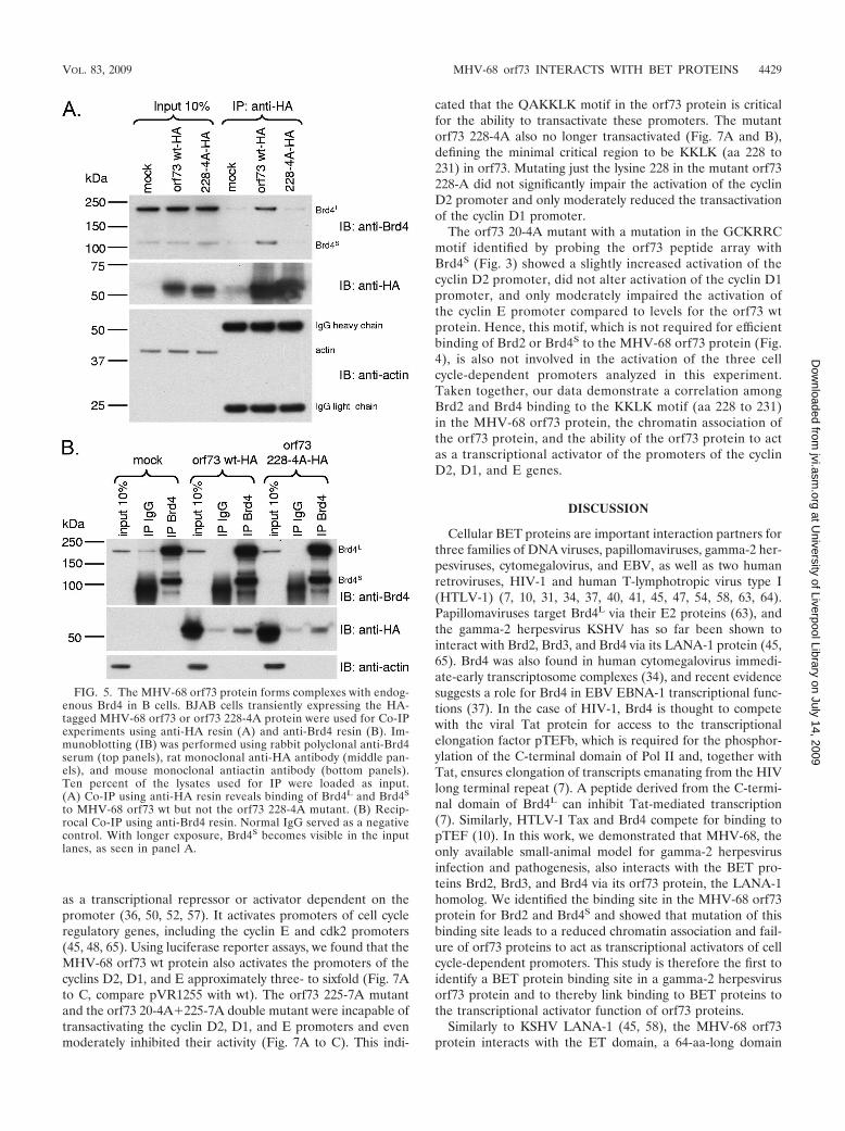
an important cell population of rhadinoviral latent infection,we investigated the interaction of the orf73 protein with en-dogenous Brd4 in B cells. To our knowledge, no antibodies todetect MHV-68 orf73 are available. Hence, human BJABBurkitt’s lymphoma cells were electroporated with expres-sion vectors for the HA-tagged MHV-68 orf73 wt or orf73228-4A protein to perform Co-IP experiments with anti-HA(Fig. 5A) and anti-Brd4 (Fig. 5B) resin. Both isoforms ofBrd4 coimmunoprecipitated with the HA-tagged orf73 wtprotein but not with the orf73 228-4A mutant protein (Fig.5A, compare lanes 5 and 6). In the reciprocal Brd4-specificimmunoprecipitation, the orf73 wt protein showed interac-tion with Brd4 (Fig. 5B, middle panel, lane 6). The MHV-68orf73 228-4A mutant protein was impaired in its binding toBrd4 compared to the wt orf73 protein (Fig. 5B, comparelane 9 to lane 6) but showed binding above backgroundlevels compared to the IgG control (Fig. 5B, compare lane9 with lane 8). This finding is in line with the observation ofsome residual binding of the orf73 228-4A mutant to EGFP-Brd4 (Fig. 4C; summarized in Fig. 6B).
Impaired Brd2 and Brd4 binding of MHV-68 orf73 mutantscorrelated with a weaker association to nuclear chromatin. Weand others have previously reported that the interaction ofKSHV LANA-1 with Brd2/4 might contribute to the associa-tion of LANA-1 with interphase chromatin or mitotic chromo-
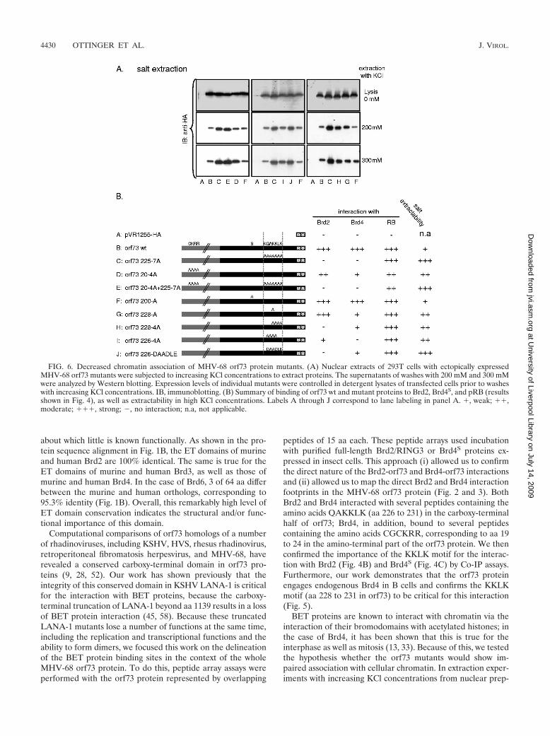
somes (58, 65). We therefore compared the associations ofMHV-68 orf73 wt and mutant proteins with nuclear interphasechromatin. To do this, cells expressing HA-tagged orf73 or oneof the mutants were lysed under hypotonic conditions andnuclear pellets subjected to several washes at increasing saltconcentrations (50, 200, and 300 mM KCl). All orf73 constructsexhibited comparable expression levels (Fig. 6, top panel). Themutants orf73 225-7A, orf73 228-4A, orf73 20-4A�225-7A, andorf73 226-DAADLE were more readily extractable at 200 and300 mM KCl than was the wt protein or the control orf73 200-Amutant. This can be seen in the higher signal intensity for thesemutant orf73 proteins on immunoblots of the 200 mM and 300mM KCl extractions (Fig. 6A, middle and bottom panels, lanes C,E, and J). The mutants orf73 228-A and orf73 226-4A showedintermediate phenotypes. The results of these experiments aresummarized in Fig. 6B and compared to the abilities of thesemutants to interact with Brd2 and Brd4S (results in Fig. 4). Over-all, we noted a correlation between a decreased binding of orf73mutants to Brd2 and Brd4S and an increased extractability in KClbuffer. These findings support a model in which the interaction ofthe MHV-68 orf73 proteins with Brd2 and/or Brd4 contributes tothe association of orf73 with cellular chromatin.
The carboxy-terminal Brd2 and Brd4 binding site inMHV-68 orf73 is critical for the transcriptional activation ofcell cycle regulatory gene promoters. KSHV LANA-1 can act
FIG. 4. Interaction of MHV-68 orf73 protein mutants with Brd2, Brd4, and Rb. (A) Schematic depiction of MHV-68 orf73 mutants generatedand used in this study. Labels A through J correspond to lane labeling in panels B, C, and D. (B) Co-IP of orf73 wt, orf73 mutants, and EGFP-Brd2.HA-tagged orf73 proteins were immunoprecipitated from lysates of 293T cells transfected with expression vectors for orf73 proteins andEGFP-Brd2 using an antibody to the HA tag. Immunoprecipitated proteins were analyzed by immunoblotting (IB) using an antibody to GFP.(C) Co-IP of orf73 wt, orf73 mutants, and EGFP-Brd4S. Experiments were performed as described for panel B. (D) Co-IP of orf73 wt, orf73mutants, and cotransfected, myc-tagged Rb protein. Transfected, c-myc-tagged Rb was immunoprecipitated with an antibody to the c-myc epitope,and immunoprecipitates were analyzed by immunoblotting using an antibody to the HA epitope.
4428 OTTINGER ET AL. J. VIROL.
at University of Liverpool Library on July 14, 2009
jvi.asm.org
Dow
nloaded from
as a transcriptional repressor or activator dependent on thepromoter (36, 50, 52, 57). It activates promoters of cell cycleregulatory genes, including the cyclin E and cdk2 promoters(45, 48, 65). Using luciferase reporter assays, we found that theMHV-68 orf73 wt protein also activates the promoters of thecyclins D2, D1, and E approximately three- to sixfold (Fig. 7Ato C, compare pVR1255 with wt). The orf73 225-7A mutantand the orf73 20-4A�225-7A double mutant were incapable oftransactivating the cyclin D2, D1, and E promoters and evenmoderately inhibited their activity (Fig. 7A to C). This indi-
cated that the QAKKLK motif in the orf73 protein is criticalfor the ability to transactivate these promoters. The mutantorf73 228-4A also no longer transactivated (Fig. 7A and B),defining the minimal critical region to be KKLK (aa 228 to231) in orf73. Mutating just the lysine 228 in the mutant orf73228-A did not significantly impair the activation of the cyclinD2 promoter and only moderately reduced the transactivationof the cyclin D1 promoter.
The orf73 20-4A mutant with a mutation in the GCKRRCmotif identified by probing the orf73 peptide array withBrd4S (Fig. 3) showed a slightly increased activation of thecyclin D2 promoter, did not alter activation of the cyclin D1promoter, and only moderately impaired the activation ofthe cyclin E promoter compared to levels for the orf73 wtprotein. Hence, this motif, which is not required for efficientbinding of Brd2 or Brd4S to the MHV-68 orf73 protein (Fig.4), is also not involved in the activation of the three cellcycle-dependent promoters analyzed in this experiment.Taken together, our data demonstrate a correlation amongBrd2 and Brd4 binding to the KKLK motif (aa 228 to 231)in the MHV-68 orf73 protein, the chromatin association ofthe orf73 protein, and the ability of the orf73 protein to actas a transcriptional activator of the promoters of the cyclinD2, D1, and E genes.
DISCUSSION
Cellular BET proteins are important interaction partners forthree families of DNA viruses, papillomaviruses, gamma-2 her-pesviruses, cytomegalovirus, and EBV, as well as two humanretroviruses, HIV-1 and human T-lymphotropic virus type I(HTLV-1) (7, 10, 31, 34, 37, 40, 41, 45, 47, 54, 58, 63, 64).Papillomaviruses target Brd4L via their E2 proteins (63), andthe gamma-2 herpesvirus KSHV has so far been shown tointeract with Brd2, Brd3, and Brd4 via its LANA-1 protein (45,65). Brd4 was also found in human cytomegalovirus immedi-ate-early transcriptosome complexes (34), and recent evidencesuggests a role for Brd4 in EBV EBNA-1 transcriptional func-tions (37). In the case of HIV-1, Brd4 is thought to competewith the viral Tat protein for access to the transcriptionalelongation factor pTEFb, which is required for the phosphor-ylation of the C-terminal domain of Pol II and, together withTat, ensures elongation of transcripts emanating from the HIVlong terminal repeat (7). A peptide derived from the C-termi-nal domain of Brd4L can inhibit Tat-mediated transcription(7). Similarly, HTLV-I Tax and Brd4 compete for binding topTEF (10). In this work, we demonstrated that MHV-68, theonly available small-animal model for gamma-2 herpesvirusinfection and pathogenesis, also interacts with the BET pro-teins Brd2, Brd3, and Brd4 via its orf73 protein, the LANA-1homolog. We identified the binding site in the MHV-68 orf73protein for Brd2 and Brd4S and showed that mutation of thisbinding site leads to a reduced chromatin association and fail-ure of orf73 proteins to act as transcriptional activators of cellcycle-dependent promoters. This study is therefore the first toidentify a BET protein binding site in a gamma-2 herpesvirusorf73 protein and to thereby link binding to BET proteins tothe transcriptional activator function of orf73 proteins.
Similarly to KSHV LANA-1 (45, 58), the MHV-68 orf73protein interacts with the ET domain, a 64-aa-long domain
FIG. 5. The MHV-68 orf73 protein forms complexes with endog-enous Brd4 in B cells. BJAB cells transiently expressing the HA-tagged MHV-68 orf73 or orf73 228-4A protein were used for Co-IPexperiments using anti-HA resin (A) and anti-Brd4 resin (B). Im-munoblotting (IB) was performed using rabbit polyclonal anti-Brd4serum (top panels), rat monoclonal anti-HA antibody (middle pan-els), and mouse monoclonal antiactin antibody (bottom panels).Ten percent of the lysates used for IP were loaded as input.(A) Co-IP using anti-HA resin reveals binding of Brd4L and Brd4S
to MHV-68 orf73 wt but not the orf73 228-4A mutant. (B) Recip-rocal Co-IP using anti-Brd4 resin. Normal IgG served as a negativecontrol. With longer exposure, Brd4S becomes visible in the inputlanes, as seen in panel A.
VOL. 83, 2009 MHV-68 orf73 INTERACTS WITH BET PROTEINS 4429
at University of Liverpool Library on July 14, 2009
jvi.asm.org
Dow
nloaded from
about which little is known functionally. As shown in the pro-tein sequence alignment in Fig. 1B, the ET domains of murineand human Brd2 are 100% identical. The same is true for theET domains of murine and human Brd3, as well as those ofmurine and human Brd4. In the case of Brd6, 3 of 64 aa differbetween the murine and human orthologs, corresponding to95.3% identity (Fig. 1B). Overall, this remarkably high level ofET domain conservation indicates the structural and/or func-tional importance of this domain.
Computational comparisons of orf73 homologs of a numberof rhadinoviruses, including KSHV, HVS, rhesus rhadinovirus,retroperitoneal fibromatosis herpesvirus, and MHV-68, haverevealed a conserved carboxy-terminal domain in orf73 pro-teins (9, 28, 52). Our work has shown previously that theintegrity of this conserved domain in KSHV LANA-1 is criticalfor the interaction with BET proteins, because the carboxy-terminal truncation of LANA-1 beyond aa 1139 results in a lossof BET protein interaction (45, 58). Because these truncatedLANA-1 mutants lose a number of functions at the same time,including the replication and transcriptional functions and theability to form dimers, we focused this work on the delineationof the BET protein binding sites in the context of the wholeMHV-68 orf73 protein. To do this, peptide array assays wereperformed with the orf73 protein represented by overlapping
peptides of 15 aa each. These peptide arrays used incubationwith purified full-length Brd2/RING3 or Brd4S proteins ex-pressed in insect cells. This approach (i) allowed us to confirmthe direct nature of the Brd2-orf73 and Brd4-orf73 interactionsand (ii) allowed us to map the direct Brd2 and Brd4 interactionfootprints in the MHV-68 orf73 protein (Fig. 2 and 3). BothBrd2 and Brd4 interacted with several peptides containing theamino acids QAKKLK (aa 226 to 231) in the carboxy-terminalhalf of orf73; Brd4, in addition, bound to several peptidescontaining the amino acids CGCKRR, corresponding to aa 19to 24 in the amino-terminal part of the orf73 protein. We thenconfirmed the importance of the KKLK motif for the interac-tion with Brd2 (Fig. 4B) and Brd4S (Fig. 4C) by Co-IP assays.Furthermore, our work demonstrates that the orf73 proteinengages endogenous Brd4 in B cells and confirms the KKLKmotif (aa 228 to 231 in orf73) to be critical for this interaction(Fig. 5).
BET proteins are known to interact with chromatin via theinteraction of their bromodomains with acetylated histones; inthe case of Brd4, it has been shown that this is true for theinterphase as well as mitosis (13, 33). Because of this, we testedthe hypothesis whether the orf73 mutants would show im-paired association with cellular chromatin. In extraction exper-iments with increasing KCl concentrations from nuclear prep-
FIG. 6. Decreased chromatin association of MHV-68 orf73 protein mutants. (A) Nuclear extracts of 293T cells with ectopically expressedMHV-68 orf73 mutants were subjected to increasing KCl concentrations to extract proteins. The supernatants of washes with 200 mM and 300 mMwere analyzed by Western blotting. Expression levels of individual mutants were controlled in detergent lysates of transfected cells prior to washeswith increasing KCl concentrations. IB, immunoblotting. (B) Summary of binding of orf73 wt and mutant proteins to Brd2, Brd4S, and pRB (resultsshown in Fig. 4), as well as extractability in high KCl concentrations. Labels A through J correspond to lane labeling in panel A. �, weak; ��,moderate; ���, strong; �, no interaction; n.a, not applicable.
4430 OTTINGER ET AL. J. VIROL.
at University of Liverpool Library on July 14, 2009
jvi.asm.org
Dow
nloaded from

arations, the orf73 constructs with mutations in their BETprotein binding sites were more readily extracted by KCl thanwas the orf73 wt protein or a control mutant (orf73 200-A),indicating that the mutant constructs had a weaker relativechromatin association than did the orf73 wt protein. This sug-gests a contribution of the orf73-BET interaction to the orf73chromatin association. Because the association with the nuclear
fraction in this assay was weakened but not completely abrogated,our data suggest that Brd2 and Brd4 are most likely not the onlymechanism of chromatin association for the MHV-68 orf73 pro-tein. In the case of KSHV LANA-1, the current model is that ofa direct histone interaction via the LANA-1 amino-terminal do-main (5), with the LANA-1 carboxy-terminal domain modulatingthe chromatin interaction to a fully functional state via its inter-
FIG. 7. The MHV-68 orf73 protein activates promoters of cell cycle regulatory genes, and the BET interaction site KKLK (aa 228 to 231)in the orf73 protein is critical for this function. Transient luciferase reporter assays were performed with murine 3T3 fibroblasts. Cells werecotransfected with 50 ng of promoter luciferase reporter plasmids together with 500 ng, 1,000 ng, or 2,500 ng of empty vector (pVR1255)or different MHV-68 orf73 constructs (Fig. 4A). Relative luciferase activities compared to that for the empty vector were calculated, andmean values � standard deviations from two representative experiments in duplicate are depicted. (A) Murine cyclin D2 promoter;(B) murine cyclin D1 promoter; (C) human cyclin E promoter.
VOL. 83, 2009 MHV-68 orf73 INTERACTS WITH BET PROTEINS 4431
at University of Liverpool Library on July 14, 2009
jvi.asm.org
Dow
nloaded from

action with additional cellular binding partners, e.g., the BETproteins Brd2 and Brd4 or the methyl-CpG-binding proteinMeCP2 or DEK (5, 27, 35, 45, 58).
KSHV LANA-1 is a promiscuous transcriptional regulatorthat regulates the promoters of cell cycle regulatory genes,including the cyclin E promoter, besides many other targetgenes (3, 45, 65). We show here that the MHV-68 orf73 pro-tein also acts as a transcriptional activator of the cyclin D2 andcyclin E promoters, two E2F-responsive promoters, as well asthe cyclin D1 promoter (Fig. 7). orf73 proteins with mutationsin their carboxy-terminal Brd2 and Brd4 binding sites (e.g., theorf73 225-7A and orf73 228-4A mutants) were impaired intheir ability to activate the cyclin D2, D1, and E promoters(Fig. 7). In contrast, mutating the core of the amino-terminalBrd4 binding footprint CKRR (mutant orf73 20-4A) did nothave a negative effect on the transactivation of the cyclin D2and D1 promoters and only moderately impaired the transac-tivation of the cyclin E promoter. Interestingly, the orf7320-4A mutant also still interacted with Brd2 and (more weakly)with Brd4S and did not show a changed chromatin associationin the salt extraction experiments at 50 mM and 200 mM KCl(Fig. 6A; also, data not shown). This suggests that the inter-action of Brd4 with the site CKRR (aa 20 to 23) in the amino-terminal domain of the MHV-68 orf73 protein, which hadbeen identified in the peptide array, is not required for theinteraction with Brd2 and Brd4S and is not essential for chro-matin association or the transcription activation function.
As with all site-directed mutations generated in the absenceof structural information, one potential caveat could be thatthey affect the proper folding of the MHV-68 orf73 protein.The preserved ability of the mutants to interact with RB wouldargue against this. In addition, aa 225 to 231 are predicted tobe part of an alpha-helical structure of orf73, and stretches ofalanines, as used with the orf73 228-4A and orf73 225-7Amutants, are known to preferentially form a helical structure;therefore, our alanine mutants should fold similarly to the wtprotein. Also, we found that the orf73 225-7A and orf73228-4A mutants retained the ability of the MHV-68 orf73 wtprotein to induce G2/M cell cycle arrest (not shown). Takentogether, these results show that the minimal orf73 228-4Amutant has retained several functional properties of theMHV-68 orf73 proteins, while losing the ability to interact withBrd2 and Brd4S and to activate transcription from three cellcycle-dependent promoters.
Both Brd2 and Brd4 have recently been identified in proteincomplexes with RNA Pol II and have been shown to activatetranscription (11, 32, 61). For Brd4, this occurs via the inter-action of Brd4 with pTEFb, a cyclin-dependent kinase, andsubsequent stimulation of RNA Pol II-dependent transcrip-tional elongation (32, 61). Papillomaviruses have been shownto interact with Brd4 via their E2 proteins, and this E2-Brd4interaction is critical for the E2 transcriptional activation func-tion (31, 40, 54). There are a number of different ways in whichpTEFb can be recruited to transcription complexes, and therecruitment via the chromatin-bound activator Brd4 is one ofthem (46). HTLV-1 has recently been shown to modulate theBrd4/pTEFb interaction through the interaction of its Tax pro-tein with the cyclin T1 subunit of pTEFb, thereby possiblycompeting with the Brd4/pTEFb interaction (10). Similarly,Brd4L has been shown to compete with HIV-1 Tat for binding
to pTEFb and to thereby modulate transcription from the HIVlong terminal repeat (7). The mechanism employed byHTLV-1 to target pTEFb therefore seems to be distinguish-able from the mechanism employed by both papillomavirusesand gamma-2 herpesviruses, which directly bind to Brd4 (45,54, 63, 64).
The MHV-68 orf73 protein is required for the establishmentof splenic latency in mice (43). The molecular mechanisms,however, have remained elusive. Importantly, MHV-68 orf73engages endogenous Brd4 in B cells (Fig. 5). Therefore, thisstudy provides a first step in understanding the functions ofMHV-68 orf73 on a molecular level. The MHV-68 orf73 pro-tein is expressed throughout the viral replication cycle (19) andhence most likely plays roles during lytic as well as duringlatent infection. By enhancing the expression of G1/S-phasecell cycle genes (Fig. 7), orf73 may create an S-phase cellularenvironment favorable to viral replication. Through its inter-action with the nuclear fraction (Fig. 6), possibly with host cellchromatin through its interaction with BET proteins, orf73may play a role in latent viral replication and genome mainte-nance. Furthermore, the functional interaction of orf73 withBET proteins may play a role in the pathogenesis of rhadino-virus-induced B-cell lymphoma, possibly through the enhance-ment of BET target gene transcription, such as that of thecyclin D1, D2, and E genes (Fig. 7). Interestingly, B-cell-spe-cific ectopic Brd2 expression in mice results in B-cell lym-phoma (26). A strength of the MHV-68 system is the possibilityto study aspects of rhadinoviral biology and pathogenesis incell culture and use the findings to design in vivo experimentswith infected mice. Experiments are under way to study thephenotype of orf73 proteins with mutations in their BET pro-tein binding sites in the context of the whole virus in vivo.
In summary, this work describes a novel transcriptional ac-tivation function of the MHV-68 orf73 protein on the cyclinD1, D2, and E promoters. Furthermore, this study identifiesbinding sites for Brd2 and Brd4S in the MHV-68 orf73 proteinand shows that the ability to activate these cell cycle promoterscorrelates with the interaction with BET proteins. In addition,binding to BET proteins appears to contribute to the interac-tion of the MHV-68 orf73 protein with cellular chromatin.
ACKNOWLEDGMENTS
We thank Peter Valentin-Weigand for helpful discussions and sug-gestions early in the project and all members of the Schulz laboratorywho contributed to discussions relating to this work. We thank Sus-anne Daenicke for expert technical assistance in the synthesis of thepeptide arrays.
M.O. was funded by grants DFG GRK 745 Mucosal Host-PathogenInteractions and DFG SPP1130. D.P. was funded by the EuropeanUnion Integrated Project INCA (LSHC-CT-2005-018704). J.P.S. wasfunded by a Royal Society University research fellowship and PublicHealth Service grant CA090208 from the National Cancer Institute.Some of the later experiments were carried out by M.O. in PeterHowley’s laboratory at Harvard Medical School and were supported bygrant R01CA116720 (to Peter Howley) from the National CancerInstitute.
REFERENCES
1. Abbate, E. A., C. Voitenleitner, and M. R. Botchan. 2006. Structure of thepapillomavirus DNA-tethering complex E2:Brd4 and a peptide that ablatesHPV chromosomal association. Mol. Cell 24:877–889.
2. Allen, R. D., III, S. Dickerson, and S. H. Speck. 2006. Identification ofspliced gammaherpesvirus 68 LANA and v-cyclin transcripts and analysis oftheir expression in vivo during latent infection. J. Virol. 80:2055–2062.
4432 OTTINGER ET AL. J. VIROL.
at University of Liverpool Library on July 14, 2009
jvi.asm.org
Dow
nloaded from
3. An, F. Q., N. Compitello, E. Horwitz, M. Sramkoski, E. S. Knudsen, and R.Renne. 2005. The latency-associated nuclear antigen of Kaposi’s sarcoma-associated herpesvirus modulates cellular gene expression and protects lym-phoid cells from p16 INK4A-induced cell cycle arrest. J. Biol. Chem. 280:3862–3874.
4. Ballestas, M. E., P. A. Chatis, and K. M. Kaye. 1999. Efficient persistence ofextrachromosomal KSHV DNA mediated by latency-associated nuclear an-tigen. Science 284:641–644.
5. Barbera, A. J., J. V. Chodaparambil, B. Kelley-Clarke, V. Joukov, J. C.Walter, K. Luger, and K. M. Kaye. 2006. The nucleosomal surface as adocking station for Kaposi’s sarcoma herpesvirus LANA. Science 311:856–861.
6. Baxter, M. K., M. G. McPhillips, K. Ozato, and A. A. McBride. 2005. Themitotic chromosome binding activity of the papillomavirus E2 protein cor-relates with interaction with the cellular chromosomal protein, Brd4. J. Vi-rol. 79:4806–4818.
7. Bisgrove, D. A., T. Mahmoudi, P. Henklein, and E. Verdin. 2007. ConservedP-TEFb-interacting domain of BRD4 inhibits HIV transcription. Proc. Natl.Acad. Sci. USA 104:13690–13695.
8. Bouchard, C., K. Thieke, A. Maier, R. Saffrich, J. Hanley-Hyde, W. Ansorge,S. Reed, P. Sicinski, J. Bartek, and M. Eilers. 1999. Direct induction of cyclinD2 by Myc contributes to cell cycle progression and sequestration of p27.EMBO J. 18:5321–5333.
9. Burnside, K. L., J. T. Ryan, H. Bielefeldt-Ohmann, B. A. Gregory, M. E.Thouless, C. C. Tsai, and T. M. Rose. 2006. RFHVMn ORF73 is structurallyrelated to the KSHV ORF73 latency-associated nuclear antigen (LANA)and is expressed in retroperitoneal fibromatosis (RF) tumor cells. Virology354:103–115.
10. Cho, W. K., M. Zhou, M. K. Jang, K. Huang, S. J. Jeong, K. Ozato, and J. N.Brady. 2007. Modulation of the Brd4/P-TEFb interaction by the humanT-lymphotropic virus type 1 tax protein. J. Virol. 81:11179–11186.
11. Crowley, T. E., E. M. Kaine, M. Yoshida, A. Nandi, and D. J. Wolgemuth.2002. Reproductive cycle regulation of nuclear import, euchromatic local-ization, and association with components of Pol II mediator of a mammaliandouble-bromodomain protein. Mol. Endocrinol. 16:1727–1737.
12. Denis, G. V., M. E. McComb, D. V. Faller, A. Sinha, P. B. Romesser, andC. E. Costello. 2006. Identification of transcription complexes that containthe double bromodomain protein Brd2 and chromatin remodeling machines.J. Proteome Res. 5:502–511.
13. Dey, A., F. Chitsaz, A. Abbasi, T. Misteli, and K. Ozato. 2003. The doublebromodomain protein Brd4 binds to acetylated chromatin during interphaseand mitosis. Proc. Natl. Acad. Sci. USA 100:8758–8763.
14. Dey, A., J. Ellenberg, A. Farina, A. E. Coleman, T. Maruyama, S. Sciortino,J. Lippincott-Schwartz, and K. Ozato. 2000. A bromodomain protein,MCAP, associates with mitotic chromosomes and affects G2-to-M transition.Mol. Cell. Biol. 20:6537–6549.
15. Ebrahimi, B., B. M. Dutia, K. L. Roberts, J. J. Garcia-Ramirez, P. Dickin-son, J. P. Stewart, P. Ghazal, D. J. Roy, and A. A. Nash. 2003. Transcriptomeprofile of murine gammaherpesvirus-68 lytic infection. J. Gen. Virol. 84:99–109.
16. Flano, E., S. M. Husain, J. T. Sample, D. L. Woodland, and M. A. Blackman.2000. Latent murine gamma-herpesvirus infection is established in activatedB cells, dendritic cells, and macrophages. J. Immunol. 165:1074–1081.
17. Flano, E., I. J. Kim, D. L. Woodland, and M. A. Blackman. 2002. Gamma-herpesvirus latency is preferentially maintained in splenic germinal centerand memory B cells. J. Exp. Med. 196:1363–1372.
18. Florence, B., and D. V. Faller. 2001. You bet-cha: a novel family of tran-scriptional regulators. Front. Biosci. 6:D1008–D1018.
19. Forrest, J. C., C. R. Paden, R. D. Allen III, J. Collins, and S. H. Speck. 2007.ORF73-null murine gammaherpesvirus 68 reveals roles for mLANA and p53in virus replication. J. Virol. 81:11957–11971.
20. Fowler, P., S. Marques, J. P. Simas, and S. Efstathiou. 2003. ORF73 ofmurine herpesvirus-68 is critical for the establishment and maintenance oflatency. J. Gen. Virol. 84:3405–3416.
21. Frank, R. 1992. Spot synthesis: an easy technique for the positionally ad-dressable, parallel chemical synthesis on membrane support. Tetrahedron48:9217–9232.
22. Frank, R., and S. Dubel. 2005. Analysis of protein interactions with immo-bilized peptide arrays synthesized on membrane supports, p. 591–608. In E.Golemis and P. Adams (ed.), Protein-protein interactions: a molecular clon-ing manual. Cold Spring Harbor Laboratory Press, Cold Spring Harbor, NY.
23. Friborg, J., Jr., W. Kong, M. O. Hottiger, and G. J. Nabel. 1999. p53inhibition by the LANA protein of KSHV protects against cell death. Nature402:889–894.
24. Fujimuro, M., F. Y. Wu, C. ApRhys, H. Kajumbula, D. B. Young, G. S.Hayward, and S. D. Hayward. 2003. A novel viral mechanism for dysregu-lation of beta-catenin in Kaposi’s sarcoma-associated herpesvirus latency.Nat. Med. 9:300–306.
25. Geng, Y., E. N. Eaton, M. Picon, J. M. Roberts, A. S. Lundberg, A. Gifford,C. Sardet, and R. A. Weinberg. 1996. Regulation of cyclin E transcription byE2Fs and retinoblastoma protein. Oncogene 12:1173–1180.
26. Greenwald, R. J., J. R. Tumang, A. Sinha, N. Currier, R. D. Cardiff, T. L.
Rothstein, D. V. Faller, and G. V. Denis. 2004. E mu-BRD2 transgenic micedevelop B-cell lymphoma and leukemia. Blood 103:1475–1484.
27. Griffiths, R., and A. Whitehouse. 2007. Herpesvirus saimiri episomal persis-tence is maintained via interaction between open reading frame 73 and thecellular chromosome-associated protein MeCP2. J. Virol. 81:4021–4032.
28. Grundhoff, A., and D. Ganem. 2003. The latency-associated nuclear antigenof Kaposi’s sarcoma-associated herpesvirus permits replication of terminalrepeat-containing plasmids. J. Virol. 77:2779–2783.
29. Hu, J., A. C. Garber, and R. Renne. 2002. The latency-associated nuclearantigen of Kaposi’s sarcoma-associated herpesvirus supports latent DNAreplication in dividing cells. J. Virol. 76:11677–11687.
30. Huang, H., J. Zhang, W. Shen, X. Wang, J. Wu, J. Wu, and Y. Shi. 2007.Solution structure of the second bromodomain of Brd2 and its specificinteraction with acetylated histone tails. BMC Struct. Biol. 7:57.
31. Ilves, I., K. Maemets, T. Silla, K. Janikson, and M. Ustav. 2006. Brd4 isinvolved in multiple processes of the bovine papillomavirus type 1 life cycle.J. Virol. 80:3660–3665.
32. Jang, M. K., K. Mochizuki, M. Zhou, H. S. Jeong, J. N. Brady, and K. Ozato.2005. The bromodomain protein Brd4 is a positive regulatory component ofP-TEFb and stimulates RNA polymerase II-dependent transcription. Mol.Cell 19:523–534.
33. Kanno, T., Y. Kanno, R. M. Siegel, M. K. Jang, M. J. Lenardo, and K. Ozato.2004. Selective recognition of acetylated histones by bromodomain proteinsvisualized in living cells. Mol. Cell 13:33–43.
34. Kapasi, A. J., and D. H. Spector. 2008. Inhibition of the cyclin-dependentkinases at the beginning of human cytomegalovirus infection specificallyalters the levels and localization of the RNA polymerase II carboxyl-terminaldomain kinases cdk9 and cdk7 at the viral transcriptosome. J. Virol. 82:394–407.
35. Krithivas, A., M. Fujimuro, M. Weidner, D. B. Young, and S. D. Hayward.2002. Protein interactions targeting the latency-associated nuclear antigen ofKaposi’s sarcoma-associated herpesvirus to cell chromosomes. J. Virol. 76:11596–11604.
36. Krithivas, A., D. B. Young, G. Liao, D. Greene, and S. D. Hayward. 2000.Human herpesvirus 8 LANA interacts with proteins of the mSin3 corepres-sor complex and negatively regulates Epstein-Barr virus gene expression indually infected PEL cells. J. Virol. 74:9637–9645.
37. Lin, A., S. Wang, T. Nguyen, K. Shire, and L. Frappier. 2008. The EBNA1protein of Epstein-Barr virus functionally interacts with Brd4. J. Virol. 82:12009–12019.
38. Maruyama, T., A. Farina, A. Dey, J. Cheong, V. P. Bermudez, T. Tamura, S.Sciortino, J. Shuman, J. Hurwitz, and K. Ozato. 2002. A mammalian bro-modomain protein, Brd4, interacts with replication factor C and inhibitsprogression to S phase. Mol. Cell. Biol. 22:6509–6520.
39. Matangkasombut, O., R. M. Buratowski, N. W. Swilling, and S. Buratowski.2000. Bromodomain factor 1 corresponds to a missing piece of yeast TFIID.Genes Dev. 14:951–962.
40. McPhillips, M. G., J. G. Oliveira, J. E. Spindler, R. Mitra, and A. A.McBride. 2006. Brd4 is required for E2-mediated transcriptional activationbut not genome partitioning of all papillomaviruses. J. Virol. 80:9530–9543.
41. McPhillips, M. G., K. Ozato, and A. A. McBride. 2005. Interaction of bovinepapillomavirus E2 protein with Brd4 stabilizes its association with chromatin.J. Virol. 79:8920–8932.
42. Mochizuki, K., A. Nishiyama, M. K. Jang, A. Dey, A. Ghosh, T. Tamura, H.Natsume, H. Yao, and K. Ozato. 2008. The bromodomain protein Brd4stimulates G1 gene transcription and promotes progression to S phase.J. Biol. Chem. 283:9040–9048.
43. Moorman, N. J., D. O. Willer, and S. H. Speck. 2003. The gammaherpesvirus68 latency-associated nuclear antigen homolog is critical for the establish-ment of splenic latency. J. Virol. 77:10295–10303.
44. Muller, H., J. Lukas, A. Schneider, P. Warthoe, J. Bartek, M. Eilers, and M.Strauss. 1994. Cyclin D1 expression is regulated by the retinoblastoma pro-tein. Proc. Natl. Acad. Sci. USA 91:2945–2949.
45. Ottinger, M., T. Christalla, K. Nathan, M. M. Brinkmann, A. Viejo-Bor-bolla, and T. F. Schulz. 2006. Kaposi’s sarcoma-associated herpesvirusLANA-1 interacts with the short variant of Brd4 and releases cells from aBrd4- and Brd2/RING3-induced G1 cell cycle arrest. J. Virol. 80:10772–10786.
46. Peterlin, B. M., and D. H. Price. 2006. Controlling the elongation phase oftranscription with P-TEFb. Mol. Cell 23:297–305.
47. Platt, G. M., G. R. Simpson, S. Mittnacht, and T. F. Schulz. 1999. Latentnuclear antigen of Kaposi’s sarcoma-associated herpesvirus interacts withRING3, a homolog of the Drosophila female sterile homeotic (fsh) gene.J. Virol. 73:9789–9795.
48. Radkov, S. A., P. Kellam, and C. Boshoff. 2000. The latent nuclear antigen ofKaposi sarcoma-associated herpesvirus targets the retinoblastoma-E2F path-way and with the oncogene Hras transforms primary rat cells. Nat. Med.6:1121–1127.
49. Rawlins, D. R., G. Milman, S. D. Hayward, and G. S. Hayward. 1985.Sequence-specific DNA binding of the Epstein-Barr virus nuclear antigen(EBNA-1) to clustered sites in the plasmid maintenance region. Cell 42:859–868.
VOL. 83, 2009 MHV-68 orf73 INTERACTS WITH BET PROTEINS 4433
at University of Liverpool Library on July 14, 2009
jvi.asm.org
Dow
nloaded from
50. Renne, R., C. Barry, D. Dittmer, N. Compitello, P. O. Brown, and D. Ganem.2001. Modulation of cellular and viral gene expression by the latency-asso-ciated nuclear antigen of Kaposi’s sarcoma-associated herpesvirus. J. Virol.75:458–468.
51. Rochford, R., M. L. Lutzke, R. S. Alfinito, A. Clavo, and R. D. Cardin. 2001.Kinetics of murine gammaherpesvirus 68 gene expression following infectionof murine cells in culture and in mice. J. Virol. 75:4955–4963.
52. Schwam, D. R., R. L. Luciano, S. S. Mahajan, L. Wong, and A. C. Wilson.2000. Carboxy terminus of human herpesvirus 8 latency-associated nuclearantigen mediates dimerization, transcriptional repression, and targeting tonuclear bodies. J. Virol. 74:8532–8540.
53. Schweiger, M. R., M. Ottinger, J. You, and P. M. Howley. 2007. Brd4-independent transcriptional repression function of the papillomavirus E2proteins. J. Virol. 81:9612–9622.
54. Schweiger, M. R., J. You, and P. M. Howley. 2006. Bromodomain protein 4mediates the papillomavirus E2 transcriptional activation function. J. Virol.80:4276–4285.
55. Sinha, A., D. V. Faller, and G. V. Denis. 2005. Bromodomain analysis ofBrd2-dependent transcriptional activation of cyclin A. Biochem. J. 387:257–269.
56. Stewart, J. P., E. J. Usherwood, A. Ross, H. Dyson, and T. Nash. 1998. Lungepithelial cells are a major site of murine gammaherpesvirus persistence. J.Exp. Med. 187:1941–1951.
57. Viejo-Borbolla, A., E. Kati, J. A. Sheldon, K. Nathan, K. Mattsson, L.Szekely, and T. F. Schulz. 2003. A domain in the C-terminal region oflatency-associated nuclear antigen 1 of Kaposi’s sarcoma-associated herpes-virus affects transcriptional activation and binding to nuclear heterochroma-tin. J. Virol. 77:7093–7100.
58. Viejo-Borbolla, A., M. Ottinger, E. Bruning, A. Burger, R. Konig, E. Kati, J. A.Sheldon, and T. F. Schulz. 2005. Brd2/RING3 interacts with a chromatin-binding domain in the Kaposi’s sarcoma-associated herpesvirus latency-associ-ated nuclear antigen 1 (LANA-1) that is required for multiple functions ofLANA-1. J. Virol. 79:13618–13629.
59. Willer, D. O., and S. H. Speck. 2003. Long-term latent murine gammaher-pesvirus 68 infection is preferentially found within the surface immunoglob-ulin D-negative subset of splenic B cells in vivo. J. Virol. 77:8310–8321.
60. Wu, S. Y., A. Y. Lee, S. Y. Hou, J. K. Kemper, H. Erdjument-Bromage, P.Tempst, and C. M. Chiang. 2006. Brd4 links chromatin targeting to HPVtranscriptional silencing. Genes Dev. 20:2383–2396.
61. Yang, Z., J. H. Yik, R. Chen, N. He, M. K. Jang, K. Ozato, and Q. Zhou.2005. Recruitment of P-TEFb for stimulation of transcriptional elongationby the bromodomain protein Brd4. Mol. Cell 19:535–545.
62. Yates, J. L., N. Warren, and B. Sugden. 1985. Stable replication of plasmidsderived from Epstein-Barr virus in various mammalian cells. Nature 313:812–815.
63. You, J., J. L. Croyle, A. Nishimura, K. Ozato, and P. M. Howley. 2004.Interaction of the bovine papillomavirus E2 protein with Brd4 tethers theviral DNA to host mitotic chromosomes. Cell 117:349–360.
64. You, J., M. R. Schweiger, and P. M. Howley. 2005. Inhibition of E2 bindingto Brd4 enhances viral genome loss and phenotypic reversion of bovinepapillomavirus-transformed cells. J. Virol. 79:14956–14961.
65. You, J., V. Srinivasan, G. V. Denis, W. J. Harrington, Jr., M. E. Ballestas,K. M. Kaye, and P. M. Howley. 2006. Kaposi’s sarcoma-associated herpes-virus latency-associated nuclear antigen interacts with bromodomain proteinBrd4 on host mitotic chromosomes. J. Virol. 80:8909–8919.
4434 OTTINGER ET AL. J. VIROL.
at University of Liverpool Library on July 14, 2009
jvi.asm.org
Dow
nloaded from