the influence of chromosomal amplification and … by professor byong ro kim the doctoral...
TRANSCRIPT

The Influence of Chromosomal
Amplification and Deletion on Clinical
Characteristics and Prognosis in Patients
with Hepatocellular Carcinoma
Yoon-Seok Chae
Department of Medicine
The Graduate school, Yonsei University

The Influence of Chromosomal
Amplification and Deletion on Clinical
Characteristics and Prognosis in Patients
with Hepatocellular Carcinoma
Directed by Professor Byong Ro Kim
The Doctoral Dissertation
Submitted to the Department of Medical Science,
the Graduate School of Yonsei University
in partial fulfillment of the requirements for the degree of
Doctor of Philosophy
Yoon Seok Chae
June 2005
This certifies that the Doctoral Dissertation
of Yoon Seok Chae is approved.
Thesis supervisor: Byong Ro Kim, M.D. Signature:
Thesis committee: Hoguen Kim, M.D. Signature:
Thesis committee: Chae Yoon Chon, M.D. Signature:
Thesis committee: Woo Jung Lee, M.D. Signature:
Thesis committee: Jin-Sung Lee, M.D. Signature:
The Graduate school
Yonsei University
June 2005
Acknowledgements
The author thanks Professor Tae Yeon Jang of Chon buk National
University for editorial assistance in English. Also, thanks Professor Woo
Jung Lee for statistical analysis and Miss. Jan Dy Lee for medical record
review. Professor Hoguen Kim teach CGH to me, help me with a many
aspects. I am indebted to director professor Byong Ro Kim for his advice
and criticisms during the preperation of this manuscription. Finally, thanks
for my parents who give life to me into this world.

- i -
TABLE OF CONTENTS
Abstract ------------------------------------------------------------------------------------ 1
I. INRODUCTION ----------------------------------------------------------------------- 2
II. MATRIALS AND METHODS ----------------------------------------------------- 3
1. Tumor specimen--------------------------------------------------------------------- 3
2. Comparative genomic hybridization analysis ---------------------------------- 3
3. Analysis of patient's clinicopathologic features
& Statistical Analysis-------------------------------------------------------------- 4
III. RESULTS ----------------------------------------------------------------------------- 5
1. Fluorescence of photomicrograph showing
the result of a CGH analysis ------------------------------------------------------ 5
2. Graphic form of rate of chromosomal loss and gain----------------------- 6
3. Avarage fluorescence ratio profiles ------------------------------------------ 6
4. Patients characteristics ------------------------------------------------------------ 7
5. Genetic alteration by CGH in HCCs --------------------------------------- 8
6. 5-year survival rate of 37 hepatoceullular carcinomas --------------------- - 8
7. Overall survival of hepatocellular carcinomas according to AFP level ---- 9
8. Genetic alterations impact on 5-year overall survival ------------------------- 9
9. Overall survival according to the loss of 16q ----------------------------------10
10. Correlation 4q and α-fetoprotein ---------------------------------------------- 10
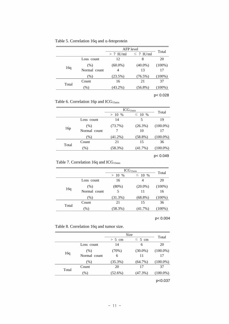
11. Correlation 16q and α-fetoprotein ---------------------------------------------- 11
13. Correlation 16p and ICG15min --------------------------------------------------- 11
14. Correlation 16q and ICG15min ---------------------------------------------------- 11
15. Correlation 16q and tumor size ------------------------------------------------- 11
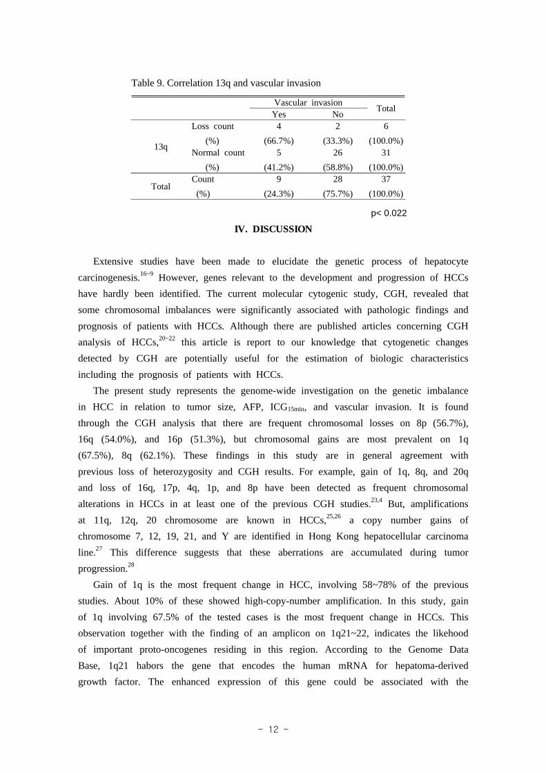
16. Correlation 13q and vascular invasion ----------------------------------------- 12
IV. DISCUSSION ---------------------------------------------------------------------- 12
V. CONCLUSION----------------------------------------------------------------------
15
REFERENCES-------------------------------------------------------------------------- 16
ABSTRACT(IN KOREAN)---------------------------------------------------------- 21

- ii -
LIST OF FIGURES
Figure 1. Schematic diagram of comparative genomic hybridization
method ----------------------------------------------------------------- 4
Figure 2. Fluorescence of photomicrograph showing the result of a CGH
analysis of the hepatoceullar carcinoma of case 1 ---------------- 5
Figure 3. The rate of chromosomal loss and gain in graphic form -- 6
Figure 4. Avarage fluorescence ratio profiles ------------------------------ 6
Figure 5. 5-year survival rate of 37 hepatoceullular carcinomas ----------- 8
Figure 6. Overall survival of hepatocellular carcinomas according to AFP
level ---------------------------------------------------------------------- 9
Figure 7. Overall survival according to the loss of 16q ----------------------10
LIST OF TABLES
Table 1.Summary of clinical data and chromosomal aberration
detected by CGH from 37 hepatomas --------------------------- 7
Table 2. Genetic alteration by CGH in HCCs ---------------------------- 8
Table 3. Genetic alterations impact on 5-year overall survival ------------- 9
Table 4. Correlation 4q and α-fetoprotein -------------------------------------10
Table 5. Correlation 16q and α-fetoprotein ----------------------------------- 11
Table 6. Correlation 16p and ICG15min ---------------------------------------- 11
Table 7. Correlation 16q and ICG15min ---------------------------------------- 11
Table 8. Correlation 16q and tumor size ------------------------------------- 11
Table 9. Correlation 13q and vascular invasion ---------------------------- 12
- 1 -
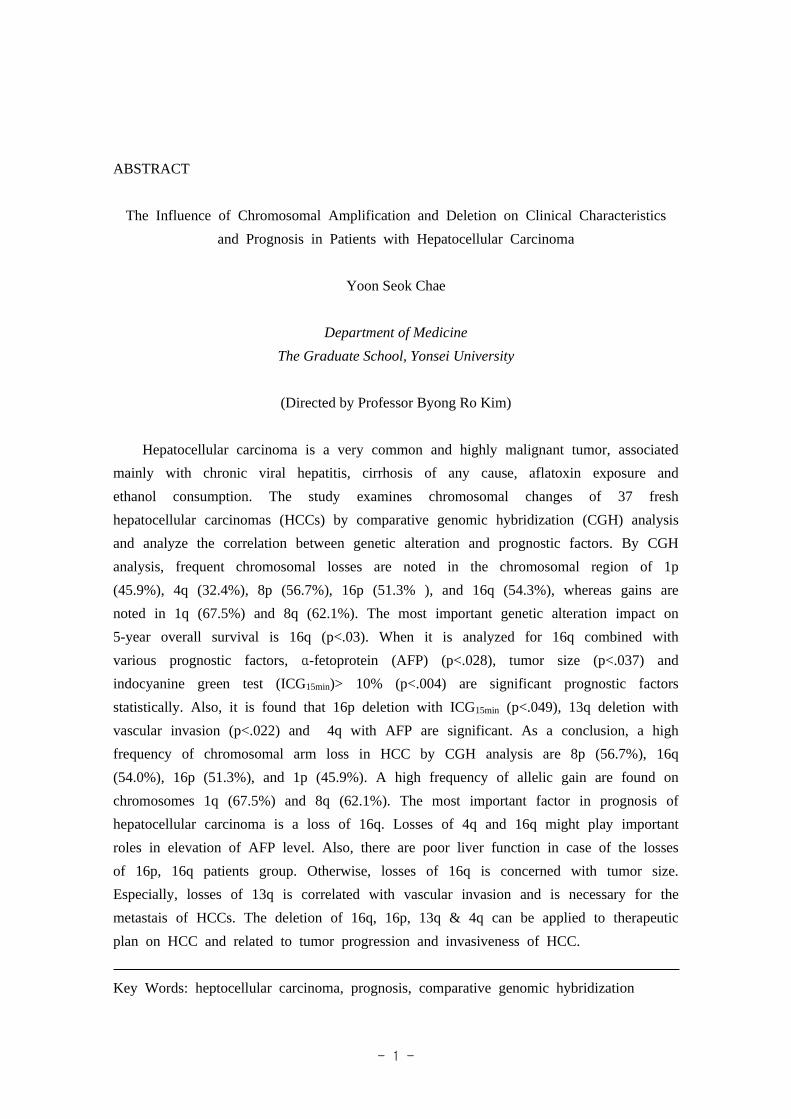
ABSTRACT
The Influence of Chromosomal Amplification and Deletion on Clinical Characteristics
and Prognosis in Patients with Hepatocellular Carcinoma
Yoon Seok Chae
Department of Medicine
The Graduate School, Yonsei University
(Directed by Professor Byong Ro Kim)
Hepatocellular carcinoma is a very common and highly malignant tumor, associated
mainly with chronic viral hepatitis, cirrhosis of any cause, aflatoxin exposure and
ethanol consumption. The study examines chromosomal changes of 37 fresh
hepatocellular carcinomas (HCCs) by comparative genomic hybridization (CGH) analysis
and analyze the correlation between genetic alteration and prognostic factors. By CGH
analysis, frequent chromosomal losses are noted in the chromosomal region of 1p
(45.9%), 4q (32.4%), 8p (56.7%), 16p (51.3% ), and 16q (54.3%), whereas gains are
noted in 1q (67.5%) and 8q (62.1%). The most important genetic alteration impact on
5-year overall survival is 16q (p<.03). When it is analyzed for 16q combined with
various prognostic factors, α-fetoprotein (AFP) (p<.028), tumor size (p<.037) and
indocyanine green test (ICG15min)> 10% (p<.004) are significant prognostic factors
statistically. Also, it is found that 16p deletion with ICG15min (p<.049), 13q deletion with
vascular invasion (p<.022) and 4q with AFP are significant. As a conclusion, a high
frequency of chromosomal arm loss in HCC by CGH analysis are 8p (56.7%), 16q
(54.0%), 16p (51.3%), and 1p (45.9%). A high frequency of allelic gain are found on
chromosomes 1q (67.5%) and 8q (62.1%). The most important factor in prognosis of
hepatocellular carcinoma is a loss of 16q. Losses of 4q and 16q might play important
roles in elevation of AFP level. Also, there are poor liver function in case of the losses
of 16p, 16q patients group. Otherwise, losses of 16q is concerned with tumor size.
Especially, losses of 13q is correlated with vascular invasion and is necessary for the
metastais of HCCs. The deletion of 16q, 16p, 13q & 4q can be applied to therapeutic
plan on HCC and related to tumor progression and invasiveness of HCC.
Key Words: heptocellular carcinoma, prognosis, comparative genomic hybridization
- 2 -
The Influence of Chromosomal Amplification and Deletion on Clinical Characteristics
and Prognosis in Patients with Hepatocellular Carcinoma
Yoon Seok Chae
Department of Medicine
The Graduate School, Yonsei University
(Directed by Professor Byong Ro Kim)
I. INTRODUCTION
Hepatocellular carcinoma is one of the most common human malignant tumors,
especially in southern and eastern Asia.1 HCC is the second leading cause of death
from cancer in Korea. It is widely accepted that hepatitis B (HBV) or C virus (HCV)
infection, subsequent chronic inflammation and hepatocyte regeneration play important
roles in the development of HCCs.2~4 However, the effect of viral infection on
hepatocellular transformation remains to be undetermined. Accordingly, it remains
unknown whether carcinogenetic process is different in HBV-positive and HCV-postitive
livers. HCCs, like many other tumors, is considered to develop and progress as a
consequence of an accumulation of genetic alterations.5 Many investigators have made
varying attempts to find genes implicated in hepatocarcinogenesis to construct a genetic
pathway in the progression of HCC.6~9 Although frequent allelic losses at loci of
chromosomes 1p, 4q, 5p, 5q, 8p, 10p, 11q, 13q, 16q, and 17p have been researched in
HCCs,10,11 most of these studies fail to provide consistent information concerning genetic
changes leading to the evolution of HCCs. Comprehensive analysis is necessary to
throughly understand the complicated genetic alterations in malignant tumors.
Determination of these comprehensive genetic changes in solid tumors is practically
difficult, because the examination of many individual genes by conventional method is
laborious and cumbersome. Screening for chromosomal regions with frequent gains and
losses is one of the steps toward the identification of genes implicated in the
development and progression of tumors. Although karyotyping provides comprehensive
information concerning structural aberrations of whole chromosomes, it is highly
specialized work and time-concerning even for experienced technicians. Moreover, it is
difficult to prepare metaphase spreads from solid tumors such as HCCs. In fact, there
are no large scale genetic studies on HCCs as far as we know. Fortunately, comparative
genomic hybridization (CGH), a recently developed technology, allows a global analysis
- 3 -
of chromosomal imbalances which may be etiologically relevant or of diagnostic and
prognostic importance.
At present, however, there is no available information concerning the relationship
between genetic alterations and clinicopathologic characteristics in HCCs. In this study,
we wish to know which genetic alternation related with prognostic factors.
II. MATERIAL & METHODS
1. Tumor specimen
Thirty-seven surgically resected HCCs were included for this study. The cases were
identified prospectively and consecutively at Yonsei University Severance Hospital
between January 1996 and December 2002 for a study of molecular markers for
HCCs. Patient information was obtained prospectively without any knowledge of genetic
alterations. The macroscopic and microscopic features of the resected specimens are
reviewed by an experienced liver pathologist who confirms the diagnosis of HCCs,
assesses the presence or absence of vascular invasion, and records the maximal diameter
of the tumor. The presence or absence of cirrhosis in the nontumorous part of the
resected specimen is also recorded. Cirrhosis is defined as the presence of complete
fibrous septa seperating regeneration nodules.12 Grading of differentiation is performed
according to Edmondson and Steiner.13 According to this classification, 20 HCCs are
categorized as well differentiated, 8 HCCs are moderate differentiated, and 9 HCCs are
poorly differentiated. Among the 37 HCC patients, 33 (89%) had liver cirrhosis in the
non-neoplastic liver. Serum hepatitis B surface antigen is positive in 31 patients (84%)
and anti-hepatitis C virus antibody is positive in 2 patients (5%). The selected tissues
are stored at -700C until DNA extraction is performed. Each tisssue is microdissected in
a cryostat to separate the tumor cells from adjacent non-neoplastic tissues. Genomic
DNA is performed by the Sodium dodecyl sulphate-proteinase K and phenol-chloroform
extraction method.14
2. Comparative genomic hybridization analysis
Genomic DNA samples from tumors are labeled with Spectrum Green deoxyuridine
triphosphate (dUTP) (Vysis Inc., Downers Grove, IL), and normal reference genomic
DNA was labeled with Spectrum Red dUTP (Vysis) using the nick translation technique.
Labeled tumor and reference DNA (200~400ng), as well as 10 μg of unlabeled human
cobalt uptake protein (Cot-1 DNA) (Vysis) are dissolved in 10μl of hybridization buffer
(50% formamide, 10% dextran sulfate, and 2x standard saline citrate) and denatured at
370C on denatured normal metaphase spreads. After hybridization for 3 days, the slides
- 4 -
are washed and counterstained with 4',6-diamino-2-phenylindole dihydrocholoride (DAPI)
in antifade solution. CGH hybridizations were analyzed using an Olympus fluorescent
microscope and the Cytovision image analysis system (Applied Imaging, Sunderland,
Tyne & Wear, UK). Three digital images (DAPI, Spectrum Green, and Spectrum Red)
were acquired from 10 to 20 metaphases in each hybridization. DNA of normal male
and DNA from tumor cell lines with known aberrations are used as control test DNA.
Green-to-red intensity ratio profiles are calculated for each chromosome and threshold
values defining gains and losses are set at 1.25 and 0.75, respectively. High level
increase in copy number (amplicon) is defined as a ratio of tumor/reference greater than
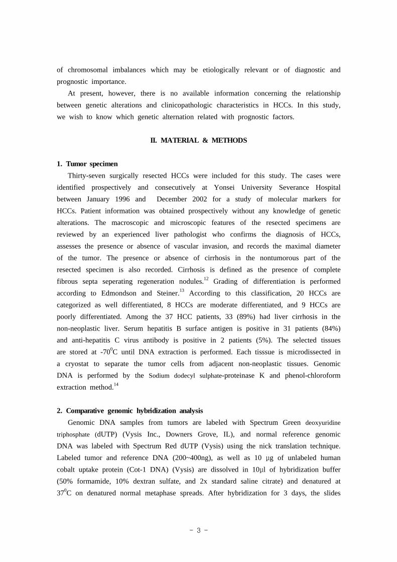
1.5. Schematic diagraphic discription is shown in Figure 1.
Figure 1. Schematic diagram of comparative genomic hybridization method.
3. Analysis of patient's clinicopathologic features & Statistical Analysis
We review clinical recording chart of 37 patients as follows; HBV infection, AFP
level (preoperation), ICG15min, tumor size, vascular invasion, and tumor stage. Statistical
analysis is performed using SPSS package version 10.0 statistical software, USA. The
Kaplan-Meier method is used to calculate survival rates and log-rank test is used to
analyze differences. Total DNA copy number aberrations, whether gains or losses,
differences in proportions between the groups are analyzed the chi-square test. For all
statistical tests, a probability p value of less than 0.05 is considered significant.

- 5 -
III. RESULTS
This study included 37 patients of 31 males and 6 females, whose average age is 51
years (range 16~66). Average follow-up period is 51 months. Serum AFP levels ranged
from less than 10 ng/ml(within reference range) in 21 patients(56.8%). The disease stage
of the HCCs is classified according to modified Union International Contre le Cancer
(UICC)15. One case (3%) is stage I (T1N0M0), 16 (45%) as stage II (T2N0M0), 11 (30%)
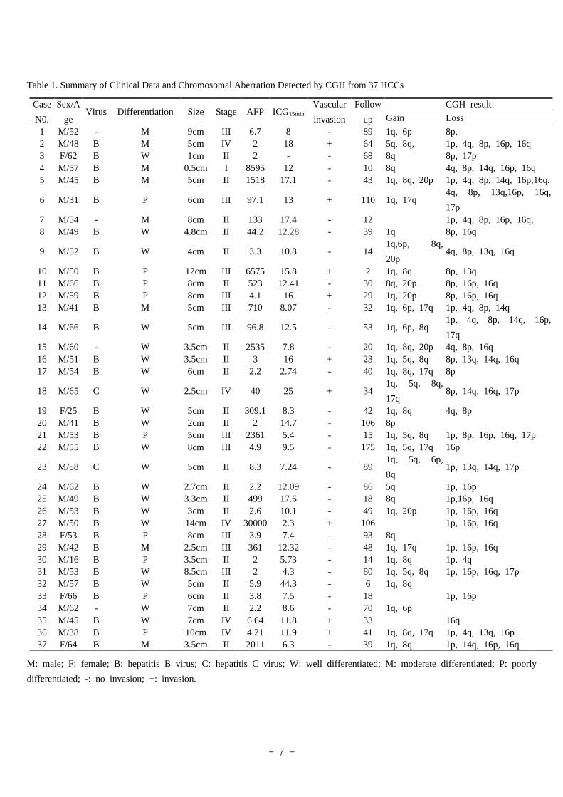
as stage III (T3N0M0), 8 (22%) as stage IV (T1~4N0M1 or T4N0M0). Table 1 summarizes
the patients' characteristics, histopathologic differentiation, tumor size, vascular invasion,
AFP, ICG15min, Stage and CGH results. Fluorescence photomicrograph of hepatocellular
carcinoma of case 1 of CGH analysis is shown in Figure 2.
Figure 2. Fluorescence of photomicrograph showing the result of a CGH analysis of the hepatoceullar
carcinoma of case 1.
A schematic summary of all chromosomal copy number aberrations is shown in Figure
3, 4. The chromosomal losses are more frequent than the gains. All patients show
chromosomal loss in at least one chromosomal arm. The frequency of chromosomal
losses is summarized in Table 2. A high frequency of chromosomal arm loss in HCC
by CGH analysis is 8p (56.7%), 16q (54.0%), 16p (51.3%), and 1p (45.9%). Moderate
frequency loss is detected at 4q (32.4.%), 14q (24.3%), and 17p (21.6%). The frequency
of chromosomal loss on the other chromosomes was less than 20%.

- 6 -
Figure 4. The CGH analysis on primary HCCs. Green lines represent gains and red
lines highlight losses. The intensity ratio of a balanced copy number is 1.0 (central
vertical line). The left-side shift indicates underrepresentations (value 0.75) while
right-side shift indicates overrepresentations (value 1.25). Primary HCC shows gains of
3p, 19p, and high copy number amplication of 4p, 4q, Losses are detected on 10p, 14q,
22q.
Figure 3. The rate of chromosomal loss and gain observed on a designated 37
non-acrocentric chromosomal arms of HCC in graphic form. Each bar represents the
percentage of loss (lower) or gain (upper) of a chromosomal arm
- 7 -
Table 1. Summary of Clinical Data and Chromosomal Aberration Detected by CGH from 37 HCCs
Case
N0.
Sex/A
geVirus Differentiation Size Stage AFP ICG15min
Vascular
invasion
Follow
up
CGH result
Gain Loss
1 M/52 - M 9cm III 6.7 8 - 89 1q, 6p 8p,2 M/48 B M 5cm IV 2 18 + 64 5q, 8q, 1p, 4q, 8p, 16p, 16q3 F/62 B W 1cm II 2 - - 68 8q 8p, 17p4 M/57 B M 0.5cm I 8595 12 - 10 8q 4q, 8p, 14q, 16p, 16q5 M/45 B M 5cm II 1518 17.1 - 43 1q, 8q, 20p 1p, 4q, 8p, 14q, 16p,16q,
6 M/31 B P 6cm III 97.1 13 + 110 1q, 17q4q, 8p, 13q,16p, 16q,
17p7 M/54 - M 8cm II 133 17.4 - 12 1p, 4q, 8p, 16p, 16q, 8 M/49 B W 4.8cm II 44.2 12.28 - 39 1q 8p, 16q
9 M/52 B W 4cm II 3.3 10.8 - 141q,6p, 8q,
20p4q, 8p, 13q, 16q
10 M/50 B P 12cm III 6575 15.8 + 2 1q, 8q 8p, 13q11 M/66 B P 8cm II 523 12.41 - 30 8q, 20p 8p, 16p, 16q12 M/59 B P 8cm III 4.1 16 + 29 1q, 20p 8p, 16p, 16q13 M/41 B M 5cm III 710 8.07 - 32 1q, 6p, 17q 1p, 4q, 8p, 14q
14 M/66 B W 5cm III 96.8 12.5 - 53 1q, 6p, 8q1p, 4q, 8p, 14q, 16p,
17q15 M/60 - W 3.5cm II 2535 7.8 - 20 1q, 8q, 20p 4q, 8p, 16q16 M/51 B W 3.5cm II 3 16 + 23 1q, 5q, 8q 8p, 13q, 14q, 16q17 M/54 B W 6cm II 2.2 2.74 - 40 1q, 8q, 17q 8p
18 M/65 C W 2.5cm IV 40 25 + 341q, 5q, 8q,
17q8p, 14q, 16q, 17p
19 F/25 B W 5cm II 309.1 8.3 - 42 1q, 8q 4q, 8p20 M/41 B W 2cm II 2 14.7 - 106 8p21 M/53 B P 5cm III 2361 5.4 - 15 1q, 5q, 8q 1p, 8p, 16p, 16q, 17p22 M/55 B W 8cm III 4.9 9.5 - 175 1q, 5q, 17q 16p
23 M/58 C W 5cm II 8.3 7.24 - 891q, 5q, 6p,
8q 1p, 13q, 14q, 17p
24 M/62 B W 2.7cm II 2.2 12.09 - 86 5q 1p, 16p25 M/49 B W 3.3cm II 499 17.6 - 18 8q 1p,16p, 16q26 M/53 B W 3cm II 2.6 10.1 - 49 1q, 20p 1p, 16p, 16q27 M/50 B W 14cm IV 30000 2.3 + 106 1p, 16p, 16q28 F/53 B P 8cm III 3.9 7.4 - 93 8q29 M/42 B M 2.5cm III 361 12.32 - 48 1q, 17q 1p, 16p, 16q30 M/16 B P 3.5cm II 2 5.73 - 14 1q, 8q 1p, 4q31 M/53 B W 8.5cm III 2 4.3 - 80 1q, 5q, 8q 1p, 16p, 16q, 17p32 M/57 B W 5cm II 5.9 44.3 - 6 1q, 8q33 F/66 B P 6cm II 3.8 7.5 - 18 1p, 16p34 M/62 - W 7cm II 2.2 8.6 - 70 1q, 6p35 M/45 B W 7cm IV 6.64 11.8 + 33 16q36 M/38 B P 10cm IV 4.21 11.9 + 41 1q, 8q, 17q 1p, 4q, 13q, 16p37 F/64 B M 3.5cm II 2011 6.3 - 39 1q, 8q 1p, 14q, 16p, 16q
M: male; F: female; B: hepatitis B virus; C: hepatitis C virus; W: well differentiated; M: moderate differentiated; P: poorly
differentiated; -: no invasion; +: invasion.
- 8 -
The frequency of chromosomal gain is summarized in Table 2.
Table 2. Genetic alterations by CGH in HCCs
Loss(%) Gain(%)1p 45.9 1q 67.54q 32.4 5q 21.68p 56.7 6p 16.213q 18.9 8q 62.114q 24.3 17q 18.916p 51.3 20p 16.216q 54.017p 21.6
A high frequency of allelic gain was found on chromosomes 1q (67.5%) and 8q
(62.1%). Other chromosomal arms have chromosomal gain frequency of less than 20%.
It is to evaluate the correlation between the change occurring on each chromosomal arm
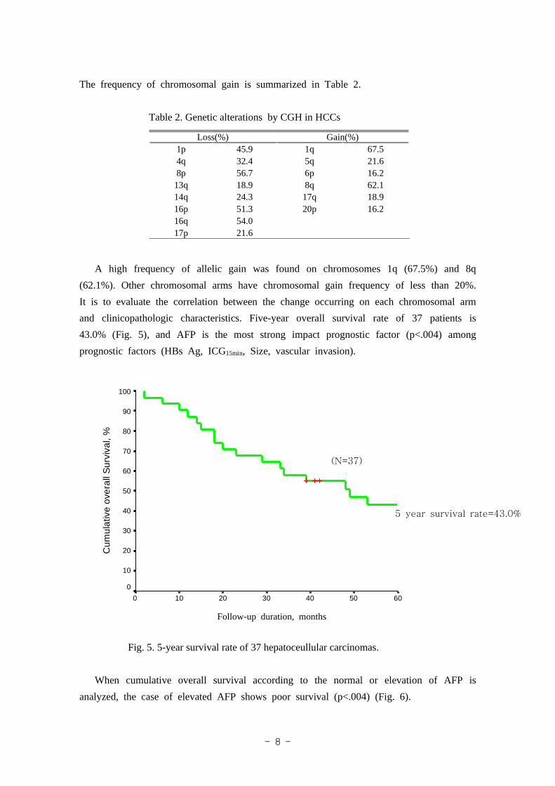
and clinicopathologic characteristics. Five-year overall survival rate of 37 patients is
43.0% (Fig. 5), and AFP is the most strong impact prognostic factor (p<.004) among
prognostic factors (HBs Ag, ICG15min, Size, vascular invasion).
F/U duration, months
6050403020100
Cum
ulat
ive
ove
rall
Sur
viva
l, %
100
90
80
70
60
50
40
30
20
10
0
Fig. 5. 5-year survival rate of 37 hepatoceullular carcinomas.
When cumulative overall survival according to the normal or elevation of AFP is
analyzed, the case of elevated AFP shows poor survival (p<.004) (Fig. 6).
Follow-up duration, months
5 year survival rate=43.0%
(N=37)

- 9 -
F/U duration, months
6050403020100
Cum
ulat
ive
Ove
rall
Su
rviv
al, %
100
90
80
70
60
50
40
30
20
10
0
Fig. 6. Overall survival of hepatocellular carcinomas according to AFP level.
Univariate analysis on chromosomal aberrations is performed for the potential factors
that are associated with patient survival. 16q chromosomal loss is significantly correlated
with 5-year overall survival (Table 3) (Fig. 7), but other chromosomal abberations are
not significant statistically.
Table 3. Genetic alterations impact on 5-year overall survival
Loss GainGene p-value Gene p-value
1p 0.73 1q 67.54q 0.16 5q 21.68p 0.46 6p 16.213q 0.90 8q 62.114q 0.80 17q 18.916p 0.95 20p 16.216q 0.0317p 21.6
Follow-up duration, months
Elevated AFP
Normal AFP
(N=37)
p=0.004

- 10 -
F/U duration, months
6050403020100
Cum
ulat
ive
over
all
Su
rviv
al, %
100
90
80
70
60
50
40
30
20
10
0
Fig. 7. Overall survival according to the loss of 16q.
As a result of efforts to find out relationship genetic alterations with prognostic factor
statistically, 4q and AFP (p<0.048) (Table 4), 16q and AFP (p<0.028) (Table 5), 16p
and ICG15min (p<0.049) (Table 6), 16q and ICG15min (p<0.004) (Table 7), 16q and size
(p<0.037) (Table 8), 13q and vascular invasion (p<0.022) (Table 9) and are correlated
with prognostic factors of HCCs.
Table 4. Correlation 4q and α-fetoprotein
AFP levelTotal
> 7 IU/ml ≤ 7 IU/ml
4q
Loss count
(%)
8
(66.7%)
4
(33.3%)
12
(100.0%)Normal count
(%)
8
(32.0%)
17
(68.0%)
25
(100.0%)
TotalCount
(%)
16
(43.2%)
21
(56.8%)
37
(100.0%)
p< 0.048
Follow-up duration, months
(N=37)
Normal 16q
Loss of 16q
p=0.03
- 11 -
Table 5. Correlation 16q and α-fetoprotein
AFP levelTotal
> 7 IU/ml ≤ 7 IU/ml
16q
Loss count
(%)
12
(60.0%)
8
(40.0%)
20
(100%)Normal count
(%)
4
(23.5%)
13
(76.5%)
17
(100%)
TotalCount
(%)
16
(43.2%)
21
(56.8%)
37
(100%)
p< 0.028
Table 6. Correlation 16p and ICG15min
ICG15min Total> 10 % ≤ 10 %
16p
Loss count
(%)
14
(73.7%)
5
(26.3%)
19
(100.0%)Normal count
(%)
7
(41.2%)
10
(58.8%)
17
(100.0%)
TotalCount
(%)
21
(58.3%)
15
(41.7%)
36
(100.0%)
p< 0.049
Table 7. Correlation 16q and ICG15min
ICG15min Total> 10 % ≤ 10 %
16q
Loss count
(%)
16
(80%)
4
(20.0%)
20
(100%)Normal count
(%)
5
(31.3%)
11
(68.8%)
16
(100%)
TotalCount
(%)
21
(58.3%)
15
(41.7%)
36
(100%)
p< 0.004
Table 8. Correlation 16q and tumor size.
SizeTotal
> 5 cm ≤ 5 cm
16q
Loss count
(%)
14
(70%)
6
(30.0%)
20
(100.0%)Normal count
(%)
6
(35.3%)
11
(64.7%)
17
(100.0%)
TotalCount
(%)
20
(52.6%)
17
(47.3%)
37
(100.0%)
p<0.037
- 12 -
Table 9. Correlation 13q and vascular invasion
Vascular invasionTotal
Yes No
13q
Loss count
(%)
4
(66.7%)
2
(33.3%)
6
(100.0%)Normal count
(%)
5
(41.2%)
26
(58.8%)
31
(100.0%)
TotalCount
(%)
9
(24.3%)
28
(75.7%)
37
(100.0%)
p< 0.022
IV. DISCUSSION
Extensive studies have been made to elucidate the genetic process of hepatocyte
carcinogenesis.16~9 However, genes relevant to the development and progression of HCCs
have hardly been identified. The current molecular cytogenic study, CGH, revealed that
some chromosomal imbalances were significantly associated with pathologic findings and
prognosis of patients with HCCs. Although there are published articles concerning CGH
analysis of HCCs,20~22 this article is report to our knowledge that cytogenetic changes
detected by CGH are potentially useful for the estimation of biologic characteristics
including the prognosis of patients with HCCs.
The present study represents the genome-wide investigation on the genetic imbalance
in HCC in relation to tumor size, AFP, ICG15min, and vascular invasion. It is found
through the CGH analysis that there are frequent chromosomal losses on 8p (56.7%),
16q (54.0%), and 16p (51.3%), but chromosomal gains are most prevalent on 1q
(67.5%), 8q (62.1%). These findings in this study are in general agreement with
previous loss of heterozygosity and CGH results. For example, gain of 1q, 8q, and 20q
and loss of 16q, 17p, 4q, 1p, and 8p have been detected as frequent chromosomal
alterations in HCCs in at least one of the previous CGH studies.23,4 But, amplifications
at 11q, 12q, 20 chromosome are known in HCCs,25,26 a copy number gains of
chromosome 7, 12, 19, 21, and Y are identified in Hong Kong hepatocellular carcinoma
line.27 This difference suggests that these aberrations are accumulated during tumor
progression.28
Gain of 1q is the most frequent change in HCC, involving 58~78% of the previous
studies. About 10% of these showed high-copy-number amplification. In this study, gain
of 1q involving 67.5% of the tested cases is the most frequent change in HCCs. This
observation together with the finding of an amplicon on 1q21~22, indicates the likehood
of important proto-oncogenes residing in this region. According to the Genome Data
Base, 1q21 habors the gene that encodes the human mRNA for hepatoma-derived
growth factor. The enhanced expression of this gene could be associated with the

- 13 -
paracrine and/or autocrine activity that supports tumor growth. CGH studies on soft
tissue sarcomas, osteosarcoma, bladder cancer,29 breast cancer,30 and the Ewing family of
tumors have also reported the presence of a recurring 1q21~22 amplicon.31~33
Amplification of the flagellar basal body rod protein(FLG) and small proline-rich protein
(SPRR3) genes, also located on 1q21, have been identified in several sarcoma cell
lines.34 An increased expression of CACY (calcyclin) and CAPL (calcium protein,
murine placental homologue) of the S-100 family calcium-binding proteins have been
mapped to the same region and implicated in tumor progression and metastasis.35
Amplification of the distal region of the long arm of chromosome 8 is frequently
seen in a variety of solid tumors, leukemias and lymphomas, and MYC a major
protooncogene being involved in over 80% of neoplasias.36 Gains at 8q24 are recurrent
in both HCC cell lines and primary tumors and most likely involves the c-MYC gene.20
The importance of the c-MYC gene in hepatocarcinogenesis has been firmly shown both
in human tumors and in a transgenic mouse model. Coexpression of c-MYC and
transforming growth factor-α enhances the development of HCC in transgenic mice
through the disruption of the pRb/EF2 (retinoblastoma tumor suppressor
protein/transcriptional regulatory protein) pathway. In addition, transforming growth
factor-α may function as a survival factor for neoplastic cells and thereby accelerate the
neoplastic process.37,38 Deregulation of c-MYC gene expression mediated by chromosome
translocations and viral integration is very common in cancer.39
CGH study on HCC that the deletion of chromosomes 8p might contribute to the
development of HCC metastasis.40 Several candidate tumor suppressor genes have been
mapped to 8p including DLC-1(deleted in liver cancer) (8p21.3~22)41 and FEZ1
(fasciculation and elongation protein zeta 1) (8p22).42 In this study, the rate of loss of 8p is
56.7%, it can conclude that 8p might harbor one or more tumor suppressor genes that
are important in HCC progression especially in the tumor metastasis, as well as other
kinds of cancers, although 8p is not significant correlation with prognostic factors.
Genetic alterations successively emerge in individual tumor cell because of genetic
instability inherent to tumor cells. Advanced tumors show more malignant characteristics
on tumor cells. It is postulated that the identification of genetic changes linked to
malignant characteristics of tumor cells allows us to estimate the prognosis of each
patient with high precision.43 In HCCs, losses of 8p, 16q, and 16p, and gains of 1q and
8q, although they are not independent prognostic factors, are associated with poor
prognosis. Loss of heterozygosity on chromosome 16 often coexists with deletions on
chromosome 4.44 Furthermore, deletion mapping suggested that there may be two tumor
suppressor genes on chromosome 16q and one putative suppressor gene located in the
region 4q26~27, all of which may play a role in the aggressive phenotype of HCC.45,46
In HCCs, inactivation of 16p has been reported, but the principal inactivation

- 14 -
mechanisms are quite diverse. The 16p gene is a cell-related gene encoding a 16p
protein that binds competitively to cyclin-dependant kinase 4 protein(Cdk 4) and thereby
inhibits the interaction of Cdk 4 with cyclin D1 to stimulate passage through the G1
phase of the cell cycle.47 The disruption of 16p-mediated cell cycle control seems to
play a role in hepatocarcinogenesis because inactivation of the 16p gene has been
reported in HCCs.48~50 This study shows that loss of 16q is significantly related with
impacting on overall 5-year survival (p<.004) among genetic alteration genes, that is,
decreased five year survival rate in 16q deletion patients.
In general, the conventional TNM staging classification51,52 is less widely used in
HCCs than other malignant tumors because the prognosis is related to the state of the
underlying liver disease as much as the extent of the tumor itself. It is compared
bewteen the pattern of genetic alterations and 16 cases of T2 and 11 cases of T3,
respectively, and it is found that there is no significant difference except a higher
incidence of 1q gain in stage III. Rather, tumor size or the presence of vascular
invasion in conjunction with measurement of the underlying liver function may be better
prognostic parameters of HCCs.
We compared the genetic alterations of 16q with total 37 cases with > 5 cm and ≤
5 cm tumor. Loss of 16q with > 5 cm tumor is 14 cases, loss of 16q with ≤ 5 cm
tumor is 6 cases, normal 16q with > 5 cm tumor is 6 cases, normal 16q with ≤ 5
cm tumor is 11 cases. It shows that high genetic losses of 16q are significantly found
in large HCCs. Guan et al22 studied the association between the incidence of
chromosomal alteration and tumor size. The incidence of gain 20q was obviously
increased in large tumors. Gain of 8q and loss of 8p showed significant difference
between small size and large size tumors. This various high genetic losses of 8q, 16q,
20q could be explained by genetic alterations which accumulated during tumor
progression.29~30
By analyzing the relationship between genomic alterations and AFP in Table 4 and
5, losses of 16q in increase of AFP is 14 cases, loss of 16q in normal of AFP is 6
cases, normal 16q in increase of AFP is 6 cases, and normal of 16q in normal of AFP
is 11 cases. And loss of 4q in increase of AFP is 8 cases, loss of 4q in normal AFP
is 4 cases, normal 4q in increase of AFP is 8 cases, and normal 4q in normal AFP is
17 cases. There is statistically a significant relationship between 4q and 16q and AFP.
AFP is considered as prognostic factor based on chromosomal alteration. AFP
concentration is correlate with differentiation of HCC and tumor size. It can be
explained that two tumor suppressor genes is on chromosome 16q and one putative
suppressor gene located in the region 4q26~27.
When the relationship between ICG15min and 16q and 16p, respectively, studies, loss
of 16q in > 10% ICG15min is 16 cases, loss of 16q in ≤ 10% ICG15min is 4 cases,

- 15 -
normal 16q in > 10% ICG15min is 5 cases, normal 16q in ≤ 10% ICG15min is 11 cases.
In addition, the cases who has loss of 13q with vascular invasion are more than
patients with no vascular invasion. This is a new detection in this study.
In summary, We document that HCC development and progression involve multiple
genetic alterations which were 8p, 16p, 16q, 1q, 8q. The frequent gain and loss of
chromosomal regions identified in this study may represent candidate regions for
potential oncogenes and tumor suppressor genes, respectively. Correlations between
genetic alterations and poor prognostic factors are shown significantly in groups of 16q
and 4q deletion with AFP, 16q and 16p deletion with ICG15min, 16q deletion with tumor
size (>5 cm), 13q deletion with vascular invasion. Especially, this study shows that the
5-year survival rate of 16q deletion patients decreases.
V. CONCLUSION
The most important factor in prognosis of hepatocellular carcinoma is a loss of 16q.
The chromosomal losses are more frequent than the gains. All patients show
chromosomal loss in at least one chromosomal arm. A high frequency of chromosomal
arm loss in HCC by CGH analysis is 8p (56.7%), 16q (54.0%), 16p (51.3%), and 1p
(45.9%). A high frequency of allelic gain was found on chromosomes 1q (67.5%) and
8q (62.1%). AFP is the most strong impact prognostic factor (p<0.004) among
prognostic factors (HBs Ag, ICG15min, Size, vascular invasion). Losses of 4q and 16q
might play important roles in elevation of AFP level. Also, there are poor liver function
in case of the losses of 16p, 16q patients group. Otherwise, losses of 16q is concerned
with tumor size. Especially, losses of 13q is correlated with vascular invasion and is
necessary for the metastais of HCCs. Losses of 16p, 16q, 13q, 4q can be applied to
therapeutic plan in hepatocellular carcinoma and their deletions are related with tumor
progression and invasiveness of hepatocellular carcinoma.
- 16 -
REFERENCES
1. Simonetti RG, Camma C, Fiorello F, Politi F, D'Amico D, Pagliro L.
Hepatocellularcarcinoma. A worldwide problem and the major risk factors. Dig Dis
Sci 1991;36:962~972.
2. Harris CC. Hepatocellular carcinogenesis: recent advances and speculations. Cancer
Cells 1990;2:146~148.
3. Tiollais P, Pourcel C, Dejean A. The hepatitis B virus. Nature 1985;317:489~495.
4. Kew MC. Hepatitis C virus and hepatocellular carcinoma. FEMS Microbiol
Rev1994;14:211~220.
5. Ohsawa N, Sakamoto M, Saito T, Kobayashi M, Hirohashi S, Numerical chromosome
aberrations in hepatocellular carcinoma detected by fluorescence in situ hybridization.
J Hepatol 1996;25:655~662.
6. Takayama T, Makuuchi M, Hifohashi S, Sakamoto M< Okazaki N, Takayasu K,
Losuge T, et al. Malignant transformation adenomatous hyperplasia to hepatocellular
carcinoma. Lancet 1990;33:1150~1153.
7. Zimmermann U, Feneus D, Mathey G, Gayral F, Franco D, Bedossa P. Chromosomal
aberrations in hepatocellular carcinomas: relationship with pathological features.
Hepatology 1997;26:1492~1498.
8. Nasarek A, Werner M, Nolte M, Klempnauer J, Georgii A. Trisomy 1 and 8 occur
frequently in hepatocellular carcinoma but not in liver cell adenoma and focal nodular
hyperplasia. A fluorescence in situ hybridization study. Virchows Arch
1995;427:373~378.
9. Piao Z, Kim H, Jeon BK, Lee WJ. Park C, Relationship between loss of
heterozygosity of tumor suppressor genes and histologic differentiation in
hepatocellular carcinoma. Cancer 1997;80(5):865~872.
10. Yumoto Y, Hada H, Morita M, Ooguchi S, Sinji N, Mitani T, et al. Loss of
heterozygosity and analysis if mutation of p53 in hepatocellular carcinoma. J Gastroen
Hepatol 1995;10:179~185.
11. Tamura S, Nakamori S, Kuroki T, Sasaki Y, Furukawa H, Ishikawa O, Imaoka S, et al.
Association of cumulative allelic losses with tumor aggressiveness in hepatocellular
carcinoma J Hepatol 1997;27:669~676.
12. Anthony PP, Ishak KG, Nayak NC, Poulsen HE, Scheuer PJ, Sobin LH. The
morphology of cirrhosis. Recommendations on definition, nomenclature, and
classification by a working group sponsored by the World Health Organization. J Clin
Pathol. 1978 May;31(5):395-414.
13. Edmondson H.A. Steiner P.E. Primary carcinoma of liver: A study of 100 cases among
48, 900 necropsies. Cancer 7(1954) 462~503.
- 17 -
14. S.E. Goelz, S.R. Hamilton, B. Vogelstein, Purification of DNA from
formaldehyde fixed and paraffin embedded human tissue. Biochem. Biophys. Res.
Commun. 130(1985) 118~126.
15. N.J.Lygidakis, G.N.J.Tytgat. Hepatobiliary & pancreatic malignancies. New York:
Thieme; 1989.
16. Takayama T, Makuuchi M, Hirohashi S, Sakamoto M, Okazaki N, Takayasu K, et al.
Malignant transformation of adenomatous hyperplasia to hepatocellular carcinoma.
Lancet. 1990 Nov 10;336(8724):1150-1153.
17. Ute Z, Danielle F, Geraldine M, Francois G, Dominique F, Pierre B. chromosomal
aberration in haptocellular carcinomas: relationship with pathological features.
Hepatology 1997;26:1492~1498.
18. Antte N, Martin W, Martin N, Jurgen K, Axel G. Trisomy 1 and 8 occur frequently in
hepatocellular carcinoma but not in liver cell adenoma and focal nodular hyperplasia.
A fluorescence in situ hybridization study. Virchows Arch 1995;427:373~378.
19. Pia Z, Kim H, Jeon BK, Lee WJ, Park K. Relationship between loss of heterozygosity
of tumor suppressor genes and histologic differentiation in hepatocellular carcinoma,
Cancer 1887;80:865~872.
20. Marchio A, Meddeb M, Pineau P, Danglot G, Tiollais P, Bernheim A, Dejean A.
Recurrent chromosomal abnormalities in hepatocellular carcinoma detected by
comparative genomic hybridization. Genes Chromosomes Cancer. 1997
Jan;18(1):59-65.
21. Wong N, Lai P, Lee SW, Fan S, Pang E, Liew CT, et al. Assessment of genetic changes
in hepatocellular carcinoma by comparative genomic hybridization analysis:
relationship to disease stage, tumor size, and cirrhosis. Am J Pathol. 1999
Jan;154(1):37-43.
22. Guan XY, Fang Y, Sham J, Kwong D, Zhang Y, Liang Q, et al. Recurrent chromosome
alterations in hepatocellular carcinoma detected by comparative genomic
hybridization. Genes Chromosomes Cancer. 2000 Oct;29(2):110-116.
23. Kusano N, Shiraishi K, Kubo K, Oga A, Okita K, Sasaki K. Genetic aberrations
detected by comparative genomic hybridization in hepatocellular carcinomas: their
relationship to clinicopathological features. Hepatology. 1999 Jun;29(6):1858-1862.
24. Nishida N, Fukuda Y, Kokuryu H, Sadamoto T, Isowa G, Honda K, et al.
Accumulation of allelic loss on arms of chromosomes 13q, 16q and 17p in the
advanced stages of human hepatocellular carcinoma. Int J Cancer. 1992 Jul
30;51(6):862-868.
25. Zitzelsberger H, Lehmann L, Werner M, Bauchinger M. Comparative genomic
hybridisation for the analysis of chromosomal imbalances in solid tumours and
haematological malignancies. Histochem Cell Biol. 1997 Oct-Nov;108(4-5):403-417.
- 18 -
26. Knuutila S, Bjorkqvist AM, Autio K, Tarkkanen M, Wolf M, Monni O, et al. DNA
copy number amplifications in human neoplasms: review of comparative genomic
hybridization studies. Am J Pathol. 1998 May;152(5):1107-1123.
27. Pang E, Wong N, Lai PB, To KF, Lau JW, Johnson PJ. A comprehensive karyotypic
analysis on a newly developed hepatocellular carcinoma cell line, HKCI-1, by spectral
karyotyping and comparative genomic hybridization. Cancer Genet Cytogenet. 2000
Aug;121(1):9-16.
28. Kato A, Kubo K, Kurokawa F, Okita K, Oga A, Murakami T. Numerical aberrations of
chromosomes 16, 17, and 18 in hepatocellular carcinoma: a FISH and FCM analysis of
20 cases. Dig Dis Sci. 1998 Jan;43(1):1-7.
29. Simon R, Burger H, Brinkschmidt C, Bocker W, Hertle L, Terpe HJ. Chromosomal
aberrations associated with invasion in papillary superficial bladder cancer. J Pathol.
1998 Aug;185(4):345-351.
30. Tirkkonen M, Tanner M, Karhu R, Kallioniemi A, Isola J, Kallioniemi OP. Molecular
cytogenetics of primary breast cancer by CGH. Genes Chromosomes Cancer. 1998
Mar;21(3):177-184.
31. Forus A, Weghuis DO, Smeets D, Fodstad O, Myklebost O, van Kessel AG.
Comparative genomic hybridization analysis of human sarcomas: I. Occurrence of
genomic imbalances and identification of a novel major amplicon at 1q21-q22 in soft
tissue sarcomas. Genes Chromosomes Cancer. 1995 Sep;14(1):8-14.
32. Szymanska J, Tarkkanen M, Wiklund T, Virolainen M, Blomqvist C, Asko-Seljavaara
S. Gains and losses of DNA sequences in liposarcomas evaluated by comparative
genomic hybridization. Genes Chromosomes Cancer. 1996 Feb;15(2):89-94.
33. Armengol G, Tarkkanen M, Virolainen M, Forus A, Valle J, Bohling T, et al. Recurrent
gains of 1q, 8 and 12 in the Ewing family of tumours by comparative genomic
hybridization. Br J Cancer. 1997;75(10):1403-1409.
34. Forus A, Weghuis DO, Smeets D, Fodstad O, Myklebost O, van Kessel AG.
Comparative genomic hybridization analysis of human sarcomas: I. Occurrence of
genomic imbalances and identification of a novel major amplicon at 1q21-q22 in soft
tissue sarcomas. Genes Chromosomes Cancer. 1995 Sep;14(1):8-14.
35. Engelkamp D, Schafer BW, Mattei MG, Erne P, Heizmann CW. Six S100 genes are
clustered on human chromosome 1q21: identification of two genes coding for the two
previously unreported calcium-binding proteins S100D and S100E. Proc Natl Acad Sci
U S A. 1993 Jul 15;90(14):6547-6551.
36. Grisham JW. Interspecies comparison of liver carcinogenesis: implications for cancer
risk assessment. Carcinogenesis. 1997 Jan;18(1):59-81.
37. Santoni-Rugiu E, Nagy P, Jensen MR, Factor VM, Thorgeirsson SS. Evolution of
neoplastic development in the liver of transgenic mice co-expressing c-myc and

- 19 -
transforming growth factor-alpha. Am J Pathol. 1996 Aug;149(2):407-428.
38. Santoni-Rugiu E, Jensen MR, Thorgeirsson SS. Disruption of the pRb/E2F pathway and
inhibition of apoptosis are major oncogenic events in liver constitutively expressing
c-myc and transforming growth factor alpha. Cancer Res. 1998 Jan 1;58(1):123-134.
39. Popescu NC. Chromosome fragility and instability in human cancer. Crit Rev Oncog.
1994;5(2-3):121-140.
40. Qin LX, Tang ZY, Sham JS, Ma ZC, Ye SL, Zhou XD, et al. The association of
chromosome 8p deletion and tumor metastasis in human hepatocellular carcinoma.
Cancer Res. 1999 Nov 15;59(22):5662-5665.
41. Yuan BZ, Miller MJ, Keck CL, Zimonjic DB, Thorgeirsson SS, Popescu NC. Cloning,
characterization, and chromosomal localization of a gene frequently deleted in human
liver cancer (DLC-1) homologous to rat RhoGAP. Cancer Res. 1998 May
15;58(10):2196-2199.
42. Ishii H, Baffa R, Numata SI, Murakumo Y, Rattan S, Inoue H, et al. The FEZ1 gene at
chromosome 8p22 encodes a leucine-zipper protein, and its expression is altered in
multiple human tumors. Proc Natl Acad Sci U S A. 1999 Mar 30;96(7):3928-3933.
43. Kusano N, Okita K, Shirahashi H, Harada T, Shiraishi K, Oga A, et al. Chromosomal
imbalances detected by comparative genomic hybridization are associated with outcome
of patients with hepatocellular carcinoma. Cancer. 2002 Feb 1;94(3):746-751.
44. Zhang WD, Hirohashi S, Tsuda H, Shimosato Y, Yokota J, Terada M, et al. Frequent
loss of heterozygosity on chromosomes 16 and 4 in human hepatocellular carcinoma.
Jpn J Cancer Res. 1990 Feb;81(2):108-111.
45. Chou YH, Chung KC, Jeng LB, Chen TC, Liaw YF. Frequent allelic loss on
chromosomes 4q and 16q associated with human hepatocellular carcinoma in Taiwan.
Cancer Lett. 1998 Jan 16;123(1):1-6.
46. Piao Z, Park C, Park JH, Kim H. Deletion mapping of chromosome 4q in
hepatocellular carcinoma. Int J Cancer. 1998 Aug 21;79(4):356-360.
47. Sherr CJ. Cancer cell cycles. Science. 1996 Dec 6;274(5293):1672-1677.
48. Kita R, Nishida N, Fukuda Y, Azechi H, Matsuoka Y, Komeda T, et al. Infrequent
alterations of the p16INK4A gene in liver cancer. Int J Cancer. 1996 Jul
17;67(2):176-180.
49. Hui AM, Sakamoto M, Kanai Y, Ino Y, Gotoh M, Yokota J, et al.
Inactivation of p16INK4 in hepatocellular carcinoma. Hepatology. 1996
Sep;24(3):575-579.
50. Biden K, Young J, Buttenshaw R, Searle J, Cooksley G, Xu DB, et al. Frequency of
mutation and deletion of the tumor suppressor gene CDKN2A (MTS1/p16) in
hepatocellular carcinoma from an Australian population. Hepatology. 1997
Mar;25(3):593-5997.

- 20 -
51. Hermanek P, Sobin LH. International Union Against Cancer: TNM
classification of malignant tumor. 4th ed. Berlin: Springer-Verlag; 1987. p. 53~55.
52. Yamamoto M, Sugahara K. Overview of the general rules for the clinical and
pathological study of primary liver cancer in Japan. Primary Liver Cancer in Japan.
Tokyo: Springer-Verlag; 1992. p. 385~401.
- 21 -
ABSTRACT(IN KOREAN)
간세포암환자의 특정 염색체의 변형과 임상양상 상관관계
<지도교수 김 병 로>
연세대학교 대학원 의학과
채 윤 석
간암은 매우 흔한 암종이며 발생하면 예후가 좋지 않은 암종이다. 간암의 발생은 만
성적인 B형 혹은 C형 간염바이러스 감염과, 술이나 아플라톡신과 같은 독성에 장시간
노출되어 발생한 간경화가 원인이 되는 것으로 생각하고 있다. 또한 간염바이러스 감염
과 여러 가지 발암물질에 노출되면 간세포 유전자의 손실 (deletion), 증폭
(duplication) 및 전좌 (translocation)등의 염색체이상이 확인되었으며 유전자의 변형
이 종양의 발생과 진행에 관련이 있기 때문에 이러한 유전자를 찾는 것이 암을 이해하
고 치료하는데 있어 매우 중요하다. 비교유전자교잡법 (Comparative Genomic
Hybridization)은 한번의 교잡(hybridization)을 통하여 종양 유전자의 복제수 변화(손
실 또는 증폭)을 정상 염색체에 mapping하여 확인 할 수 있다. 따라서 본 연구에서는
CGH를 통한 간암의 유전자 변형을 찾아내고 이를 바탕으로 간암의 예후인자인 α
-fetoprotein, ICG15min, Tumor size, TACE, Multiplicity, Capsular invasion,
Vascular invasion, Satellite nodule등과 연관지어 분석함으로써 어떠한 유전자가
예후인자와 관계가 있고 그 예후인자는 예후인자로써 의미가 있는지 알아보고자 하
였다. 1996년 1월부터 2002년 12월까지 37예의 간암을 대상으로 CGH를 하였다.
그리고 의무기록을 열람하여 해당환자의 임상적 특징을 후향적으로 조사 하였다.
CGH 결과 가장 흔한 염색체 손실은 8p (56.7%), 16q (54.3%), 16p (51.3%), 1p
(45.9%), 그리고 4q(32.4%) 순이었다. 반면 획득은 1q (67.5%), 8q (62.1%) 이었
다. 5년 생존율과 가장 관련이 있는 유전자 이상은 16q 손실이었다.(p<.03) 그리고
16q 유전자 손실과 예후인자들과의 통계학적 상관관계는 α-fetoprotein (p<.028), 종양
크기 (p<.037), 그리고 ICG15min > 10% (p<.004)등이 통계학적으로 의미가 있었다. 그 외
13q 유전자 손실과 혈관침범 (p<.022), 그리고 4q 손실과 α-fetoprotein (p<.049)이 의미가
있었다.
결론적으로 간암의 발생과 예후에 가장 관련된 유전자 변형은 16q 이며, 13q 유전자 손
실이 있는 간암은 혈관침범 가능성이 높아 예후도 좋지 않을것으로 생각되며 여러 유전
자중 4q 손실은 간암의 가장 중요한 예후인자인 α-fetoprotein 증감과 관련이 깊은 것으로
생각된다. 그러므로 16q, 16p, 13q, 4q 손실은 간암의 치료방향의 설정 및 예후 예측에 유
용할 것으로 생각된다.
핵심되는말: 간암, 예후인자, 비교유전자교잡법