the in vivo aggregation of chick embryo ribosomes in response to low temperature
TRANSCRIPT

Voh27, No. 4,1967 BIOCHEMICAL AND BIOPHYSICAL RESEARCH COMMUNICATIONS
THE IN VIVO AGGREGATION OF CHICK EMBRYOIRIBOSOMES IN RESPONSE TO LOW TEMPERATURE
2 Tom Humphreys, and E. Bell
Department of Biology, M.I.T., Cambridge, Mass.
Received April II , 1967
Ribosomes which have been isolated from the cell can aggregate in vitro
under a number of conditions, such as extremes of pH and salt concentration.
(Hamilton and Peterman, 1959; Tissieres, et al 1959; and others). In vivo
ribosomal aggregates apparently are mostly polysomes with the ribosomes held
together by the messenger RNA which they are translating. An aggregate of
four ribosomes which fit into neither of these categories was isolated from
chick embryo feathers (Humphreys, et al 1964). The number of these curious
aggregates in a homogenate was unaffected by any of the conditions which arti-
ficially cause aggregation of extracted monomer ribosomes. Nor did these
aggregates break down in response to small quantities of ribonuclease or par-
ticipate in protein synthesis in the way characteristic of polysomes. Because
the aggregates appeared to increase in the feather during morphogensis and
then to disappear during an important stage in the keratinization of the
feather, it was hypothesized that these aggregates were special polysomes in-
volved in the storage of messenger RNA for the developmental regulation of
protein synthesis. This hypothesis was further supported by the first appear-
ance of long-lived messenger RNA when these unusual aggregates disappeared as
though the latter were yielding the messenger RNA. This original hypothesis
and correlation now appear incorrect.
i. supported by NSF grants No. GB 614 and 4144
2. present address: Department of Biology, UCSD, La Jolla, California
443
Vol. 27, No. 4, 1967 BIOCHEMICAL AND BIOPHYSICAL RESEARCH COMMUNICATIONS
A number of experiments indicated that the ribosomes of these unusual
aggregates did not contain messenger RNA in any form (Humphreys, unpublished
data). No messenger can be demonstrated either physically or by stimulation in
in vitro protein synthesizing systems under numerous conditions. However, the
true nature-of these aggregates was first suspected by Byers (1966) when his
electron micrographs revealed sheets of four-ribosome aggregates in chick
embryo tissue which had been cooled to 0°C for other purposes. Our experiments
quickly confirmed that the four-ribosome aggregates were formed inthe cells of
the chick embryo feather in response to the low temperatures used in our experi-
mentsduring the dissection of the feathers. The physical or chemical nature of
this cold induced aggregation and the reasons for its disappearance during
development are unknown.
MATERIALS AND METHODS
Feathers were collected from embryos of 12 days incubation. Procedures
for isolation of polysomes from feathers dissected at 0°C have been previously
described (Humphreys, et al, 1964). The dissection of feathers at O°C usually
took from one to three hours.
Embryos from which feathers were to be dissected at 37°C were washed in
Weymouth medium with two per cent fetal calf serum at 37°C. The feathers were
removed in the same medium and were held at 37°C until all feathers were
collected. This dissection was always completed within one hour. The feathers
were then washed two more times in the medium, centrifuged, resuspended in
hypotonic buffer at O°C as previously described. All subsequent operations
were at 0°C as previously described.
RESULTS
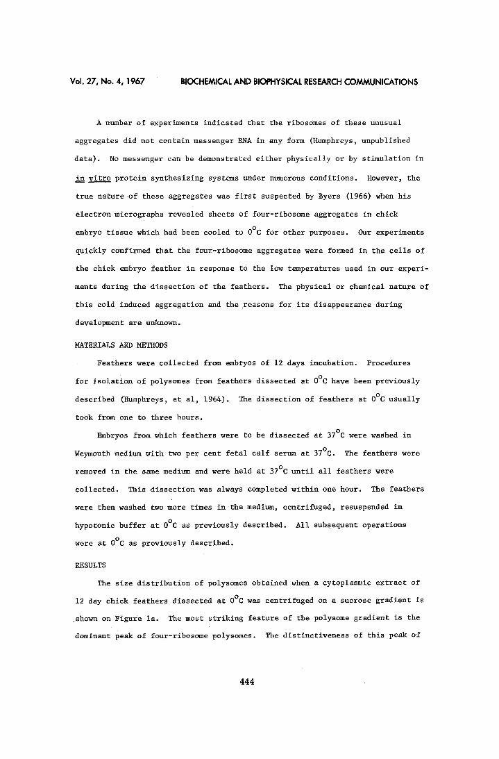
The size distribution of polysomes obtained when a cytoplasmic extract of
12 day chick feathers dissected at 0°C was centrifuged on a sucrose gradient is
shown on Figure la. The most striking feature of the polysome gradient is the
dominant peak of four-ribosome polysomes. The distinctiveness of this peak of
444

V o l . 2 7 , N o . 4 , 1 9 6 7 B I O C H E M I C A L A N D B I O P H Y S I C A L R E S E A R C H C O M M U N I C A T I O N S
7.0
6.0
5.0
4.0
3.0
~ 2.o_ ~ Le
d 1.0
0.5
a
I I I I
? / 4 ' s
~ L _ l [ I 1 [ 1 I I I I 2 4 6 8 I0 12 14 16 18 20 22
Fraction No.
6.0
5.0
4.0
5.0
1.5":-
o 1 . 0 -
0 . 5 -
I I I I I I i I I I I b
i
~ I I i I I I I 2 4 6 8 I0 12. 14 16 18 20 22.
Fraction No.
Figure i. Cytoplasmic extracts prepared from embryo feathers dissected at 0 C and sedimented for 3 hours at 24,000 rpm on 15 to 30% linear sucrose gradients (a) no ribonuclease (b) 0.1ugm ribonuclease per ml.
four-ribosome polysomes was enhanced greatly if the extract was treated with 0.i
~gm ribonuclease. This treatment dissociated all the polysomes except the major-
ity of the four-ribosome polysomes (Figure ib). Previous studies show that
this ribonuclease-resistant peak does not synthesize protein although it is
composed of aggregates Of four-ribosomes (Humphreys, et al, 1964; Bell, etJal,
1965).
If the feathers were dissected from chick embryos in Tyrode's solution
which had been maintained at 37°C during the operations, nearly all ribosomes
extracted from these feathers were monomers; there were virtually no polysomes.
This was taken to indicate that polysomes broke down during incubation of the
feathers in Tyrode's at 37°C.
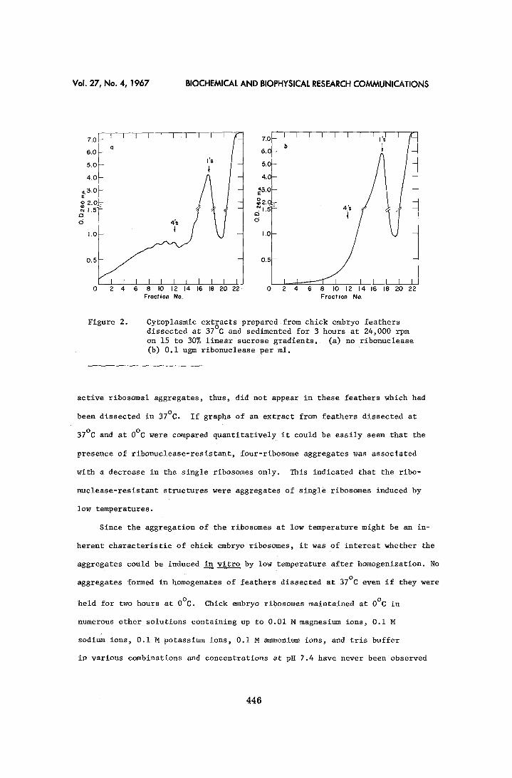
If, instead, the feathers were dissected at 37°C in a complete nutrient
medium with 2% serum, a considerable portion of ribosomes were in aggregates
(Figure 2a). However, in this case the peak of four-ribosome aggregates did
not predominate. Moreover, when the extract was treated with ribonuclease all
ribosomes became monomers (Figure 2b). The ribonuclease-resistant peak of in-
445
Vol . 27 , No . 4 , 1 9 6 7 B IOCHEMICAL A N D BIOPHYSICAL RESEARCH C O M M U N I C A T I O N S
7 . 0 [ - l a I I I l I I I I I l ~ 7.0
5.0 lls 5.0
°I A/t ~:s.o ~.3.o
I. 0 I.O
I I I I I I I I I 0 2 4 6 8 I0 12 14 16 18 20 22 0
Fraction No.
I I I I I I I I ,~I I
2 4 6 8 I0 12 14 16 18 20 22 Fraction No.
Figure 2. Cytoplasmic extracts prepared from chick embryo feathers dissected at 37°C and sedimented for 3 hours at 24,000 rpm on 15 to 30% linear sucrose gradients. (a) no ribonuclease (b) 0.i ugm ribonuelease per mlo
active ribosomal aggregates, thus, did not appear in these feathers which had
been dissected in 37°C. If graphs of an extract from feathers dissected at
37°C and at 0°C were compared quantitatively it could be easily seen that the
presence of ribonuclease-resistant, four-ribosome aggregates was associated
with a decrease in the single ribosomes only. This indicated that the ribo-
nuclease-resistant structures were aggregates of single ribosomes induced by
low temperatures.
Since the aggregation of the ribosomes at low temperature might be an in-
herent characteristic of chick embryo ribosomes, it was of interest whether the
aggregates could be induced in vitro by low temperature after homogenization. No
aggregates formed in homogenates of feathers dissected at 37°C even if they were
held for two hours at O°C. Chick embryo ribosomes maintained at 0°C in
numerous other solutions containing up to 0.01M magnesium ions, 0.i M
sodium ions, 0.i M potassium ions, 0.i M arm~onium ions, and tris buffer
in various combinations and concentrations at pH 7.4 have never been observed
446
Vol. 27, No. 4, 1967 BIOCHEMICAL AND BIOPHYSICAL RESEARCH COMMUNICATIONS
to form aggregates of four ribosomes.
DISCUSSION
These data show that the ribonuclease-resistent aggregates of four
ribosomes appearing in chick feathers (Humphreys; et al, 1964; Bell, et al,
1965), are formed in vivo in response to low temperature. The apparent
tissue specificity of the aggregation noted earlier (Humphreys, et al, 1964)
was probably due to the rapidity with which the tissues other than feather
could be dissected. This conclusion has been affirmed by electron microscopy
which shows that the aggregates are not present in normal chick tissueS, but
appear rapidly in several other tissues when cooled (Byers, 1966). The basic
mechanisms causing this aggregation or its physiological function in the chick
embryo, which normally experiences some cooling in development, are obscure.
REFERENCES
Bell, E., T. Humphreys, H. S. Slater, and C. E. Hall, Science 148,
1739 (1964).
Byers, B., J. Cell Biol. 30, CI (1966).
Humphreys, T., S. Penman, and E. Bell, Biochem. Biophys. Res. Comm.
17, 618 (1964).
Hamilton, M. G., and M. L. Peterman, J. of Biol. Chem. 234, 1441 (1959).
Tissieres, A., J. D. Watson, E. Schlessinger, and E. R. Hollingworth,
J. of Mol. Biol. !, 221 (1959).
447