the in vitro respiratory toxicity of cristobalite-bearing ... · the in vitro respiratory toxicity...
TRANSCRIPT

Environmental Research 145 (2016) 74–84
Contents lists available at ScienceDirect
Environmental Research
http://d0013-93
n Corrfor MinGerman
E-m
journal homepage: www.elsevier.com/locate/envres
The in vitro respiratory toxicity of cristobalite-bearing volcanic ash
David E. Damby a,b,c,n, Fiona A. Murphy d, Claire J. Horwell a, Jennifer Raftis b,Kenneth Donaldson b
a Institute of Hazard, Risk and Resilience, Department of Earth Sciences, Durham University, Durham, United Kingdomb The University of Edinburgh/MRC Centre for Inflammation Research, The Queen’s Medical Research Institute, Edinburgh, United Kingdomc Department of Earth and Environmental Sciences, Ludwig-Maximilians Universität München, Munich, Germanyd MRC Toxicology Unit, University of Leicester, Leicester, United Kingdom
a r t i c l e i n f o
Article history:Received 24 June 2015Received in revised form5 November 2015Accepted 17 November 2015Available online 6 December 2015
Keywords:Volcanic ashCrystalline silicaCristobaliteToxicity
x.doi.org/10.1016/j.envres.2015.11.02051/& 2015 The Authors. Published by Elsevie
espondence to: Department of Earth and Eneralogy, Petrology and Geochemistry, Thery.ail address: [email protected]
a b s t r a c t
Ash from dome-forming volcanoes poses a unique hazard to millions of people worldwide due to anabundance of respirable cristobalite, a crystalline silica polymorph. Crystalline silica is an establishedrespiratory hazard in other mixed dusts, but its toxicity strongly depends on sample provenance. Pre-vious studies suggest that cristobalite-bearing volcanic ash is not as bio-reactive as may be expected for adust containing crystalline silica. We systematically address the hazard posed by volcanic cristobalite byanalysing a range of dome-related ash samples, and interpret the crystalline silica hazard according tothe mineralogical nature of volcanic cristobalite. Samples are sourced from five well-characterized dome-forming volcanoes that span a range of magmatic compositions, specifically selecting samples rich incristobalite (up to 16 wt%). Isolated respirable fractions are used to investigate the in vitro response ofTHP-1 macrophages and A549 type II epithelial cells in cytotoxicity, cellular stress, and pro-inflammatoryassays associated with crystalline silica toxicity. Dome-related ash is minimally reactive in vitro for arange of source compositions and cristobalite contents. Cristobalite-based toxicity is not evident in theassays employed, supporting the notion that crystalline silica provenance influences reactivity. Macro-phages experienced minimal ash-induced cytotoxicity and intracellular reduction of glutathione; how-ever, production of IL-1β, IL-6 and IL-8 were sample-dependent. Lung epithelial cells experiencedmoderate apoptosis, sample-dependent reduction of glutathione, and minimal cytokine production. Wesuggest that protracted interaction between particles and epithelial cells may never arise due to effectiveclearance by macrophages. However, volcanic ash has the propensity to incite a low, but significant, andsample-dependent response; the effect of this response in vivo is unknown and prolonged exposure mayyet pose a hazard.
& 2015 The Authors. Published by Elsevier Inc. This is an open access article under the CC BY license(http://creativecommons.org/licenses/by/4.0/).
1. Introduction
Given the established hazard posed by respirable crystallinesilica (CS) in occupational settings, the presence of cristobalite, aCS polymorph, in ash emerged as a primary health concern duringthe 1980 eruption of Mount St. Helens, USA (Green et al., 1981),and again during the 1995-present eruption of Soufrière Hillsvolcano, Montserrat (Baxter et al., 1999; Wakefield, 2000), gainingthe attention of governments and international public health andtoxicology experts (Baxter et al., 2014). As a result, detection of CSin volcanic ash has been undertaken in response to many
r Inc. This is an open access article
vironmental Sciences Sectionesienstr. 41, 80333 Munich,
.de (D.E. Damby).
eruptions worldwide, and is central to rapid hazard assessmentscarried out by the International Volcanic Health Hazard Network(e.g., Damby et al., 2013; Horwell et al., 2013a). However, theconsequence of regular exposure to volcanic cristobalite is stillunknown, even in areas with prolonged exposure; for example,the densely populated regions surrounding Mt. Sakurajima, wherepersistent small eruptions frequently expose the population to ashcontaining up to 5 wt% cristobalite (reviewed by Hillman et al.(2012)).
Comprehensive risk assessments to determine the long-termrisk of silicosis arising from chronic exposure to cristobalite-bearing ash have been conducted only for the eruptions of MountSt. Helens and Soufrière Hills (Horwell and Baxter, 2006). As anon-occupational risk of silicosis had never been recognized priorto the Mount St. Helens eruption, these assessments relied onoccupational approaches, for which determination of exposure to
under the CC BY license (http://creativecommons.org/licenses/by/4.0/).

D.E. Damby et al. / Environmental Research 145 (2016) 74–84 75
crystalline silica is central; however, the toxicity of volcanic cris-tobalite had not yet been established (Baxter et al., 2014). Despitesubstantial variability in discrete in vitro and in vivo toxicologyresults for volcanic ash to date (see Baxter et al. (2014) and Hor-well and Baxter (2006) for a full review), there is a tacit assump-tion in the literature that observed toxicity may result from thepresence of CS (e.g., Sanders et al., 1983; Wehner et al., 1986).However, no notable intra-study differences in reactivity amongsamples with variable cristobalite contents have been observed(e.g., Housley et al., 2002; Vallyathan et al., 1983; Wilson et al.,2000).
The respiratory hazard posed by CS in other mixed dusts isdependent on the provenance of the sample, leading CS reactivityto be deemed a ‘variable entity’ (Donaldson and Borm, 1998).Volcanic cristobalite is particularly abundant in ash from dome-forming volcanoes (Damby, 2012), where mounds of viscous lavapile up over the vent. The active dome environment facilitates thein situ crystallization of cristobalite in these lavas through vapour-phase deposition in cracks and pore space and through devi-trification of volcanic glass (Baxter et al., 1999; Horwell et al.,2013b). The presence of cristobalite in ash, therefore, requires in-corporation of existing lavas during an eruption, and can compriseupwards of 25 wt% of the ash (Damby, 2012; Horwell et al., 2014).Its abundance depends on dome residence time and lava extrusionrate as well as the eruptive style by which the ash is generated(Horwell et al., 2014). For example, ash from the initial 2008eruption of Chaitén volcano contained �2 wt% cristobalite,whereas subsequent eruptions after dome growth began con-tained 13–19 wt% (Horwell et al., 2010).
The influence of source environment and mineralogy on toxi-city has recently been considered for volcanic cristobalite,whereby natural substitutions of aluminium for silicon are shownto affect the chemistry and structure of volcanic cristobalite re-lative to pure-phase samples (Damby et al., 2014; Horwell et al.,2012). Horwell et al. (2012) suggest that these substitutions couldpotentially reduce the toxicity of volcanic cristobalite, as alumi-nium has been shown experimentally to quell CS toxicity (Duffinet al., 2001). Further, Horwell et al. (2012) show that discrete ashgrains from Soufrière Hills volcano are mineralogically hetero-geneous, effectively reducing the surface area of cristobalite at theparticle surface.
Despite the present geological understanding, interpreting andcontextualizing the hazard posed by different and/or new volcaniceruptions has been difficult. Direct inter-study comparisons areproblematic as variably sourced ash comprises different miner-alogical constituents and eruptive histories, and because previoustoxicological assessments typically focus on single locations. Onlytwo studies have considered multiple source volcanoes: the first,by Vallyathan et al. (1984), considered three volcanoes (Mount St.Helens, El Chichón, and Galunggung); however, their respirablesamples only contained �1.5 to 2.0% CS; the second, by Horwellet al. (2013a), benchmarks cristobalite-free ash from the 2010 and2011 eruptions of Eyjafjallajökull and Grímsvötn, Iceland against a
Table 1Sample information for the respirable ash panel. All ash samples were generated in thmethod of Le Blond et al. (2009) and has a73 wt% error.
Sample Source Ash type Eruption
COL Colima, Mexico Vulcanian explosion unknowMER Merapi, Indonesia Dome collapse 11–19/07MER.PDC Merapi, Indonesia Pyroclastic density current 05/11/20MSH Mount St. Helens, USA Vent explosion 08/03/20SHVa Soufrière Hills, Montserrat Dome collapse 05/06/19UNZ Unzen, Japan Dome collapse 03/04/19
a SHV is the same as sample MRA5/6/99 in Horwell et al. (2003).
cristobalite-rich sample from Soufrière Hills volcano. Inter-studycomparisons are further hindered by differences in experimentaldesign. For example, haemolysis assays on Soufrière Hills ash byCullen et al. (2002) and Wilson et al. (2000) both use sheep ery-throcytes and a top particle dose of 20–30 mg ml�1 whereasHorwell et al. (2013a) use fresh human erythrocytes and a top doseof 1 mg ml�1. Consequently, Soufrière Hills ash has ranged from0 to 45% haemolysis in studies that cannot be directly compared.Within these studies, samples from the same volcano, and evensame eruptive phase, have elicited different responses (e.g., Hor-well et al. (2013a) and Wilson et al. (2000)).
Here, we systematically evaluate the in vitro response elicitedby respirable cristobalite-bearing volcanic ash from five dome-forming volcanoes, specifically selecting samples rich in CS, tounderstand the toxicity of volcanic cristobalite. This is first studyto specifically investigate the reactivity of volcanic cristobalite aswell as the first to consider the toxicity of ash derived from anexceptional range of magmatic compositions. We assess the ash-induced decrease in viability and pro-inflammatory activation ofmacrophages and alveolar type II epithelial cells. Macrophages areclassically considered a crucial cell type in the initiation and pro-gression of pulmonary inflammation due to their wide spectrumof pro-inflammatory cytokine production and direct role in parti-cle clearance, and type II epithelial cells are thought to play a keyrole in pulmonary inflammation and fibrogenesis, including inresponse to CS exposure (Driscoll et al., 1993). Ash samples fromMount St. Helens and Soufrière Hills volcano (discussed above)contextualize the present study due to the wealth of previoushealth-focused studies (see Horwell and Baxter (2006) for a re-view). Further cristobalite-bearing samples are analysed from Mt.Unzen (Japan), Volcán de Colima (Mexico) and Mt. Merapi (In-donesia) due to their location in densely populated areas andhistory of violent eruptions, factors which led to their designationas Decade Volcanoes by the United Nations as part of a disaster-reduction effort; thus, understanding the respiratory hazard posedby these volcanoes is crucial.
2. Methods
2.1. Samples of volcanic ash
Volcanic ash samples were sourced from 5 dome-formingvolcanoes, ranging from basaltic-andesite to dacite in composition(Table 1). Samples were selected from a larger sample suite tomaximize the CS contents of the samples analysed (see Damby(2012)). Mineralogically, these samples primarily comprise volca-nic glass, feldspars, pyroxenes, amphiboles, Fe–Ti oxides, andcrystalline silica polymorphs (see Supplementary material). Allsamples were generated from eruptions involving dome materialbut were derived from a range of eruptive styles due to the dif-ferent eruptive natures of the volcanoes studied. Two sampleswere selected from Merapi to consider the effect of different
e presence of a volcanic dome. Cristobalite abundance was determined by the IAS
date Collection date Distance to source (km) Cristobalite (wt%)
n 21/03/2010 o0.2 5.7/1998 09/08/1998 0.2 2.710 30/11/2010 17.0 6.205 10/03/2005 6.5 15.399 05/06/1999 4.0 15.793 03/04/1993 5.0 7.3

D.E. Damby et al. / Environmental Research 145 (2016) 74–8476
fragmentation mechanisms on the particle hazard and are dis-tinguished by the designation ‘PDC’, which denotes the samplederived from a pyroclastic density current (PDC) deposit ratherthan ashfall. Further information on each volcano can be found inthe Supplementary material.
Respirable fractions were isolated at SAFENANO, Institute ofOccupational Medicine, Edinburgh. Approximately 5 g of bulkvolcanic ash was aerosolised with HEPA-filtered compressed airusing an AERO PA100 particle aerosoliser. The aerosol was passedthrough a gravitational separator where large particles and ag-glomerates sedimented from the aerosol in accordance with Sto-ke's Law. Particles below a maximum theoretical aerodynamicdiameter of 6 mm (i.e., within the respirable range; Brown et al.,2013), calculated for a specific airflow and particle density in thesystem, then entered a sampling chamber for collection.
2.2. Particle size distribution and morphologies
The particle size distribution of the respirable particulateaerosols was analysed concurrent with separation by two real-time instruments: a Fast Mobility Particle Sizer (FMPS; model3091, TSI Inc.) and an Aerodynamic Particle Sizer (APS; model3320, TSI Inc.). The FMPS measures particles from 5.6 to 560 nmbased on electrical mobility and the APS measures particles from500 to 20,000 nm by light scattering. By overlapping the two in-strument readings at their detection boundaries, a total range of5.6 nm to 20 mm was analysed. Data were recorded over a 15–30 min period.
Size-classified respirable ash was imaged by scanning electronmicroscopy (SEM) to confirm the upper particle size cut-off and toconsider particle morphology and cristobalite habit. Cristobalitewas confirmed by energy dispersive X-ray spectroscopy (EDS).Imaging and chemical analysis was carried out on a Hitachi SU-70FEG SEM equipped with an Oxford Instruments EDS system (IN-CAx-act LN2-free analytical Silicon Drift Detector) in the GJ RussellMicroscopy Facility, Department of Physics, Durham University.
2.3. Quantification of crystalline silica phases
The abundance of CS as cristobalite, quartz and tridymite wasquantified by X-ray diffraction (XRD) using the IAS method of LeBlond et al. (2009). This method allows for quantification of singlephases without prior determination of a sample's bulk mineralogy.Experiments were performed on an Enraf-Nonius X-ray dif-fractometer with an INEL curved position sensitive detector at theNatural History Museum, London, UK. Single phases were quan-tified as outlined in Le Blond et al. (2009) using Nonius GUFIsoftware. Phase quantifications are an average of 2 experimentsand have73 wt% error.
2.4. Treatment of cell cultures with respirable ash
The human leukaemic monocytic THP-1 cell line was main-tained in continuous suspension using RPMI 1640 medium con-taining 10% heat-inactivated FBS (PAA Laboratories, Ltd., UK)supplemented with 2 mM L-Glutamine, penicillin (100 units ml�1)and streptomycin (100 mg ml�1). Cells were seeded at a density of5�105 cells ml�1 in 12-well plates and differentiated to express amacrophage phenotype with 10 ng ml�1 PMA (Sigma-Aldrich, UK)for 48 h. Prior to particle treatment, the media was replaced withserum-free RPMI media (supplemented as above) for 24 h.
A549, a human adenocarcinoma-derived type II alveolar epi-thelial cell line, was maintained in continuous culture in DMEMgrowth media with 10% FBS and supplemented with L-Glutamine,penicillin and streptomycin as per the THP-1 cell media. To pre-pare cells for particle treatment, cells were trypsinized using a
0.25% solution and allowed to re-adhere overnight. Treatment wasperformed on 1 ml of cells at a density of 5�105 cells ml�1 in eachwell of a 12-well plate. Both cell lines were maintained at 37 °C ina 5% CO2 environment.
Cells were treated for 24 h with four concentrations of volcanicash and control particles: 5, 10, 25 and 50 mg cm�2. DQ12 quartzwas used as a positive control, as CS is of primary concern andDQ12 toxicity is well established in the assays employed and isutilised extensively in previous ash toxicity studies (see Horwelland Baxter (2006)). TiO2 (rutile) was used as a negative control.Volcanic ash was suspended in the appropriate cell-line mediawithout FBS and sonicated for 10 min to disaggregate the particles.Mass was used as the metric since mass is the current metric forCS and nuisance dusts in the workplace globally.
2.5. Assessment of cytotoxicity
The extent of cytolysis was determined by lactate dehy-drogenase (LDH) release. Culture medium was transferred to1.5 ml microcentrifuge tubes and spun at 12,000g for 10 min toremove any cell debris and remaining particulate. 35 ml of cell-freeculture supernatant was transferred in triplicate to a 96 well plateand each well diluted with 70 ml of phosphate buffered solution(PBS). 100 ml of LDH test reagent (Roche Diagnostics, Mannheim,Germany) was added to each well. The absorbance was de-termined at 490 nm after 30 min using a Synergy HT microplatereader (BioTek Instruments, Inc., VT, USA). The level of LDH releaseis expressed as a percentage of total LDH release from completecell lysis by Triton X-100. Data are expressed as the average of3 experiments.
Cell viability was determined using the alamarBlues cell pro-liferation assay. Following particle treatment, the supernatant wasremoved and the cells washed with PBS. 300 ml of PBS and 30 ml ofalamarBlues (Invitrogen, Life Technologies, CA, USA) was added toeach well. THP-1 cultures were incubated for 1 h and A549 cul-tures for 30 min at 37 °C in 5% CO2. The solution was spun at12,000g for 5 min, as the particles interfered with the readings,and transferred to a 96 well plate to be read spectro-photometrically at 570 nm. Cell viability is expressed as a per-centage of base line viability (no particle treatment) using TritonX-100 as complete cell death. Data are expressed as the average of3 experiments.
2.6. Determination of apoptosis, necrosis, and cellular stress
The extent of apoptosis, necrosis and endogenous antioxidantproduction was determined by fluorescence-activated cell sorting(FACS). FITC-conjugated Annexin V staining was used to determinethe percentage of cells actively undergoing apoptosis. Propidiumiodide (PI) was used to distinguish viable from non-viable (ne-crotic) cells. Monochlorobimane (MCB) was used to measure in-tracellular concentrations of glutathione (GSH), the principal in-tracellular low-molecular-weight thiol.
Supernatants from post-exposure THP-1 and A549 cell cultureswere collected and spun at 1500 RPM for 3 min to recover non-adherent cells. Adhered cell monolayers were washed with PBS(without Ca and Mg) �2 and trypsinized (5 min, 37 °C, 5% CO2),the trypsin neutralised with culture media, and recovered cellswere added to previously collected cells. Collected cells werewashed �3 at 1500 RPM for 3 min and re-suspended in Annexinbuffer (Hank’s Balanced Salt Solution with 2.5 mM CaCl2, filtersterilised). Cells were stained with 5 ml FITC-Annexin V and 1 mlMCB per 195 ml cell suspension and incubated for 10 min. Cellswere spun at 1500 RPM for 3 min and re-suspended in 200 mlAnnexin buffer. PI (1 ml) was added immediately prior to flow cy-tometry. Unstained cells and unstained particle-treated cells were

0
1
2
3
4
5
6
0 1 2 3 4 5 6 7 8 9 10
Rel
ativ
e P
artic
le C
once
ntra
tion
(num
ber
%)
Particle Diameter (µm)
COLMER
MER.PDC
MSHUNZ
FMP
SA
PS
SHV
Par
ticle
Con
cent
ratio
n(v
olum
e %
)
0
10
20
30
40
50
Fig. 1. Particle size distributions for isolated respirable fractions. Size data forparticles smaller than 0.5 mm are collected by FMPS and data above by APS. SampleSHV was not isolated specifically for this study and therefore does not haveequivalent data; however, PSD data from Horwell et al. (2003) are included forcomparison.
D.E. Damby et al. / Environmental Research 145 (2016) 74–84 77
analysed to control for differences in size and granularity resultingfrom phagocytosis of ash. Untreated stained cells (negative vehiclecontrols) were analysed, as were a necrotic control (10 mM H2O2),an apoptotic control (amine beads at 50 mg ml�1), and a GSHcontrol (N-ethylmaleimide; NEM).
2.7. Assessment of the pro-inflammatory response
Enzyme-linked immunosorbent assays (ELISA) were used forthe quantification of acute-phase cytokines IL-1β, IL-6 and che-mokine IL-8 produced following particle treatment using cyto-kine-specific ELISA DuoSet kits (R&D Systems, Abingdon, UK) ac-cording to the manufacturer's protocol. Cytokine concentrationswere determined spectrophotometrically at 450 nm via extra-polation of a recombinant cytokine protein standard curve.
2.8. Imaging of cell-particle interaction
Differentiated THP-1 cells were imaged by light microscopyfollowing ash treatment to determine whether particles werebeing internalised. Only THP-1 cells were imaged as A549 ag-gregated during preparation. Cytospin preparations were made bycentrifuging �40 to 60,000 cells in 300 ml of 0.1% BSA/sterile sal-ine solution at 300 RPM for 3 min at room temperature using aCytospin 4 centrifuge (Thermo Scientific, UK). Air-dried slideswere fixed in 100% methanol and stained with eosin (Quick-Diff,Dade Behring, Inc.) and haematoxylin (REASTAIN Quick-Diff Blue),and then submerged in xylene for 30 s. DPX mounting media(Sigma-Aldrich, Poole, UK) was added directly to the cell popula-tion and slides were covered with a glass cover slip. Images werecaptured at 100� magnification using QCapture Pro software(Media Cybernetics, Inc., Bethesda, MD).
2.9. Statistical analysis
Statistical analysis was performed using GraphPad PRISMsoftware (San Diego, CA). Multiple comparisons were conductedusing two-way analysis of variance (ANOVA) with Tukey's post-test where appropriate. In all cases, tests were considered sig-nificant when pr0.05.
3. Results
3.1. Particle characterisation
Sizing of the size-separated samples yielded distributions ran-ging from �0.08 mm to 7.0 mm (Fig. 1); the distribution was similarfor all samples. A bi-modal distribution arises from combiningresults from FMPS and APS (Fig. 1); however, this is likely due toinstrumentation and is not considered further. The gradual slopetowards the upper size cut-off (�7.0 mm) is likely a physical ar-tefact as the probability of re-suspension decreases towards thedefined particle cut-off (6 mm). The lower cut-off (�0.08 mm) isnot likely representative of a true minimum particle size as par-ticles are subject to aggregation.
SEM imaging of the isolated ash substantiates the particle sizedata, and reveals little difference in general morphology amongsamples (Fig. 2a and b). Particles are poorly vesiculated, sub-an-gular and blocky with varying amounts of nano-scale particlesadhering to the surfaces of larger particles. Only scarce mineralfibres were observed in any of the samples. Cristobalite was pre-sent in all respirable samples, ranging from �3 to 16 wt% (Table 1).Minor quartz was detected in samples SHV, MSH and UNZ. Notridymite was observed in any sample. SEM backscattered imagingof ash in cross section confirms that discrete ash grains can
contain cristobalite intimately associated with other mineralphases and glass (Fig. 2c) whereas others solely comprise cristo-balite (Fig. 2d). Further, all cristobalite contains aluminium(Fig. 2d), extending the observations of Horwell et al. (2012) to therespirable fraction.
3.2. Cytotoxicity of differentiated THP-1 and A549 cells
All samples induced some degree of decreased viability in THP-1 macrophages at 24 hours as measured by metabolism of thealamarBlues reagent (Fig. 3a). However, the response to ash didnot follow a typical dose-dependent decrease as exemplified byDQ12 quartz. The decrease in viability is similar to that elicited byTiO2 (consistent 85% viability over the same dose range). A similarresponse was seen for LDH release (Fig. 3b), where treatment re-sulted in substantially lower release than DQ12 and marginallylower release than TiO2.
A549 cell viability determined by the alamarBlues assayshowed little effect of particle treatment compared to DQ12 andTiO2 (Fig. 3c). Ash samples MER, COL, UNZ elicited more LDH re-lease than TiO2 but less than DQ12 (Fig. 3d).
3.3. Assessment of apoptosis and necrosis
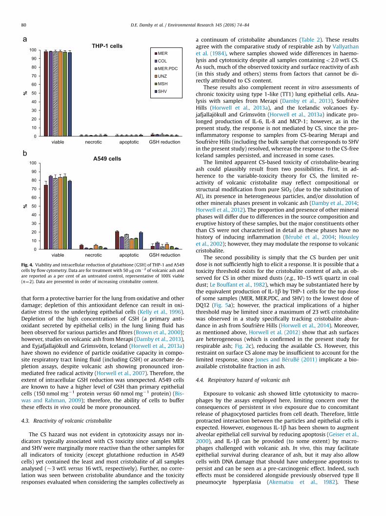
Differentiated THP-1 cells experienced limited reduction inviability following particle treatment as determined by FACSanalysis (Fig. 4); THP-1 cell necrosis and apoptosis were roughlyequivalent and negligible. A549 cells experienced a �20% reduc-tion in viability, which was largely attributed to apoptosis (Fig. 4);all samples resulted in a significant level of apoptosis over un-treated control.
3.4. Intracellular oxidative stress
No reduction in GSH abundance was observed with ash treat-ment in THP-1 cells compared to untreated cells (Fig. 4). However,an ash-induced reduction was observed for A549 cells (Fig. 4),with UNZ and COL causing the most pronounced depletion relativeto controls. The extent of the decrease was more pronounced forhigher doses; in general, the effect was more prominent that thatcaused by DQ12 quartz but substantially less than that produced

Fig. 2. Scanning electron micrographs of respirable volcanic ash. Selected SEM micrographs of respirable volcanic ash samples (a) UNZ and (b) SHV, which are representativeof all samples investigated. Backscattered SEM images of ash sample SHV in cross section showing (c) heterogeneous patches of cristobalite labelled ‘c’ (cristobalite crystalboundaries are demarcated), and (d) a particle comprised entirely of cristobalite. Inset: EDS spot analysis of cristobalite showing the presence of aluminium and sodium. Allimages were collected at 6.0 kV and 14 mm working distance.
D.E. Damby et al. / Environmental Research 145 (2016) 74–8478
by NEM, an effective oxidiser of GSH which rapidly depletes itfrom the cell cytosol.
3.5. Pro-inflammatory response
The pro-inflammatory response of differentiated THP-1 cellswas sample and cytokine dependent. The top two doses (25 and50 mg cm�2) of SHV, MER and MER.PDC resulted in the productionof IL-1β above the untreated control and were equivalent to thelowest dose (5 mg cm�2) of DQ12 (Fig. 5a). Treatment by all sam-ples except for MER resulted in IL-6 production comparable to theuntreated control (Fig. 5b). One repeat experiment (of n¼3) forMER resulted in similar levels to all other samples, resulting inlarge error bars and perhaps emphasizing the heterogeneous mi-neralogy of the ash samples. Cells treated with both UNZ and MERproduced significantly more IL-8 than untreated cells in a dose-dependent response (Fig. 5c). Levels for the top dose (50 mg cm�2)of these samples were also greater than that induced by equivalentconcentrations of DQ12. Although MER induced significant IL-8production, results for MER.PDC were largely negative.
The response of A549 epithelial cells to insult was largely ne-gative (Fig. 5d–f). Little to no release of IL-1β or IL-6 occurred,especially compared to that induced by DQ12. Production of IL-8for all particle treatments (ash and DQ12) did not differ sig-nificantly from an untreated control (p40.05), suggesting mini-mal treatment-dependent production of IL-8 over a basal level.
3.6. Particle-Cell imaging
Light microscope imaging of untreated and ash-treated differ-entiated THP-1 cells in cytospin preparations shows that particles
were internalised by cells (Fig. 6). Using the techniques here, it wasnot possible to determine if there was a preferential uptake orretention of certain mineral phases relative to others.
4. Discussion
4.1. Differentiated THP-1 cell response
Phagocytosis of particles by THP-1 macrophages resulted inminimal cytotoxicity, as evidenced by low levels of LDH release,minimal suppression of cellular metabolism, and negligibleapoptosis and necrosis. These results agree with previous studieson alveolar macrophages, which generally show no ash-dependentchanges in viability (e.g., Fruchter et al., 1980; Green et al., 1982;Huang et al., 2004; Martin et al., 1984); however, one study hasshown moderate cytotoxicity to macrophages exposed to MountSt. Helens ash (McLemore et al., 1984). THP-1 cells also appear tohave increased intracellular GSH levels following ash exposure.Macrophages are known to have lower levels of intracellular GSHthan other cell types (e.g., 10 nmol mg�1 protein compared to150 nmol mg�1 protein in A549 cells) (Biswas and Rahman, 2009),and perhaps increase production to cope with insult. This wouldhave the effect of increasing antioxidant defence in response toexposure. Collectively, these cytotoxicity and cellular stress datasuggest that clearance by macrophages should be effective withlittle impairment to the cell at plausible exposures.
It is not possible to assign the pro-inflammatory response ofTHP-1 macrophages to CS abundance alone. Production of IL-1β,which is important in the development of silica-related disease(Mossman and Glenn, 2013), was negligible for MSH (as also

0 10 20 30 40 50
80
70
60
50
40
30
20
10
0
particle concentration (µg cm )-2
% L
DH
rele
ase
0 10 20 30 40 50
201816141210
86
0
42
% L
DH
rele
ase
particle concentration (µg cm )-2
SHVCOL MSH UNZMER MER.PDC TiO2 DQ12
0 10 20 30 40 50
8070605040302010
0
90100110
% c
ellu
lar v
iabi
lity
particle concentration (µg cm )-2
THP-1 cells
0 10 20 30 40 50
8070605040302010
0
90100
% c
ellu
lar v
iabi
lity
particle concentration (µg cm )-2
A549 cells
Fig. 3. Cytotoxicity of ash samples towards differentiated THP-1 and A549 cells. Cytotoxicity of THP-1 cells following exposure to respirable ash and controls measured by(a) metabolism of the alamarBlue reagent and (c) LDH release. Cytotoxicity of A549 cells measured by (b) metabolism of the alamarBlue reagent and (d) LDH release. Errorbars are the standard error of the mean. *pr0.05, **pr0.01.
D.E. Damby et al. / Environmental Research 145 (2016) 74–84 79
reported for Mount St. Helens ash by Bonner et al. (1998) andHuang et al. (2004)), which contains the most CS (together withSHV, within error), yet pronounced for MER, which contains theleast CS. These data support the adherence of volcanic cristobaliteto the ‘variable entity’ description of CS (Donaldson and Borm,1998), as proposed by Horwell et al. (2012), whereby ‘inherentcharacteristics and external factors' may affect the propensity of CSto incite IL-1β production.
An increase in the production of IL-8, a potent chemoattractantand activator of neutrophils, was only observed for low CS-contentsamples UNZ and MER; a similar inability to stimulate neutrophilchemotactic activity was previously observed for Mount St. Helensash (Martin et al., 1984). IL-8 transcription is under the control ofNuclear Factor-κB (NF-κB), a transcriptional regulator proteincomplex that becomes activated following CS exposure (Schinsand Donaldson, 2000); NF-κB has been shown to play a role indiscriminating between inflammogenic and non-inflammogenicquartz varieties (Duffin et al., 2001). However, like IL-1β produc-tion, IL-8 production does not correlate with CS content. Since NF-κB is an oxidative stress-responsive transcription factor involvedin the pro-inflammatory effects of many pathogenic particles(Schins and Donaldson, 2000), again, CS cannot be independentlyimplicated.
4.2. A549 cell response
In general, A549 cells experienced a moderate reduction in cellviability in response to ash treatment. These data agree with the
results of Wilson et al. (2000), where exposure of A549 cells tothree samples of respirable ash from Soufrière Hills resulted in asimilar decrease in viability to both DQ12 and TiO2, which werenot statistically different in their study. Results for individual ashsamples, however, did not always correspond among assays. Forexample, COL was the most cytotoxic by LDH and alamarBlues
assays but induced the lowest amount of apoptosis and necrosis.The propensity of all samples towards apoptosis rather than ne-crosis aligns with previous work using Mount St. Helens ash onlung epithelial cells (Agopyan et al., 2004), and may help explainlow haemolysis results for ash in previous studies (e.g., Dambyet al., 2013; Horwell et al., 2013a) as membranolysis would con-stitute necrosis.
Despite the decrease in viability, no pro-inflammatory responseby A549 cells was observed above untreated control. Exposure ofA549 cells to pure-phase crystalline and amorphous silica is ex-pected to result in the release of IL-8 and IL-6, with CS being themost potent inducer of IL-8 and amorphous silica of IL-6 (Hetlandet al., 2001; Monteiller et al., 2007). Contrary to the above studies,the amorphous component of volcanic ash failed to incite pro-duction of IL-6, and the CS component of volcanic ash did notresult in the production of IL-8 above untreated control, especiallyconsidering that both MSH and SHV comprised �15 wt% cristo-balite. However, variability in the response of cultured cells toexposure is well known, and the response can vary over time(Poland et al., 2014).
Lung lining fluid contains high concentrations of antioxidants
A549 cells
0
10
20
30
40
50
60
70
80
90
100
viable necrotic apoptotic GSH reduction
%
THP-1 cellsMER
COL
MER.PDC
UNZ
MSH
SHV
0
10
20
30
40
50
60
70
80
90
100
viable necrotic apoptotic GSH reduction
%
Fig. 4. Viability and intracellular reduction of glutathione (GSH) of THP-1 and A549cells by flow cytometry. Data are for treatment with 50 mg cm�2 of volcanic ash andare reported as a per cent of an untreated control, representative of 100% viable(n¼2). Data are presented in order of increasing cristobalite content.
D.E. Damby et al. / Environmental Research 145 (2016) 74–8480
that form a protective barrier for the lung from oxidative and otherdamage; depletion of this antioxidant defence can result in oxi-dative stress to the underlying epithelial cells (Kelly et al., 1996).Depletion of the high concentrations of GSH (a primary anti-oxidant secreted by epithelial cells) in the lung lining fluid hasbeen observed for various particles and fibres (Brown et al., 2000);however, studies on volcanic ash fromMerapi (Damby et al., 2013),and Eyjafjallajökull and Grímsvötn, Iceland (Horwell et al., 2013a)have shown no evidence of particle oxidative capacity in compo-site respiratory tract lining fluid (including GSH) or ascorbate de-pletion assays, despite volcanic ash showing pronounced iron-mediated free radical activity (Horwell et al., 2007). Therefore, theextent of intracellular GSH reduction was unexpected. A549 cellsare known to have a higher level of GSH than primary epithelialcells (150 nmol mg�1 protein versus 60 nmol mg�1 protein) (Bis-was and Rahman, 2009); therefore, the ability of cells to bufferthese effects in vivo could be more pronounced.
4.3. Reactivity of volcanic cristobalite
The CS hazard was not evident in cytotoxicity assays nor in-dicators typically associated with CS toxicity since samples MERand SHV were marginally more reactive than the other samples forall indicators of toxicity (except glutathione reduction in A549cells) yet contained the least and most cristobalite of all samplesanalysed (�3 wt% versus 16 wt%, respectively). Further, no corre-lation was seen between cristobalite abundance and the toxicityresponses evaluated when considering the samples collectively as
a continuum of cristobalite abundances (Table 2). These resultsagree with the comparative study of respirable ash by Vallyathanet al. (1984), where samples showed wide differences in haemo-lysis and cytotoxicity despite all samples containingo2.0 wt% CS.As such, much of the observed toxicity and surface reactivity of ash(in this study and others) stems from factors that cannot be di-rectly attributed to CS content.
These results also complement recent in vitro assessments ofchronic toxicity using type 1-like (TT1) lung epithelial cells. Ana-lysis with samples from Merapi (Damby et al., 2013), SoufrièreHills (Horwell et al., 2013a), and the Icelandic volcanoes Ey-jafjallajökull and Grímsvötn (Horwell et al., 2013a) indicate pro-longed production of IL-6, IL-8 and MCP-1; however, as in thepresent study, the response is not mediated by CS, since the pro-inflammatory response to samples from CS-bearing Merapi andSoufrière Hills (including the bulk sample that corresponds to SHVin the present study) resolved, whereas the response to the CS-freeIceland samples persisted, and increased in some cases.
The limited apparent CS-based toxicity of cristobalite-bearingash could plausibly result from two possibilities. First, in ad-herence to the variable-toxicity theory for CS, the limited re-activity of volcanic cristobalite may reflect compositional orstructural modification from pure SiO2 (due to the substitution ofAl), its presence in heterogeneous particles, and/or dissolution ofother minerals phases present in volcanic ash (Damby et al., 2014;Horwell et al., 2012). The proportion and presence of other mineralphases will differ due to differences in the source composition anderuptive history of these samples, but the major constituents otherthan CS were not characterised in detail as these phases have nohistory of inducing inflammation (Bérubé et al., 2004; Housleyet al., 2002); however, they may modulate the response to volcaniccristobalite.
The second possibility is simply that the CS burden per unitdose is not sufficiently high to elicit a response. It is possible that atoxicity threshold exists for the cristobalite content of ash, as ob-served for CS in other mixed dusts (e.g., 10–15 wt% quartz in coaldust; Le Bouffant et al., 1982), which may be substantiated here bythe equivalent production of IL-1β by THP-1 cells for the top doseof some samples (MER, MER.PDC, and SHV) to the lowest dose ofDQ12 (Fig. 5a); however, the practical implications of a higherthreshold may be limited since a maximum of 23 wt% cristobalitewas observed in a study specifically tracking cristobalite abun-dance in ash from Soufrière Hills (Horwell et al., 2014). Moreover,as mentioned above, Horwell et al. (2012) show that ash surfacesare heterogeneous (which is confirmed in the present study forrespirable ash; Fig. 2c), reducing the available CS. However, thisrestraint on surface CS alone may be insufficient to account for thelimited response, since Jones and BéruBé (2011) implicate a bio-available cristobalite fraction in ash.
4.4. Respiratory hazard of volcanic ash
Exposure to volcanic ash showed little cytotoxicity to macro-phages by the assays employed here, limiting concern over theconsequences of persistent in vivo exposure due to concomitantrelease of phagocytosed particles from cell death. Therefore, littleprotracted interaction between the particles and epithelial cells isexpected. However, exogenous IL-1β has been shown to augmentalveolar epithelial cell survival by reducing apoptosis (Geiser et al.,2000), and IL-1β can be provided (to some extent) by macro-phages challenged with volcanic ash. In vivo, this may facilitateepithelial survival during clearance of ash, but it may also allowcells with DNA damage that should have undergone apoptosis topersist and can be seen as a pre-carcinogenic effect. Indeed, sucheffects must be considered alongside previously observed type IIpneumocyte hyperplasia (Akematsu et al., 1982). These

COLMER
MER.PDC
MSHSHV
UNZDQ12
Untrea
ted0
500
1000
1500
2000
2500
3000IL
-1β
(pg
ml-1
)
COLMER
MER.PDC
MSHSHV
UNZDQ12
Untrea
ted0
1
2
3
4
5
IL-1β
(pg
ml-1
)
COLMER
MER.PDC
MSHSHV
UNZDQ12
Untrea
ted0
1020304050607080
IL-6
(pg
ml-1
)
COLMER
MER.PDC
MSHSHV
UNZDQ12
Untrea
ted0
1
2
3
4
IL-6
(pg
ml-1
)
COLMER
MER.PDC
MSHSHV
UNZDQ12
Untrea
ted0
200
400
600
800
1000
IL-8
(pg
ml-1
)
COLMER
MER.PDC
MSHSHV
UNZDQ12
Untrea
ted0
500
1000
1500
2000
2500
IL-8
(pg
ml-1
)
5102550
x104
x104
x104
x104
THP-1 cells A549 cells
Fig. 5. Cytokine production by THP-1 and A549 cells. Production of pro-inflammatory markers IL-1β, IL-6 and IL-8 by differentiated THP-1 (a–c) and A549 (d–f) cells at 24 hafter treatment with 5, 10, 25 and 50 mg cm�2 volcanic ash. Error bars are the standard error of the mean. *pr0.05, **pr0.01.
D.E. Damby et al. / Environmental Research 145 (2016) 74–84 81
expectations may extend to volcanic ash in general, as in vitroexposure to cristobalite-free Eyjafjallajökull ash had minimal ef-fect on innate immunity (Monick et al., 2013), comprising non-statistical impairment of alveolar macrophage function and nodecrease in airway epithelial cell integrity.
Previous studies have shown that exposure to both CS and non-CS bearing ash correlates with an increased risk of bacterial in-fection (e.g., Castranova et al., 1982; Monick et al., 2013), whichmay be augmented by acute inflammatory effects induced byparticle exposure (Sandberg et al., 2012). Opportunistic bacterialinfections, like tuberculosis, are widespread in many countrieswith active volcanism, especially Indonesia, which ranks fifth in
the world for pulmonary tuberculosis (World Health Organisation,2011), and may be exacerbated by crystalline silica exposure(Balmes, 1990). Evidence from the present study, and those dis-cussed above, indicates a general potential for ash to induce (orexacerbate) inflammatory disorders, irrespective of CS content; assuch, we suggest further consideration of complications resultingfrom ash exposure.
5. Conclusion
The overall effects of a range of cristobalite-bearing ash werelow and did not correlate to cristobalite abundance. It is likely a

Volcanicash
10 µm 10 µm
50 µm
Fig. 6. Uptake of ash by THP-1 cells. Light micrographs of the uptake of volcanic ash by THP-1 cells: (a) untreated cells, (b) cells treated with COL, and (c) low magnificationview of cells with internalized ash particles (arrows). Images are representative of all samples.
Table 2Summary of the correlation (slope and coefficient of determination) betweentoxicity endpoints and cristobalite content for all samples used in the presentstudy. Individual plots comparing CS to discrete endpoints available in the Sup-plementary material.
Indicator of toxicity THP-1 cells A549 cells
Slope R2 value Slope R2 value
Cytokine/chemokine productionIL-1β � 0.0194 n/a n/aIL-6 � 0.3387 � 0.0059IL-8 � 0.3206 � 0.0301
Cellular viabilityFACS � 0.5347 þ 0.0854LDH release � 0.0058 � 0.4871AlamarBlue þ 0.5698 þ 0.2900
Oxidative capacity n/a n/a � 0.3373
D.E. Damby et al. / Environmental Research 145 (2016) 74–8482
combination of both ‘intrinsic and external factors' and dosimetrythat accounts for the suppressed reactivity of volcanic cristobalite.However, these data, in conjunction with other studies, indicatethe potential for volcanic ash to initiate an inflammatory responsewithout seriously compromising lung cell function or integrity.Consequently, prolonged exposure may yet pose a hazard, and thegeneral potential of ash to incite a response should be consideredin risk assessments of long-term complications arising fromchronic exposure.
Critically, the results of the present study may not be fully re-presentative of the overall respiratory hazard at the locationsstudied; as previous reports have emphasized, volcanic ash is aheterogeneous dust, the physicochemical components of whichcan vary considerably, even during a discrete eruption (e.g., Hor-well et al., 2013a). The influence of this variability is further evi-denced here by the disparity in response between explosive andPDC samples. Therefore, volcanic ash should continue to be con-sidered on a case-by-case basis to best inform the hazard posedand most effectively aid mitigation work of disaster managersglobally.

D.E. Damby et al. / Environmental Research 145 (2016) 74–84 83
Acknowledgements
DED thanks the Christopher Moyes Memorial Foundation forresearch support and acknowledges the AXA Research Fund Grant“Risk from volcanic ash in the Earth system” and ERC AdvancedInvestigator Grant no. 247076 (EVOKES) for support duringmanuscript preparation. CJH acknowledges a Natural EnvironmentResearch Council (NERC) Postdoctoral Research Fellowship (Grantno. NE/C518081/2). Thanks to Peter Baxter for providing advice onhealth impacts, Ed Llewellin for constructive comments on anearly version of this manuscript, Anja Schinwald for laboratoryassistance, Jens Najorka for overseeing XRD analyses, and to Gor-don Fern and Steve Hankin for separation of the fine fraction.Thanks to Nick Varley, Setsuya Nakada and the Cascades VolcanoObservatory for providing samples from Colima, Unzen and MountSt. Helens, respectively. We thank the GJ Russell Microscopy Fa-cility at Durham University for SEM assistance. KD has carried outpaid consultancy regarding the toxicology of the products of in-dustries producing particles and fibres, but none of this has con-cerned volcanic ash particles. The other authors declare no po-tential competing financial interests.
Appendix A. Supplementary material
Supplementary data associated with this article can be found inthe online version at http://dx.doi.org/10.1016/j.envres.2015.11.020.
References
Agopyan, N., Head, J., Yu, S., Simon, S.A., 2004. Trpv1 receptors mediate particulatematter-induced apoptosis. Am. J. Physiol.: Lung Cell. Mol. Physiol. 286, 563–572.
Akematsu, T., Dodson, R.F., Williams Jr, M.G., Hurst, G.A., 1982. The short-term ef-fects of volcanic ash on the small airways of the respiratory system. Environ.Res. 29, 358–370.
Balmes, J., 1990. Silica exposure and tuberculosis: an old problem with some newtwists. J. Occup. Med. 32, 114–115.
Baxter, P.J., Bonadonna, C., Dupree, R., Hards, V.L., Kohn, S.C., Murphy, M.D., et al.,1999. Cristobalite in volcanic ash of the soufriere hills volcano, montserrat,british west indies. Science 283, 1142–1145.
Baxter, P.J., Searl, A., Cowie, H.A., Jarvis, D., Horwell, C.J., 2014. Evaluating the Re-spiratory Health Risks of Volcanic Ash at the Eruption of the Soufrière HillsVolcano, Montserrat, 1995–2010. Geological Society of London, United King-dom, Memoir.
Bérubé, K.A., Jones, T.P., Housley, D.G., Richards, R.J., 2004. The respiratory toxicityof airborne volcanic ash from the soufriere hills volcano, montserrat. Miner.Mag. 68, 47–60.
Biswas, S., Rahman, I., 2009. Environmental toxicity, redox signaling and lung in-flammation: the role of glutathione. Mol. Asp. Med. 30, 60–76.
Bonner, J.C., Rice, A.B., Lindroos, P.M., O’Brien, P.O., Dreher, K.L., Rosas, I., et al., 1998.Induction of the lung myofibroblast pdgf receptor system by urban ambientparticles from mexico city. Am. J. Respir. Cell Mol. Biol. 19, 672–680.
Brown, D.M., Beswick, P.H., Bell, K.S., Donaldson, K., 2000. Depletion of gluathioneand ascorbate in lung lining fluid by respirable fibres. Ann. Occup. Hyg. 44,101–108.
Brown, J.S., Gordon, T., Price, O., Asgharian, B., 2013. Thoracic and respirable particledefinitions for human health risk assessment. Part. Fibre Toxicol. 10, 12.
Castranova, V., Bowman, L., Shreve, J.M., Jones, G.S., Miles, P.R., 1982. Volcanic ash:toxicity to isolated lung cells. J. Toxicol. Environ. Health 9, 317–325.
Cullen, R.T., Jones, A.D., Miller, B.G., Donaldson, K., Davis, J.M.G., Wilson, M., et al.,2002. Toxicity of Volcanic Ash From Montserrat. Institute of OccupationalMedicine, Edinburgh, TM/02/01.
Damby, D.E., 2012. From Dome to Disease: The Respiratory Toxicity of VolcanicCristobalite (e-theses). Durham University, United Kingdom, pp. 1–258.
Damby, D.E., Horwell, C.J., Baxter, P.J., Delmelle, P., Donaldson, K., Dunster, C., et al.,2013a. The respiratory health hazard of tephra from the 2010 centennialeruption of merapi with implications for occupational mining of deposits. J.Volcanol. Geotherm. Res. 261, 376–387.
Damby, D.E., Llewellin, E.W., Horwell, C.J., Williamson, B.J., Najorka, J., Cressey, G.,et al., 2014. The alpha-beta transition in volcanic cristobalite. J. Appl. Crystal-logr. 47, 1205–1215.
Donaldson, K., Borm, P.J.A., 1998. The quartz hazard: a variable entity. Ann. Occup.Hyg. 42, 287–294.
Driscoll, K.E., Hassenbein, D.G., Carter, J., Poynter, J., Asquith, T.N., Grant, R.A., et al.,
1993. Macrophage inflammatory proteins 1 and 2: expression by rat alveolarmacrophages, fibroblasts, and epithelial cells and in rat lung after mineral dustexposure. Am. J. Respir. Cell Mol. Biol. 8, 311–318.
Duffin, R., Gilmour, P.S., Schins, R.P.F., Clouter, A., Guy, K., Brown, D.M., et al., 2001.Aluminium lactate treatment of dq12 quartz inhibits its ability to cause in-flammation, chemokine expression, and nulcear factor-κb activation. Toxicol.Appl. Pharmacol. 176, 10–17.
Fruchter, J.S., Robertson, D.E., Evans, J.C., Olsen, K.B., Lepel, E.A., Laul, J.C., et al., 1980.Mount st. Helens ash from the 18 may 1980 eruption: chemical, physical, mi-neralogical, and biological properties. Science 209, 1116–1125.
Geiser, T., Jarreau, P.H., Atabai, K., Matthay, M.A., 2000. Interleukin-1 beta augmentsin vitro alveolar epithelial repair. Am. J. Physiol.: Lung Cell. Mol. Physiol. 279,L1184–L1190.
Green, F.H.Y., Vallyathan, V., Mentnech, M.S., Tucker, J.H., Merchant, J.A., Kiessling,P., et al., 1981. Is volcanic ash a pneumoconiosis risk? Nature 293, 216–217.
Green, F.H.Y., Bowman, V., Castranova, V., Dollberg, D.D., Elliot, J.A., Fedan, J.S., et al.,1982. Health implications of the mount st. Helens’ eruption: laboratory in-vestigations. Ann. Occup. Hyg. 26, 921–933.
Hetland, R.B., Schwarzel, P.E., Johansen, B.V., Myran, T., Uthus, N., Refsnes, M., 2001.Silica-induced cytokine release from a549 cells: importance of surface areaversus size. Hum. Exp. Toxicol. 20, 46–55.
Hillman, S.E., Horwell, C.J., Densmore, A.L., Damby, D.E., Fubini, B., Ishimine, Y.,et al., 2012. Sakurajima volcano: a physico-chemical study of the health con-sequences of long-term exposure to volcanic ash. Bull. Volcanol. 74, 913–930.
Horwell, C.J., Sparks, R.S.J., Brewer, T.S., Llewellin, E.W., Williamson, B.J., 2003. Thecharacterisation of respirable volcanic ash from the soufriere hills volcano,montserrat, with implications for health hazard. Bull. Volcanol. 65, 346–362.
Horwell, C.J., Baxter, P.J., 2006. The respiratory health hazards of volcanic ash: areview for volcanic risk mitigation. Bull. Volcanol. 69, 1–24.
Horwell, C.J., Fenoglio, I., Fubini, B., 2007. Iron-induced hydroxyl radical generationfrom basaltic volcanic ash. Earth Planet. Sci. Lett. 261, 662–669.
Horwell, C.J., Le Blond, J.S., Michnowicz, S.A.K., Cressey, G., 2010. Cristobalite in arhyolitic lava dome: evolution of an ash hazard. Bull. Volcanol. 72, 249–253.
Horwell, C.J., Williamson, B.J., Donaldson, K., Le Blond, J.S., Damby, D.E., Bowen, L.,2012. The structure of volcanic cristobalite in relation to its toxicity; relevancefor the variable crystalline silica hazard. Part. Fibre Toxicol. 9, 44.
Horwell, C.J., Baxter, P.J., Hillman, S.E., Calkins, J.A., Damby, D.E., Delmelle, P., et al.,2013a. Physicochemical and toxicological profiling of ash from the 2010 and2011 eruptions of eyjafjallajökull and grímsvötn volcanoes, iceland using arapid respiratory hazard assessment protocol. Environ. Res. 127, 63–73.
Horwell, C.J., Williamson, B.J., Llewellin, E.W., Damby, D.E., Le Blond, J.S., 2013b. Thenature and formation of cristobalite at the soufrière hills volcano, montserrat:implications for the petrology and stability of silicic lava domes. Bull. Volcanol.75, 696.
Horwell, C.J., Hillman, S.E., Cole, P.D., Loughlin, S.C., Llewellin, E.W., Damby, D.E.,et al., 2014. Controls on Variations in Cristobalite Abundance in Ash Generatedby the Soufrière Hills Volcano, Montserrat in the Period 1997 to 2010. Geolo-gical Society, London, Memoirs.
Housley, D.G., Bérubé, K.A., Jones, T.P., Anderson, S., Pooley, F.D., Richards, R.J., 2002.Pulmonary epithelial response in the rat lung to instilled montserrat respirabledusts and their major mineral components. Occup. Environ. Med. 59, 466–472.
Huang, Y.-C.T., Li, Z., Harder, S.D., Soukup, J.M., 2004. Apoptotic and inflammatoryeffects induced by different particles in human alveolar macrophages. Inhal.Toxicol. 16, 863–878.
Jones, T., BéruBé, K., 2011. The bioreactivity of the sub-10 μm component of vol-canic ash: Soufrière hills volcano, montserrat. J. Hazard. Mater. 194, 128–134.
Kelly, F.J., Blomberg, A., Frew, A., Holgate, S.T., Sandstrom, T., 1996. Antioxidantkinetics in lung lavage fluid following exposure of humans to nitrogen dioxide.Am. J. Respir. Crit. Care Med. 157, 1027–1033.
Le Blond, J.S., Cressey, G., Horwell, C.J., Williamson, B.J., 2009. A rapid method forquantifying single mineral phases in heterogeneous natural dust using x-raydiffraction. Powder Diffr. 24, 17–23.
Le Bouffant, L., Daniel, H., Martin, J.C., Bruyère, S., 1982. Effect of impurities andassociated minerals on quartz toxicity. Ann. Occup. Hyg. 26, 625–634.
Martin, T.R., Ayars, G., Butler, J., Altman, L.C., 1984. The comparative toxicity ofvolcanic ash and quartz: effects on cells derived from the human lung. Am. Rev.Respir. Dis. 130, 778–782.
McLemore, T.L., Mauldin, J.E., Marshall, M.V., Teague, R., Ford, G., Busbee, D.L., et al.,1984. Biological effects of mount saint helens volcanic ash on cultured humanalveolar macrophages. J. Am. Coll. Toxicol. 3, 25–35.
Monick, M.M., Baltrusaitis, J., Powers, L.S., Borcherding, J.A., Caraballo, J.C., Mu-dunkotuwa, I., et al., 2013. Effects of eyjafjallajökull volcanic ash on innateimmune system responses and bacterial growth in vitro. Environ. Health Per-spect. 121, 691–698.
Monteiller, C., Tran, L., MacNee, W., Faux, S., Jones, A., Miller, B., et al., 2007. Thepro-inflammatory effects of low-toxicity low-solubility particles, nanoparticlesand fine particles, on epithelial cells in vitro: The role of surface area. Occup.Environ. Med. 64, 609–615.
Mossman, B.T., Glenn, R.E., 2013. Bioreactivity of the crystalline silica polymorphs,quartz and cristobalite, and implications for occupational exposure limits (oels).Crit. Rev. Toxicol., 1–29.
Poland, C.A., Miller, M.R., Duffin, R., Cassee, F., 2014. The elephant in the room:Reproducibility in toxicology. Part. Fibre Toxicol. 11, 42.
Sandberg, W.J., Låg, M., Holme, J.A., Friede, B., Gualtieri, M., Kruszewski, M., et al.,2012. Comparison of non-crystalline silica nanoparticles in il-1β release frommacrophages. Part. Fibre Toxicol. 9, 32.

D.E. Damby et al. / Environmental Research 145 (2016) 74–8484
Sanders, C.L., Rhoads, K., Mahaffey, J.A., 1983. Long-term reactivity of lung andmediastinal lymph nodes following intratracheal instillation of sandy loam soilor mount st. Helens volcanic ash. Environ. Res. 32, 188–198.
Schins, R.P.F., Donaldson, K., 2000. Nuclear factor kappa-b activation by particlesand fibers. Inhal. Toxicol. 12, 317–326.
Vallyathan, V., Mentnech, M.S., Stettler, L.E., Dollberg, D.D., Green, F.H.Y., 1983.Mount st helens’ volcanic ash: hemolytic activity. Environ. Res. 30, 349–360.
Vallyathan, V., Robinson, V., Reasor, M., Stettler, L., Bernstein, R., 1984. Comparativein vitro cytotoxicity of volcanic ashes from mount st. Helens, el chichon, and
galunggung. J. Toxicol. Environ. Health 14, 641–654.Wakefield, J., 2000. An eruption of silicosis. Environ. Health Perspect. 108, A302.Wehner, A.P., Dagle, G.E., Clark, M.L., Buschbom, R.L., 1986. Lung changes in rats
following inhalation exposure to volcanic ash for two years. Environ. Res. 40,499–517.
Wilson, M.R., Stone, V., Cullen, R.T., Searl, A., Maynard, R.L., Donaldson, K., 2000.In vitro toxicology of respirable montserrat volcanic ash. Occup. Environ. Med.57, 727–733.
World Health Organisation, 2011. Tuberculosis in the South-East Asia Region.