the in vitro and in vivo anti-inflammatory properties and
TRANSCRIPT

The in vitro and in vivo anti-inflammatory properties and cytotoxicity of
extracts of Euphorbia hirta
By
Okobi Eko Ekpo
Submitted in partial fulfilment of the requirements for the degree
Doctor of Philosophy (PhD)
In the Faculty of Health Sciences,
Department of Anatomy
University of Pretoria
South Africa
Supervisor: Prof. E Pretorius
Co-Supervisor: Dr. M. Bester
Department of Anatomy,
Faculty of Health Sciences
October 2008
©© UUnniivveerrssiittyy ooff PPrreettoorriiaa

ii
ABSTRACT
Asthma is considered one of the most common respiratory complaints in the world
today but a medical cure for this condition is currently not available. The use of
herbal medicines to treat asthma has however been reported and Euphorbia hirta
is one such herb. The alkaloids, flavonoids, glycosides, sterols, tannins and
triterpenoids in E. hirta appear to exert the anti-asthma effects reported.
In the first part of this study, the aqueous, acetone, dichloromethane and hexane
extracts of E. hirta were evaluated for their effects on the lysosomal membrane
integrity, cell viability and cell number of MRC-5 cell-line using the NR/MTT/CV
assay. Hydrocortisone was used as a pharmaceutical control. The differences
between the effects of the different extracts were investigated and the effects of
the extracts were compared with hydrocortisone. Results obtained showed that
hydrocortisone was relatively toxic to the MRC-5 cells whereas all four extracts
studied showed very limited cytotoxic effects, with the aqueous extracts generally
exhibiting the least effects.
In the second part of this study, the effects of the aqueous E. hirta extract on the
blood coagulation system and general airway wall microstructure and
ultrastructure were investigated using the BALB/c mouse asthma model.
Hydrocortisone was also used as a pharmaceutical control. Parameters studied
included inflammatory cell population in peripheral blood and their migration into
the lung parenchyma; platelet aggregation and fibrin fibre morphology; fibroblast
and mucous cell proliferation; alveolar cell numbers, lamellar body formation as

iii
well as filopodia formation. The animal weights were continuously being monitored
throughout the study.
Results from the animal studies showed that the aqueous extract of E. hirta had
limited effects on changes in the animal weights and did not cause fragility of
blood fibrin fibres nor change the integrity and morphology of the platelets in the
mice as seen in those treated with hydrocortisone. E. hirta extracts also
significantly reduced the number of active inflammatory cells (especially
neutrophils, eosinophils and basophils); restored the histological alterations
observed in respiratory structures studied and had diverse, dose-dependent
beneficial ultrastructural effects like reduction of smooth muscle hypertrophy,
inhibition of macrophages into the airway parenchyma, among others.
The final judgment and conclusion of this study was that the aqueous E. hirta
extract did not show cytotoxic effects and could be used for the treatment of
asthma in the BALB/c mice at doses ranging 25-62.5mg/kg. Further research
leading to clinical trials is recommended after testing the potency of equivalent
doses of this extract in other animal asthma models.
iv
DECLARATION
I, Okobi Ekpo hereby declare that this thesis entitled:
“The in vitro and in vivo anti-inflammatory properties and cytotoxicity of extracts of
Euphorbia Hirta”
which I herewith submit to the University of Pretoria for the Degree of Doctor of
Philosophy in Anatomy, is my own original work and has never been submitted for
any academic award to any other tertiary institution for any degree.
__________________________ ___________________________
Date Okobi Ekpo
Department of Anatomy, Faculty of Health Sciences,
University of Pretoria,
Pretoria
South Africa

v
ACKNOWLEDGEMENTS
I would like to first and foremost thank God almighty, for the gift of life and for the
good health He permitted me to enjoy throughout the period of my studies and
research for this degree. I also thank Him for provision, knowledge and wisdom for
this project.
I also acknowledge and thank the following individuals and institutions:
The Anatomy Department of the University of Pretoria, for granting me the
opportunity to undertake and complete my study and Prof Meiring (Head of the
department) for administrative assistance and funding for the project.
Prof. Etheresia Pretorius, my supervisor, for all her academic and intellectual input
to the project as well as for her mentorship, guidance, constant encouragement
and patience with me during all stages of this project. I enjoyed working with her!
Dr. Megan Bester my co-supervisor for her academic and technical guidance
especially in the cell culture work, as well as her painstaking review, corrections
and suggestions towards improving the overall quality of the final draft.
Maurius Loots and Gert Lewis for their respective technical assistance, Eureke
Smit for helping with the blood count and coagulation studies and Grace Ngwenya
for helping with the cell culture work and light microscopy tissue processing. Also,
my thanks go to all the 2006 Honours students in the Histology/Cell Biology
Section of the Department who helped with the collection of specimens during the
animal studies.

vi
Frank van der Kooy and Angelique Joubert both of the Department of Botany, UP,
for their assistance with the identification and preparation of the plant material.
The staff of the Onderstepoort Animal Care facility (Biomedical Research Centre,
Faculty of Veterinary Sciences of the University of Pretoria) who helped with my
animal studies, especially Dr. Auer, Patrick Selahle and Lebo Sentle
The Unit of Microscopy and Micro-analysis of the University of Pretoria, for use of
their facilities and technical assistance. Special thanks to Chris van der Merwe and
Alan Hall.
LAUTECH Ogbomosho Nigeria, for financial support with special thanks to Dr.
Toyin Oyewo, Dr Wole Omotosho and Mr. Ajibade.
Present and former staff of the Anatomy Unit of the MBS Department at the
University of the Western Cape, especially Drs. Fisher, Kotze, Akpa and Rasool
for their understanding, co-operation and support.
All my friends - Uche, Kemi Udom (late), Sister Taiwo, Mashishi, Utstas Jimoh,
Innosemi, Solo, Bassey, Nkasi, Utem, Evidence, Davies, Mayowa, Sunday Falae,
Moses, Avwioro, Raji, Opus, Bobo, Dr. Oladejo, Koffi, Opus, Andy, Ernest, Amadi,
Odey, Amaka, KK Ojo - for their moral support.
My special thanks go to Mr. and Mrs. Grace and Innocent Oboma for their diverse
assistance.
To my mother, Ma’ Veronica Ekpo and my siblings – Isu, Nneoyi, Maxwell, Tessy,
Eddy, Titus, Mercy and Maria; and my extended family, I say thanks for their love,
emotional support and prayers.

vii
Last, but indeed not least, my family: my wife Christy for her true love, care,
emotional support, prayers and constant encouragement; and my son Dave for
bearing with my spending so much time working on my thesis rather than playing
with him.

viii
DEDICATION
To the memory of my former life coach and loving father, Chief Eko Ekpo Offem
who laid for me, a solid foundation for morality, character, discipline and hard work
but did not live long enough to see how these have helped to shape me.

ix
LIST OF PUBLICATIONS
Pretorius E, Ekpo OE, Smit E (2007): Comparative ultrastructural analyses
platelets and fibrin networks using the murine model of asthma. Experimental and
Toxicologic Pathology 59 (2): 105-114.
Pretorius E, Humphries P, Ekpo OE, Smit E, van der Merwe CF (2007):
Comparative ultrastructural analysis of mouse, rabbit and human platelets and
fibrin networks. Microsc. Res Tech. 70(9): 823-7.
Ekpo OE and Pretorius E (2007): Euphorbia hirta and its anti-inflammatory
properties: news and views. South African Journal of Science 103 (5, 6): 201-203.
Submitted for Publication Oberholzer HM, Pretorius E, Smit E, Ekpo OE, Humphries P, Auer RE (2007):
Investigating the effect of Withania somnifera, selenium and hydrocortisone on
blood count and bronchial lavage of Balb/c mice. (Scandinavian Journal of
Laboratory Animal Science)
Ekpo OE and Pretorius E (2007): Using the Balb/c asthmatic mouse model to
investigate the effects of hydrocortisone and an herbal asthma medicine on animal
weight. (Scandinavian Journal of Laboratory Animal Science)

x
TABLE OF CONTENTS
TITLE PAGE i
ABSTRACT ii
DECLARATION iv
ACKNOWLEDGEMENTS v
DEDICATION viii
TABLE OF CONTENTS x
LIST OF TABLES xviii
LIST OF FIGURES xix
LIST OF ABBREVIATIONS AND SYMBOLS xxv
CHAPTER ONE: General Introduction 1
1.1 General Introduction 2
CHAPTER TWO: Literature Review 9
2.1. Asthma: an introduction 10
2.2. Development and expression of asthma 11
2.2.1. Host factors 12
2.2.1.1. Genetics 12
2.2.1.2. Obesity 13
2.2.1.3. Sex 13
2.2.2. Environmental factors 13
2.2.2.1. Allergens 14
2.2.2.2. Infections 15
2.2.2.3. Occupational sensitizers 16

xi
2.2.2.4. Tobacco smoke 17
2.2.2.5. Outdoor/indoor air pollution 18
2.2.2.6. Diet 19
2.3. Classification of asthma 19
2.3.1. Extrinsic (allergic or atopic) asthma 19
2.3.2. Intrinsic (non-allergic or non-atopic) asthma 20
2.3.3. Occupational asthma 20
2.3.4. Exercised-induced asthma (EIA) 21
2.4. Epidemiology of asthma 22
2.5. Aetiology and pathophysiology of asthma 22
2.6. Asthma and genetics 23
2.7. The inflammatory process of asthma 25
2.7.1. The main mediators of asthma 28
2.7.1.1. Chemokines 28
2.7.1.2. Cysteinyl leukotrienes 28
2.7.1.3. Cytokines 29
2.7.1.4. Histamine 29
2.7.1.5. Nitric oxide (NO) 29
2.7.1.6. Prostaglandin D2 29
2.7.2. Cellular influx during asthma 30
2.7.2.1. Mast cells 30
2.7.2.2. Eosinophils 30
2.7.2.3. T-lymphocytes 30
2.7.2.4. Dendritic cells: 30
2.7.2.5. Macrophages 30
2.7.2.6. Neutrophils 31
2.8. Structural changes in asthmatic airways 31
2.9. Treatment of asthma 32
2.9.1. Controller medications 33
2.9.1.1. Hydrocortisone (HC) 35
2.9.2. Reliever medications 37
2.9.3. Complementary and Alternative Medicine (CAM) 37

xii
2.9.3.1. Herbal remedies and medicines for asthma 40
2.9.3.2. Effects of vitamins and other food supplements 44
2.9.3.3. Euphorbia hirta 44
2.10. Animal asthma models 46
2.10.1. BALB/c mouse models 49
2.11. Cell cultures 50
2.12. Aims and objectives of study. 52
CHAPTER THREE: Effects of HC and extracts of E. hirta on the fibroblast
MRC-5 cell line
54
3.1. Introduction 55
3.2. Hypothesis 57
3.3. Aims of study 57
3.4. Materials 58
3.4.1. MRC-5 cell line 58
3.4.2. HC and E. hirta 58
3.4.3. Media, supplements, reagents and plastic ware 59
3.5. Methods 62
3.5.1. Cultivation, maintenance and preservation of the MRC-5
fibroblast cell line
60
3.5.2. Exposure of cells to the treatment agents 64
3.5.3. The combined NR, MTT and CV (NR/MTT/CV) bioassay 63
3.6. Data management and statistical analysis 64
3.6.1. Pilot study with HC 65
3.6.2. Treatment with the aqueous extract of E. hirta 67
3.6.3.
Effects of acetone and acetone extracts of E hirta on MRC-5
cells 68
3.6.4. Effects of DCM and DCM extracts of E hirta on MRC-5 cells 72
3.6.5.
Effects of hexane and hexane extracts of E hirta on MRC-5
cells 75
3.6.6.
Analysis of the effects of the different solvents, compared with
their E. hirta extracts and solvent extracts compared with water 81

xiii
extract.
3.6.6.1. Comparative effects in the NR Assays 80
3.6.6.2. Comparative effects in the MTT Assays 80
3.6.6.3. Comparative effects in the CV Assays 80
3.6.7. Comparison between all organic solvent extracts and the
aqueous extract of E. hirta.
82
3.6.8. Comparison between treatment with HC and treatments with all
three organic solvent extracts of E. hirta.
83
3.6.8.1. Comparative effects in the NR Assay 83
3.6.8.2. Comparative effects in the MTT Assay 84
3.6.8.3. Comparative effects in the CV Assay 84
3.6.9. Comparative effects between the three organic solvent extracts 85
3.7. Discussion 87
3.8. Conclusion 90
CHAPTER FOUR: Animal Experiments and Weight Studies 92
4.1. Introduction 93
4.1.1. Aim Of Study 93
4.2. Materials and methods 93
4.2.1. Materials 93
4.2.1.1. BALB/c Mice 93
4.2.1.2. Hydrocortisone (HC) 93
4.2.1.3. Euphorbia hirta 94
4.2.1.4. Reagents and equipment 95
4.2.2. Methods 95
4.2.2.1. Animal care and grouping 95
4.2.2.2. Experimental procedure 96
4.2.2.2.1. Sensitization 97
4.2.2.2.2. Nebulization 97
4.2.2.3. Administration of the test agents 98
4.3. Results and discussion 101
4.3.1. General effects of HC and E. hirta on asthmatic mice 101
4.3.2. Effects of HC and E. hirta on body weights of asthmatic mice 101

xiv
4.3.2.1. Analysis of intra-group weight changes on selected days 105
4.3.2.1.1. Early and late effects of immunization 107
4.3.2.1.2. Effects of first time treatment on nebulization 111
4.3.2.1.3. Midterm and late effects of continuous treatment 112
4.3.2.1.4. Effects of repeated nebulization on weight changes 112
4.3.2.1.5.
Early and terminal post-nebulization treatment effects after
nebulization on weight changes 113
4.3.2.2. Comparison of progressive inter-group weight changes 113
4.3.2.2.1. Control versus asthma group 113
4.3.2.2.2. Control versus treatment groups 114
4.3.2.2.3. Asthma versus other groups 114
4.3.2.2.4. Low dose versus high dose groups 116
4.3.2.2.5. Cortisone versus E. hirta groups 116
4.4. Conclusion 118
CHAPTER FIVE: Analysis of Inflammatory Leukocytes 120
5.1. Introduction 121
5.1.1. Eosinophils 122
5.1.2. Lymphocytes 124
5.1.3. Neutrophils 125
5.1.4 Aim of study 126
5.2. Materials and Methods 126
5.3. Results 126
5.4. Discussion 130
5.4.1. Effects of E. hirta aqueous extract 130
5.4.2. Effects of hydrocortisone 132
5.4.3. Effects of treatment on monocyte count 132
5.5. Conclusion 134
CHAPTER SIX: Ultrastructural studies of the Blood coagulating system 136
6.1. Introduction 137
6.1.1. Aim of study 140
6.2. Materials and methods 140

xv
6.2.1. Inducing and treating asthma in the BALB/c mice 140
6.2.2. Preparation of fibrin clots 141
6.2.3. Preparation of washed fibrin clot for Scanning Electron
Microscopy
142
6.3. Results 142
6.4. Discussion 147
6.5. Conclusion 151
CHAPTER SEVEN: Inflammatory Cell Infiltration and Structural Changes in
the Airways
153
7.1. Introduction 154
7.2. Mixed inflammatory infiltrate in the lung parenchyma 155
7.2.1. Eosinophils 156
7.2.2. Lymphocytes 156
7.2.3. Mast cells 157
7.2.4. Basophils 157
7.2.5. Macrophages 158
7.2.6. Polymorphonuclear neutrophils 158
7.2.7. Fibroblasts 159
7.2.8. Myofibroblasts 159
7.2.9. Dendritic cells 160
7.2.10. Aim of study 161
7.3. Materials and methods 161
7.3.1. Materials 161
7.3.1.1. BALB/c mice 161
7.3.1.2. Hydrocortisone (HC) 161
7.3.1.3. Euphorbia hirta 162
7.3.1.4. Reagents and equipment 162
7.3.1.5. Light microscopy 163
7.3.2. Transmission electron microscopy (TEM) 163
7.4. Results 164
7.4.1. Light microscopy 164
7.4.2. Transmission electron microscopy (TEM) analysis 172

xvi
7.4.2.1. Cell types 172
Fibroblasts 172
Lymphocytes 172
Monocytes 172
Neutrophils 172
Macrophages 173
Plasma cells 173
7.4.2.2. Other structures 173
Collagen fibres 173
Lamellar bodies 173
Thick alveolar walls 173
Mitochondria 173
Striations 173
Mucous-secreting structures 173
Smooth muscles 173
Filopodia 174
7.5. Discussion 181
7.5.1. Cellular structures 181
Fibroblasts 181
Lymphocytes 182
Neutrophils 183
Monocytes 184
Macrophages 184
Plasma cells 185
Pneumocytes 186
7.5.2. Other structures 187
Collagen deposition 187
Lamellar bodies 188
Alveolar wall thickness 190
Mitochondria 190
Striations 191
Mucus-secreting structures 191
Smooth muscle cells 192

xvii
Filopodia 193
7.6. Conclusion 196
CHAPTER EIGHT: Concluding Discussion 199
8.1. Concluding discussion 210
REFERENCES 210
APPENDICES 287

xviii
LIST OF TABLES
Table 3.1: The effects of all solvent and extracts of E hirta as determined
by the NR/MTT/CV assays.
79
Table 3.2: Summary of the comparative effects of different E hirta solvent
extracts as well as HC, as determined by the NR/MTT/CV
assays
86
Table 4.1: Groups for the short-term and long-term studies 96
Table 4.2: Day of weighing and expected effects on weight changes 99
Table 4.3a: Intra-group mean weights on selected reference days (ST
study)
106
Table 4.3b: Intra-group mean weights on selected reference days (LT study) 106
Table 4.4: Percentage weights relative to baseline weights 107
Table 5.1: Summary of the mean values obtained from white blood cell
counts for all experimental groups
127
Table 5.2: Summary of the percentage values obtained from white blood
cell counts for every exposure group
128
Table 7.1: Summary of the comparative effects of HC and E hirta extract
treatments on inflammatory cell infiltration and structural
changes in the airways as determined by TEM analysis
195

xix
LIST OF FIGURES
Figure 3.1: The effect of 0-5.56 μM HC on lysosomal membrane integrity,
cell viability and number measured using the NR/MTT/CV
assay. Data expressed as mean (n=4) ± SD. *Differences are
significant p ≤ 0.05
66
Figure 3.2: The effect of 0-11.11mg/ml aqueous extract of E. hirta on
lysosomal membrane integrity, cell viability and number
measured using the NR/MTT/CV assay. Data expressed as
mean (n=4) ± SD. *Differences are significant p≤0.05
68
Figure 3.3: The effect of (a) 0-11.11% of acetone solvent, and (b): 0-
11.11mg/ml acetone extract of E. hirta, on lysosomal
membrane integrity, cell viability and number measured using
the NR/MTT/CV assay. Data expressed as mean (n=2) ± SD. *
Differences are significant at p ≤ 0.05.
70
Figure 3.4: Comparison of the effects of 24-hour exposure to the carrier
acetone and E. hirta acetone extracts on the MRC-5 cell line;
a) NR, b) MTT and c) CV assay. * Differences are significant
for the NR and CV assays, p ≤ 0.05.
71
Figure 3.5: The effect of (a) 0-11.11% DCM and (b) 0-11.11mg/ml DCM
extract of E. hirta on lysosomal membrane integrity, cell
viability and number measured using the NR/MTT/CV assay.
Data expressed as mean (n=2) ± SD. *No significant
differences at p ≤ 0.05
73
Figure 3.6: Comparison of the effects of 24-hour exposure to the carrier
DCM and DCM extracts of E. hirta on the MRC-5 cell line; a)
NR, b) MTT and c) CV assay. * Differences significant at p ≤
0.05
74

xx
Figure 3.7: The effect of (a) 0-11.11% carrier hexane and (b) 0-
11.11mg/ml hexane extract of E. hirta on lysosomal membrane
integrity, cell viability and number measured using the
NR/MTT/CV assay. Data expressed as mean (n=2) ± SD.
*Significant differences were seen at p ≤ 0.05
76
Figure 3.8: Comparison of the effects of 24-hour exposure to hexane and
hexane extracts of E. hirta on the MRC-5 cell line; a) NR, b)
MTT and c) CV assay.* Differences are significant for the NR,
MTT and CV assays respectively as indicated; p ≤ 0.05
78
Figure 3.9a-
c:
Comparison of the effects of 24-hour exposure to HC;
aqueous, acetone, DCM and hexane extracts of E. hirta on
lysosomal membrane integrity, cell viability and number
measured using the NR/MTT/CV assay. Data expressed as
mean (n=2) ± SD. * Differences are significant at p ≤ 0.05
81
Figure 4.1: Histogram illustrating weight changes during the ST study for
the control, asthma, high and low hydrocortisone, high and low
E. hirta treatment groups
109
Figure 4.2: Histogram illustrating weight changes during the LT study for
the control, asthma, high and low hydrocortisone, high and low
E. hirta treatment groups
109
Figure 4.3: Line graph illustrating the trend of changes in weight during the
ST study for the control, asthma, high and low hydrocortisone,
high and low E. hirta treatment groups
110
Figure 4.4: Line graph illustrating the trend of changes in weight during the
LT study for the control, asthma, high and low hydrocortisone,
high and low E. hirta treatment groups
110

xxi
Figure 5.1a: Histogram showing the mean values obtained from white blood
cell counts per group
129
Figure 5.1b: Histogram showing the percentage white blood cell counts per
group
129
Figure 6.1. (a) Control fibrin network with thick, major fibres as well as
thin, minor fibres. Label A ¼ thick, major fibres; Label B ¼ thin,
minor fibres. (b) Control platelet aggregate forming dense,
round aggregate with pseudopodia.
143
Figure 6.2. (a) Fibrin network from asthmatic BALB/c mice showing thick,
major fibres as well as thin, minor fibres. Label A ¼ thick,
matted, major fibres; Label B ¼ thin, minor fibre forming a
dense network. (b) Platelet aggregate from asthmatic BALB/c
mice forming course, granular aggregate.
143
Figure 6.3. (a) Fibrin network from LHC (100 mg/kg) asthmatic BALB/c
mice, forming flimsy fibrin network. Label A ¼ flimsy fibres with
breakages. (b) Platelet aggregate from LHC (100 mg/kg)
asthmatic BALB/c mice. Label A ¼ granular platelet aggregate.
144
Figure 6.4. (a) Fibrin network from HHC treated (125 mg/kg) asthmatic
BALB/c mice network. Label A ¼ Flimsy fibre network. (b)
Platelet aggregate from high dose HC-treated (125 mg/kg)
asthmatic BALB/c mice forming granular aggregate
146
Figure 6.5. (a) Fibrin network from asthmatic BALB/c mice treated with E.
hirta (0.01 ml of 62.5mg/kg plant material) showing fibrin
network with thick, major fibres as well as thin, minor fibres.
Label A ¼ thick, major fibres; Label B ¼ thin, minor fibres. (b)
Round aggregate with pseudopodia. Label A ¼ pseudopodia
146

xxii
Figure 7.1: Histological cross sections of control mice lungs showing A)
and B) thin and continuous alveolar walls; regular shaped
alveoli. H and E staining. Magnification X 100
166
Figure 7.2: Histological cross sections of asthma mice lungs showing A)
thick and discontinuous alveolar walls and B) thick smooth
muscle mass in walls of distal airways indicated with arrows. H
and E staining. Magnification X 100
167
Figure 7.3: Histological cross sections of the lungs of mice treated with
HHC showing A) and B) alveoli that are relatively small but
numerous; alveolar walls moderately thick, alveoli compact
and irregular in shape; alveolar walls discontinuous; smooth
muscle thickenings of terminal airways seen, shown with
arrows. H and E staining. Magnification X 100
168
Figure 7.4: Histological cross sections of the lungs of mice treated with low
HC showing A) and B) alveolar walls discontinuous and
irregular; smooth muscle, masses seen in walls of distal
airways) H and E staining. Magnification X 100
169
Figure 7.5: Histological cross sections of the lungs of mice treated with
high EH showing A) and B) thick alveolar walls; alveoli very
compact and irregular in shape; alveolar walls discontinuous
(shown with arrows in B). H and E staining. Magnification X
100
170
Figure 7.6: Histological cross sections of the lungs of mice treated with low
EH showing A) thin alveolar walls; less compact and more
patent alveoli; alveolar walls are only partially discontinuous
(shown with arrows) and B) thinner alveolar walls; alveoli less
compact and irregular in shape; alveolar walls only partially
171

xxiii
discontinuous (arrowed). H and E staining. Magnification X 100
Figure 7.7: TEM micrograph of lung tissue from control mice showing A =
Fibroblast (F), scanty collagen fibres [x7500]; B = Macrophage
and lymphocytes in interstitium [x9800]; C = Collagen fibres
[x13000]; D = Neutrophil, macrophage [x7500]; E = Fibroblast,
neutrophils scanty fibres x5900; F = Lymphocytes and
macrophage in lung interstitium [x4300]
175
Figure 7.8: TEM micrograph of lung tissue from asthma mice showing A =
Types 1 and 2 (T1; T2); thick walls [x3600]; B = Fibroblasts (F),
many collagen fibres, smooth muscle [x5900]; C = Thick walls
(TW), Type 1 cells (T1); macrophage (Ma) [x4300]; D =
Macrophage (Ma), collagen fibres, smooth muscle [x9800]; E =
Large Lymphocytes (LL); fibres (F), mitochondria (Mi)
[x18000]; F = Many collagen fibres (CF), fibroblast, striations
[x5900]
176
Figure 7.9: TEM micrograph of lung tissue from asthma mice treated with
LHC showing A = Monocyte (Mo), lymphocytes (L) [X5900]; B
= Fibroblast, collagen fibres (CF) [X7500]; C = Mucous
secreting structures [X4300]; D = Thick walls, lamellar bodies
LB, Types 1, 2 cells [X3600]; E = Lymphocyte with vesicles
(LV), Types 1, 2 cells [X2800]; F = Thick walls, macrophages
(M), T2 cells [x3600]
177
Figure 7.10: TEM micrograph of lung tissue from asthma mice treated with
HHC showing A = Fibroblast and scanty collagen fibres
[x9800], B = Fibroblast and scanty collagen fibres [x9800], C =
Fibroblasts, few fibres, many mitochondria [x9800], D =
Fibroblast (F), relatively more fibres (CF) [x9800], E =
Unmyelinated nerves and collagen fibres [x13000], F = Very
scanty collagen fibres, red blood cells [x4300]
178

xxiv
Figure 7.11: TEM micrograph of lung tissue from asthma mice treated with
HEH showing A = Highly fibrous lung parenchyma, fibroblast
[x5900]; B = Highly fibrous lung parenchyma, fibroblast
[X5900]; C = Fibroblasts (F) and fibres [x7500]; D = Smooth
muscle (SM) cell, many fibres [x9800]
179
Figure 7.12: TEM micrograph of lung tissue from asthma mice treated with
LEH showing A = Mucus gland (Mu) and many fibres [x3600];
B = Fibroblast (F), many fibres [x13000]; C = Alveolar walls,
Type 1 (T1) cells, lamellar bodies [x4300]; D = Bronchioles:
Plasma cell (P), lymphocyte (L), Rough ER [x4300]; E =
Bronchial seromucous gland and cell [x7500]; F = Bronchial
artery containing red blood cells [x5900]
180

xxv
LIST OF ABBREVIATIONS AND SYMBOLS AHR Airway hyperresponsiveness
ASM Airway smooth muscle
Al (OH)3 Aluminium trioxide
ANOVA Analysis of Variance
AS Asthma
ACD Atopic contact dermatitis
PWDs Percentage weight differences
BTG Beta-thromboglobulin
BALF Bronchoalveolar lavage fluid
Ca2+ Calcium ion
CD4+ Cluster of differentiation 4
cm2 Centimetre squared
CAM Complimentary and Alternative Medicine
Conc. Concentration
CT Control
CV Crystal Violet
DCM Dichloromethane
DMSO DImethyl sulphoxide
DNA Deoxyribonucleic acid
DPBS Dulbecco's Phosphate Buffered Saline
ddH2O Double distilled and deionized water
EMEM Eagles Minimum Essential Medium
EIA Exercise induced asthma
ECP Eosinophil cationic protein

xxvi
EDTA Ethylene diamine tetra acetate
ECM Extracellular matrix
FCS Foetal Calf Serum
FER Food efficiency ratio
GM-CSF Granulocyte monocyte colony stimulating factor
HBSS Hanks Balanced salt solution
HC Hydrocortisone
HASMC Human airway smooth muscle cells
Hu-PBMC Human peripheral blood Mononuclear cells
IFN-γ Interferon-gamma
IL Interleukin
IgE Immunoglobulin E
ICS Inhaled corticosteroids
IFN-a Interferon-alpha
KCl Potassium chloride
KH2PO4 Potassium dihydrogen phosphate
Kg Kilogram
LT Long-term
LEH Low EH
LHC Low HC
M Molar
MIP Macrophage inflammatory protein
MBP Major basic protein
MPM Malleable Protein Matrix
Mg/ml Milligram/Millilitre

xxvii
MTT [3-(4, 5-dimethylthiazol-zyl) 2, 5-dimethyl tetrazolium bromide]
NR Neutral Red
NA Not available
NOS Nitric Oxide Synthase
NSD No significant difference
Na2HPO4 Disodium hydrogen phosphate
NaHCO3 Sodium hydrogen carbonate
OsO4 Osmium tetraoxide
OVA Ovalbumin
PAIact Plasminogen activator inhibitor
pH Measure of the acidity or basicity
PBS Phosphate Buffered Saline
PHA Phytohemagglutinin
PA Plasminogen activator
PRP Platelet rich plasma
PAF Platelet-activating factor
PF Pulmonary fibrosis
SEM Scanning Electron Microscope
ST Short-term
SD Standard deviation
SD Significant difference
SO Superoxide
Th1 T helper type 1 lymphocytes
TNF-α Tumour Necrosis Factor-α
TEM Transmission Electron Microscope

xxviii
TGF Transforming Growth Factor
µ Micro
β Beta
μl Micro litre
g Gram
% Percent
≤ Equal or less than
µg/mL Microgram per millilitre
0C Degree Celsius

1
CHAPTER ONE
General Introduction

2
1.1. General Introduction
The hallmark of biomedical research over the years has been the investigation of
the causes and nature of diseases as well as development of medication for
treatment of these diseases. Whereas research involving cell culture and animal
diseases could be carried out with some ethical prescription, many invasive
procedures employed for studying human diseases are often subjected to intense
scrutiny and the use of human subjects and specimens usually requires a very
high degree of thorough ethical clearance. Gene therapy (especially germ-line
gene therapy) for various diseases can only be administered in line with arduous
clinical protocols especially because of possibilities of serious trans-generational
side effects.
Studies on diseases can be investigated at a molecular, sub-cellular, cellular,
tissue, organ or even systemic level. There are difficulties in many drug
development processes because of the limited understanding of the molecular
pathogenesis of various diseases. Molecular and cell culture studies have thus far
provided useful information and knowledge about the causes and progressions of
many human diseases. The use of human cells in culture experiments has also
provided insights into the various mechanisms involved in human diseases even
though not all in vitro conditions have been successfully reproduced in in vivo
models.
In spite of many reported shortcomings, cell culture experiments have remained
useful means of studying biosystems since the researcher is able to alter
experimental conditions to suite the desired research objectives. In general, the
use of in vitro model systems makes it easy to conduct reproducible experiments

3
under controlled conditions such as measuring cellular damage, screening
dosages and evaluating substance cytotoxicity. Another major advantage of in
vitro studies is that only small amounts of the test compound are usually required
to perform several statistically significant experiments. Furthermore, a wide range
of bioassay systems is already available for use in cell culture experiments.
Cell viability assays are used to measure the proportion of viable cells affected by
the treatment of cells in culture with different test agents. Most of the assays rely
on the fact that there is a breakdown of cell membranes to allow the uptake of a
dye to which cells are normally impermeable or the release of a dye that is usually
retained by cells. The Neutral Red (NR) assay distinguishes viable from non-viable
cells because only viable cells absorb the NR dye and sequester it in their
lysosomes. The MTT assay is based on the principle that a yellow water-soluble
dye MTT [3-(4, 5-dimethylthiazol-zyl) 2, 5-dimethyl tetrazolium bromide] is reduced
by live cells in culture to a purple or blue product (MTT-formazan) which can be
quantified using absorbance readings (Mosmann, 1988). Densitometric readings
of Crystal Violet (CV) are directly related to the number of viable cells. It thus
implies that cell culture assays could be used to effectively determine cell viability,
membrane integrity as well as cell number, all of which find application in the
determination of cytotoxicity as well as dose effects. In this study, cell number, cell
viability and lysosomal membrane integrity were determined using a combined
NR/MTT/CV assay.
In most cases, in vitro experiments are used for preliminary investigations prior to
in vivo studies with animals. Test doses determined to be safe from in vitro studies
are further used in animal experiments and then eventually in drug trials. The need

4
to effectively represent or reproduce human diseases in animals led to the
development of animal models of human diseases. Understanding the molecular
processes that lead to the manifestations of human-like disease symptoms in
these animal models became even more of a possibility as researchers were
permitted within limits to subject animals to more invasive techniques than could
be used for humans.
Modelling human diseases in animals has greatly enhanced knowledge about
human diseases in general. A guinea-pig system that was utilized for more than 90
years has contributed to the basic understanding of physiological and
immunological processes involved in allergic respiratory sensitization (Karol,
1994). Currently, animal models exist for many human diseases like Alzheimer's
disease (Richardson and Burns, 2002) and cancer in nude mice (Hunter and
Williams, 2002); diabetes, obesity and asthma in Sprague-Dawley rats (Schaan
and Machado, 2006; Speakman et al., 2007). Animal models of mice, guinea pigs,
rats, dogs, cats, monkeys, sheep, and horses have been developed in order to
study disease pathogenesis and to discover effective drug treatments (Epstein,
2004a, b). However, since the first demonstration of allergic mouse asthma was
reported in 1994, mice have become one of the most extensively studied model
systems (Epstein, 2006).
In the case of asthma, animal models have been developed over the years with
the aim of understanding the exact mechanisms causing the disease and with a
view to developing drugs that can cure the associated chronic inflammatory
condition. Although all of the currently available animal models of asthma have
their strengths and weaknesses, the mouse asthma model appears to be the most

5
commonly used and seemingly preferred model of the disease. Different strains of
mice have been adequately modelled and a broad spectrum of molecular and
immunological tools (including gene deletion technology) is now available
particularly for studying the balance between Th1 and Th2 responses (Kips et al.,
2003). Inbred strains of mice may also be useful for studies on genetic
susceptibility and predisposition to asthma (Herman, 2002).
Th1 responses are proinflammatory in nature and tend to be responsible for killing
intracellular parasites or pathogens like viruses and certain bacteria as well as for
perpetuating autoimmune responses. Th1 cells secrete interleukin (IL)-2 and
interferon (IFN)-γ which are responsible for these responses. On the other hand,
Th2 cells secrete interleukins (IL)-4, IL-5 and IL-13 and Th2-responses are more
anti-inflammatory or humoural in nature, causing allergic diseases and asthma.
They are associated with the promotion of IgE and eosinophilic proliferation in
atopy and are important in the inhibition of macrophage activation. Th-2 responses
are thus essential for antibody-mediated immunity (Mosmann and Coffman, 1989;
Tadao et al., 2004).
Excessive proinflammatory responses can lead to uncontrolled tissue damage and
excessive Th2 responses will counteract the Th1-mediated microbicidal action.
Because Th1 and Th2 cells cross-regulate each other, the Th1/Th2 theory predicts
that allergic diseases like asthma develop when there are too many Th2 cells and
not enough Th1 cells. Accordingly, allergy results from an imbalance in favour of a
Th2 response, and is negatively regulated by Th1 cells (Gereda et al., 2000;
Berger 2000).

6
Epstein (2006) reported that despite differences between the allergic mice models
and the human disease, mice models develop features of the clinical disease
(albeit with several notable caveats) and are therefore useful for testing novel
therapeutic agents aimed at reducing lung inflammation, mucus hypersecretion,
airway hyperresponsiveness (AHR) and immunoglobulin E (IgE) levels. Mouse
models are especially beneficial because many different materials and methods
can be used to study the disease pathology from numerous and complementary
angles. For instance numerous models from different mice strains are available for
laboratory experimentation, a large variety of different antigens and many different
routes of administration exist for introducing experimental substances. In addition,
even the non-allergic (intrinsic) form of asthma could be modelled using strains of
mice (t-bet knockout or t-bet deficient transgenic mice) in which asthma-like
symptoms occur spontaneously (Epstein, 2006).
All the above factors seem to allude to the possible preference of the mouse
model. Moreover, the use of other animals like horses, dogs, sheep and primates
is limited due to several factors such as size, difficulty to handle, availability of
large sample sizes and general costs making the mouse a much better alternative.
In general, the efficient use of animal models would surely advance the
recognition, treatment and prevention of asthma (Karol, 1994; Epstein 2006).
Asthma is considered one of the most common respiratory complaints and affects
the respiratory passages and indeed the whole lung. Triggers of an asthma attack
may include among others, animal skin, hair and feathers, cockroaches, infections,
dust and house mites, exercise, pollen and outdoor molds, smoke, strong odours
and sprays, tobacco smoke, weather, occupational dust and vapours such as

7
plastic, wood, metals and grains; air pollutants such as cigarette smoke, auto
exhaust and sulphur dioxide. Incidence of asthma appears to be on the increase
worldwide despite improving therapeutic advances. The pathophysiology of
asthma involves airway inflammation, hyperresponsiveness and bronchospasm,
mucus hypersecretion and airway remodelling (Schieken, 2002). The need to
develop effective treatment and management regimens for asthma still remains a
high clinical priority.
Although corticosteroids are considered among the best medication for asthma,
much of the world’s population and the poor in particular, seem to rely on herbal
remedies and other traditional means for the management and treatment of the
disease. The use of herbal remedies for the treatment of many diseases has
gained popularity globally as part of the complimentary and alternative medicine
(CAM) revolution (Chevrier et al, 2005).
A number of herbal CAMs have reportedly been tested experimentally for their
effects on allergy, asthma or other inflammatory conditions. These include the
Echinacea family (Echinacea augustifolia, Echinacea pallida, and Echinacea
purpurea); garlic (Allium sativu); angelica (Angelica archangelica); chamomile
flower (Chamomilla recutita); ephedra (Ephedra sinica) and gingko (Gingko
biloba). Others include red grape seed extract, licorice root (Glycyrrhiza glabra),
peppermint oil and leaf (Mentha piperitae), stinging nettle root and leaf (Urtica
dioica) and ginseng (Panax ginseng), among others (Bielory, 2004).
Other known anti-asthma herbs include Astragalus membraneous, Ammi visnaga,
Brassica spp, Boswellia serrata (frankincence), Convallaria majalis, Commiphora
myrrha, Datura stramonium, Euphorbia hirta (E. hirta), Grindelia robusta, Ephedra
8
vulgaris, Lobelia inflata, Marrubium vulgare, Petasites hybridus, Polygala senega,
Sanguinaria canadensis, Seleneicerus grandifluros, Symplocarpus goetidus,
Thymus vulgaris, Tylophora astmatica, Verbascum thapsus and Viburnum opulus.
Interestingly, most patients adopting CAM interventions use them to complement
conventional care rather than as the sole form of treatment (Eisenberg et al., 1993;
Astin, 1998 and Eisenberg et al., 1998)
In parts of Africa, Asia and Australia the herb E. hirta (known commonly as asthma
weed or commercially as Euphorbia pilulifera) has been reportedly used for
treatment of numerous diseases including allergies, bronchitis, asthma,
hypertension, oedema, worm infestation, amoebic dysentery, conjunctivitis, and
syphilis. Very little documented information is currently available on the biomedical
properties and mechanism of action of this plant especially with respect to its use
against asthma.
In this study therefore, the possible medicinal effects of E. hirta herb were
explored. Extracts of this plant were used in cell culture assays to determine
cytotoxicity and later administered to asthmatic BALB/c mice to determine possible
improvements in a number of observed parameters in the treated animals
compared to untreated animals. Changes in the blood coagulation system, total
eosinophil count as well as lung ultrastructure were examined and compared.
Results from this study are expected to provide more information on some dose-
related effects of the herb E. hirta in experimental asthmatic conditions.

9
CHAPTER TWO
Literature Review

10
2.1. Asthma: an introduction
The word "asthma" is derived from the Greek word "Panos," which means
“panting” or “laboured breathing” possibly referring to the airway obstruction often
associated with this condition. This term has been used by such ancient medical
pioneers as Hippocrates, Galen and Bernardino Ramazziniin, in their description
of the airway condition that causes wheezing, chest tightness and obstructs the
airways. (Rosner, 1981; Marketos and Ballas, 1982). There has been continuous
revision of the scientific description of asthma as more knowledge of asthma
pathogenesis becomes available. A comprehensive description would consider
asthma as a syndrome in which genetic predisposition and environmental factors
interact to produce complex inflammatory reactions in respiratory passages such
as airway hyper-responsiveness (AHR), mucus overproduction, proliferation and
infiltration of inflammatory cells and airway wall remodelling, among others (Kon
and Kay, 1999).
An operational description of asthma adopted by a team of experts is as follows:
“asthma is a chronic inflammatory disorder of the airways in which many cells and
cellular elements play a role. The chronic inflammation is associated with airway
hyperresponsiveness that leads to recurrent episodes of wheezing,
breathlessness, chest tightness and coughing, particularly at night or in the early
morning. These episodes are usually associated with widespread, but variable,
airflow obstruction within the lung that is often reversible either spontaneously or
with treatment” (GINA, 2006).
The working definition of asthma, as proposed in the Expert Panel Report of the
National Heart, Lung and Blood Institute is as follows:

11
“Asthma is a chronic inflammatory disorder of the airways in which many cells and
cellular elements play a role, in particular, mast cells, eosinophils, T-lymphocytes,
neutrophils and epithelial cells. In susceptible individuals, this inflammation causes
recurrent episodes of wheezing, breathlessness, chest tightness, and cough,
particularly at night and in the early morning. These episodes are usually
associated with widespread, but variable airflow obstruction that is either often
reversible spontaneously or with treatment. The inflammation also causes an
associated increase in the existing bronchial hyperresponsiveness to a variety of
stimuli” (NHLBI, 1997).
All the descriptions given above are based on current knowledge about asthma
and are likely to change when more information becomes available on the
pathogenesis of this airway condition.
2.2. Development and expression of asthma
A number of factors influence the development of asthma and others trigger
asthma symptoms; although some factors do both. The factors which influence
the development of asthma (referred to as host factors) are primarily genetic while
those which trigger asthma symptoms are usually environmental factors (Busse
and Lemanske 2001). However, the mechanisms by which these factors influence
the development and expression of asthma are complex and interactive. For
example, genes likely interact both with other genes and with environmental
factors to determine asthma susceptibility (Holgate, 1999; Ober, 2005). In addition,
certain characteristics have been linked to an increased risk for asthma, but are
not themselves true causal factors. These include racial, ethnic differences and
socioeconomic variation in asthma prevalence. A higher prevalence of asthma is

12
found in developed rather than in developing nations. In populations from
developed countries, there is a higher prevalence of atopic asthma in affluent
populations compared to non-atopic asthma in poor populations. This may reflect
lifestyle differences such as exposure to allergens and access to health care
(Aligne, 2000; McGeady, 2004).
2.2.1. Host factors
2.2.1.1. Genetics
Asthma can be inherited and research has shown that multiple genes may be
involved in the pathogenesis of asthma (Holloway et al., 1999; Wiesch et al.,
1999). It has also been suggested that different genes may be involved in different
ethnic groups. The search for genes linked to the development of asthma has
focused on four major areas: production of allergen specific IgE antibodies (atopy);
expression of airway hyperresponsiveness; generation of inflammatory mediators,
such as cytokines, chemokines, and growth factors; and determination of the ratio
between Th1 and Th2 immune responses (as relevant to the hygiene hypothesis
of asthma) (Strachan 1989). The tendency to produce an elevated level of total
serum IgE is co-inherited with airway hyperresponsiveness and the gene
governing airway hyperresponsiveness is located near a major locus that
regulates serum IgE levels on chromosome 5q (Postma et al., 1995).
Some genes do not predispose individuals to asthma but are associated with the
response to asthma treatments. For example, variations in the gene encoding the
beta-adrenoreceptor have been linked to differences in subjects’ responses to β2-
agonists (Israel et al., 2004). Other genes of interest modify the responsiveness to
glucocorticosteroids (Ito et al., 2006) and leukotriene modifiers (In et al., 1997).

13
These genetic markers will likely become important not only as risk factors in the
pathogenesis of asthma but also as determinants of responsiveness to treatment
(Lane et al., 1994; In et al., 1997; Drazen and Weiss, 2002; Israel et al., 2004;
Tattersfield and Hall et al., 2004).
2.2.1.2. Obesity
Obesity has also been shown at the molecular level to be a risk factor for asthma.
Certain mediators such as leptins may affect airway function and increase the
likelihood of asthma development (Shore and Fredberg, 2005; Beuther et al.,
2006).
2.2.1.3. Sex
Male sex is a risk factor for asthma in children and prior to the age of fourteen (14)
the prevalence of asthma is nearly twice as great in boys as in girls (Horwood et
al., 1985). As children get older, the differences narrows and by adulthood the
prevalence of asthma is greater in women than in men. The reasons for this sex-
related difference are not clear. However, lung size is smaller in males than in
females at birth (Martinez et al., 1995) but larger in adulthood (Weiss, 1998).
2.2.2. Environmental factors
There is some overlap between environmental factors that influence the risk of
developing asthma and factors that cause asthma symptoms (occupational
sensitizers for example belong in both categories). However, there are some
important causes of asthma symptoms such as air pollution and some allergens
that have not been clearly linked to the development of asthma.

14
2.2.2.1. Allergens
Although allergens are known to cause asthma exacerbations, their specific role in
the development of asthma is still not clear. The prevalence of sensitization with
allergens derived from house dust mites and cockroaches appears to correlate
directly with exposure (Wahn et al., 1997; Huss et al., 2001). Although some data
suggest that exposure to house dust mite allergens may be a causal factor in the
development of asthma (Sears et al., 2003), other studies tend to disagree
(Charpin et al., 1991; Sporik, 1995). Cockroach infestation has been shown to be
an important cause of allergic sensitization, particularly in inner-city homes
(Rosenstreich et al., 1997).
In the case of animal-borne allergens, some epidemiologic studies have shown
that early exposure to dogs and cats may protect a child against allergic
sensitization or the development of asthma (Platts-Mills et al., 2001; Melen et al.,
2001; Ownby et al., 2002; Almqvist et al., 2003; Gern et al., 2004). Other studies
have however suggested that such exposure may increase the risk of allergic
sensitization (Ownby et al., 2002; Celedon et al., 2002) and so this issue remains
unresolved. Research has also shown that the prevalence of asthma is reduced in
children raised in a rural setting, which may be linked to the presence of endotoxin
in these environments (Braun-Fahrlander, 2003).
Findings from this study seem to indicate that prolonged environmental exposure
to microbial products as assessed by the measurement of endotoxin levels in
mattress dust is associated with the development of tolerance toward ubiquitous
allergens found in natural environments. Mechanisms relating to the recognition of
these microbial compounds by the innate immune system and the regulation of the

15
resulting inflammatory responses through adaptive immunity are likely to be of key
importance for the development of atopic childhood asthma.
2.2.2.2. Infections
During infancy, a number of viruses have been associated with the inception of the
asthmatic phenotype. Respiratory syncytial virus (RSV) and parainfluenza virus
produce a pattern of symptoms including bronchiolitis that parallel many features
of childhood asthma (Sigurs et al., 2000; Gern and Busse, 2002). A number of
long-term prospective studies of children admitted to the hospital with documented
RSV have shown that approximately 40% will continue to wheeze or have asthma
into later childhood (Sigurs et al., 2000). On the other hand, evidence also
indicates that certain respiratory infections early in life, including measles and
sometimes, even RSV, may protect against the development of asthma (Shaheen
et al., 1996; Stein et al., 1999). The data did not allow specific conclusions to be
drawn.
The “hygiene hypothesis” of asthma suggests that exposure to infections early in
life influences the development of a child’s immune system along a “nonallergic”
pathway, leading to a reduced risk of asthma and other allergic diseases. Although
the hygiene hypothesis continues to be investigated, this mechanism may explain
observed associations between family size, birth order, day-care attendance, and
the risk of asthma. For example, young children with older siblings and those who
attend day care are at increased risk of infections, but enjoy protection against the
development of allergic diseases, including asthma later in life (Ball et al., 2000; Illi
et al., 2001; de Meer et al., 2005).

16
The interaction between atopy and viral infections appears to be a complex
relationship (Zambrano et al., 2003) in which the atopic state can influence the
lower airway response to viral infections. Viral infections in turn can then influence
the development of allergic sensitization, and interactions can occur when
individuals are exposed simultaneously to both allergens and viruses (Venables
and Chan-Yeung, 1997; Zambrano et al., 2003; Malo et al., 2004).
2.2.2.3. Occupational sensitizers
Over 300 substances have been associated with occupational asthma (Newman,
1995; Fabbri et al., 1997; Venables and Chan-Yeung, 1997; Chan-Yeung and
Malo, 1999; Malo et al., 2004), which is defined as asthma caused by exposure to
an agent encountered in the work environment. These substances include highly
reactive small molecules that may cause an alteration in airway responsiveness,
and that stimulate the production of IgE. Occupational asthma occurs
predominantly in adults (Chan-Yeung and Malo, 1994; Bernstein et al., 1999) and
occupational sensitizers are estimated to cause about 1 in 10 cases of asthma
among adults of working age (Nicholson et al., 2005). It is now known that asthma
is the most common occupational respiratory disorder in industrialized countries
(Blanc and Toren 1999) and occupations associated with a high risk for
occupational asthma include farming and agricultural work, painting (including
spray painting), cleaning work, and plastic manufacturing (Venables and Chan-
Yeung, 1997).
Two types of occupational asthma can be distinguished according to the presence
or absence of a latency period, the type with latency being the most common. This
type develops after a period of exposure ranging from a few weeks to several

17
years. Occupational asthma with latency includes all instances of immunologic
asthma, although the immunologic mechanism for some agents has yet to be
identified. Occupational asthma without a latency period on the other hand follows
exposure to high concentrations of irritant gases, fumes, or chemicals on one or
several occasions (Brooks et al., 1985).
Most occupational asthma is immunologically mediated and has a latency period
of months to years after the onset of exposure Sastre et al., (2003). IgE-mediated
allergic reactions and cell-mediated allergic reactions are involved (Frew et al.,
1998). Levels above which sensitization frequently occurs have been proposed for
many occupational sensitizers. However, the factors that cause some people but
not others to develop occupational asthma in response to the same exposures
have not been identified. Very high exposures to inhaled irritants may cause
“irritant induced asthma” (formerly called the reactive airways dysfunctional
syndrome) even in non-atopic persons. Atopy and tobacco smoking may increase
the risk of occupational sensitization, but screening individuals for atopy is of
limited value in preventing occupational asthma (Bernstein, 1993). The most
important method of preventing occupational asthma is elimination or reduction of
exposure to occupational sensitizers.
2.2.2.4. Tobacco smoke
Tobacco smoking is associated with accelerated decline of lung function in people
with asthma, increases asthma severity, may render patients less responsive to
treatment with inhaled (Chalmers et al., 2002) and systemic (Chaudhuri et al.,
2003) glucocorticosteroids, and reduces the likelihood of asthma being controlled
(Bateman et al., 2004). Exposures to tobacco smoke both prenatally and after
18
birth are associated with measurable harmful effects including a greater risk of
developing asthma-like symptoms in early childhood. However, evidence of
increased risk of allergic diseases is uncertain (Strachan and Cook, 1998). Studies
of lung function immediately after birth have shown that maternal smoking during
pregnancy has an influence on lung development (Martinez et al., 1995) and other
studies have shown that infants of smoking mothers are four times more likely to
develop wheezing illnesses in the first year of life (Dezateux et al., 1999). In
contrast to the latter, there is little evidence that maternal smoking during
pregnancy has an effect on allergic sensitization in children (Strachan and Cook,
1998). Exposure to environmental tobacco smoke (passive smoking) was also
found to increase the risk of lower respiratory tract illnesses in infancy (Nafstad et
al., 1997) and childhood (AAPCEH, 1997).
2.2.2.5. Outdoor/indoor air pollution
The role of outdoor air pollution in causing asthma remains controversial
(American Thoracic Society, 2000). Children raised in a polluted environment were
found to have diminished lung function (Gauderman et al., 2004), but the
relationship of this loss of function to the development of asthma is not known.
Outbreaks of asthma exacerbations have been shown to occur in relation to
increased levels of air pollution, and this may be related to a general increase in
the level of pollutants or to specific allergens to which individuals are sensitized
(Anto et al., 1999; Marks et al., 2001; Chen et al., 2004). Indoor pollutants, e.g.,
smoke and fumes from gas and biomass fuels used for heating and cooling,
moulds and cockroach infestations have also been associated with different airway
conditions.

19
2.2.2.6. Diet
The role of diet, particularly breast-feeding, in relation to the development of
asthma has been extensively studied and, in general, the data reveal that infants
fed formulas of intact cow's milk or soy protein have a higher incidence of
wheezing illnesses in early childhood compared with those that have been fed
breast milk (Friedman and Zeiger, 2005). Some data also suggests that certain
characteristics of Western diets, such as increased use of processed foods and
decreased intake of antioxidants (in the form of fruits and vegetables), increased
n-6 polyunsaturated fatty acid (found in margarine and vegetable oil), and
decreased n-3 polyunsaturated fatty acid (found in oily fish) intakes have
contributed to the recent increases in asthma and atopic disease (Devereux and
Seaton, 2005).
2.3. Classification of asthma
Asthma can be categorized into four: extrinsic (allergic or atopic), intrinsic (non-
allergic), occupational and exercised-induced asthma.
2.3.1. Extrinsic (allergic or atopic) asthma
This form of asthma is IgE-mediated, atopy-associated and usually begins in
childhood or early adolescence. Atopy is the genetic predisposition for the
development of IgE-mediated response to common aeroallergens and has been
described as the strongest predisposing factor for developing asthma (NHLBI
1997; Nadel and Busse, 1998). Allergic asthma is the most common form of
asthma and is characterized by reversible obstruction of airway, bronchospasm,
infiltration of inflammatory cells into lung tissues, airway hyper-responsiveness

20
(AHR), mucus overproduction and over-expression of Th2-mediated cytokines
among others (Kon and Kay, 1999; Renauld, 2001).
2.3.2. Intrinsic (non-allergic or non-atopic) asthma
A number of factors could cause intrinsic asthma but its onset is usually during
adulthood (NHLBI, 2003). There is little or no IgE-mediation and the observed
bronchoconstriction and airway hyper-responsiveness could possibly be due to
stimulation of airway postganglionic parasympathetic nerve endings by inhaled
antigens, leading to the release of acetylcholine which then binds to M3 muscarinic
receptors to sustain the process (Jacoby et al., 2001). Neutrophils instead of
eosinophils appear to be the most prominent cell type in this form of asthma (Sur
et al., 1993; Amin et al., 2000) and therefore the mechanism of non-allergic
asthma could be said to be associated more with smooth muscle constriction and
less with inflammatory response.
2.3.3. Occupational asthma
Occupational asthma is often considered as a temporary form of asthma caused
by occupational exposure to workplace materials (animal products, biological
enzymes, plastic resins, wood dusts and metal particles). The airway
inflammation, bronchial hyperresponsiveness and clinical signs of asthma
observed after inhalation of these workplace materials can be reduced by removal
of the causative agent (NHLBI 1997; Venables and Chan-Yeung 1997) but the
asthmatic conditions could persist even after removal of the causative agent(s)
(Venables and Chan-Yeung, 1997).

21
2.3.4. Exercise-induced asthma (EIA)
This form of asthma is characterized by narrowing of the airways when triggered
by vigorous activity, often beginning 5-10 minutes after a brief exercise. Patients
with EIA have airways that are overly sensitive to sudden changes in temperature
and humidity, especially when breathing colder, drier air. One explanation for this
is that during strenuous activity, people tend to breathe through their mouths,
allowing the cold, dry air to reach the lower airways without passing through the
warming, humidifying effect of the nose (Jeffery 1999).
A type of EIA called “ski asthma” has been modelled in dogs (Davis et al., 2002)
and in horses (Davis et al., 2005) and results compare closely with findings from
studies in human winter athletes. Macrophage, lymphocyte and eosinophil
concentrations were raised (Karjalainen et al., 2000) and there was expression of
airway cytokines (Davis et al., 2005). The inflammatory mechanisms involved in
EIA have also been investigated in human subjects (Hallstrand et al., 2005) and it
appears activation of mast cells by osmotic stimuli through high-affinity IgE
receptors is one of the mechanisms for exercise-induced bronchoconstriction
(Robinson, 2004).
Other identified forms or sub-types of asthma include aspirin sensitive asthma
(Stevenson 1984; Nasser et al., 1996; Szczeklik et al., 2000; Szczeklik and
Stevenson, 2003; Szczeklik et al., 2004), severe infant asthma (Balfour-Lynn,
1999) and ‘steroid resistant’ asthma (Woolcock, 1993). The exact mechanism
underlying steroid resistance is uncertain, but abnormalities in glucocorticoid
receptor number, defective glucocorticoid receptor binding, or abnormalities in the
glucocorticoid-glucocorticoid receptor complex binding to DNA have been

22
implicated for the poor response to corticosteroid therapy in affected patients
Spahn and Leung (1999).
2.4. Epidemiology of asthma
Asthma is considered one of the most common respiratory complaints in the world
today. According to the World Health organization (WHO) estimates,
approximately 300 million people worldwide currently have asthma and 255 000
people died of asthma in 2005. It is projected that by 2025, an additional 100
million people will suffer from asthma due, in part, to growing urbanization and
pollution (Masoli et al., 2004). Researchers have not yet determined the cause of
this increase in asthma prevalence. Worldwide, the rate of asthma is increasing
significantly, rising by 50 percent every decade. It is estimated that asthma
accounts for about one in every 250 deaths worldwide (Masoli et al., 2004) and
deaths due to asthma are projected to increase by almost 20% in the next 10
years unless urgent action is taken (WHO, 2006). Among the many socio-
economic costs of asthma in many countries is the loss of economic work hours as
asthma sufferers stay away from work whenever their condition requires them to
be hospitalized (Karr et al., 1978; Thompson, 1984; Barnes et al., 1996; Sculpher
and Price, 2003).
2.5. Aetiology and pathophysiology of asthma
The aetiology of asthma is complex and multifactorial (Maddox and Schwartz,
2002); the exact mechanisms inducing and regulating this condition are poorly
understood (Hamelmann and Gelfand, 2001). It is however known that the disease
is elicited by allergic reactions to certain agents (Jarjour and Kelly, 2002) and that
there is a strong correlation between increased serum immunoglobulin type E

23
(IgE) levels and the progression and severity of asthma (Anupama et al., 2005).
The failure of current therapies to cure asthma stems from the poor understanding
of its mechanism.
IgE is the initiator of the airway inflammatory cascade that produces the classic
early and late phase airway response to an inhaled allergen. Airway inflammation
is initiated when an inhaled allergen forms a crosslink with a mast cell or basophil-
bound IgE. Linking of the allergen and receptor-bound IgE provokes mast
cell/basophil degranulation and release of inflammatory mediators including
histamine, prostaglandins, tryptase and leukotrienes as well as such cytokines as
IL-4, IL-5 and IL-13 (Travis et al., 2002; Puxeddu and Levi-Schaffer, 2004).
Together, these mediators are responsible for mucosal oedema and smooth
muscle contraction that are characteristic of the early asthma response (Fahy
1997).
As more information became available on asthma over the years, perception of the
asthmatic condition shifted from that of a disease primarily characterized by
altered bronchial smooth muscle function, bronchoconstriction and airway
hyperresponsiveness to that of a disease mainly characterized by acute, sub
acute, and/or chronic inflammation driven by a variety of agents (Davies et al.,
1997; Drazen, 1998). The chronic airway inflammation seen in asthma adversely
affects normal lung function as a result of which many new treatments have
focused on control of the underlying inflammation.
2.6. Asthma and genetics
Several studies on asthma seem to suggest that genetic predisposition and
environmental factors interact to produce asthma even though more studies are

24
required to define the exact manner of the interactions between genes and the
environment as well as provide information on how gene therapy can provide the
much-needed cure for the disease. Understanding gene-environmental interaction
would facilitate risk prognostication, improve preventive strategies and develop
targeted interventions in people with asthma (Yang et al., 2007).
Using genetic linkage techniques, the human chromosome 5q31 has been
identified as the region likely to contain the genes related to asthma and asthma-
related phenotypes (Hoffjan and Ober, 2002; Hoffjan et al., 2003). Other studies
have also suggested the possibility of asthma being an inheritable condition with
about half of its causative factor considered to be due to genetic susceptibility and
the other half due to environmental factors (Duffy et al., 1990; Palmar et al., 2000).
Studies of twins have shown that concordance rates for asthma are significantly
higher in monozygotic twins than in dizygotic twins, and that the heritability of
asthma may be as high as 75% (Duffy et al., 1990).
Five asthma susceptibility genes have already been identified and include
ADAM33, PHF11, DPP10, GRPA and SPINK5 (Walley et al., 2001; Van
Eerdewegh et al., 2002; Zhang et al., 2003; Allen et al., 2003; Laitinen et al.,
2004). ADAM33 seems to function in airway remodelling and hyperresponsiveness
(Van Eerdewegh et al., 2002) while the expression of DPP10, GRPA and SPINK5
in terminally differentiating epithelium seems to suggest that these genes deal with
threat or damage from the external environment (Cookson, 2004). Other genes
exert their effect within the cells that make up the mucosa like IL13, which modifies
mucus production and FCεRI-β, which modifies the allergic trigger on mast cells
25
(Cookson, 2004). The chromosome 13q14 gene PHF11 was identified as a locus
for IgE levels in asthma (Wills-Karp and Ewart, 2004).
2.7. The inflammatory process of asthma
The inflammatory process of asthma involves a wide range of cell types and
cellular mediators. Asthma was originally described as an inflammatory disease
that predominantly involves the central airways. Pathological and physiological
evidence suggests that the inflammatory process extends beyond the central
airways to the peripheral airways and lung parenchyma (Tulic et al., 2001). The
presence of airway inflammation appears to be a consistent feature in asthma and
the pattern of inflammation in the airways appears to be similar in most clinical
forms of asthma. The relationship between the severity of asthma and the intensity
of inflammation not clearly understood (Cohn et al., 2004; Bousquet et al., 2004).
The process of inflammation in asthma is described by an inflammatory cascade
which is divisible into seven phases viz: sensitization, stimulation, cell signalling,
migration, cell activation, tissue stimulation or damage and resolution. The
sensitization or antigen presentation phase occurs as a result of presentation of
antigens to T-lymphocytes usually by dendritic cells, monocytes and even B-
lymphocytes (Holt et al., 1999). There is increasing evidence that the underlying
mechanism driving and maintaining the asthmatic inflammatory process is an
abnormal or inadequately regulated CD4+ T-cell immune response to otherwise
harmless environmental antigens (Miller, 2001). Over-expression of Th2-mediated
cytokines including IL-4, IL-5, IL-13 and TNF-α, as well as chemokines such as
eotaxin and RANTES (regulated upon activation, normal T cell expressed and

26
secreted) was observed in the airways of allergic asthmatics (Kon and Kay, 1999;
Renauld, 2001; Zimmermann et al., 2003).
The T-lymphocytes respond by changing from naive lymphocytes to allergic type
of cells (called T-Helper 2 or TH-2 cells) which produce cytokines interleukins IL-4,
IL-5, IL-9 and IL-13 (Barnes et al., 1998). The released cytokines influence
conversion of B-lymphocytes to plasma cells that produce IgE that are specific for
the particular antigen (Maddox and Schwartz, 2002). The IgE then attach mostly to
mast cells where it can bind allergens, thereby completing the first step in the
inflammatory cascade.
A number of factors usually stimulate an exacerbation of asthma, including
allergens and environmental agents, mostly through the triggering of mast cells.
Studies show that early exposure of genetically predisposed individuals to indoor
aeroallergens, occupational antigens and respiratory viral infections sensitizes
them to certain allergens (Holt and Macaubas, 1997). Recent studies suggest that
IgE and the triggered mast cells can cause long-term asthmatic inflammation.
Mast cell activation causes degranulation and leads to the release of such
mediators as histamine, tryptase, platelet-activating factor (PAF), leukotriene-C4,
Prostaglandin B2 (Wenzel et al., 1988; Wenzel et al., 1990) and such cytokines as
IL-4, IL-5 and IL-13 (Puxeddu and Levi-Schaffer, 2004).
Allergen stimulation activates a complex communication network in which
signaling cells issue biological commands that lead to recruitment of inflammatory
cells into the airways. Th2 cytokines such as IL-4/IL-13 are involved in cell
signaling and signal transducers and activators of transcription-6 (STAT6) is a
cytoplasmic factor which plays a vital role in Th2 cell differentiation (Mullings et al.,

27
2001). Increase in eosinophil numbers and T lymphocytes in the bronchial mucosa
and bronchoalveolar lavage (BAL) fluid are distinctive features of the inflammatory
response in patients with asthma and appear to correlate with the severity of the
disease (Walker et al., 1991; Caramori et al., 2005; Tillie-Leblond et al., 2005).
Inflammatory cells only function after they have been activated and this occurs at
the site of inflammation when they are exposed to cytokines and other potential
activators including IL-1, IL-5, tumour necrosis factor-alpha (TNF-α), and
chemokines such as eotaxin and IL-8 (Fireman, 2003). The major cellular
components in late-phase allergic asthma appear to be eosinophils known to
contribute greatly to the initiation and maintenance of the allergic response
(Gleich, 2000; Dombrowicz et al., 2001). Under the influence of IL-5,
undifferentiated bone marrow eosinophils differentiate and migrate to the area of
allergic inflammation in the airways via a variety of interactions with integrins and
adhesion proteins, through the influence of chemoattractant substances (Busse
and Lemanske 2001; Prescott 2003; Lampinen et al., 2004).
The inflammatory processes of asthma lead to tissue alterations (including
stimulation and damage) at the level of the epithelium, basement membrane,
smooth muscle and nerves (Laitinen and Laitinen, 1994). At the site of
inflammation, eosinophils release cationic proteins, mainly the major basic protein
(MBP) and the eosinophil cationic protein (ECP) besides several cytokines,
eosinophil peroxidase, oxygen metabolites and proteolytic enzymes. MBP has a
rapid and highly cytotoxic effect on airway epithelial cells, damaging the airway
mucosa and its associated nerves, causing epithelial shedding, increased
epithelial permeability to external agents, hypersecretion of mucus, smooth muscle
28
contraction and increased vascular permeability (Gleich, 2000; Dombrowicz et al.,
2001; Kay et al., 2004).
Besides the roles of various migrant cells, the structural cells of the airways also
produce inflammatory mediators that contribute to the persistence of airway
inflammation in various ways including contributing to the release of the over 100
different mediators now recognized to be involved in asthma and that mediate
complex inflammatory responses in the airways (Barnes et al., 1998). Various
airway structural cells involved in the pathogenesis of asthma make useful
contributions. Airway smooth muscle and epithelial cells are capable of expressing
multiple inflammatory proteins in asthma and release cytokines, chemokines, and
lipid mediators (Chung, 2000). Endothelial cells of the bronchial circulation play a
role in recruiting inflammatory cells from the circulation into the airway while
fibroblasts and myofibroblasts produce connective tissue components, such as
collagens and proteoglycans that are involved in airway remodelling. Reflex
triggers in the airways may activate airway cholinergic nerves and cause
bronchoconstriction and mucus secretion, causing sensory nerves to possibly
release inflammatory neuropeptides (Groneberg et al., 2004).
2.7.1. The main mediators of asthma
2.7.1.1. Chemokines
These are involved in the recruitment of inflammatory cells into the airways and
are mainly expressed in airway epithelial cells (Miller, 2004).
2.7.1.2. Cysteinyl leukotrienes
These are potent bronchoconstrictors and proinflammatory mediators mainly
derived from mast cells and eosinophils. They are the only mediators whose

29
inhibition has been associated with an improvement in lung function and asthma
symptoms (Leff, 2001).
2.7.1.3. Cytokines
These are the main drivers of the inflammatory process in asthma and determine
its severity (Barnes, 2002). Key cytokines include IL-1 and TNF-α which amplify
the inflammatory response, and GM-CSF, which prolongs eosinophil survival in
the airways. Th2-derived cytokines include IL-5, which is required for eosinophil
differentiation and survival; IL-4, which is important for Th2 cell differentiation; and
IL-13, needed for IgE formation.
2.7.1.4. Histamine
This substance is released from mast cells and contributes to bronchoconstriction
and to the inflammatory response. Histamine can cause inflammation directly
(Andriopoulou et al., 1999) and indirectly (Marone et al., 1999) as well as smooth
muscle contraction (Schmidt et al., 1999).
2.7.1.5. Nitric oxide (NO)
This is a potent vasodilator, is produced predominantly from the action of inducible
nitric oxide (NO) synthase in airway epithelial cells (Ricciardolo et al., 2004).
Exhaled NO is increasingly being used to monitor the effectiveness of asthma
treatment, because of its reported association with the presence of inflammation in
asthma (Smith and Taylor, 2005).
2.7.1.6. Prostaglandin D2
Prostaglandin D2 is a bronchoconstrictor derived predominantly from mast cells
and is involved in Th2 cell recruitment to the airways.

30
2.7.2. Cellular influx during asthma
2.7.2.1. Mast cells
Activated mucosal mast cells release bronchoconstrictor mediators (histamine,
cysteinyl leukotrienes, prostaglandin D2) (Galli et al., 2005). Increased mast cell
numbers in airway smooth muscle may be linked to airway hyperresponsiveness
(Robinson, 2004).
2.7.2.2. Eosinophils
Eosinophils numbers are always increased in the airways and these cells are
known to release basic proteins and growth factors that may damage airway
epithelial cells and cause airway remodelling (Kay et al., 2004).
2.7.2.3. T-lymphocytes
These cells are usually present in increased numbers in the airways and these
cells release the cytokines IL-4, IL-5, IL-9, and IL-13 that orchestrate eosinophilic
inflammation and IgE production by B lymphocytes (Larche et al., 2003; Akbari et
al., 2006).
2.7.2.4. Dendritic cells
These cells sample allergens from the airway surface and migrate to regional
lymph nodes, where they interact with regulatory T cells and ultimately stimulate
production of Th2 cells from naïve T cells (Kuipers and Lambrecht, 2004).
2.7.2.5. Macrophages
These cells are increased in number in the airways and may be activated by
allergens through low-affinity IgE receptors to release inflammatory mediators and
cytokines that amplify the inflammatory response (Peters-Golden, 2004).

31
2.7.2.6. Neutrophils
Neutrophil population is usually found to be increased in the airways, in sputum of
patients with severe asthma and in smoking asthmatics, but the pathophysiological
role of these cells is uncertain and their increase could be due to
glucocorticosteroid therapy (Wenzel, 2003).
2.8. Structural changes in asthmatic airways
In addition to the inflammatory response of asthma, there are characteristic
structural changes, often described as airway remodelling, that may represent
repair in response to chronic inflammation. The release of Th2-mediated cytokines
is known to also cause persistent inflammatory cell recruitment and to induce
structural changes in airway walls, such as increased basement membrane
thickness, increased collagen deposition, smooth muscle hypertrophy and goblet
cell hyperplasia (Chiapara et al., 2001). Some of these changes are related to the
severity of the disease and may result in relatively irreversible narrowing of the
airways (Vignola et al., 2003; James et al., 2005). Tschumperlin and Drazen
(2001) studied the role of mechanical stimuli in airway remodelling. Airway
hyperresponsiveness is linked to both inflammation and repair of the airways and
is partially reversible with therapy; its mechanisms are poorly understood
(Hargreave et al., 1986; Elias, 2000).
Increase in thickness of the airway walls may result from subepithelial fibrosis
(deposition of collagen fibres and proteoglycans under the basement membrane)
as well as from hypertrophy (increased size of individual cells) and hyperplasia
(increased cell division) of the airway smooth muscle (Hirst et al., 2004).
Subepithelial fibrosis is a cardinal feature of bronchial asthma. It is known that

32
deposition of collagens I, III, and V, fibronectin and tenascin-C in the basal lamina
contributes to this process (Roche, 1989; Laitinen et al., 1997). Airway smooth
muscle contraction in response to multiple bronchoconstrictor mediators and
neurotransmitters is the predominant mechanism of airway narrowing (Black,
2004) and is largely reversed by bronchodilators. In contrast, airway thickening
due to structural changes (often termed “remodelling) is not fully reversible by
current therapy. Trifilieff et al., (2001) reported that data from their studies
suggested that glucocorticoids, although potent anti-inflammatory agents, may not
be potent in reducing the lung remodelling process associated with asthma.
Mucus hypersecretion results from increased numbers of goblet cells in the airway
epithelium and increased size of submucosal glands. It may lead to luminal
occlusion (“mucus plugging”) and is a product of increased mucus secretion and
inflammatory exudates (Barnes, 2004). Airway oedema usually results from
increased microvascular leakage in response to inflammatory mediators.
Thickening of the airway wall by oedema and structural changes amplifies airway
narrowing (Wang et al., 2003) and in severe asthma, it is not only the bronchial
walls that are affected but also the bronchial epithelium, which is usually damaged
and sloughed (Laitinen et al., 1985; Lozewicz et al., 1990; Benayoun et al., 2003).
Although the exact reason for epithelial shedding is not clear, it is possible that it
occurs in response to abnormal changes in the structural integrity of both cell-to-
cell and cell-to-basement membrane attachments.
2.9. Treatment of asthma
There are difficulties in the drug development process for the treatment of asthma
because of the limited understanding of molecular pathogenesis of various

33
diseases including asthma (Umetsu and DeKruyff, 2004). For now, there is no
known treatment for asthma (Sheffer et al., 1992) and available medications only
control or relieve its progression. Controller medications are taken daily on a long-
term basis to keep asthma under clinical control mainly through their anti-
inflammatory effects. Reliever medications are quick-acting agents used to reverse
bronchoconstriction and to relieve asthmatic symptoms. Great variability in patient
responses to current asthma therapy has been reported and this is possibly due to
genetic and genomic variability (Pahl et al., 2006). It is expected that an ideal
medication for asthma should act to inhibit the activities of specific inflammatory
mediators or to inhibit specific processes like cell differentiation, cell proliferation,
cell migration and cytokine production, all of which are associated with different
aspects of asthma.
2.9.1. Controller medications
Although asthma treatment can be inhaled, taken orally or injected, the inhaled
therapy seems to be most effective. Controller medications are effective in
decreasing airway hyperresponsiveness (The Childhood Asthma Management
Program Research Group, 2000), controlling airway inflammation (Jeffery et al.,
1992), reducing frequency and severity of exacerbations (Pauwels et al., 1997),
and reducing asthma mortality (Suissa et al., 2000). However, when controller
medications are discontinued, deterioration of clinical control follows within weeks
to months in some patients (Waalkens et al., 1993; Jayasiri and Perera, 2005).
Controller medications include leukotriene modifiers like cysteinylleukotriene 1
(CysLT1) receptor antagonists (e.g. montelukast, pranlukast and zafirlukast) and a
5-lipoxygenase inhibitor (like zileuton). Clinical studies have demonstrated that

34
leukotriene modifiers have a small and variable bronchodilator effect but are
capable of reducing symptoms like cough (Dicpinigaitis et al., 2002), improving
lung function and reducing airway inflammation and asthma exacerbations
(Lipworth 1999; Drazen et al., 1999; Barnes and Miller, 2000).
The long-acting inhaled β2-agonists (e.g. formoterol and salmeterol) only work
effectively against airway inflammation in asthma when combined with inhaled
glucocorticosteroids and inhaled short-acting β2-agonists (salbutamol, terbutaline,
and bambuterol) (Lemanske et al., 2001; Lazarus et al., 2001). In this way, the
two processes fundamental to asthma (bronchoconstriction and inflammation) are
addressed via the additive or synergistic effects of these combined therapy (Pahl,
2006). The bronchodilator theophylline has modest anti-inflammatory properties
when given in a lower dose (Sullivan et al., 1994; Kidney et al., 1995; Barnes,
2003) while treatment of mild-to-moderate allergic asthma usually involves the use
of such medication as tranilast, repirinast, tazanolast, pemirolast, ozagrel,
celatrodast, amlexanox, and ibudilast (Kurosawa, 1994). The association of
intranasal and inhaled corticosteroids (ICS) with systemic side effects is still an
important issue against their use and recent research focuses on development of
soft corticosteroids that are capable of separating local activity from these side
effects (Bodor and Buchwald, 2006).
The inadequacy of using ICS alone to control asthma has led to the development
and introduction of combination products usually consisting of an ICS and a long-
acting beta-2 agonist. The two combination products currently available are
Symbicort (budesonide/formoterol in a single inhaler) and Seretide

35
(salmeterol/fluticasone) using fixed or adjustable dosing options (Lötvall, 2004;
Miller-Larsson and Selroos, 2006).
For unknown reasons the inflammation and underlying mechanisms seen in
asthma may not cease even after medication. A better understanding of the
resolution process could help explain the differences among mild, moderate and
severe asthma and lead to the development of more effective therapies (Woolley
et al., 1996).
The biological product omalizumab is a recombinant, humanized, monoclonal anti-
IgE that has been reported to be capable of enhancing asthma control in patients
with difficult-to-treat asthma. Omalizumab binds to the portion of the circulating IgE
recognized by the high-affinity IgE receptor on the surface of the mast cell or
basophil. Formation of omalizumab-IgE complexes reduces (in a dose-dependent
manner) the amount of free IgE available to crosslink with an allergen, minimizes
effector cell activation and greatly attenuates release of inflammatory mediators
(Boulet, 1997; Busse et al., 2001; Buhl et al., 2002).
2.9.1.1. Hydrocortisone (HC)
The in vitro effects of HC on cells in culture as well as the in vivo effects in
experimental animals have been well established. This steroidal anti-inflammatory
medication has long been reported to be highly effective in the spontaneous
treatment of Trichinella and Trichuris in mice infected with Trichinella spiralis and
Trichuris muris (Campbell, 1968). In a later study, the interaction of HC with the
German homeopathic remedy pseudomonas aeruginosa in cultured peripheral
mononuclear blood cells was investigated (Kunze and Hartmann, 1997). In
another study, the effects of treatment of pulmonary fibrosis (PF) with HC in rats

36
were compared with those seen in treatment with some Chinese herbal medicines
(Dai et al., 2004). Furthermore, a low-dose HC treatment was found to have the
most remarkable effects of improving the biological indices of lung injury,
inflammatory mediators and pathological changes in rats with early septic shock
(Zhou et al., 2004).
Besides herbal products, functional foods and so-called nutraceuticals such as
whey proteins have also been studied for their biomedical effects in comparison
with HC. The gel-like Malleable Protein Matrix (MPM) is a novel fermented whey
protein with potent anti-inflammatory activity. In a study by Beaulieu et al., (2007),
MPM demonstrated anti-inflammatory activity in an atopic contact dermatitis
mouse model (ACD) with no side-effects or neutrophil extravasation in tissue
compared to HC.
In other studies, the anti-oxidant and anti-inflammatory activities of crude extracts
of oregano (origanum vulgare L) were studied in a mouse model. The anti-
inflammatory activities of the oregano extracts tended to be weaker than those of
HC used as pharmaceutical control (Yoshino et al., 2006).
Effects of corticosteroids on cells in culture have been studied. Kraft et al.,
(2001) reported that IL-4, IL-13 and dexamethasone significantly increased
fibroblast proliferation in subjects with mild asthma whereas IFN-γ did not
significantly alter airway fibroblast proliferation.
In this study, HC was used as a pharmaceutical control for E. hirta in a BALB/c
mouse asthma model.

37
2.9.2. Reliever medications
These are medications that act quickly to relieve bronchoconstriction and
accompanying acute symptoms. Beta-2 agonists act on redundant receptors on
the muscle of the bronchioles and cause them to relax. Rapid-acting inhaled β2-
agonists are the medications of choice for relief of acute bronchoconstriction and
for the pre-treatment of exercise-induced bronchoconstriction in both adults and
children of all ages (Tattersfield et al., 2001). Examples include salbutamol and
terbutaline. Anticholinergic bronchodilators (ipratropium bromide and oxitropium
bromide), short-acting oral β2-agonists and theophylline are also being used as
reliever medications. In addition to its bronchodilator effect, theophylline is known
to be capable of attenuating inflammation in asthma (Sullivan et al., 1994). It is
considered that continuous use of reliever medications is a sign of deteriorating
asthma control and indicates the need to re-assess treatment.
2.9.3. Complementary and alternative medicine (CAM)
Over the years, complementary and alternative treatment modalities used for most
inflammatory diseases have gained popularity because these procedures are
purported to show clinical efficacy with minimal side effects compared to
mainstream treatments (Chevrier et al., 2005). In the case of asthma, Ming-Chun
et al., (2005) concluded that anti-asthma herbal medicine intervention appears to
be a safe and effective alternative medicine for treating asthma.
Traditional medical practices especially in most developing countries remain an
integral part of the primary healthcare system of these nations albeit informally.
The failure of relevant authorities to recognise and integrate these traditional
practices into the mainstream healthcare system led to many patients embracing

38
both allopathic with traditional practices. It thus seemed that only the patient
integrated the two systems and this is known to have attendant consequences. It
is only imaginable how many lives have been lost to the adverse complications of
drug-plant extract interactions that may have occurred.
The WHO traditional medicine strategy 2002-2005 reported (in Fink 2002) some
adverse effects of using prescribed drugs and herbal products like gingseng and
Saint John’s wort. Hodges and Kam (2002) also reported adverse effects such as
increased bleeding tendencies and drug interactions associated with patient use of
herbal products before surgery. Anaesthetists and surgeons are therefore required
to specifically inquire about the use of herbal medicines during pre-operative
assessment and ensure that an interval of at least two weeks is allowed between
drug use and surgery.
It is however, commendable that many countries of the world have come to terms
with the reality of extensive use of traditional medicine by their citizens leading to
the formulation of regulatory policies on these practices (Hodges and Kam, 2002).
Corticosteroids, the most potent nonspecific anti-inflammatory agents, produce
substantial improvement in objective lung functions of patients with asthma and
are the cornerstone of asthma treatment. However, prolonged use of systemic
corticosteroids also induces serious systemic adverse effects (Leonard and Sur,
2002) resulting in the demand for complementary and alternative therapies
(Partridge et al., 2003).
Although most CAMs are known to be effective for asthma, their methodological
shortcomings need to be addressed so that their scientific integrity can be

39
validated (Yu et al., 1991; Federspil and Vettor, 2000). Research shows that most
patients adopting CAM interventions use them to complement conventional care
rather than as the sole form of treatment (Eisenberg et al., 1993; Astin, 1998;
Eisenberg et al., 1998).
Complementary and alternative therapies such as acupuncture, homeopathy,
Ayurvedic medicine, ionizers, other massage therapy, hypnotherapy, yoga,
osteopathy, chiropractic manipulation, speleotherapy, herbal medicine and dietary
supplements continue to gain popularity as modalities for the treatment of asthma
(Bielory et al., 2004). Apart from studies involving herbal medicine, homeopathy
and dietary supplements, there are only very few studies in literature from which
conclusions about the efficacy of other forms of CAMs can be drawn.
A single controlled trial of chiropractic spinal manipulation failed to show benefit of
this therapy in asthma (Balon and Mior, 2004). One study of the Butyeko breathing
method suggested minor benefit but a later study of two physiologically contrasting
breathing techniques showed similar improvements in reliever and inhaled
glucocorticosteroids use in both groups, suggesting that perceived improvement
with these methods are the result of non-physiological factors (Slater et al., 1999).
Studies on the use of acupuncture for treatment of asthma have also been
reported (Jobst, 1996; Anmin, 1998). Hypnotherapy has been used for decades as
an important tool in ameliorating asthma, improving ventilatory capacity and
promoting relaxation with no recourse to pharmacologic agents and their side
effects (Hanley, 1974; Aronoff et al., 1975).
40
2.9.3.1. Herbal remedies and medicines for asthma
Phytomedicine (the use of plants or plant parts for therapeutic purposes) is an
ancient discipline and practice used worldwide. According to a 1985 World Health
Organization report, an estimated 80% of the world’s population relies on herbal
remedies for primary health care (Farnsworth et al., 1985). Herbs have been
widely used over the years for the treatment of asthma especially in rural
communities of most poor nations. There are now claims worldwide that herbal
remedies and other alternative medical practices could be used to combat a
number of chronic diseases like diabetes, cancer, HIV/AIDS, arthritis and asthma.
The present study investigates the possible effects of extracts from the herb E.
hirta (used for the traditional treatment of asthma) on the MRC-5 cell line and
asthmatic BALB/c mice. Like the currently used pharmaceutical products, herbs
are expected to function effectively in reversing the various symptomatic
conditions that characterize the onset and progression of asthma without causing
too many side effects. (Bielory and Lupoli, 1999) found that many medicinal plants
provided relief for symptoms as much as, or better than the effects of the known
allopathic medicines used in the study.
Herbal products in different preparations or packaged in homeopathic remedies
have been reported in literature as part of CAM (Slader et al., 2006). Botanicals
such as Boswellia serrata, Petasites hybridus, Astragalus membranaceus,
Echinacea angustifolia and Ananas comosus (common pineapple) and specific
extracts such as bromelain from pineapple are being investigated as therapeutic
agents in inflammatory conditions such as ulcerative colitis, multiple sclerosis and
asthma (Gupta et al., 1998; Miller, 2001; Mialovyts’ka, 2003; Shinto et al., 2004;
41
Patwardhan and Gautam, 2005; Guggi and Bernkop-Schnurch, 2005; Hale et al.,
2005; Bellavite, 2006).
Eupatilin (a pharmacologically active flavonoid in the herb Artemisia asiatica) was
found to block multi-signal pathways and Ca2+ influx in the mast cells activated by
specific antigen/antibody reaction (Kim et al., 2005). Bromelain is an extract from
the juice and stems of pineapples commonly used clinically as an anti-
inflammatory agent in rheumatoid arthritis, soft tissue injuries, colonic inflammation
and chronic pain. This botanical extract is normally delivered as a powder either
encapsulated in gelatine or prepared in an enteric-coated tablet. Bromelain is
available in combination with other natural products or as a single stand-alone
product. Bromelin treatment was shown to inhibit and modulate critical
components of the allergic airway disease response in a murine model of allergic
airway disease (AAD), which include influx of lymphocytes and eosinophils into the
lung, reduction of T-lymphocytes and bronchoalveolar lavage (BAL) IL-13 levels
(Hale et al., 2002; Secor Jr., 2005). Bromelain thus appears to be capable of
reducing the inflammation of cells associated with asthma.
Ephedra (Ephedra sinica; pharmacopeial name: Herba ephedrae) also known as
ma huang is commonly used in the treatment of asthma, bronchitis, and nasal
congestion due to its high content of ephedrine, ephedra (Goodman et al., 1993;
Hutchins, 2001). Gingkolides are derived from the botanical Gingko (Gingko
biloba; pharmacopeial name: Folium ginkgo) and have long been shown to inhibit
the development of bronchial hyper-reactivity in a small study of asthmatic
patients; and to inhibit eosinophil influx into animal airways induced by platelet-
activating factor (PAF) or antigen exposure (Guinot et al., 1987; Coyle et al., 1988;

42
Braquet and Hosford, 1991). In an in vitro model of mast cell activation, an extract
of ginseng (Panax ginseng; pharmacopeial name: Radix Ginseng) was found to
inhibit mast cell activation in a dose-dependent fashion; and to generate and
release TNF-α and IL-6 (Jeong et al., 2001).
Even the effects of plant extracts on possible airway remodelling following chronic
asthma have been studied. DA-9201 (an ethanolic extract of black rice, Oryza
sativa) was found to significantly reduce total serum and bronchoalveolar lavage
fluid (BALF) IgE levels, eosinophilia, inflammatory cell infiltration, mucus
hypersecretion and significantly reduced subepithelial collagen deposition (Lee,
2006). Studies by Chaabi et al., (2006) showed that extracts from Euphorbia
stenoclada displayed anti-proliferative activity on cultured human airway smooth
muscle (HASM) due to the presence of quercetin (Chaabi et al., 2007).
Huntley and Ernst (2000) reviewed seventeen randomized controlled trials on
herbal remedies used for asthma over three decades including six using traditional
Chinese medicines, eight using traditional Indian preparations and three using
other preparations. A number of findings were made viz: a total of 775 asthmatics
were involved and each received different herbal remedies and also, each of the
studies used different herbal preparation. Nine of the trials were described as
double blind. The overall methodological quality was poor; only three studies (all of
which used traditional Indian remedies and lasted for four weeks or more) were of
acceptable quality (Shivpuri et al., 1972; Mathew and Shivpuri, 1974; Gupta et al.,
1979). Only one of these studies involving 123 asthmatics showed a benefit with
respect to lung function (Mathew and Shivpuri, 1974).

43
Barak et al., (2002) studied the effect of five herbal remedies possessing immune-
enhancing properties and sold as food additives on the production of cytokines.
These remedies included Sambucol Black Elderberry extract, Sambucol Active
Defence Formula and Sambucol for Kids (with known antiviral properties), Protec
and Chizukit N (containing propolis and Echinacea, claimed to be immune
enhancers). Only the three Sambucol formulations were found to be very effective
in activating the healthy immune system by increasing inflammatory and anti-
inflammatory cytokines production (Barak et al., 2002). An experiment with BALB/c
mice treated with Silymarin (a complex mixture of flavonoids routinely isolated
from the seed and fruits of the common milk thistle Silybum marinum) for 5
consecutive days showed that the CD4+ and CD8+ T-helper cell population was
increased (Johnson et al., 2002).
In many low-income and developing nations of the world, herbal products are
abundant and are freely available in the open market but their safety has always
been of great concern especially because of poor regulatory standards in most of
these poor countries. An example is a study by Obi et al., (2006) in which some
randomly sampled Nigerian herbal products screened for their heavy metal
content were found to contain elevated amounts of heavy metals. Generally, there
is a lack of control of quantity and quality of the components in most herbal
remedies although many have fewer side effects than current therapy (Bielory and
Lupoli, 1999).
In spite of reported shortcomings, the use of herbal products seems to have been
deeply entrenched in many health and medical cultures and traditions and hence
there is need for them to be studied in order to elaborate their mechanisms of

44
action. It is expected that these herbal products will at least show potent anti-
inflammatory and bronchodilator effects without any toxicity and side effects.
2.9.3.2. Effects of vitamins and other food supplements
Different food supplements and vitamins present in either fruits or vegetables or in
isolated preparations have been reported to relieve different aspects of asthma
progression. Some of the substances studied include vitamin C (Ting et al., 1983;
Forastiere et al., 2000); lycopene in tomatoes (Neuman et al., 2000); vitamin B6
(Collipp et al., 1975; Reynolds and Natta, 1985), magnesium (Rylander et al.,
1997; Hill et al., 1997) and fish oil supplements (Dry and Vincent, 1991; Broughton
et al., 1997; Mickleborough et al., 2006). The use of intravenous treatment with
multiple nutrients, including magnesium, for acute and chronic asthma has also
been reported (Shrader, 2004). Findings showed that pulmonary function
improved progressively with longer periods of treatment and thus this treatment
with multiple nutrients may be of considerable benefit to asthma patients.
2.9.3.3. Euphorbia hirta
E. hirta (Euphorbiacea) is a plant with great anti-inflammatory potential (Dickshit,
1943; Hazleton and Hellerman, 1954; Watt and Breyer-Brandwijk, 1962; Le
Strange, 1977; Wong, 1980; Lanhers, 1990, 1991; Skidmore-Roth, 2001; Lindsey,
et al., 2002). Although the cytotoxic potential of the plant has been studied, little is
known of the effects of the plant on cellular function and morphology.
E. hirta is found worldwide but it is also indigenous to Africa. In East and West
Africa, extracts of the decoction of the flowering and fruiting plant have long been
used (and are still being used) for the treatment of asthma and respiratory tract
infections and are sometimes combined with bronchial sedatives like Grindelia
45
robusta in preparations for inhalation (Oliver, 1959; Kokwaro, 1976). Euphorbia
hirta is also used for the treatment of coughs, chronic bronchitis and pulmonary
disorders; for relieving hay fever and catarrh; as an anti-hypertensive agent,
analgesic, anti-pyretic and sedative; and the diuretic properties of the plant have
also been reported (Dickshit, 1943; Hazleton and Hellerman, 1954; Watt and
Breyer-Brandwijk, 1962; Le Strange, 1977; Wong, 1980; Lanhers, 1990, 1991).
E. hirta contains a great number of active ingredients including alkaloids,
flavonoids, glycosides, sterols, tannins and triterpenoids (Gupta and Garg, 1966;
Atallah and Nicholas, 1972; Sofowora, 1984; Galvez et al., 1992). The exact
mechanisms by which E. hirta relieves asthma are not clear, but significant and
dose-dependent anti-inflammatory effects have been observed (Martinez-Vazquez
et al., 1999). Research also showed that aqueous extracts of E. hirta strongly
reduced the release of prostaglandins I2, E2, and D2 in rats (Hiermann and Bucar,
1994).
Despite the array of chemical compounds identified in E. hirta and the diverse
local medicinal uses of the plant, very little pharmacological evaluations have
been carried out to ascertain the rationale behind most of the folkloric claims of its
efficacy (Johnson et al., 1999). However, E. hirta contains a bioflavonoid,
Quercitrin (3-rhamnosylquercetin), which is usually converted to Quercetin (3-O-
alpha-L-rhamnopyranoside - Quercetrin) in the alimentary canal and it seems this
is the compound that has great therapeutic potential. Quercitrin possesses
antioxidant properties as well as anti-inflammatory activities and is the
glycosylated form of Quercetin (Comalada et al., 2005). Another flavonoid in E.

46
hirta, Myricitrin also seems to be a powerful anti-oxidant, inhibiting Nitric Oxide
Synthase (NOS).
The sterols 24-methylene-cycloartenol and β-sitosterol exert significant and dose-
dependent anti-inflammatory activity (Martinez-Vazquez et al., 1999). The
triterpene β-amyrin also seems to have anti-inflammatory effects. The combined
effects of β-amyrin, 24-methylene-cycloartenol and β-sitosterol, may therefore
account for the potent dose-dependent anti-inflammatory activity of E. hirta.
Unfortunately, many of the other components of E. hirta extracts have not been
studied sufficiently to know if they too might have anti-inflammatory effects.
However, its effectiveness in treating asthma probably lies in the synergistic
relationships between particularly the flavonoids, sterols and triterpenoids. The
current research therefore investigates the effect of E. hirta on the different
parameters in a BALB/c murine model with HC used as a pharmaceutical control
for treatment of asthma.
2.10. Animal asthma models
Animal disease models are pathological states or induced injuries that present with
signs and symptoms similar to conditions seen in humans. The use of animal
models enables researchers to investigate disease states and perform potentially
harmful procedures that would otherwise be unethical to carry out on humans,
living or dead. Virtually all medical advances over the last century have been
made using animals despite many diverse limitations.
Effective animal models usually provide useful information for understanding the
mechanism of action of the modelled disease and are normally used for in vivo
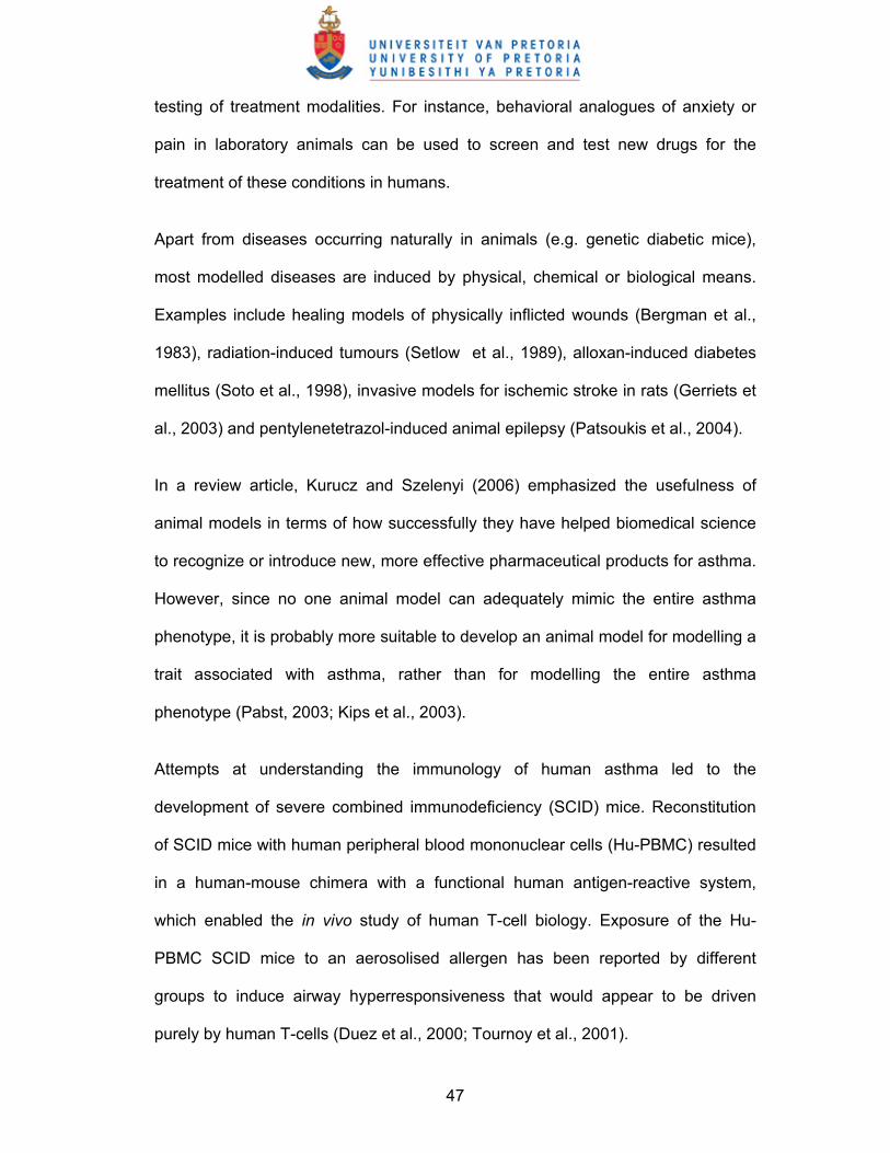
47
testing of treatment modalities. For instance, behavioral analogues of anxiety or
pain in laboratory animals can be used to screen and test new drugs for the
treatment of these conditions in humans.
Apart from diseases occurring naturally in animals (e.g. genetic diabetic mice),
most modelled diseases are induced by physical, chemical or biological means.
Examples include healing models of physically inflicted wounds (Bergman et al.,
1983), radiation-induced tumours (Setlow et al., 1989), alloxan-induced diabetes
mellitus (Soto et al., 1998), invasive models for ischemic stroke in rats (Gerriets et
al., 2003) and pentylenetetrazol-induced animal epilepsy (Patsoukis et al., 2004).
In a review article, Kurucz and Szelenyi (2006) emphasized the usefulness of
animal models in terms of how successfully they have helped biomedical science
to recognize or introduce new, more effective pharmaceutical products for asthma.
However, since no one animal model can adequately mimic the entire asthma
phenotype, it is probably more suitable to develop an animal model for modelling a
trait associated with asthma, rather than for modelling the entire asthma
phenotype (Pabst, 2003; Kips et al., 2003).
Attempts at understanding the immunology of human asthma led to the
development of severe combined immunodeficiency (SCID) mice. Reconstitution
of SCID mice with human peripheral blood mononuclear cells (Hu-PBMC) resulted
in a human-mouse chimera with a functional human antigen-reactive system,
which enabled the in vivo study of human T-cell biology. Exposure of the Hu-
PBMC SCID mice to an aerosolised allergen has been reported by different
groups to induce airway hyperresponsiveness that would appear to be driven
purely by human T-cells (Duez et al., 2000; Tournoy et al., 2001).

48
Studies on asthma modelling have been extended to new areas like exercise-
induced asthma, occupational asthma as well as to less studied aspects of
modelled asthma such as airway remodelling. For instance, the sheep asthma
model was reported to have features that correlated with the structural changes
observed in the remodelling of lung tissues seen in chronic asthmatics. In their
study, Snibson et al., (2005) repeatedly challenged the lungs of sheep with the
house dust mite (known to be the major trigger of asthma in up to 80% of cases) in
order to induce many of the distinguishing features of airway remodelling often
seen in asthmatics. These include increased collagen deposition and airway
smooth muscle bulk, mast cell and goblet cell hyperplasia as well as epithelial cell
hypertrophy.
One interesting study on modelling exercise-induced asthma involved athletic
dogs whose airway conditions after exercise compared closely with the airways of
susceptible humans. Athletes who play sports in cold weather, particularly skaters
and cross-country skiers, are known to have an increased prevalence of lower
airway disease that is hypothesized to result from repeated penetration of
incompletely conditioned air into the lung periphery (Davis et al., 2002) usually
with significantly more airway inflammation than their sedentary counterparts (Sue-
Chu et al., 1999).
Racing sled dogs like humans also perform strenuous exercises under frigid
conditions and studies by Davis et al., (2002) suggest that the racing sled dog
could be a useful naturally occurring animal model of the analogous human “ski
asthma”, a term used to describe a syndrome of non-atopic airway inflammation
and hyper-reactivity in elite winter athletes (Sue-Chu et al., 1999). Results from a

49
later study with horses also suggest that cold weather exercise can lead to
asthma-like airway disease through the local induction of cytokines typical of the
TH2 phenotype (Davis et al., 2005).
Treatment of induced asthma in animal models with botanicals has been reported
in literature. Johnson et al., (2002) exposed asthmatic BALB/c mice to the
botanical Silymarin and found that it caused increased T-lymphocyte population.
Similarly, treatment of BALB/c mice with DA-9601 (a formulated ethanol extract of
Artemisia asiatica Nakai or Asteraceae) was found to cause reduced IgE level,
recruitment of inflammatory cells into the BAL fluid and lung tissues, expression of
CD40, CD40L and VCAM-1 molecules, goblet cell hyperplasia and production of
various cytokines (Kim et al., 2006).
In general, the use of animal models of human diseases in scientific investigations
will continue since they appear to be far more beneficial. In the case of asthma,
animal models progressively remain useful through the elucidation of disease
mechanisms and development of medications that are often more active than
previously used ones. For studies in mice, a broad spectrum of molecular and
immunological tools is available (Bice et al., 2000).
2.10.1. BALB/c mouse models
BALB/c asthma models are widely studied and findings are reported in the
literature. Viana et al., (2002) studied the response of BALB/c mice to the fungus
stachybotrys chartarum in causing asthma-like conditions. Concurrent exposure of
BALB/c mice to ovalbumin and ozone appeared to cause increased goblet-cell
hyperplasia in a study by Last et al., (2004). DA-9201 (an ethanolic extract of black
rice Oryza sativa L.) appeared to play an important role in attenuating the

50
progression of airway inflammation and remodelling in BALB/c mice sensitized to
ovalbumin (OVA) (Lee et al., 2006).
The asthmatic mouse models of C57/BL, BALB/c, 129J, DBA/2, C3H/HeJ and
CBA/J strains have also been studied (Joachim et al., 2003; Shinigawa and
Kojima, 2003). BALB/c mice are chosen for this study primarily because so much
is already known about their immune responses and genetics from literature.
Despite the few identified shortcomings, results from previous studies have shown
that the BALB/c mouse model still manages to paint a good pattern of the human
airway disease better than any other model (Gleich et al., 1988, Zhao et al., 2000,
Blyth et al., 2000; Leigh et al., 2002; McMillan and Lloyd, 2004; Johnson et al.,
2004; Jungsuwadee et al., 2004).
BALB/c mice have long been used for other studies in immunology (Alvarez et al.,
2004; Chen et al., 2005), drug dose testing (Fayer and Fetterer, 1995),
carcinogenesis (McIntire and Princler, 1969; Pawlowski et al., 1979; Gambotto et
al., 2000) as well as microbiology and infection studies (Adjei et al., 1993; Sheu et
al., 1999; Singh et al., 2000; Padigel and Farrell, 2005).
2.11. Cell cultures
Cell culture systems are useful means of evaluating different biological processes
in vivo prior to undertaking further studies with animals or final studies in human
subjects. Many drugs are screened for toxicity and dosage using cell cultures. Cell
culture studies could be done either with primary cultures or with cell-lines
depending on the objectives of the study and availability. In the present study, the
MRC-5 cell-line was used for testing toxicity of the test substances in order to
determine the appropriate doses.

51
The MRC-5 cell line is a normal finite cell line derived from human embryonic or
foetal diploid lung fibroblasts (Jacobs et al., 1970; Freshney, 2000). The plethora
of literature on the MRC-5 cell line could be indicative of its excellent biological
properties. Diverse studies are carried out on these cells, ranging from their use as
control cells in experiments involving other cells or cell lines, to their use for
cytotoxicity testing, drug potency testing, medicinal plant testing and vaccine
production. In one study, Yang et al., (1997) found that cadmium induced oxidative
cellular damage in cultured MRC-5 cells. In another study, extracts of thirty - three
plants commonly used for the treatment of malaria by traditional healers in west
tropical Africa were tested for antiplasmodial activity and cytotoxicity using MRC-5
cells (Zirihi et al., 2005). In addition, extracts prepared from the leaves of seven
Panamanian tropical forest plants were found to have direct virucidal as well as
intracellular antiviral activity against both DNA and RNA viruses. Antiviral activity
was achieved with extract concentrations significantly lower than those required to
produce cytotoxic effects. In addition, extracts were less toxic to normal MRC-5
cells than to the tumour cells tested (Romin et al., 2006).
A study by Maes et al., (2004) showed that PX-6518 exhibits minimal toxicity for
mammalian cells including MRC-5 cells. Wu et al., (2005) used MRC-5 cell culture
systems for the production of a smallpox vaccine and their findings suggest that
optimization of growth conditions for MRC-5 cells resulted in enhanced vaccinia
virus production. In another study, extracts of Commelina diffusa and Spathodea
campanulata used as wound-healing agents in Ghana also exhibited significant
antioxidant activity by protecting MRC-5 cells from hydrogen peroxide-induced
oxidant injury at concentrations between one and ten microgram per millilitre (1-
10µg/mL) (Mensah et al., 2006). MRC-5 cells have also been used as control cells

52
in many studies (Chang et al., 2002; Merlin et al., 2002; Conforti et al., 2006),
among others.
In conclusion, it appears from the reviewed literature that a large number of
studies have been done on the mechanism and treatment of asthma using
different experimental models, therapeutic modalities and substances. The present
study is expected to contribute to the general body of knowledge on asthma
treatment by examining the effects of a locally-used medicinal plant on a murine
model. It is hoped that sooner than later, a cure for asthma will be developed from
all the effort of past and current researchers.
2.12. Aims and objectives of study
The general aims and objectives of this study therefore include the following:
a. To test for possible cytotoxic effects of the aqueous, acetone, hexane
and dichloromethane extracts of Euphorbia hirta on the MRC-5 cell line
using the combined NR/MTT/CV assay for lysosomal membrane
integrity, cell viability and cell number, with a view to determining safe
doses for use in the animal experiments.
b. To evaluate the possible dose-dependent therapeutic effects of the
aqueous extract of E.hirta in the BALB/c mouse model by studying its
effects on white blood cell count, platelet aggregation, fibrin network
pattern and fibre morphology using the scanning electron microscope
(SEM).
c. To use light and transmission electron microscopic (TEM) techniques to
investigate histological and ultrastructural changes in the airways and

53
lungs including inflammatory cell migration, collagen fibre distribution,
alveolar wall thickness, smooth muscle hyperplasia as well as mucous
gland distribution.
d. To determine body weight changes in asthmatic BALB/c mice treated
with the aqueous extract of E.hirta and comparing results with the
control mice.
e. To compare findings from all experiments involving treatment with the
aqueous extract of E.hirta with findings from experiments involving
treatment with hydrocortisone in both the cell culture and the animal
studies.

54
CHAPTER THREE
Effects of HC and extracts of E. hirta on the fibroblast MRC-5 cell line

55
3.1. Introduction
Asthma is one of the most common respiratory complaints in the world as it affects
an estimated 300 million people worldwide. The marked increase in the
prevalence of asthma has made it a worldwide health concern and global figures
are expected to rise to 400 million by the year 2025 (Masoli et al., 2004).
Over the years, corticosteroids have been very effective in the treatment of asthma
but serious systemic adverse effects have been associated with prolonged use of
these medications (Leonard and Sur, 2002). With increasing popularity of
complementary and alternative medicine, research geared towards developing
more effective alternative anti-asthma agents appears to be gaining much
attention.
Herbal medicines are purported to show clinical efficacy with minimal side effects
compared to mainstream treatments (Chevrier et al., 2005). A number of studies
support the use of herbal medicines in asthma and as reported by Ming-Chun et
al., (2005), herbal medicine intervention appears to be safe and effective
alternative methods for treating asthma. In addition, many pharmaceutical
products have been developed from traditionally used plant products (McRae et
al., 2007). Continuous investigation of how herbal medicines work could lead to
the development of additional effective medication for the treatment of asthma,
allergies and many other health conditions (Bielory and Lupoli, 1999). The present
study aims at investigating the possible toxic effects of E. hirta on the MRC-5 cell
line.
The MRC-5 cell line is one of the most common cell lines used for cytotoxicity
studies. This cell line is even more suitable for modelling in vitro conditions of a

56
respiratory tract disease like asthma because as it is derived from normal human
diploid foetal lung fibroblasts. The advantages of using in vitro model systems are
that cellular damage is easy to measure, the system is simple and easy to
reproduce, only small amounts of the compound are required and several
statistically significant experiments under controlled conditions can be undertaken.
In one previous study involving the MRC-5 cell line, the biocompatibility of the
Erlanger silver intravenous catheter was investigated. The possible acute
cytotoxicity of extracts from the catheter was studied using MRC-5 cells and
lymphocytes. The extracts were not cytotoxic to MRC-5 human fibroblasts or to
sensitized phytohemagglutinin (PHA)-stimulated human lymphocytes (Greil, 1999).
In another study, the physiological concentrations of the dipeptide L-carnosine
were found to prolong the lifespan of MRC-5 cells and strongly reduce the normal
features of senescence (Holliday and McFarland, 2000).
MRC-5 cells have also been used in many other studies including screening of
medicinal plants for cytotoxicity (Zirihi et al., 2005), antimicrobial and antioxidant
activities (Mensah et al., 2006); in vaccine production (Wu et al., 2006); in virology
studies (McCarter and Robinson, 1997; Maes et al., 2004); as well as in
inflammation studies (Hase et al., (2003). Others include carcinogenicity studies
(Merlin et al., 2002; Tomatsu et al., 2003); studies on the damaging effects of
heavy metals (Yang et al., 1997), stem cell research (Rieske et al., 2005) as well
as studies on radiation-induced damage effects (Jones et al., 2006).
In this study, different types of bioassay systems were used to measure
cytotoxicity in the MRC-5 cell line. Cell number, cell viability and lysosomal
membrane integrity were determined using the Crystal Violet (CV); 3-(4, 5-

57
dimethylthiazol-zyl) 2, 5-dimethyl tetrazolium bromide (MTT); and Neutral Red
(NR) assays. HC was used as a pharmaceutical control for the plant extract
because of its known anti-asthma effects. A study by McLendon et al., (2006)
showed that different cortisol concentrations did not induce structural damage in
MRC-5 cells throughout the experiment.
A pilot cell culture study was carried out to determine the appropriate
concentration of HC to be used as control and later, the final cell culture tests were
done using the aqueous and organic (hexane, dichloromethane, acetone) extracts
of E. hirta. The individual effects of each solvent were also evaluated.
3.2. Hypothesis
This study was carried out to test the hypothesis that extracts of E. hirta have
limited cytotoxic effects on cultured MRC-5 cells and such effects are not only
dose-dependent but vary between the different solvent extracts used.
3.3. Aims of study
To evaluate the possible cytotoxic effects of E. hirta extracts using in vitro
assays (NR, MTT and CV) to analyze the viability and lysosomal membrane
integrity of MRC-5 cells as well as MRC-5 cell numbers.
To compare the effects of the aqueous and organic solvent (acetone,
dichloromethane and hexane) extracts of E. hirta on the MRC-5 cell line.
To compare the effects of HC with the effects of the different extracts of E.
hirta extract on the MRC-5 cell line.
To provide information on the concentrations of E. hirta and HC that are
safe to use in the animal studies

58
3.4. Materials
3.4.1. MRC-5 cell line
The MRC-5 lung permanent cell line is a normal, finite cell line derived from
human embryonic diploid lung fibroblasts (Jacobs et al., 1970; Freshney, 2000).
MRC-5 cells are common and commercially available and were obtained from
Highveld Biological Company, Johannesburg, South Africa.
3.4.2. HC and E. hirta
HC was used as a pharmacologic control for the extracts of E. hirta in this study.
The drug was purchased from Elwierda Apteek/Pharmacy Wirdapark, Pretoria,
South Africa. One hundred milligrams (100mg) of the powdered HC were
dissolved in 2ml of injection water giving a concentration of 50mg/ml. This solution
has a molarity of 0.138M and the MRC-5 cells were exposed to five times
progressive dilutions of HC in the following molar concentrations: (0.00 μM,
22.8μM, 3.8μM, 0.63μM, 0.16μM and 0.0263μM). The pilot study showed that the
significant cytotoxic effects of HC on MRC-5 cells mostly affected the CV and NR
assays.
E. hirta was obtained from fields in the Gezina area of Pretoria, South Africa and
was identified and verified by staff in the Department of Botany, University of
Pretoria, South Africa by comparison with a voucher specimen of the plant
available at the H.G.W.J. Schweikerdt Herbarium of the University. Aqueous and
organic solvent (acetone, hexane and dichloromethane) extracts of the plant used
for the various experiments were prepared in the Cell Biology Laboratory of the
Department of Anatomy, University of Pretoria. The aboveground parts of the plant
were dried at room temperature for one week and a fine powder was then

59
prepared and was subdivided into four 50 g samples. Each 50 g sample was
extracted with distilled water and the three different organic compounds (acetone,
dichloromethane and hexane), filtered and then dried on a rotary evaporator at
40°C.
A concentration of 0.1g/ml (each 50g sample dissolved in 500ml distilled water or
500ml solvent) was prepared for each solvent and the samples were stored frozen
as the stock solutions. Later the stored samples were thawed and fifty microlitres
(50μl) of the working solution added to the culture wells in five times progressive
dilutions to determine the effects of the different extracts on the MRC-5 cell line.
3.4.3. Media, supplements, reagents and plastic ware
Eagles Minimum Essential Medium (EMEM) powder, Hanks Balanced salt solution
(HBSS) and Foetal Calf Serum (FCS) were from Highveld Biological Company,
Johannesburg, South Africa. Sartorius cellulose acetate membrane filters 0.22μm
were from National Separations, Johannesburg, South Africa. Fixatives, acids and
organic solvents, such as glutaraldehyde, hydrochloric acid (HCl), acetic acid,
isopropanol, and formic acid were analytical grade and were purchased from
Merck, Johannesburg, South Africa. Streptomycin sulphate, penicillin G (sodium
salt), Amphotericim B and Trypsin were obtained from Life Technologies
Laboratory supplied by Gibco BRL Products, Johannesburg, South Africa.
Ethylene diamine tetra acetate (EDTA), Dimethyl sulphoxide (DMSO), potassium
chloride (KCl), potassium dihydrogen phosphate (KH2PO4), disodium hydrogen
phosphate (Na2HPO4), sodium chloride (NaCl) and sodium hydrogen carbonate
(NaHCO3) were from Merck, Johannesburg, South Africa.
MTT [1- (4, 5-Dimethylthiazol-2-yl) -3, 5- diphenylformazan] and Crystal Violet

60
(CV) powder were obtained from Sigma-Aldrich, Atlasville, South Africa. Bovine
serum albumin (BSA) was from Boehringer Mannheim, Randburg South Africa.
Water was double distilled and deionised (ddH2O) with a Continental Water
System and sterilized by filtration through a Millex 0.2μm filter. Glassware was
sterilized at –1400C in a Prestige Medical Autoclave (Series 2100). Twenty four
(24-well) and 96-well 25cm2 and 75cm2 cell culture flasks, 10ml and 5ml pipettes,
15ml and 50ml centrifuge tube, micro centrifuge tubes were obtained from
NUNCTM and supplied by AEC- Amersham, Johannesburg, South Africa.
3.5. Methods
3.5.1. Cultivation, maintenance and preservation of the MRC-5 fibroblast cell
line
The MRC-5 fibroblast cell line was maintained in EMEM supplemented with 5%
FCS and 1% antibiotic solution (EMEM/ 5% FCS). An antibiotic stock solution was
prepared by mixing 10,000U/ml Penicillin G (sodium salt), 10,000μg/ml
Streptomycin sulphate and 25μg/ml Amphotericim B in 0.85% saline. A volume of
10 ml of the working solutions was added to 1 litre of the prepared medium. The
antibiotic solution was kept at -10 oC and thawed when needed. The media was
sterilized by filtration through a 0.22μm membrane filters under aseptic conditions
in a laminar flow cabinet. Aliquots of 100 ml were prepared and the medium was
stored at 4oC and warmed to 37oC before use.
The MRC-5 cells were plated at a concentration of 5X10⁴ cells per ml in 25cm2
and 75cm2 cell culture flasks and were maintained at 37°C and 5% CO2 in a CO2 -
water-jacketed incubator from Forma Scientific. The cell culture medium was
changed every three days or when the medium had become acidic. The cells were

61
later passaged once confluent with a 0.05% trypsin solution. A 10X Trypsin/EDTA
stock solution of 5g/l Trypsin, 2g/l EDTA.4Na was prepared by mixing 0.25g
Trypsin, 0.1g EDTA and 0.425g NaCl in 50ml Hanks buffer. Hanks buffer was
prepared by dissolving 9.86g/ l Hanks salt and 0.35g/l NaHCO3 in ddH2O. A
working solution of 1X was prepared by diluting the stock solution with DPBS that
was stored at -10 oC and warmed up at 370C before use. The solutions were
filtered through a 0.22μm membrane filter under aseptic conditions.
The cells were passaged by firstly removing the medium from the confluent
monolayer. A volume of 0.08 ml/cm2 trypsin working solution was added and the
flask was placed at 370C for 5-10 minutes. The medium containing the detached
cells was added to 10 ml medium. The cells were collected by centrifugation in a
BTL Bench centrifuge from Baird and Tatlock at 6000xg (2000 rpm) for 5 minutes.
The medium was removed and the cells were suspended in 10 ml fresh medium.
The centrifugation step was repeated and the number of cells was determined by
counting a 10μl aliquot of cells using a haemocytometer from Brand supplied by
Merck, Johannesburg, South Africa.
The MRC-5 fibroblasts were either used for experiments described below or stored
at –70°C. For storage, the cells were suspended in cell culture freezing medium at
a concentration of 5x10⁶ cells per ml. The freezing medium was prepared by
adding 10% DMSO and EMEM/5%FCS. One millilitre of the cell suspension was
transferred to the 1.5ml freezing vials and stored by slow freezing (the vials were
wrapped with tissue paper and placed into a large Styrofoam box) in a –70°C
freezer.

62
The cells were stored for an indefinite period of time, with minimum loss of
viability. The vials containing the MRC-5 fibroblasts were thawed rapidly by stirring
the vial in warm water at 370C. The cells were suspended in EMEM containing 5%
FCS to a volume of 15 ml. The cells were collected by centrifugation, the
supernatant removed and the cells suspended in fresh medium. This step was
repeated twice before the cells were suspended in a final volume of 3ml culture
medium and plated in 25cm2 cell culture flasks. For each experiment, cells were
plated at a cell concentration of 2x10⁴ cells per ml in 24 well plates with the culture
area of 1.9cm2/well and were kept for 24 hours at 37°C and 5% CO2 before
conducting each experiment.
3.5.2. Exposure of cells to the treatment agents
Different experiments were done using MRC-5 cells. Some cells were exposed to
HC and others were treated with a 0.1g/ml concentration of each of the stock
solutions (aqueous, acetone, dichloromethane and hexane). In all experiments,
cells in the first set of wells represented the controls and were left untreated. A five
times dilution was done for the remaining wells by first adding 50μl of the
treatment agents to the set of wells next to the control wells, mixing properly
before transferring 100μl by pipette from these set of wells to the next, and again
removing another 100μl to the next set of wells. The process was repeated in
series for all the wells and the 100μl removed from the last set of wells was
discarded.
After 24 hours of exposure cell number, cell viability and lysosomal membrane
integrity were determined using the combined NR/MTT/CV assay. All experiments
conformed to the ethical standards of the University of Pretoria.

63
3.5.3. The combined NR, MTT and CV (NR/MTT/CV) bioassay
In this study, cell viability, lysosomal membrane integrity and cell number were
determined almost simultaneously using the combined NR/MTT/CV bioassay of
Chiba and others (Chiba et al., 1998) with only a slight modification.
Briefly, 50µl of a 50µg/ml NR dye solution was first added to each well for uptake
into lysosomes of viable cells. After an incubation period of 1-2 hours, 50µl of a
2.5mg/ml MTT solution was added. The yellow water-soluble MTT dye is
converted by mitochondrial dehydrogenase in viable cells to insoluble purple/blue
formazan (Mosmann, 1983).
After a further 1-2 hours, the culture medium was carefully removed, the plate
carefully blotted dry and the cells rapidly fixed in 1% each of acetic acid and
formalin in water for a few seconds. The fixative was decanted and the plate
blotted dry before 100µl of the NR solvent (1% acetic acid in 50% ethanol) was
added for extraction of NR. The extracted dye was then transferred to a 96-well
plate. Likewise the formazan crystals were extracted by adding 100µl dimethyl
sulphoxide (DMSO) and the extracted dye transferred to the 96-well plate.
The plates were later washed with water and dried before the attached cells were
stained with 1% CV for 20 minutes. The CV dye was thereafter removed, the
plates washed thoroughly with tap water, dried and the dye extracted with 100µl of
a 10% acetic acid solution and transferred to the 96-well plate as well. Absorbency
was measured at 570 nm, 450 nm and 590 nm using an EL900 plate reader for
the NR, MTT and CV assays respectively (Borenfreund et al., 1990).

64
3.6. Data management and statistical analysis
The average of four independent experiments was determined and the data was
expressed as percentage of the unexposed control wells. Graphs were plotted to
show the effects of HC and the different plant extracts on lysosomal membrane
integrity, cell viability and number. Tables were combined to obtain one final graph
that is a summary of all experiments. All data was expressed as mean ± standard
deviation (SD) and analysed for statistical significance using the Analysis of
Variance (ANOVA) statistical package. A confidence level of 95% (i.e. p< 0.05)
was considered statistically significant.
For the CV, MTT and NR assays, a one-way Analysis of Variance (ANOVA) was
used to determine the presence or otherwise of statistically significant differences
between unexposed control cells and the different dosage concentrations. For the
solvent only experiments, a one-way ANOVA was used to compare between
control and solvent extract dosage while a two-way ANOVA was used to compare
each concentration of solvent with the solvent and plant extract.
In order to determine the differences in effects between the solvent only assays
and the solvent and plant extract assays, comparisons were made between group-
matched concentrations. The concentration range was designated ‘0-5’ where ‘0’
represents the control concentration, ‘1’ the lowest concentration and ‘5’ the
highest concentration respectively. The 2-way ANOVA gives the results of
statistically significant differences between corresponding concentrations in the
solvent assays and the solvent with plant extract assays respectively.

65
3.6.1. Pilot study with HC
MRC-5 cells were exposed to a concentration range of 0-5.56 μM HC for 24 hours
after which lysosomal membrane integrity, cell viability and number was
determined using the NR/MTT/CV assay. From the data presented in figure 3.3, it
is evident that HC had no effect on cell viability (MTT assay) but did cause a
decrease in lysosomal membrane integrity and cell number. At concentrations of
1.11μM and 5.56μM, a significant decrease in NR (p ≤ 0.05) was observed while at
5.56μM, a significant decrease in CV (p ≤ 0.05) was observed.
Analysis of the mean results obtained showed that only the two highest
concentrations of HC had statistically significant effects compared with the controls
for the NR assay. There was no significant difference between the different dose
concentrations for the MTT assay but a statistically significant difference was seen
in the CV assay between control and the highest concentration.

66
Figure 3.1: The effect of 0-5.56 μM HC on lysosomal membrane integrity, cell viability and number
measured using the NR/MTT/CV assay. Data expressed as mean (n=4) ± SD. *Differences are
significant p ≤ 0.05 compared to the least concentration.
In addition, there was no statistically significant difference between the three
lowest concentrations but differences were observed between the highest NR
concentration and the three lowest concentrations as well as between the second
highest NR concentration and the three lowest concentrations respectively. In the
case of the CV assay, statistically significant differences were seen only when the
three lowest concentrations were compared with the highest concentration.
*

67
3.6.2. Treatment with the aqueous extract of E. hirta.
The aqueous extract of most plants generally contain lipophobic compounds such
as polyphenolic compounds, triterpenes and flavonoids, and most of these
compounds have potent anti-inflammatory and antioxidant properties (Lanhers et
al., 1991; Sharma et al., 2007; Fecka and Turek, 2007). MRC-5 cells were
exposed to 0-11.11 mg/ml of the aqueous extract of E. hirta for 24 hours after
which lysosomal membrane integrity, cell viability and number were determined
using the NR/MTT/CV assay.
The aqueous fraction of E. hirta had no significant effect on lysosomal membrane
integrity. For the MTT assay that measures cell viability. There was a significant
decrease in cell viability when the 0.02mg/ml and 0.09mg/ml concentrations were
compared to the 2.22mg/ml and 11.11mg/ml (p ≤ 0.05 and p ≤ 0.05) respectively.
However, from 0.44 - 11.11mg/ml concentrations, the aqueous extract of E. hirta
caused an increase in value for the CV assay from 100% to 300% compared to
the control (p ≤ 0.05). This finding appeared to imply that the aqueous extract of E.
hirta could cause a three fold increase in cell number. This is very unlikely an
increase in cell number, since such an increase is usually associated with a
corresponding increase in the number of mitochondria and lysosomes and
therefore an increase in the values for the NR and MTT assays. It is possible that
the plant proteins bind either the tissue culture surface or to the surface of the
MRC-5 cells resulting in false high values.
However, the MTT assay mean showed statistically significant differences
between the control and least concentration, as well as between the third highest
and the highest concentration.

68
ASSAYS OF E.hirta AQUEOUS EXTRACT
0
50
100
150
200
250
300
350
400
0.00 0.02 0.09 0.44 2.22 11.11
Concentration (mg/ml)
% a
vera
ge o
f con
trol
NR WaterEHMTT WaterEHCV WaterEH
Figure 3.2: The effect of 0-0.11.11mg/ml aqueous extract of E. hirta on lysosomal membrane
integrity, cell viability and number measured using the NR/MTT/CV assay. Data expressed as mean
(n=4) ± SD. * Differences are significant p ≤ 0.05 compared to lower concentrations and the control.
3.6.3. Effects of acetone and acetone extracts of E hirta on MRC-5 cells
Acetone extracts of plants generally contain glycerides, sterols, sterol esters,
carotenoids, lipid soluble vitamins, monogalactoside diglycerides and sterol
glycosides (Gouveia and Empis, 2003; Nyström et al, 2007). Acetone used as
carrier itself can be cytotoxic to cells; therefore, the cytotoxicity of acetone in MRC-
5 cells was determined. MRC cells were exposed to 0-11.11% acetone for 24
hours before lysosomal membrane integrity, cell viability and number was
determined.
* *
*
*

69
For acetone only assays (Figure 3.3a), following exposure to 11.11%, there was
no significant difference in lysosomal membrane integrity and cell viability.
However, the cell number appeared to rise initially to 125.4% before decreasing
steadily until percentage concentration 2.22%; increasing again to 108.9% at the
highest concentration. The differences found in cell number were significantly
different from the controls as well as from 0.09% and 0.44% concentrations.
The MRC-5 cells in culture were also exposed to 0 - 11.11 mg/ml acetone extract
of E. hirta (Figure 3b) and results show a steady progression between the different
concentrations. Again, no statistical differences were observed between the
control (without extract) and the different volumes/concentrations of the plant
extract added to the cell culture medium of MRC-5 cells (p ≤ 0.05 for both NR and
MTT assays) (Figure 3.3b). Except at the 0.02 mg/ml concentration with 47%
significant increase in cell number, all other differences in volumes/concentration
were not significant (p ≤ 0.05).
Two way ANOVA analysis showed significant differences in the NR and CV assay
when corresponding concentrations of the acetone only and the acetone extract
treatments were compared (p ≤ 0.05 and p ≤ 0.05)(Figure 3.4b and c). No
statistical differences were observed for the MTT assay (Figure 3.4c).

70
ACETONE ONLY ASSAYS
0
20
40
60
80
100
120
140
160
0.00 0.02 0.09 0.44 2.22 11.11
Percentage Concentration
% A
vera
ge o
f Con
trol
NR ActMTT ActCV Act
Figure 3.3: The effect of (a) 0-11.11% of acetone solvent, and (b): 0-11.11 mg/ml acetone extract
of E. hirta, on lysosomal membrane integrity, cell viability and number measured using the
NR/MTT/CV assay. Data expressed as mean (n=2) ± SD. * Differences are significant at p ≤ 0.05
compared to lower concentrations and the control.
ASSAYS OF ACETONE EXTRACTS OF E. hirta
0
20
40
60
80
100
120
140
160
180
0.00 0.02 0.09 0.44 2.22 11.11
Concentration (mg/ml)
% a
vera
ge o
f con
trol
NR ActEHMTT ActEHCV ActEH
*
*

71
NEUTRAL RED ASSAYS
0
20
40
60
80
100
120
140
160
0.00 0.02 0.09 0.44 2.22 11.11
Percentage Concentration
% A
vera
ge o
f Con
trol
NR ActNR ActEH
MTT ASSAYS
0
20
40
60
80
100
120
140
0.00 0.02 0.09 0.44 2.22 11.11
Percentage Concentration
% A
vera
ge o
f Con
trol
MTT ActMTT ActEH
CRYSTAL VIOLET ASSAY
0
20
40
60
80
100
120
140
160
180
0.00 0.02 0.09 0.44 2.22 11.11
Percentage Concentration
Per
cent
age
Ave
rage
of C
ontro
l
CV ActCV ActEH
Figure 3.4: Comparison of the effects of 24-hour exposure to the carrier acetone and E. hirta
acetone extracts on the MRC-5 cell line; a) NR, b) MTT and c) CV assay. *Differences are
significant (p ≤ 0.05) for NR and CV assays, each compared to ActEH.
*
* *

72
3.6.4. Effects of dichloromethane (DCM) and DCM extracts of E hirta on
MRC-5 cells
Most DCM plant extracts usually contain sesquiterpene lactones lychnopholide,
centratherin, goyazensolide and 15-desoxygoyazensolide (Grael et al., 2005),
terpene and naphthalene derivatives (Jing et al., 2007) as well as β-Sitosterol, 5,
6-Trihydroxy-3′, 7-dimethoxyflavone, Daucosterin and Ursolic acid (Moghaddam et
al., 2007). The MRC-5 cells were exposed to dichloromethane (DCM) alone as
well as to DCM of E. hirta.
After 24 hours exposure to 0 - 11.11% DCM (Figure 3.5a) and DCM containing 0-
11.11 mg/ml plant extract (Figure 3.5b), lysosomal membrane integrity, cell
viability and number was determined by the NR, MTT and CV assays respectively.
For the DCM treatment (Figure 3.5a), a one-way ANOVA showed that there was
no significant difference (p ≤ 0.05) between the control (no DCM) and other
concentrations for all three assays. Likewise, no significant differences were
observed (Figure 3.5b) for all three assays of the DCM extracts of E. hirta.
When treatment with DCM was compared to the DCM extracts of E. hirta using
two-way ANOVA analysis (Figure 3.6), no significant differences were seen in the
NR and MTT assays. However, the two-way ANOVA showed that only the
difference between the second highest concentrations (2.22%) of the CV assays in
the two treatments was statistically significant. Comparisons between the two
treatments at other concentrations were not statistically significant.

73
DICHLOROMETHANE ONLY ASSAYS
0
20
40
60
80
100
120
140
0.00 0.02 0.09 0.44 2.22 11.11
Percentage Concentration
% a
vera
ge o
f con
trol
NR DiCMMTT DiCMCV DiCM
ASSAYS OF DICHLOROMETHANE EXTRACTS
OF E. hirta
0
20
40
60
80
100
120
140
160
180
0.00 0.02 0.09 0.44 2.22 11.11
Concentration (mg/ml)
% a
vera
ge o
f con
trol
NR DiCMEHMTT DiCMEHCV DiCMEH
Figure 3.5: The effect of (a) 0-11.11% DCM and (b) 0-11.11 mg/ml DCM extract of E. hirta, on
lysosomal membrane integrity, cell viability and number measured using the NR/MTT/CV assay.
Data expressed as mean (n=2) ± SD. *No significant differences at all concentrations.
74
NEUTRAL RED ASSAYS
0
20
40
60
80
100
120
140
160
180
0.00 0.02 0.09 0.44 2.22 11.11
Percentage Concentration
Perc
enta
ge A
vera
ge o
f Con
trol
NR DiCMEHNR DiCM
MTT ASSAYS
0
20
40
60
80
100
120
140
160
0.00 0.02 0.09 0.44 2.22 11.11
Percentage concentration
Per
cent
age
Ave
rage
of C
ontro
l
MTT DiCMEHMTT DiCM
CRYSTAL VIOLET ASSAY
0
20
40
60
80
100
120
140
160
0.00 0.02 0.09 0.44 2.22 11.11
Percentage Concentration
Per
cent
age
Ave
rage
of C
ontr
ol
CV DiCMEHCV DiCM
Figure 3.6: Comparison of the effects of 24-hour exposure to the carrier DCM and DCM
extracts of E. hirta on the MRC-5 cell line; a) NR, b) MTT and c) CV assay. * Differences
significant at p ≤ 0.05 compared to the highest concentration.
*

75
3.6.5. Effects of hexane and hexane extracts of E hirta on MRC-5 cells
In most plant extracts, hexane extracts mainly contain terpenes, terpenic alcohols,
terpenic aldehydes and ketones, xylene, dimethylformamide, and hexadecanoic
acid (Quinn et al., 2007). The hot hexane extract of E. hirta was found to contain
alpha and beta amyrin, sitosterol and taraxerone (Atallah and Nichola, 1972).
MRC-5 cells were exposed in vitro to 0-11.11% hexane alone as well as 0 -
11.11mg/ml hexane extracts of E. hirta for 24 hours. The effects of the hexane
solvent alone on lysosomal membrane integrity, cell viability and number were
determined using the combined NR/MTT/CV assays. Each assay in the two
different experiments was tested for possible statistically significant differences
between the control (no hexane or hexane extract of E hirta) and the different
concentrations added. A two way ANOVA was used to determine differences
between hexane alone and hexane E. hirta extract.
The one-way ANOVA test showed that treatment of MRC-5 cells with the hexane
solvent alone did not show statistically significant differences between the different
concentrations for the NR, MTT and CV assays (Figure 3.7a). Similarly, treatment
with the hexane E hirta extract showed no significant differences at all
concentrations for all three assays following (Figure 3.7b).

76
HEXANE ONLY ASSAYS
0
20
40
60
80
100
120
140
160
0.00 0.02 0.09 0.44 2.22 11.11
Percentage Concentration
% A
vera
ge o
f Con
trol
NR HexMTT HexCV Hex
ASSAYS OF HEXANE EXTRACTS OF E. hirta
0
20
40
60
80
100
120
140
0.00 0.02 0.09 0.44 2.22 11.11
Concentration (mg/ml)
% a
vera
ge o
f con
trol
NR HexEHMTT HexEHCV HexEH
Figure 3.7: The effect of (a) 0-11.11% carrier hexane and (b) 0-11.11 mg/ml hexane extract of E.
hirta on lysosomal membrane integrity, cell viability and number measured using the NR/MTT/CV
assay. Data expressed as mean (n=2) ± SD. No significant differences at all concentrations.

77
Comparison between the hexane-treated MRC-5 cells and those treated with the
hexane extract of E. hirta was done for the different assay types (Figure 3.8a-c)
using the 2-way ANOVA analysis. Corresponding NR values showed statistically
significant differences (p ≤ 0.05) at all the concentrations compared. On the other
hand, no statistically significant differences were observed between the hexane
only and the hexane extract treatments in the MTT assays except at 2.22%
concentration. This appears to indicate that the effects of MTT were relatively
similar in both treatments.
For the CV assays, all percentage average of control values in the hexane only
treatment were higher than corresponding values of the hexane extract treatment
at the same concentration range (except the highest concentration), as shown in
Figure 3.8c. The differences between these values were also statistically
significant (p ≤ 0.05) except at the highest percentage concentration (11.11%). At
this concentration, the value of the percentage average of control for the ‘hexane
only’ treatment decreased while that for the hexane extracts treatment increased.

78
NEUTRAL RED ASSAYS
0
20
40
60
80
100
120
140
0.00 0.02 0.09 0.44 2.22 11.11
Percentage Concentration
% A
vera
ge o
f Con
trol
NR HexNR HexEH
MTT ASSAYS
0
20
40
60
80
100
120
0.00 0.02 0.09 0.44 2.22 11.11
Percentage Concentration
% A
vera
ge o
f Con
trol
MTT HexMTT HexEH
CRYSTAL VIOLET ASSAYS
0
20
40
60
80
100
120
140
0.00 0.02 0.09 0.44 2.22 11.11
Percentage Concentration
% A
vera
ge o
f Con
trol
CV HexCV HexEH
Figure 3.8: Comparison of the effects of 24-hour exposure to hexane and hexane extracts of E.
hirta on the MRC-5 cell line; a) NR, b) MTT and c) CV assay.* Differences are significant (p ≤
0.05) as indicated for hexane only, compared to the hexane extract.
******
* ** *
*
*

79
3.6.6. Analysis of the effects of the different solvents, compared with their E.
hirta extracts and solvent extracts compared with water extract.
A summary of all the effects of treatment of MRC-5 cells with different solvents
and extracts of E hirta as determined using the NR/MTT/CV assays is given in
table 3.1:
Table 3.1: Effects of all solvent and E hirta extractas determined by NR/MTT/CV assays.
Solvent only
Assay Water Acetone DCM Hexane
NR NA NSD NSD NSD
MTT NA NSD NSD NSD
CV NA SD (⇑ highest conc
only) NSD NSD
E hirta Extract
Assay Water Acetone DCM Hexane
NR NSD NSD NSD NSD
MTT SD (⇓ highest
conc only) NSD NSD NSD
CV
SD (⇑ highest
two concs.
only)
SD (⇑ lowest conc only) NSD
NSD
Solvent versus solvent E. hirta extract
Assay Water Acetone DCM Hexane
NR NA
SD (⇑ in the 2nd lowest
conc only, compared to
ActEH)
NSD
SD in all (⇑ for
hexane in all except
2nd lowest conc)
MTT NA NSD NSD
SD (⇓ for hexane;
2nd highest conc.
only)
CV NA
SD ( ⇑ 2nd and 3rd
highest concs only,
compared to ActEH)
SD (⇑ in the 2nd
highest conc only,
compared to DCM)
All SD except
highest conc (⇑ for
hexane)
Key: ⇓ = decrease ⇑ = increase

80
3.6.6.1. Comparative effects in the NR assays
Only the differences between the effects of acetone and the acetone extract at the
second highest concentration (2.22mg/ml) as well as all comparisons between
hexane and hexane extracts were statistically significant (p ≤ 0.05). Comparisons
between hexane and hexane extracts all showed statistically significant
differences.
3.6.6.2. Comparative effects in the MTT assays
Only the highest two concentrations (0.118 mg/ml and 0.588 mg/ml) of the
aqueous extracts of E. hirta were significantly different (p ≤ 0.05) when compared
to the control. In addition, the second highest concentrations (2.22 mg/ml) of the
hexane and hexane extract of E. hirta showed statistically significant difference (p
≤ 0.05).
3.6.6.3. Comparative effects in the CV assays
Most of the comparisons in the CV assays showed statistically significant
differences. Statistically significant difference (p ≤ 0.05) was observed in the
acetone only treatment when the highest concentration was compared with the
control as well as in the acetone extracts treatment when the lowest concentration
was compared with the control. The highest two concentrations in the aqueous
extract treatment also showed significant differences (p≤0.05) compared with the
controls. In the hexane only treatment, all comparisons except at the highest
concentration showed statistically significant differences (p ≤ 0.05). In addition,
only the second and third highest concentrations in the acetone versus acetone
extract as well as the second highest concentration in the dichloromethane versus

81
dichloromethane extract comparisons showed statistically significant differences (p
≤ 0.05).
NR ASSAYS FOR HYDROCORTISONE AND ALL PLANT EXTRACTS
0
50
100
150
200
0.000 0.001 0.005 0.024 0.118 0.588Percentage concentration
% A
vera
ge o
f con
trol
NR HCNR WaterEHNR ActEHNR HexEHNR DiCMEH
MTT ASSAYS FOR HYDROCORTISONE
AND ALL PLANT EXTRACTS
0
40
80
120
160
0.000 0.001 0.005 0.024 0.118 0.588Percentage concentration
% A
vera
ge o
f con
trol
MTT HCMTT WaterEHMTT ActEHMTT HexEHMTT DiCMEH
CRYSTAL VIOLET ASSAYS FOR
HYDROCORTISONE AND ALL PLANT EXTRACTS
0
50
100
150
200
250
300
350
400
0.000 0.001 0.005 0.024 0.118 0.588Percentage concentration
% A
vera
ge o
f con
trol
CV HCCV WaterEHCV ActEHCV HexEHCV DiCMEH
Figure 3.9a-c: Comparison of the effects of 24-hour exposure to HC, aqueous, acetone, DCM
and hexane extracts of E. hirta on lysosomal membrane integrity, cell viability and number
measured using the NR/MTT/CV assay. Data expressed as mean (n=2) ± SD. * Differences are
significant at p ≤ 0.05 compared to the hydrocortisone treatment.
82
3.6.7. Comparison between all organic solvent extracts and the aqueous
extract of E. hirta.
At all concentrations and in all three assay systems used in this study, all the
organic solvent (acetone, DCM and hexane) extracts used in this study generally
showed statistically significant differences (p ≤ 0.05) when compared with the
aqueous E. hirta extract (Figure 3.9a-c). Some differences in the NR and CV
assays in the acetone and water extract comparisons were however not significant
at some concentrations (table 3.2).
NR values were relatively higher at most concentrations of all solvent extracts,
indicating less toxic effects on lysosomal membrane integrity. However, most of
the values for the MTT were much lower at all concentrations, indicating lower cell
viability with the aqueous E. hirta extract treatment.
In the CV assays, there appeared to be a spike in cell numbers (that significantly
differed from results obtained in the CV assays with organic solvent extracts)
following addition of 2.22 mg/ml and 11.11 mg/ml aqueous E. hirta extract. The
observed spike in cell numbers could not possibly be due to cell number increase
and so the aqueous E. hirta extract could be said to have similar effects on cell
number as other extracts.

83
3.6.8. Comparison between treatment with HC and treatments with all three
organic solvent extracts of E. hirta.
Most herbal preparations of E. hirta used by traditional healers for the treatment of
asthma are in aqueous solution, hence the need to investigate the effects of the
aqueous extract. The organic solvent extracts were used in this study to determine
their suitability for use. Different organic solvents may have effects that differ
greatly as observed in previous studies (Jung et al., 2006), from the effects of the
water extract or known anti-inflammatory medications like HC.
The effects of each of the different E. hirta extracts used in this study were
compared with the effects of HC on the MRC-5 cells for every concentration of
each assay type. The two-way ANOVA test was used to determine statistically
significant differences (at p ≤ 0.05) while graphs of the percentage averages of
control plotted against concentrations (figure 9a-c) were used to compare the
effects of the treatments.
3.6.8.1. Comparative effects in the NR assay
Figure 3.9a shows that for the NR assays, the hexane extract followed by the
acetone extract, had effects that were similar in pattern to the effects of HC at all
concentrations. The water extract showed a wide variation in pattern while the
dichloromethane extract showed a sigmoid graphical pattern.
The observed graphical differences were tested statistically and results showed
that only the differences between the HC and the aqueous extract treatments at all
concentrations were statistically significant. The acetone extract showed
significant differences only in the second lowest concentration; the hexane extract

84
showed significant differences between in the two lowest concentrations while the
dichloromethane extract showed significant differences only in the two highest
concentrations (table 3.2).
3.6.8.2. Comparative effects in the MTT assay
The graphs for the MTT assays (figure 3.9b) show similar graph patterns for both
the acetone and hexane extracts as HC for all concentrations. The water extract
showed a marked variation in pattern while the dichloromethane extract only
showed variation in pattern at concentration 0.118mg/ml that later normalized at
the highest concentration. In spite of the observed similarities in graphical patterns
to the HC treatment, statistical analysis showed significant differences between
the HC treatment and treatment with all the plant extracts at the different
concentrations (table 3.2).
3.6.8.3. Comparative effects in the CV assay
The graphs of the acetone and the dichloromethane extract treatments at all
concentrations show relative similarity in the patterns for the CV assays (figure
3.9c). In addition, only the graphs of these two treatments appear to resemble the
HC graph. The water extract had a pattern that widely varied from the HC graph
like in the other two assays while the hexane extracts had a graph pattern that
differed from all other treatments.
Statistical analysis showed significant differences mostly in the two highest
concentrations for all the three plant extract treatments relative to the HC
treatment (table 3.2). In case of the aqueous extract, the lowest concentrations
also showed significant differences when compared. Similarly, the differences

85
between HC and the hexane and DCM extracts treatment were also significant at
the third lowest concentrations.
3.6.9. Comparative effects between the three organic solvent extracts
Comparisons between the different organic solvent extracts of E. hirta showed
differences that were not statistically significant at p ≤ 0.05 in most cases. Such
differences were observed mostly in comparisons between the other two extract
solvents and the DCM extract. Comparison between acetone and hexane showed
only one significant difference at the lowest concentration. Other comparisons are
given in table 3.2.

86
Table 3.2: Summary of the comparative effects of different E hirta solvent extracts as well as HC, as determined by the NR/MTT/CV assays.
Aqueous versus Solvent Extracts of E hirtaAssay Water Acetone
vs. Water extract
DCMvs.
Water extract
Hexanevs.
Water extract NR NA
SD (⇑ highest 3
concs. only)
SD (All) SD (All)
MTT NA SD (All) SD (All) SD (All)
CV NA SD (⇑ lowest 2
concs. only)
SD (All) SD (All)
E hirta Solvent Extracts comparedAssay Water Acetone
vs. DCM
Acetonevs.
Hexane
Hexanevs.
DCM NR NA SD (highest 3
concs only; ⇑ for
DCM)
NSD SD (highest 3
concs only;⇑ for
DCM)
MTT NA SD (2nd highest
conc only; ⇑ for
DCM)
SD (lowest conc.
only;⇑ for ACT)
SD (highest 2
concs; lowest
conc. only;⇑ for
DCM)
CV NA SD (highest
conc. only;⇓ for
DCM)
NSD SD (lowest
conc. only;⇑ for
DCM)
Aqueous and Solvent E. hirta extracts versus HC Assay Water vs. HC Acetone vs. HC DCM vs. HC Hexane vs. HC
NR SD (All) SD (2nd lowest
conc. only; ⇑ for
ACT)
SD (highest 2
concs. only;;⇑ for
DCM)
SD (lowest 2
concs. only;⇑ for
HEX)
MTT SD (All) SD (All) SD (All except
2nd lowest
conc.;⇑ for DCM)
SD (All)
CV SD (All except the 2nd
and 3rd lowest
concentrations;⇑ for
water)
SD (highest 2
concs. only;⇑ for
ACT)
SD (highest 3
concs. only;⇑ for
DCM)
SD (highest 3
concs. only;⇑ for
HEX)
Key: ⇓ = decrease ⇑ = increase

87
3.7. Discussion
Approximately 80% of the people in the world’s developing countries rely on
traditional medicines for their primary health care needs, and about 85% of
traditional medicine involves the use of plant extracts (Vieira and Skorupa, 1993).
Since most of these traditional plant remedies are used without expert
prescriptions, there is need for scientific validation as well as in vitro and/or in vivo
evaluation to understand their claimed effectiveness. Over the years, cell culture
systems have provided the means for such scientific validation, commonly by
evaluating medicinal plants for cytotoxicity (Zirihi et al., 2005).
The MTT assay is based on the reduction of the yellow coloured 3-(4, 5-
dimethylthiazol-2-yl)-2, 5-diphenyltetrazolium bromide (MTT) by the enzyme
mitochondrial dehydrogenase of metabolically active cells to a blue formazan
which can be spectrophotometrically measured. Using the MTT colorimetric assay,
the percentage of survived cells after treatment with the plant extract could be
determined. Thus, the MTT assay measures cell viability.
The neutral red (NR) assay depends on the uptake of the neutral red dye solution
into the lysosomes of viable cells and this uptake is proportional to the number of
viable cells within the well and is usually expressed as percentage uptake by
control cells. Thus, the NR assays measure lysosomal membrane integrity. The
CV assay is a simple assay useful for obtaining quantitative information about the
relative density of cells adhering to multi-well cluster dishes. The crystal violet dye
stains DNA and upon solubilization, the amount of dye taken up by the monolayer
can be quantitated in a spectrophotometer or plate reader. Thus, the CV assay
measures protein levels and is an indirect measure of the cell number.

88
This study was carried out to test for the cytotoxic effects of the different extracts
of E. hirta on the MRC-5 cell line using the NR, MTT and CV bioassay systems.
The aqueous and organic solvent extracts (acetone, hexane and dichloromethane)
were used. Separate experiments were carried out with the organic solvents alone
to determine their individual effects and compare them with the effects of their
corresponding plant extracts. In addition, the effects of the different extracts were
compared with the effects of HC.
HC was chosen as a pharmaceutical control for the plant extract because of its
known anti-asthma effects. The pilot study showed that HC like other treatment
agents caused statistically significant differences between most of the
concentrations in the NR assay indicating that its effect on lysosomal membrane
integrity was concentration dependent.
The differences observed between the concentrations in the MTT assays of most
of the treatments in this study were not statistically significant, implying that cell
viability was not dependent on concentration difference. The crystal violet (CV)
assays however showed statistically significant differences between
concentrations mostly in the water, HC and acetone treatments, indicating that cell
number differences could be concentration dependent.
The concentration dependence of the effects of treatment agents in bioassay
systems has been previously studied. Findings from this particular study are
similar to many reported findings from previous studies. In a study by Castro-
Garza et al., (2007) the effects of Triton X-100 (known to dissolve the cell
membrane leading to cell death) on a macrophage (THP-1 cell line) monolayer
were investigated. The study found that the effects depended on the concentration

89
used, starting with a 0.05% concentration and reaching a maximum at 0.1%,
measured with the CV assay. Another study by Greil et al., (1999) reported that a
polyvinyl chloride (PVC) extract concentration of 15% killed MRC-5 cells
completely but did not cause a decrease in the neutral-red uptake of the MRC-5
cells in the NR test. In addition, PVC-extract concentrations of 4.5% or less
showed no cytotoxicity on MRC-5 cells in the MTT test.
The biological effects of the individual organic solvents used to prepare the plant
extracts (acetone, dichloromethane and hexane) appear to differ from the effects
of the plant extracts prepared with them. Results from this study show that
acetone treatment differed significantly (p ≤ 0.05) from treatment with the acetone
extract of E.hirta. Similar results were observed for the hexane solvent versus
hexane extract comparisons but the differences between the dichloromethane
solvent and the DCM extract were not statistically significant.
A previous study showed that out of all the active solvent extracts, the DCM
extracts of Baccharis grisebachii and Pluchea sagittalis had the greatest inhibitory
activity (Pérez-García et al., 2001). The cells were stimulated using hydrogen
peroxide, phorbol-12-myristate-13-acetate (PMA) or formyl-methionyl-leucyl-
phenylalanine (FMLP) for reactive oxygen species (ROS) generation and sodium
nitroprusside (SNP) or PMA for reactive nitrogen species (RNS) generation
In the present study, the effects of the different organic solvent extracts were also
found to differ significantly (p ≤ 0.05) from those of the aqueous extract.
Observations of differences in effects of the different extracts of the same plant as
seen in this study are consistent with previous studies. One such study by
Šipailienė et al., (2005) showed that the methanol extract had the most effective
90
antimicrobial activity followed by the acetone extracts whereas the aqueous
extracts either showed the lowest effectiveness or did not possess inhibitory effect
in all eight bacterial test cultures studied.
3.8. Conclusion
At the concentrations used in this study, treatment of MRC-5 cells with HC in the
pilot study showed toxic effects with increasing concentration. On the other hand,
acetone, dichloromethane and hexane solvents showed very limited toxic effects
on the MRC-5 cells in vitro, and less toxic effects were observed when the cells
were exposed to the acetone, dichloromethane and hexane extracts of E. hirta.
However, the aqueous extract showed the least toxic effects compared to all three
solvent extracts.
The above results prove the hypothesis set for this cell culture study. Further
studies done with different assay probes at different concentrations of the plant
extracts or using the individual active compounds, as well as mechanism-of-action
studies will be required.

91
CHAPTER FOUR
Animal Experiments and Weight Studies

92
4.1. Introduction
The use of animals for modelling human diseases has been considered in the
literature review chapter and the various procedures for inducing airway reaction
to allergenic stimulation have been reported in literature. The wide variety of
available sensitization and challenge protocols as well as the time point of
assessment and readout systems has been reviewed by Kumar and Foster
(2002). In addition, a comprehensive review of murine models of asthma by Kips
et al., (2003) considered a number of critical factors that may affect experimental
modelling of asthma in animals by introducing variation in results obtained.
The age of animals or differential response of the airways to various chemical
agents used during the sensitization and challenge (nebulization) stages of the
experiment could cause such variation. Also important as other sources of
variation are the strain of mice used as well as the route and timing of exposure to
allergen (Kips et al., 2003).
The procedures used in evaluating the experimental outcomes of modeled asthma
also vary slightly depending on researcher discretion and aim of experiment, all
targeted at establishing the usefulness of the asthma mouse model. Epithelial
shedding, airway smooth muscle hypertrophy and hyperplasia, overproduction of
mucus and airway inflammation have all been examined in different previous
studies. In some of the review articles studied, there seemed to be suggestions
that separate modelling of the various phenotypes or traits of asthma may be
better than attempting to model the entire disease in a single experiment. Kips et
al., 2003 concluded that the specific characteristics of the various asthma
phenotypes could be clearly defined if modeled separately.

93
Besides the plethora of research on the inflammation aspect of asthma, studies
that focus on other phenotypes of asthma such as studies on the ultrastructure of
airway epithelium, shedding of epithelium (Shahana et al., 2003; Shebani et al.,
2005) as well as airway remodelling (Bischof et al., 2003; Snibson et al., 2005)
have been reported.
4.1.1. Aim of study
The aim of this present study was therefore to induce asthma in BALB/c mice
using a previously described procedure (albeit with slight variations) and attempt
to treat the condition with the aqueous extract of E. hirta. The focus in this chapter
will be to determine the effects of hydrocortisone as well as the aqueous extract of
E. hirta on progressive changes in the body weights of the BALB/c mice as this
reflects the general wellbeing of the animals.
4.2. Materials and methods
4.2.1. Materials
4.2.1.1. BALB/c Mice
A total of sixty (60) six-week-old male BALB/c mice (mean weight 20g) were used
in this study. These mice have previously been used to investigate a number of
parameters involved in asthma (Bice et al., 2000).
4.2.1.2. Hydrocortisone (HC)
HC was used in this study as a pharmaceutical control for the extracts of E. hirta
as used in the cell culture study. One hundred milligrams (100mg) of the sterile
powder (Brand name Solu-Cortef®) which contains HC sodium succinate as the
active ingredient was dissolved in 2 ml of bacteriostatic injection water, giving a
94
concentration of 50mg/ml. In this study, a high dose (125mg/kg) and a low dose
(100mg/kg) of the 50mg/ml HC preparation were used. Fresh solution was
prepared for each injection.
4.2.1.3. Euphorbia hirta
Only the aqueous plant extract of E. hirta was used for the animal studies. The
plant material was collected in the Gezina region of Pretoria South Africa. A
herbarium specimen was prepared and compared to an authentic specimen in the
HGJW Sweikerdt herbarium at the University of Pretoria. The aboveground parts
of the plant were allowed to dry at room temperature for one week in the Cell
Biology laboratory of the Department of Anatomy, University of Pretoria, and the
material was grounded into a fine powder. Shortly after, 50 grams of the sample
was extracted in 500ml of double distilled water after which it was filtered, and
dried on a rotary evaporator at 40 °C. A stock aqueous solution of 50mg/ml plant
extract was prepared and stored in a fridge until used.
The doses of 62,5mg/kg and 25mg/kg were prepared from the stock solution and
administered orally to the mice. This implied that each animal (average weight
20g) received 0.01 ml of plant material. This dose was decided upon after studying
literature that mentioned physiological doses suggested by herbalists. Typically, a
teaspoon of the herb is added to a teacup volume of water and allowed to simmer
for 20 minutes (Lindsey et al., 2002). Alternatively, an extract of the plant could be
prepared and the recommended adult dose range of the fluid extract is 0.2-0.3ml,
taken three times daily and of the infusion, 120-300mg three times daily
(Skidmore-Roth, 2001).

95
4.2.1.4. Reagents and equipment
The reagents and equipments used for this study include ovalbumin (OVA)
purchased from Sigma-Aldrich Co., Phosphate Buffered Saline (PBS), alum, NaCl,
KCI, Na2HPO4.H2O, KH2PO4, DPSS, KLAVA ultrasonic nebulizer, oral-pharyngeal
canula, injection syringes and needles.
4.2.2. Methods
This study was divided into short-term (ST) and long-term (LT) phases to assess
both initial and advanced effects of the induced asthma on body weight changes,
blood cell count, airway activity, airway morphology, among other characteristics.
4.2.2.1. Animal care and grouping
All the mice used in this study were obtained from the Biomedical
Research Centre (Faculty of Veterinary Sciences of the University of Pretoria) and
maintained in a pathogen-free environment at the Onderstepoort Animal Care
facility. A temperature range of 20-24°C, a relative humidity of 40-60% and a 12-
hour day light and 12-hour night were maintained. Polycarbonate Type III cages
were obtained from Tecniplast and only one mouse was housed per cage
containing autoclaved wood shavings as bedding and elite white facial tissue
paper as enrichment (to reduce male mouse aggressive behaviour during
handling). Animals were provided with OVA-free food (Balanced EpolT mice cubes
and pellets obtained from EPOL - a division of Rainbow Farms PTY LTD, South
Africa) and pre-boiled tap water was made available ad libitum, one bottle per
cage. Mice were allowed to acclimatize for seven days before the experiments
commenced. All experimental protocols complied with the requirements of the
University of Pretoria’s Animal Use and Care Committee (UPAUCC).

96
The mice were randomly assigned to two main groups, each for the short-term
(ST) and (LT) long-term phases of the study. Each main group consisted of thirty
animals and further assigned randomly into six sub-groups (each containing 5
mice) according to the treatment to be given – control (CT), asthma (AS), high HC-
treated (HHC), low HC-treated (LHC), high E. hirta – treated (HEH) and low E.
hirta - treated (LEH) groups respectively. Altogether, there were twelve groups as
listed in table 4.1
Table 4.1: Groups for the short-term and long-term studies. Groups for the short-term studies Control (CT) 5 mice; pure controls; neither sensitized nor challenged.
Asthma (AS) 5 mice; asthmatic controls; sensitized and challenged but not treated.
High HC (HHC) 5 mice sensitized and challenged, treated with high dose of HC.
Low HC (LHC) 5 mice sensitized and challenged, treated with low dose of HC.
High EH (HEH) 5 mice sensitized and challenged, treated with high dose of E. hirta plant extract.
Low EH (LEH ) 5 mice sensitized and challenged, treated with low dose of E. hirta plant extract.
Groups for the long-term studies Control (CT) 5 mice; pure controls; neither sensitized nor challenged.
Asthma (AS) 5 mice; asthmatic controls; sensitized and challenged but not treated.
High HC (HHC) 5 mice; sensitized and challenged mice, treated with high dose of HC.
Low HC (LHC) 5 mice; sensitized and challenged mice, treated low dose of HC.
High EH (HEH) 5 sensitized and challenged mice, treated with high dose of E. hirta plant extract.
Low EH (LEH ) 5 mice; sensitized and challenged, treated with low dose of E. hirta plant extract.
4.2.2.2. Experimental procedure
The process of inducing asthma involved sensitization (immunization) and
nebulization (allergen challenge). The study was extended beyond day 18 (the
long-term phase) to evaluate the effects of a much longer exposure of the mice to

97
the test agents (HC and E. hirta extract). All animals except those in the CT group
were sensitized and nebulized before treatment with the two test agents. Asthma
was induced in the AS group and the animals were left untreated whereas animals
in all other test groups were treated with HC or E. hirta extract after exposure to
the same asthma-inducing conditions as the animals in the AS group.
4.2.2.2.1. Sensitization
Sensitization (also described as immunization) is the procedure that prepared the
immune system of the animals for the subsequent exposure to allergens in the
nebulization chamber. Sensitization was done on days 0 and 5 respectively via
intraperitoneal injection of a solution of 25mg OVA emulsified in 2mg aluminium
hydroxide [Al (OH)3] and dissolved in 0.5ml of 0.9% saline solution. All mice except
those in the control and asthma groups (CT for LT and ST) were sensitized and all
sensitized animals were allowed a duration of one week before exposure
(challenge).
4.2.2.2.2. Nebulization
The nebulization procedure served to challenge the sensitized immune system of
the animals to the presence of allergens. This procedure was carried out one week
after immunization and involved placing the mice (except those in the pure control
groups) in a Plexiglas chamber and exposing them to fumes generated via a
KLAVA ultrasonic nebulizer from a 1% OVA in PBS solution (1mg OVA in 100 ml
PBS).
In order to induce acute onset of asthma, eligible mice were nebulized for two
consecutive 30-minute periods daily with one-hourly intervals on days 13, 14 and

98
15 for the short-term groups; and repeated on days 34, 35 and 36 for the long-
term groups. The animals were carefully observed and monitored for basic
asthmatic symptoms (wheezing and difficulty to breath) shortly after nebulization
up to 10 minutes before treatment. Blood cell count was carried out as an
additional source of proof of the presence of asthma.
4.2.2.3. Administration of the test agents
HC dosages of 75mg/kg (LHC) and 125mg/kg (HHC) were used in this study and
administered to the mice was used in this study and administered to eligible short-
term and long-term animal study groups via intraperitoneal injection. On the other
hand, the HEH (62.5mg/kg) and LEH (25mg/kg) dosages of the plant extract were
administered orally to the mice in designated groups. Each animal received the
same dose of the treatment agents twice daily, with an hour’ interval allowed
between treatments.
For groups involved in the short-term studies, administration of treatment agents
was done on days 15 (about 30 minutes after last nebulization), 16 and 17.
Animals involved in the short-term studies, were terminated on day 18 to end the
short-term studies. For groups involved in the long-term studies, administration of
treatment agents was on days 15 (about 30 minutes after last nebulization), 16,
17, 18, 22, 25, 29 and 32. Treatment was stopped after day 32 to repeat
nebulization (on days 34, 35, 36) and was continued daily for one week from days
39, 40, 41, 42, 43, 44 and 45.
A number of factors informed the choice of reference days on which to analyze
and study the possible effects of the different experimental interventions. The
selection took into account the days on which animals were sensitized (days 0 and

99
5), nebulized (days 13-15) as well as treated (days 15-17 for ST studies; days 22,
25, 29, 32, 39, 40, 41, 42, 43, 44, 45 for the LT studies respectively).
The reference days chosen for analysis included days 6, 13, 15, 18, 26, 32, 37, 42
and 46, as shown in table 4.2.
Table 4.2: Day of weighing and expected effects on weight changes DAY Expected effects of procedures on weight changes
1 Baseline weight
6 Early effects of immunization
13 Late effects of immunization (just before first nebulization)
15 Effects of first nebulization
18 Effects of first batch of treatment on nebulization (just before sacrifice)
26 Midterm effects of continuous treatment
32 Late effects of continuous treatment
37 Acute effects of repeated nebulization on weight changes
42 Early effects of post-nebulization treatment on weight changes
46 Terminal effects of post-nebulization treatment on weight changes
All treated animals were also observed for basic asthmatic symptoms (wheezing
and difficulty to breath) shortly after nebulization, 10 minutes before treatment as
well as one hour after each treatment exposure. As one of the criteria for the
assessment of general health, animals were weighed at the beginning of the
experiment and daily before any other routine procedures were carried out.
Weighing continued until day 46 of the long-term study when the last batch of
animals was terminated. The mean weights for all the groups on each day were
expressed in grams ± standard deviation.
Mean weight values on day 1 were considered baseline weights and a 2-tailed
paired-sample t-test was used to determine differences between the baseline

100
weights and other weight values recorded on all the reference days in each group.
Since the starting weights varied between the groups, mean weight values per
group per day were converted into percentages of the starting weights and the
respective percentage weight differences (PWDs) were determined relative to the
starting or baseline weights. This offered a more appropriate means of determining
weight differences between groups as opposed to using the original weight values.
Values for the percentage weight differences from baseline weights (PWDs) were
obtained using the formula:
PWD = [Mean weightn - Mean weightb] x 100;
Mean baseline weightb
Where:
Mean weightn = mean weight of specified group on selected day and
Mean weightb = mean baseline weight on the specified group.
At the end of the different stages of the experiment, animals were terminated by
skilled UPBRC technical personnel on the morning of days 18 (ST) and 46 (LT)
either by bleeding them to death or via cervical dislocation depending on whether
a lot of blood was required for coagulation studies or not. During the course of the
experiment, only one out of the five animals in the AS (LT) group died on day 1
and pathology report did not associate the cause of death with the experimental
procedures. A second animal in the low HC (ST) group was perceived to be
unhealthy by the technical personnel and was thus excluded from the experiment
and terminated on experimental day 4.

101
4.3. Results and discussion
4.3.1. General effects of HC and E. hirta on asthmatic mice
Asthma was induced in BALB/c mice using OVA, the mice were then treated with
high and low dosages of HC and E. hirta. Basic asthmatic symptoms including
reduced physical activity, general discomfort, difficulty of breathing and wheezing,
were made throughout the study by the principal investigator.
Although these symptoms were present in most of the animals just after the
nebulization, these symptoms gradually eased out. Greater improvement was
however observed after about 30 minutes following treatment with either HC or the
plant extract.
4.3.2. Effects of HC and E. hirta on body weights of asthmatic mice
The co-existence or relationship between airway inflammation (including that seen
in asthma) with either excessive body weight, anomalous body mass index, or
obesity has been reported (Camargo et al., 1999; Hakala et al., 2000; Aaron et
al., 2004; Weiss and Shore 2004; Beuther et al., 2006), but the underlying
mechanisms remain obscure. Although it is known that obesity worsens asthmatic
conditions, the cause-effect relationship (i.e. whether one of these two conditions
can lead to the other) is not understood.
Shore (2006) in a review article, argued that the relationship between asthma and
weight changes as reported in most previous studies, does not address the
direction of causality, adding that one possible interpretation of these studies could
be that asthma leads to obesity, perhaps because asthmatics adopt a sedentary
lifestyle to avoid respiratory symptoms during exercise. A different opinion by

102
Hayman (2006) was that any factor that causes inflammation could lead to weight
gain that could in turn lead to more inflammation.
In adult humans, a reduction in excessive body weight by medical treatment and
surgical procedures has resulted in a reduction of asthma symptoms, medication
usage, and severity, and an improvement of lung function, indicating a possible
causal relationship (Macgregor and Greenburg, 1993; Dixon et al., 1999; Stenius-
Aarniala et al., 2000).
Animal models of mice, guinea pigs, rats, dogs, cats, monkeys, sheep, and horses
have been developed to study disease pathogenesis and for drug discovery
(Epstein, 2004a, b). Since the first demonstration of allergic mouse asthma was
reported in 1994, mice have become one of the most extensively studied model
systems (Epstein, 2006). BALB/c mice were used in this study primarily because
so much is already known about their immune responses and genetics from
literature. Results from previous studies have shown that despite a few
shortcomings, the BALB/c mouse model still manages to paint a better pattern of
the human airway disease than any other model (Gleich et al., 1988, Zhao et al.,
2000, Blyth et al., 2000; Leigh et al., 2002; McMillan and Lloyd, 2004; Johnson et
al., 2004; Jungsuwadee et al., 2004).
Animal models of weight studies have also been reported in literature (Harris et al.,
1998; Retana-Ma´rquez et al., 2003; Snibson et al., 2005) and the use of different
plant extracts or herbal preparations in managing weight changes have also been
reported. For instance, a study using plant extracts showed that the leaves of
Syzygium cordatum did not cause any weight changes in diabetic rats
(Musabayane et al., 2005) unlike the significant weight gains reported in

103
ovariectomised mice treated with extracts from the plant Onobrychis ebenoides
(Dontas et al., 2006). Another study by Kyungah et al., (2004) showed that
different medicinal plant extracts did not have significant effect on the body weight
compared to the control group.
In this study, the possible effects of the plant extract E. hirta on asthma and
weight change was investigated. E. hirta (Euphorbiacea) is found worldwide and
in many parts of Africa. Extracts or a decoction of the flowering and fruiting plant
have long been used (and are still being used) in East and West Africa and
elsewhere for the treatment of many conditions (Oliver 1959; Hazleton and
Hellerman 1954; Watt and Breyer Brandwijk 1962; Kokwaro, 1976; Le Strange
1977; Wong 1980; Lanhers 1990; 1991). The medicinal properties of E. hirta are
possibly due to its content of many active ingredients including alkaloids,
flavonoids, glycosides, sterols, tannins and triterpenoids (Gupta and Garg, 1966;
Atallah and Nicholas, 1972; Sofowora, 1984; Galvez et al., 1993). Flavanoids are
well known to have a high antioxidant activity (Kandaswami and Middleton,
1994).
The (bio) flavonoid in E. hirta, Quercitrin (3-rhamnosylquercetin) is usually
converted to Quercetin (3-O-alpha-L-rhamnopyranoside - Quercetrin) in the
alimentary canal and appears to be the compound that has given this plant its
great therapeutic potential. Quercitrin is the glycosylated form of Quercetin and
possesses antioxidant as well as anti-inflammatory properties (Comalada et al.,
2005). Another flavonoid in E. hirta, Myricitrin also seems to be a powerful Nitric
Oxide Synthase-inhibiting anti-oxidant. In addition, the sterols 24-methylene-
cycloartenol and β-sitosterol have been reported to exert significant and dose-

104
dependent anti-inflammatory activity while the triterpene β-amyrin also showed
anti-inflammatory effects (Martinez-Vazquez et al., 1999).
Free radicals are created in the body as a by-product of energy released by cells.
Excessive amounts of free radicals can cause a wide range of diseases but
antioxidants help the body fight and neutralize these reactive groups. Asthma has
long been associated with an overall increase in reactive groups and oxidative
stress (Barnes, 1990; Kharitinov et al., 1994; Nadeem et al., 2003). It is possible
that one way by which E. hirta functions for the treatment of asthma is through
synergistic anti-inflammatory and antioxidant activities of especially the flavonoids,
sterols and triterpenoids (Park and Lee, 2006). Since many weight dynamics in
especially asthmatics are related to oxidative stress (Fenster et al., 2004; Johnson
et al., 2007), the presence of antioxidants in E. hirta could have positive effects in
eliminating free radicals associated with weight processes in animals treated with
extracts of this plant.
The direct or indirect effects of drugs on weight changes have been reported. Sixty
percentage of male wistar rats that received cortisone (5 mg/day) were reported to
have diminished gain of body weight and total body protein Hausberger and
Hausberger (1958). Similarly, a marked decrease in weight was observed in
another study in mice receiving HC (Borovitskaya et al., 1971). In addition,
following administration of a high-dose of systemic dexamethasone for 3 days,
Kumar et al., (1997) reported a marked catabolic effect with weight loss in rats.
Another study with Prednisolone showed that the drug caused reduced body
weight in mice and guinea pigs (Nagao et al., 2004) while other studies with
antipsychotic drugs showed varying results (Ganguli 1999; Goudie et al., 2002). In

105
line with the above, this current study investigates the possibility of HC and E. hirta
having effects on weights of asthmatic BALB/c mice.
Day 6 was one day after the last immunization and therefore the acute effects of
immunization could be determined on this day. Day 13 was analyzed because any
possible effects of the immunization procedure on changes in animal weights
could manifest on this day. Similarly, day 15 was considered because it is the last
day of the first batch of nebulization and the day when administration of HC and
the plant extract commenced. On this day, the acute effects of nebulization on
weight changes could be determined. Finally, the weights recorded on day 18 (the
last day of the ST studies) could provide information about the effects of all
treatments given on days 15, 16 and 17.
Days of analysis for the LT studies included days 18, 26, 32, 37, 42 and 46 since
administration of treatment agents continued on days 22, 25, 29, 32 and later days
39, 40, 41, 42, 43, 44, 45. Day 18 was studied to assess the early effects of
treatments on animal weights. Day 26 was chosen to assess the midterm effects
while day 32 was chosen to assess the late effects of treatments. Since treatment
was suspended while the second batch of nebulization was done on days 34-36,
treatment effects evaluated after day 36 will be related to the reciprocal effects of
nebulization and treatment on the animal weights.
4.3.2.1. Analysis of intra-group weight changes on selected days
Tabulated summaries for mean weights of animals in the ST and LT study groups
are given in (Table 4.3a and b). The complete daily data are given in the appendix.
The mean weights in each experimental group that are significantly lower (p ≤

106
0.05) than the mean baseline or starting weights of same group on Day 1, are
indicated by the asterisk symbol in tables 4.3a and b.
Table 4.3a: Intra-group mean weights on selected reference days (ST study)
Table 4.3b: Intra-group mean weights on selected reference days (LT study)
DAY Mean Weights (g) ± SD CT AS HHC LHC HEH LEH
1 19.86±2.36 19.08±2.36 18.40±2.40 18.78±1.71 18.07±1.44 20.09±2.34
6 19.92±2.30 20.17±2.09* 19.47±2.00* 19.69±1.20* 19.19±1.54* 20.82±2.05*
13 20.53±2.46* 21.16±1.80* 20.39±1.77* 20.19±1.27* 19.88±1.55* 21.87±2.07*
15 20.74±2.19* 21.61±1.73* 21.07±1.93* 20.51±1.08* 20.27±1.55* 22.14±2.08*
18 21.11±2.12* 21.90±1.73* 19.79±1.44* 19.64±1.03 20.34±1.48* 22.15±2.13*
26 21.83±2.00* 22.63±1.42* 19.83±1.71* 19.94±1.09* 20.80±1.42* 21.96±1.78*
32 22.56±1.92* 23.52±1.43* 20.73±1.58* 20.72±1.15* 21.78±1.46* 23.50±2.16*
37 22.69±2.08* 23.53±1.43* 21.13±1.57* 21.00±0.87* 22.10±1.56* 23.79±2.15*
42 23.04±2.18* 23.87±1.43* 21.26±1.76* 21.54±1.28* 22.61±1.49* 24.21±2.40*
46 22.86±1.98* 24.46±1.64* 20.90±1.22* 21.31±1.32* 23.09±1.32* 24.37±2.31*
DAY Mean Weights (g) ± SD CT AS HHC LHC HEH LEH
1 22.47±0.71 18.29±1.85 21.43±1.53 18.47±2.82 18.89±0.76 17.95±2.56*
6 22.59±0.75 19.57±1.50* 22.23±1.26* 20.69±0.98* 19.59±0.66* 19.42±2.36*
13 23.01±0.64 20.62±1.42* 22.87±0.93* 21.19±0.72* 20.23±0.76* 20.26±2.25*
15 23.40±0.61* 20.98±1.33* 23.43±0.97* 21.72±0.64* 20.83±0.49* 20.84±2.24*
18 23.63±0.57* 21.10±1.81* 22.18±0.97* 19.85±0.82 20.22±0.27* 20.51±2.41*
*difference significant compared with weight on day 1 (baseline weight) in same group
The absolute mean weight values were not used when comparing data by group
since the starting weights vary between groups. Instead, respective percentage
values (PWD) were determined as described in Section 4.2.2.3. Thus, all baseline
weights became equivalent to 100% and all original mean weight values that were
greater than the baseline weights became percentage values higher than 100%
and vice versa for all lower original mean weight values. The respective change in
percentage weights between groups on the same reference day were then
compared by evaluating their closeness to (or their comparability with) the
percentage weight values of the group under reference (Table 4.4).

107
Table 4.4: Percentage weights relative to baseline weights
ST Study
Days CT AS HHC LHC HEH LEH
1 100.00 100.00 100.00 100.00 100.00 100.00
6 100.54 106.99 103.70 112.03 103.72 108.18
13 102.39 112.72 106.70 114.73 107.11 112.82
15 104.15 114.73 109.31 117.60 110.29 116.07
18 105.15 115.39 103.47 107.45 107.08 114.28
LT study
Days Control Asthmatic HHC LHC HEH LEH
1 100.00 100.00 100.00 100.00 100.00 100.00
6 100.30 105.69 105.82 104.86 106.22 103.60
13 103.34 110.86 110.79 107.50 110.04 108.87
15 104.42 113.25 114.52 109.23 112.21 110.19
18 106.28 114.74 107.55 104.57 112.56 110.22
26 109.92 118.60 107.76 106.16 115.13 109.31
32 113.56 123.23 112.64 110.33 120.54 116.97
37 114.21 123.31 114.83 111.84 122.32 118.43
42 115.97 125.06 115.55 114.72 125.13 120.51
46 115.08 128.18 113.60 113.46 127.81 121.30
Line and column graphs were used to illustrate weight change trends within
groups and to compare patterns between the different groups (figure 4.1). Intra-
group comparisons of weight changes on selected days showed that most of the
mean weights in each experimental group were significantly lower (p≤0.05) than
the mean baseline (or starting weights) of the same group on Day 1 as indicated
by the asterisk symbol (table 4.3a and b).
4.3.2.1.1. Early and late effects of immunization
Any possible effects of immunization on weight change could be evaluated on day
6. Since all animals received treatment only on a later day, any exaggerated
experimental effects on day 6 could only be due to the immunization procedure.
Analyses of animal weights in all the groups show that the mean weights on day 6
were significantly higher than baseline weights (table 4.3a and b). Unlike all other

108
weight values on same day, values for the controls in both the short-term and
long-term study groups did not differ significantly from the respective baseline
weights. Thus, the segment in the histograms for the controls corresponding to the
period after immunization is relatively linear (figures 4.3 and 4.4). Since
histograms for all other groups show remarkable percentage weight increase, it
could be deduced that the early sensitization (immunization) procedure caused
some weight gain in the mice.
Similarly, it is reasonable to examine the possible late effects of immunization on
animal weights on day 13 because one week‘s interval was allowed between the
first immunization procedure on day 5 and the next nebulization on day 13.
Results show that on this day, there was remarkable weight gain in most of the
groups but since a similar pattern was observed in the (unexposed) long-term
controls, it could imply that the general weight gain observed may not be due to
factors other than the immunization procedure. Previous studies involving different
forms of animal sensitization showed mixed results, most of which seemed to be
species and strain-specific (Curtis et al., 1990; Huneau et al., 1991; Saldanha et
al., 2004).

109
Percentage weight changes on selected days
90
95
100
105
110
115
120
Day1 Day6 Day13 Day15 Day18
Selected days
% w
eigh
ts
CTASHHC LHCHEH LEH
Figure 4.1: Histogram illustrating weight changes during the ST study for the control,
asthma, high and low hydrocortisone, high and low E. hirta treatment groups.
Percentage weight changes on selected days
90
95
100
105
110
115
120
Day1 Day13 Day18 Day32 Day42
Selected days
% w
eigh
ts
CTASHHC LHCHEH LEH
Figure 4.2: Histogram illustrating weight changes during the LT study for the control,
asthma, high and low hydrocortisone, high and low E. hirta treatment groups.

110
Weight Change Trends
90
100
110
120
Day 1 Day 6 Day 13 Day 15 Day 18
Selected Days
Per
cent
age
Wei
ghts
CTASHHCLHCHEHLEH
Figure 4.3: Line graph illustrating the trend of changes in weight during the ST study for
the control, asthma, high and low hydrocortisone, high and low E. hirta treatment groups.
Weight Change Trends
90
95
100
105
110
115
120
125
130
135
Day1 Day6 Day13 Day15 Day18 Day26 Day32 Day37 Day42 Day46
Selected Experimental Days
Perc
enta
ge W
eigh
ts
CTASHHC LHCHEHLEH
Figure 4.4: Line graph illustrating the trend of changes in weight during the LT study for
the control, asthma, high and low hydrocortisone, high and low E. hirta treatment groups.

111
Nebulization was a very important experimental procedure in this study intended to
induce the onset of asthma. The first batch of nebulization was on days 13, 14 and
15 before treatment on day 15. The acute effects of nebulization on animal
weights could therefore be determined from the weight data taken on day 15
before treatment commenced. Results from tables 4.3a and b show that weights in
all the groups on this day were significantly (p ≤ 0.05) higher than baseline
weights.
Figures 4.3 and 4.4 show that the histograms for all the groups in both the short
and long-term studies were almost linear between day 13 and 15 in most of the
groups although slight weight increases were observed. It could therefore be
suggested that nebulization had minimal weight gain effects compared to
immunization.
4.3.2.1.2. Effects of first time treatment on nebulization
The early effects of HC and E. hirta treatment on the nebulization procedure could
be evaluated on day 18. Treatment on day 18 was expected to alter the ‘asthmatic’
effects of nebulization.
Results from table 4.3a and b and the histograms (figures 4.3 and 4.4) indicate
that animals treated with HC and E. hirta in both the short and long-term studies
lost varying amounts of weight on day 18, most of which was statistically
significant. However, the short and long-term control and asthma groups continued
to gain relatively small amounts of weight on this day, indicating that treatment
with HC remarkably reduced the cumulative weights gained following prior
experimental procedures (immunization and nebulization), and in some cases to
values approximate to those of Day 6. On the other hand, weights of the two E.

112
hirta groups remained relatively similar to values on Day 15 in the LT study group.
During the LT study, weights in the low E. hirta group reduced on Day 26 and later
increased steadily along with weights in other groups. All these results tend to
suggest that the weight gain induced mostly by prior experimental procedures like
immunization and nebulization could be reduced remarkably by treatment with HC.
4.3.2.1.3. Midterm and late effects of continuous treatment
Treatment continued during Days 22, 25, 29 and 32 of the long-term study and
was suspended during days 34, 35 and 36 in order to repeat the nebulization
procedure. The midterm effects of continuous treatment could be assessed from
data obtained on Day 26. Except for the LEH group, animals in most of the groups
re-gained weight (albeit slightly) on this day after the plunge on day 18. The rate
of weight gain was however more marked between days 26 and 32, indicating that
a longer period of continuous treatment had remarkable cumulative effects on
‘weight recovery’.
Although treatment with HC was observed to cause a sharp weight loss on day 18,
prolonged treatment appeared to initiate and sustain an increase in the rate of
weight gain as observed between days 26 - 32 (figures 4.3 and 4.4). HC is known
to enhance improvement in the asthmatic condition (Landstra et al., 2003) while
prednisolone was found to reduce body weight in mice and guinea pigs (Nagao et
al., 2004).
4.3.2.1.4. Effects of repeated nebulization on weight changes
Effects of initial nebulization observed on Day 15 had minimal weight gain effects
compared to immunization. Repeated immunization ended on day 36 of the LT
term study and effects of repeating this process were determined on Day 37.
113
Results from table 2 and the graphs show that weight increase was only very
slightly in almost all the test groups, confirming the minimal effects of nebulization
on weight gain.
4.3.2.1.5. Early and terminal post-nebulization treatment effects after
nebulization on weight changes
After nebulization, treatment resumed during days 39, 40, 41, 42, 43, 44, 45.
Acute post-nebulization effects could be determined on day 42. Results from the
histograms show that weights increased sharply between day 37 and 42 instead of
causing a sharp reduction following treatment after repeated nebulization. This
effect was however not sustained after day 42 as animals in especially the two HC
groups lost weight slightly while E. hirta animals gained weight slightly. The reason
for the weight disparity between the treatment groups on Day 46 is not clear but
the weight loss effect observed in the HC-treated animals is consistent with one
previous finding (Bernick and Zipkin, 1967).
4.3.2.2. Comparison of progressive inter-group weight changes
4.3.2.2.1. Control versus asthma group
Throughout the study, percentage mean weights in the control groups were
generally lower than in the asthma group as shown in Figures 4.3 and 4.4. The
weight gain in the controls was at a relatively slower rate compared to other
groups since animals in this group were not exposed to any procedure. The rate of
weight gain in the asthma group was faster than in other groups at different
periods during the study indicating that the effects of the different experimental
procedures on weight change were more pronounced in the asthma group.

114
4.3.2.2.2. Control versus treatment groups
Percentage mean weights in all the treatment groups were higher than the control
weights during most of the ST study period. During the LT study period however,
weights in the two HC groups were lower than control weights from Day 26
following prolonged treatment. Weights remained relatively low until the end of the
study albeit at about the same rate of weight change. The weight gain in the two
EH groups were consistently higher than the controls throughout the study. These
results suggest that long-term administration of HC causes a low but sustained
weight gain pattern while prolonged administration of E. hirta extracts causes only
a slight but consistent weight gain.
4.3.2.2.3. Asthma versus other groups
The percentage weight values for the asthma group were higher than all other
groups by Day 18. During the short-term study, the weights were lower than some
of the groups and almost of same value as others. Figures 3 and 4 shows that
there is inconsistent weight change patterns during the short-term period of the
experiment but the weights in the asthma group were highest during the long-term
study period.
The higher weight values in the asthma group and the consistent rate of weight
gain in this group throughout the study indicate that immunization causes a high
steady and sustained rate of weight gain in mice, which can only be reduced by
treatment. On the other hand, challenge causes only a mild effect on weight
change.
During the ST study, administration of the low doses of both HC and E. hirta
appeared to be less effective in reducing weights to values below the weights in

115
the asthma group but the rate of weight gain in the two E. hirta groups appears to
approximate that in the asthma group during the long-term period. This implies
that treatment with E. hirta causes less reduction in the weight gained due to
induced asthmatic conditions, than treatment with HC. This finding is in line with a
previous study using extracts of six medicinal plants Cordyceps militaris (CM),
Paecilomyces japonia (PJ), Phellinus linteus (PL), Ganoderma lucidum (GL),
Grifola frondosa (GF), and Panax ginseng (PG).
In this particular study, body weight, weight gain and food efficiency ratio (FER)
were found to have no significant effect on the in treated mice compared to the
control group (Jung et al., 2004). Also, extracts of the Brazilian plant Cissus
sicyoides were found to further reduce the weight loss caused by alloxan in
diabetic animals (Viana et al., 2004) implying that Cissus sicyoides extracts are
less effective in weight reduction.
In figures 4.1 and 4.2, the lower the bars in the graph, the more effective is the
treatment represented by the bar likely to cause reduction in weight. It was found
that the lower doses of the treatment agents used in this study were more effective
than the higher doses. The reason for this is unclear but the “low dose of
100mg/kg” used in this study is only described in the context of a relative weight,
when considering the higher dose of 125 mg/kg. In a previous study by
Hausberger and Hausberger (1958) variable results were reported produced
following a 5 mg/day cortisone dose.

116
4.3.2.2.4. Low dose versus high dose groups
During the short-term study period, there appeared to be no clear weight change
pattern as seen in the long-term period. Weight gain was much higher in the “high
dose groups” implying that the weights tended towards the values observed in the
asthmatic group. The low dose weight data for both the low HC and low E. hirta
groups were however closer in value to the control group, indicating a low rate of
weight gain compared to the corresponding high dose groups. As mentioned
previously, these results indicate that 100mg/kg body weight dose of HC and
25mg/kg dose of E. hirta had a more effective impact on weight loss than the
corresponding higher doses (125 mg/kg HC and 62.5 mg/kg E. hirta) respectively.
4.3.2.2.5. Cortisone versus E. hirta groups
Weight patterns were generally irregular during the short-term period. However,
the plant extract groups appeared to have higher weight values that closely
approximated the asthma group for most of the long-term study duration. These
results again showed that cortisone administration produces a lower rate of weight
gain than does administration of E. hirta extracts, indicating that after the animals
became asthmatic, the HC-treatment was more effective in restoring conditions to
normal (near control) states than E. hirta-treatment.
A number of factors including motivation, eating behaviour, amount of activity
(especially exercise), overall health, metabolism and stress can cause weight loss
or gain. Excessive body weight increases the risk of asthma (Camargo et al.,
1999) and obese individuals with asthma may improve their lung-function
symptoms and overall health status by engaging in a weight loss program. A
controlled study found that weight loss resulted in significant decreases in

117
episodes of shortness of breath, increases in overall breathing capacity, and
decreases in the need for medication to control symptoms (Stenius-Aarniala et al.,
2000).
The effects of various forms of activity (experimental procedures, food intake,
exercise, physical and chemical stress) on weight changes have been studied in
humans and animals alike. Findings from these studies tend to suggest that weight
changes that occur especially in relation to stress are usually in response to
internal changes in animal physiology as induced by the stress exposure (Retana-
Ma´rquez et al., 2003). In one study using restraint stress, rats lost weight and
remained hypophagic until a few days after the stressor had ended (Harris et al.,
1998).
In yet another study, stress effects on body weight were observed only with
repeated exposure to the stressors, and less body weight gain (but not body
weight loss) was observed compared to the control group in animals subjected to
stress by immobilization or by immobilization plus tail shocks during three days.
The loss in body weight observed in other studies by Ottenweller et al., (1992) and
Marti et al., (1994) was due to a decrease in food intake.
During most of this study, animals experienced consistent weight increase but
there were periods of weight reduction after weight gain. Some of the observed
weight gain in all the groups in this study could be attributed to a number of factors
including the various experimental interventions. Specifically, any weight changes
in the control groups at any time during the study were assumed to be due to
changes in normal body metabolic responses, growth as well as changes in food

118
and fluid consumption patterns. The control animals had no form of experimental
intervention and were provided food and water ad libitum.
In yet another study, stress effects on body weight were observed only with
repeated exposure to the stressors, and less body weight gain (but not body
weight loss) was observed compared to the control group in animals subjected to
stress by immobilization or by immobilization plus tail shocks during three days.
The loss in body weight observed in other studies by Ottenweller et al., 1992 and
Marti et al., 1999 was due to a decrease in food intake. One interesting
observation was that weights in the control and asthma groups were generally
among the lowest throughout the study, suggesting that the progressive higher
animal weights observed in all other groups were caused by the different
experimental procedures.
4.4. Conclusion
A guided stage-to-stage analysis of all data was undertaken to avoid excluding
any possible contributions to weight change by the different experimental
interventions, especially because the causes of weight changes are multifactorial.
Data obtained show that animals in the control groups initially experienced weight
losses that steadily became marginal weight increments (see appendix tables).
The reason for the early weight losses is not clear but beyond day 6, mean
weights in the control groups became regular and progressively higher throughout
the remaining duration of the study. Data also show that animals in both the ST
and LT control groups experienced slow but steady progressive weight increments
throughout the duration of the study. In addition, mean weights in all other groups
also increased progressively in value. After day 6, none of the animals lost weight

119
below their starting (baseline) weights on the selected days and only at certain
stages during the study did animals lose weight relative to the weight values
recorded on the previous study days.
It could be concluded that both immunization and nebulization had positive weight
gain effects on the animals but the effects were more pronounced following
immunization but were only minimal following nebulization. These effects were
however modulated variously by treatment with the test agents (HC and E. hirta
extracts). Prolonged treatment with HC remarkably reduced the cumulative weight
gained following prior experimental procedures (immunization and nebulization),
followed by a slow and sustained increase in the rate of weight gain. On the other
hand, prolonged administration of E. hirta causes only a minimal reduction in
weight gained due to induced asthmatic conditions. In addition, the lower doses
were found to be more effective in lowering weights than the high doses.
The above conclusions are based on the assumption that the control group values
represented animals with normal, uninterrupted, physiological states and the
asthma group values represented the animals with “the asthmatic symptoms”.
Further studies with specific defined weight-related experimental goals would be
required to clearly determine the possible effects of varying doses of especially the
E. hirta plant extracts on animal weights, as well as to confirm the effects of HC
observed in this study.

120
CHAPTER FIVE
Analysis of Inflammatory Leukocytes

121
5.1. Introduction
The formed elements of blood (particularly white blood cells) play key roles in the
pathogenesis and progression of asthma especially the inflammation aspect of the
disease. The process of asthmatic inflammation is described by an inflammatory
cascade of seven phases beginning with sensitization (Wenzel et al., 1994).
Sensitization involves presentation of antigens to T-lymphocytes usually by
dendritic cells, monocytes and even B-lymphocytes (Holt et al., 1999) and is
usually followed by other phases including stimulation, cell signaling, migration,
cell activation, tissue damage and resolution. It is known that one of the
predominant inflammatory cells recruited into asthmatic lung tissues is eosinophils,
but neutrophils and macrophages have also been found to be elevated in the
bronchoalveolar lavage fluid (BALF) and lung tissues (McKay et al., 2004).
Eosinophils are the predominant cells in the late phase of allergic inflammation
and some authors have shown that neutrophils are also observed in bronchial
biopsies and BALF from asthmatics although in relatively low numbers (Bradley et
al., 1991; Lacoste et al., 1993; Persson et al., 1993). Studies by Lommatzsch et
al., (2006) show that monocyte and neutrophil counts increased significantly 18
hours after challenge and returned to control levels 42 hours after challenge.
Neutrophils (but not monocytes) were significantly decreased 3 days after
challenge, before returning to control levels after 7 days. Lymphocyte counts did
not change significantly at all time points. In contrast, eosinophils were significantly
elevated at all time points, with a maximum 42 hours after challenge.
A study using the wild mouse model presented many of the characteristic features
of allergic asthma, including eosinophil and lymphocyte infiltration, mucus

122
production and airway hyper reactivity (Gama Landgraf et al., 2003; Gama
Landgraf et al., 2004). In another study, total white blood count (WBC) showed a
significant increase following treatment of male albino rats with extracts of garlic.
Neutrophil, lymphocyte and monocyte counts were significantly higher than in the
control group whereas basophil count did not differ significantly from the controls.
Eosinophil count on the other hand showed no significant change when compared
with the control (Iranloye, 2002).
5.1.1. Eosinophils
The association of airway eosinophilic inflammation and asthma has been known
for more than 100 years and the number of eosinophils in bronchial tissues, in
bronchoalveolar lavage and in sputum correlates with symptoms, bronchial
hyperresponsiveness and airflow obstruction (Bousquet et al., 1990). A significant
positive correlation was found to exist between the clinical severity of bronchial
asthma and total peripheral eosinophil count (TPEC) (Koshak and Alamoudi,
1999). One previous study documents a significant positive correlation between
the clinical severity of bronchial asthma and eosinophil counts (Kamafar et al.,
1999). Another study demonstrated a relationship between reduced sputum
eosinophil apoptosis and increased clinical severity of chronic stable asthma,
providing additional evidence that eosinophil apoptosis may be important in the
resolution of eosinophilic airway inflammation in asthma (Duncan et al., 2003).
Eosinophils in bone marrow are undifferentiated bone cells. These cells usually
differentiate and migrate to the area of allergic inflammation in the airways via a
variety of interactions with integrins and adhesion proteins under influence of
chemo-attractant substances and interleukin-5 (IL-5) (Busse and Lemanske 2001;
Prescott 2003; Lampinen et al., 2004).

123
Eosinophils appear to be the major cellular components in late-phase allergic
asthma and contribute greatly to the initiation and maintenance of the allergic
response (Dombrowicz and Capron, 2001 and Gleich, 2000). Increase in
eosinophil numbers and T-lymphocytes in the bronchial mucosa and BALF are
distinctive features of the inflammatory response in patients with asthma and
appear to correlate with the severity of the disease (Walker et al., 1991, Caramori
et al., 2005 and Tillie-Leblond et al., 2005). Eosinophilia in the BALF of asthmatic
patients is associated with production of IL-5, which plays a critical role in the
differentiation, infiltration, and activation of pulmonary eosinophils (Wills-Karp and
Karp, 2004).
There is convincing evidence that early phase bronchoconstriction in asthma is
attributable to IgE-mediated mast cell degranulation (Bingham and Austen, 2000).
In contrast, the underlying mechanisms of the late asthmatic response are still in
dispute. Eosinophils, the most characteristic leukocyte subpopulation within
allergen-challenged airways (Virchow et al., 1995) are one example for this debate
(Williams, 2004). Animal studies have suggested a role for eosinophils in the
development of late phase bronchoconstriction (Cieslewicz et al., 1999). However,
a specific reduction of endobronchial and peripheral eosinophils did not affect the
development of a late asthmatic response in human asthma (Leckie et al., 2000).
In humans, eosinophil numbers are always increased in the airways of asthmatics
and these cells are known to release basic proteins and growth factors that may
damage airway epithelial cells and cause airway remodelling (Kay et al., 2004).
Data from one previous study suggests that in both human and experimental
asthma, the recruitment of eosinophils peaks approximately 2 days after allergen
124
exposure (Lommatzsch et al., 2006). Other human studies have shown that
eosinophils and IL-5 levels in BALF increase between 4 and 24 hours after
allergen challenge (Teran et al., 1999). Notably, hypereosinophilia can even
prevent airway dysfunction, possibly because of the release of anti-inflammatory
mediators by eosinophils (Kobayashi et al., 2003). Repeated exposure of
sensitized mice to ovalbumin aerosol induced airway inflammation characterized
by eosinophil infiltration in lung tissue, trachea, and BALF and development of
airway hyperresponsiveness of methacholine and serotonin (Hessel et al., 1994;
Hessel et al., 1997; De Bie et al., 1996).
5.1.2. Lymphocytes
The role of lymphocytes in the asthmatic process has been well documented.
There is increasing evidence that the underlying mechanism driving and
maintaining the asthmatic inflammatory process is an abnormal or inadequately
regulated CD4+ T-cell immune response to otherwise harmless environmental
antigens (Miller, 2001). Inflammatory cells only function after they have been
activated and this occurs at the site of inflammation when the cells are exposed to
cytokines and other potential activators including interleukins IL-1, IL-5, tumour
necrosis factor-alpha (TNF-α) and such chemokines as eotaxin and IL-8 (Fireman,
2003).
T-lymphocytes respond to inflammation by changing from naïve lymphocytes to
allergic type of cells (called T-Helper 2 or TH-2 cells) which produce IL-4, IL-5, IL-9
and IL-13 (Barnes et al., 1998). The released cytokines influence conversion of B-
lymphocytes to plasma cells known to produce antigen specific IgE as well as
orchestrate eosinophilic inflammation (Maddox and Schwartz 2002; Larche et al.,

125
2003 and Akbari et al., 2006). The IgE molecules then attach mostly to mast cells
where they bind allergens to complete the first step in the inflammatory cascade.
Over-expression of the Th2-mediated cytokines like IL-4, IL-5, IL-13, and TNF-α as
well as such chemokines as eotaxin and RANTES was observed in the airways of
allergic asthmatics (Kon and Kay, 1999; Renauld, 2001). T-lymphocyte numbers in
the bronchial mucosa and BALF are also usually high in airways of asthmatics
(Walker et al., 1991, Caramori et al., 2005 and Tillie-Leblond et al., 2005).
Current hypotheses hold that lymphocytes are recruited into the lung after allergen
challenge to orchestrate the activity and differentiation of various effector cells
(Hamid and Cameron 2000). Migration of lymphocytes from peripheral blood may
contribute to the 15-fold increase of assorted types found in the BALF and lung
parenchyma of sensitized and challenged Brown Norway rats (Schuster et al.,
2000).
5.1.3. Neutrophil
Neutrophils are considered the dominant leukocyte subpopulation within airways
during status asthmaticus (Lamblin et al., 1998) and are known to be more closely
related to airway obstruction and asthma severity than eosinophils (Jatakanon et
al., 1999; Ordonez et al., 2000). Neutrophils are usually found to be higher in
count only in non-atopic asthmatic patients (Tobin 2001) and increased neutrophil
numbers in airway mucosa or recovered from within the lumen has also been
described in sudden-onset fatal asthma (Sur et al., 1993; Carroll et al., 1996).
Results from one study showed that neutrophils predominated more frequently
than eosinophils as the major inflammatory cell in sputum from patients with acute

126
exacerbation asthma (Fahy et al., 1995). Another study showed that neutrophils
are the dominant inflammatory leukocyte characterizing airway inflammation in
acute severe asthma that requires mechanical ventilation, and that IL-8 is an
important mediator of the observed neutrophilia (Ordoñez et al., 2000).
5.1.4 Aim of study
Since the number of inflammatory cells is known to increase in most forms of
asthma, the aim of this study was to determine the presence of this effect in the
BALB/c mice as a way of establishing their asthmatic status. In addition the effects
of treatment of the “asthmatic” mice with the high and low doses of both HC and E.
hirta were studied to determine which of the doses of these treatment agents used
in this study had potential beneficiary effects in the asthmatic mouse model of
asthma.
5.2. Materials and methods
Blood samples of all mice from each group were collected by cardiac puncture on
day 46 of the study, pooled and used to prepare histological blood smears with
Giemsa Wright staining technique. BALF was not used in this study. Five fields in
each blood smear slide were evaluated and hundred (100) leukocytes were
counted. Under X 100 magnification, cells were identified by standard morphology
and counted manually. The cells are categorized into the respective leukocyte
sub-population (lymphocytes, neutrophils, eosinophils, basophils and monocytes).
5.3. Results
Results from this study (table 5.1 and figure 5.1a), show that most values for cell
counts in all the treated groups are relatively lower than the values for the control

127
group (except neutrophil count in the HHC group). The percentage values were
not however the lowest in the CT group. In addition, the AS group had the highest
total leukocyte count while the lowest total count was observed in the LHC group
and not the control group as would have been expected.
Table 5.1: Summary of the mean values obtained from white blood cell counts for all experimental groups
Group/ Cell type CT AS LHC HHC LEH HEH Total Neutrophil 39 336 36 91 24 31 557
Eosinophil 13 104 10 3 8 4 142
Basophil 7 58 3 2 3 1 74
Lymphocyte 144 1203 41 81 70 82 1621
Monocyte 203 737 32 63 89 113 1237
Total 406 2438 122 240 194 231 3631
The highest mean cell count recorded (1203 lymphocytes) was in the asthma
group while the lowest cell count recorded (one basophil) was in the HEH group.
In all the groups, the mean basophil counts were consistently the least followed by
eosinophils (except in the AS group).
Generally, lymphocyte and monocyte counts were higher when compared to other
cell types, followed by neutrophils. Interestingly, monocyte counts were the highest
in three groups viz: CT, LEH, HEH groups and represented 50%, 45.88% and
48.92% respectively. Comparison according to dose shows that apart from
eosinophil and basophil counts which were slightly higher than observed in all
other treatment groups, the low dose treatment groups (LHC and LEH) generally
had fewer cells than their corresponding high dose groups (HHC and HEH).
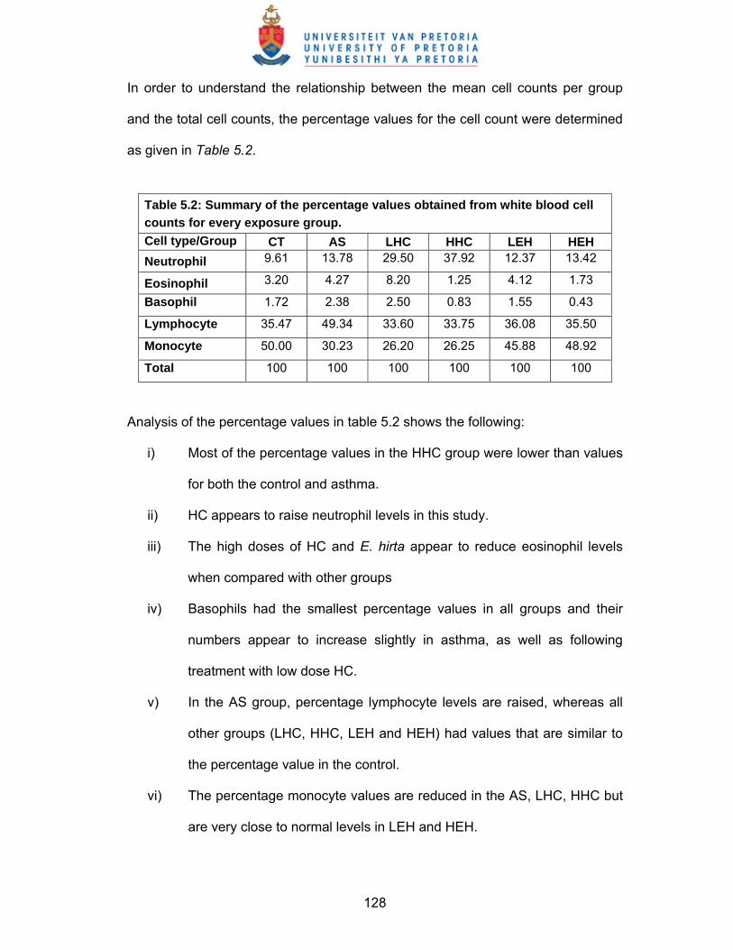
128
In order to understand the relationship between the mean cell counts per group
and the total cell counts, the percentage values for the cell count were determined
as given in Table 5.2.
Table 5.2: Summary of the percentage values obtained from white blood cell counts for every exposure group. Cell type/Group CT AS LHC HHC LEH HEH Neutrophil 9.61 13.78 29.50 37.92 12.37 13.42
Eosinophil 3.20 4.27 8.20 1.25 4.12 1.73
Basophil 1.72 2.38 2.50 0.83 1.55 0.43
Lymphocyte 35.47 49.34 33.60 33.75 36.08 35.50
Monocyte 50.00 30.23 26.20 26.25 45.88 48.92
Total 100 100 100 100 100 100
Analysis of the percentage values in table 5.2 shows the following:
i) Most of the percentage values in the HHC group were lower than values
for both the control and asthma.
ii) HC appears to raise neutrophil levels in this study.
iii) The high doses of HC and E. hirta appear to reduce eosinophil levels
when compared with other groups
iv) Basophils had the smallest percentage values in all groups and their
numbers appear to increase slightly in asthma, as well as following
treatment with low dose HC.
v) In the AS group, percentage lymphocyte levels are raised, whereas all
other groups (LHC, HHC, LEH and HEH) had values that are similar to
the percentage value in the control.
vi) The percentage monocyte values are reduced in the AS, LHC, HHC but
are very close to normal levels in LEH and HEH.

129
vii) Monocytes had the highest percentage of all the cells in the LEH and
HEH groups.
White Blood Cell Count
0
200
400
600
800
1000
1200
1400
CT AS LHC HHC LEH HEH
Experimental Groups
Cel
l num
bers
NeutrophilEosinophilBasophilLymphocyteMonocyte
Figure 5.1a: Histogram showing the mean values obtained from white
blood cell counts per group
White Blood Cell Count
0
10
20
30
40
50
60
CT AS LHC HHC LEH HEH
Experimental Groups
Perc
enta
ge c
ell n
umbe
rs
NeutrophilEosinophilBasophilLymphocyteMonocyte
Figure 5.1b: Histogram showing the percentage white blood cell counts
per group

130
5.4. Discussion
In this study, most values for cell counts in all the treated groups were relatively
lower than the values for the control group. Since higher total cell counts could
indicate the likely presence of inflammation, in this case experimental asthmatic
inflammation, it could be concluded that mice in the “asthma” group (with the
highest total leukocyte count) were actually asthmatic via inflammation.
Values for mean cell count in all other groups (except the control) were generally
lower than in the asthma group but this is not necessarily an indication that all the
treatment agents had a potential to relieve asthmatic effects. However the
percentage values (table 5.2) show that only treatment with HHC could
considerably reduce the numbers of most of the cells (except neutrophils). The
LHC and HEH (in order) treatments also appeared to have limited positive effects
at reducing asthma-induced leukocyte surge.
Basophils are said to be absent in healthy airways but present in asthmatic
airways under a variety of circumstances (Hamid et al., 2003). In this study,
basophil numbers were seen to be consistently the lowest of all cell types
evaluated in all the treatment groups. This finding is therefore in line with findings
from previous studies.
The respective effects of HC and E. hirta aqueous extract on the different
subpopulations of leukocytes are discussed in the following sections.
5.4.1. Effects of E. hirta aqueous extract
The potential use of natural products to inhibit eosinophilic inflammation in both
experimental models (Lopes-Martins et al., 2002; Rogerio et al., 2003) and human

131
diseases (Gupta et al., 1998; Takano et al., 2004) has been demonstrated. Many
natural products have been considered more useful because they are purported to
exert their positive effects with little or no side effects like some known
pharmaceutical products.
A study by (Lee et al., 2006) revealed a significantly increased percentage of
eosinophils in BALF of OVA-exposed animals. However, treatment of the animals
with DA-9201, an extract from rice, resulted in a dose-dependent reduction in
eosinophil count. Another study by Secor et al., (2005) utilized a well-established
OVA-induced model of allergic airway disease (AAD) to demonstrate the effects of
extract of bromelain in reducing CD4+ and CD8+ T cell as well as eosinophil
counts in BALF of mice.
A different study showed that the initial eosinophilia observed in OVA-exposed
mice was reduced following treatment with Vimang, an aqueous extract of the
stem bark of Mangifera indica (Sá-Nunes et al., 2006). In this case, the airway
inflammation was induced by Toxocara canis and resulted in an IL-5-dependent
systemic eosinophilia that mimicked the features observed in asthma (Faccioli et
al., 1996; Sá-Nunes et al., 2006).
In this present study, both the high and low doses of E. hirta extract were effective
in lowering the cell counts of specifically lymphocytes, eosinophils and basophils
known to be active in inflammation, in line with previous studies which showed that
the predominant inflammatory cells in asthmatic inflammation include eosinophil,
T-lymphocytes, mast cells and basophils (Denburg 1991; Kepley et al., 2001).
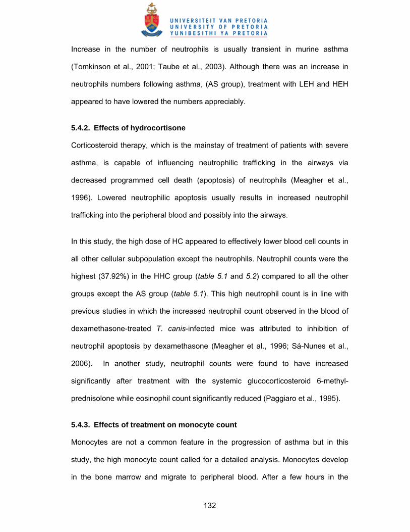
132
Increase in the number of neutrophils is usually transient in murine asthma
(Tomkinson et al., 2001; Taube et al., 2003). Although there was an increase in
neutrophils numbers following asthma, (AS group), treatment with LEH and HEH
appeared to have lowered the numbers appreciably.
5.4.2. Effects of hydrocortisone
Corticosteroid therapy, which is the mainstay of treatment of patients with severe
asthma, is capable of influencing neutrophilic trafficking in the airways via
decreased programmed cell death (apoptosis) of neutrophils (Meagher et al.,
1996). Lowered neutrophilic apoptosis usually results in increased neutrophil
trafficking into the peripheral blood and possibly into the airways.
In this study, the high dose of HC appeared to effectively lower blood cell counts in
all other cellular subpopulation except the neutrophils. Neutrophil counts were the
highest (37.92%) in the HHC group (table 5.1 and 5.2) compared to all the other
groups except the AS group (table 5.1). This high neutrophil count is in line with
previous studies in which the increased neutrophil count observed in the blood of
dexamethasone-treated T. canis-infected mice was attributed to inhibition of
neutrophil apoptosis by dexamethasone (Meagher et al., 1996; Sá-Nunes et al.,
2006). In another study, neutrophil counts were found to have increased
significantly after treatment with the systemic glucocorticosteroid 6-methyl-
prednisolone while eosinophil count significantly reduced (Paggiaro et al., 1995).
5.4.3. Effects of treatment on monocyte count
Monocytes are not a common feature in the progression of asthma but in this
study, the high monocyte count called for a detailed analysis. Monocytes develop
in the bone marrow and migrate to peripheral blood. After a few hours in the

133
bloodstream, they migrate to tissues such as spleen, liver, lung, and even bone
marrow tissue, where they mature into macrophages (Van Furth et al, 1973).
Monocytes are the main ‘effector’ cells of the immune system and play a crucial
role in defence mechanisms. These cells help other white blood cells to remove
dead or damaged tissues, destroy tumour cells and regulate immunity against
foreign substances. Therefore, a relatively high monocyte count in peripheral
blood may not necessarily be an indication of pathology but usually occurs only in
response to chronic infections. However, a very low monocyte count could
increase the risk especially of bacterial infections.
A previous study by Rinehart et al., (1974) showed that a 16μg/ml concentration of
hydrocortisone succinate (cortisone) could significantly impair random migration of
monocytes and so treatment with glucocorticoids is capable of causing a marked
reduction in monocyte count in peripheral blood. In the present study, monocyte
counts were highest in the “asthma” group followed by the control group but values
were remarkably lower in both HC groups.
In this study, a high monocyte count was observed in the asthma group compared
to other groups but this value represented only 30.23% of the total cell count in the
AS group and was much lower than the 50% value in the CT group. It is not too
clear why the control group had the highest percentage value of monocytes. In the
case of the 30.23% monocytes in the AS group, an automatic immune response to
the prolonged presence of foreign agents (e.g. OVA used in the airway challenge
procedure) in the body system of challenged mice can implicated. The remarkably
lower counts (with their relatively low percentage values) observed in both the
HHC and LHC groups could be due to the effects of cortisone treatment as

134
previously reported (Rinehart et al., 1974) and this could possibly be due to
reduced cellular participation in host defence, which is then taken over by the
administered cortisone.
The effects of medicinal plants on monocyte counts have also been reported.
Extracts of the Mediterranean desert shrub Teucrium polium L. (Labiatae) for an
example, were found to increase monocyte count in a dose-dependent manner
while lowering lymphocyte counts (Khleifat et al., 2002). Garlic extracts
administered to albino rats also caused significant increase in monocyte counts
(Iranloye, 2002). Results from this study showed that monocyte counts in the HEH
and LEH groups were lower than counts in the control and ‘asthma’ groups but
their respective percentage values were only slightly lower than in the control
group but higher than values of the asthma group .
This disparity in the monocyte numbers is not clearly understood but lower cell
counts could be indicative of the involvement of the E. hirta extract in reduction of
cellular involvement in immune processes.
5.5. Conclusion
Results from the white blood cell counts indicate that asthma was indeed induced
in the BALB/c mice. E. hirta extract was effective in lowering the number of active
inflammatory cells remarkably lymphocytes, eosinophils and basophils. Both the
high and low doses of E. hirta also caused as much percentage increase in
neutrophil numbers as in the asthmatic mice while mean monocyte counts were
also lowered by treatment with the E. hirta extract.

135
In summary, E. hirta extract caused reduction of raised leukocyte numbers
observed in asthmatic mice but in this study such effects were more pronounced
following treatment with HC.

136
CHAPTER SIX
Ultrastructural Studies of the Blood Coagulating System

137
6.1. Introduction
In vivo animal models have been used successfully during the past few years to
study diseases like asthma (Epstein, 2004a, b). The murine model in particular, is
used successfully because mice allow for a variety of in vivo immunological
applications (Bice et al., 2000). According to Epstein (2006), the allergic asthma as
observed in an experimental mouse model is a reliable, clinically relevant facsimile
of the human disease. Furthermore, antigen-induced mouse allergic asthma is a
useful model for testing novel therapeutics (Epstein, 2006) and has been used for
testing many novel agents aimed at reducing lung inflammation, mucus
hypersecretion, airway hyperresponsiveness and IgE profiles.
It appears however, that only few non-immunologic studies with the BALB/c
mouse model are available in literature. Many studies on the involvement of a wide
range of cells in the typical immunological processes of asthma have been done
but in the present study, the possible involvement of blood platelets
(thrombocytes) in asthma was explored. The ultrastructure of the components of
the coagulation process (haemostasis) especially the cellular components involved
(e.g. platelets, fibrin networks) was examined for any significant treatment effects.
Comparing the effects of HC and phytomedicines on platelet and fibrin formation
and morphology using the murine model might give researchers insight into how
these products affect the coagulation system.
Platelets have traditionally been associated with disorders of the cardiovascular
system, where they are known to be involved in the maintenance of haemostasis
as well as in the initiation of the repair process following tissue injury (Herd and
Page, 1994). Many cardiovascular diseases could be attributed to excessive

138
platelet aggregation, which in turn has a critical role in thrombus formation (Lee et
al., 1998).
A number of stimuli activate platelets resulting in the expression and/or activation
of surface receptors, secretion of vaso-active substances, their adhesion,
aggregation and thrombus formation (Lazerus et al., 2003). In conditions like
allergic asthma, platelets participate by acting as inflammatory cells, releasing
mediators, spasmogens and/or by interacting with other inflammatory cell types.
Some of these mediators are enzymes active in the coagulation cascade. Platelet
activation may be due to damage of the vessel wall or activation of the
endothelium by chemicals, cytokines and by the inflammatory processes (Camera
et al., 1999; Butenas and Mann, 2002) typically involved in conditions like allergic
asthma.
Interest in the use of phytomedicines for the treatment of diseases like asthma has
greatly increased over the past years. One plant extract used for asthma is E. hirta
(Euphorbiacea), a plant with great anti-inflammatory potential (Dickshit 1943;
Hazleton and Hellerman, 1954; Watt and Breyer-Brandwijk, 1962; Le Strange
1977; Wong 1980; Lanhers 1990, 1991; Skidmore-Roth 2001; Lindsey et al.,
2002). Although the cytotoxicity potential of E. hirta has been studied, only few
pharmacological evaluations have been carried out to ascertain the rationale
behind most of the folkloric claims of its efficacy (Johnson 1999) and not much is
known about the effects of extracts of the plant on cellular function and
morphology.
The effects of some medicinal plants or natural products on platelets and the blood
coagulation system have been documented in literature. Plant products like garlic

139
(Rahman and Billington, 2000) and tomato (Dutta-Roy et al., 2001) have been
shown to be potentially beneficial in protecting against cardiovascular diseases by
inhibiting platelet aggregation. In addition, Mekhfi et al., (2004) examined the in
vitro effects of aqueous extracts of five medicinal plants (Arbutus unedo, Urtica
dio¨ıca, Petroselinum crispum, Cistus ladaniferus and Equisetum arvense) used
for cardiovascular diseases. In their study on rat platelet aggregation induced
separately by thrombin and ADP, they found that extracts derived from all five
tested plants elicited a dose-dependent inhibitory activity on platelet aggregation.
Effects of different traditional herbal medicines have been reported. The anti-
thrombic Korean herbal medicine, Dae-Jo-Whan (DJW), consists of 11 herbs of
Rehmanniae Radix, Hominis Placenta, Testudinis Carapax, Eucommiae Cortex,
Asparagi Radix, Phellodendri Cortex, Achyranthis Radix, Liriopis Tuber, Angelicae
Sinensis Radix, Ginseng Radix, and Schizandrae Fructus. DJW was reported to
have inhibitory effects on collagen-and ADP-induced blood platelet aggregation,
thrombin-induced conversion of fibrinogen and fibrinolysis in in vitro experiments
(Min, 1997). The effects observed with total DJW extract were stronger than the
additive effects since the sum of the single contributions was lower than the effects
of the total.
In other studies, extracts of Andrographis paniculata (Acanthaceae) used as a
traditional medicine in India, China, Thailand, and Scandinavia, remarkably
decreased and inhibited platelet aggregation induced by thrombin in a
concentration-and time-dependent manner (Thisoda et al., 2006).
Tetramethylpyrazine, one of the active ingredients of the Chinese herbal medicine
Chuanxiong was reported to demonstrate unique antiplatelet characteristics -

140
selective inhibition of platelet activation, aggregation and thrombus formation
under high shear rate conditions (Li et al., 2001).
6.1.1. Aim of study
The aim of this study was to describe the possible contribution of the blood
coagulation system to the general pathology of asthma as well as investigate the
effects of treatment with HC and E. hirta on this aspect of the asthma pathology.
The effects of the plant extracts were compared with the effects of HC, a known
anti-inflammatory pharmaceutical used for the treatment of asthma. Presently, not
much is reported on the effects of E. hirta extracts on the morphology of the
coagulation system. Since the plant is known to be used for asthma, the BALB/c
mouse asthma model thus provides a medium for investigating the effects of
phytomedicines (in this case E. hirta) on the blood coagulation process in the
presence of asthma, and particularly the effects on the ultrastructure of platelets
and fibrin networks.
6.2. Materials and methods
6.2.1. Inducing and treating asthma in the BALB/c mice
Ethical clearance was obtained for the animal studies. Mice were divided into the
following groups: control mice, asthmatic mice, mice exposed to low dose
(100mg/kg) HC, mice exposed to higher dose (125mg/kg) HC, mice exposed to
physiologically comparable levels of E. hirta (0.01 ml of 62.5mg/kg plant material).
The BALB/c mice were sensitized and challenged before treatment with either
E.hirta or cortisone. Sensitization (on day 0 and day 5) of mice was via
intraperitoneal injection of a mixture of 25mg OVA (grade V; Sigma-Aldrich) and
2mg of Aluminium hydroxide [Al (OH)3] dissolved in 0.5ml of 0.9% saline solution.

141
All mice except those in the control group were sensitized. Nebulization (or
challenge) was done twice daily on days 13, 14 and 15, each for 1 hour using 1%
OVA in PBS (i.e. 1mg OVA in 100 ml PBS).
The plant material was prepared as described (in section 4.2.1.3). The stock
solution of 50mg/ml was prepared into the dose solutions of 62,5mg/kg and
25mg/kg respectively. Administration of low and high doses of the treatment
agents (E. hirta and HC) was done on days 15 -18, and again on days 22, 25, 29
and 32. Nebulization was repeated on days 34-36 before the treatment resumed
daily on days 39 - 45. Animals were terminated on day 45.
6.2.2. Preparation of fibrin clots
On day 45 during termination, 100 - 500 μl of blood per group was drawn from the
hearts of the mice in each of the groups and 11μl citrate for every 100 μl of blood,
was added. The blood samples were then centrifuged at 1000 rpm for 2 minutes to
obtain platelet rich plasma (PRP). Human thrombin (provided by The South
African National Blood Services) was used to prepare fibrin clots (Pretorius et al.,
2006). The thrombin is 20 U/ml and is made up in biological buffer containing
0.2% human serum albumin. When thrombin is added to PRP, fibrinogen is
converted to fibrin and intracellular platelet components e.g. transforming growth
factor (TGF), platelet derived growth factor (PDGF) and fibroblastic growth factor
(FGF) are released into the coagulum.
Ten micro litres (10μl) of mouse PRP was mixed with 10 μl of human thrombin.
The PRP and thrombin mix was transferred immediately with a pipette tip to a 0.2
µm millipore membrane to form the coagulum (fibrin clot) on the membrane. This
millipore membrane was then placed in a Petri dish on filter paper dampened with

142
PBS to create a humid environment at 37 ºC for 10 minutes. The millipore
membranes with the coagula were later placed in PBS and magnetically stirred for
120 minutes to remove any blood proteins trapped within the fibrin network
(Pretorius et al., 2006).
6.2.3. Preparation of washed fibrin clot for scanning electron microscopy (SEM)
Washed fibrin clots were fixed in 2.5% glutaraldehyde in the buffer DPBS
(Dulbecco's Phosphate buffered saline) at a pH of 7.4 for 1 hour. Each fibrin clot
was rinsed thrice in phosphate buffer for 5 minutes before being fixed for 1 hour
with 1% Osmium tetra oxide (OsO4.). The samples were rinsed thrice with distilled
water for 5 minutes and later dehydrated serially in 30%, 50%, 70%, 90% ethanol
as well as three times with 100% ethanol.
The Scanning Electron Microscopy (SEM) procedures were completed by critical
point drying of the material, mounting and examining the tissue with a JEOL 6000F
FEGSEM.
6.3. Results
Platelet and fibrin structure morphology of each of the mice in each group were
investigated. Morphology was found to be constant for a particular group and
homogenous for each sample and for each test field. SEM stubs were analyzed
systematically to cover the whole fibrin network area for each animal. Figures
6.1(a) and (b) show fibrin networks and platelet aggregates from control mice.
Both thick, major fibres (label A) and a thin network of minor fibres (label B) are
present in the controls.
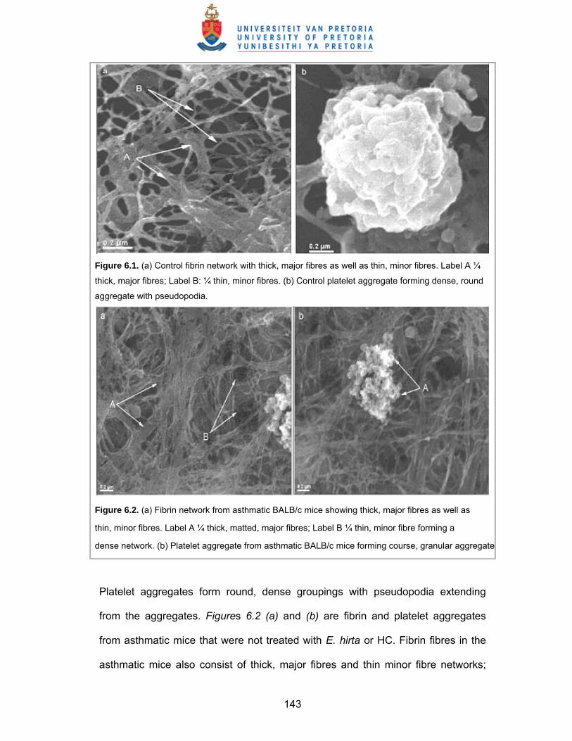
143
Figure 6.1. (a) Control fibrin network with thick, major fibres as well as thin, minor fibres. Label A ¼
thick, major fibres; Label B: ¼ thin, minor fibres. (b) Control platelet aggregate forming dense, round
aggregate with pseudopodia.
Figure 6.2. (a) Fibrin network from asthmatic BALB/c mice showing thick, major fibres as well as
thin, minor fibres. Label A ¼ thick, matted, major fibres; Label B ¼ thin, minor fibre forming a
dense network. (b) Platelet aggregate from asthmatic BALB/c mice forming course, granular aggregate
Platelet aggregates form round, dense groupings with pseudopodia extending
from the aggregates. Figures 6.2 (a) and (b) are fibrin and platelet aggregates
from asthmatic mice that were not treated with E. hirta or HC. Fibrin fibres in the
asthmatic mice also consist of thick, major fibres and thin minor fibre networks;

144
however, major fibres seem to have a matted appearance [label A of Figure 6.2
(a)] and seem to be fused longitudinally.
Minor fibre networks are more prominent [label B of Figure 6.2 (a)] and cover the
major fibre network. Figure 6.2 (b) shows an aggregation of platelets; however,
they differ from the control aggregates in that they look more granular and do not
clump together closely. Furthermore, although pseudopodia are visible [label A of
Figure 6.2 (b)], they are much smaller and less bulbous than pseudopodia of the
control animals.
Figure 6.3. (a) Fibrin network from LHC (100 mg/kg) asthmatic BALB/c mice, forming flimsy fibrin
network. Label A ¼ flimsy fibres with breakages. (b) Platelet aggregate from LHC (100 mg/kg)
asthmatic BALB/c mice. Label A ¼ granular platelet aggregate.
Figures 6.3 (a) and (b) show fibrin and platelet aggregates of the mice treated with
LHC. Fibrin fibres appeared flimsy and breakages are present and can be seen
[Figure 6.3 (a) label B]. When viewing the networks using the SEM, the fibres tend
to break just as the electron bundle moving over a region. This suggests that these
fibres are much more prone to breakages than those of the controls or even those
found in the asthmatic mice. Platelet aggregates were also granular (similar to the

145
untreated asthmatic mice) and the aggregates did not form the tight round, dense
aggregates that were seen in the controls. Rather, a loosely associated aggregate
with small pseudopodia is seen [Figure 6.3 (b) label B].
The HHC samples [Figure 6.4(a) and (b)] also showed a flimsy fibre network easily
prone to breakages and both major and minor fibres are seen. These fibre
networks have a similar appearance as the untreated asthmatic mice, where the
fine minor fibres form a much thicker covering than is found in the controls.
Platelet aggregates also did not have a tight round appearance and appeared
granular and not attached closely to each other as seen in the controls [Figure
6.4(b)]. Figure 6.5(a) and (b) shows a fibrin network and platelet aggregate from
mice that were treated with E. hirta.
Both major and minor fibres are present (Figure 6.5a labels A and B); however,
fibres were much more stable and did not break as easily as the HC groups and
the minor fibres are much less prominent than those of the asthmatic mice group
are. Figure 6.5b shows a platelet aggregate. Aggregates from E. hirta look similar
to that of the controls. The aggregate is much more condensed and round while
the pseudopodia are more bulbous and more similar to that of the controls.

146
Figure 6.4. (a) Fibrin network from HHC treated
(125 mg/kg) asthmatic BALB/c mice network.
Label A ¼ Flimsy fibre network. (b) Platelet
aggregate from high dose HC-treated (125
mg/kg) asthmatic BALB/c mice forming granular
aggregate.
Figure 6.5. (a) Fibrin network from asthmatic
BALB/c mice treated with E. hirta (0.01 ml of
62.5mg/kg plant material) showing fibrin network
with thick, major fibres as well as thin, minor fibres.
Label A ¼ thick, major fibres; Label B ¼ thin, minor
fibres. (b) Round aggregate with pseudopodia.
Label A ¼ pseudopodia.

147
6.4. Discussion
Although platelets have traditionally been associated with disorders of the
cardiovascular system, they also play an important physiological role in allergic
processes and immunological mechanisms. Platelets play an important and
fundamental part in asthma, as inflammatory processes, typically involved in
asthma, activate them. Furthermore, platelet-activating factor (PAF) as well as
platelet factor 4 (PF4) and also thrombin itself, fibrinogen, fibrin, are all known to be
involved in asthma. Importantly, platelets contribute to the adhesion of eosinophils
to inflamed endothelium of patients with allergic asthma. Platelet depletion is also
known to reduce PAF, suggesting that PAF plays a central role in the processes
by which platelets facilitate the induction of eosinophil accumulation, which is
central in asthma.
In addition, PAF is involved in increased vascular permeability and platelets have
been shown to be in contact with the vasculature of the bronchi of patients with
asthma and in this way, platelets evoke contraction of smooth muscles of the
respiratory passages that directly leads to an asthma attack. Fibrin and platelets
are therefore two important factors in the disease. Fibrinogen itself is also widely
recognised as a marker for systemic inflammation as it is considered an acute
phase protein. Pithcford et al., (2004) mentioned that there is evidence of platelet
recruitment to the lungs of asthmatics after allergen exposure, suggesting that
platelets participate in various aspects of asthma. Morley et al., (1984) has earlier
suggested that platelet activation may contribute to airway remodelling in asthma.
Furthermore, the association between thrombin, fibrin and asthma seems to be the
following:

148
• Thrombin is known to increase airway smooth muscle contraction ex vivo
(Panettieri et al., 1995).
• Increased thrombin generation occurs in the airway of patients with asthma
(Gabazza et al., 1999). Thrombin may play a role in the pathogenesis of
airway remodelling.
• Human platelets can produce PAF upon thrombin stimulation in the lungs
(Touqui et al., 1985).
• Fibrinogen can be produced by lung epithelia because of inflammatory
stimulus (Lawrence and Simpson-Haidaris, 2004).
• Fibrin degradation products have also been found to increase pulmonary
vascular smooth muscle contraction (Kern et al., 1986).
• Airway fibrin deposition occurs in inflammatory disorders of the lung, and it
is known that fibrin inhibits surfactant function (Wagers et al., 2003).
• Fibrin is typically formed at sites of vascular damage (Touqui et al., 1985).
• Extra-vascular thrombin, fibrinogen, and fibrin have been found in the
sputum of patients with asthma (Banach-Wawrzenczyk et al., 2000;
Pizzichini et al., 1996; Wagers et al., 2004).
• Wagner and co-workers in 2004 hypothesized that airway
hyperresponsiveness seen in asthma is largely the result of decreased
stability of airways and subsequent airway closure secondary to the
formation of fibrin on the distal airway surface. According to the authors the
coagulation system and fibrinolytic system proteins is associated with the
pathogenesis of airway hyperresponsiveness in asthma.
• There is decrease in plasminogen activator (PA) activity in asthma (Wagers
et al., 2004).

149
• Activity of PAIact (plasminogen activator inhibitor) is increased in
homogenates of lung tissue of mice with allergic airway inflammation,
thereby potentially promoting the accumulation of fibrin by suppressing
fibrinolysis (Wagers et al., 2004).
In control BALB/c mice we find major and minor fibrin networks (Figure 6.1a); this
is similar to previous findings in humans (Pretorius et al., 2006). In addition, the
morphology of platelet aggregates appears as a collection of platelets that has a
dense, round shape. This is also similar to human platelet aggregates (Pretorius
et al., 2006). However, in the asthmatic mice the minor fibres seem to be more
dense and covering the major fibres (as seen in Figure 6.2a). This might be the
reason why in asthma, an accumulation of fibrin is seen in airways (Wagers et al.,
2004); perhaps because the fibrin mass is more dense and that the fibrin networks
are not disaggregated so quickly (fibrinolysis) and stay in the airways for longer
periods, forming fibrin plugs. It is known that surfactant function is inhibited in
asthma (Wagers et al., 2003), possibly because of the denser fibrin network. In
addition, degradation products of fibrin have been found to increase smooth
muscle contraction. This might be because the fine minor fibrin network inhibits
the surfactant to function optimally and interferes with smooth muscle function.
It is known that glucocorticosteroids are the most useful class of drugs employed
in the treatment of patients with allergic asthma (Lantero et al., 1997; Pretorius,
2005). There is also increasing evidence that fibrin(ogen) physiology is affected
by glucocorticoids. After fibrin is produced via the thrombin (intrinsic and extrinsic)
pathway, it undergoes fibrinolysis. This process is under the control of
plasminogen activators (PAs), which are serine proteases that convert the

150
proenzyme plasminogen to active plasmin, a broad-spectrum proteolytic enzyme
that readily degrades fibrin as well as extracellular matrix glycoproteins including
laminin, vitronectin, fibronectin and proteoglycans (Bator et al., 1998).
Platelets also seem to be affected by glucocorticoid treatment (Tutluoglu et al.,
2005). Platelets have the capacity to release mediators with potent inflammatory
or anaphylactic properties; these mediators include PF4 and beta-thromboglobulin
(BTG) (Tutluoglu et al., 2005). BTGs are also chemokines that play an important
role in mediating cell recruitment and activation necessary for inflammation and
the repair of tissue damage. Plasma levels of PF4 and BTG also show changes in
chronic inflammatory diseases such as asthma; and Tutluoglu and co-workers
(2005) found that plasma levels of PF4 among patients with an asthma attack were
significantly higher than those of controls and a further increase in plasma PF4
levels was detected after steroid therapy. From these results it appears that both
the low and high HC dosages produce flimsy fibrin networks that break easily and
that are not as stable as was found in the control fibrin networks (Figure 6.3a and
6.4a). In addition, the platelet aggregates do not have the typical round, compact
shape; rather the platelet aggregates are widely spread, granular, and not tightly
associated with each other (Figure 6.3b and 6.4b); also, aggregates appear more
like those found in the asthmatic mouse.
An interesting observation was that the extract of E. hirta, did not cause the fibrin
networks to be as flimsy and fragile (Figure 6.5a). However, the fine minor fibres
seem to be more prominent than in the controls and more similar to those found in
the asthmatic mice. Platelet aggregates showed the same morphology as those of
the control mice, without the widely spread granular appearance found in HC
151
treated mice (Figure 6.3b and 6.4b). However, further studies need to be done to
determine the effect of the plant on lymphocytic lung infiltrates; Ag-specific
production of IL-4 and IL-5 from spleen and lung cells in vitro, elevated levels of
IgG1 as well as expression of Th2 cytokine RNA in lungs.
6.5. Conclusion
Asthma is a very complex condition, with many physiological factors playing a role
in its presentation. However, it appears that platelets and products of the
coagulation cascade form an intricate and important part of asthma and not only
affect the presentation of the condition itself but also interact with the typical
choice of treatment, namely glucocorticoid therapy. The question that arises is
how the platelet activation process and the coagulation cascade contribute to
asthma in the presence of other pharmaceutical products or phytomedicine.
It is known from previous research that the E. hirta is widely used for the treatment
of asthma probably because it contains chemical ingredients that have anti-
inflammatory properties. However, very little information is available of the effect of
the plant on cellular systems of the body and the exact mechanism through which
it plays a role in the treatment of asthma. The current research seems to indicate
that treatment with E. hirta does not make fibrin fibres of mice as fragile as seen in
treatment with HC; also, treatment with E. hirta did not change the integrity and
morphology of the platelets as observed for treatment with HC.
This work focused on a small aspect of the coagulation process and now leaves
more questions regarding the effect of HC on the coagulation process as well as
the effects and the exact mechanisms of E. hirta action on asthma in general and
on other cellular systems of the body. Further ultrastructural studies are

152
suggested especially involving the use of transmission electron microscopy to
examine particularly platelet morphology.
We conclude that E. hirta does not affect the fragility of mouse fibrin and that it
prevents the minor fibres from forming a dense netlike layer over the major fibres,
as is seen in untreated asthmatic mice. This ultrastructural morphology might give
a better insight into asthma and possible new treatment regimes for it.

153
CHAPTER SEVEN
Inflammatory Cell Infiltration and Structural Changes in the Airways

154
7.1. Introduction
Asthmatic inflammation usually affects the entire respiratory tract, from the nose
down to the most distal (and therefore much smaller) respiratory passages as well
as the alveolar tissue (Martin, 2002; Togias, 2003), causing a wide range of
structural changes. Some of the common changes include thickening of the
reticular basement membrane (Roche et al., 1989; Wilson and Li, 1997),
thickening of the smooth muscle layer as well as infiltration of different cell types.
Thickening of the basement membrane could be due to deposition of collagen
types I, III and V (Roche et al., 1989).
Cell infiltration into the airway has also been considered a remarkable feature of
asthma that causes structural changes. Some studies have indicated that the
predominant cells in bronchial asthma were eosinophils and lymphocytes (Hoshi et
al., 1995) and yet other studies have shown that CD4 T-lymphocytes, eosinophils
and mast cells were the predominant cells in asthma (Saetta and Turato, 2001).
Other studies reported the presence of eosinophils, neutrophils and macrophages
in lung tissues (McKay et al., 2004). Infiltration of T-cells, macrophages and
eosinophils into the proximal and distal lung tissues has also been reported in rare
cases of sudden asthma death (dying within one hour of onset of symptoms) (Faul
et al., 1997) and in soybean dust induced asthma (Synek et al., 1996).
The distribution pattern of eosinophilic infiltration was found to differ between the
central and peripheral airways, suggesting important functional consequences
(Saetta and Turato, 2001). A comprehensive review by Epstein (2006) stated that
peril-bronchial and peril-vascular infiltrates were evident in all asthma models but
the cellular composition, severity of overall inflammation and location of infiltrates

155
was protocol-dependent. The study also showed that although the protocol-
dependent differences in asthma histopathology are poorly understood, and could
provide new insights into the heterogeneity of disease.
Many previous studies have shown that the entire length of the airways is involved
in asthma (Huber and Koessler 1922; Dunnill 1960; Dunnill et al., 1969; Kuwano et
al., 1993). Using surgically resected lung tissue (Taha et al., 1999), trans-bronchial
biopsies (Kraft et al., 1999) and autopsy lung specimens (Christodoulopoulos et
al., 2000), more severe inflammatory and structural changes were observed in
small airways (Faul et al., 1997) and in peripheral airways and lung parenchyma
(Kraft et al., 1996) of ‘asthmatic’ persons when compared to the larger airways.
Regional variations were also observed in inflammatory cell distribution within
‘asthmatic’ airways (Haley et al., 1998).
The types of inflammatory cells present in the airway walls and spaces could
indicate the pattern of asthmatic progression. The present study examines the
effects of HC and extracts of the medicinal herb E. hirta on the structural changes
and cellular disposition and characteristics in especially the distal respiratory
passages using the Balb/c mouse asthma model.
7.2. Mixed inflammatory infiltrate in the lung parenchyma
A mixture of inflammatory cells, notably eosinophils, lymphocytes, mast cells,
basophils, macrophages, fibroblasts and platelets usually infiltrates the
subepithelial layers of respiratory passages. Results from previous studies are
given in the following sections.

156
7.2.1. Eosinophils
Tissue eosinophilia is a characteristic of asthma but it is not necessarily specific to
asthma (Lacoste et al., 1993). The classic descriptions of asthma pathology
include the presence of eosinophils (and neutrophils) in both the lamina propria
and the airway lumen of patients with asthma (Kaliner et al., 1976). Biopsy
specimens from ‘mild asthma’ patients have confirmed the presence of eosinophils
in the mucosa, often beneath the basement membrane as well as in the epithelium
(Djukanovic et al., 1990).
Eosinophils also appear to be important cells of airway remodelling as they
release growth factors (Ohno et al 1992, Walz et al 1993), elastase (Lungarella et
al., 1992) and metalloproteases (Ohno et al., 1997) that are all involved in the
process of tissue remodelling and pulmonary fibrosis (Schlick et al., 1993), mostly
through fibroblast stimulation (Pincus et al., 1987). Recruited eosinophils become
activated during segmental allergen challenge and then release products that
cause contraction of the human bronchial smooth muscle and increased vascular
permeability (Collins et al., 1993; Rabe et al., 1994; Shaver et al 1997).
7.2.2. Lymphocytes
T-cells possibly play a role in controlling the chronic inflammation of allergic and
non-allergic asthma via the release of Th2-cytokines (Ying et al., 1995, Robinson
et al., 1992; Del Prete et al., 1993). After allergen challenge, the population of
activated T-cells appears to increase in ‘asthmatic’ subjects (Robinson et al.,
1993; Bentley et al., 1993) with only a few B-cells present in the bronchi.

157
7.2.3. Mast cells
Mast cells are found in the bronchi of normal subjects and “asthmatics” alike
(Pesci et al., 1993; Koshino et al., 1995) that are often degranulated in the airways
of “asthmatics” in both their stable phase and after allergen challenge (Laitinen
1985; Beasley et al., 1989). Mast cells appear to be critical “trigger” cells during
episodes of acute asthma (Broide et al., 1991). These cells may be involved in
airway remodelling because they appear to play an important role in pulmonary
fibrosis (Jordana et al., 1993; Chanez et al., 1993) especially because they are
potential sources of products that stimulate migration and proliferation of
fibroblasts (Ruoss et al., 1991; Nagata et al., 1992). Research has also shown that
mast cell lines can release components of basement membranes such as laminin
and collagen IV (Thompson et al., 1991) as well as angiogenic growth factors
(Meininger and Zetter, 1992). Mast cell products have also been reported to have
anti-inflammatory properties (Green et al., 1993; Tyrell et al., 1995).
7.2.4. Basophils
The role of basophils in the pathogenesis of asthma appears poorly defined
(Denburg, 1998). There has been some controversy over the extent to which
basophils participate in the inflammatory processes of asthma (Walls et al., 2001)
even though these cells are known to be responsible for the mediator release
observed during the asthmatic response (Lichtenstein and Bochner, 1991). After
anti-IgE challenge, most human blood basophils release histamine and other
inflammatory mediators without degranulating (Youssef et al., 2007). Though often
regarded as circulating counterparts of the mast cell, many basophils infiltrate the
tissues and fluids that bathe mucosal surfaces (Walls, 2001).

158
7.2.5. Macrophages
A study by Calhoun et al (1992) showed that activation of alveolar macrophages
occurred immediately after antigen challenge, indicating a major role for
macrophages in asthma. Alveolar macrophages have been reported to exhibit
enhanced capacity to release pro-inflammatory cytokines (John et al., 1998). In
addition, macrophage numbers increased in the airways and may be activated by
allergens through low-affinity IgE receptors to release inflammatory mediators and
cytokines that amplify the inflammatory response (Peters-Golden, 2004).
Phagocytic cells such as polymorphonuclear leukocytes and macrophages,
respond to a variety of membrane stimulants via the production and extracellular
release of a number of reactive oxygen reduction products (Santos et al., 2004).
Alveolar macrophages may also contribute to inflammation via the release of
reactive oxygen species such as superoxide (SO) anion, hydroxyl radicals,
hydrogen peroxide, singlet oxygen and hypohalous acids, which may cause
cellular injury and alterations in airway functions (Freeman and Crapo, 1982;
Barnes 1990). These reactive groups may be involved in the regulation of airway
remodelling through secretion of growth-promoting factors for fibroblasts,
cytokines as well as growth factors possibly involved in fibrosis (Kovacs and
DiPietro, 1994).
7.2.6. Polymorphonuclear neutrophils
The role of neutrophils in stable asthma remains unclear but neutrophil numbers
were noted to have increased after an allergen challenge in the airways in the
different forms of asthma during the late-phase reaction (Koh et al., 1993;
Montefort et al., 1994). In one study, both bronchoalveolar lavage (BAL) and lung
159
tissue digestion were used to determine rat lung inflammatory cell contents
following a 4-hour exposure to 2-ppm ozone. Immediately following the exposure,
the neutrophil content of the lung tissue significantly increased and by three hours
post-exposure, reached a value fourfold higher than air-exposed controls.
Although lavage-recovered neutrophils were elevated 24 hour afterwards, tissue
neutrophil numbers had returned to control values. This transient elevation of
tissue neutrophils directly correlated with an elevation and subsequent decline of
airway hyperresponsiveness (DeLorme et al., 2002).
7.2.7. Fibroblasts
Fibroblasts are often found in connective tissue and are responsible for the
production of collagen, reticular and elastic fibres as well as for the synthesis of
proteoglycans and glycoproteins of the amorphous intercellular substance
(Sheppard and Harrison, 1992). Although these cells are regarded as fixed cells of
connective tissue origin, they retain the capacity for growth and proliferation and
are pluripotent cells (precursors for various other cell types), including smooth
muscle cells (Gizycki et al., 1997).
7.2.8. Myofibroblasts
Myofibroblasts may potentially contribute to the regulation of bronchial
inflammation via the release of cytokines (Zhang et al., 1996) and to tissue
remodelling via the release of extracellular matrix (ECM) components such as
elastin, fibronectin and laminin (Leslie et al., 1992). During the late-phase reaction
after allergen challenge, myofibroblasts increase from the normal of 2% of cells to
approximately 15% within a day (Gizycki et al., 1997). In asthma, myofibroblasts
are increased in numbers beneath the reticular basement membrane and there is

160
an association between their numbers and the thickness of reticular basement
membrane (Brewster et al., 1993; Gabbrielli et al., 1993).
7.2.9. Dendritic cells
Dendritic cells are receiving increasing scientific and clinical interest due to their
key role in anti-cancer host responses and potential use as biological adjuvants in
tumour vaccines, as well as their involvement in the immunobiology of tolerance
and autoimmunity (Satthaporn and Eremin, 2001). Dendritic cells are a major
source of many cytokines, namely, interferon-alpha (IFN-a), IL-1, IL-6, IL-7, IL-12
and IL-15 and also produce macrophage inflammatory protein (MIP1g), all of
which are important in the elicitation of a primary immune response (Macatonia et
al., 1995; Cella et al., 1996; Mohamadzadeh et al., 1996).
In humans, there is a network of dendritic cells within the epithelium of the
conducting airways (Jahnsen et al., 2001). These cells may be critically important
to the induction of immune responses within the airways as they are specialized in
antigen processing and presentation. In animals, the dendritic cell population in
the airway epithelium is renewed every 48 to 72 h (Hance, 1993) and it appears
that the number of dendritic cells can be altered after exposure to both topical and
systemic corticosteroids (Moser et al., 1995).
Dendritic cell numbers are greater in the airways of asthmatics compared with
those of control subjects (Tunon-De-Lara et al., 1993; Moller et al., 1996). Their
role in asthma remains under investigation. Mouse models and culture systems of
human cells have been used to investigate the implications of the dendritic cell-
epithelial interaction for the pathogenesis of asthma (Lambrecht and Hammad,
2003).

161
7.2.10. Aim of study
The aim of this study was to determine the presence of inflammatory cells in the
airway walls and spaces in especially the distal respiratory structures using the
BALB/c mouse asthma model, and to compare the findings in the control (CT),
asthmatic (AS), HC-treated and E. hirta-treated specimens. Differences in cellular
characteristics as well as structural changes in the airways were also analyzed
and compared. Although the amount of inflammatory cells in the airway walls was
not determined, their possible relationship to the findings in peripheral blood cell
counts in chapter five was analyzed.
7.3. Materials and methods
7.3.1. Materials
7.3.1.1. BALB/c Mice
A total of sixty (60) six-week-old male BALB/c mice (mean weight 20g) were used
in this study.
7.3.1.2. Hydrocortisone (HC)
HC was used in this study as a pharmaceutical control for the extracts of E. hirta
as used iin the cell culture study. One hundred milligrams (100 mg) of the sterile
powder (Brand name Solu-Cortef®) which contains HC sodium succinate as the
active ingredient was dissolved in 2 ml of bacteriostatic injection water, giving a
concentration of 50mg/ml. In this study, a high dose (125mg/kg) and a low dose
(100mg/kg) of the 50mg/ml HC preparation were used. Fresh solution was
prepared for each injection.

162
7.3.1.3. Euphorbia hirta
Only the aqueous plant extract of E. hirta was used for the animal studies. The
plant material was collected in the Gezina region of Pretoria South Africa. A
herbarium specimen was prepared and compared to an authentic specimen in the
HGJW Sweikerdt herbarium at the University of Pretoria. The aboveground parts
of the plant were allowed to dry at room temperature for one week in the Cell
Biology laboratory of the Department of Anatomy, University of Pretoria, and the
material was grounded into a fine powder. Shortly after, 50 grams of the sample
was extracted in 500ml of double distilled water after which it was filtered, and
dried on a rotary evaporator at 40 °C. A stock aqueous solution of 50mg/ml plant
extract was prepared and stored in a fridge until used.
The doses of 62,5mg/kg and 25mg/kg were prepared from the stock solution and
administered orally to the mice. This implied that each animal (average weight
20g) received 0.01 ml of plant material. This dose was decided upon after studying
literature that mentioned physiological doses suggested by herbalists. Typically, a
teaspoon of the herb is added to a teacup volume of water and allowed to simmer
for 20 minutes (Lindsey et al., 2002). Alternatively, an extract of the plant could be
prepared and the recommended adult dose range of the fluid extract is 0.2-0.3ml,
taken three times daily and of the infusion, 120-300mg three times daily
(Skidmore-Roth, 2001).
7.3.1.4. Reagents and equipment
The reagents and equipments used for this study include ovalbumin (OVA)
purchased from Sigma-Aldrich Co., Phosphate Buffered Saline (PBS), alum,
formaldehyde, assorted clearing agents, glutaraldehyde, epoxy resin, 1% OsO4,

163
cacodylate buffer, Agar 100 Resin, DPSS, KLAVA ultrasonic nebulizer, oral-
pharyngeal canula, injection syringes and needles, light microscope (Olympus
Japan binocular BH2 (BHTU) model) and transmission electron of microscope
(Multi-purpose TEM (Philips 301), 100kV, eucentric goniometer stage, heating
holder).
7.3.2. Light microscopy
For light microscopy, samples from the lungs were processed using basic
histological techniques. Tissues were fixed in 10% formaldehyde for 24 hours and
later dehydrated in increasing grades of ethanol, cleared in xylene and infiltrated
with molten paraffin wax. Thereafter, the specimens were embedded in paraffin
wax to form tissue blocks and five-micron thick sections were cut from the blocks
and stained with Haematoxylin and Eosin (H and E) dyes. The stained sections
were later observed with the light microscope (Olympus Japan binocular BH2
(BHTU) model).
7.3.3. Transmission electron microscopy (TEM)
All specimens were taken to the central Laboratory for Microscopy and
Microanalysis at the University of Pretoria. Intrapulmonary tissues were cut into
very small sizes and fixed in glutaraldehyde for one hour. The samples were then
put in buffer (0.075M sodium potassium phosphate at pH = 7.4) for 15 minutes and
later washed twice in same buffer. Thereafter, samples were placed in a
secondary fixative (1% osmium tetra-oxide solution: OsO4) for one hour. Following
OsO4 fixation, the tissues were rinsed again thrice in the buffer as described
earlier and later dehydrated through 30%, 50%, 70%, 90%, and then three
changes of 100% ethanol. The dehydrated samples were later embedded in resin

164
and ultra-thin sections prepared and stained with uranyl acetate for 15 minutes
followed by 10 minutes staining with lead citrate. Later the stained specimens
were allowed to dry for a few minutes before examination under the multi-purpose
transmission electron microscope (Philips 301 100kV, eucentric goniometer stage,
heating holder). Ultra-photomicrographs of the internal morphology of the
intrapulmonary respiratory tract were also taken.
7.4. Results 7.4.1. Light microscopy
At an objective magnification of X10, the following photomicrographs were taken
for the different study groups to show the possible effects of the two treatment
agents on alveolar sizes, alveolar wall thickness, as well as the presence of other
features as the presence of smooth muscle masses in the terminal bronchioles.
Results from the light microscopy studies show that the CT specimens had
relatively thin and continuous (interconnected) alveolar walls with the alveoli being
of regular shapes. In contrast, alveoli appear irregularly shaped in the asthma
group, their walls very thick and discontinuous, with smooth muscle masses seen
in the walls of distal airways. These findings appear to confirm the presence of
asthma induced in mice of the AS group.
In the case of specimens of mice treated with the HHC, the alveoli were relatively
compact, relatively small and of irregular shapes. Alveolar walls were also
discontinuous and moderately thick. Smooth muscle thickenings of terminal
airways were also seen. The LHC treatment specimens showed alveoli that were
relatively thick and irregular in shape with walls that were discontinuous at some
regions. Smooth muscle masses were also seen in the walls of distal airways.

165
These findings tend to indicate that at the level of light microscopy, both doses of
HC did not ameliorate the effects of asthma on the lung microstructure in
specimens of this group in terms of the characteristics earlier mentioned.
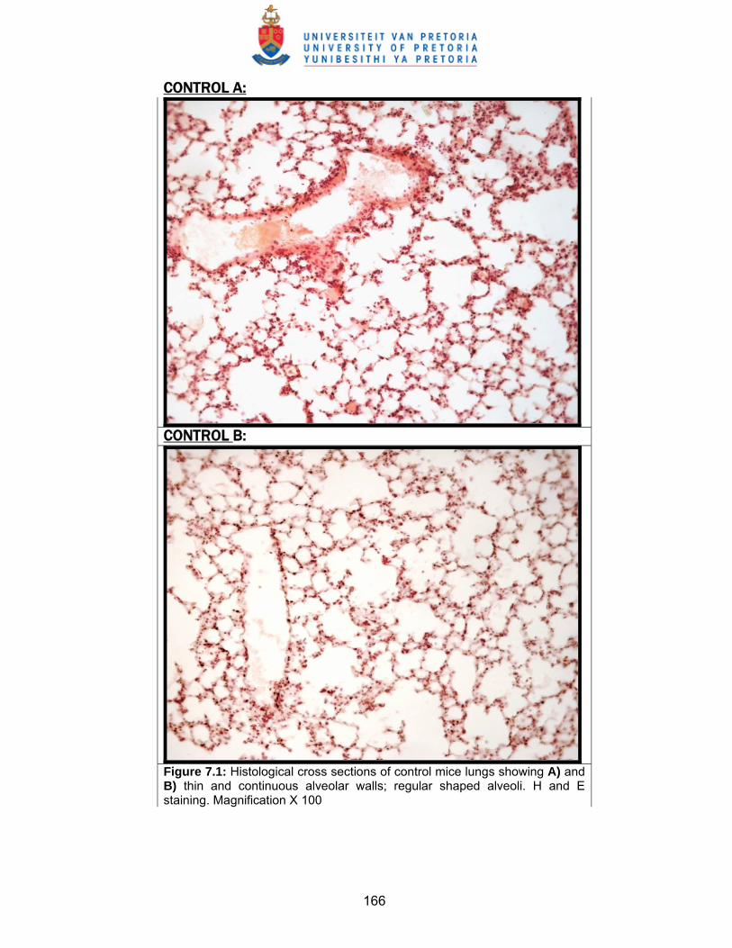
166
CONTROL A:
CONTROL B:
Figure 7.1: Histological cross sections of control mice lungs showing A) and B) thin and continuous alveolar walls; regular shaped alveoli. H and E staining. Magnification X 100

167
ASTHMA A:
ASTHMA B:
Figure 7.2: Histological cross sections of asthma mice lungs showing A) thick and discontinuous alveolar walls and B) thick smooth muscle mass in walls of distal airways indicated with arrows. H and E staining. Magnification X 100

168
HHC A:
HHC B:
Figure 7.3: Histological cross sections of the lungs of mice treated with HHC showing A) and B) alveoli that are relatively small but numerous; alveolar walls moderately thick, alveoli compact and irregular in shape; alveolar walls discontinuous; smooth muscle thickenings of terminal airways seen, shown with arrows. H and E staining. Magnification X 100.
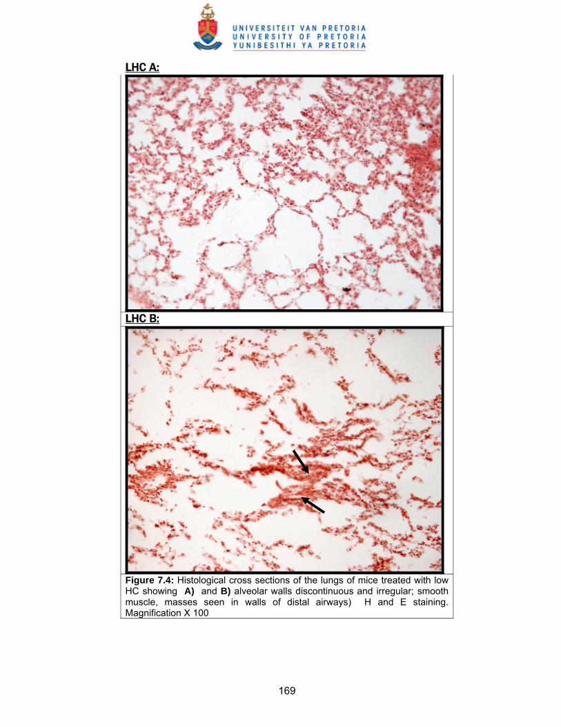
169
LHC A:
LHC B:
Figure 7.4: Histological cross sections of the lungs of mice treated with low HC showing A) and B) alveolar walls discontinuous and irregular; smooth muscle, masses seen in walls of distal airways) H and E staining. Magnification X 100

170
HEH A:
HEH B:
Figure 7.5: Histological cross sections of the lungs of mice treated with high EH showing A) and B) thick alveolar walls; alveoli very compact and irregular in shape; alveolar walls discontinuous (shown with arrows in B). H and E staining. Magnification X 100

171
LEH A:
LEH B:
Figure 7.6: Histological cross sections of the lungs of mice treated with low EH showing A) thin alveolar walls; less compact and more patent alveoli; alveolar walls are only partially discontinuous (shown with arrows) and B) thinner alveolar walls; alveoli less compact and irregular in shape; alveolar walls only partially discontinuous (arrowed). H and E staining. Magnification X 100
172
The HEH treatment specimens showed compact, irregularly shaped alveoli having
thick and discontinuous walls. In contrast, the low E. hirta specimens had thinner
alveolar walls, less compact and more patent alveoli, with the alveolar walls only
partially discontinuous. The features seen in the LEH specimens resemble those
of the CT group specimens and therefore suggest that treatment with the LEH
appears effective in ameliorating any histological alterations observed in the lung
parenchyma in the presence of asthmatic conditions.
7.4.2. Transmission electron microscopy (TEM) analysis
Transmission electron micrographs revealed a variety of intra cellular and
extracellular structures shown in the labelled figures below.
7.4.2.1. Cell types
Results from the photomicrographs below show that different cell types are
present in distal respiratory passages and in lung parenchyma as follows:
Fibroblasts: were scanty in the CT specimens as well as in the low dose
treatment specimens. However, these cells were as many in the “asthmatic” (AS)
specimens as were in the high dose treatment groups.
Lymphocytes: were few in the specimens of the CT and LHC groups but were
relatively scanty in specimens of the AS, HHC and HEH groups.
Monocytes: were absent in specimens from animals treated with the LHC.
Neutrophils: were generally not prominent but only very few were seen in CT
group specimens.

173
Macrophages: were numerous in specimens of the asthmatic and LHC groups but
only a few were present in specimens of the CT, HHC as well as in the HEH and
LEH specimens.
Plasma cells: were scanty or absent in specimens of all groups except those from
the low dose E.hirta treatment group. These cells were characterized by the
elaborate presence of rough endoplasmic reticulum in their cytoplasm.
7.4.2.2. Other structures
Collagen fibres: were scanty in specimens from the CT and LHC groups. The
remaining groups had varying amounts of collagen fibres.
Lamellar bodies: are usually associated with type 2 pneumocytes (great alveolar
cells) and in this study, lamellar bodies were present in specimens of the low HC
and low E. hirta doses.
Thick alveolar walls: were more prominent in specimens of the LHC group and
less thick in the CT specimens.
Mitochondria: were prominent in specimens of the asthma and HHC groups.
Striations: were observed as special structures in some of the asthma group
specimens, much similar to those observed in a typical striated muscle.
Mucous-secreting structures: were prominent in specimens of the LHC and LEH
groups.
Smooth muscles: were relatively well developed in bronchiolar specimens of the
asthma group compared to other treatment groups.

174
Filopodia: were seen in fibroblasts as cytoplasmic extensions only in specimens
of the LHC and the HEH groups.

175
CONTROL
Figure 7.7: TEM micrograph of lung tissue from control mice showing A = Fibroblast (F), scanty collagen fibres [x7500]; B = Macrophage and lymphocytes in interstitium [x9800]; C = Collagen fibres [x13000]; D = Neutrophil, macrophage [x7500]; E = Fibroblast, neutrophils scanty fibres x5900; F = Lymphocytes and macrophage in lung interstitium [x4300].
Macrophage
Fibroblast
Neutrophil
Lymphocytes
Fibroblast
Macrophage
Lymphocytes
C
E F
D
BA

176
ASTHMA
Figure 7.8: TEM micrograph of lung tissue from asthma mice showing A = Types 1 and 2 (T1; T2); thick walls [x3600]; B = Fibroblasts (F), many collagen fibres, smooth muscle [x5900]; C = Thick walls (TW), Type 1 cells (T1); macrophage (Ma) [x4300]; D = Macrophage (Ma), collagen fibres, smooth muscle [x9800]; E = Large Lymphocytes (LL); fibres (F), mitochondria (Mi) [x18000]; F = Many collagen fibres (CF), fibroblast, striations [x5900].
T2
T1
T1
T2
A
F
B
T1
Ma
TW
T1
C
Ma
Collagen Fibres
Smooth muscle
D
L L
CF Mi
E
Dividing Fibroblast
CF
Striations
F

177
LHC
Figure 7.9: TEM micrograph of lung tissue from asthma mice treated with LHC showing A = Monocyte (Mo), lymphocytes (L) [X5900]; B = Fibroblast, collagen fibres (CF) [X7500]; C = Mucous secreting structures [X4300]; D = Thick walls, lamellar bodies LB, Types 1, 2 cells [X3600]; E = Lymphocyte with vesicles (LV), Types 1, 2 cells [X2800]; F = Thick walls, macrophages (M), T2 cells [x3600].
L
L
L
L
L
L
A
Fibroblast with filopodia
CF
B
Mucous gland
Mucus cell
Bronchiolar epithelial cell nuclei
C
LB
T2 T1
Thick walls
D
T1 LV
T1
T2
E
T2
M
M SM F

178
HHC
Figure 7.10: TEM micrograph of lung tissue from asthma mice treated with HHC showing A = Fibroblast and scanty collagen fibres [x9800], B = Fibroblast and scanty collagen fibres [x9800], C = Fibroblasts, few fibres, many mitochondria [x9800], D = Fibroblast (F), relatively more fibres (CF) [x9800], E = Unmyelinated nerves and collagen fibres [x13000], F = Very scanty collagen fibres, red blood cells [x4300]
A B
Fibroblast
C D
E F

179
HEH
Figure 7.11: TEM micrograph of lung tissue from asthma mice treated with HEH showing A = Highly fibrous lung parenchyma, fibroblast [x5900]; B = Highly fibrous lung parenchyma, fibroblast [X5900]; C = Fibroblasts (F) and fibres [x7500]; D = Smooth muscle (SM) cell, many fibres [x9800]
SM
F
F
BA
DCF
F F SM

180
LEH
Figure 7.12: TEM micrograph of lung tissue from asthma mice treated with LEH showing A = Mucus gland (Mu) and many fibres [x3600]; B = Fibroblast (F), many fibres [x13000]; C = Alveolar walls, Type 1 (T1) cells, lamellar bodies [x4300]; D = Bronchioles: Plasma cell (P), lymphocyte (L), Rough ER [x4300]; E = Bronchial seromucous gland and cell [x7500]; F = Bronchial artery containing red blood cells [x5900]
F Mu
BA
C
T1
Lamellar bodies
Lamellar bodies
P
P
P
L
L L
RER
D
Mucus cell nucleus
E Endothelial cell nuclei
Red blood cells
F

181
7.5. Discussion
The airway wall consists of epithelial, interstitial, nervous and immunological
components (Plopper 1996, Pinkerton 1997, Evans et al., 1999) all of which
actively interact with one another (Kips et al., 2003). The common structural
changes that characterize the asthmatic condition include an increase in epithelial
goblet cells, mucous gland hyperplasia, subepithelial fibrosis (with deposition of
collagen and fibronectin), smooth muscle hypertrophy and/or hyperplasia,
formation of new vessels (Ordonez et al., 2001; Kips et al., 2003), among others.
Cell migration into the airway lumen and parenchyma is a common and very
important feature of asthma and a wide range of cells are known to be involved. In
prior experiments discussed in chapter five of this study, results from blood
parameters have confirmed the presence of asthmatic conditions in the
experimental mice used in this study.
7.5.1. Cellular structures
Fibroblasts
Results obtained from this study show that fibroblasts were scanty in both the LHC
and LEH specimens respectively, almost as much as was seen in the CT
specimens. This finding appears to indicate that treatment with the LHC and LEH
could lower fibroblast proliferation. On the other hand, the HHC and HEH
specimens had more fibroblasts but only as much as in the ‘asthmatic’ specimens.
This finding may suggest that the high dose treatment with both HC and E. hirta
has minimal or no effect on fibroblast proliferation since about same amount of
fibroblasts was seen in the AS specimens.

182
It is not clear why the low dose treatments were more effective in reducing
fibroblast proliferation in this study but previous studies have long shown that
glucocorticoids inhibit fibroblast proliferation albeit through obscure molecular
mechanisms (Kruse et al., 1978; Ponec et al., 1979; Ramalingam et al., 1997).
The effects of many botanical extracts on fibroblast proliferation showed similar
results. Van Wyk et al (1994) demonstrated that areca nut extract was toxic to
cultured fibroblasts and inhibited their proliferation in a concentration-dependent
manner. Valentiner et al (2003) also showed that six out of seventeen ’potentiated’
plant extracts, had no effect on fibroblast proliferation while eleven of the plant
extracts had a dose-dependent inhibitory effect.
Lymphocytes
These cells are among the most prominent cell types during asthma. The
‘compartmental’ distribution of these cells was studied in the Brown Norway rat
asthma model and lymphocyte numbers were observed to have increased in the
BALF and in the lung parenchyma (Schuster et al., 2000). The CD8 subtype of T-
lymphocytes is also known to form a significant component of alveolar wall
inflammatory infiltrate (Finkelstein et al., 1995; Saetta et al., 1999).
Lymphocyte migration to the airways and into airway tissue appears to occur via
an indirect mechanism and the effects of the treatment agents used in this study
on lymphocyte migration have been studied. It was found in one study, that in spite
of their widespread effects during asthma, glucocorticoids were capable of
reducing the effects of cytokine-induced recruitment and survival of inflammatory
cells in general, of which the lymphocytes are a part (Schwiebert, et al., 1996).

183
In this study, a few lymphocytes were seen in the lung parenchyma of the LHC
dose specimens but in all other groups, including the AS group, there were much
fewer lymphocytes. The implication of this finding for the high HC group may
possibly be that HC doses above the low dose used in this study possibly inhibit
cytokine-induced lymphocyte migration, resulting in less lymphocytes being
attracted to the airways unlike as observed in the low HC specimens in which the
effect on inhibition seemed minimal.
The exact mechanisms by which the E. hirta extract acts on lymphocytes are not
clear but both the high and low doses of this extract appear to exert similar pattern
of effects on lymphocyte migration to the airway interstitium probably via alteration
of adhesion molecules. Previous studies have shown that Bromelain, a natural
proteinase preparation derived from pineapple stem, selectively removed certain
cell surface molecules that possibly affected lymphocyte migration and activation
following in vitro treatment (Hale and Haynes, 1992).
Neutrophils
Neutrophils were not seen in specimens of all other groups (including the asthma
group) but very few were found in the CT group specimens. Neutrophils normally
have a high turnover and rapid clearance via apoptosis, followed by macrophage
phagocytosis (Cox et al., 1995; DeLeo 2004). The few neutrophils present in the
CT specimens could be naïve residual cells normally attracted to the airways prior
to an infection or inflammation. In the AS and all other treatment groups, an initial
neutrophil accumulation could have occurred, followed by either migration away or
apoptotic elimination from airways. A previous study by (DeLorme et al., 2002)
showed that airway tissue neutrophil numbers were capable of reverting to control

184
values after 24 hours airway exposure to ozone. This could explain the
observation for the AS group but not for the different treatment interventions. It is
unclear whether the transient elevation of tissue neutrophils in the airways and the
accompanying changes apply only to ozone exposure or is of general application.
The mechanism through which the different treatment applications contributed to
the absence of neutrophils in the airways is not clear, but glucocorticoid treatment
is generally known to inhibit neutrophil apoptosis in the airways (Cox 1995)
implying that neutrophil numbers were supposed to be much higher in the airway
tissue of the HC treated specimens. It thus appears that in this study even
treatment with either E. hirta or HC did not prevent the rapid elimination of
neutrophils from the airways nor promote their retention in the airway and lung
parenchyma.
Monocytes
In this study, monocytes were only seen in the LHC specimens but absent in all
other specimens. This could be because in asthma, circulating monocytes are
mobilized via differentiation into the macrophage pool that addresses any
prevailing inflammatory conditions in the airways and even down to the alveoli
(Landsman and Jung, 2007). Thus, there would be only few or no monocytes in
especially the distal respiratory structures in all the other specimen groups. It is
possible that unlike E. hirta, HC alters the monocyte-macrophage balance in a
reverse dose-dependent manner.
Macrophages
There is much information in literature about the dynamics of macrophage
involvement in non-asthmatic forms of inflammation than in asthmatic

185
inflammation. Macrophage infiltration into the airway and lung parenchyma has
only been identified as a common feature in COPD and bronchitis (Saetta et al.,
1993; Barnes, 2002) but little has been reported about their involvement in
asthma. Results from this study showed that there were relatively more
macrophages in specimens of the AS and LHC groups than in specimens of the
CT, HHC as well as the HEH and LEH.
The presence of only a few macrophages in the CT specimens and relatively more
of the cells in the AS group is understandable but in all the other treatment groups,
it seems that only a few of the mobilized macrophages were able to penetrate the
airway walls while the rest possibly remained within the airway spaces, including
the alveolar spaces. The HHC and both doses of E. hirta plant extract appear to
inhibit migration of the macrophages into the airway parenchyma while the LHC
appeared to have little or no inhibitory effect on the ability of macrophages to
penetrate the airway parenchyma.
Plasma cells
These cells are essential immunological cells in asthma and are known to be
derived from B-lymphocytes. Under the influence of cytokines, plasma cells
produce antigen specific IgE. Allergens induce T cells to activate B cells, which
then develop into plasma cells that produce and release more of the IgE
antibodies (Maddox and Schwartz, 2002). After their role in early immune
reactions, plasma cells appear to migrate finally to the bone marrow where they
survive without undergoing any further proliferation (Manz et al., 1997; Slifka et al.,
1998).

186
In this study, plasma cells were easily identified by the elaborate presence of
rough endoplasmic reticula in their cytoplasm. Plasma cell numbers were either
very scanty or absent in all treatment specimens except those of the LEH group.
The AS group also contained but only a few plasma cells in the airway and lung
parenchyma, an indication that these cells are only prominent and active at the
early stage of the inflammation process and not resident in the airways.
The presence of a few cells observed in the LEH group could indicate that this
dose was inadequate to promote migration of plasma cells away from the airways
but it is not clear whether this dose rather promoted the migration of the plasma
cells into the airway interstitium. On the other hand, though plasma cells ultimately
migrate away from the airways, treatment with the HEH as well as with both HC
doses appears to be effective in promoting this migration, hence the fewness or
absence of these cells in specimens of these treatment groups.
Pneumocytes
These alveolar cells are resident structural cells and therefore not expected to vary
remarkably in number between the specimens of the respective treatment groups.
In most species, there are generally more type-II cells than are type-I cells in the
alveoli even though type-I cells cover 95% of the alveolar surface (Berthiaume et
al., 2006). In the rat lungs for instance, type-II cells cover approximately 3.6%
while type-I pneumocytes cover 96.4% of the entire alveolar surface (Travis and
Tucker, 1986). In this study, there were more of the flat type-I cells than the
cuboidal type-II cells in the alveolar parenchyma of all specimens indicating that
the numerical composition of these resident cells was not distorted by the different
experimental interventions.

187
7.5.2. Other structures
Collagen deposition
In the subepithelial "basement membrane" (SBM), collagen deposition is a known
feature in the pathophysiology of asthma. Submucosal collagen deposition has
also been studied especially because this airway region is larger and anatomically
closer to smooth muscles than it is to the subepithelial "basement membrane” (Chu
et al., 1998). The authors suggested from their findings that large airway collagen
deposition might not be a key contributor to the symptoms and pathophysiology of
asthma.
The present study explored the distal airways and the lung parenchyma. Collagen
fibre deposition was minimal in the CT specimens as well as in specimens of the
LHC groups. Specimens of the AS group had a high amount of fibre deposition
compared to others. The remaining groups had varying amounts of collagen fibre
deposition, albeit higher than observed in the CT specimens indicating that
treatment with E.hirta extract as well as with the HHC did not cause significant
reduction in collagen fibre deposition even after a possible increase at the onset of
asthma prior to treatment. The LHC specimens had minimal collagen deposition
possibly because the low dose was ineffective in reducing collagen deposition.
The exact mechanism involved in the reduction of collagen fibre deposition
following HC or E. hirta treatment is not understood. Results from previous studies
have been indeterminate regarding the effects of steroids on airway remodelling.
Laitinen et al (1992) and Jeffery et al (1992) demonstrated that inhaled
corticosteroids reduced airway inflammation, but could not reduce the thickness of
the SBM in mild/moderate asthmatics. In contrast, other studies (Trigg et al., 1994;

188
Olivieri et al., 1997; Sont et al., 1997) showed reduction of the thickness of
bronchial subepithelial basement membrane after a few months of inhaled
corticosteroid treatment in mild/moderate asthmatics. Not much information is
available on the effects of plant extracts on collagen deposition in airway
parenchyma and no previous studies are available on the effects of E. hirta.
However, extracts of Ecklonia Cava (a polyphenol/phlorotannin derived from
Brown Algae) have been reported to cause reduction in airway epithelial
hyperplasia by 75% as well as reduced collagen-causing fibrosis in lung
interstitium by 20% (Levine, 2007).
Lamellar bodies
Alveolar type II cells synthesize, store and secrete pulmonary surfactant (King,
1974) and the final secretory product is segregated as concentric lamellar bodies,
which are destined for secretion into the alveolar space to become part of the
surfactant-lining layer (Chevalier and Collet, 1972). The presence of many lamellar
bodies could therefore indicate increased surfactant secretion.
In this study, lamellar bodies in type II cells were more abundant only in the LHC
and LEH specimens. A previous study showed cortisol-accelerated synthesis of
pulmonary surfactant in the human fetal lung as early as the second trimester
(Ekelund et al., 1975). Cortisol also caused a marked stimulation of synthesis and
accumulation of lamellar body phosphatidylcholine in lung explants from both 21-
day and 28-day fetal rabbits (Mendelson and Synder, 1985). Similarly, Asabe et al
(2007) obtained results which suggested that maternal dexamethasone (a
glucocorticoid) treatment accelerates the maturation of the surfactant system,
especially the expression of lamellar bodies in type II pneumocytes, even in

189
hypoplastic lungs induced by oligohydramnios. It is not clear if the stimulatory
effect of HC observed in this study was dose dependent.
There is no information available in literature on the effects of E.hirta extract
administration on either the formation of lamellar bodies or surfactant production.
However, the effects of other herbal products on the surfactant system have been
reported. Amygdalin, which occurs naturally in a number of plant materials but
usually commercially obtained from the kernel of Prunus armeniaca L. (apricot
pits) was found to partially protect type II alveolar epithelial cells from the effects of
hyperoxia-induced lung injury (Huaping et al., 2004). In another study,
“Bakumondo-to” (a traditional Chinese medicinal prescription consisting of six
herbs (Ophiopogonis tuber, Pinelliae tuber, Zizyphi fructus, Glycyrrhizae radix,
Ginseng radix, and Oryzae fructus), was found to have characteristic secretory-
enhancing effect on pulmonary surfactant and normalizes hypersecretion by the
inhibition of superoxide (Miyata et al., 1999).
The absence of lamellar bodies in the asthmatic group specimens as well as
specimens of both higher doses of the test agents used in this study is not
understood. In the asthmatic group, it seems that induction of asthma in the mice
did not influence a high type II cellular activity by way of lamellar body formation
and surfactant secretion. This seeming absence of surfactant-producing cells
possibly relates to the reasons the airways remain constricted and the alveoli
collapsed during an asthmatic attack unlike what is seen in normal lungs. It is not
clear why in this study there was a dose-related disparity in the effects of
treatment on the presence of lamellar bodies. However, the absence of lamellar
bodies in the high dose specimens could mean that treatment with these high
190
doses does not stimulate lamellar body formation and surfactant production in
spite of other positive anti-asthmatic effects associated with these dose levels.
Alveolar wall thickness
Another feature observed in this study was the relative thinness of the alveolar
walls in specimens of the CT group as well as the moderate thickness in all other
groups with the thickest walls seen in specimens of the LHC group. This finding
tends to suggest that all other treatment agents and doses had varying degrees of
effect in reducing alveolar wall thickness except the low HC treatment, which
appears to have a mild effect on collagen fibre deposition in the alveolar wall.
Mitochondria
An important role for mitochondria in the pathogenesis of inflammation and in the
development of bronchial asthma has been reported (Heinzmann et al., 2003). In
this study, increase in mitochondria number in the different cells studied showed
that there were relatively higher numbers of mitochondria in the cells of the asthma
and HHC specimens. Damage to mitochondria in asthma could affect clearance of
any free radicals released in asthmatic inflammation and increased mitochondria
numbers could point to the high-energy requirement of these cells. The number
and activity of mitochondria in smooth muscle cells from patients with asthma was
found to increase due to an altered calcium homeostasis that increases
mitochondrial biogenesis (Trian et al., 2007). The reason why there were higher
mitochondria numbers in the asthmatic group is not clear but much energy is
apparently required in asthma by cells to respond to the inflammatory reactions
associated with the sudden onset of experimental asthma and this will involve
mitochondrial activity.
191
Results of experiments by (Lowe et al., 1954) have long indicated that the number
of mitochondria per cell is significantly reduced in rat liver cells following parenteral
administration of HC. It is therefore not understood why the HHC specimens had
relatively higher numbers of mitochondria compared to specimens from other
treatment groups. Perhaps organism specificity and activity levels are possible
implicating factors since in this case HC was administered as a treatment for the
asthma induced in the mice.
Striations
The presence of striated muscle cells in non-neoplastic lung parenchyma is an
uncommon observation (Aterman and Patel, 1970; Chellam 1988) even though
these cells have been previously detected in pulmonary anomalies. Studies with
human specimens have shown many cases of proliferation of these cells in non-
neoplastic lung tissue (Fraggetta et al., 2000) and the authors believed these
findings are more common than usually reported.
Striations seen in this study were only present in specimens of the AS group and
were much similar to those observed in a typical striated muscle. It is not clear why
in this study, such rare finding occurred only in the AS group and therefore the
association of this structure with asthma is uncertain. Although the exact origin of
these cells is speculative, a wide morphogenetic error has been suggested (Drut
et al., 1988; Fraggetta 2000). No previous studies on experimental asthma have
shown similar results.
Mucus-secreting structures
Mucus plugs occur in airways of all sizes, from the second-generation airway to
bronchioles (Roche et al., 1989). Although only relatively few mucous-secreting

192
structures were identified in the asthma group specimens, these structures were
very prominent in specimens of the LHC and LEH groups respectively but absent
in specimens of the respective high doses and in the controls. This finding tends to
suggest that the low doses of the two treatment agents are ineffective in
ameliorating or eliminating mucous cell proliferation and possibly mucus secretion.
The presence of relatively few mucous-secreting structures in the AS group under
the TEM may not imply that mucous secretion was not a feature of this model of
asthma. It is possible that mucus secretion is not dependent solely on the number
of cells but on the metabolic potency of the few mucous cells and glands present.
Smooth muscle cells
Smooth muscle cells surround the airways from the trachea down to the alveolar
ducts and their constriction could be induced by a variety of stimuli. It is not
understood why more smooth muscle is present in the airways of patients with
asthma, although the commonly held paradigm suggests that mediators released
during chronic inflammation induce airway smooth muscle (ASM) proliferation
(Panettieri 1998; Hirst et al., 2000). The precise role of this muscle in the
pathogenesis of asthma is not certain but it is known that ASM contraction
certainly causes acute narrowing of the airway and airflow obstruction in asthma,
and airways (Panettieri, 2004; Solway, 2007).
In this study, smooth muscle mass increased prominently in specimens of the AS
group while specimens of other groups exhibited varying degrees of muscle
hypertrophy that possibly relate to the respective treatment doses. This finding
conforms to information from previous studies. Treatment with HC and the extract
of E. hirta appear to be effective in preventing airway smooth muscle cell

193
proliferation. Glucocorticoids have been shown to inhibit the proliferation and
migration of airway myocytes and suppress their expression of a number of
proinflammatory cytokines (Bonacci et al., 2006; Solway, 2007). In one previous
study with herbal products, the ethanolic extract of a Malagasy species Euphorbia
stenoclada (ES) (Euphorbiaceae), traditionally used as a herbal remedy against
asthma and acute bronchitis, was tested to evaluate possible anti-proliferative
activity on human airway smooth muscle cells (HASMC) (Chaabi et al., 2007). This
extract was found to completely abolish the interleukin-1β (IL-1β)-induced
proliferation of HASMC. It is therefore possible that the absence of airway smooth
muscle proliferation in the E. hirta group reflects similar effects as seen with the
Euphorbia stenoclada treatment.
Filopodia
Filopodia are thin, dynamic cell extensions comprising tight bundles of long actin
filaments covered with cell membrane. Filopodia allow cells to explore their
environment (extracellular matrix and surfaces of other cells), to identify
appropriate targets for adhesion, and then generate guidance cues and traction
forces to move the cell body (Jacinto and Wolpert, 2001). The presence of these
structures in cells may indicate a high cellular activity. In this study, only
specimens of the LHC group had fibroblasts with many filopodia. It is not
understood whether this low dose treatment promotes filopodia formation but its
inability to prohibit filopodia formation appears certain. The absence of filopodia in
the fibroblasts of the HHC dose specimens could indicate that this dose inhibits
filopodia formation by fibroblast.

194
A previous study showed that Piperlactam S inhibited filopodia formation in
macrophages among other effects (Chiou et al., 2003). Piperlactam S is an
alkaloid isolated from Piper kadsura, an anti-inflammatory Chinese herbal
medicine used for the treatment of asthma and rheumatic arthritis (Han et al.,
1990). Since E. hirta extract also contains alkaloids, the absence of filopodia in the
specimens of both the high and low doses of E. hirta extract in this study possibly
denotes inhibition of filopodia formation by the extract via a mechanism similar to
that described for Piperlactam S by Chiou et al (2003).
A summary of all the findings is given in table 7.1.

195
Table 7.1: Summary of the comparative effects of HC and E hirta extract treatments on inflammatory cell infiltration and structural changes in the airways as determined by TEM analysis.
Cellular structures
CT AS LHC HHC LEH HEH
Fibroblasts Very few Many Scanty Few Scanty Few
Lymphocytes Very few Very few Few Very few Very few Very few
Neutrophils Very few Absent Absent Absent Absent Absent
Monocytes Absent Absent Very few Absent Absent Absent
Macrophages Very few Many Few Very few Very few Very few
Plasma cells Scanty Few Scanty Scanty Very few Scanty
Pneumocytes More type-I cells than type-II cells in all specimens; treatment did not
alter the cell ratio.
Other structures
CT AS LHC HHC LEH HEH
Collagen fibre deposition
Minimal More than
in CT Few
More
than in
CT
More
than in
CT
More
than in
CT
Lamellar bodies Absent Absent Many Absent Many Absent
Alveolar wall thickness
Very
thin
Very
Thick Thickest Thick Thick Thick
Mitochondria Few Many Few Many Few Few
Striations Absent Present Absent Absent Absent Absent
Mucus-secreting structures
Absent Few Many Absent Absent Many
Smooth muscle cells
Few Many Few Few Few Few
Filopodia in fibroblasts
Absent Absent Present Absent Absent Absent

196
7.6. Conclusion
The general effects of both treatment agents show that treatment with either E.
hirta or HC did not prevent the rapid elimination of neutrophils from the airways nor
promote their retention in the airway and lung parenchyma whereas lymphocyte
migration into the lung parenchyma was found to be inhibited by both HC and E.
hirta in a dose-dependent manner. In addition, HC treatment appeared to be more
effective than E. hirta in promoting monocyte differentiation into macrophages,
albeit in a dose dependent manner.
This study also showed that the numerical composition of alveolar pneumocytes
was not distorted by the different experimental interventions and abnormal muscle
striations were observed only in the AS specimens but all treatment agents and
doses were found to have varying effects on alveolar wall thickness. Treatment
with both HC and E. hirta also appeared to abolish smooth muscle hypertrophy.
There were relatively more mitochondria in specimens of the ‘asthmatic’ mice than
others and treatment with the LHC appeared more effective in promoting lamellar
body formation and plasma cell migration into the airway parenchyma but caused
reduced fibroblast proliferation. Treatment with the HHC dose appeared to
promote migration of plasma cells into the airway parenchyma but inhibited
filopodia formation by fibroblasts as well as migration of macrophages into the
airway parenchyma. The HHC treatment also appeared to inhibit mucous cell
proliferation but did not reduce collagen fibre deposition. In addition, there were
relatively more mitochondria in the HHC specimens than in others.

197
The LEH extract not only caused a reduction in fibroblast proliferation but also
inhibited the migration of macrophages into the airway parenchyma as well as
filopodia formation by fibroblasts. However, treatment with this dose of E. hirta
promoted lamellar body formation whereas treatment with the HEH dose appeared
promote plasma cell migration into the airway parenchyma but inhibits migration of
macrophages into the airway parenchyma. This HEH dose also inhibits filopodia
formation by fibroblasts as well as mucous cell proliferation but did not reduce
collagen fibre deposition.
When compared to the white cell counts in tables 5.1 and 5.2, no correlations were
seen in terms of abundance. Neutrophils, lymphocytes and monocytes were all
more abundant in the asthma group than in other cells, extreme variations from
this were observed in the peripheral blood cell counts.

198
CHAPTER EIGHT
Concluding Discussion

199
8.1. Concluding discussion
The search for permanent treatment medications and modalities for many disease
conditions with no known cure has been the focus of most current scientific
investigations in many parts of the world and these usually employ different
experimental probes, methodologies and models. For asthma with a complex
aetiology and pathogenesis, developing a cure will involve not only understanding
how genetic predisposition, environmental and other factors interact but also
detailed investigation of how the different components of the disorder especially
the chemical and cellular inflammatory processes as well as airway remodelling
could be prevented or controlled. It is clear over the years that the best way to
study the progression of asthma is via the use of in vivo animal models, which
have also helped with drug development efforts.
A wide spectrum of herbal and homeopathic products is now being widely
investigated in many parts of the world for their biomedical efficacies using in vivo
and in vitro systems and many pharmacological and biochemical studies
investigate the mechanisms of action of these products.
The focus of this study was the investigation of in vitro and in vivo anti-
inflammatory properties and cytotoxicity effects of Euphorbia hirta extracts.
Hydrocortisone (HC) was used as a pharmaceutical control for the plant extract
because of its known anti-asthma effects.
The rationales for this study included following:
• Knowledge of the high chances (about 75%) of heritability (genetic
predisposition) of asthma (Duffy et al., 1990).

200
• The knowledge that the prevalence of asthma is on the increase even in
many poor communities of the world due to increasing environmental
pollution especially from industrialization (Masoli et al., 2004).
• The knowledge that approximately 80% of the people in the world’s
developing countries rely on traditional medicines (including plant remedies)
for their primary health care needs (Vieira and Skorupa, 1993).
• The knowledge that about 85% of traditional medicine involves the use of
plant extracts (Vieira and Skorupa, 1993) and in most cases without expert
prescriptions.
• The possibility that cases of asthma in these poor rural communities could
be treated with herbal medicines with no prescriptions as has been the
practice.
• The need for in vitro and in vivo evaluation of a known anti-asthma herbal
medicine (E. hirta) for scientific validation of its claimed potency in folk
medicine.
The in vitro effects were evaluated by way of cytotoxicity testing of the aqueous,
acetone, dichloromethane and hexane extracts of the plant on the MRC-5 cell line
as previously reported (Zirihi et al., 2005). The Neutral Red (NR), MTT [1- (4, 5-
Dimethylthiazol-2-yl) -3, 5- diphenylformazan] and Crystal Violet (CV) assays were
used for the studies. Screening the plant extracts for cytotoxicity was necessary to
establish the safe dose range for use in the animal studies. The aqueous, acetone,
dichloromethane and hexane extracts of the whole plant material were prepared
and tested on the MRC-5 cell line.

201
Different solvents used for extraction of plant materials isolate different
compounds. The aqueous extracts will normally contain most of the polar
compounds (e.g. polyphenols, triterpenes and flavonoids) while the solvent
extracts will isolate compounds based on their polarities (e.g. glycerides,
carotenoids, sterol compounds, lipid soluble vitamins, β-Sitosterol). Separate
experiments were carried out with the organic solvents alone to determine their
individual effects and compare them with the effects of their corresponding plant
extracts.
At all concentrations for water, acetone, hexane and dichloromethane, no
significant differences were observed compared to the control (no solvent or no
solvent plant extract). This is possibly an indication that extracts of E. hirta contain
few if any, toxic compounds and thus their biological activity could be due to the
presence of anti-oxidant compounds. Futher studies on the use of plasmids in
hydrogen peroxide protection systems could provide further information in this
regard.
Further studies are also recommended with different assay systems at other
concentrations of the plant extracts or with the isolated active compounds. In
addition, apoptosis and morphology of the MRC-5 could be studied by flow
cytometry and with fluorescence microscopy. The MRC-5 cell line has a few
limitations: it is susceptible to a wide range of human viruses and only 42-46
population doublings are possible before the onset of decline in proliferation and
eventually senesce. Other cell lines that could be used to test for E. hirta toxicity
include the BEAS-2B cell line was derived from normal human bronchial
epithelium especially because the lung epithelium is not simply a passive barrier

202
but plays an active role in immune and inflammatory responses to toxic stress
through the release of inflammatory cytokines. The WI-38 is another cell line that
could also be used since it is also derived from lung tissue and has a fibroblast-like
morphology.
The in vivo studies involved the use of the BALB/c asthma mouse model to
investigate the effects of only the aqueous extract of E. hirta treatment on
inflammatory and structural changes in the airways after asthma was induced. The
processes of inducing asthma included sensitization followed by airway challenge.
Although the entire procedure used in this study has been previously used, it was
necessary to evaluate the possible effects of these initial experimental procedures
on animal weights. This investigation was informed by the reported obscurity in the
relationship between airway inflammation (including that seen in asthma) with
excessive body weight, anomalous body mass index, or obesity (Camargo et al.,
1999; Hakala et al., 2000; Aaron et al., 2004; Weiss and Shore 2004; Beuther et
al., 2006).
Both immunization and nebulization caused weight gain effects in the mice but the
effects were more pronounced following immunization and only minimal following
nebulization. These effects were however modulated variously by treatment with
the test agents (HC and E. hirta extracts). Prolonged treatment with HC
remarkably reduced the cumulative weight gained following prior experimental
procedures (immunization and nebulization), followed by a slow and sustained
increase in the rate of weight gain. On the other hand, prolonged administration of
E. hirta causes only a minimal reduction in weight gained due to induced asthmatic

203
conditions. In addition, the lower doses were found to be more effective in lowering
weights than the high doses.
Any weight loss associated with E. hirta treatment could be due to the effects of
their constituent antioxidants in eliminating free radicals associated with weight
gain since many weight dynamics in especially asthmatics are related to oxidative
stress (Fenster et al., 2004; Johnson et al., 2007). Further studies with specific
defined weight-related experimental goals would be required to clarify some of the
effects reported above.
In chapter 5, the role of inflammatory cells in the BALB/c asthma model was
investigated. Most of these cells generate free radicals which when in excessive
amounts can cause a wide range of diseases. Asthma has long been associated
with an overall increase in reactive groups and oxidative stress (Barnes, 1990;
Kharitinov et al., 1994; Nadeem et al., 2003). It is possible that one way by which
E. hirta functions for the treatment of asthma is through synergistic anti-
inflammatory and antioxidant activities of especially the flavonoids, sterols and
triterpenoids (Park and Lee, 2006).
Blood smears were prepared and a white blood cell count was undertaken.
Results showed that In general, treatment with both the high and low doses of the
E. hirta extracts effectively reduced the number of active inflammatory cells
(neutrophils, eosinophils and basophils) and the high dose of HC appeared to
effectively lower counts in all other cellular subpopulations except in the neutrophil
smears.

204
Although a large number of blood smears was used in the determination of cell
counts and results were pooled, the use of other techniques e.g. flow cytometry in
addition to the methods used in this study could have been explored. In addition,
bronchoalveolar lavage fluid (BALF) analysis could provide additional cell count
data relating to the presence of inflammatory cells in the airway passages. Finally,
assay systems like the Cellular Allergen Stimulation Test (CAST), Eosinophil
Cationic Protein (ECP) Assay and the ECP fluoroenzyme immunoassay could
provide information on whether or not high numbers of particular cells in the blood
translates into the release of cytokines into the bloodstream following induced
asthma.
In chapter 6, the effects of both HC and E. hirta on the blood coagulation system
were reported. This aspect of the study was motivated by the lack of previous
studies in literature on this aspect of asthma pathogenesis. Platelets play an
important and fundamental part in asthma, as inflammatory processes, typically
involved in asthma, activate them. Furthermore, platelet-activating factor (PAF) as
well as platelet factor 4 (PF4) and also thrombin itself, fibrinogen, fibrin, are all
known to be involved in asthma. The ultrastructural outlook of the coagulation
process (haemostasis) especially the cellular components involved (e.g. platelets,
fibrin networks) was examined for any significant treatment effects and these were
compared to the effects of HC and phytomedicines on platelet and fibrin formation
and morphology using the murine model to give researchers insight into how these
products affect the coagulation system.
Findings from this study showed that treatment with E. hirta did not cause fragility
of blood fibrin fibres in the mice and did not change the integrity and morphology

205
of the platelets as seen in treatment with hydrocortisone. In addition, E. hirta
prevented the minor fibres from forming a dense netlike layer over the major
fibres, as is seen in untreated asthmatic mice. Knowledge of the ultrastructural
morphology might give new insights into asthma pathology and possible new
treatment regimes for it. Only a small aspect of the coagulation process was
explored and this did not address the question of the mechanisms of action of both
E. hirta and HC on the coagulation pathway. The morphological observations from
this study however provide a clue on other possible effects. Further ultrastructural
studies are suggested especially involving the use of transmission electron
microscopy techniques to process and examine particularly platelet morphology.
Measurement of other coagulation parameters in the animals e.g. coagulation
times may also be necessary.
A major limitation of this study was however the size of the BALB/c mice even
though the BALB/c asthma model has been widely acclaimed to be a reliable
clinical facsimile of the human asthma (Epstein, 2006). Mice do not have enough
blood to allow for long-term studies of individual mice because they have to be
terminated and blood samples from many mice pooled to obtain about 900µ of
blood required for a single coagulation study. The development of a rabbit asthma
model appears to be the way forward especially because the coagulation factors,
platelets and fibrin networks of rabbits have been reported to be similar to those of
humans than are those of the mice (Humphries et al., 2007; Pretorius et al.,
2007a).
In chapter 7, the effects of E. hirta and HC on possible inflammatory cell migration
into the respiratory airway walls and lung parenchyma were reported. The effects

206
of these treatment agents on the general airway wall ultrastructure were also
reported. Parameters studied included lymphocyte and plasma cell migration into
the lung parenchyma, fibroblast and mucous cell proliferation, alveolar
pneumocyte numbers, lamellar body formation, filopodia formation as well as
migration of macrophages into the airway parenchyma were examined.
The light microscopic studies showed very thick and discontinuous alveolar walls
in the asthma group, with smooth muscle masses seen in the walls of distal
airways. These findings appeared to confirm the presence of asthma induced in
the asthmatic mice. Treatment with HC did not however appear to reverse the
asthmatic effects whereas the low E. hirta appeared to be effective in ameliorating
the histological alterations observed in the respiratory structures studied. The
histological findings were complemented with ultrastructural studies and results
showed a variety of effects of treatment by both the high and low doses of E. hirta
and hydrocortisone on different inflammatory cells, collagen fibre deposition,
filopodia formation by fibroblasts, lamellar body formation, mitochondria population
and smooth muscle hypertrophy. One strange finding in this study was the
presence of abnormal muscle striations only in the ‘asthmatic’ specimens.
The summary of the remarkable ultrastructural findings in this study included the
following (also see table 7.1):
• Both low doses of HC and E. hirta appeared to lower fibroblast proliferation
whereas the high doses increased fibroblast proliferation
• HC doses above the low dose used in this study possibly inhibit cytokine-
induced lymphocyte migration, resulting in less lymphocytes being attracted
to the airways

207
• Treatment with either E. hirta or HC did not prevent the rapid elimination of
neutrophils from the airways nor promote their retention in the airway and
lung parenchyma.
• Monocytes appear to have differentiated extensively into the macrophage
pool that usually addresses inflammatory conditions in the airways and
even down to the alveoli (Landsman and Jung, 2007).
• Treatment with both HC and E. hirta extract appear to inhibit migration of
the macrophages into the airway parenchyma.
• Treatment with both HC and E. hirta appeared to promote plasma cell
migration hence the fewness or absence of these cells in treatment
specimens.
• The numerical composition of alveolar pneumocytes was not distorted by
the different experimental interventions.
• Treatment with HC and E.hirta extract did not generally cause significant
reduction in collagen fibre deposition.
• Only treatment with the low HC and E.hirta extract doses appeared to
increase lamellar body formation in type II cells.
• Only the high doses of HC and E.hirta extract appeared to be effective in
reducing mucous cell proliferation and possibly mucus secretion.
• Striations were seen only present in the AS specimens group and were
much similar to those observed in a typical striated muscle.
• Treatment with both HC and E.hirta extract appeared to be effective in
preventing airway smooth muscle cell proliferation.
• Filopodia were absent in the fibroblasts of all other specimens but those of
the low HC dose.

208
Most of the findings above were made from observations with the TEM and the
use of additional methods like special staining techniques could have revealed
more information. For an example, immunohistochemical staining techniques
could show the specific types of collagen and smooth muscle actin proteins
present in the specimens. In addition, quantitative analyses involving
histomorphometric techniques to measure micro-distances or ultra-distances in
specimens could provide numerical data that support the morphological
observations described above. Such quantitative analyses could also be applied to
investigate the effects of E. hirta extracts on the ultrastructure and function of the
liver and kidney.
Although most of the goals set for this research were accomplished, there remain
areas, which still need to be investigated. For example, there is need to explore
the effects of the isolated active ingredients present in E. hirta on the different
parameters examined in this study. It is also necessary to study the effects of the
plant extracts on free radical scavenging systems (e.g. the Horse-radish system)
since the pathogenesis of asthma appears to involve the activities of free radicals
(Chanez et al., 1990; Andreadis et al., 2003). In addition, the effects of varying the
durations of especially the sensitization and nebulization stages probably need to
be investigated to possibly establish the exact time span needed to produce
complete asthmatic effects in the chosen animal model. These findings would help
optimize these animal models.
Finally, since the E. hirta extracts produced many positive effects in the
ultrastructure of many cells and tissues of treated asthmatic mice, it may be
necessary to investigate the possible effects of these extracts on asthma-induced

209
damage to the bronchiolar epithelium. These cells are the first cells of contact and
many studies have shown that ploughing of this epithelium is a common feature of
asthma and potentially harmful to the airway.
The conclusion therefore is that the aqueous E. hirta extract is non-toxic and could
be used for the treatment of asthma in the BALB/c mice. Further studies are
however required in the different areas recommended above.

210
REFERENCES
Aaron SD, Fergusson D, Dent R, Chen Y, Vandemheen KL and Dales RE (2004):
Effect of weight reduction on respiratory function and airway reactivity in obese
women. Chest 125: 2046-52.
Adjei AA, Matsumoto Y, Kina T, Takamine F, Yonabaru M, Asato L, Yokoyama H,
Imamura T and Yamamoto S (1993): Protection of BALB/c mice against
methicillin-resistant Staphylococcus aureus infection by intraperitoneal
administration of nucleoside-nucleotide mixture. Tohoku Journal of Experimental
Medicine 169 (3): 179-86.
Akbari O, Faul JL, Hoyte EG, Berry GJ, Wahlstrom J, Kronenberg M, DeKruyff RH
and Umetsu DT (2006): CD4+ invariant T-cell-receptor+ natural killer T cells in
bronchial asthma. New England Journal of Medicine 11: 1117-29.
Aligne CA, Auinger P, Byrd RS and Weitzman M (2000): RIsk factors for pediatric
asthma: Contributions of poverty, race and urban residence American Journal of
Respiration and Critical Care Medicine 162 (3): 873-77.
Allen JN and Davis WB (1994): State of the Art: The eosinophilic lung diseases.
American Journal of Respiration and Critical Care Medicine 150: 1423-38.
Allen M, Heinzmann A, Noguchi E, Abecasis G, Broxholme J, Ponting CP,
Bhattacharyyha S, Tinsley J, Zhang Y, Holt R, et al (2003): Positional cloning of a
novel gene influencing asthma from chromosome 2q14. Natural Genetics 35: 258-
63.
Almqvist C, Egmar AC, van Hage-Hamsten M, Berglind N, Pershagen G, Nordvall
SL, et al. (2003): Heredity, pet ownership and confounding control in a population-
based birth cohort. Journal of Allergy Clinical Immunology 111(4): 800-6.
211
Alvarez R, Harrod KS, Shieh W-J, Zaki S and Tripp RA (2004): Human
metapneumovirus persists in BALB/c mice despite the presence of neutralizing
antibodies. Journal of Virology 78 (24): 14003-11.
American Academy of Pediatrics Committee on Environmental Health
Environmental (1997): Tobacco smoke: a hazard to children. Pediatrics 99(4):
639-42.
American Thoracic Society (2000): What constitutes an adverse health effect of air
pollution? Official statement of the American Thoracic Society. Am Journal of
Respiration and Critical Care Medicine 161(2 Pt 1): 665-73.
Anmin Chen (1998): Traditional Chinese medicine in treatment of bronchitis and
bronchial asthma. Journal of Traditional Chinese Medicine 18(1): 71-6.
Anmin K, Lodviksdottir D, Janson C et al. (2000): Inflammation and structural
changes in the airways of patients with atopic and non-atopic asthma. BHR Group
American Journal of Respiration and Critical Care Medicine 162: 2295-301.
Andreadis AA, Hazen SL, Comhair SAA and Erzurum SC (2003): Oxidative and
nitrosactive events in asthma. Free Radical Biology and Medicine 35 (3): 213-225.
Andriopoulou P, Navarro P, Zanetti A, Lampugnani MG and Dejana E (1999):
Histamine induces tyrosine phosphorylation of endothelial cell-to-cell adherens
junctions. Arteriosclerosis Thrombosis and Vascular Biology 19(10): 2286-97.
Anon (2003): Ephedra and ephedrine for weight loss and athletic performance
enhancement: clinical efficacy and side effects. Evidence report/technology
assessment (Sum) 76: 1-4.

212
Anto JM, Soriano JB, Sunyer J, Rodrigo MJ, Morell F, Roca J, et al (1999): Long-
term outcome of soybean epidemic asthma after an allergen reduction
intervention. Thorax 54(8): 670-4.
Anupama N, Sharma MV, Nagaraja HS and Bhat MR (2005): The serum
immunoglobulin E level reflects the severity of bronchial asthma. Thai Journal of
Physiological Sciences 18(3): 35-40.
Aronoff GM, Aronoff S and Peck L (1975): Hypnotherapy in the treatment of
bronchial asthma. Annals of Allergy 34(6): 356-62.
Asabe K, Oka Y, Kai H and Shirakusa T (2007): Effect of maternal
dexamethasone treatment on the type II pneumocytes in hypoplastic lung by
oligohydramnios: an ultrastructural study. Pediatric Surgery International 23 (10):
967-70.
Astin JA (1998): Why patients use alternative medicine: results of a national study.
Journal of American Medical Association 279: 1548-53.
Atallah AM and Nicholas HJ (1972): Triterpenoids and steroids of Euphorbia
pilulifera. Phytochemistry 2: 1860-68.
Aterman K and Patel S (1970): Striated muscle in lung. American Journal of
Anatomy 28: 341-49.
Balfour-Lynn, I (1999): Difficult asthma: beyond the guidelines. Archives of
Diseases in Childhood 80: 201-6.
Ball TM, Castro-Rodriguez JA, Griffith KA, Holberg CJ, Martinez FD and Wright AL
(2000): Siblings, day-care attendance, and the risk of asthma and wheezing during
childhood. New England Journal of Medicine 343(8): 538-43.

213
Balon JW and Mior SA (2004): Chiropractic care in asthma and allergy. Annals of
Allergy and Asthma Immunology 93 (2 Suppl 1): S55-60.
Banach-Wawrzenczyk E, Dziedziczko A and Rosc D (2000): Fibrinolytic system in
bronchial asthma after prednisone treatment. Polski merkuriusz lekarski: organ
Polskiego Towarzystwa Lekarskiego 7: 9-11.
Barak V, Birkenfeld S, Halperin T and Kalickman I (2002): The effect of herbal
remedies on the production of human inflammatory and anti-inflammatory
cytokines. Isreali Medical Association Journal 4 (11): 919-22. Comment in: Isreali
Medical Association Journal 4(11): 944-6.
Barnes BJ. Reactive oxygen species and airway inflammation (1990): Free
Radical Biology and Medicine 9: 235-43.
Barnes NC and Miller CJ (2000): Effect of leukotriene receptor antagonist therapy
on the risk of asthma exacerbations in patients with mild to moderate asthma: an
integrated analysis of zafirlukast trials. Thorax 55(6): 478-83.
Barnes PJ (2002): Cytokine modulators as novel therapies for asthma. Annual
Review of Pharmacology and Toxicology 42: 81-98.
Barnes PJ (2002): New treatments for COPD. National Review of Drug Discovery
1437--46.
Barnes PJ (2003): Theophylline: new perspectives for an old drug. American
Journal of Respiratory Critical Care Medicine 167(6): 813-8.
Barnes PJ (2004): Perspective: Small airways in COPD. New England Journal
Medicine 350 (26): 2635-7.
214
Barnes PJ, Chung KF and Page CP (1998): Inflammatory mediators of asthma: an
update. Pharmacological Review 4: 515-96.
Barnes PJ, Jonsson B and Klim JB (1996): The costs of asthma European Journal
of Respiration 9: 636-42.
Bateman ED, Boushey HA, Bousquet J, Busse WW, Clark TJ, Pauwels RA, et al.
(2004): Can guideline-defined asthma control be achieved? The gaining optimal
asthma control study. American Journal of Respiratory Critical Care Medicine
170(8): 836-44.
Bator JM, Cohen RL and Chambers DA (1998): Hydrocortisone regulates the
dynamics of plasminogen activator and plasminogen activator inhibitor expression
in cultured murine keratinocytes. Experimental Cell Research 242(1): 110-9.
Beasley R, Roche WR, Roberts JA and Holgate ST (1989): Cellular events in the
bronchi in mild asthma and after bronchial provocation. American Review
Respiratory Diseases 139: 806-17.
Beaulieu J, Dupont C and Lemieux P (2007): Anti-inflammatory potential of a
malleable matrix composed of fermented whey proteins and lactic acid bacteria in
an atopic dermatitis model. Journal of Inflammation 4: 6.
Bellavite P, Ortolani R and Conforti A (2006): Immunology and homeopathy.
Experimental studies on animal models. Evidence Based Complementary and
Alternative Medicine 3: 171-86.
Benayoun L, Druilhe A, Dombret MC, Aubier M and Pretolani M (2003): Airway
structural alterations selectively associated with severe asthma. American Journal
of Respiratory Critical Care Medicine 167: 1360-68.

215
Bentley AM, Meng Q, Robinson DS, Hamid Q, Kay AB and Durham SR (1993):
Increases in activated T lymphocytes, eosinophils, and cytokine mRNA expression
for interleukin-5 and granulocyte/macrophage colony-stimulating factor in bronchial
biopsies after allergen inhalation challenge in atopic asthmatics. American Journal
of Respiratory Cell and Molecular Biology 8: 35-42.
Berger A (2000): Th1 and Th2 responses: what are they? Science commentary.
British Medical Journal 321(7258): 424.
Bergman A, Yanai J, Weiss J, Bell D and David MP (1983): Acceleration of wound
healing by topical application of honey: An animal model. American Journal of
Surgery 145 (3): 374-6.
Bernick S and Zipkin I (1967): Histochemical study of bone in hydrocortisone-and
fluoride-treated rats. Journal of Dental Research 46 (6): 1404-11.
Bernstein IL (1993), ed. Asthma in the workplace. New York: Marcel Dekker.
Bernstein IL, Chan-Yeung M, Malo JL and Bernstein DI (1999): Definition and
classification of asthma. In: Bernstein IL, Chan-Yeung M, Malo JL, Bernstein DI,
eds. Asthma in the workplace. New York: Marcel Dekker 1-4.
Berthiaume Y, Voisin G and Dagenais A (2006): The alveolar type I cells: the new
knight of the alveolus? The Journal of physiology 572(Pt 3): 609-10.
Beuther DA, Weiss ST and Sutherland ER (2006): Pulmonary perspective: Obesity
and asthma. American Journal of Respiratory and Critical Care Medicine 174:
112-119.
Bice DE, Seagrave J and Green FHY (2000): Animal models of asthma: Potential
usefulness for studying health effects of inhaled particles. Inhalation Toxicology 12
(9): 829-862.
216
Bielory L (2004): Complementary and alternative interventions in asthma, allergy,
and immunology. Annals of Allergy, Asthma and Immunology 93 (Suppl 1):S45-
S54.
Bielory L and Lupoli K (1999): Herbal interventions in asthma and allergy. Journal
of Asthma 36(1):1-65.
Bielory L, Russin J and Zuckerman GB (2004): Clinical efficacy, mechanisms of
action, and adverse effects of complementary and alternative medicine therapies
for asthma. Allergy and Asthma Proceedings: 25(5): 283-91.
Bingham CO and Austen KF (2000): Mast-cell responses in the development of
asthma. The Journal of Allergy and Clinical Immunology 105: S527-34.
Bischof RJ, Snibson K, Shaw RJ and Meeusen ENT (2003): Induction of allergic
inflammation in the lungs of sensitized sheep after local challenge with house dust
mite. Clinical and Experimental Allergy 33: 367-75.
Black JL (2004): Asthma - more muscle cells or more muscular cells? American
Journal of Respiratory and Critical Care Medicine 169(9): 980-1.
Blanc PD and Toren K (1999): How much adult asthma can be attributed to
occupational factors? American Journal of Medicine 107(6): 580-7.
Blyth DI, Wharton TF, Pedrick MS, Savage TJ and Sanjar S (2000): Airway
subepithelial fibrosis in a murine model of atopic asthma: suppression by
dexamethasone or anti-interleukin-5 antibody. American Journal of Respiratory
Cell and Molecular Biology 23: 241- 6.
Bodor N and Buchwald P (2006): Corticosteroid design for the treatment of
asthma: structural insights and the therapeutic potential of soft corticosteroids.
Current pharmaceutical design 12(25): 3241-60.
217
Bonacci JV, Schuliga M, Harris T and Stewart AG (2006): Collagen impairs
glucocorticoid actions in airway smooth muscle through integrin signalling. British
Journal of Pharmacology 149: 365-73.
Borenfreund E, Babich H and Martin-Alguacil N (1990): Rapid chemosensitivity
assay with human normal and tumour cells in vitro. In Vitro Cellular and
Developmental Biology 26: 1030-4.
Borish L, Mascali JJ, Dishuck J, Beam WR, Martin RJ and Rosenwasser LJ
(1992): Detection of alveolar macrophage-derived IL-1 beta in asthma: Inhibition
with corticosteroids. The Journal of Immunology 149 (9): 3078-82.
Borovitskaya MP, Volodchenko NI, Vysokoostrovskaya IB and Zhukova NA
(1971): Side effects of hydrocortisone and prednisolone on mice of various age
groups. Bulletin of Experimental Biology and Medicine 72 (6): 22-4.
Boulet LP, Chapman KR, Côte J, et al. (1997): Inhibitory effects of an anti-IgE
antibody E25 on allergen-induced early asthmatic response. American Journal of
Respiratory and Critical Care Medicine 155: 1835-40.
Bousquet J, Chanez P, Lacoste JY, Barneon G, Ghavanian N, Enander I, Venge
P, Ahlstedt S, Simony-Lafontaine J, Godard P and Michel FB (1990): Eosinophilic
inflammation in asthma. New England Journal of Medicine 323: 1033-9.
Bousquet J, Jeffery PK, Busse WW, Johnson M and Vignola AM (2000): Asthma:
From bronchoconstriction to airways inflammation and remodelling. American
Journal of Respiratory and Critical Care Medicine 161(5): 1720-45.
Bradley BL, Azzawi M, Jacobson M, Assoufi B, Collins JV, Irani AM, Schwartz LB,
Durham SR, Jeffery PK and Kay AB (1991): Eosinophils, T-lymphocytes, mast
cells, neutrophils and macrophages in bronchial biopsy specimens from atopic
subjects with asthma: comparison with biopsy specimens from atopic subjects

218
without asthma and normal control subjects and relationship to bronchial
hyperresponsiveness. The Journal of Allergy and Clinical Immunology 88: 661-74.
Braquet P and Hosford D (1991): Ethnopharmacology and the development of
natural PAF antagonists as therapeutic agents. Journal of Ethnopharmacology 32:
135-9.
Braun-Fahrlander C (2003): Environmental exposure to endotoxin and other
microbial products and the decreased risk of childhood atopy: evaluating
developments since April 2002. Current Opinions on Allergy and Clinical
Immunology 3(5): 325-9.
Brewster CE, Howarth PH, Djukanovic R, Wilson J, Holgate ST and Roche WR
(1990): Myofibroblasts and subepithelial fibrosis in bronchial asthma. American
Journal of Respiratory Cell and Molecular Biology 3: 507-11.
Broide D H, Gleich GJ, Cuomo AJ, Coburn DA, Federman EC, Schwartz LB and
IS Wasserman (1991): Evidence of ongoing mast cell and eosinophil
degranulation in symptomatic asthma airway. Journal of Allergy and Clinical
Immunology 88: 637-48.
Brooks SM, Weiss MA and Bernstein IL (1985): Reactive airways dysfunction
syndrome (RADS): persistent asthma syndrome after high level irritant exposures.
Chest 88: 376-84.
Broughton KS, Johnson CS, Pace BK, Liebman M and Kleppinger KM (1997):
Reduced asthma symptoms with n-3 fatty acid ingestion are related to 5-series
leukotriene production. The American Journal of Clinical Nutrition 65: 1011-7.
Buhl R, Hanf G, Soler M, et al. (2002): The anti-IgE antibody omalizumab
improves asthma-related quality of life in patients with allergic asthma. The
European Respiratory Journal 20: 1088-94.

219
Busse W, Corren J, Lanier BQ, McAlary M, Fowler-Taylor A, Cioppa. GD, van As
A and Gupta N (2001): Omalizumab, anti-IgE recombinant humanized monoclonal
antibody, for the treatment of severe allergic asthma. The Journal of Allergy and
Clinical Immunology 108: 184-90.
Busse WW and Lemanske RF (2001): Asthma. New England Journal of Medicine
5: 350-62.
Butenas S and Mann KG (2002): Blood coagulation. Biochemistry 67(1): 3-12.
Calhoun WJ, Reed HE, Moest DR, Stevens CA (1992): Enhanced superoxide
production by alveolar macrophages and air-space cells, airway inflammation, and
alveolar macrophage density changes after segmental antigen
bronchoprovocation in allergic subjects. American Review of Respiratory Diseases
145(2 Pt 1): 317-25.
Camargo CA Jr, Weiss ST, Zhang S, Willett WC, Speizer FE (1999): Prospective
study of body mass index, weight change, and risk of adult-onset asthma in
women. Archives of Internal Medicine.159: 2582-8.
Campbell WC (1968): Effect of anti-inflammatory agents on spontaneous cure of
Trichinella and Trichuris in mice. The Journal of Parasitology 54(3): 452-6.
Camera M, Giesen PL, Fallon J, Aufiero BM, Taubman M, Tremoli E and
Nemerson Y (1999): Cooperation between VEGF and TNF-alpha is necessary for
exposure of active tissue factor on the surface of human endothelial cells.
Arteriosclerosis, Thrombosis, and Vascular Biology 19: 531-37.
Caramori G, Pandit A and Papi A (2005): Is there a difference between chronic
airway inflammation in chronic severe asthma and chronic obstructive pulmonary
disease? Current Opinions on Allergy and Clinical Immunology 5: 77-83.

220
Carroll N, Carello S, Cooke C and James A (1996): Airway structure and
inflammatory cells in fatal attacks of asthma. The European Respiratory Journal:
9(4): 709-15.
Castro-Garza J, Barrios-Garci´a HB, Cruz-Vega DE, Said-Ferna´ndez S,
Carranza-Rosales P, Molina-Torres CA and Vera-Cabrera L (2007): Use of a
colorimetric assay to measure differences in cytotoxicity of Mycobacterium
tuberculosis strains. Journal of Medical Microbiology 56: 733-37.
Celedon JC, Litonjua AA, Ryan L, Platts-Mills T, Weiss ST and Gold DR (2002):
Exposure to cat allergen, maternal history of asthma and wheezing in first 5 years
of life. Lancet 360 (9335): 781-2.
Cella, M, Scheidegger D, Palmer-Lehmann K, Lane P, Lanzavecchia A and Alber
G (1996): Ligation of CD40 on dendritic cells triggers production of high levels of
interleukin-12 and enhances T cell stimulatory capacity: T-T help via APC
activation. The Journal of Experimental Medicine 184: 747-52.
Chaabi M, Freund-Michel V, Frossard N, Randriantsoa A, Andriantsitohaina R and
Lobstein A (2007): Anti-proliferative effect of Euphorbia stenoclada in human
airway smooth muscle cells in culture. Journal of Ethnopharmacology 109(1): 134-
139.
Chalmers GW, Macleod KJ, Little SA, Thomson LJ, McSharry CP and Thomson
NC (2002): Influence of cigarette smoking on inhaled corticosteroid treatment in
mild asthma. Thorax 57(3): 226-30.
Chanez P, Lacoste JY, Guillot B, Giron J, Barneon G, Enander I, Godard P, Michel
FB and Bousquet J (1993): Mast cell's contribution to the fibrosing alveolitis of the
scleroderma lung. A Review of Respiratory Diseases 147: 1497-1502.

221
Chang HC, Hung WC, Huang MS and Hsu HK (2002): Extract from the leaves of
Toona sinensis roemor exerts potent antiproliferative effect on human lung cancer
cells. American Journal of Chinese Medicine 30(2-3): 307-14.
Chan-Yeung M and Malo JL (1994): Aetiological agents in occupational asthma.
The European Journal of Respiration 7(2): 346-71.
Chan-Yeung M and Malo JL (1999): Table of the major inducers of occupational
asthma. In: Bernstein IL, Chan-Yeung M, Malo JL and Bernstein DI, eds. Asthma
in the workplace. New York: Marcel Dekker 683-720.
Chanez P, Dent G, Yukawa T, Barnes PJ and KF Chung (1990): Generation of
oxygen free radicals from blood eosinophils from asthma patients after stimulation
with PAF or phorbol ester. The European respiratory journal 3: 1002-7.
Charpin D, Birnbaum J, Haddi E, Genard G, Lanteaume A, Toumi M, et al. (1991):
Altitude and allergy to house-dust mites. A paradigm of the influence of
environmental exposure on allergic sensitization. American Review of Respiratory
Diseases 143(5 Pt 1): 983-6.
Chaudhuri R, Livingston E, McMahon AD, Thomson L, Borland W and Thomson
NC (2003): Cigarette smoking impairs the therapeutic response to oral
corticosteroids in chronic asthma. American Journal of Respiratory and Critical
Care Medicine 168 (11): 1308-11.
Chellam VG (1988): Rhabdomyomatous dysplasia of the lung: A case report with
review of the literature. Pediatric Pathology 8: 391-4.
Chen LL, Tager IB, Peden DB, Christian DL, Ferrando RE, Welch BS, et al.
(2004): Effect of ozone exposure on airway responses to inhaled allergen in
asthmatic subjects. Chest 125(6): 2328-35.

222
Chen X, Oppenheim JJ and Zack Howard OM (2005): BALB/c mice have more
CD4, CD25, T regulatory cells and show greater susceptibility to suppression of
their CD4, CD25, responder T cells than C57BL/6 mice. Journal of Leukocyte
Biology 78: 114-21.
Cheng-Feng Y, Han-Ming S, Yi S, Zhi-Xiong Z and Choon-Nam O (1997):
Cadmium-induced oxidative cellular damage in human fetal lung fibroblasts (MRC-
5 Cells). Environmental Health Perspectives 105(7): 712-16.
Chevalier G and Collet AJ (1972): In vivo incorporation of choline- 3H, leucine-3H
and galactose-3H in alveolar type II pneumocytes in relation to surfactant
synthesis: a quantitative radioautographic study in mouse by electron microscopy.
Anatomical Records 174: 289-310.
Chevrier MR, Ryan AE, Lee DYW, Zhongze M, Yan ZW and Via CS (2005):
Efficacy and tolerability of antiasthma herbal medicine intervention in adult patients
with moderate-severe allergic asthma. Clinical and Diagnostic Laboratory
Immunology 12 (5): 575-80.
Chiba K, Kawakami K, Tohyama K (1998): Simultaneous evaluation of cell viability
by Neutral Red, MTT and Crystal violet staining assays of the same cells.
Toxicology in Vitro 12: 251-258.
Chiou W-F, Shum AY-C, Peng C-H, Chen C-F and Chou C-J (2003): Piperlactam
S suppresses macrophage migration by impeding F-actin polymerization and
filopodia extension. European Journal of Pharmacology 458 (1-2) 1: 217-25.
Christodoulopoulos P, Leung DY, Elliott MW, Hogg JC, Muro S, Toda M, Laberge
S and Hamid QA (2000): Increased number of glucocorticoid receptor-beta-
expressing cells in the airways in fatal asthma. The Journal of allergy and clinical
immunology 106: 479-484.

223
Chu HW, Halliday JL, Martin RJ, Leung DYM, Szefler S J and Wenzel SE (1998):
Collagen deposition in large airways may not differentiate severe asthma from
milder forms of the disease. American Journal of Respiratory and Critical Care
Medicine 158 (6): 1936-44.
Chung KF (2000): Airway smooth muscle cells: contributing to and regulating
airway mucosal inflammation? The European Respiratory Journal: 15(5): 961-8.
Cieslewicz G, Tomkinson A, Adler A, Duez C, Schwarze J, Takeda K, Larson KA,
Lee JJ, Irvin C and Gelfand EW (1999): The late, but not early, asthmatic
response is dependent on IL-5 and correlates with eosinophil infiltration. The
Journal of Clinical Investigation 104: 301-8.
Cohn L, Elias JA and Chupp GL (2004): Asthma: mechanisms of disease
persistence and progression. Annual Review of Immunology 22: 789- 815.
Collins DS, Dupuis R, Gleich GJ, Bartemes KR, Koh YY, Pollice M, Albertine KH,
Fish JE and Peters SP (1993): Immunoglobulin E-mediated increase in vascular
permeability correlates with eosinophilic inflammation. American Review of
Respiratory Diseases 147: 677-83.
Collipp PJ, Goldzier S III, Weiss N, et al. (1975): Pyridoxine treatment of childhood
bronchial asthma. Annals of Allergy 35: 93-7.
Comalada M, Camuesco D, Sierra S, Ballester I, Xaus J, Galvez J and Zarzuelo A
(2005): In vivo quercitrin anti-inflammatory effect involves release of quercetin
which inhibits inflammation through down-regulation of the NF-kappa B pathway.
European Journal of Immunology 35: 584-92.
Conforti F, Loizzo M, Statti G, Houghton P and Menichini F (2006): Biological
properties of different extracts of two Senecio species. International Journal of
Food Sciences and Nutrition 57(1-2): 1-8.

224
Cookson W (2004): The immunogenetics of asthma and eczema: a new focus on
the epithelium. Natural Review of Immunology 4: 978-88.
Cox G (1995): glucocorticoid treatment inhibits apoptosis in human neutrophils.
American Association of Immunologists 154: 4719-25.
Cox G, Crossley J and Xing Z (1995): Macrophage engulfment of apoptotic
neutrophils contributes to the resolution of acute pulmonary inflammation in vivo.
American Journal of Respiratory Cell and Molecular Biology 12: 232-37.
Coyle AJ, Urwin SC, Page CP, Touvay C, Villain B and Braquet P (1988): The
effect of the selective PAF antagonist BN 52021 on PAF- and antigen-induced
bronchial hyper-reactivity and eosinophil accumulation. European Journal of
Pharmacology 148: 51-8.
Curtis GH, Patrick MK, Catto-Smith AG and Gall DG (1990): Intestinal anaphylaxis
in the rat. Effect of chronic antigen exposure. Gastroenterology 98: 1558 - 66.
Dai LJ, Hou J and Cai HR (2004): Experimental study on treatment of pulmonary
fibrosis by Chinese drugs and integrative Chinese and Western medicine.
Zhongguo Zhong Xi Yi Jie He Za Zhi 24(2): 130-2. [Article in Chinese].
Davies RJ, Wang J, Abdelaziz MM, Calderon MA, Khair O, Devalia JL and
Rusznak C (1997): New insights into the understanding of asthma. Chest 111: 2-
10.
Davis MS, Malayer JR, Vandeventer L, Royer CM, McKenzie EC and Williamson
KK (2005): Cold weather exercise and airway cytokine expression. Journal of
Applied Physiology 98: 2132-36.

225
Davis MS, McKiernan B, McCullough S, Nelson S Jr., Mandsager RE, Willard M
and Dorsey K (2002): Racing Alaskan Sled Dogs as a Model of "Ski Asthma".
American Journal of Respiratory and Critical Care Medicine 166: 878 - 82.
De Bie JJ, Hessel EM, Van Ark I, Van Esch B, Hofman G, Nijkamp FP and Van
Oosterhout AJM (1996): Effect of dexamethasone and endogenous corticosterone
on airway hyperresponsiveness and eosinophilia in the mouse. British Journal of
Pharmacology 119: 1484 -90.
de Meer G, Janssen NA and Brunekreef B (2005): Early childhood environment
related to microbial exposure and the occurrence of atopic disease at school age.
Allergy 60(5): 619-25.
Del Prete GF, De Carli M, D'Elios MM, Maestrelli P, Ricci M, Fabbri L and
Romagnani S (1993): Allergen exposure induces the activation of allergen-specific
Th2 cells in the airway mucosa of patients with allergic respiratory disorders.
European Journal of Immunology 23: 1445-49.
DeLeo FR (2004): Modulation of phagocyte apoptosis by bacterial pathogens.
Apoptosis 9: 399-413.
DeLorme MP, Yang H, Elbon-Copp C, Gao X, Barraclough-Mitchell H and Bassett
DJ (2002): Hyperresponsive airways correlate with lung tissue inflammatory cell
changes in ozone-exposed rats. Journal of Toxicology and Environmental Health
(A) 11: 1453-70.
Denburg JA (1998): Basophils and mast cells in airway inflammation and asthma.
Canadian Respiratory Journal (Suppl A): 41-4.
Denburg JA (1996): The inflammatory response. American Journal of Respiratory
and Critical Care Medicine 153: 11-13.

226
Devereux G and Seaton A (2005): Diet as a risk factor for atopy and asthma.
Journal of Allergy and Clinical Immunology 115(6): 1109-17.
Dezateux C, Stocks J, Dundas I and Fletcher ME (1999): Impaired airway function
and wheezing in infancy: the influence of maternal smoking and a genetic
predisposition to asthma. American Journal of Respiratory and Critical Care
Medicine 159(2): 403-10.
Dickshit RAO (1943): Effect of Euphorbia hirta on the cardiovascular system.
Proceedings of Indian Science Conference 349.
Dicpinigaitis PV, Dobkin JB and Reichel J (2002): Antitussive effect of the
leukotriene receptor antagonist zafirlukast in subjects with cough-variant asthma.
Journal of Asthma 39(4): 291-7.
Dixon JB, Chapman L and O'Brien P (1999): Marked improvement in asthma after
Lap-Band surgery for morbid obesity. Obesity Surgery 9: 385-9.
Djukanovic R, Roche WR, Wilson JW, Beasley CR, Twentyman OP, Howarth RH
and Holgate ST (1990): Mucosal inflammation in asthma. American Review of
Respiratory Diseases 142: 434-57.
Djukanovic R, Wilson JW, Britten KM, Wilson SJ, Walls AF, Roche WR, Howarth
PH and Holgate ST (1990): Quantitation of mast cells and eosinophils in the
bronchial mucosa of symptomatic atopic asthmatics and healthy control subjects
using immunohistochemistry. American Review of Respiratory Diseases 142: 863-
71.
Dombrowicz D and Capron M (2001): Eosinophils, allergy and parasites. Current
Opinions in Immunology 13: 16-20.

227
Dontas I, Halabalaki M, Moutsatsou P, Mitakou S, Papoutsi Z, Khaldi L, Galanos A
and Lyritis GP (2006): Protective effect of plant extract from Onobrychis ebenoides
on ovariectomy-induced bone loss in rats. Maturitas 53: 234 - 42.
Drazen JM (1998): New directions in asthma drug therapy. Hospital Practice 33:
25-6.
Drazen JM and Weiss ST (2002): Genetics: inherit the wheeze. Nature 418
(6896): 383-4.
Drazen JM, Israel E and O'Byrne PM (1999): Treatment of asthma with drugs
modifying the leukotriene pathway. New England Journal of Medicine 340(3): 197-
206.
Drut RM, Quijano G, Drut R and Las Heras J (1988): Rhabdomyomatous
dysplasia of the lung. Pediatric Pathology 8 (4): 385-90.
Dry J and Vincent D (1991): Effect of a fish oil diet on asthma: results of a 1-year
double-blind study. International Archives of Allergy and Applied Immunology 95:
156-7.
Duez C, Kips J, Pestel J, Tournoy K, Tonnel AB and Pauwels R (2000): House
dust mite-induced airway changes in hu-SCID mice. American Journal of
Respiratory and Critical Care Medicine 161: 200-6.
Duffy DL, Martin NG, Battisttutta D, Hopper JL and Matthews JD. (1990): Genetics
of asthma and hayfever in Australian twins. American Review of Respiratory
Diseases 142: 1351-8.
Duncan CJA, Lawrie A, Blaylock MG, Douglas JG and Walsh GM (2003):
Reduced eosinophil apoptosis in induced sputum correlates with asthma severity.
The European Respiratory Journal 22: 484-90.

228
Dunnill MS (1960): The pathology of asthma, with special reference to changes in
the bronchial mucosa. Journal of Clinical Pathology 13: 27-33.
Dunnill MS, Massarella GR and Anderson JA (1969): A comparison of the
quantitative anatomy of the bronchi in normal subjects, in status asthmaticus, in
chronic bronchitis and in emphysema. Thorax 24: 176-9.
Dutta-Roy AK, Crosbie L and Gordon MJ (2001): Effect of tomato extract on
human platelet aggregation in vitro. Platelets 12: 218-27.
Eisenberg DM, Davis RB, Ettner SL, Appel S, Wilkey S, Van Rompay M and
Kessler RC (1998): Trends in alternative medicine use in the United States, 1990-
1997: results of a follow-up national survey. Journal of the American Medical
Association 280: 15-1575.
Eisenberg DM, Kessler RC, Foster C, Norlock FE, Calkins DR and Delbanco TL
(1993): Unconventional medicine in the United States: prevalence, costs, and
patterns of use. New England Journal of Medicine 328: 246-52.
Ekelund L, Arvidson G, Emanuelsson H, Myhrberg H and Åstedt B (1975): Effect
of cortisol on human fetal lung in organ culture. Cell and Tissue Research 163 (3):
263-72.
Elias JA (2000): Airway Remodelling in Asthma: Unanswered Questions.
American Journal of Respiratory and Critical Care Medicine 161(3): 168-171.
Epstein MM (2004a): Do mouse models of allergic asthma mimic clinical disease?
International Archives of Allergy and Applied Immunology 133: 84-100.
Epstein MM (2004b): Modelling allergic asthma: in vitro assays to virtual patients.
Drug Discovery Today. Disease Models 1: 387-94.

229
Epstein MM (2006): Are mouse models of allergic asthma useful for testing novel
therapeutics? Experimental and Toxicologic Pathology 57 (2): 41-4.
Evans MJ, Van Winkle LS, Fanucchi MV and Plopper CG (1999): The attenuated
fibroblast sheath of the respiratory tract epithelial-mesenchymal trophic unit.
American Journal of Respiratory Cell and Molecular Biology 21: 655-7.
Fabbri LM, Caramori G and Maestrelli P (1997): Etiology of occupational asthma.
In: Roth RA, ed. Comprehensive toxicology: toxicology of the respiratory system.
Cambridge: Pergamon Press 425-35.
Faccioli LH, Mokwa VF, Silva CL, Rocha GM, Araujo JI, Nahori MA and Vargaftig
BB (1996): IL-5 drives eosinophils from bone marrow to blood and tissues in a
guinea-pig model of visceral larva migrans syndrome. Medical Inflammation 5: 24-
31.
Fahy JV, Fleming E, Wong HH, et al. (1997): The effect of an anti-IgE monoclonal
antibody on the early-and late-phase responses to allergen inhalation in asthmatic
subjects. American Journal of Respiratory and Critical Care Medicine 155: 1828-
34.
Fahy JV, Kim KW, Liu J and Boushey HA (1995): Prominent neutrophilic
inflammation in sputum from subjects with asthma exacerbation. Journal of Allergy
and Clinical Immunology 95(4): 843-52.
Farnsworth NR, Akerele 0, Bingel AS, Soejarto DD and Guo Z (1985): Medicinal
plants in therapy. Bullettin of the World Health Organization 63: 965-81.
Faul JL, Tormey VJ, Leonard C, Burke CM, Farmer J, Horne SJ and Poulter LW
(1997): Lung immunopathology in cases of sudden asthma death. The European
Respiratory Journal 10: 301-7.

230
Fayer R and Fetterer R (1995): activity of Benzimidazoles against
cryptosporidiosis in neonatal BALB/c mice. Journal of Parasitology 81(5): 794-5.
Fecka I and Turek S (2007): Determination of water-soluble polyphenolic
compounds in commercial herbal teas from Lamiaceae: peppermint, melissa and
sage. Journal of Agricultural and Food Chemistry 55(26): 10908-17.
Federspil G and Vettor R (2000): Can scientific medicine incorporate alternative
medicine? Journal of Alternative and Complementary Medicine 6: 241-4.
Fenster CP, Darley-Usmar VM, Landar AL, Gower BA, Weinster RL, Hunter GR
and Patel RP (2004): Weight loss and race modulate nitric oxide metabolism in
overweight women. Free Radical Biology and Medicine 37: 695-702.
Fink S (2002): Going public: international efforts spotlight traditional,
complementary and alternative medicine. American Journal of Public Health 92
(11): 1734-9.
Finkelstein R, Fraser RS, Ghezzo H and Cosio MG (1995): Alveolar inflammation
and its relationship to emphysema in smokers. American Journal of Respiratory
and Critical Care Medicine 152: 1666-72.
Fireman P (2003): Understanding asthma pathophysiology. Allergy and Asthma
Proceedings 24(2): 79-83.
Forastiere F, Pistelli R, Sestini P, Fortes C, Renzoni E, Rusconi F, Dell'Orco V,
Ciccone G and Bisanti L (2000): Consumption of fresh fruit rich in vitamin C and
wheezing symptoms in children. SIDRIA Collaborative Group, Italy (Italian Studies
on Respiratory Disorders in Children and the Environment). Thorax 55: 283-8.

231
Fraggetta F, Davenport M, Magro G, Cacciaguerra S and Nash R (2000): Striated
muscle cells in non-neoplastic lung tissue: A clinicopathologic study. Human
Pathology 31 (12): 1477-81.
Freeman BA and Crapo JD (1982): Biology of disease: free radicals and tissue
injury. Laboratory Investigation 47: 412-28.
Freshney, RI (2000): Culture of animal cells: a manual of basic technique. Wiley
Publishers, New York.
Frew A, Chang JH, Chan H, Quirce S, Noertjojo K, Keown P, et al. (1998): T-
lymphocyte responses to plicatic acid-human serum albumin conjugate in
occupational asthma caused by western red cedar. J Allergy Clin Immunol 101(6
Pt 1):841-7.
Friedman NJ and Zeiger RS (2005): The role of breast-feeding in the development
of allergies and asthma. Journal of Allergy and Clinical Immunology 115(6): 1238-
48.
Frigas E and Gleich GJ (1986): The eosinophil and the pathophysiology of
asthma. Journal of Allergy and Clinical Immunology 77: 527-37.
Fukakusa M, Bergeron C, Tulic MK, Fiset P-O, Dewachi O A, Laviolette M, Hamid
Q and Chakir J (2005): Oral corticosteroids decrease eosinophil and CC
chemokine expression but increase neutrophil, IL-8, and IFN-g-inducible protein
10 expression in asthmatic airway mucosa. Journal of Allergy and Clinical
Immunology 115: 280-6.
Gabazza EC, Taguchi O, Tamaki S, Takeya H, Kobayashi H, Kobayashi T, Hataji
O, Urano H, Zhou H, Suzuki K and Adachi Y (1999): Thrombin in the airways of
asthmatic patients. Lung 177: 253-62.

232
Gabbrielli S, Di Lollo S, Stanflin N and Romagnoli P (1994): Myofibroblast and
elastic and collagen fibre hyperplasia in the bronchial mucosa: a possible basis for
the progressive irreversibility of airway obstruction in chronic asthma. Pathologica
86: 157-60.
Galli SJ, Kalesnikoff J, Grimbaldeston MA, Piliponsky AM, Williams CM, Tsai M
(2005): Mast cells as "tunable" effector and immunoregulatory cells: recent
advances. Annual Review of Immunology 23: 749-86.
Galvez J, Crespo ME, Jimenez J, Suarez A and Zarzuelo A (1993): Antidiarrhoeic
activity of quercitrin in mice and rats. The Journal of Pharmacy and Pharmacology
45(2): 157-9.
Gama Landgraf R, Sirois P and Jancar S (2003): Differential modulation of murine
lung inflammation by bradykinin B (1) and B(2) selective receptor antagonists.
European Journal of Pharmacology 460: 75-83.
Gama Landgraf R, Steil AA, Sirois P and Jancar S (2004): Modulation of allergic
and immune complex-induced lung inflammation by bradykinin receptor
antagonists. Inflammation Research 53: 78-83.
Gambotto A, Dworacki G, Cicinnati V, Kenniston T, Steitz J, Tüting T, Robbins P D
and DeLeo A B (2000): Immunogenicity of enhanced green fluorescent protein
(EGFP) in BALB/c mice: identification of an H2-Kd-restricted CTL epitope. Gene
Therapy 7: 2036-40.
Ganguli R (1999): Weight gain associated with antipsychotic drugs. Journal of
Clinical Psychiatry 60 (21): 20-4.
Gauderman WJ, Avol E, Gilliland F, Vora H, Thomas D, Berhane K, et al. (2004):
The effect of air pollution on lung development from 10 to 18 years of age. New
England Journal of Medicine 351(11): 1057-67.

233
Gereda JE, Leung DYM, Thatayatikom A, Streib JE, Price MR, Klinnert MD, et al.
(2000): Relationship between house dust endotoxin exposure, type 1 T-cell
development, and allergen sensitisation in infants at high risk of asthma. Lancet
355: 1680-3.
Gern JE and Busse WW (2002): Relationship of viral infections to wheezing
illnesses and asthma. Natural Review of Immunology 2(2): 132 - 8.
Gern JE, Reardon CL, Hoffjan S, Nicolae D, Li Z, Roberg KA, et al. (2004): Effects
of dog ownership and genotype on immune development and atopy in infancy.
Journal of Allergy and Clinical Immunology 113(2): 307-14.
Gerriets T, Li F, Silva MD., Meng Xi, Brevard M, Sotak CH and Fisher M (2003):
The macrosphere model: Evaluation of a new stroke model for permanent middle
cerebral artery occlusion in rats. Journal of Neuroscience Methods 122: 201-11.
Gizycki M, Adelroth E, Rodgers A, O'Byrne P and Jeffery P (1997): Myofibroblast
involvement in allergen-induced late response in mild asthma. American Journal of
Respiratory Cell and Molecular Biology 16: 664-73.
Gleich GJ (2000): Mechanisms of eosinophil-associated inflammation. Journal of
Allergy and Clinical Immunology 105: 651-63.
Gleich GJ, Flavahan NA, Fujisawa T and Vanhoutte PM (1988): The eosinophil as
a mediator of damage to respiratory epithelium: a model for bronchial
hyperreactivity. Journal of Allergy and Clinical Immunology 81: 776-81.
Global Strategy for Asthma Management and Prevention, Global Initiative for
Asthma (GINA) 2006. Retrieved from: http://www.ginasthma.org.
Goodman LS et al. (1993): Goodman and Gilman's the pharmacological basis of
therapeutics, 8th Ed. New York, MacMillan: 213-4.

234
Goudie AJ, Smith J and Halford J (2002): Characterization of olanzapine-induced
weight gain in rats. Journal of Psychopharmacology 16: 291-296.
Gouveia L and Empis J (2003): Relative stabilities of microalgal carotenoids in
microalgal extracts, biomass and fish feed: effect of storage conditions. Innovative
Food Science and Emerging Technologies 4 (2): 227-33.
Grael CFF, Albuquerque S and Lopes JLC (2005): Chemical constituents of
Lychnophora pohlii and trypanocidal activity of crude plant extracts and of isolated
compounds. Fitoterapia 76(1): 73-82.Green WF, Konnaris K and Woolcock AJ
(1993):
Effect of salbutamol, fenoterol, and sodium cromoglycate on the release of heparin
from sensitized human lung fragments challenged with Dermatophagoides
pteronyssinus allergen. American Journal of Respiratory Cell and Molecular
Biology 8: 518-21.
Greil J, Spies T, Boswald M, Bechert T, Lugauer S, Regenfus A, Guggenbichler
JP (1999): Analysis of the Acute Cytotoxicity of the Erlanger Silver Catheter.
Infection 27 (1): 34-7.
Groneberg DA, Quarcoo D, Frossard N and Fischer A (2004): Neurogenic
mechanisms in bronchial inflammatory diseases. Allergy 59(11): 1139-52.
Grootendorst DC, Sont JK, Willems LN, Kluin-Nelemans JC, Van Krieken JH,
Veselic-Charvat M and.Sterk PJ (1997): Comparison of inflammatory cell counts in
asthma: induced sputum vs bronchoalveolar lavage and bronchial biopsies.
Clinical and Experimental Allergy 7: 769-79.
Guggi D and Bernkop-Schnurch A. (2005): Improved paracellular uptake by the
combination of different types of permeation enhancers. International Journal of
Pharmaceutics 288: 141-50.

235
Guinot P, Brambilla C, Duchier J, Braquet P, Bonvoisin B and Cournot A (1987):
Effect of BN 52063, a specific PAF-acether antagonist, on bronchial provocation
test to allergens in asthmatic patients: a preliminary study. Prostaglandins 34: 723-
31.
Gupta DR and Garg SK (1966): A chemical examination of Euphorbia hirta Linn.
Bulletin of the Chemical Society of Japan 3911: 2532-4.
Gupta I, Gupta V, Parihar A, Gupta S, Ludtke R, Safayhi H, Ammon HP (1998):
Effects of Boswellia serrata gum resin in patients with bronchial asthma: results of
a double-blind, placebo-controlled, 6-week clinical study. European Journal of
Medical Research 3: 511-4.
Gupta S, George P, Gupta V, Tandon VR and Sundaram KR (1979): Tylophora
indica in bronchial asthma: a double-blind study. Indian Journal of Med Research
69: 981-9.
Hakala K, Stenius-Aarniala B, Sovijarvi A (2000): Effects of weight loss on peak
flow variability, airways obstruction, and lung volumes in obese patients with
asthma. Chest 118: 1315-21.
Hale LP and Haynes BF (1992): Bromelain treatment of human T cells removes
CD44, CD45RA, E2/MIC2, CD6, CD7, CD8, and Leu 8/LAM1 surface molecules
and markedly enhances CD2-mediated T cell activation. Journal of Immunolology
149: 3809-16.
Hale LP, Greer PK and Sempowski GD (2002): Bromelain treatment alters
leukocyte expression of cell surface molecules involved in cellular adhesion and
activation. Clinical Immunology 104: 183-90.
Hale LP, Greer PK, Trinh CT and Gottfried MR (2005): Treatment with oral
bromelain decreases colonic inflammation in the IL-10-deficient murine model of
inflammatory bowel disease. Clinical Immunology 116: 135-42.

236
Haley KJ, Sunday ME, Wiggs BR, Kozakewich HP, Reilly JJ, Mentzer SJ,
Sugarbaker DJ, Doerschuk CM and Drazen JM (1998): Inflammatory cell
distribution within and along asthmatic airways. American Journal of Respiratory
and Critical Care Medicine 158: 565-72.
Hallstrand TS, Moody MW, Wurfel MM, Schwartz LB, Henderson WR Jr and
Aitken ML (2005): Inflammatory Basis of Exercise-induced Bronchoconstriction.
American Journal of Respiratory and Critical Care Medicine 172: 679-86.
Hamelmann E and Gelfand EW (2001): IL-5 induced airway eosinophilia - the key
to asthma. Immunology Reviews 179: 182-91.
Hamid Q, Tulic' MK, Liu MC, MD and Moqbel R (2003): Inflammatory cells in
asthma: Mechanisms and implications for therapy. Journal of Allergy and Clinical
Immunology 111 (1): 5 – 17.
Hamid QA and Cameron LA (2000): Recruitment of T cells to the lung in response
to antigen challenge. Journal of Allergy and Clinical Immunology 106: 227-34.
Hance AJ (1993): Pulmonary immune cells in health and disease: dendritic cells
and Langerhans' cells. The European Respiratory Journal 6: 1213-20.
Hanley FW (1974): Individualized hypnotherapy of asthma. American Journal of
Clinical Hypnotherapy 16(4): 275.
Hargreave FE, Dolovich J, O'Byrne PM, Ramsdale EH and Daniel EE (1986): The
origin of airway hyperresponsiveness. Journal of Allergy and Clinical Immunology
78 (5, 1): 825-32.
Harris RB, Zhou J, Youngblood BD, Rybkin II, Smagin GN and Ryan DH (1998):
Effect of repeated stress on body weight and body composition of rats fed low-
and high-fat diets. American Journal of Physiology 275 (6, 2): 1928-38.

237
Hase S, Richter A, Zhou XY, Aslam A, Buckley MG, Mceuen AR and Walls AF
(2003): Generation of inflammatory cytokines by human lung fibroblasts and
myofibroblasts in response to tryptase and agonists of protease-activated
receptor-2 (part-2). Clinical and Experimental Allergy 33: 1464-68.
Hausberger FX and Hausberger BC (1958): Effect of insulin and cortisone on
weight gain, protein and fat content of rats. American Journal of Physiology 193:
455-60.
Hazleton LW and Hellerman RC (1954): Studies on the pharmacology of E
pilulifera. Journal of American Pharmacological Association 40: 474-6.
Heinzmann A, Thoma C, Dietrich H and Deichmann KA (2003): Identification of
common polymorphisms in the mitochondrial genome. Allergy 58 (8): 830-1.
Herd CM and Page CP (1994): Pulmonary immune cells in health and disease:
platelets. The European Respiratory Journal 7(6): 1145-60.
Herman GE (2002): Mouse Models of Human Disease: Lessons Learned and
Promises to Come: Institute of Laboratory Animal Research Journal 43 (2).
Retrieved July 2005 from http://dels.nas.edu/ilar_n/ilarjournal/43_2/herman.shtml.
Hessel EM, Van Oosterhout AJM, Hofstra CL, Garssen J, Van Loveren H,
Savelkoul HFJ and Nijkamp FP (1994): Repeated ovalbumin inhalation causes
bronchial hyperresponsiveness and infiltration in sensitized mice. American
Review of Respiratory Diseases 149: A754.
Hessel EM, Van Oosterhout AJM, Van Ark I, Van Esch B, Hofman G, Van Loveren
H, Savelkoul HFJ and Nijkamp FP (1997): Development of airway
hyperresponsiveness is dependent of IFN-g and independent of infiltration.
American Journal of Respiratory Cell and Molecular Biology 16: 325-35.

238
Hiermann A, Bucar F (1994): Influence of some traditional medicinal plants of
Senegal on prostaglandin biosynthesis. Journal of Ethnopharmacology 422 111-6.
Hill J, Micklewright A, Lewis S and Britton J (1997): Investigation of the effect of
short-term change in dietary magnesium intake in asthma. The European
Respiratory Journal 10: 2225-9.
Hirst SJ, Martin JG, Bonacci JV, Chan V, Fixman ED, Hamid QA, et al. (2004):
Proliferative aspects of airway smooth muscle. Journal of Allergy and Clinical
Immunology 114(2): S2-17.
Hirst SJ, Walker TR and Chilvers ER (2000): Phenotypic diversity and molecular
mechanisms of airway smooth muscle proliferation in asthma. The European
Respiratory Journal 16: 159-77.
Hodges PJ and Kam PC (2002): The peri-operative implications of herbal
medicines. Anaesthesia 57(9): 889-99.
Hoffjan S and Ober C (2002): Present status on the genetic studies of asthma.
Current Opinions in Immunology 14: 709-17.
Hoffjan S, Nicolae D and Ober C (2003): Association studies for asthma and
atopic diseases: a comprehensive review of the literature. Respiratory Research
4:14.
Holgate ST (1999): Genetic and environmental interaction in allergy and asthma.
Journal of Allergy and Clinical Immunology 104(6): 1139-46.
Holliday R and McFarland GA (2000): A Role for Carnosine in Cellular
Maintenance. Biochemistry (Moscow) 65 (7): 843-848.

239
Holloway JW, Beghe B and Holgate ST (1999): The genetic basis of atopic
asthma. Clinical and Experimental Allergy 29(8): 1023-32.
Holt PG and Macaubas C (1997): Development of long term tolerance versus
sensitisation to environmental allergens during the perinatal period. Current
Opinions in Immunology 9: 782-7.
Holt PG, Macaubas C, Stumbles PA and Sly PD (1999): The role of allergy in the
development of asthma. Nature 402: 12-17.
Horwood LJ, Fergusson DM and Shannon FT (1985): Social and familial factors in
the development of early childhood asthma. Pediatrics 75(5): 859-68.
Hoshi H, Ohno I, Honma M, Tanno Y, Yamauchi K, Tamura G and Shirato K
(1995): IL-5, IL-8 and GM-CSF immunostaining of sputum cells in bronchial
asthma and chronic bronchitis. Clinical and Experimental Allergy 25: 720-8.
Hoshino M and Nakamura Y (1996): Anti-inflammatory effects of inhaled
beclomethasone dipropionate in nonatopic asthmatics. The European Respiratory
Journal 9(4): 696-702.
Huaping Z, Liwen C, Wenbin L and Hanchu L (2004): Effect of amygdalin on the
proliferation of hyperoxia-exposed type II alveolar epithelial cells isolated from
premature rat. Journal of Huazhong University of Science and Technology
(Medical Sciences) 24 (3): 223-5.
Huber HL and Koessler KK (1922): The pathology of bronchial asthma. Archives of
Internal Medicine 30: 689-760.
Humphries P, Smit E and Pretorius E (2007): Effects of aspartame on certain
coagulation factors of the rabbit model (in press).

240
Huneau J-F, Coste M and Tome D (1991): Effect of chronic antigen exposure in
growth and intestinal histamine content of sensitized rats. Gastroenterologie
Clinique et Biologique 15: 525-8.
Hunter KW and Williams RW (2002): Complexities of cancer research: Mouse
genetic models. Institute of Laboratory Animal Research Journal 43: 80-8.
Huntley A and Ernst E (2000): Herbal medicines for asthma: a systematic review.
Thorax 55: 925-9.
Huss K, Adkinson NF, Eggleston PA, Dawson C, Van Natta ML, Hamilton RG
(2001): House dust mite and cockroach exposure are strong risk factors for
positive allergy skin test responses in the Childhood Asthma Management
Program. Journal of Allergy and Clinical Immunology 107(1): 48-54.
Hutchins GM (2001): Dietary supplements containing ephedra alkaloids. New
England Journal of Medicine 344:1095-1096, author reply 1096-7.
Hyman M (2006): Ultrametabolism: the simple plan for automatic weight loss.
Simon and Schuster (Scribner) Inc: New York.
Illi S, von Mutius E, Lau S, Bergmann R, Niggemann B, Sommerfeld C, et al.
(2001): Early childhood infectious diseases and the development of asthma up to
school age: a birth cohort study. British Medical Journal 322(7283): 390-5.
In KH, Asano K, Beier D, Grobholz J, Finn PW, Silverman EK, et al. (1997):
Naturally occurring mutations in the human 5-lipoxygenase gene promoter that
modify transcription factor binding and reporter gene transcription. Journal of
Clinical Investigation 99(5): 1130-7.

241
Iranloye BO (2002): Effect of chronic garlic feeding on some haematological
parameters (Short communication). African Journal of Biomedical Research 5 (1-
2): 81-2.
Israel E, Chinchilli VM, Ford JG, Boushey HA, Cherniack R, Craig TJ, et al. (2004):
Use of regularly scheduled albuterol treatment in asthma: genotype-stratified,
randomised, placebo-controlled crossover trial. Lancet 364(9444): 1505-12.
Ito K, Chung KF and Adcock IM (2006): Update on glucocorticoid action and
resistance. Journal of Allergy and Clinical Immunology 117(3): 522-43.
Jacinto A and Wolpert L (2001): Filopodia. Current Biology 11 (16): 634-5.
Jacobs JP, Jones CM and Baille JP (1970): Characteristics of a human diploid cell
designated MRC-5. Nature 227: 168-170.
Jacoby DB, Castello RM, Fryer AD (2001): Eosinophil recruitment to the airway
nerves. Journal of Allergy and Clinical Immunology 107 (2): 211-8.
Jahnsen FL, Moloney ED, Hogan T, Upham JW, Burke CM and Holt PG (2001):
Rapid dendritic cell recruitment to the bronchial mucosa of patients with atopic
asthma in response to local allergen challenge. Thorax 56: 823-6.
James A (2005): Airway remodelling in asthma. Current Opinion in Pulmonary
Medicine 11(1): 1-6.
Jarjour NN and Kelly EA (2002): Pathogenesis of asthma. The Medical clinics of
North America 86: 925-936.
Jatakanon A, Uasuf C, Maziak W, Lim S, Chung KF and Barnes PJ (1999):
Neutrophilic inflammation in severe persistent asthma. American Journal of
Respiratory and Critical Care Medicine 160: 1532-9.

242
Jayasiri B and Perera C (2005): Successful withdrawal of inhaled corticosteroids in
childhood asthma. Respirology 10: 385-8.
Jeffery PK (1999): Differences and similarities between chronic obstructive
pulmonary disease and asthma. Clinical and Experimental Allergy 29 (2): 14-26.
Jeffery PK, Godfrey RW, Adelroth E, Nelson F, Rogers A and Johansson SA
(1992): Effects of treatment on airway inflammation and thickening of basement
membrane reticular collagen in asthma. A quantitative light and electron
microscopic study. American Review of Respiratory Diseases 145(4 Pt 1): 890-9.
Jeong HJ, Koo HN, Myung NI, Shin MK, Kim JW, Kim DK, Kim KS, Kim HM and
Lee YM: (2001): Inhibitory effects of mast cell-mediated allergic reactions by cell
cultured Siberian Ginseng. Immunopharmacology and Immunotoxicology 23: 107-
17.
Jing N, Zhi-Ming L, Xiang-Ming W, You-Ming X and Qing-Wen W (2007): Analysis
of compounds in dichloromethane extractives for Sawara Falsecypress
(Chamaecyparis pisifera) outer heartwood. Journal of Forestry Research 18 (3):
208-12.
Joachim RA, Quarcoo D, Arck PC, Herz U, Renz H and Klapp BF (2003): Stress
Enhances Airway Reactivity and Airway Inflammation in an Animal Model of
Allergic Bronchial Asthma. Psychosomatic Medicine 65: 811-5.
Jobst KA (1996): Acupuncture in asthma and pulmonary disease: an analysis of
efficacy and safety. Journal of Alternative and Complementary Medicine 2(1): 179-
206.
John M, Lim S, Seybold J, Jose P, Robichaud A, O'Connor B, Barnes PJ and
Chung KF (1998): Inhaled corticosteroids increase interleukin-10 but reduce
macrophage inflammatory protein-1alpha, granulocyte-macrophage colony-
stimulating factor, and interferon-gamma release from alveolar macrophages in

243
asthma. American Journal of Respiratory and Critical Care Medicine 157(1): 256-
62.
Johnson JB, Summer W, Cutler RG, Martin B, Hyun D-H, Dixit VD, Pearson M,
Nassar M, Tellejohan R, Maudsley S, Carlson O, John S, Laub DR and Mattson
MP (2007): Alternate day calorie restriction improves clinical findings and reduces
markers of oxidative stress and inflammation in overweight adults with moderate
asthma. Free Radical Biology and Medicine 42: 665-74.
Johnson JR, Wiley RE, Fattouh R, Swirski FK, Gajewska BU, Coyle AJ, et al.
(2004): Continuous exposure to house dust mite elicits chronic airway
inflammation and structural remodelling. American Journal of Respiratory and
Critical Care Medicine 169: 378-85.
Johnson PB, Abdurahman EM, Tiam EA, Abdu-Aguye I and Hussaini IM (1999):
Euphorbia hirta leaf extracts increase urine output and electrolytes in rats. Journal
of Ethnopharmacology 65 (1): 63-9.
Johnson VJ, Osuchowski MF, He Q and Sharma RP (2002): Physiological
responses to a natural antioxidant flavonoid mixture, silymarin, in Balb/c mice;
alterations in thymic differentiation correlate with changes in c-myc gene
expression. Planta Medica 68: 961-5.
Jones P, Benghuzzi H, Tucci M, Richards L, Harrison G and Patel R (2006):
Pathophysiological responses of MRC-5 cells exposed to various doses of x-ray
radiation. Biomedical Sciences Instrumentation 42: 72-7.
Jordana M (1993): Mast cells and fibrosis - who's on first? American Journal of
Respiratory Cell and Molecular Biology 8: 7-8.
Jung CH, Seog HM, Choi IW, Park MW and Cho HY (2006): Antioxidant properties
of various solvent extracts from wild ginseng leaves. Food Science and
Technology 39 (3): 266-74.
244
Jung K, Kim I-H and Han D (2004): Effect of medicinal plant extracts on forced
swimming capacity in mice. Journal of Ethnopharmacology 93 (1): 75-81.
Jungsuwadee P, Dekanb G, Stingla G and Epstein MM (2004): Inhaled
dexamethasone differentially attenuates disease relapse and established allergic
asthma in mice. Clinical Immunology 110: 13-21.
Juniper EF, Kline PA, Vanzieleghem MA, Ramsdale EH, O'Byrne PM and
Hargreave FE (1990): Effect of long-term treatment with an inhaled corticosteroid
(budesonide) on airway hyperresponsiveness and clinical asthma in nonsteroid-
dependent asthmatics. American Review of Respiratory Diseases 142(4): 832-6.
Kaliner MA, Blennerhassett J and Austen KF (1976): Bronchial asthma. In:
Meischer PA and Muller-Eberhard HJ (eds). Textbook of Immunopathology. New
York: Grune and Stratton, 387.
Kamafar HZ, Koshak EE and Milaat WA (1999): Is there a role for automated
eosinophil count in asthma severity assessment? Journal of Asthma 36 (2): 153-8.
Kandaswami C and Middleton E Jr. (1994): Free radical scavaging and antioxidant
activity of plant flavonoids. Advances in Experimental Medicine and Biology 366:
351-376.
Karjalainen EM, Laitinen A, Sue-Chu M, Altraja A, Bjermer L and Laitinen LA
(2000): Evidence of airway inflammation and remodelling in ski athletes with and
without bronchial hyperresponsiveness to methacholine. American Journal of
Respiratory and Critical Care Medicine 161: 2086-91.
Karol MH (1994): Animal models of occupational asthma. The European
Respiratory Journal 7: 555-68.
245
Karr RM, Davies RJ, Butcher BT, Lehrer SB, Wilson MR, Dharmarajan V, et al.
(1978): Occupational asthma. Journal of Allergy and Clinical Immunology 61(1):
54-65.
Kay AB, Phipps S and Robinson DS (2004): A role for eosinophils in airway
remodelling in asthma. Trends in Immunology 25(9): 477-82.
Kepley CL, Mcfeeley PJ, Oliver JM and Lipscomb MF (2001):
Immunohistochemical Detection of Human Basophils in Postmortem Cases of
Fatal Asthma. American Journal of Respiratory and Critical Care Medicine 164 (6):
1053-58.
Kern DF, Saldeen K, Saldeen T, Malik AB (1986): Pulmonary vascular effects of a
fibrinogen-derived vasoactive peptide. Thrombosis Research 42: 783-8.
Kharitinov SA, Yates D, Robbins RA, Logan-Sinclair R, Shinebourne EA and
Barnes PJ (1994): Increased nitric oxide in exhaled air of asthmatic patients.
Lancet 342: 133-5.
Khleifat K, Shakhanbeh J, Tarawneh K (2002): The Chronic Effects of Teucrium
polium on some blood parameters and histopathology of liver and kidney in the rat.
Turkish J ournal of Biology (26): 65-71.
Kidney J, Dominguez M, Taylor PM, Rose M, Chung KF, Barnes PJ (1995):
Immunomodulation by theophylline in asthma. Demonstration by withdrawal of
therapy. American Journal of Respiratory and Critical Care Medicine 151(6): 1907-
14.
Kim JY, Kim DY, Lee YS, Lee BK, Lee K-H and Ro JY (2006): DA-9601, Artemisia
Asiatica herbal extract, ameliorates airway inflammation of allergic asthma in mice.
Molecules and Cells 22(1): 104-12.

246
Kim JY, Kwon EY, Lee YS, Kim WB and Ro JY (2005): Eupatilin blocks mediator
release via tyrosine kinase inhibition in activated guinea pig lung mast cells.
Journal of Toxicology and Environmental Health 6(8): 2060-77.
King RJ (1974): The surfactant system of the lungs. Federal Proceedings 33:
2238-47.
Kips JC, Anderson GP, Fredberg JJ, Herz U, Inman MD, Jordana M, Kemeny DM,
Lo¨ tvall J, Pauwels RA, Plopper CG, Schmidt D, Sterk PJ, Van Oosterhout AJM,
Vargaftig BB and Chung KF (2003): Murine models of asthma. The European
Respiratory Journal 22: 374-82.
Kobayashi T, Iijima K and Kita H (2003): Marked airway eosinophilia prevents
development of airway hyper-responsiveness during an allergic response in IL-5
transgenic mice. Journal of Immunology 170: 5756-63.
Koh YY, Dupuis R, Pollice M, Albertine KH, Fish JE and Peters SP (1993):
Neutrophils recruited to the lungs of humans by segmental antigen challenge
display a reduced chemotactic response to leukotriene B4. American Journal of
Respiratory Cell and Molecular Biology 8: 493-9.
Kokwaro JO (1976): Medicinal plants in East Africa East African Literature. Bureau
Nairobi 1-25.
Kon OM and Kay AB (1999): T cells and chronic asthma. International Archives of
Allergy and Immunology 118: 133-5.
Koshak EA and Alamoudi OS (1999): Do eosinophil counts correlate differently
with asthma severity by symptoms versus peak flow rate? Annals of Allergy,
Asthma and Immunology 83 (6): 567-71.

247
Koshino T, Arai Y, Miyamoto Y, Sano Y, Takaishi T, Hirai K, Ito K and Morita Y
(1995): Mast cell and basophil number in the airway correlate with the bronchial
responsiveness of asthmatics. International Archives of Allergy and Immunology
107:378-9.
Kovacs E and DiPietro L (1994): Fibrogenic cytokines and connective tissue
production. The FASEB Journal: Official publication of the Federation of American
Societies for Experimental Biology 8: 854-61.
Kraft M, Djukanovic R, Wilson S, Holgate ST and Martin RJ (1996): Alveolar tissue
inflammation in asthma. American Journal of Respiratory and Critical Care
Medicine 154: 1505-10.
Kraft M, Lewis C, Pham D and Chu HW (2001): IL-4, IL-13 and dexamethasone
augment fibroblast proliferation in asthma. Journal of Allergy and Clinical
Immunology 107: 602-6.
Kraft M, Martin RJ, Wilson S, Djukanovic R and Holgate ST (1999): Lymphocyte
and eosinophil influx into alveolar tissue in nocturnal asthma. American Journal of
Respiratory and Critical Care Medicine 159: 228-34.
Kruse NJ, Rowe DW, Fujimoto WJ and Bornstein P (1978): Biochimica et
Biophysica Acta 540: 101-16.
Kuipers H and Lambrecht BN (2004): The interplay of dendritic cells, Th2 cells and
regulatory T cells in asthma. Current Opinions in Immunology 16(6): 702-8.
Kumar A, Sorkness RL, Kaplan MR and Lemanske RF Jr. (1997): Chronic,
episodic, reversible airway obstruction after viral bronchiolitis in rats. American
Journal of Respiratory and Critical Care Medicine 155: 130-4.

248
Kumar RK and Foster PS (2002): Modelling allergic asthma in mice; pitfalls and
opportunities. American Journal of Respiratory Cell and Molecular Biology 27:
267-72.
Kunze R and Hartmann J (1997): Eliminating Hydrocortisone-induced Immune
Suppression with Pseudomonas aeruginosa in vitro. Explore 8(2). Retrieved
October 3, 2005, from http://www.explorepub.com/articles/bio-therapy2.html.
Kurosawa M (1994): Anti-allergic drug use in Japan - the rationale and the clinical
outcome. Clinical and Experimental Allergy 24: 299-306.
Kurucz I and Szelenyi I (2006): Current Animal Models of Bronchial Asthma. Curr
ent Pharmaceutical Design 12(25): 3175-94.
Kuwano K, Bosken CH, Pare PD, Bai TR, Wiggs BR and Hogg JC (1993): Small
airways dimensions in asthma and in chronic obstructive pulmonary disease.
American Review of Respiratory Diseases 148: 1220-25.
Lacoste JY, Bousquet J, Chanez P, Van Vyve T, Simony-Lafontaine J, Lequeu N,
Vic P, Enander I, Godard P and Michel FB (1993): Eosinophilic and neutrophilic
inflammation in asthma, chronic bronchitis and chronic obstructive pulmonary
disease. Journal of Allergy and Clinical Immunology 92: 537-48.
Laitinen A, Altraja A, Kampe M, Linden M, Virtanen I and Laitinen LA (1997):
Tenascin is increased in airway basement membrane of asthmatics and
decreased by an inhaled steroid. American Journal of Respiratory and Critical
Care Medicine 156: 951-8.
Laitinen LA, Heino M, Laitinen A, Kava T and Haahtela T (1985): Damage of the
airway epithelium and bronchial reactivity in patients with asthma. American
Review of Respiratory Diseases 131: 599-606.

249
Laitinen LA., Laitinen A and Haahtela T (1992): A comparative study of the effects
of an inhaled corticosteroid, budesonide, and a 2-agonist, terbutaline, on airway
inflammation in newly diagnosed asthma: a randomized double-blind, parallel-
group controlled trial. Journal of Allergy and Clinical Immunology 90: 32-42.
Laitinen T, Polvi A, Rydman P, Vendelin J, Pulkkinen G, Salmikangas P, Makela
S, Rehn M, Pirskanen A, Rautanen A, et al. (2004): Characterisation of a common
susceptibility locus for asthma-related tratits. Science 304: 300-4.
Lamblin C, Gosset P, Tillie-Leblond I, Saulnier F, Marquette CH, Wallaert B and
Tonnel AB (1998): Bronchial neutrophilia in patients with noninfectious status
asthmaticus. American Journal of Respiratory and Critical Care Medicine 157:
394-402.
Lambrecht B, Pauwels R and Bullock G (1996): The dendritic cell: its potent role in
the respiratory immune response. Cell Biology International 20: 111-20.
Lambrecht BN and Hammad H (2003): The other cells in asthma: dendritic cell
and epithelial cell crosstalk. Current Opinions in Immunology 9(1): 34-41.
Lampinen M, Carlson M, Hakansson LD and Venge P (2004): Cytokine regulated
accumulation of eosinophils in inflammatory diseases. Allergy 59: 793-805.
Landsman L and Jung S (2007): Lung Macrophages Serve as Obligatory
Intermediate between Blood Monocytes and Alveolar Macrophages. The Journal
of Immunology 179: 3488-94.
Landstra AM, Boezen HM, Postma DS and van Aalderen WM (2003): Effect of
intravenous hydrocortisone on nocturnal airflow limitation in childhood asthma.
The European Respiratory Journal 21: 627-32.

250
Lane SJ, Arm JP, Staynov DZ and Lee TH (1994): Chemical mutational analysis of
the human glucocorticoid receptor cDNA in glucocorticoid-resistant bronchial
asthma. American Journal of Respiratory Cell and Molecular Biology 11(1): 42-8.
Lanhers MC, Fleurentin J, Cabalion P, Rolland A, Dorfman P, Misslin R and Pelt
JM (1990): Behavioral effects of Euphorbia hirta L: sedative and anxiolytic
properties. J ournal of Ethnopharmacology 292: 189-98.
Lanhers MC, Fleurentin J, Dorfman P, Mortier F and Pelt JM (1991): Analgesic
antipyretic and anti-inflammatory properties of Euphorbia hirta. Planta Medica
57(3): 225-31.
Lantero S, Sacco O, Scala C, Spallarossa D and Rossi GA (1997): Stimulation of
blood mononuclear cells of atopic children with the relevant allergen induces the
release of eosinophil chemotactic activity that includes IL-3 IL-5 and GM-CSF.
Journal of Asthma 34: 141-152.
Larche M, Robinson DS and Kay AB (2003): The role of T lymphocytes in the
pathogenesis of asthma. Journal of Allergy and Clinical Immunology 111(3): 450-
63.
Last JA, Ward R, Temple L and Kenyon NJ (2004): Ovalbumin-induced airway
inflammation and fibrosis in mice also exposed to ozone. Inhalation Toxicology 16
(1): 33-43.
Lawrence SO and Simpson-Haidaris PJ (2004): Regulated de novo biosynthesis
of fibrinogen in extrahepatic epithelial cells in response to inflammation.
Thrombosis and haemostasis 92 (2): 234-43.
Lazarus AH, Song S and Crow AR (2003): Understanding platelet function through
signal transduction. Transfusion Medicine Reviews 17(1): 45-56.

251
Lazarus SC, Boushey HA, Fahy JV, Chinchilli VM, Lemanske RF, Jr., Sorkness
CA, et al. (2001): Long-acting beta2-agonist monotherapy versus continued
therapy with inhaled corticosteroids in patients with persistent asthma: a
randomized controlled trial. Journal of the American Medical Association
285(20):2583-93.
Le Strange R (1977): A history of herbal plants Angus and Robertson. London 1-
50.
Leckie MJ, ten Brinke A, Khan J, Diamant Z, O'Connor BJ, Walls CM, Mathur AK,
Cowley HC, Chung KF, Djukanovic R, Hansel TT, Holgate ST, Sterk PJ and
Barnes PJ (2000): Effects of an interleukin-5 blocking monoclonal antibody on
eosinophils, airway hyper-responsiveness, and the late asthmatic response.
Lancet 356: 2144-8.
Lee SH, Choi SM, Sohn YS, Kang KK and Yoo M (2006): Effect of Oryza sativa
extract on the progression of airway inflammation and remodelling in an
experimental animal model of asthma. Planta Medica 72(5): 405-10.
Lee, J.Y., Lee, M.Y., Chung, S.M., Chung, J.H., (1998): Chemically induced
platelet lysis causes vasoconstriction by release of serotonin. Toxicology and
applied pharmacology 149: 235-42.
Leff AR (2001): Regulation of leukotrienes in the management of asthma: biology
and clinical therapy. Annual Review of Medicine 52:1-14.
Leigh R, Ellis R, Wattie J, Southam DS, de Hoogh M, Gauldie J, O'Byrne PM and
Inman MD (2002): Dysfunction and remodelling of the mouse airway persist after
resolution of acute allergen-induced airway inflammation. American Journal of
Respiratory Cell and Molecular Biology 27: 526-35.
Lemanske RF, Sorkness CA, Mauger EA, Lazarus SC, Boushey HA, Fahy JV, et
al. (2001): Inhaled corticosteroid reduction and elimination in patients with

252
persistent asthma receiving salmeterol: a randomized controlled trial. Journal of
the American Medical Association 285 (20): 2594-603.
Leonard P and Sur S (2002) Asthma: future directions. The Medical clinics of
North America 86: 1131-56.
Leslie KO, Mitchell J and Low R (1992): Lung myofibroblasts. Cell Motility and the
Cytoskeleton 22: 92-8.
Levine SA (2007): Ecklonia Cava Extract - Superior polyphenol antioxidant.
Retrieved April 2007 from http://www.nutricology.com.
Li M, Handa S, Ikeda Y and Goto S (2001): Specific inhibiting characteristics of
tetramethylpyrazine, one of the active ingredients of the Chinese herbal medicine
'Chuanxiong,' on platelet thrombus formation under high shear rates. Thrombosis
Research 104: 15-28.
Lichtenstein LM and Bochner BS (1991): The role of basophils in asthma. Annals
of the New York Academy of Sciences 629 (1): 48-61.
Lindsey KL, Motei ML and Jager AK (2002): Screening South African food plants
for anti-oxidant activity. Journal of Food Science 67: 2129-31.
Lipworth BJ (1999): Leukotriene-receptor antagonists. Lancet 353 (9146): 57-62.
Litinen A and Laitinen LA (1994): Airway morphology: epithelium/basement
membrane. American Journal of Respiratory and Critical Care Medicine 150: 14-
17.
Lommatzsch M, Julius P, Kuepper M, Garn H, Bratke K, Irmscher S, Luttmann W,
Renz H, Braun A and Virchow JC (2006): The course of allergen-induced

253
leukocyte infiltration in human and experimental asthma. Journal of Allergy and
Clinical Immunology 118: 91-7.
Lopes-Martins RA, Pegoraro DH, Woisky R, Penna SC and Sertie JA (2002): The
anti-inflammatory and analgesic effects of a crude extract of Petiveria alliacea L.
(Phytolaccaceae). Phytomedicine 9: 245-8.
Lötvall J (2004): Combination therapy in asthma--fixed or variable dosing in
different patients? Current Medical Research and Opinion 20(11): 1711-27.
Lowe CU, Mackinney D and Sarkaria D (1955): Effects of cortisone on rat liver
mitochondria. Journal of Biophysics and Biochemical Cytology 1: 237-44.
Lozewicz S, Wells C, Gomez E, Ferguson H, Richman P, Devalia J and Davies RJ
(1990): Morphological integrity of the bronchial epithelium in mild asthma. Thorax
45: 12-15.
Lungarella G, Menegazzi R, Gardi C, Spessotto P, de Santi MM, Bertoncin P,
Patriarca P, Calzoni P and Zabucchi G (1992): Identification of elastase in human
eosinophils: immunolocalization, isolation, and partial characterization. Archives of
Biochemistry and Biophysics 292: 128-35.
Macatonia SE, Hosken NA, Litton M, Vieira P, Hsieh CS, Culpepper JA, Wysocka
M, Trinchieri G, Murphy KM and O'Garra A (1995): Dendritic cells produce IL-12
and direct the development of Th1 cells from naive CD4+ T cells. Journal of
Immunology 154: 5071-9.
Macgregor AM and Greenberg RA (1993): Effect of surgically induced weight loss
on asthma in the morbidly obese. Obesity Surgery 3: 15-21.
Maddox I and DA Schwartz (2002): The pathophysiology of asthma. Annual
Review of Medicine 53: 477-98.

254
Maes L, Berghe DV, Germonprez N, Quirijnen L, Cos P, De Kimpe N and Van
Puyvelde L (2004): In Vitro and In Vivo activities of a triterpenoid saponin extract
(PX-6518) from the Plant maesa balansae against visceral leishmania species.
Antimicrobial Agents and Chemotherapy 48(1): 130-6.
Malo JL, Lemiere C, Gautrin D and Labrecque M (2004): Occupational asthma.
Curr ent Opinion in Pulmonary Medicine 10(1): 57-61.
Manz RA, Thiel A and Radbruch A (1997): Lifetime of plasma cells in the bone
marrow. Nature 388: 133.
Marketos SG and Ballas CN (1982): Bronchial asthma in the medical literature of
Greek antiquity. J Asthma 19(4): 263-9.
Marks GB, Colquhoun JR, Girgis ST, Koski MH, Treloar AB, Hansen P, et al.
(2001): Thunderstorm outflows preceding epidemics of asthma during spring and
summer. Thorax 56 (6): 468-71.
Marone G, Granata F, Spadaro G, Onorati AM and Triggiani M (1999):
Antiinflammatory effects of oxatomide. Journal of Investigational Allergology and
Clinical Immunology 9(4): 207-14.
Marti O, Marti J and Armario A (1994): Effects of chronic stress on food intake in
rats: influence of stressor intensity and duration of daily exposure. Physiology and
Behaviour 55: 747-53.
Martin RJ (2002): Therapeutic significance of distal airway inflammation in asthma.
Journal of Allergy and Clinical Immunology 109: S447-60.
Martinez FD, Wright AL, Taussig LM, Holberg CJ, Halonen M and Morgan WJ
(1995): Asthma and wheezing in the first six years of life. The Group Health
Medical Associates. New England Journal of Medicine 332(3): 133-8.

255
Martinez-Vazquez M, Apan TOR, Lazcano ME and Bye R (1999): Anti-
inflammatory active compounds from the n-hexane extract of Euphorbia hirta.
Revista de la Sociedad Quimica de Mexico 433 (4): 103-5.
Masoli M, Fabian D, Holt S and Beasley R (2004): The global burden of asthma:
executive summary of the GINA Dissemination Committee Report. Allergy 59 (5):
469-78.
Mathew KK and Shivpuri DN (1974): Treatment of asthma with alkaloids of
Tylophora indica: a double blind study. Aspects of Allergy and Applied
Immunology 7: 166-79.
McCarter YS and Robinson A (1997): Comparison of MRC-5 and primary rabbit
kidney cells for the detection of herpes simplex virus. Archives of Pathology and
Laboratory Medicine 121 (2) 122-4.
McGeady SJ (2004): Immunocompetence and allergy. Paediatrics 113 (4): 1107-
13.
McIntire KR and Princler GL (1969): Prolonged adjuvant stimulation in germ-free
BALB/c mice: development of plasma cell neoplasia. Immunology 17(3): 481-7.
McKay A, Leung BP, McInnes IB, Thomson NC and Liew FY (2004): A novel anti-
inflammatory role of simvastatin in a murine model of allergic asthma. Journal of
Immunology 172: 2903-8.
McLendon C, Tucci M, Hamadain and Benghuzzi (2006): Differential effects of
cortisol on MRC-5 fibroblasts and hypertrophic LL-29 fibroblasts. Biomedical
Sciences Instrumentation 42: 344-9.
McMillan SJ and Lloyd CM (2004): Prolonged allergen challenge in mice leads to
persistent airway remodelling. Clinical and Experimental Allergy 34: 497-507.

256
McRae J, Yang Q, Crawford R and Palombo E (2007): Review of the methods
used for isolating pharmaceutical lead compounds from traditional medicinal
plants. Environmentalist 27: 165–74.
Meagher LC, Cousin JM, Seckl JR and Haslett C (1996): Opposing effects of
glucocorticoids on the rate of apoptosis in neutrophilic and eosinophilic
granulocytes. J ournal of Immunology 156(11): 4422-8.
Meininger CJ and Zetter BR (1992): Mast cells and angiogenesis. Seminars in
Cancer Biology 3: 73-9.
Mekhfi H, El Haouari M, Legssyer A, Bnouham M, Aziz M, Atmani F, Remmal A
and Ziyyat A (2004): Platelet anti-aggregant property of some Moroccan medicinal
plants. Journal of Ethnopharmacology 94: 317-22.
Melen E, Wickman M, Nordvall SL, van Hage-Hamsten M and Lindfors A (2001):
Influence of early and current environmental exposure factors on sensitization and
outcome of asthma in pre-school children. Allergy 56(7): 646-52.
Mendelson CR and Synder JM (1985): Effect of cortisone on the synthesis of
lamellar body glycerophospholipids in fetal rabbit lung tissue in vitro. Biochimica et
Biophysica Acta 834 (1): 85-94.
Mensah AY, Houghton PJ, Dickson RA, Fleischer TC, Heinrich M and Bremner P
(2006): In Vitro evaluation of effects of two Ghanaian plants relevant to wound
healing. Phytotherapy Research 20 (11): 941-4.
Merlin JL, Marchal S, Ramacci C, Berlion M and Poullain MG (2002):
Enhancement of fotemustine (Muphoran) cytotoxicity by amifostine in malignant
melanoma cell lines. Preclinical Report. Anti-Cancer Drugs 13(2): 141-7.

257
Mialovyts'ka OA. (2003): Effect of phlogenzym in long-term treatment of patients
with multiple sclerosis. Sprava Lik (3-4): 109-13.
Mickleborough TD, Lindley MR, Ionescu AA and Fly AD (2006): Protective effect of
fish oil supplementation on exercise-induced bronchoconstriction in asthma. Chest
129: 39-49.
Miller AL (2001): The etiologies, pathophysiology and alternative/complementary
treatment of asthma. Alternative Medicine Reviews 6(1): 20-47.
Miller AL and Lukacs NW (2004): Chemokine receptors: understanding their role in
asthmatic disease. Immunology and Allergy Clinics of North America 24(4): 667-
83.
Miller-Larsson A and Selroos O (2006): Advances in asthma and COPD treatment:
Combination therapy with inhaled corticosteroids and long-acting ß [2]-agonists.
Curr Pharmaceutical Design 12 (25): 3261-79.
Min SY (1997): A study on anti-thrombic action of Korean Herbal Medicine, Dae-
Jo-Whan and its herbs. Thesis of Master, Dongguk University Graduate School,
Kyungju, Korea.
Ming-Chun W, Chun-Hua W, Zhao-Qiu H, Kamal S, Jimmy K, Su-Ting X, Dong-
Zhen M, Ji-Bin D, Guo-Hua L, Hugh S, Meyer K and Xiu-Min L (2005): Efficacy
and tolerability of antiasthma herbal medicine intervention in adult patients with
moderate-severe allergic asthma. Journal of Allergy and Clinical Immunology 116
(3): 517-24.
Miyata T, Isohama Y, Tai S, Kai H and Takahama K (1999): Pathopharmacological
evaluation of Bakumondo-to (Maimengdongtang) as a curative for chronic
inflammatory airway diseases. In: Watanabe H, Shibuya T, editors.
Pharmacological research on traditional herbal medicine. Amsterdam: Harwood
Academic Publishers, 121-47.

258
Moghaddam FM, Farimani MM, Salahvarzi S and Amin G (2007): Chemical
constituents of dichloromethane extract of cultivated Satureja khuzistanica.
Evidence Based Complementary and Alternative Medicine 4(1): 95-98.
Mohamadzadeh M, Poltorak AN, Bergstressor PR, Beutler B and Takashima A
(1996): Dendritic cells produce macrophage inflammatory protein-1 gamma, a new
member of the CC chemokine family. Journal of Immunology 156: 3102-6.
Moller GM, Overbeek SE, Van Helden-Meeuwsen CG, Van Haarst JM, Prens EP,
Mulder PG, Postma DS and Hoogsteden HC (1996): Increased numbers of
dendritic cells in the bronchial mucosa of atopic "asthmatic" patients:
downregulation by inhaled corticosteroids. Clinical and Experimental Allergy 26:
517-424.
Montefort S, Gratziou C, Goulding D, Polosa R, Haskard DO, Howarth PH,
Holgate ST and Carroll MP (1994): Bronchial biopsy evidence for leukocyte
infiltration and upregulation of leukocyte-endothelial cell adhesion molecules 6
hours after local allergen challenge of sensitized "asthmatic" airways. Journal of
Clinical Investigation 93: 1411-21.
Morley J, Sanjar S and Page CP (1984): The platelet in asthma. Lancet 1: 1142-4.
Moser M, De Smedt T, Sornasse T, Tielemans F, Chentoufi AA, Muraille E, Van
Mechelen M, Urbain J and Leo O (1995): Glucocorticoids down-regulate dendritic
cell function in vitro and in vivo. European Journal of Immunology 25: 2818-24.
Mosmann T (1983): Rapid colorimetric assay for cellular growth and survival:
application to proliferation and cytotoxicity assays. Journal of Immunological
Methods 65: 55-63.
Mosmann R and Coffman RL (1989): Th1 and Th2 cells: different patterns of
lymphokine secretion lead to different functional properties. Annual Review of
Immunology 7: 145–73.

259
Mullings RE, Wilson SJ, Puddicombe SM, et al. (2001): Signal transducer and
activator of transcription 6 (STAT-6) expression and function in asthmatic
bronchial epithelium. Journal of Allergy and Clinical Immunology 108: 832-8.
Musabayane CT, Mahlalela N, Shode FO and Ojewole JAO (2005): Effects of
Syzygium cordatum (Hochst) [Myrtaceae] leaf extract on plasma glucose and
hepatic glycogen in streptozotocin-induced diabetic rats. Journal of
Ethnopharmacology 97: 485-90.
Nadeem A, Chhabra SK, Masood A and Raj HG (2003): Therapeutic
augmentation of the antioxidant defenses might be beneficial. Journal of Allergy
and Clinical Immunology 11: 72-8.
Nadel JA and Busse WW (1998): Asthma. American Journal of Respiratory and
Critical Care Medicine 157: 5130-8.
Nafstad P, Kongerud J, Botten G, Hagen JA and Jaakkola JJ (1997): The role of
passive smoking in the development of bronchial obstruction during the first 2
years of life. Epidemiology 8(3): 293-7.
Nagao K, Akabane H, Masuda T, Komai M, Tanaka H and Nagai H (2004): Effect
of MX-68 on airway inflammation and hyper-responsiveness in mice and guinea
pigs. The Journal of Pharmacy and Pharmacology 56(2): 187-96.
Nagata Y, Matsumura F, Motoyoshi H, Yamasaki H, Fukuda K and Tanaka S
(1992): Secretion of hyaluronic acid from synovial fibroblasts is enhanced by
histamine: a newly observed metabolic effect of histamine. The Journal of
Laboratory and Clinical Medicine 120: 707-12.
Nasser SM, Pfister R, Christie PE, Sousa AR, Barker J, Schmitz-Schumann M, et
al. (1996): Inflammatory cell populations in bronchial biopsies from aspirin -
sensitive asthmatic subjects. American Journal of Respiratory and Critical Care
Medicine 153(1): 90-6.

260
National Heart, Lung and Blood Institute (NHLBI) (2003): Report of a working
group of extramural experts, entitled "Future Research Directions in Asthma". April
9-10.
National Heart, Lung and Blood Institute. Expert Panel Report 2 (1997): Guidelines
for The diagnosis and management of asthma. Bethesda, MD: NIH Publication No:
97-4051.
Neuman I, Nahum H and Ben-Amotz A (2000): Reduction of exercise-induced
asthma oxidative stress by lycopene, a natural antioxidant. Allergy 55: 1184-9.
Newman LS (1995): Occupational asthma. Diagnosis, management, and
prevention. Clinics in Chest Medicine 16(4): 621-36.
Nguyen LT, Lim S, Oates T and Chung KF (2005): Increase in airway neutrophils
after oral but not inhaled corticosteroid therapy in mild asthma. Respiratory
Medicine 99: 200-7.
Nicholson PJ, Cullinan P, Taylor AJ, Burge PS and Boyle C (2005): Evidence
based guidelines for the prevention, identification and management of
occupational asthma. Journal of Occupational and Environmental Medicine 62(5):
290-9.
Nyström L, Paasonen A, Lampi A-M and Piironen V (2007): Total plant sterols,
steryl ferulates and steryl glycosides in milling fractions of wheat and rye. Journal
of Cereal Science 45(1): 106-15.
Ober C (2005): Perspectives on the past decade of asthma genetics. Journal of
Allergy and Clinical Immunology 116(2): 274-8.
Obi E, Akunyili DN, Ekpo B and Orisakwe OE (2006): Heavy metal hazards of
Nigerian herbal remedies. The Science of the Total Environment 369(1-3): 35-41.

261
Ohno I, Lea RG, Flanders KC, Clark DA, Banwatt D, Dolovich J, Denburg J,
Harley CB, Gauldie J and Jordana M (1992): Eosinophils in chronically inflamed
human upper airway tissues express transforming growth factor beta 1 gene (TGF
beta 1). Journal of Clinical Investigation 89: 1662-8.
Ohno I, Ohtani H, Nitta Y, Suzuki J, Hoshi H, Honma M, Isoyama S, Tanno Y,
Tamura G, Yamauchi K, Nagura H and Shirato K (1997): Eosinophils as a source
of matrix metalloproteinase-9 in "asthmatic" airway inflammation. American
Journal of Respiratory Cell and Molecular Biology 16: 212-9.
Oliver B (1959): Medicinal Plants in Nigeria (first edition). College of Arts Science
and Technology Ibadan Nigeria 138-149.
Olivieri D, Chetta A, Del Donno M, Bertorelli G, Casalini A, Pesci A, Testi R and
Foresi A (1997): Effect of short-term treatment with low-dose inhaled fluticasone
propionate on airway inflammation and remodelling in mild asthma: a placebo-
controlled study. American Journal of Respiratory and Critical Care Medicine 155:
1864-71.
Ordonez CL, Khashaya R, Wong HH, Ferando R, Wu R, Hyde DM, Hotchkiss JA,
Zhang Y, Novikov A, Dolganov G and Fahy JV (2001): Mild and moderate asthma
is associated with airway goblet cell hyperplasia and abnormalities in mucin gene
expression. American Journal of Respiratory and Critical Care Medicine 163: 517-
23.
Ordoñez CL, Shaughnessy TE, Matthay MA and Fahy JV (2000): Increased
neutrophil numbers and IL-8 levels in airway secretions in acute severe asthma
clinical and biologic significance. American Journal of Respiratory and Critical
Care Medicine 161 (4): 1185-90.
Ottenweller JE, Servatius RJ, Tapp WN, Drastal SD, Bergen MT and Natelson BH
(1992): A chronic state in rats: Effects of repeated stress on basal corticosterone
and behaviour. Physiology and Behaviour 51: 689-98.

262
Ownby DR, Johnson CC and Peterson EL (2002): Exposure to dogs and cats in
the first year of life and risk of allergic sensitization at 6 to 7 years of age. Journal
of the American Medical Association 288(8): 963-72.
Pabst R (2003): Animal models for asthma: controversial aspects and unsolved
problems. Pathobiology 70: 252-4.
Padigel UM and Farrell JP (2005): Control of Infection with Leishmania major in
susceptible BALB/c mice lacking the common-chain for fcr is associated with
reduced production of IL-10 and TGF-ß by parasitized cells. The Journal of
Immunology 174: 6340-5.
Paggiaro PL, Bancalari AL, Giannessi D, Bernini W, Lazzerini G, Sicari R, Bacci E,
Dente FL, Vagaggini B and De Caterina R (1995): Effects of systemic
glucocorticosteroids on peripheral neutrophil functions in asthmatic subjects: an ex
vivo study. Mediators of Inflammation 4: 251-6.
Pahl A (2006): Strategies for the Improvement of Asthma Therapy (Editorial).
Current Pharmaceutical Design 12(25): 3173-4.
Pahl A, Benediktus E and Chialda L (2006): Pharmacogenomics of Asthma.
Current Pharmaceutical Design 12(25): 3195-206.
Palmar LJ, Burton PR, Faux JA, James AL, Musk AW and Cookson WO (2000):
Independent inheritance of serum Immunoglobulin E concentrations and airway
responsiveness. American Journal of Respiratory and Critical Care Medicine 161:
1836-43.
Panettieri RA Jr (1998): Cellular and molecular mechanisms regulating airway
smooth muscle proliferation and cell adhesion molecule expression. American
Journal of Respiratory and Critical Care Medicine 158 (Part 3): S133-40.

263
Panettieri RA Jr (2004): Airway smooth muscle: immunomodulatory cells? Allergy
and Asthma Proceedings 25: 381-6.
Panettieri RA Jr, Hall IP, Maki CS and Murray RK (1995): alpha-thrombin
increases cytosolic calcium and induces human airway smooth muscle cell
proliferation. American Journal of Respiratory Cell and Molecular Biology 13: 205-
16.
Park SJ and Lee YC (2006): Antioxidants as Novel Agents for Asthma. Mini
Reviews in Medicinal Chemistry 6 (2): 235-240.
Partridge MR, Dockrell M and Smith NM (2003): The use of complementary
medicines by those with asthma. Respiratory Medicine 97: 436-8.
Patsoukis N, Zervoudakis G, Georgiou CD, Angelatou F, Matsokis NA and
Panagopoulos NT (2004): Effect of pentylenetetrazol-induced epileptic seizure on
thiol redox state in the mouse cerebral cortex. Epilepsy Research 62 (1): 65-74.
Patwardhan B and Gautam M (2005): Botanical immunodrugs: scope and
opportunities. Drug Discovery Today 10: 495-502.
Pauwels RA, Lofdahl CG, Postma DS, Tattersfield AE, O'Byrne P, Barnes PJ, et
al. (1997): Effect of inhaled formoterol and budesonide on exacerbations of
asthma. Formoterol and Corticosteroids Establishing Therapy (FACET)
International Study Group. New England Journal of Medicine 337(20): 1405-11.
Pawlowski A, Haberman HF and Menon IA (1979): The effects of hyalurodinase
upon tumor formation in BALB/c mice painted with 7, 12-dimethylbenz-(a)
anthracene. International Journal of Cancer 23(1): 105-9.

264
Pérez-García F, Marín E, Adzet T and Cañigueral S (2001): Activity of plant
extracts on the respiratory burst and the stress protein synthesis. Phytomedicine 8
(1): 31-8.
Persson MG, Friberg SG, Hedqvist P and Gustafsson LE (1993): Endogenous
nitric oxide counteracts antigen-induced bronchoconstriction. European Journal of
Pharmacol ogy 249: R7-8.
Pesci A, Foresi A, Bertorelli G, Chetta A and Oliveri D (1993): Histochemical
characteristics and degranulation of mast cells in epithelium and lamina propria of
bronchial biopsies from "asthmatic" and normal subjects. American Review of
Respiratory Diseases 147: 684-9.
Peters-Golden M (2004): The alveolar macrophage: the forgotten cell in asthma.
American Journal of Respiratory Cell and Molecular Biology 31(1): 3-7.
Pincus SH, Ramesh KS and Wyler DJ (1987): Eosinophils stimulate fibroblast
DNA synthesis. Blood 70: 572-4.
Pinkerton KE, Avadhanam KP, Peake JL and Plopper CG (1997):
Tracheobronchial airways. In: Sipes IG, McQueen CA, Gandol AJ, eds.
Comprehensive Toxicology: Toxicology of the Respiratory System. New York,
Pergamon Press, Oxford, 23-44.
Pitchford SC, Riffo-Vasquez Y, Sousa A, Momi S, Gresele P, Spina D and Page
CP (2004): Platelets are necessary for airway wall remodelling in a murine model
of chronic allergic inflammation. Blood 103 (2): 639-47.
Pizzichini E, Pizzichini MM, Efthimiadis A, Evans S, Morris MM, Squillace D,
Gleich GJ, Dolovich J, Hargreave FE (1996): Indices of airway inflammation in
induced sputum: reproducibility and validity of cell and fluid-phase measurements.
American Journal of Respiratory and Critical Care Medicine 154: 308-17.

265
Platts-Mills T, Vaughan J, Squillace S, Woodfolk J and Sporik R (2001):
Sensitisation, asthma, and a modified Th2 response in children exposed to cat
allergen: a population-based cross-sectional study. Lancet 357(9258): 752-6.
Plopper CG (1996): Structure and function of the lung. In: Jones TC, Dungworth
DL, Mohr U, eds. Respiratory System, 2nd ed. New York, Springer; 135-50.
Ponec M, Kempenaar JA, Van Der Meulen-Van Harskamp, GA and Bachra BN
(1979): Effects of glucocorticosteroids on cultured human skin fibroblasts. IV.
Specific decrease in the synthesis of collagen but no effect on its hydroxylation.
Biochemical Pharmacol ogy 28: 2777-83.
Postma DS, Bleecker ER, Amelung PJ, Holroyd KJ, Xu J, Panhuysen CI, et al.
(1995): Genetic susceptibility to asthma-bronchial hyperresponsiveness co-
inherited with a major gene for atopy. New England Journal of Medicine 333 (14):
894-900.
Prescott SL (2003): New concepts of cytokines in asthma. Is the Th2/Th1
paradigm out of the window? Journal of Paediatrics and Child Healh 39: 575-9.
Pretorius E, Briedenhann S, Marx J and Franz RC (2006): Structural changes in
the fibrin network of a Pretoria family with dysfibrinogenaemia: a scanning
electron microscopical study Ultrastructural Pathology 303: 167-76.
Pretorius E. (2005): Adverse reactions with particular focus on asthma occurring in
a dental practice. In: Miroslav Tersa ed Trends in Asthma Research Nova
Biomedical Books.
Pretorius E, Humphries P, Ekpo OE, Smit E, van der Merwe CF (2007):
comparative ultrastructural analyses of mouse, rabbit and human platelets and
fibrin networks. Microscopy Research and Techniques 70(9): 823-7.

266
Puxeddu I and Levi-Schaffer F (2004): Mast cells and eosinophils: the hallmark of
asthma. Paediatric Respiratory Review (5A): 31-4.
Quinn BP, Bernier U R and Booth MM (2007): Identification of compounds from
Etonia rosemary (Conradina etonia). Journal of Chromatography A1160 (1-2):
306-10.
Rabe KF, Munoz NM, Vita AJ, Morton BE, Magnussen H and Leff AR (1994):
Contraction of human bronchial smooth muscle caused by activated human
eosinophils. American Journal of Physiology 267: L326-34.
Rahman K and Billington D (2000): Dietary supplementation with aged garlic
extract inhibits ADP-induced platelet aggregation in humans. Human Nutrition and
Metabolism Research Communication 130: 2662-5.
Ramalingam A, Hirai A and Thompson EA (1997): Glucocorticoid Inhibition of
Fibroblast Proliferation and Regulation of the Cyclin Kinase Inhibitor p21Cip.
Molecular Endocrinology 11: 577-86.
Remberger K and Hubner G (1974): Rhabdomyomatous dysplasia of the lung.
Virchows Arch A Pathol Anath Histol 363:363-9.
Renauld JC (2001): New insights into the role of cytokines in asthma. Journal of
Clinical Pathology 54: 577-89.
Retana-Ma´rquez S, Bonilla-Jaime H, Va´zquez-Palacios G, Domi´nguez-Salazar
E, Marti´nez-Garcia R and Vela´zquez-Moctezuma J (2003): Body weight gain and
diurnal differences of corticosterone changes in response to acute and chronic
stress in rats. Psychoneuroendocrinology 28: 207-27.

267
Reynolds RD and Natta CL (1985): Depressed plasma pyridoxal phosphate
concentrations in adult asthmatics. American Journal of Clinical Nutrition 41: 684-
8.
Ricciardolo FL, Sterk PJ, Gaston B and Folkerts G (2004): Nitric oxide in health
and disease of the respiratory system. Physiology Reviews 84(3): 731-65.
Richardson JA and Burns DK (2002): Mouse models of Alzheimer's disease: A
quest for plaques and tangles. Journal of the Institute of Laboratory Animal
Research 43: 89-99.
Rieske P, Krynska B and Azizi SA (2005): Human fibroblast-derived cell lines have
characteristics of embryonic stem cells and cells of neuro-ectodermal origin.
Differentiation 73 (9-10): 474-83.
Rinehart JJ, Balcerzak SP, Sagone AL and LoBuglio AF (1974): Effects of
corticosteroids on Human Monocyte Function. Journal of Clinical Investigation
54(6): 1337-43.
Robinson D, Hamid Q, Bentley A, Ying S, Kay AB and Durham SR (1993):
Activation of CD41 T cells, increased Th2-type cytokine mRNA expression, and
eosinophil recruitment in bronchoalveolar lavage after allergen inhalation
challenge in patients with atopic asthma. Journal of Allergy and Clinical
Immunology 92: 313-24.
Robinson DS (2004): The role of the mast cell in asthma: induction of airway
hyperresponsiveness by interaction with smooth muscle? Journal of Allergy and
Clinical Immunology 114(1): 58-65.
Robinson DS, Hamid Q, Ying S, Tsicopoulos A, Barkans J, Bentley AM, Corrigan
C, Durham SR and Kay AB (1992): Predominant Th2-like bronchoalveolar T-
lymphocyte population in atopic asthma. New England Journal of Medicine 326:
298-304.

268
Roche WR, Beasley R, Williams JH and Holgate ST (1989): Subepithelial fibrosis
in the bronchi of asthmatics. Lancet 1: 520-4.
Rogerio AP, Sá-Nunes A, Albuquerque DA, Aníbal FF,Medeiros AI, Machado ER,
Souza AO, Prado JC Jr and Faccioli LH (2003): Lafoensia pacari extract inhibits
IL-5 production in toxocariasis. Parasite Immunology 25: 393-400.
Roming TL, Weber ND, Murray BK, North JA, Wood SG, Hughes BG and Cates
RG (2006): Antiviral activity of Panamanian plant extracts. Phytotherapy Research
6 (1): 38 - 43.
Rosenstreich DL, Eggleston P, Kattan M, Baker D, Slavin RG, Gergen P, et al.
(1997): The role of cockroach allergy and exposure to cockroach allergen in
causing morbidity among inner-city children with asthma. New England Journal of
Medicine 336(19): 1356-63.
Rosner F (1981): Moses Maimonides' treatise on asthma. Thorax 36: 245-51.
Ruoss SJ, Hartmann T and Caughey GH (1991): Mast cell tryptase is a mitogen
for cultured fibroblasts. Journal of Clinical Investigation 88: 493-9.
Rylander R, Dahlberg C and Rubenowitz E (1997): Magnesium supplementation
decreases airway responsiveness among hyperreactive subjects. Magnesium-
Bulletin 19: 4-6.
Saetta M and Turato G (2001): Airway pathology in asthma. The European
Respiratory Journal 18: (34): 18-23.
Saetta M, Baraldo S, Corbino L, et al. (1999): CD8+ve cells in the lungs of
smokers with chronic obstructivepulmonary disease. American Journal of
Respiratory and Critical Care Medicine 160: 711-7.

269
Saetta M, Di Stefano A, Maestrelli P, Ferraresso A, Drigo R, Potena A, Ciaccia A
and Fabbri LM (1993): Activated T-lymphocytes and macrophages in bronchial
mucosa of subjects with chronic bronchitis. American Review of Respiratory
Diseases 147(2): 301-6.
Saldanha JCS, Gargiulo DL, Silva SS, Carmo-Pinto FH, Andrade MC, Alvarez-
Leite JI, Teixeira MM and Cara DC (2004): A model of chronic IgE-mediated food
allergy in ovalbumin-sensitized mice. Brazilian Journal of Medical and Biological
Research 37(6): 809-16.
Santos LC, Honda NK, Carlos IZ and Vilegas W (2004): Intermediate reactive
oxygen and nitrogen from macrophages induced by Brazilian lichens. Fitoterapia
75 (5): 473-9.
Sá-Nunes A , Rogerio AP, Medeiros AI, Fabris VE, Andreu GP, Rivera DG,
Delgado R and Faccioli LH (2006): Modulation of eosinophil generation and
migration by Mangifera indica L. extract (Vimang). Preliminary report International
Immunopharmacology 6: 1515-23.
Sastre J, Vandenplas O and Park HS (2003): Pathogenesis of occupational
asthma. The European Respiratory Journal 22(2): 364-73.
Satthaporn S and Eremin O (2001): Scientific Review - Dendritic cells (I):
Biological functions. Journal of the Royal College of Surgeons of Edinburgh 46: 9-
20.
Schaan BD and Machado UF (2006): Glucose transporters in animal models of
diabetes and hypertension. American Journal of Physiology and Renal Physiology
291: F702-3.
Schieken LS (2002): Asthma pathophysiology and the scientific rationale for
combination therapy. Allergy and Asthma Proceedings 23: 247-51.
270
Schlick W (1993): Current issues in the assessment of interstitial lung disease.
Monaldi Archives for Chest Disease 48: 237-44.
Schmidt D, Ruehlmann E, Branscheid D, Magnussen H and Rabe KF (1999):
Passive sensitization of human airways increases responsiveness to leukotriene
C4. European Respiratory Journal 14(2): 315-9.
Schuster M, Tschernig T, Krug N and Pabst R (2000): Lymphocytes migrate from
the blood into the bronchoalveolar lavage and lung parenchyma in the asthma
model of the brown Norway rat. American Journal of Respiratory and Critical Care
Medicine 161: 558-66.
Schwiebert LA, Beck LA, Stellato C, Bickel CA, Bochner BS and Schleimer RP
(1996): Glucocorticosteroid inhibition of cytokine production: relevance to
antiallergic actions. Journal of Allergy and Clinical Immunology 97: 143-52.
Sculpher MJ and Price M (2003): Measuring costs and consequences in economic
evaluation in asthma. Respiratory Medicine 97 (5): 508-20.
Sears MR, Greene JM, Willan AR, Wiecek EM, Taylor DR, Flannery EM, et al.
(2003): A longitudinal, population-based, cohort study of childhood asthma
followed to adulthood. New England Journal of Medicine 349(15): 1414-22.
Secor Jr ER, Carson IV WF, Cloutier MM, Guernsey LA, Schramm CM, Wu CA
and Thrall RS (2005): Bromelain exerts anti-inflammatory effects in an ovalbumin-
induced murine model of allergic airway disease. Cellular Immunology 237: 68-75.
Setlow RB, Woodhead AD and Grist E (1989): Animal model for ultraviolet
radiation-induced melanoma: Platyfish-swordtail hybrid. Proceedings of the
National Academy of Sciences USA (Genetics) 86: 8922-6.

271
Shahana S, Bj¨ornsson E, Ludviksdotttir D, Jansson C, Nettelbladt O, Venge P
and Roomans GM (2003): Ultrastructure of bronchial biopsies from patients with
atopic and non-atopic asthma. American Journal of Respiratory and Critical Care
Medicine 167: 450-71.
Shaheen SO, Aaby P, Hall AJ, Barker DJ, Heyes CB, Shiell AW, et al. (1996):
Measles and atopy in Guinea-Bissau. Lancet 347(9018): 1792-6.
Sharma NK, Dey S, Prasad R (2007): In vitro antioxidant potential evaluation of
Euphorbia hirta L. Pharmacology online 1: 91-98.
Shaver JR, Zangrilli JG, Cho SK, Cirelli RA, Pollice M, Hastie AT, Fish JE and
Peters SP (1997): Kinetics of the development and recovery of the lung from IgE-
mediated inflammation: dissociation of pulmonary eosinophilia, lung injury, and
eosinophil-active cytokines. American Journal of Respiratory and Critical Care
Medicine 155: 442-8.
Shebani E, Shahana S, Janson C, Roomansa GM, on behalf of the BHR group1
(2005): Attachment of columnar airway epithelial cells in asthma. Tissue and Cell
37:145-52.
Sheffer AL, Bousquet J, Busse WW, et al. (1992): International consensus report
on diagnosis and treatment of asthma. European Journal of Respiration 5: 601-41.
Sheppard MN and Harrison NK (1992): New perspectives on basic mechanisms in
lung disease: Lung injury, inflammatory mediators, and fibroblast activation in
fibrosing alveolitis. Thorax 47: 1064-74.
Sheu B-S, Yang H-B, Wu J-J, Huang A-H, Lin X-Z and Su I-J (1999): Development
of helicobacter pylori infection model in BALB/c mice with domestic cagA-Positive
and-negative strains in Taiwan. Digestive Diseases and Sciences 44(5): 68-875.

272
Shinigawa K and Kojima M (2003): Mouse model of airway remodelling: strain
differences. American Journal of Respiratory and Critical Care Medicine 168: 959-
67.
Shinto L, Calabrese C, Morris C, Sinsheimer S and Bourdette D (2004):
Complementary and alternative medicine in multiple sclerosis: survey of licensed
naturopaths. Journal of Alternative and Complementary Medicine 10: 891-7.
Shivpuri DN, Sighal SC and Parkash D (1972): Treatment of asthma with an
alcoholic extract of tylophora indica: a crossover doubleblind study. Annals of
Allergy 30: 497-512.
Shore SA (2006): Obesity and asthma: cause for concern. Current Opinion in
Pharmacology 6 (3): 230-6.
Shore SA and Fredberg JJ (2005): Obesity, smooth muscle and airway
hyperresponsiveness. Journal of Allergy and Clinical Immunology 115(5): 925-7.
Shrader WA (2004): Short and long term treatment of asthma with intravenous
nutrients. Journal of Nutrition 3: 6-13.
Sigurs N, Bjarnason R, Sigurbergsson F and Kjellman B (2000): Respiratory
syncytial virus bronchiolitis in infancy is an important risk factor for asthma and
allergy at age 7. American Journal of Respiratory and Critical Care Medicine
161(5): 1501-7.
Simon HU and Blaser K (1995): Inhibition of programmed eosinophil death: A key
pathogenic event for eosinophilia? Immunology Today 16: 53.
Singh B, Nayak BP, Rao KVS and Sharma P (2000): Immune responses
mediating survival of naive BALB/c mice experimentally infected with lethal rodent

273
malaria parasite, Plasmodium yoelii nigeriensis. Microbes and infection 2(5): 473-
80.
Šipailiene A, Venskutonis PR, Šarkinas A and Cypiene V (2005): The
Antimicrobial Action of the Fractions of Methanol, Acetone and Aqueous Extracts.
Veterinarija Ir Zootechnika T 32:54.
Skidmore-Roth L (2001): Mosby's Handbook of Herbs and Natural Supplements.
Mosby Inc.
Slader CA, Reddel HK, Jenkins CR., Armour CL. and Bosnic-Anticevich SZ
(2006): Complementary and alternative medicine use in asthma: who is using
what? Respiratory 11: 373-87.
Slater JW, Zechnich AD and Haxby DG (1999): Second-generation antihistamines:
a comparative review. Drugs 57(1): 31-47.
Slifka MK, Antia R, Whitmire JK and Ahmed R (1998): Humoral immunity due to
long-lived plasma cells. Immunity 8: 363.
Smith AD and Taylor DR (2005): Is exhaled nitric oxide measurement a useful
clinical test in asthma? Current Opinion in Allergy Clinical Immunology 5(1): 49-56.
Snibson KJ, Bischof RJ, Slocombe RF and Meeusen ENT (2005): Airway
remodelling and inflammation in sheep lungs after chronic airway challenge with
house dust mite. Clinical and Experimental Allergy 35: 146-52.
Sofowora A (1984): Medical Plants and Traditional Medicine in Africa. First Edition,
Chichester UK: John Wiley and Sons, 114-213.
Solway J and Irvin CG (2007): Airway Smooth Muscle as a Target for Asthma
therapy (editorials). New England Journal of Medicine 356: 13.

274
Sont JK, van Krieken JH JM, Evertse CE, Neeskens P, Willems LNA and Sterk J
(1997): Additional reduction in airway subepithelial reticular layer thickness can be
obtained by a treatment strategy aimed at improving bronchial
hyperresponsiveness (BHR) in asthma. American Journal of Respiratory and
Critical Care Medicine 155: A502.
Soto CP, Perez BL, Favari LP and Reyes JL (1998): Prevention of Alloxan-
Induced diabetes mellitus in the rat by silymarin - cardiovascular complications of
diabetes mellitus: from glucose to insulin and back. Comparative Biochemistry and
Physiology - Part C: Pharmacology, Toxicology and Endocrinology 119 (2): 125-9.
Spahn JD and Leung DYM (1999): Steroid-Resistant Asthma. Pathogenesis and
Management. Resident and Staff Physician 45(3): 11-22.
Speakman J, Hambly C, Mitchell S and Król E (2007): Animal models of obesity.
Obesity Reviews 8 (Suppl 1): 55-61.
Sporik R, Ingram JM, Price W, Sussman JH, Honsinger RW and Platts-Mills TA
(1995): Association of asthma with serum IgE and skin test reactivity to allergens
among children living at high altitude: Tickling the dragon's breath. American
Journal of Respiratory and Critical Care Medicine 151(5): 1388-92.
Stein RT, Sherrill D, Morgan WJ, Holberg CJ, Halonen M, Taussig LM, et al.
(1999): Respiratory syncytial virus in early life and risk of wheeze and allergy by
age 13 years. Lancet 354 (9178): 541-5.
Steinke JW and Borish L (2001): Th2 cytokines and asthma - Interleukin-4: its role
in the pathogenesis of asthma, and targeting it for asthma treatment with
interleukin-4 receptor antagonists. Respiratory Research 2: 66-70.
Stenius-Aarniala B, Poussa T, Kvarnstrom J, Gronlund EL, Ylikahri M and
Mustajoki P (2000): Immediate and long-term effects of weight reduction in obese

275
people with asthma: randomised controlled study. British Medical Journal 320:
827-32.
Stevenson DD (1984): Diagnosis, prevention, and treatment of adverse reactions
to aspirin and nonsteroidal anti-inflammatory drugs. Journal of Allergy and Clinical
Immunology 74(4 Pt 2): 617-22.
Strachan DP (1989): Hay fever, hygiene and household size. British Medical
Journal 299(6710): 1259-60.
Strachan DP and Cook DG (1998): Health effects of passive smoking, parental
smoking and childhood asthma: longitudinal and case control studies. Thorax
53(3): 204-12.
Strachan DP and Cook DG (1998): Health effects of passive smoking. Parental
smoking and allergic sensitisation in children. Thorax 53(2): 117-23.
Sue-Chu M, Larsson L, Moen T, Rennard SI and Bjermer L (1999): Bronchscopy
and bronchoalveolar lavage findings in cross-country skiers with and without "ski
asthma." The European Journal of Respiration 13: 626-32.
Suissa S, Ernst P, Benayoun S, Baltzan M and Cai B (2000): Low-dose inhaled
corticosteroids and the prevention of death from asthma. New England Journal of
Medicine 343(5): 332-6.
Sullivan P, Bekir S, Jaffar Z, Page C, Jeffery P and Costello J (1994): Anti-
inflammatory effects of low-dose oral theophylline in atopic asthma. Lancet
343(8904): 1006-8.
Sur S, Crotty TB, Kephart GM, Hyma BA, Colby TV, Reed CE, Hunt LW and
Gleich GJ (1993): Sudden-onset fatal asthma: A distinct entity with few eosinophils

276
and relatively more neutrophils in the airway submucosa? American Reviews of
Respiratory Diseases 148: 713-9.
Synek M, Anto JM, Beasley R, Frew AJ, Halloway L, Lampe FC, Lloreta JL,
Sunyer J, Thornton A and Holgate ST (1996): Immunopathology of fatal soybean
dust-induced asthma. The European Journal of Respiration 9: 54-7.
Szczeklik A and Stevenson DD (2003): Aspirin-induced asthma: advances in
pathogenesis, diagnosis and management Journal of Allergy and Clinical
Immunology 111(5): 913-21.
Szczeklik A, Nizankowska E and Duplaga M (2000): Natural history of aspirin-
induced asthma. AIANE Investigators. European Network on Aspirin-Induced
Asthma. The European Journal of Respiration 16(3): 432-6.
Szczeklik A, Sanak M, Nizankowska-Mogilnicka E and Kielbasa B (2004): Aspirin
intolerance and the cyclooxygenase-leukotriene pathways. Current Opinion in
Pulmonary Medicine 10(1): 51-6.
Tadao E, Lei C and Taro S (2004): Th1/Th2 Theory in allergic rhinitis and asthma.
Progress in Medicinal Chemistry 24(12): 3167-72 (Article in Japanese).
Taha RA, Minshall EM, Miotto D, Shimbara A, Luster A, Hogg JC and Hamid QA
(1999): Eotaxin and monocyte chemotactic protein-4 mRNA expression in small
airways of asthmatic and nonasthmatic individuals. Journal of Allergy and Clinical
Immunology 103: 476-83.
Takano H, Osakabe N, Sanbongi C, Yanagisawa R, Inoue K, Yasuda A, Natsume
M, Baba S, Ichiishi Ei-I and YoshikawaT (2004): Extract of Perilla frutescens
enriched for rosmarinic acid, a polyphenolic phytochemical, inhibits seasonal
allergic rhinoconjunctivitis in humans. Experimental Biology and Medicine 229:
247-54.

277
Tattersfield A, Löfdahl CG, Postma DS, Eivindson A, Schreurs AGM, Rasidakis A
and Ekström T (2001): Comparison of formoterol and terbutaline for as-needed
treatment of asthma: A randomised trial. In: Lancet 357: 257-61.
Tattersfield AE and Hall IP (2004): Are beta2-adrenoceptor polymorphisms
important in asthma-an unravelling story? Lancet 364(9444): 1464-6.
Taube C, Dakhama A, Takeda K, Nick JA and Gelfand EW (2003): Allergen-
specific early neutrophil infiltration after allergen challenge in a murine model.
Chest 123: 410-1.
Teran LM, Carroll MP, Shute JK and Holgate ST (1999): Interleukin 5 release into
asthmatic airways 4 and 24 hours after endobronchial allergen challenge: Its
relationship with eosinophil recruitment. Cytokine 11: 518-22.
The childhood Asthma Management Program Research Group (2000): Long-term
effects of budesonide or nedocromil in children with asthma. New England Journal
of Medicine 343(15): 1054-63.
Global Strategy for Asthma Management and Prevention (2006): The GINA
reports 2006. Retrieved April 2006 from www.ginasthma.org.
Thisoda P, Rangkadilok N, Pholphana N, Worasuttayangkurn L, Ruchirawat S and
Satayavivad J (2006): Inhibitory effect of Andrographis paniculata extract and its
active diterpenoids on platelet aggregation. European Journal of Pharmacology
553: 39-45.
Thompson HL, Burbelo PD, Gabriel G, Yamada Y and Metcalfe DD (1991): Murine
mast cells synthesize basement membrane components: a potential role in early
fibrosis. Journal of Clinical Investigation 87: 619-23.

278
Thompson S (1984): On the social cost of asthma. European Journal of
Respiratory Diseases Supplement 136: 185-91.
Tillie-Leblond I, Gosset P and Tonnel AB (2005): Inflammatory events in severe
acute asthma. Allergy 1: 23-9.
Ting S, Mansfield LE and Yarbrough J (1983): Effects of ascorbic acid on
pulmonary functions in mild asthma. Journal of Asthma 20: 39-42.
Tobin MJ (2001.): Asthma, airway biology, and allergic rhinitis in AJRCCM 2000.
American Journal of Respiratory and Critical Care Medicine 164: 1559-80.
Togias A (2003): Rhinitis and asthma: evidence for respiratory system integration.
Journal of Allergy and Clinical Immunology 111: 1171-83.
Tomatsu M, Ohnishi-Kameyama M and Shibamoto N (2003): Aralin, a new
cytotoxic protein from Aralia elata, inducing apoptosis in human cancer cells.
Cancer Letters 199 19-25.
Tomkinson A, Cieslewicz G, Duez C, et al. (2001): Temporal association between
airway hyperresponsiveness and airway eosinophilia in ovalbumin-sensitized
mice. American Journal of Respiratory and Critical Care Medicine 163: 721-30.
Touqui L, Hatmi M and Vargaftig BB (1985): Human platelets stimulated by
thrombin produce platelet-activating factor (1-O-alkyl-2-acetyl-sn-glycero-3-
phosphocholine) when the degrading enzyme acetyl hydrolase is blocked.
Biochemical Journal 229: 811-16.
Tournoy KG, Kips JC and Pauwels RA (2001): The allergen-induced airway
hyperresponsivenes in a human mouse chimera model of asthma is T cell and IL-4
and IL-5 dependent. Journal of Immunology 166: 6985-91.
279
Travis EL and Tucker SL (1986): The relationship between functional assays of
radiation response in the lung and target cell depletion. British Journal of Cancer
53: 304-19.
Travis, WD et al. (2002): Non-neoplastic disorders of the lower respiratory tract/
Publisher Washington, DC: American Registry of Pathology: Armed Forces
Institute of Pathology; Bethesda, MD: Universities Associated for Research and
Education in Pathology.
Trian T, Benard G, Begueret H, Rossignol R, Girodet PO, Ghosh D, Ousova O,
Vernejoux JM, Marthan R, Tunon-de-Lara JM and Berger P (2007): Bronchial
smooth muscle remodelling involves calcium-dependent enhanced mitochondrial
biogenesis in asthma. The Journal of Experimental Medicine 204 (13): 3173-81
Trifilieff A, El-Hashim A and Bertrand C (2000): Time course of inflammatory and
remodelling events in a murine model of asthma: effect of steroid treatment.
American Journal of Physiology: Lung Cellular and Molecular Physiology 279 (6):
L1120-28.
Trigg CJ, Manolitsis ND, Wang J, Calderon MA, McAulay A and Jordan SE (1994):
Placebo-controlled immunopathologic study of four months of inhaled
corticosteroids in asthma. American Journal of Respiratory and Critical Care
Medicine 150: 17-22.
Tschumperlin DJ and Drazen JM (2001): Mechanical stimuli to airway remodelling.
Am American Journal of Respiratory and Critical Care Medicine 164: S90-4.
Tulic MK, Christodoulopoulos P and Hamid Q (2001): Small airway inflammation in
asthma. Respiratory Research 2: 333-9.
Tunon-De-Lara JM, Redington AE, Bradding P, Church MK, Hartley JA, Semper
AE and Holgate ST (1996): Dendritic cells in normal and "asthmatic" airways:

280
expression of the alpha subunit of the high affinity immunoglobulin E receptor (Fc
epsilon RI-alpha). Clinical and Experimental Allergy 26: 648-55.
Tutluoglu B, Gurel CB, Ozdas SB ,Musellim B, Erturan S, Anakkaya AN, Kilinc G,
Ulutin T (2005): Platelet function and fibrinolytic activity in patients with bronchial
asthma. Clinical and Applied Thrombosis/Hemostasis 11(1): 77-81.
Tyrell DJ, Kilfeather S and Page CP (1995): Therapeutic uses of heparin beyond
its traditional role as an anticoagulant. Trends in Pharmacological Sciences 16:
198-204.
Umetsu D and DeKruyff R (2004): Editorial overview: novel concepts in the
pathogenesis and therapy of allergy and asthma. Springer Seminars in
Immunopathology 25 (3-4): 231-6.
Valentiner U, Weiser M, Moll I and Schumacher U (2003): The Effect of
Homeopathic Plant Extract Solutions on the Cell Proliferation of Human
Cutaneous Fibroblasts in vitro. Research in Complementary and Classical Natural
Medicine 10: 122-7.
Van Eerdewegh P, Little RD, Dupuis J, Del Mastro RG, Falls K, Simon J, Torrey D,
Pandit S, McKenny J, Braunschweiger K et al. (2002): Association of the ADAM33
with asthma and bronchial hyperresponsiveness. Nature 418 (6896): 426-30.
Van Furth R, Diesselhoff-den Dulk MC and Mattie H (1973): Quantitative study on
the production and kinetics of mononuclear phagocytes during an acute
inflammatory reaction. Journal of Experimental Medicine 138: 1314 -30.
van Wyk CW, Olivier A, de Miranda CM, van der Bijl P and Grobler-Rabie AF
(1994): Observations on the effect of areca nut extracts on oral fibroblast
proliferation. Journal of Oral Pathology and Medicine (4): 145-8.

281
Venables MK and Chan-Yeung M (1997): Occupational asthma. Lancet 349:1465-
9.
Viana GSB, Medeiros ACC, Lacerda AMR, Leal LKAM, Vale TG and José de A
Matos F (2004): Hypoglycemic and anti-lipemic effects of the aqueous extract from
Cissus sicyoides. BioMed Central Pharmacology 4: 4–9.
Viana ME, Coates NH, Gavett SH, Selgrade MK, Vesper SJ and Ward MDW
(2002): An extract of stachybotrys chartarum causes allergic asthma-like
responses in BALB/c mouse model. Toxicological Sciences 70: 98-109.
Vieira RF and LA Skorupa (1993): Brazilian medicinal plants gene bank. Acta
Horticultura 330: 51-8.
Vignola AM, Mirabella F, Costanzo G, Di Giorgi R, Gjomarkaj M, Bellia V, et al.
(2003): Airway remodelling in asthma. Chest 123(3): 417-22.
Vilanova JR, Burgos-Bretones I, Aguirre TM, et al. (1983): Rhabdomyomatous
dysplasia of lung and congenital diaphragmatic hernia. Journal of Pediatric
Surgery 18: 201-3.
Virchow JC Jr, Walker C, Hafner D, Kortsik C, Werner P, Matthys H and Kroegel C
(1995): T cells and cytokines in bronchoalveolar lavage fluid after segmental
allergen provocation in atopic asthma. American Journal of Respiratory and
Critical Care Medicine 151: 960-8.
Waalkens HJ, Van Essen-Zandvliet EE, Hughes MD, Gerritsen J, Duiverman EJ,
Knol K, et al. (1993): Cessation of long-term treatment with inhaled corticosteroid
(budesonide) in children with asthma results in deterioration. The Dutch CNSLD
Study Group. American Reviews of Respiratory Diseases 148(5): 1252-7.

282
Wagers S, Norton R, Bates J and Irvin CG (2003): Role of fibrin in determining
airway closure. Chest 123: 362-3.
Wagers SS, Norton RJ, Rinaldi LM, Bates JHT, Sobel BE and Irvin CG (2004):
Extravascular fibrin, plasminogen activator, plasminogen activator inhibitors, and
airway hyperresponsiveness. Journal of Clinical Investigation 114(1): 104-11.
Wahn U, Lau S, Bergmann R, Kulig M, Forster J, Bergmann K, et al. (1997):
Indoor allergen exposure is a risk factor for sensitization during the first three
years of life. J Allergy Clin Immunol 99(6 Pt 1): 763-9.
Walker C, Kaegi MK, Braun P and Blaser K (1991): Activated T cells and
eosinophilia in bronchoalveolar lavages from subjects with asthma correlated with
disease severity. The Journal of Allergy and Clinical Immunology 88: 935-42.
Walley AJ, Chavanas S, Moffatt MF, Esnouf RM, Ubhi B, Lawrence R, Wong K,
Abecasis GR, Jones EY, Harper JI et al. (2001): Gene polymorphism in Netherton
and common atopic disease. Nature Genetics 29: 175-8.
Walls AF, He S, Buckley MG and McEuen AR (2001): Roles of the mast cell and
basophil in asthma. Clinical and Experimental Allergy Reviews 1 (2): 68-72.
Walz TM, Nishikawa BK, Malm C and Wasteson A (1993): Production of
transforming growth factor alpha by normal human blood eosinophils. Leukemia 7:
1531-7.
Wang L, McParland BE and Pare PD (2003): The functional consequences of
structural changes in the airways: implications for airway hyperresponsiveness in
asthma. Chest 123 (3): 356S-62S.
Watt MJ and Breyer-Brandwijk MG (1962): The Medicinal and poisonous plants of
Southern and Eastern Africa, 2nd Edition London. E and S Livingstone, 408-10.

283
Weiss ST (1998): Environmental risk factors in childhood asthma. Clinical and
Experimental Allergy 28 (5): 29-34.
Weiss ST and Shore S (2004): Obesity and asthma: directions for research.
American Journal of Respiratory and Critical Care Medicine 169: 963-8.
Wenzel S (2003): Mechanisms of severe asthma. Clinical and Experimental
Allergy 33(12): 1622-8.
Wenzel SE (1994): Asthma as an inflammatory disease. Annals of Allergy 72: 261-
71.
Wenzel SE, Fowler AA and Schwartz LB (1988): Activation of pulmonary mast cell
by bronchoalveolar allergen challenge. In vivo release of histamine and tryptase in
atopic subjects with or without asthma. American Reviews of Respiratory Diseases
137: 1002-8.
Wenzel SE, Larsen GL, Johnson K et al. (1990): Elevated levels of Leukotriene C4
in bronchoalveolar lavage fluid from atopic asthmatic after endobronchial allergen
challenge. American Reviews of Respiratory Diseases 142: 112-9.
WHO Media centre, 2006: Asthma. Fact sheet No.307 August 2006. Retrieved in
February 2007 from www.who.int/mediacentre/factsheets/fs307/en/index.html.
Wiesch DG, Meyers DA and Bleecker ER (1999): Genetics of asthma. The Journal
of Allergy and Clinical Immunology 104(5): 895-901.
Williams TJ (2004): The eosinophil enigma. Journal of Clinical Investigation 113:
507-9.
Wills-Karp M and Ewart, SL (2004): Time to draw breathe: asthma susceptibility
genes are identified - Reviews of Nature Genetics 5: 376-87.

284
Wills-Karp M and Karp CL (2004): Eosinophils in asthma: remodelling a tangled
tale. Science 305: 1726-9.
Wilson JW and Li X (1997): The measurement of reticular basement membrane
and submucosal collagen in "asthmatic" airways. Clinical and Experimental Allergy
27: 63-371.
Wong TF (1980): Medicinal plants of Mauritius. E NDA Publications No 10 Dakar
Senegal.
Woolcock AJ (1993): Steroid resistant asthma: what is the clinical definition? The
European Journal of Respiration 6: 743-7.
Woolley KL, Gibson PG and Carty K (1996): Eosinophil apoptosis and the
resolution of airway inflamation in asthma. American Journal of Respiratory and
Critical Care Medicine 154: 237-43.
Wu F, Reddy K, Nadeau I, Gilly J, Terpening S and Clanton DJ (2005):
Optimization of a MRC-5 cell culture process for the production of a smallpox
vaccine. Cytotechnology 49: 95-107.
Yang C-F, Han-Ming Shen, Shen Y, Zhuang Z-X and Ong CN (1997): Cadmium-
induced oxidative cellular damage in human fetal lung fibroblasts (MRC-5 Cells).
Environmental Health Perspectives 105 (7): 712-16.
Yang IA, Savarimuthu S, Kim ST, Holloway JW, Bell SC, Fong KM (2007): Gene-
environmental interaction in asthma. Mechanisms of allergy and adult asthma.
Current Opinion in Allergy and Clinical Immunology 7(1): 75-82.
Ying S, Durham SR, Corrigan CJ, Hamid Q and Kay AB (1995): Phenotype of cells
expressing mRNA for Th2-type (interleukin 4 and interleukin 5) and Th1-type
(interleukin 2 and interferon gamma) cytokines in bronchoalveolar lavage and

285
bronchial biopsies from atopic "asthmatic" and normal control subjects. American
Journal of Respiratory Cell and Molecular Biology 12: 477-87.
Yoshino K, Higashi N and Koga K (2006): Anti-oxidant and anti-inflammatory
activities of oregano extracts. Journal of Health Science 52 (2): 169-73.
Youssef LA, Schuyler M, Gilmartin L, Pickett G, Bard JDJ, Tarleton CA,
Archibeque T, Qualls C, Wilson BS and Oliver JM (2007): Histamine release from
the basophils of control and asthmatic subjects and a comparison of gene
expression between "releaser" and "non-releaser" basophils. The Journal of
Immunology 178: 4584-94.
Yu HP, Fu J and Li PS (1991): Experimental and clinical study of activated platelet
in allergic asthma [in Chinese]. Zhonghua Nei Ke Za Zhi 30: 546-9; 593-4.
Zambrano JC, Carper HT, Rakes GP, Patrie J, Murphy DD, Platts-Mills TA, et al.
(2003): Experimental rhinovirus challenges in adults with mild asthma: response to
infection in relation to IgE. The Journal of Allergy and Clinical Immunology 111(5):
1008-16.
Zhang S, Howarth PH and Roche WR (1996): Cytokine production by cell cultures
from bronchial subepithelial myofibroblasts. The Journal of Pathology 180: 95-101.
Zhang Y, Leaves NI, Anderson GG, Ponting CP, Broxholme J, Holt R, Edser P,
Bhattacharyyha S, Dunham A, Adcock IM et al. (2003): Positional cloning of a
quantitative trait locus on chromosome 13q14 that influences immunoglobulin E
levels and asthma. Nature Genetics 34: 181-6.
Zhao GD, Yokoyama A, Kohno N, Sakai K, Hamada H and Hiwada K (2000):
Effect of suplatast tosilate (IPD-1151T) on a mouse model of asthma: inhibition of
eosinophilic inflammation and bronchial hyperresponsiveness. International
Archives of Allergy and Immunology 121(2): 116-22.
286
Zhou T, Fan XM, Wang YQ, Qi YJ, Chen H and Qian SY (2004): Effects of
different doses of hydrocortisone on acute lung injury in rats with early septic
shock induced by Escherichia coli. Zhonghua Er Ke Za Zhi 42(9): 644-8. [Article in
Chinese].
Zimmermann N, King NE, Laporte J, Yang M, Mishra A, Pope SM, Muntel EE,
Witte DP, Pegg AE, Foster PS, Hamid Q and Rothenberg ME (2003): Dissection
of experimental asthma with DNA microarray analysis identifies arginase in
asthma pathogenesis. Journal of Clinical Investigation 111: 1863-74.
Zirihi GN, Mambu L, Gu´ed´e-Guina F, Bodo B and Grellier P (2005): In vitro
antiplasmodial activity and cytotoxicity of 33 West African plants used for
treatment of malaria. Journal of Ethnopharmacology 98: 281-5.