the impact of nanotechnology in achieving site- …
TRANSCRIPT

www.wjpr.net Vol 8, Issue 8, 2019.
Ghassan et al. World Journal of Pharmaceutical Research
61
THE IMPACT OF NANOTECHNOLOGY IN ACHIEVING SITE-
SPECIFIC DRUG IMPACT OF NANOTECHNOLOGY AGAINST
CANCER: AN EVALUATION OF SITE-SPECIFIC DELIVERY OF
NANOMATERIALS FOR CANCER CELL TARGETING
Ghassan Elagib* and L. V. Huixia
State Key Laboratory of Natural Medicines, School of Pharmacy, Department of
Pharmaceutics, School of Pharmacy, China Pharmaceutical University, 24 Tongjiaxiang,
Nanjing 210009, China.
ABSTRACT
Nanotechnology is serving as an alternative way to overcome several
limitations of conventional anti-cancer therapy in recent times. Among
various nanosystems intended to annihilate cancer cells, just a
predetermined number of them have undergone clinical trials. It is
expected that progress in the development of nanotechnology-based
anti-cancer drugs will give present day, individualized cancer therapies
guaranteeing low morbidity and mortality. One significant aspect of
cancer therapy is the site-specificity of the nanodrugs. There has been
several attempts to design drugs in a way which enhances the drug’s
targeting potential while keeping the side effects very low. This review
paper seeks to discuss nanotechnology for targeted drug delivery
systems addressing challenges facing the fight against cancer therapy.
We also offer a general summary of the advantages and challenges
with general classes of drug delivery systems. Herein, we will further
explore the novel strategies for targeted drug delivery and the advantages of using
nanotechnology-based delivery techniques for cancer therapy and diagnosis. A discussion on
ligands, receptors and advanced drug targeting will be presented in this review. Overall, this
paper aims to provide a concise source of literature for drug delivery researchers.
KEYWORDS: Conventional anti-cancer therapy, individualized cancer therapies, site-
specificity, targeted drug delivery.
World Journal of Pharmaceutical Research SJIF Impact Factor 8.074
Volume 8, Issue 8, 61-92. Review Article ISSN 2277– 7105
*Corresponding Author
Dr. Ghassan Elagib
State Key Laboratory of
Natural Medicines, School
of Pharmacy, Department of
Pharmaceutics, School of
Pharmacy, China
Pharmaceutical University,
24 Tongjiaxiang, Nanjing
210009, China.
Article Received on
14 May 2019,
Revised on 05 June 2019,
Accepted on 26 June 2019
DOI: 10.20959/wjpps20198-15368

www.wjpr.net Vol 8, Issue 8, 2019.
Ghassan et al. World Journal of Pharmaceutical Research
62
INTRODUCTION
Cancer is one of the main sources of mortality in the world due to genetics and awful
lifestyles such as exorbitant liquor intake and cigarette smoking, among others.[1,2]
In recent
years, various novel anti-cancer drugs, having pharmacological action including apoptosis,
dysfunction in cell cycle, gene transcription and inhibition of angiogenesis process have been
developed.[3]
Nonetheless, tumor treatment continues to rely on joint technique of surgical
mediation, radiation or chemotherapy. These techniques are still accompanied with a lot of
difficulties in that anti-cancer drugs are toxic; they have poor selectivity, probability of
cancer recurrence and the induction of drug-resistant cancer. In any case, research has
demonstrated that these impediments can be overcome by utilizing new nanotechnology-
based techniques.[4,5]
Various nano-materials comprising of engineered biodegradable polymers, such as chitosan
(CS), polycaprolactone (PCL) or polylactic-co-glycolic acid (PLGA), lipids (liposomes,
nanoniosomes, solid-lipid nanoparticles), mesoporous silica nanoparticles (MSNs), micelles,
quantum spots (QDs), carbon nanotubes (CNTs) and iron oxide attractive nanoparticles
(MNPs) have been employed for drug delivery.[6-12]
Liposomes are self-assembled nano-or
microparticles or colloidal carriers that form when certain lipids are hydrated in aqueous
media. To enhance liposomal properties and increase the half-life, surface adjustment has
been made with poly(ethylene glycol) and different agents so as to disallow their rapid
clearance from circulation by cells of the mononuclear phagocyte system (MPS) to create
long-circulating liposomes. Of late, multifunctional liposomes have been intended to
specifically target cells or tissues of interest utilizing antibodies, aptamers, or different
ligands as targeting agents.[13,14]
On the other hand, polymeric nanoparticles are characterized
as sub-micron-sized colloidal systems (1-1000nm) that can be created from an assortment of
natural or synthetic polymers (biodegradable or non-biodegradable) of different composition.
Leverage of utilizing polymeric nanoparticles is the improvement of novel methodologies to
give different functionalities to the nanoparticle for targeted delivery.[15,16]
Another relevant
nano-based drug delivery agent is known as Quantum Dot (QD). QDs are small nanoparticles
with typical diameters of a couple of nanometers (regularly <10 nm) which comprise of II–
VI or sometimes III– V semiconductors in a core– shell structure. They demonstrate positive
optical properties and a good resistance towards photobleaching which are exploited for
biomedical imaging. Although their real application lies in the field of imaging, they are
widely utilized for transfection due to their nature and size.[17]
Carbon nanotubes have also

www.wjpr.net Vol 8, Issue 8, 2019.
Ghassan et al. World Journal of Pharmaceutical Research
63
been portrayed as enlongated tubular nanostructures of graphene sheets with extraordinary
physical, mechanical, and chemical properties.[18,19]
Two distinct sorts are known: single
walled carbon nanotubes (SWCNTs) and multi-walled carbon nanotubes (MWCNTs), with
widths of a couple of nanometers and lengths up to 1 mm.[20]
Their trademark property is their
high proportion of length to breadth. They have been used as productive biosensors, as
substrates for coordinated cell growth, as supports for the adhesion of liposaccharides to
impersonate the cell membrane, for transfection, and for controlled drug release.[20]
Table1
summarizes the nano-carriers usually combined with targeting agents for drug delivery, their
qualities and shortcomings.
It is obvious that advances in nanotechnology have contributed essentially to the
improvement of novel nano-scale carriers in the field of medication delivery.[21-25]
These
nanoparticles are of fitting sizes (10-150 nm) capable of infiltrating vessels and gathering in
specific tissues (e.g., tumors) and furthermore for their surfaces to be functionalized with
particular ligands for targeting effects, giving a promising stage for drug delivery with
improved therapeutic efficacy. To upgrade the viability of chemotherapeutics and diminish
their side effects, different nano-particle(NP)- based medication delivery methodologies have
been broadly studied.[26-30]
Targeting of NP-based chemotherapeutics to tumors depends to a
great extent on the improved permeability and retention (EPR) impact and the execution of
NP surfaces with germane ligands which empower NPs to explicitly bind with disease cells.
Current DDS ordinarily utilize vehicles to convey therapeutics keeping in mind the end goal
to enhance the drug solubility, lessen toxicity, improve the half-life, restrict bio-distribution,
achieve specific targeting, control drug release, and reduce immunogenicity. A perfect drug
delivery vehicle ought to be biocompatible, biodegradable, simple to alter, and targeted
towards a particular illnesses. An extensive variety of vehicles have been utilized as
controlled DDS, e.g., liposomes[31]
, nanoparticles (NPs)[32]
, micelles[33]
, hydrogels[34,35]
,
fibers[36,37]
, and films.[38]
Concretely, nano- and micromaterials embellished with targeting
ligands or molecules, such as peptides, antibodies, aptamers, and proteins, that are specific to
the receptors expressed or overexpressed on aberrant cells and their circumventing
microenvironments have been widely studied in targeted drug delivery.[39,40]
Lamentably, very few of these nanomedicines have shown clinical efficacy due to
consequential challenges associated with the trafficking and targeting in vivo. Drug targeting
strategies can be classified as either passive or active. Passive targeting is typically dependent
www.wjpr.net Vol 8, Issue 8, 2019.
Ghassan et al. World Journal of Pharmaceutical Research
64
on enhanced permeability and the retention (EPR) effect caused by leaky vasculature and
poor lymphatic drainage. The EPR effect is an interesting method, by which macromolecules
and nanoparticles escape from the blood stream and specially gather more in tumors as
opposed to in normal tissues. It happens most of the time in solid tumors, in which blood
vessels are usually defective. Also, a faulty lymphatic drainage can lead to the loss of its
ability to clear invading substances. These recognizing anatomical and pathophysiological
characteristics of solid tumors have been for the most part used in tumor targeting. A large
variety of nanoparticles utilize the enhanced permeation and retention (EPR) effect while
intrinsic and physical stimuli as well as various nanomaterial properties and surface
modification for cellular routing. Be that as it may, passive targeting is chance-dependent.
Hence, active targeting, particularly molecular targeting, has been the area of concern in drug
delivery research of late. Up to date, various surface markers have been found on abnormal
cells or around their microenvironment. Integrins, folate, growth factors, and cytokines
enhance the possibilities for counter-ligand functionalized drug vehicles to detect cells and
tissue targets. With the capacity to bind to particular surface markers, circulating drug
carriers may have a higher inclination to attach and accumulate at the site of cancer cells.
Likewise, various cell-penetrating molecules have been found to enhance the drug delivery
efficiency.[41]
Undoubtedly, the combined effect of both active and passive targeting for drug
delivery can produce amplified results.
This review paper will focus on using nanotechnology for targeted drug delivery systems
addressing challenges facing the fight against cancer therapy. A number of issues are raised
which include: immune reactions, drug resistance, biological hurdles (micro environment of
tumor tissues), treatment affecting healthy cells, cancer drugs being cleared from the body by
reticulo-endothelium system (RES) before reaching target point and recurrence of cancer.
Table. 1: A summary of the merits and demerits of nanomaterials conjugated to
targeting moieties.
Nanomaterial Advantages Disadvantages References
Liposomes
Biodegradable, Biocompatible, and
Flexible Can form capsules and as well
as deliver both watery andlipid-soluble
medications The drugs Are delivered at
a particular site and also Facilitate
sustained release of drugs Drugs
encapsulated are protected from
unfriendly environmental conditions
They have a short half-
life and relatively weak
stability The carriers are usually
leaky in nature and form into solid
when they fuse together Carriers
which carry lipids experience
oxidation and hydrolysis Some
liposomal constituents can lead to
[13, 42-44]
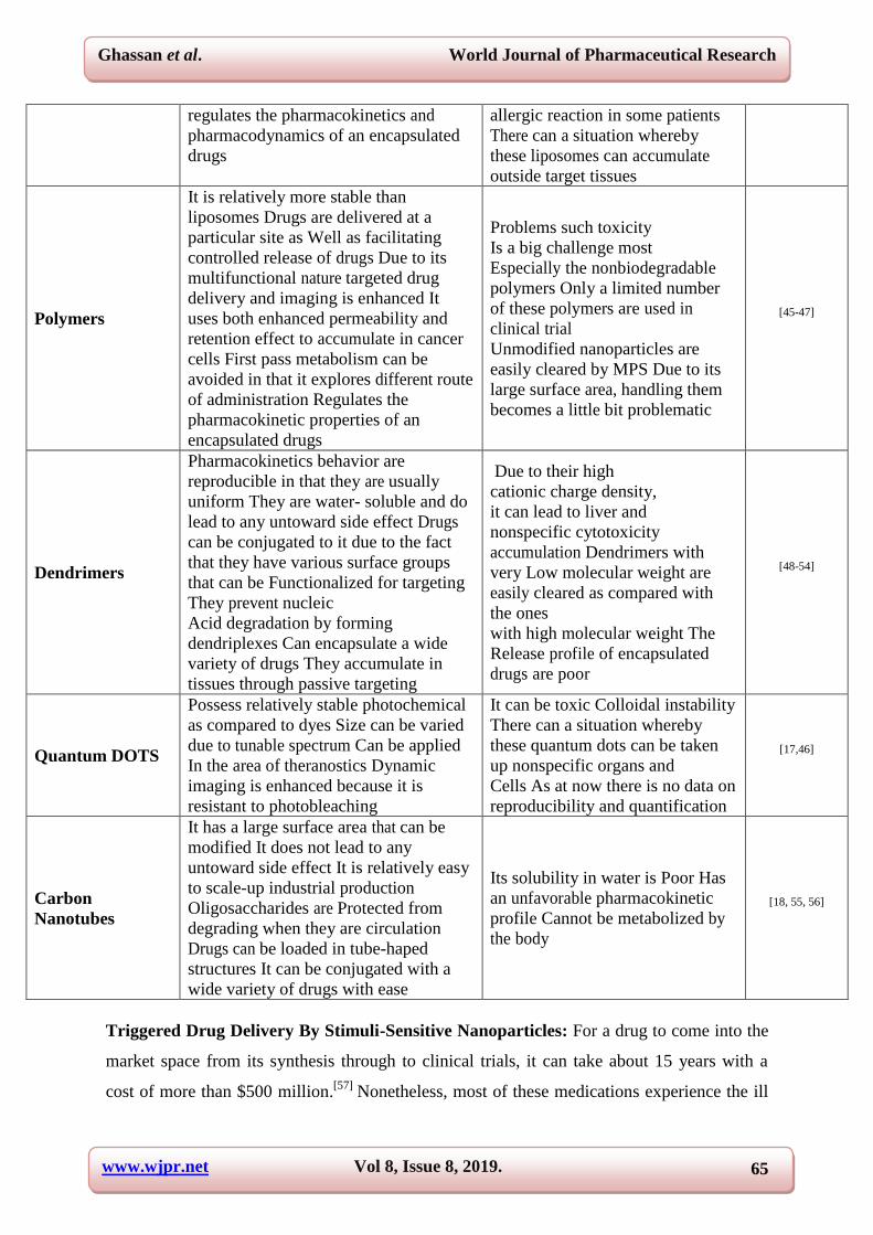
www.wjpr.net Vol 8, Issue 8, 2019.
Ghassan et al. World Journal of Pharmaceutical Research
65
regulates the pharmacokinetics and
pharmacodynamics of an encapsulated
drugs
allergic reaction in some patients
There can a situation whereby
these liposomes can accumulate
outside target tissues
Polymers
It is relatively more stable than
liposomes Drugs are delivered at a
particular site as Well as facilitating
controlled release of drugs Due to its
multifunctional nature targeted drug
delivery and imaging is enhanced It
uses both enhanced permeability and
retention effect to accumulate in cancer
cells First pass metabolism can be
avoided in that it explores different route
of administration Regulates the
pharmacokinetic properties of an
encapsulated drugs
Problems such toxicity
Is a big challenge most
Especially the nonbiodegradable
polymers Only a limited number
of these polymers are used in
clinical trial
Unmodified nanoparticles are
easily cleared by MPS Due to its
large surface area, handling them
becomes a little bit problematic
[45-47]
Dendrimers
Pharmacokinetics behavior are
reproducible in that they are usually
uniform They are water- soluble and do
lead to any untoward side effect Drugs
can be conjugated to it due to the fact
that they have various surface groups
that can be Functionalized for targeting
They prevent nucleic
Acid degradation by forming
dendriplexes Can encapsulate a wide
variety of drugs They accumulate in
tissues through passive targeting
Due to their high
cationic charge density,
it can lead to liver and
nonspecific cytotoxicity
accumulation Dendrimers with
very Low molecular weight are
easily cleared as compared with
the ones
with high molecular weight The
Release profile of encapsulated
drugs are poor
[48-54]
Quantum DOTS
Possess relatively stable photochemical
as compared to dyes Size can be varied
due to tunable spectrum Can be applied
In the area of theranostics Dynamic
imaging is enhanced because it is
resistant to photobleaching
It can be toxic Colloidal instability
There can a situation whereby
these quantum dots can be taken
up nonspecific organs and
Cells As at now there is no data on
reproducibility and quantification
[17,46]
Carbon
Nanotubes
It has a large surface area that can be
modified It does not lead to any
untoward side effect It is relatively easy
to scale-up industrial production
Oligosaccharides are Protected from
degrading when they are circulation
Drugs can be loaded in tube-haped
structures It can be conjugated with a
wide variety of drugs with ease
Its solubility in water is Poor Has
an unfavorable pharmacokinetic
profile Cannot be metabolized by
the body
[18, 55, 56]
Triggered Drug Delivery By Stimuli-Sensitive Nanoparticles: For a drug to come into the
market space from its synthesis through to clinical trials, it can take about 15 years with a
cost of more than $500 million.[57]
Nonetheless, most of these medications experience the ill

www.wjpr.net Vol 8, Issue 8, 2019.
Ghassan et al. World Journal of Pharmaceutical Research
66
effects of side effect, poor adsorption, poor solubility, high medication dosing, minimum
efficiency, and uncontrolled, non-specific delivery with high cytotoxicity, which constrain
their uses. For chemotherapeutic medications, the worry ought to be more noteworthy in light
of the fact that the vast majority of the anticancer medications are exceptionally toxic to the
healthy cells and harm the healthy cells alongside the cancer cells, which brings about
unwanted reactions in the body.[58]
These issues can be overcome and toxicity can be
decreased if the medications are delivered through a vehicle that conveys the medications
exactly on request. To accomplish the exact mission, we require a drug delivery system
(DDS) that conveys the medications to our target cells without influencing the healthy cells in
a controlled way.[59,60]
This is exactly what stimuli-sensitive drug delivery systems seek to
achieve.
Nicolò Mauro et al delivered a twofold system organized with graphene oxide-containing
nanogels as photothermal operators for the treatment of colorectal malignancy. Hyaluronic
acid/polyaspartamide-based twofold system nanogels utilized as potential treatment for
colorectal malignancy. Graphene oxide, on account of the enormous aromatic surface zone,
permits to effectively load high measure of irinotecan (33.0% w/w) and gives to the system
hyperthermic properties when irradiated with a near infrared (NIR) laser beam. The release of
antitumor medication is affected both by the pH of the outside medium and the NIR light
process. In vitro biological experiment on human colon cancer cells (HCT 116), uncovered
that nanogels are taken-up by the disease cells and, within the sight of the antitumor
medication, and could deliver a synergistic hyperthermic/cytotoxic impact. At long last, 3D
tests showed that it is conceivable to conduct thermal ablation of solid tumors after the intra-
tumoral administration of nanogels.[61]
For example, Wang et al designed a general technique to accomplish photoreactions in view
of triplet-triplet annihilation up conversion (TTA-UC) and Förster resonance energy transfer
(FRET). PLA-PEG micellar nanoparticles containing in their centers hydrophobic
photosensitizer and annihilator atoms which, when animated with green light, would
experience TTA-UC. The upconverted energy was then transferred by FRET to a
hydrophobic photocleavable group (DEACM), which was at the core. The DEACM was
clung to (and consequently inactivated) the cell-binding peptide cyclo-(RGDfK), which was
bound to the PLA-PEG chain. Cleavage of DEACM by FRET reactivated the PLA-PEG-
bound peptide and enabled it to move from the molecule center to the surface. TTA-UC

www.wjpr.net Vol 8, Issue 8, 2019.
Ghassan et al. World Journal of Pharmaceutical Research
67
followed by FRET permitted photo controlled binding of cell attachment with green light
LED irradiation at low irradiance for brief periods. These are appealing properties in photo-
triggered systems.[62]
Fig. 1: Efficient Triplet-Triplet Annihilation Based Up conversion For Nanoparticle
Phototargeting. Reproduced with permission from reference (62) (Wang et al ).
Copyright 2018 American Chemical Society.
No endeavor has been made to investigate thermo-responsive polymers for medicate
conveyance at nearer to the tumor tissue temperature and furthermore convey drugs within
the intracellular compartment. Along these lines, the improvement of new polymer scaffolds
that are equipped for conveying drugs at 40-43 °C and totally inactive at ≤37 °C would be
extremely helpful to upgrade the local drug concentration at tumor tissue over normal healthy
tissues. Kashyap et al outlined double responsive polymer nano-scaffolds for directing
anticancer medications both at the tumor site and intracellular compartments. The double
responsive polymer scaffold was observed to be fit for loading water insoluble medications
like doxorubicin (DOX), and fluorescent probe-like Nile Red. The medication release
kinetics uncovered that DOX was protected in the core shell assembly at typical body
temperature (beneath LCST, ≤ 37 °C). At nearer to diseased tissue temperature (above
LCST,∼43 °C), the polymeric scaffold experienced burst release to convey 90% of loaded
medications inside 2 h. At the intracellular condition (pH 7.4, 37 °C) within the sight of
esterase enzyme, the amphiphilic copolymer ruptured in a slow and controlled way to
discharge >95% of the medications in 12 h. In this way, both burst release of cargo at the
tumor microenvironment and control delivery at intracellular compartments were proficient

www.wjpr.net Vol 8, Issue 8, 2019.
Ghassan et al. World Journal of Pharmaceutical Research
68
in a solitary polymer system. Cytotoxicity assay of DOX-loaded polymer were performed on
breast (MCF-7) and cervical tumor (HeLa) cells. Among the two cell lines, the DOX-loaded
polymers indicated improved termination in breast tumor cells. Moreover, the cell take-up of
the DOX was examined by confocal and fluorescence microscopy. The present research
describes another enzyme and thermal responsive polymer scaffold approach for DOX
delivery in cancer cells.[63]
Fig. 2: Enzyme and Thermal Dual Responsive Amphiphilic Polymer Core-Shell
Nanoparticle to Treat Cancer Cells. Reproduced with permission from reference (63)
(Kashyap et al). Copyright 2018 American Chemical Society.
Examples of stimuli-responsive nanotherapeutics
Stimulus factor Non-formulation Active-compound Cancer cells Reference
Magnetic
Resonance
Supramolecular
nanofibres Pemetrexed Glioma
[64]
Electric field
Citrate-Apatite
Nanocrystals
with carbonate
bleomycin
hydrochlorid MCF-7 cells
[65]
Redox potential HCN polymeric
nanoparticles Camptothecin
HER2
positive
cancer cells
[66]
light Chitosan-based
nanoparticles
5-aminolaevulinic
acid
Oral
cancer
cells
[67]
pH
Multifunctional
metal-phenolic
nanoparticles
Doxorubicin Hep G2 cells [68]

www.wjpr.net Vol 8, Issue 8, 2019.
Ghassan et al. World Journal of Pharmaceutical Research
69
Methods Used for Targeted Drug Delivery
Targeted Drug Delivery: Conventional chemotherapy can result in uncontrolled distribution
of anticancer medications, which can cause harmful side effect in healthy cells and
tissue.[21,69,70]
Hence, it is imperative to create a drug delivery system with the aim of binding
to disease cells while healthy cells are avoided.[25,71]
A number of biomolecules, including
hyaluronic acid (HA)[72,73]
folic acid[74]
, peptides[75]
, and monoclonal antibodies[76]
have been
produced as targeting ligands for specific receptors on cancer cells.
Influence of the architecture of actively-targeted NPs: The conjugation of ligands on the
surface of NPs changes their characteristics.[77,78]
They lose both their rotational and
translational freedom to free molecules, the new targeted element accomplishes enhanced
avidity on account of increased valency.[79-81]
Correspondingly, the properties of the NP like
size, geometry, surface properties (charge and hydrophobicity), and composition (NP
material) likewise influence the behavior of the targeted constructs. To completely
comprehend the properties of actively-targeted NPs, it is imperative to determine how the
physicochemical properties of the NPs influence the interactions with the targets.
The ligand density: Since an increased valency permits cooperative effects, the density of
the targeting molecules on the surface of NPs greatly affects the impact of their affinity for
the substrate. Thermodynamically, the binding of a ligand to its substrate can result to
binding of its neighbors inclusive.[81,82]
Biologically, the several interactions of the NP with
the cell membrane force the clustering and local concentration of receptors. Subsequently, the
membrane is wrapped and internalized. All these impediments detach the NP from the cell
surface which results in an increased avidity. This permits the utilization of various relatively
low affinity ligands to effectively bind targets with high avidity.[83]
In vitro, an increased
ligand density improves cell take-up.[84]
Be that as it may, this increase in affinity is not
generally linear. At times, the helpful impact of the ligand can saturate and further increments
in ligand density can effectively affect cell binding.[85,86]
This effect can be clarified in view
of improper orientation of the ligand, steric prevention of neighboring molecules or
competitive behaviors for the binding of the receptor.
The NP size and shape: Size and shape of the nanomaterial must be mulled when designing
targeted NPs. Spherical particles and smaller sizes presents with higher curvatures can be
hazardous for post-synthesis ligand functionalization.

www.wjpr.net Vol 8, Issue 8, 2019.
Ghassan et al. World Journal of Pharmaceutical Research
70
Surface and ligand charge: From a synthesized point of view, the charge of the
unfunctionalized NP and that of the ligand can influence the conjugation yield and the spatial
display of the ligand on the surface.[87]
Both repulsive as well as attractive forces between the
surface of the NPs and the ligand can affect with their conjugation[87,88]
or ultimately,
influence the final ligand structure and conformation. PEG is mostly used to diminish the
effect; however this can result in an increase in final particle size and also complicate
synthesis.[85]
The last surface charge will influence the therapeutic effect of the targeted on
NPs. In spite of the interaction between cationic NPs and negatively charged cell membrane,
the NP demonstrates an increase in cellular binding and uptake abilities, in a non-specific
manner.[89]
Since most ligands are charged molecules, the NP surface charge is dependent on
the ligand densities, materials, and NP formulation strategies.
Surface hydrophobicity: Other than surface charge, hydrophobicity can likewise influence
the architecture of the ligand display.[84]
This can have genuine impacts since most polymeric
NPs have hydrophobic cores.[90]
The last surface hydrophobicity of the NPs can likewise
influence non-specific interactions with cells. From one viewpoint, actively targeted NPs
without steric stabilization appear to lose their substrate-binding capability when proteins
adsorb on their surface.[91]
Then again, while PEG surface-functionalization can defer
adsorption of opsonins what's more, plasma proteins, the utilization of long or dense PEG
chains can likewise keep ligands from achieving their targets.
Ligands Used for Targeted Drug Delivery: Riboflavin (RF) is an important vitamin for
cellular metabolism. Lately, it has been demonstrated that RF is internalized through RF
transporters which are exceptionally over-expressed by prostate and breast tumor cells, and
additionally by angiogenic endothelium. Beztsinna et al exhibited an enhanced synthesis
protocol for making tailor-made amphiphilic phospholipid-based RF derivatives utilizing
phosphoramidite chemistry. In vitro take-up investigations demonstrated that RfdiC14-
containing liposomes were unequivocally internalized in HUVEC, PC3, and A431 cells, in a
specific manner.[92]
In another development HA, a begning, biodegradable, naturally occurring polysaccharide,
has been widely joined with drug carriers since it has a high binding ability toward its
essential receptor, CD44, which is over-expressed on various sorts of tumor cells (e.g., breast,
ovarian, and lung cells).[93-96]
Nonetheless, HA-based carriers can likewise have nonspecific
interaction with normal cells, which can lead to a decrease in targeting specificity toward

www.wjpr.net Vol 8, Issue 8, 2019.
Ghassan et al. World Journal of Pharmaceutical Research
71
cancer cells. To solve this issue is to join low-fouling materials, for example, poly (ethylene
glycol) (PEG)[97,98]
, into targeted carriers in order to prevent any interaction between these
health cells and diminish uptake by phagocytes thereby improving circulation in-vivo.
A metal-phenolic capsule with high targeting and low nonspecific cell binding properties has
been developed by Ju et al. The capsules were made by covering phenolic-functionalized
hyaluronic acid (HA) and poly (ethylene glycol) (PEG) on calcium carbonate template,
accompanied by cross-connecting the phenolic groups with metal ions and expelling the
template. The joining of HA fundamentally improved binding and interaction with a CD44
overexpressing (CD44+) cancer cell line, while the incorporation of PEG decreased
nonspecific association with a CD44 minimal expressing (CD44-) cell line. Besides, high
specific targeting to CD44+, cells can be adjusted with low nonspecific binding to CD44-
cells basically by utilizing an optimized feed-ratio of HA and PEG to change the content of
HA and PEG joined into the capsule. Loading an anticancer medication (i.e., doxorubicin)
into the acquired capsule brought about fundamentally higher cytotoxicity to CD44+ cells yet
bring down cytotoxicity to CD44-cells.[99]
Aptamers
It is characterized as a short DNA or RNA or an even peptide molecule that has affinity for a
specific receptor on the tumor cell. Basically, aptamers can be classified into two
1. DNA or RNA aptamers: They are usually short strands of oligonucleotides.
2. Peptide aptamers: They consist of one or more short variable peptide domains, attached at
both ends to a protein scaffold.
On account of their exceptional conformational structures that begin from intramolecular
Watson-Crick interactions, Aptamers demonstrate high affinity and specificity. Candidates
are screened from extensive oligonucleotide libraries with random sequences by exploiting
the nucleic acid sequence. Binders are selected and specifically enhanced at the expense of
non-binders utilizing the polymerase chain response. Aptamers that bind strongly to small
molecules and proteins have been identified. The biggest advantage of Aptamers is their
ability to isolate high affinity ligands against a number of substrates, however they possess
other advantages, thus their reproducible synthesis and simplicity of their chemical
derivatives give room for Aptamers to be used as ligands for targeted NPs.[100]

www.wjpr.net Vol 8, Issue 8, 2019.
Ghassan et al. World Journal of Pharmaceutical Research
72
Abnous et al, developed a novel chemotherapy drug- free DNA nanocomplex made up of
three medicinal aptamers (IDA, AS1411 and apMNK2F) intended for treatment of cancer
cells. The MTT assay revealed, PC-3 and 4T1 cells are target cells and CHO cells are non-
target cells both treated with apMNK2F-AS1411-IDA complex (DNA nanocomplex),
together with AS1411, IDA and apMNK2F alone. Internalization of apMNK2FAS1411-IDA
complex was investigated by fluorescence imaging and flow cytometry analysis. In the last
stride, the introduced DNA nanocomplex was applied to prevent cancerous growth in vivo.
The after effects of the internalization assay revealed that the created apMNK2F-AS1411-
IDA complex was internalized into PC-3 and 4T1 cells, however not into CHO cells. The
after effects of internalization assay were affirmed by MTT assay. apMNK2F-AS1411-IDA
complex was more cytotoxic in PC-3 and 4T1 cells (target) and less cytotoxic in CHO cells
(non-target). Likewise, the DNA nanocomplex could viably smother the development of
tumors in vivo.[101]
Fig. 3. A Novel Chemotherapy Drug-Free Delivery System Composed Of Three
Therapeutic Aptamers For The Treatment Of Prostate And Breast Cancers In-Vitro
And In-Vivo.
Reproduced with permission from reference[102]
(Abnous et al). Copyright 2018 Elsevier Inc.
Affibodies
These are small proteins engineered to bind to a large number of cancer tissues with high
affinity, imitating monoclonal antibodies. They are therefore a member of the family of
antibody mimetics. These molecules are used as biopharmaceutical drugs for cancer therapy.
Tyrosine Kinase receptor HER3 has become a therapeutic target in various cancers including
prostate, breast and ovarian because it can activate the P13K/Akt pathway by dimerization
with HER2 and also mediating drug resistance. An improved efficacy of HER3-targeted

www.wjpr.net Vol 8, Issue 8, 2019.
Ghassan et al. World Journal of Pharmaceutical Research
73
therapeutics would consequently profit an extensive variety of patients. This investigation by
Schardt et al assessed the capability of multivalent presentation, through protein engineering.
It was used to improve the viability of HER3-targeted affibodies as contrasting options to
monoclonal counter acting agent therapeutics. Evaluation of multivalent affibodies on an
assortment of malignant cell lines uncovered their expansive capacity to enhance inhibition of
Neuregulin (NRG)- induced HER3 and Akt phosphorylation contrasted with monovalent
analogs. Designed multivalency prevented cell growth by affibodies as single agent and also
as part of combination treatment techniques. Mechanistic investigations uncovered that
designed multivalency improved affibody-mediated HER3 downregulation in different tumor
cell types. By and large, these outcomes feature the guarantee of engineered multivalency as
a general method for improved therapeutic effect of HER3-focused on therapeutics against a
number of cancers.[103]
Another study by Hoppmann et al, showed a straightforward and generalizable methodology
for lessening the renal uptake of Affibody molecules while sustaining their tumor uptake.
Radiolabeled DOTA-HSA-ZHER2: 342 conjugates showed specific cell uptake into SKOV3 cell
cultures. Positron emission tomography (PET) examinations were performed in SKOV3
tumor-bearing mice utilizing 64
Cu-DOTA-HSA-ZHER2: 342. High tumor uptake values (>14%
ID/g at 24 and 48 h) and high liver concentrations as well as low kidney concentrations were
noticed. Biodistribution studies and single-photon emission computed tomography (SPECT)
examinations utilizing 111
In-DOTA-HSA-ZHER2: 342 validated these outcomes. At 24 h post
injection, the biodistribution information uncovered high tumor (16.26% ID/g) and liver
(14.11% ID/g) concentrations and generally low kidney concentration (6.06% ID/g).[104]
Monoclonal Antibodies: Monoclonal antibodies thus mAb or moAb are antibodies that are
made by indistinguishable immune cells that are all clones of a unique parent cell. These
antibodies have specific affinity to cancer cells making them helpful as against polyclonal
antibodies with numerous epitopes. Anhorn et al, developed a target-oriented nanoparticle in
light of biodegradable human serum albumin (HSA) loaded with cytostatic drug doxorubicin.
The surface of the nanoparticles was covalently modified by attaching trastuzumab. HER2
overexpressed breast cancer cells showed high cellular up-take as well as binding of these
nanoparticles. The specific transport of the cytostatic drug doxorubicin with this
nanoparticulate formulation into the HER2 overexpressing breast cancer cells, their release,
and biological activity was demonstrated. The outcomes showed that these cell-type specific

www.wjpr.net Vol 8, Issue 8, 2019.
Ghassan et al. World Journal of Pharmaceutical Research
74
drug-loaded nanoparticles could enhance tumor therapy.[105]
Monoclonal antibody (RG 7155)
developed by Ries et al and this monoclonal antibody is used to block CSF-1 receptor one of
the known proto-oncogenes. In-vivo study revealed that CSF-1 blockade diminishes F4/80+
tumor-associated macrophages.[106]
Peptides: Peptides are either natural biological or artificially fabricated short chains of amino
acids monomers connected by peptide bonds. Peptides can be incorporated into drugs to
convey drugs at the cancer tissue or cell. Due to their shorter chains they can be made to form
smaller molecular sizes and simpler three-dimensional structures thereby easing up synthesis
and conjugation, enhancing their stability and making them resistant to the environment. The
stated advantages above, in combination with improved screening techniques to isolate
ligand-substrate have contributed immensely to the role of peptides as targeting moieties in
the past decade. Arginine-glycine-aspartic (RGD) is a motif found on a number extracellular
matrix (ECM) and plasma proteins.[107]
RGD gained much attention in research when it was
found out that it has specific binding sites for fibronectin (FN) and the FN receptor.[108]
Laminin, vitronectin (VN), fibrinogen (Fg), von Willebrand factor (vWF), osteopontin etc are
glycoproteins found in ECM and they serve us RGD-adhesive proteins.[109]
RGD is vital in
both cell recognition and cell adhesion and has been utilized in both tumor therapy and tissue
engineering by either recombinant means or chemical methods. By chemical means RGD-
peptides and RGD-mimetics can be used to restyle liposomes, polymers and peptides so that
therapeutic agents would have a better biological response. Additionally, RGD-peptides were
utilized in gene delivery by viral and non-viral vectors.[110]
Advantages: There are 2 types of RGD-containing peptides based on structure and sequence
and they are linear and cyclic RGD peptides. Cyclic RGD peptides are preferred over the
linear RGD peptides due to their higher activity. The merits of cyclic peptides are that they
have higher affinity for integrin receptors and also resist proteolysis.[111,112]
RGD peptides
possess some advantages us targeting agent for cancer therapy[113,114]
: (i) RGD is smaller in
nature and easier; (ii) the use of RGD reduces immune reactions; (iii) synthesis of RGD
peptides is relatively simple and inexpensive, makes it easier to be translated into clinical
trials; RGD has important regulatory functions in many biological activities they are; actin
formation in skeletal muscle, cell attachment, focal-adhesion formation with integrins, and
cell spreading.[115]

www.wjpr.net Vol 8, Issue 8, 2019.
Ghassan et al. World Journal of Pharmaceutical Research
75
A phenomenal method developed by Kumal et al was used to convey a platinum (IV)
medication to prostate cancer cells by developing glutathione-stabilized (Au@GSH) gold
nanoparticles. Glutathione (GSH) is known for its antioxidant properties, which inhibit
cancer cells. Due to its antioxidant properties as well as its high surface-area-to-volume ratio
of Au@GSH NPs was able to convey platinum (IV) drug by targeting it to its receptor thus
neuropilin-1 receptor (Nrp-1). A lethal dose of a platinum (IV) drug coupled with the Nrp-1-
targeting peptide (CRGDK) was able to specifically deliver to prostate disease cells in vitro.
The targeted peptide binds specifically to the Nrp-1 receptor, prompting improved cell take-
up and cell toxicity levels. These nanocarriers were nontoxic, however displayed high
cytotoxicity and an increase in therapeutic effect when functionalized with a targeting peptide
and medication. The uptake of drug-loaded nanocarriers depended on the interaction with
Nrp-1 in cell lines expressing high (PC-3) as well as low (DU-145) levels of Nrp-1, as
affirmed via inductively coupled plasma mass spectrometry and confocal microscopy. The
nanocarriers have powerful anticancer action, through upregulation of nuclear factor kappa-B
(NF-κB) protein (p50 and p65) expression and activation of NF-κB-DNA-binding
activity.[116]
Fig. 4. Neuropilin-1-Targeted Gold Nanoparticles Enhance The Therapeutic Efficacy of
Platinum (ΙV) Drug For Prostate Cancer Treatment. Reproduced with permission from
reference[116]
(Kumar et al) Copyright 2018 American Chemical Society.
Proteins: They consist of one or more polypeptides bound to ligands to target cancer cells.
More often than not, proteins are used as small molecules to convey drugs to their target sites.
However, proteins are often interchanged with peptides but what distinguish these two is on
the basis of their size. A number of naturally-occurring proteins have endogenous targets that
can be used for therapeutic purposes. The three-dimensional shape of proteins provides
www.wjpr.net Vol 8, Issue 8, 2019.
Ghassan et al. World Journal of Pharmaceutical Research
76
affinity for specific substrates, and therefore non-antibody proteins can be used as targeting
moieties.
Lipoprotein transport lipids in the human body. It is made up of lipoproteins, phospholipids,
cholesterol esters, free cholesterol, and protein. The composition of lipoproteins in human
body possesses chylomicrons, very low density lipoprotein (VLDL), low density lipoprotein
(LDL), and high density lipoprotein (HDL). Lipoprotein-mimetics have gained interest in
cancer therapy because it targets cells naturally and also minimal immune reactions. The
main reason LDL and HDL identify the corresponding receptor of target cell is due to
alipoproteins expressed on their surfaces. Lipoprotein receptor can be classified into
lipoprotein receptor (LDLR) and scavenger receptor type A (SRA). Lipoprotein receptors are
found on a number of tumor cells making it necessary as targeting for treatment and
diagnosis. It is worth noting that lipoprotein receptors are also present on the surfaces of non-
neoplastic diseases. Due diligence must be done when treating cancer cells so that cell
toxicity can be reduced to the barest minimum. Ullal et al covalently conjugated small-
molecule medication to a magnetic nanoparticle which was then utilized as a read-out for
target expression and drug-binding affinity. Poly(ADP-ribose) polymerase (PARP) was used
as an inhibition model framework, which was designed to deal with recognized differential
expression of PARP in scant cells with astounding correlation to highest quality levels, the
capacity to emulate drug pharmacodynamics ex vivo through competitive target drug binding,
and the possibility to perform such measurements in clinical samples.[117]
Intracellular protein delivery is a critical tool for both therapeutic and basic applications.
Successful protein delivery confronts two noteworthy difficulties: proficient cell take-up and
avoiding endosomal sequestration. Herein, a general procedure for direct conveyance of
useful proteins to the cytosol utilizing nanoparticle-stabilized capsules (NPSCs) was reported
by Tang et al. These NPSCs are framed and stabilized via supramolecular cooperation
between the nanoparticle, the protein cargo, and the fatty acid capsule interior. The NPSCs
are ∼130 nm in distance across and highlight low harmfulness and phenomenal stability in
serum. The NPSCs were efficacious protein carriers which were exhibited through the
delivery of completely utilitarian caspase-3 to HeLa cells with concomitant apoptosis.
Conveyance of green fluorescent protein (GFP) affirmed cytosolic delivery and also
intracellular targeting of the conveyed protein, exhibiting the utility of the system for both
therapeutic and imaging applications.[118]

www.wjpr.net Vol 8, Issue 8, 2019.
Ghassan et al. World Journal of Pharmaceutical Research
77
Inorganic Targeting Agents: Plasmon resonance (SPR) band on the surfaces of inorganic
nanoparticles such as gold, silver and copper make them display brilliant colours.[119-123]
The
very reason why inorganic nanomaterials are so special for biomedical applications is due to
the tunability of optoelectronic properties which is size and shape-dependent.[124-127]
Silver
has a long history for medicinal use.[128,129]
It was used in world war ӀӀ to treat burns.[130]
Aside burns, silver nanoparticles can also be used as a biocide against microbial infections
and diabetic skin ulcers.[128]
The Ag+ ion present in silver nanoparticles contribute immensely
in their biological activity. Platinum nanoparticles are not well known for their medicinal
purposes, instead platinum compounds are known for their anti-tumor action (cis-platin and
its derivatives).[131]
The advantage of using platinum nanoparticle is when modified structural
can minimize the cytotoxicity of drugs. Other inorganic nanoparticles include the use of
metal oxides such as SiO2 with functionalized surfaces and Fe3O4 (magnetic nanoparticles) as
a vector for targeted delivery of drugs and genes with reasonably low toxicity.[132]
These
nanoparticles can be easily synthesized and well characterized in the lab. The boron and
gadolinium nanoparticles can be used to treat cancer cells and also have neutron capture
therapy (NCT).
Circumventing Major Drug Delivery Problems Through Enhanced Targeting
In spite of gigantic endeavors made toward finding novel materials and biomolecule markers
for targeted drug delivery systems (DDS), not many of them are really specific after
intravenous infusion and targeting is chance-dependent. Active and passive targeting
techniques need exogenous medication vehicles to disperse and voyage in circulation for long
to be able to go through the leaky vasculature or recognize the surface markers. Be that as it
may, the circulating environment, in which many medication vehicles cannot have a
sufficiently long circulating time to accomplish targeted binding, is amazingly
complicated.[133,134]
Moreover, the human body has an innate defense mechanism for
invasion. For instance, the reticuloendothelial system (RES) quickly perceives foreign bodies
and pulverizes them by well-rehearsed biological processes. The RES, likewise called the
mononuclear phagocyte systems, includes essentially bone marrow progenitors, blood
monocytes, and tissue macrophages.[135]
Furthermore, the EPR impact is some way or another
heterogeneous in the tumor microenvironments and differs among patients.[136]
For instance,
hypoxic locales of solid tumor for the most part don't display EPR impacts in view of poor
angiogenesis.[137]
Considering the complexity and sophistication of in vivo conditions,
conventional active and passive targeting methods are woefully inadequate. Subsequently,
www.wjpr.net Vol 8, Issue 8, 2019.
Ghassan et al. World Journal of Pharmaceutical Research
78
creating novel DDS with really specific targeting is an impressive test for current medicine
and nanotechnology. In recent times, cellular backpacks are attached to drug loaded particles
on cell surface thereby enhancing protection of therapeutic agents as well as protecting
cellular integrity. These backpacks prevent RES elimination, off site release of drugs and
finally improves retention time of drugs in circulation. Michael F. Rubner et al utilized
unique properties of macrophages for targeted drug delivery. Herein, HA (hyaluronic acid)
was used to coat macrophages which can strongly bind to CD44 receptor expressed in many
cancer cells. Compared to the conventional spherical particles it was found out that flat PEM-
based disk were able to attach themselves to cell surface as compared to the spherical disks.
Ligand-receptor interaction is one beneficial strategy of attaching particles to circulating
cells. Identifying surface markers and binding ligands would be very useful for ligand-based
drug carrier design. However, binding efficiency and in vivo specific binding within blood
circulation still remains big hurdle. Intelligent targeting agents are required to aid drugs to
bypass the RES and target the required cells.
Reversing Multi-Drug Resistance Through Targeted Delivery
For an effective tumor treatment, there exist four extreme issues that should be attended to
and one of such issues is multidrug resistance (MDR), the others are recurrence, late stage
diagnosis and aggressive metastasis. MDR of cancer cells results in around 90%
chemotherapeutic failure of patients with metastatic tumors, and along these lines it remains a
challenge for a fruitful chemotherapy treatment of cancer. Typically, the MDR of tumor cells,
caused by the malfunctioning of genes, for the most part originates from either intrinsic high
expression of ATP-binding cassette (ABC) transporter proteins or acquired resistance in
malignancy cells by the stimulation of anticancer medications to overexpress ABC
transporters.[138-140]
Novel nanotechnology-based methodologies toward treatment of MDR
malignancies expect the engagement of nanoparticles keeping in mind the end goals which
include increased intracellular medication accumulation, silence of efflux transporters genes
and inhibiting MDR-related proteins and factors.[141]
In this a mussel-inspired engineering methodology by Jianxiang Zhang et al may quite
advance cell take-up and tissue retention of NPs. In this methodology, the catechol moiety is
covalently moored onto biodegradable NPs. In this way, created NPs can be adequately
internalized by sensitive and multidrug resistant tumor cells, and some healthy cells, bringing
about amazingly potentiated in vitro activity when an antitumor medication is packaged.

www.wjpr.net Vol 8, Issue 8, 2019.
Ghassan et al. World Journal of Pharmaceutical Research
79
Additionally, the recently engineered NPs bear the cost of increased tissue retention post
local or oral delivery. This biomimetic approach is promising for making utilitarian
nanomaterials for medicate conveyance, immunization, and cell therapy.[142]
Mix treatment
utilizing proteins and small molecules gives access to synergistic treatment procedures.
Rotello et al loaded paclitaxel into the hydrophobic center of the NPSC and self-gathered
caspase-3 and nanoparticles on the capsule surface. The subsequent combination NPSCs
indicated higher cytotoxicity than both of the single operating agent NPSCs, with synergistic
activity set up utilizing combination index value. Simultaneous delivery of small molecule
drugs and proteins reduce drug administration as a result of prevented multidrug
resistance.[143]
Targeted Delivery for Theranostics
This term was coined to define ongoing efforts in clinics to develop more specific,
individualized therapies for various diseases, and to combine diagnostic and therapeutic
capabilities into a single agent. This innovation counteracts repeat of tumor in that a more
exact diagnosis and treatment is established. Herein, the role of drug targeting agents can be
described as pivotal. They are capable of orchestrating every single move which is picked up
by the diagnostic system in and around the target cells. This means that a good theranostic
system requires a highly efficient drug targeting agent for enhancement.
In this, a one of a kind sort of redox-sensitive NCPs was built with manganese ion (Mn2+)
and dithiodiglycolic acid as the disulfide (SS)- containing organic bridging ligand. The Mn-
SS NCPs when obtained with a mesoporous structure could be effectively loaded with
doxorubicin (DOX), a chemotherapeutic agent. The yielded MnSS/DOX nanoparticles are
covered with a layer of polydopamine (PDA) and after that modified by poly(ethylene glycol)
(PEG). In such a Mn-SS/DOX@PDA-PEG NCP structure, the disulfide linkage (SS) inside
dithiodiglycolic acid can be cleaved in the presence of glutathione (GSH), resulting in a
proficient redox-responsive dissociation of NCPs following drug release. In the meantime,
Mn2+ in Mn SS/DOX@PDA-PEG NCPs would offer a strong T1 contrast in magnetic
resonance (MR) imaging, Upon intravenous injection, these Mn-SS/DOX@PDAPEG NCPs
demonstrate effective tumor homing, as uncovered by MR imaging, and offer a clearly
enhanced in vivo remedial result contrasted with that accomplished with free DOX by
Zhuang Liu et al.[144]

www.wjpr.net Vol 8, Issue 8, 2019.
Ghassan et al. World Journal of Pharmaceutical Research
80
Thus, a protein-polymer bio-conjugate-covered multifunctional upconversion nanosystem,
Comprising of upconversion nanoparticles (UCNPs) center, custom-made amphiphilic
protein-polymer bio-conjugate shell, and photosensitizer zinc phthalocyanine (ZnPc) as well
as antitumor medication doxorubicin co-loaded inside, the nanomaterial was created for
consolidated photodynamic treatment (PDT) and chemotherapy. In this framework, UCNPs
core could change over entering near-infrared light to visible light for synchronous cell
fluorescence imaging and photodynamic therapy by activating ZnPc to produce cytotoxic
ROS, while the protective shell of bovine serum albumin poly (ε-caprolactone) (BSA-PCL)
offered magnificent water solubility, great stability, and low cytotoxicity. The ROS creation
test demonstrated that this nanosystem could effectively produce singlet oxygen under NIR
irradiation. A cellular uptake study exhibited that extreme fluorescence outflow of the
UCNPs could be seen in HeLa cells, demonstrating their real-time imaging capability.
Essentially, the combined therapy UCNP system proved to be an enhanced tumor cell killing
ability as compared to single PDT or chemotherapy system by Zhongyun Liu et al.[145]
In another specific circumstance, liposomes, artificially prepared from a lamellar phase lipid
bilayer, have been presented as reasonable nano-carriers for UCNPs. Here, we made a hybrid
nano-carrier comprising of Er3+ and Yb3+ co-doped NaGdF4 UCNPs that were encapsulated
in the aqueous phase of the liposomes and the capability of the nano-carriers for drug
delivery which appeared by co-loading the model anticancer medication doxorubicin (DOX).
Under 980 nm excitation, a decline of the green up-conversion emission of the NaGdF4:
Er3+, Yb3+ UCNPs was watched when DOX was co-loaded with the UCNPs in the liposome
nanocarrier. This extinguishing impact is doled out to the energy transfer between the
contributor UCNP and the acceptor DOX and is most critical, since it takes into consideration
the spectral checking of the DOX loading and release from the liposome nano-carriers. Along
these lines, the medication loading, release, and spectral observing properties of the liposome
nano-carriers were altogether characterized enabling us to evaluate their future potential as
theranostic nano-carriers. Huang et al.[146]
CONCLUSION AND FUTURE PROSPECT
Nanotechnology has helped to curb some of the problems listed above in the fight against
cancer. It is worthy of note that not a single technology can easily prevent these problems.
However, it is necessary that the individual technologies are harnessed to explore their
advantages. Going to the future, a more combined approach can be looked at to tackle the
www.wjpr.net Vol 8, Issue 8, 2019.
Ghassan et al. World Journal of Pharmaceutical Research
81
problems associated with the underlining problems that affect the fight against cancer cells.
Most importantly, drug targeting should be pivotal in our quest for an ideal cancer therapy.
Herein, we have offered a thorough discussion concerning site-specific targeting for
enhanced drug delivery. Our review was able to outline the important aspects of drug
delivery. We also pointed out the major problems that have been overcome with targeted
delivery of drugs. It is an undeniable fact that researchers need to pay much attention in order
to explore innovative ways to improve on the existing strategies to chauffeur drugs to the
required destinations.
ACKNOWLEDGMENT
We the authors would want to acknowledge financial support from National Natural Science
Foundation of China (No. 81273469, 81501582 and 81573379). Operating Funds for Basic
Sciences Research in Central Universities (No. 2015PY011) also supported this research. A
very big thank you to Professor Bo Wang for his unflinching support. Again we want to
appreciate Presidential Scholarship of China Pharmaceutical University for supporting our
education.
COMPETING INTEREST
The authors declare that there are no competing interests.
REFERENCES
1. Butler D. Translational research: crossing the valley of death. Nature News, 2008;
453(7197): 840-2.
2. Gao J, Feng S-S, Guo Y. Nanomedicine against multidrug resistance in cancer treatment.
Nanomedicine, 2012; 7(4): 465-8.
3. Sikora K. The impact of future technology on cancer care. Clinical Medicine, 2002; 2(6):
560-8.
4. Chidambaram M, Manavalan R, Kathiresan K. Nanotherapeutics to overcome
conventional cancer chemotherapy limitations. Journal of pharmacy & pharmaceutical
sciences, 2011; 14(1): 67-77.
5. Sakamoto J. van de, Ven AL, Godin B, et al. Enabling individualized therapy through
nanotechnology Pharmacol Res., 2010; 62: 57-89.
6. Tang X, Liang Y, Feng X, Zhang R, Jin X, Sun L. Co-delivery of docetaxel and
Poloxamer 235 by PLGA–TPGS nanoparticles for breast cancer treatment. Materials
Science and Engineering: C., 2015; 49: 348-55.

www.wjpr.net Vol 8, Issue 8, 2019.
Ghassan et al. World Journal of Pharmaceutical Research
82
7. Zhao X, Chen Q, Li Y, Tang H, Liu W, Yang X. Doxorubicin and curcumin co-delivery
by lipid nanoparticles for enhanced treatment of diethylnitrosamine-induced
hepatocellular carcinoma in mice. European Journal of Pharmaceutics and
Biopharmaceutics, 2015; 93: 27-36.
8. Cheng Y-J, Luo G-F, Zhu J-Y, Xu X-D, Zeng X, Cheng D-B, et al. Enzyme-induced and
tumortargeted drug delivery system based on multifunctional mesoporous silica
nanoparticles. ACS applied materials & interfaces, 2015; 7(17): 9078-87.
9. Binkhathlan Z, Shayeganpour A, Brocks DR, Lavasanifar A. Encapsulation of P-
glycoprotein inhibitors by polymeric micelles can reduce their pharmacokinetic
interactions with doxorubicin. European Journal of Pharmaceutics and Biopharmaceutics,
2012; 81(1): 142-8.
10. Wang H, Liu Z, Gou Y, Qin Y, Xu Y, Liu J, et al. Apoptosis and necrosis induced by
novel realgar quantum dots in human endometrial cancer cells via endoplasmic reticulum
stress signaling pathway. International journal of nanomedicine, 2015; 10: 5505.
11. Anbarasan B, Babu SV, Elango K, Shriya B, Ramaprabhu S. pH responsive release of
doxorubicin to the cancer cells by functionalized multi-walled carbon nanotubes. Journal
of nanoscience and nanotechnology, 2015; 15(7): 4799-805.
12. Yallapu MM, Othman SF, Curtis ET, Gupta BK, Jaggi M, Chauhan SC. Multi-functional
magnetic nanoparticles for magnetic resonance imaging and cancer therapy. Biomaterials,
2011; 32(7): 1890-905.
13. Akbarzadeh A, Rezaei-Sadabady R, Davaran S, Joo SW, Zarghami N, Hanifehpour Y, et
al. Liposome: classification, preparation, and applications. Nanoscale research letters,
2013; 8(1): 102.
14. Li J, Wang X, Zhang T, Wang C, Huang Z, Luo X, et al. A review on phospholipids and
their main applications in drug delivery systems. Asian journal of pharmaceutical
sciences, 2015; 10(2): 81-98.
15. Brigger I, Dubernet C, Couvreur P. Nanoparticles in cancer therapy and diagnosis.
Advanced drug delivery reviews, 2002; 54(5): 631-51.
16. van Vlerken LE, Amiji MM. Multi-functional polymeric nanoparticles for tumour-
targeted drug delivery. Expert opinion on drug delivery, 2006; 3(2): 205-16.
17. Fang M, Peng C-w, Pang D-W, Li Y. Quantum dots for cancer research: current status,
remaining issues, and future perspectives. Cancer biology & medicine, 2012; 9(3): 151.
www.wjpr.net Vol 8, Issue 8, 2019.
Ghassan et al. World Journal of Pharmaceutical Research
83
18. Kirkpatrick DL, Weiss M, Naumov A, Bartholomeusz G, Weisman RB, Gliko O. Carbon
nanotubes: solution for the therapeutic delivery of siRNA? Materials, 2012; 5(2):
278-301.
19. Liu Z, Tabakman S, Welsher K, Dai H. Carbon nanotubes in biology and medicine: in
vitro and in vivo detection, imaging and drug delivery. Nano research, 2009; 2(2):
85-120.
20. Prato M, Kostarelos K, Bianco A. Functionalized carbon nanotubes in drug design and
discovery. Accounts of chemical research, 2007; 41(1): 60-8.
21. Peer D, Karp JM, Hong S, Farokhzad OC, Margalit R, Langer R. Nanocarriers as an
emerging platform for cancer therapy. Nature nanotechnology, 2007; 2(12): 751-60.
22. Davis ME, Shin DM. Nanoparticle therapeutics: an emerging treatment modality for
cancer. Nature reviews Drug discovery, 2008; 7(9): 771-82.
23. Kuang T, Liu Y, Gong T, Peng X, Hu X, Yu Z. Enzyme-responsive nanoparticles for
anticancer drug delivery. Current Nanoscience, 2016; 12(1): 38-46.
24. Petros RA, DeSimone JM. Strategies in the design of nanoparticles for therapeutic
applications. Nature reviews Drug discovery, 2010; 9(8): 615-27.
25. Farokhzad OC, Langer R. Impact of nanotechnology on drug delivery. ACS nano, 2009;
3(1): 16-20.
26. Chen H-H, Huang W-C, Chiang W-H, Liu T-I, Shen M-Y, Hsu Y-H, et al. pH-
Responsive therapeutic solid lipid nanoparticles for reducing P-glycoprotein-mediated
drug efflux of multidrug resistant cancer cells. International journal of nanomedicine,
2015; 10: 5035.
27. Ding C, Liu Y, Wang T, Fu J. Triple-stimuli-responsive nanocontainers assembled by
watersoluble pillar [5] arene-based pseudorotaxanes for controlled release. Journal of
Materials Chemistry B., 2016; 4(16): 2819-27.
28. Chiang Y-T, Cheng Y-T, Lu C-Y, Yen Y-W, Yu L-Y, Yu K-S, et al. Polymer–liposome
complexes with a functional hydrogen-bond cross-linker for preventing protein
adsorption and improving tumor accumulation. Chemistry of Materials, 2013; 25(21):
4364-72.
29. Huang W-C, Chen S-H, Chiang W-H, Huang C-W, Lo C-L, Chern C-S, et al. Tumor
Microenvironment-Responsive Nanoparticle Delivery of Chemotherapy for Enhanced
Selective Cellular Uptake and Transportation within Tumor. Biomacromolecules, 2016;
17(12): 3883-92.

www.wjpr.net Vol 8, Issue 8, 2019.
Ghassan et al. World Journal of Pharmaceutical Research
84
30. Allen TM, Cullis PR. Drug delivery systems: entering the mainstream. Science, 2004;
303(5665): 1818-22.
31. Allen TM, Cullis PR. Liposomal drug delivery systems: from concept to clinical
applications. Advanced drug delivery reviews, 2013; 65(1): 36-48.
32. Parveen S, Misra R, Sahoo SK. Nanoparticles: a boon to drug delivery, therapeutics,
diagnostics and imaging. Nanomedicine: Nanotechnology, Biology and Medicine, 2012;
8(2): 147-66.
33. Kedar U, Phutane P, Shidhaye S, Kadam V. Advances in polymeric micelles for drug
delivery and tumor targeting. Nanomedicine: Nanotechnology, Biology and Medicine,
2010; 6(6): 714-29.
34. Ashley GW, Henise J, Reid R, Santi DV. Hydrogel drug delivery system with predictable
and tunable drug release and degradation rates. Proceedings of the national academy of
sciences, 2013; 110(6): 2318-23.
35. Hamidi M, Azadi A, Rafiei P. Hydrogel nanoparticles in drug delivery. Advanced drug
delivery reviews, 2008; 60(15): 1638-49.
36. Cui W, Zhou Y, Chang J. Electrospun nanofibrous materials for tissue engineering and
drug delivery. Science and Technology of Advanced Materials, 2010; 11(1): 014108.
37. Yoo HS, Kim TG, Park TG. Surface-functionalized electrospun nanofibers for tissue
engineering and drug delivery. Advanced drug delivery reviews, 2009; 61(12): 1033-42.
38. Prausnitz MR, Langer R. Transdermal drug delivery. Nature biotechnology, 2008; 26(11):
1261-8.
39. Keefe AD, Pai S, Ellington A. Aptamers as therapeutics. Nature reviews Drug discovery,
2010; 9(7): 537-50.
40. Torchilin VP. Multifunctional nanocarriers. Advanced drug delivery reviews, 2012; 64:
302-15.
41. Snyder EL, Dowdy SF. Cell penetrating peptides in drug delivery. Pharmaceutical
research, 2004; 21(3): 389-93.
42. Kura AU, Fakurazi S, Hussein MZ, Arulselvan P. Nanotechnology in drug delivery: the
need for more cell culture based studies in screening. Chemistry Central Journal, 2014;
8(1): 46.
43. Thulasiramaraju T, Babu A, Arunachalam A, Prathap M, Srikanth S, Sivaiah P.
Liposome: A novel drug delivery system. International Journal of Biopharmacuties, 2012;
2229: 7499.

www.wjpr.net Vol 8, Issue 8, 2019.
Ghassan et al. World Journal of Pharmaceutical Research
85
44. Thakor AS, Gambhir SS. Nanooncology: the future of cancer diagnosis and therapy. CA:
a cancer journal for clinicians, 2013; 63(6): 395-418.
45. Mahapatro A, Singh DK. Biodegradable nanoparticles are excellent vehicle for site
directed in-vivo delivery of drugs and vaccines. Journal of nanobiotechnology, 2011;
9(1): 55.
46. Guo S, Huang L. Nanoparticles containing insoluble drug for cancer therapy.
Biotechnology advances, 2014; 32(4): 778-88.
47. Jawahar N, Meyyanathan S. Polymeric nanoparticles for drug delivery and targeting: A
comprehensive review. International Journal of Health & Allied Sciences, 2012; 1(4):
217.
48. Nanjwade BK, Bechra HM, Derkar GK, Manvi F, Nanjwade VK. Dendrimers: emerging
polymers for drug-delivery systems. European Journal of Pharmaceutical Sciences, 2009;
38(3): 185-96.
49. Marvaniya H, Parikh P, Patel V, Modi K, Sen D. Dendrimer nanocarriers as versatile
vectors in gene delivery. J Chem Pharm Res, 2010; 2(3): 97-108.
50. Cheng Y, Xu Z, Ma M, Xu T. Dendrimers as drug carriers: applications in different
routes of drug administration. Journal of pharmaceutical sciences, 2008; 97(1): 123-43.
51. Florence AT, Hussain N. Transcytosis of nanoparticle and dendrimer delivery systems:
evolving vistas. Advanced drug delivery reviews, 2001; 50: S69-S89.
52. Gillies ER, Frechet JM. Dendrimers and dendritic polymers in drug delivery. Drug
discovery today, 2005; 10(1): 35-43.
53. Dufes C, Uchegbu IF, Schätzlein AG. Dendrimers in gene delivery. Advanced drug
delivery reviews, 2005; 57(15): 2177-202.
54. Yavuz B, Bozdağ Pehlivan S, Ünlü N. Dendrimeric systems and their applications in
ocular drug delivery. The Scientific World Journal, 2013; 2013.
55. Sokolova V, Epple M. Inorganic nanoparticles as carriers of nucleic acids into cells.
Angewandte Chemie International Edition, 2008; 47(8): 1382-95.
56. Varkouhi AK, Foillard S, Lammers T, Schiffelers RM, Doris E, Hennink WE, et al.
SiRNA delivery with functionalized carbon nanotubes. International journal of
pharmaceutics, 2011; 416(2): 419-25.
57. Wilding IR, Bell JA. Improved early clinical development through human microdosing
studies. Drug discovery today, 2005; 10(13): 890-4.
58. Uhrich KE, Cannizzaro SM, Langer RS, Shakesheff KM. Polymeric systems for
controlled drug release. Chemical reviews. 1999; 99(11): 3181-98.

www.wjpr.net Vol 8, Issue 8, 2019.
Ghassan et al. World Journal of Pharmaceutical Research
86
59. Duncan R. The dawning era of polymer therapeutics. Nature reviews Drug discovery,
2003; 2(5): 347-60.
60. Balmert SC, Little SR. Biomimetic Delivery with Micro‐and Nanoparticles. Advanced
materials, 2012; 24(28): 3757-78.
61. Fiorica C, Mauro N, Pitarresi G, Scialabba C, Palumbo FS, Giammona G. Double-
networkstructured graphene oxide-containing nanogels as photothermal agents for the
treatment of colorectal cancer. Biomacromolecules, 2017; 18(3): 1010-8.
62. Wang W, Liu Q, Zhan C, Barhoumi A, Yang T, Wylie RG, et al. Efficient triplet–triplet
annihilation-based upconversion for nanoparticle phototargeting. Nano letters, 2015;
15(10): 6332-8.
63. Kashyap S, Singh N, Surnar B, Jayakannan M. Enzyme and Thermal Dual Responsive
Amphiphilic Polymer Core–Shell Nanoparticle for Doxorubicin Delivery to Cancer Cells.
Biomacromolecules, 2015; 17(1): 384-98.
64. Lock LL, Li Y, Mao X, Chen H, Staedtke V, Bai R, et al. One-component supramolecular
filament hydrogels as theranostic label-free magnetic resonance imaging agents. ACS
nano, 2017; 11(1): 797-805.
65. Zhao D, Wu M, Huang D, Liang Z, Wei Z, Li Z. Parametric optimization of electric field
strength for cancer electrochemotherapy on a chip-based model. Theranostics, 2018; 8(2):
358.
66. KC RB, Chandrashekaran V, Cheng B, Chen H, Peña MMO, Zhang J, et al. Redox
potential ultrasensitive nanoparticle for the targeted delivery of camptothecin to HER2-
positive cancer cells. Molecular pharmaceutics, 2014; 11(6): 1897-905.
67. Yang S-J, Lin C-F, Kuo M-L, Tan C-T. Photodynamic detection of oral cancers with
highperformance chitosan-based nanoparticles. Biomacromolecules, 2013; 14(9): 3183-
91.
68. Liang H, Li J, He Y, Xu W, Liu S, Li Y, et al. Engineering multifunctional films based on
metalphenolic networks for rational pH-responsive delivery and cell imaging. ACS
Biomaterials Science & Engineering, 2016; 2(3): 317-25.
69. Debbage P. Targeted drugs and nanomedicine: present and future. Current pharmaceutical
design, 2009; 15(2): 153-72.
70. Byrne JD, Betancourt T, Brannon-Peppas L. Active targeting schemes for nanoparticle
systems in cancer therapeutics. Advanced drug delivery reviews, 2008; 60(15): 1615-26.
71. Ossipov DA. Nanostructured hyaluronic acid-based materials for active delivery to
cancer. Expert opinion on drug delivery, 2010; 7(6): 681-703.

www.wjpr.net Vol 8, Issue 8, 2019.
Ghassan et al. World Journal of Pharmaceutical Research
87
72. Arpicco S, Milla P, Stella B, Dosio F. Hyaluronic acid conjugates as vectors for the active
targeting of drugs, genes and nanocomposites in cancer treatment. Molecules, 2014;
19(3): 3193-230.
73. Mattheolabakis G, Milane L, Singh A, Amiji MM. Hyaluronic acid targeting of CD44 for
cancer therapy: from receptor biology to nanomedicine. Journal of drug targeting, 2015;
23(7-8): 605-18.
74. Xia W, Low PS. Folate-targeted therapies for cancer. Journal of medicinal chemistry,
2010; 53(19): 6811-24.
75. Svensen N, Walton JG, Bradley M. Peptides for cell-selective drug delivery. Trends in
pharmacological sciences, 2012; 33(4): 186-92.
76. Weiner LM, Surana R, Wang S. Monoclonal antibodies: versatile platforms for cancer
immunotherapy. Nature Reviews Immunology, 2010; 10(5): 317-27.
77. Kamaly N, Xiao Z, Valencia PM, Radovic-Moreno AF, Farokhzad OC. Targeted
polymeric therapeutic nanoparticles: design, development and clinical translation.
Chemical Society Reviews, 2012; 41(7): 2971-3010.
78. Shi J, Xiao Z, Kamaly N, Farokhzad OC. Self-assembled targeted nanoparticles:
evolution of technologies and bench to bedside translation. Accounts of chemical
research, 2011; 44(10): 1123-34.
79. Jiang W, Kim BY, Rutka JT, Chan WC. Nanoparticle-mediated cellular response is
sizedependent. Nature nanotechnology, 2008; 3(3): 145-50.
80. Wang J, Tian S, Petros RA, Napier ME, DeSimone JM. The complex role of
multivalency in nanoparticles targeting the transferrin receptor for cancer therapies.
Journal of the American Chemical Society, 2010; 132(32): 11306-13.
81. Mammen M, Choi S-K, Whitesides GM. Polyvalent interactions in biological systems:
implications for design and use of multivalent ligands and inhibitors. Angewandte
Chemie International Edition. 1998; 37(20): 2754-94.
82. Bertrand N, Colin P, Ranger M, Leblond J. Designing polymeric binders for
pharmaceutical applications. Supramolecular Systems in Biomedical Fields, Royal
Society of Chemistry, Cambridge UK, 2013: 483-517.
83. Weissleder R, Kelly K, Sun EY, Shtatland T, Josephson L. Cell-specific targeting of
nanoparticles by multivalent attachment of small molecules. Nature biotechnology, 2005;
23(11): 1418-23.

www.wjpr.net Vol 8, Issue 8, 2019.
Ghassan et al. World Journal of Pharmaceutical Research
88
84. Gu F, Zhang L, Teply BA, Mann N, Wang A, Radovic-Moreno AF, et al. Precise
engineering of targeted nanoparticles by using self-assembled biointegrated block
copolymers. Proceedings of the National Academy of Sciences, 2008; 105(7): 2586-91.
85. Stefanick JF, Ashley JD, Kiziltepe T, Bilgicer B. A systematic analysis of peptide linker
length and liposomal polyethylene glycol coating on cellular uptake of peptide-targeted
liposomes. ACS nano, 2013; 7(4): 2935-47.
86. Elias DR, Poloukhtine A, Popik V, Tsourkas A. Effect of ligand density, receptor density,
and nanoparticle size on cell targeting. Nanomedicine: nanotechnology, biology and
medicine, 2013; 9(2): 194-201.
87. Vincent A, Babu S, Heckert E, Dowding J, Hirst SM, Inerbaev TM, et al. Protonated
nanoparticle surface governing ligand tethering and cellular targeting. ACS nano, 2009;
3(5): 1203-11.
88. Kocbek P, Obermajer N, Cegnar M, Kos J, Kristl J. Targeting cancer cells using PLGA
nanoparticles surface modified with monoclonal antibody. Journal of controlled release,
2007; 120(1): 18-26.
89. Zhao F, Zhao Y, Liu Y, Chang X, Chen C, Zhao Y. Cellular uptake, intracellular
trafficking, and cytotoxicity of nanomaterials. Small, 2011; 7(10): 1322-37.
90. Wu J, Chu C-C. Block copolymer of poly (ester amide) and polyesters: synthesis,
characterization, and in vitro cellular response. Acta biomaterialia, 2012; 8(12): 4314-23.
91. Salvati A, Pitek AS, Monopoli MP, Prapainop K, Bombelli FB, Hristov DR, et al.
Transferrinfunctionalized nanoparticles lose their targeting capabilities when a
biomolecule corona adsorbs on the surface. Nature nanotechnology, 2013; 8(2): 137-43.
92. Beztsinna N, Tsvetkova Y, Bartneck M, Lammers T, Kiessling F, Bestel I. Amphiphilic
phospholipid-based riboflavin derivatives for tumor targeting nanomedicines.
Bioconjugate chemistry, 2016; 27(9): 2048-61.
93. Dreaden EC, Morton SW, Shopsowitz KE, Choi J-H, Deng ZJ, Cho N-J, et al. Bimodal
tumortargeting from microenvironment responsive hyaluronan layer-by-layer (LbL)
nanoparticles. ACS nano, 2014; 8(8): 8374-82.
94. Ganesh S, Iyer AK, Morrissey DV, Amiji MM. Hyaluronic acid based self-assembling
nanosystems for CD44 target mediated siRNA delivery to solid tumors. Biomaterials,
2013; 34(13): 3489-502.
95. Lee MS, Lee JE, Byun E, Kim NW, Lee K, Lee H, et al. Target-specific delivery of
siRNA by stabilized calcium phosphate nanoparticles using dopa–hyaluronic acid
conjugate. Journal of Controlled Release, 2014; 192: 122-30.

www.wjpr.net Vol 8, Issue 8, 2019.
Ghassan et al. World Journal of Pharmaceutical Research
89
96. Lee J-E, In I, Lee H, Lee KD, Jeong JH, Park SY. Target delivery and cell imaging using
hyaluronic acid-functionalized graphene quantum dots. Molecular pharmaceutics, 2013;
10(10): 3736-44.
97. Vllasaliu D, Fowler R, Stolnik S. PEGylated nanomedicines: recent progress and
remaining concerns. Expert opinion on drug delivery, 2014; 11(1): 139-54.
98. Otsuka H, Nagasaki Y, Kataoka K. PEGylated nanoparticles for biological and
pharmaceutical applications. Advanced drug delivery reviews, 2012; 64: 246-55.
99. Ju Y, Cui J, Sun H, Müllner M, Dai Y, Guo J, et al. Engineered metal-phenolic capsules
show tunable targeted delivery to cancer cells. Biomacromolecules, 2016; 17(6): 2268-76.
100. Noeske J, Buck J, Fürtig B, Nasiri HR, Schwalbe H, Wöhnert J. Interplay of ‘induced
fit’and preorganization in the ligand induced folding of the aptamer domain of the
guanine binding riboswitch. Nucleic acids research, 2006; 35(2): 572-83.
101. Abnous K, Danesh NM, Ramezani M, Yazdian-Robati R, Alibolandi M, Taghdisi SM. A
novel chemotherapy drug-free delivery system composed of three therapeutic aptamers
for the treatment of prostate and breast cancers in vitro and in vivo. Nanomedicine:
Nanotechnology, Biology and Medicine, 2017.
102. Abnous K, Danesh NM, Ramezani M, Yazdian-Robati R, Alibolandi M, Taghdisi SM. A
novel chemotherapy drug-free delivery system composed of three therapeutic aptamers
for the treatment of prostate and breast cancers in vitro and in vivo. Nanomedicine:
Nanotechnology, Biology and Medicine, 2017; 13(6): 1933-40.
103. Schardt JS, Oubaid JM, Williams SC, Howard JL, Aloimonos CM, Bookstaver ML, et
al. Engineered multivalency enhances affibody-based HER3 inhibition and
downregulation in cancer cells. Molecular pharmaceutics, 2017; 14(4): 1047-56.
104. Hoppmann S, Miao Z, Liu S, Liu H, Ren G, Bao A, et al. Radiolabeled Affibody−
Albumin Bioconjugates for HER2-Positive Cancer Targeting. Bioconjugate chemistry,
2011; 22(3): 413-21.
105. Anhorn MG, Wagner S, Kreuter Jr, Langer K, von Briesen H. Specific targeting of
HER2 overexpressing breast cancer cells with doxorubicin-loaded trastuzumab-modified
human serum albumin nanoparticles. Bioconjugate chemistry, 2008; 19(12): 2321-31.
106. Ries CH, Cannarile MA, Hoves S, Benz J, Wartha K, Runza V, et al. Targeting
tumorassociated macrophages with anti-CSF-1R antibody reveals a strategy for cancer
therapy. Cancer cell, 2014; 25(6): 846-59.
107. Colombo M, Bianchi A. Click chemistry for the synthesis of RGD-containing integrin
ligands. Molecules, 2010; 15(1): 178-97.

www.wjpr.net Vol 8, Issue 8, 2019.
Ghassan et al. World Journal of Pharmaceutical Research
90
108. Pierschbacher MD, Ruoslahti E. Cell attachment activity of fibronectin can be duplicated
by small synthetic fragments of the molecule. Nature. 1984; 309(5963): 30-3.
109. Kim J, Nam HY, Kim T-i, Kim P-H, Ryu J, Yun C-O, et al. Active targeting of RGD –
conjugated bioreducible polymer for delivery of oncolytic adenovirus expressing shRNA
against IL-8 mRNA. Biomaterials, 2011; 32(22): 5158-66.
110. Temming K, Schiffelers RM, Molema G, Kok RJ. RGD-based strategies for selective
delivery of therapeutics and imaging agents to the tumour vasculature. Drug resistance
updates, 2005; 8(6): 381-402.
111. Verrier S, Pallu S, Bareille R, Jonczyk A, Meyer J, Dard M, et al. Function of linear and
cyclic RGD-containing peptides in osteoprogenitor cells adhesion process. Biomaterials,
2002; 23(2): 585-96.
112. Frochot C, Di Stasio B, Vanderesse R, Belgy M-J, Dodeller M, Guillemin F, et al.
Interest of RGD-containing linear or cyclic peptide targeted tetraphenylchlorin as novel
photosensitizers for selective photodynamic activity. Bioorganic chemistry, 2007; 35(3):
205-20.
113. Hilgenbrink AR, Low PS. Folate receptor-mediated drug targeting: from therapeutics to
diagnostics. Journal of pharmaceutical sciences, 2005; 94(10): 2135-46.
114. Bellis SL. Advantages of RGD peptides for directing cell association with biomaterials.
Biomaterials, 2011; 32(18): 4205-10.
115. Hwang DS, Sim SB, Cha HJ. Cell adhesion biomaterial based on mussel adhesive
protein fused with RGD peptide. Biomaterials, 2007; 28(28): 4039-46.
116. Kumar A, Huo S, Zhang X, Liu J, Tan A, Li S, et al. Neuropilin-1-targeted gold
nanoparticles enhance therapeutic efficacy of platinum (IV) drug for prostate cancer
treatment. ACS nano, 2014; 8(5): 4205-20.
117. Ullal AV, Reiner T, Yang KS, Gorbatov R, Min C, Issadore D, et al. Nanoparticle-
Mediated measurement of target–drug binding in cancer cells. ACS nano, 2011; 5(11):
9216-24.
118. Tang R, Kim CS, Solfiell DJ, Rana S, Mout R, Velázquez-Delgado EM, et al. Direct
delivery of functional proteins and enzymes to the cytosol using nanoparticle-stabilized
nanocapsules. ACS nano, 2013; 7(8): 6667-73.
119. Jain PK, El-Sayed IH, El-Sayed MA. Au nanoparticles target cancer. nano today, 2007;
2(1): 18-29.
120. Liz-Marzán LM. Nanometals: formation and color. Materials today, 2004; 7(2): 26-31.

www.wjpr.net Vol 8, Issue 8, 2019.
Ghassan et al. World Journal of Pharmaceutical Research
91
121. Jain PK, Lee KS, El-Sayed IH, El-Sayed MA. Calculated absorption and scattering
properties of gold nanoparticles of different size, shape, and composition: applications in
biological imaging and biomedicine. The journal of physical chemistry B, 2006; 110(14):
7238-48.
122. Stöber W, Fink A, Bohn E. Controlled growth of monodisperse silica spheres in the
micron size range. Journal of colloid and interface science. 1968; 26(1): 62-9.
123. Brongersma ML. Nanoscale photonics: Nanoshells: gifts in a gold wrapper. Nature
materials, 2003; 2(5): 296.
124. Eustis S, El-Sayed MA. Why gold nanoparticles are more precious than pretty gold:
noble metal surface plasmon resonance and its enhancement of the radiative and
nonradiative properties of nanocrystals of different shapes. Chemical society reviews,
2006; 35(3): 209-17.
125. Alivisatos P. The use of nanocrystals in biological detection. Nature biotechnology,
2004; 22(1): 47.
126. Katz E, Willner I. Integrated nanoparticle–biomolecule hybrid systems: synthesis,
properties, and applications. Angewandte Chemie International Edition, 2004; 43(45):
6042-108.
127. Whitesides GM. The'right'size in nanobiotechnology. Nature biotechnology, 2003;
21(10): 1161.
128. Silver S, Phung LT, Silver G. Silver as biocides in burn and wound dressings and
bacterial resistance to silver compounds. Journal of Industrial Microbiology and
Biotechnology, 2006; 33(7): 627-34.
129. Wadhera A, Fung M. Systemic argyria associated with ingestion of colloidal silver.
Dermatology Online Journal, 2005; 11(1).
130. Klasen H. Historical review of the use of silver in the treatment of burns. I. Early uses.
Burns, 2000; 26(2): 117-30.
131. Hall MD, Mellor HR, Callaghan R, Hambley TW. Basis for design and development of
platinum (IV) anticancer complexes. Journal of medicinal chemistry, 2007; 50(15): 3403-
11.
132. Xu ZP, Zeng QH, Lu GQ, Yu AB. Inorganic nanoparticles as carriers for efficient
cellular delivery. Chemical Engineering Science, 2006; 61(3): 1027-40.
133. Torchilin VP, Trubetskoy VS. Which polymers can make nanoparticulate drug carriers
longcirculating? Advanced drug delivery reviews. 1995; 16(2-3): 141-55.
www.wjpr.net Vol 8, Issue 8, 2019.
Ghassan et al. World Journal of Pharmaceutical Research
92
134. Greenwald RB, Choe YH, McGuire J, Conover CD. Effective drug delivery by
PEGylated drug conjugates. Advanced drug delivery reviews, 2003; 55(2): 217-50.
135. Alexis F, Pridgen E, Molnar LK, Farokhzad OC. Factors affecting the clearance and
biodistribution of polymeric nanoparticles. Molecular pharmaceutics, 2008; 5(4): 505-15.
136. Fang J, Nakamura H, Maeda H. The EPR effect: unique features of tumor blood vessels
for drug delivery, factors involved, and limitations and augmentation of the effect.
Advanced drug delivery reviews, 2011; 63(3): 136-51.
137. Tatum JL. Hypoxia: importance in tumor biology, noninvasive measurement by imaging,
and value of its measurement in the management of cancer therapy. International journal
of radiation biology, 2006; 82(10): 699-757.
138. Luqmani Y. Mechanisms of drug resistance in cancer chemotherapy. Medical Principles
and Practice, 2005; 14(Suppl. 1): 35-48.
139. Szakács G, Paterson JK, Ludwig JA, Booth-Genthe C, Gottesman MM. Targeting
multidrug resistance in cancer. Nature reviews Drug discovery, 2006; 5(3): 219-34.
140. Gottesman MM. Mechanisms of cancer drug resistance. Annual review of medicine,
2002; 53(1): 615-27.
141. Piktel E, Niemirowicz K, Wątek M, Wollny T, Deptuła P, Bucki R. Recent insights in
nanotechnology-based drugs and formulations designed for effective anti- cancer therapy.
Journal of nanobiotechnology, 2016; 14(1): 39.
142. Chen K, Xu X, Guo J, Zhang X, Han S, Wang R, et al. Enhanced intracellular delivery
and tissue retention of nanoparticles by mussel-inspired surface chemistry.
Biomacromolecules, 2015; 16(11): 3574-83.
143. Kim CS, Mout R, Zhao Y, Yeh Y-C, Tang R, Jeong Y, et al. Co-delivery of protein and
small molecule therapeutics using nanoparticle-stabilized nanocapsules. Bioconjugate
chemistry, 2015; 26(5): 950-4.
144. Zhao J, Yang Y, Han X, Liang C, Liu J, Song X, et al. Redox-Sensitive Nanoscale
Coordination Polymers for Drug Delivery and Cancer Theranostics. ACS Applied
Materials & Interfaces, 2017; 9(28): 23555-63.
145. Nissinen T, Nakki S, Laakso H, Kuciauskas D, Kaupinis A, Kettunen MI, et al. Tailored
dual PEGylation of inorganic porous nanocarriers for extremely long blood circulation in
vivo. ACS applied materials & interfaces, 2016; 8(48): 32723-31.
146. Huang Y, Hemmer E, Rosei F, Vetrone F. Multifunctional liposome nanocarriers
combining upconverting nanoparticles and anticancer drugs. The Journal of Physical
Chemistry B., 2016; 120(22): 4992-5001.