the impact of implant abutment material on the...
TRANSCRIPT

I . S . S . N 0 0 7 0 - 9 4 8 4
w w w . e d a - e g y p t . o r g
EGYPTIANDENTAL JOURNAL
Vol. 62, 1:12, April, 2016
* Lecturer, Fixed Prosthodontics Department, Faculty of Oral and Dental Medicine, Cairo University, Egypt.** Assisstant Professor, Fixed Prosthodontics Department, Faculty of Oral and Dental Medicine, Cairo University,
Egypt.
THE IMPACT OF IMPLANT ABUTMENT MATERIAL ON THE FRACTURE RESISTANCE OF ALL CERAMIC CROWNS
Shereen M. El Sayed *and Zeinab N. Emam**
ABSTRACT
Statement of problem: The fracture of implant-supported restorations, especially of the veneering layer, is a common problem in dentistry. Monolithic ceramics might help solve this problem.
Objectives: The purpose of this in vitro study was to investigate the interaction of implant-abutment materials on the fracture resistance of all ceramic crowns and inspect their mode of failure.
Methods: Thirty implant fixtures replacing missing mandibular first premolar were embedded into epoxy resin blocks. Thirty implants were divided into two equal groups (n=15) according to the abutment used. Group I: prefabricated titanium abutments (n=15), Group II: prefabricated zirconia abutments (n=15). Abutments were tightened to their corresponding fixtures. Each group will be further subdivided into three equal sub groups (n=5) according to the superstructure material type. Sub-group A: (n=5): mandibular premolar crown was constructed from monolithic yttrium tetragonal zirconia poly-crystals, subgroup B: (n=5): mandibular premolar crown was constructed from monolithic lithium disilicate glass ceramics and subgroup C: (n=5): mandibular premolar crown was constructed from monolithic zirconia reinforced lithium silicate glass ceramics. All the crowns were fabricated using CEREC in lab CAD/CAM system. All samples underwent cyclic loading for 240000 cycles using computer controlled material testing machine. Following the cyclic loading test, Fracture resistance test was done by compressive mode of load applied at the same assembly of fatigue test set up with upper movable compartment of universal testing machine traveling at cross-head speed of 1mm/min. The load required to fracture was recorded in Newton The fractured crowns were photographed and examined under digital microscope. Data was collected, tabulated and statistically analyzed.
Results: Two-way ANOVA test was used to study the effect of ceramic type, abutment type and their interaction on fracture resistance. Monolithic Zirconia showed the statistically significant highest mean fracture resistance (740.8±67.7N), e. max CAD showed statistically significant lower mean value (667.2±65.5N). Suprinity showed statistically significantly lowest mean fracture resistance (589.8±59.8N). Zirconia abutment showed statistically significantly higher mean fracture resistance(700.6±86.3N )than Titanium abutment(631.3±73.6N)

(2) Shereen M. El Sayed and Zeinab N.EmamE.D.J. Vol. 62, No. 2
INTRODUCTION
Implant -supported single crowns are a valid and a well-established alternative to conventional fixed partial dentures (FPDs) for single-tooth replacement1. Many studies have shown a high success rate over 10 years for implant supported crowns, with high standard of operator and patient satisfaction 2. Although the numerous improvements in the fabrication and design of titanium abutments, it may cause a grayish discoloration of the peri-implant mucosa especially for patient with gummy smile. Introducing ceramic abutments are reported to minimize soft tissue shadowing due to their color, enhanced translucency, excellent biocompatibility and may lead to optimal aesthetic results in combination with all-ceramic crowns3 .
Computer-aided design/computer-aided manu-facturing (CAD/CAM) systems have rapidly and widely used for construction of all-ceramic restora-tions as well as frameworks for implant supported prosthesis 4, 5.
All-ceramic restorations are now available instead of metal ceramic ones, which suffered from some disadvantages both aesthetically and clinically in prosthetic treatment of tooth loss 6,
7. All-ceramic restorations are also used in the construction of large restorations in the posterior region with the development of high-resistant oxide ceramics. Among the available all-ceramic restorations, zirconia (zirconium dioxide) has gained particular popularity and is now commonly used 8, 9. Zirconia combines almost all the advantages of dental materials in one single material both
in terms of mechanical, physico-chemical and aesthetic properties 10, 11. A glass ceramic based on lithium disilicate has been developed. Densely arranged needle-like lithium disilicate crystals at a concentration of 70% by volume with a length of 4μm and a diameter of 0.5μm are uniformly distributed in a glass matrix. This Interlocking structure hinders crack propagation and elevates the flexural strength of lithium disilicate ceramic to 300-400 MPa 12.
Lithium disilicate glass ceramics could be typically fabricated through a combination of the lost wax technique and heat pressed techniques or milled with CAD/CAM systems. The IPS e.max CAD block is a partially crystallized block consisting of 40% lithium meta-silicate crystals, allowing the material to be easily milled. After processing the blue block into the desired dental restoration, a recrystallization process takes place at 850ºC for 10 minutes, through which the lithium meta-silicate is transformed into lithium disilicate crystals. This transformation provides the restoration with its final mechanical and aesthetic properties. According to the manufacturer’s data, the flexural strength of fully crystallized IPS e.max CAD is about 360 MPa 12.
Since the fracture resistance of lithium disilicate glass ceramic is in general less than zirconia, higher fracture resistance on implant abutments is anticipated with zirconia.
Recently introduced ceramic Vita Suprinity (Vita, Zahnfabrik, Bad Sackingen, Germany) is a lithium silicate ceramic enriched with zirconia
Conclusions: Within the limitation of this study, Superior fracture resistance was achieved for Monolithic zirconia based crowns combined with both titanium and zirconia abutment and may be clinically beneficial in high load areas such as premolar and molar region.
KEYWORDS: yttrium tetragonal zirconia poly-crystals, lithium disilicate glass ceramics, zirconia reinforced lithium silicate glass ceramics, CAD/CAM, implant abutment, implant superstructure, Cyclic loading, fracture resistance.

THE IMPACT OF IMPLANT ABUTMENT MATERIAL ON THE FRACTURE RESISTANCE (3)
(approx..10%). This new glass ceramic features a special fine grained and homogenous structure, which guarantees excellent material quality, consistent high load capacity and excellent translucency. The major problems related to veneered zirconia based restoration as well as veneered lithium disilicate based restoration was chipping or delamination of this weak veneering layer when subjected to functional loads (flexural strength 30 to 100MPa) 13.
With the introduction of monolithic restorations made entirely of zirconia or lithium disilicate. Beuer et al 14 showed in an in vitro study that anatomically contoured zirconia crowns demonstrated higher resistance to static loading tests than veneered zirconia crowns.
Growing in aesthetic demands resulted in more interest in cement retained prosthesis rather than screw retained where no screw access holes are used giving more esthetic restoration and ease of retrieval 15. Therefore, the purpose of this study is to evaluate the Impact of implant abutment material on the fracture resistance of all ceramic crowns.
MATERIALS AND METHODS
Implant and abutment preparation
Thirty implant fixtures with a diameter of 3.7mm and length of 10 mm (Spectra system, Implant Direct, U.S.A) replacing missing mandibular first premolar were embedded into epoxy resin blocks using autopolymerizing polyester resin (Polypoxy 700, polymer, chemical industries for construction Co., CIC, Egypt). Thirty implants were divided into two equal groups (n=15) according to the abutment used. Fifteen prefabricated titanium abutments and fifteen prefabricated zirconia abutments (Spectra system, Implant Direct, USA) were tightened to their corresponding implant fixtures. A torque wrench was used to tighten the abutments to the implants with a torque of 35 Ncm according to manufacturer’s instructions. All the abutments used
were 7 mm axial height, 3° taper and a circular 0.8 mm deep chamfer finish line.
Each group was further subdivided into three equal subgroups (n=5) according to the superstructure material type; Subgroup A: mandibular premolar crown was constructed from monolithic yttrium tetragonal zirconia polycrystals (YTZP, Incoris TZI, Sirona). Subgroup B: mandibular premolar crown was constructed from lithium disilicate glass ceramics (IPS e.max CAD, Ivoclar Vivadent). Subgroup C: mandibular premolar crown was constructed from zirconia reinforced lithium silicate glass ceramics (Vita Suprinity, Vita Zahnfabrik). All the crowns were fabricated using Cerec inLab CAD/CAM system (Sirona, Germany). Sample grouping is shown in table 1.
Superstructure construction
Zirconia and titanium abutments were scanned using in lab scanner (InEos, Sirona, Germany). All ceramic crowns were designed using the CAD/CAM system software (InLab SW4.0, Sirona). All-ceramic crowns were designed with 1.5mm thickness for axial walls, 1.5mm thickness for the cusp area and 1mm thickness for the fissure area.
Ten YTZP all-ceramic crowns were milled using CAD/CAM milling machine (Cerec inLab MC XL, Sirona, Germany). The YTZP crowns were then sintered for 7.5 hours in the Zyrcomat furnace (Vita Zahnfabrik, Bad Sackingen, Germany). Ten IPS e-max CAD all-ceramic crowns were milled using the same CAD/CAM milling machine. After milling, the crowns were crystallized in Programat P500 furnace (Ivoclar, Vivadent, Schaan Lieichtenstein) at 850°C for thirty minutes. Ten Vita Suprinity all-ceramic crowns were milled using the same CAD/CAM milling machine. The crowns were crystallized at 840°C for eight minutes in the Vita Vacumat furnace (Vita Zahnfabrik, Bad Sackingen, Germany) then the crowns were finished and polished according to manufacturer´s instructions.

(4) Shereen M. El Sayed and Zeinab N.EmamE.D.J. Vol. 62, No. 2
Superstructure cementation
The fitting surfaces of the IPS e-max CAD and Vita Suprinity crowns were etched using 4.5% hydrofluoric acid (IPS ceramic etching gel, Ivoclar Vivadent) for twenty seconds and then rinsed thoroughly. The fitting surfaces of the YTZP crowns were airabraded with Al2 O3 particles (100µm, 1bar). The abutments and the fitting surfaces of the crowns were silanized (Monobond plus, IvoclarVivadent) and the crowns were cemented to their corresponding abutments using adhesive resin cement 16 (Multilink, Ivoclar Vivadent). A static load of 3Kg was applied on the top of each cemented crown using a specially constructed loading device. The excess cement was removed and light curing was done from each side for 20 seconds using light curing unit (Mini LED, 1250 mW/cm2, Satelec, Acteon). The load was applied for ten minutes.
Cyclic Loading
All samples were individually mounted in the lower fixed compartment of a computer controlled materials testing machine (Model 3345; Instron Industrial Products, Norwood,MA, USA) with a load cell of (5 KN) and data were recorded using computer software (Instron® Bluehill Lite Software).
The samples underwent cyclic loading for 240000 cycle which is equivalent to twelve months clinical service 17 by means of a metallic rod with round tip of 3.8 mm diameter which was attached to the upper movable compartment of the machine and applied occlusally at the middle of crown in such way to rest on the inclined planes of buccal and lingual cusps, with tin foil sheet in-between load applicator and sample to achieve homogenous stress distribution and minimization of the transmission of local force peaks. Load profile was in the form of a sine wave at a rate of (1 Hz). The rate was used as equivalent to the average masticatory cycle of (0.8–1.0 s) 18
The load was cycled at first between a specified
maximum (89 N) and small but non-zero minimum (10N) to avoid lateral dislocation of the loading tip during the test. This value (i.e.89 N) is within the average biting force in patient who had a crown replacing a single premolar 19.
Fracture resistance test
Following the cyclic loading test, all samples were individually mounted on a computer controlled materials testing machine (Model LRX-plus; Lloyd Instruments Ltd., Fareham, UK) with a loadcell of 5 kN and data were recorded using computer software (Nexygen-MT; Lloyd Instruments). Samples were secured to the lower fixed compartment of testing machine by tightening screws. Fracture resistance test was done by compressive mode of load applied at the same assembly of fatigue test set up with upper movable compartment of testing machine traveling at cross-head speed of 1mm/min. The load required to fracture was recorded in Newton. Fig. (2)
Failure mode analysis:
After fracture resistance testing, the fractured crowns were photographed and examined under digital microscope with gradual increase in magnification (until 50x) to analyze the failure mode 20.
TABLE (1) Sample Grouping.
Abutment type Ceramic type Total
Zirconia n=15
Incoris TZI (n=5)
30 samples
e.max CAD (n=5)
Vita Suprinity (n=5)
Titaniumn=15
Incoris TZI (n=5)
e.max CAD (n=5)
Vita SUprinity (n=5)

THE IMPACT OF IMPLANT ABUTMENT MATERIAL ON THE FRACTURE RESISTANCE (5)
Statistical analysis:
Data were presented as mean, standard deviation (SD), median, range and 95% Confidence Interval (95% CI) values (Table 2). Two-way ANOVA test was used to study the effect of ceramic type,
abutment type and their interactions on fracture resistance. Bonferroni’s post-hoc test was used for pair-wise comparisons when ANOVA test is significant. The significance level was set at P ≤ 0.05. Statistical analysis was performed with IBM® SPSS® Statistics Version 20 for Windows.
RESULTS:
All the tested samples survived the 240000 cycles of cyclic loading. The results of the two-way ANOVA showed that ceramic type and abutment type had a statistically significant effect on mean fracture resistance. The interaction between the two variables had no statistically significant effect on mean fracture resistance. Since the interaction between the two variables is non-significant, so the variables are independent from each other (Table 3).
Fig. (1): Fracture resistance testing.
TABLE (2) Descriptive statistics of fracture resistance values (N)
Ceramic typeAbutment
typeMean SD Median Minimum Maximum
95% CILower bound
Upper bound
SuprinityZirconia 614.5 65.5 602.9 548.3 696.1 533.2 695.8
Titanium 565.1 47.1 559.1 517.4 625.3 506.6 623.6
EmaxZirconia 709.2 45.9 711.1 637.1 753.2 652.2 766.3
Titanium 625.2 25.4 615.4 600.0 663.2 593.6 656.8
Monolithic Zirconia
Zirconia 777.9 53.0 782.4 693.4 833.4 712.1 843.8
Titanium 703.6 63.7 682.6 628.5 799.2 624.5 782.7
TABLE (3): Two-way ANOVA results for the effect of different variables on mean fracture resistance
Source of variationType III Sum
of Squaresdf Mean Square F-value P-value
Effect size (Partial Eta Squared)
Observed Power
Ceramic type 113953.5 2 56976.7 21.2 <0.001* 0.638 1.000
Abutment type 35977.1 1 35977.1 13.4 0.001* 0.358 0.939
Ceramic type x Abutment type interaction
1601.1 2 800.5 0.3 0.745 0.024 0.092
df: degrees of freedom = (n-1), *: Significant at P ≤ 0.05

(6) Shereen M. El Sayed and Zeinab N.EmamE.D.J. Vol. 62, No. 2
Effect of the crown material on the fracture resistance
Regardless of the abutment material, Monolithic Zirconia showed the statistically significant highest mean fracture resistance. Emax showed statistically significant lower mean value. Suprinity showed statistically significant lowest mean fracture resistance. Table (4)
Similarly with each type of abutment, Monolithic Zirconia showed the statistically significant highest mean fracture resistance. Emax showed statistically significant lower mean value. Suprinity showed statistically significant lowest mean fracture resistance. Table (5), fig.(2)
TABLE (4): Mean, standard deviation (SD) values and results of comparison between ceramic types regardless of abutment type
Suprinity Emax Monolithic
ZirconiaP-value
Mean SD Mean SD Mean SD<0.001*
589.8c 59.8 667.2b 65.5 740.8 a 67.7
* Significant at P ≤ 0.05, Different superscripts in the same row are statistically significantly different
Effect of abutment material on fracture resistance
Regardless of ceramic type, Zirconia abutment showed statistically significant higher mean fracture
resistance than Titanium abutment. Table (6).
Table (5): Mean, standard deviation (SD) values and results of comparison between ceramic types with each abutment type
Abutment type
Suprinity EmaxMonolithic
ZirconiaP-value
Mean SD Mean SD Mean SD
Zirconia 614.5 c 65.5 709.2 b 45.9 777.9 a 53.0 <0.001*
Titanium 565.1 c 47.1 625.2 b 25.4 703.6 a 63.7 0.001*
*: Significant at P ≤ 0.05, Different superscripts in the same row are statistically significantly different
TABLE (6): Mean, standard deviation (SD) values and results of comparison between the two abutment types regardless of ceramic type
Zirconia TitaniumP-value
Mean SD Mean SD
700.6 86.3 631.3 73.6 0.001*
*: Significant at P ≤ 0.05
Similarly with each ceramic type, Zirconia abutment showed statistically significant higher mean fracture resistance than Titanium abutment. Table (7), fig.(3)
Fig. (2): Bar chart representing mean and standard deviation values for fracture resistance of ceramic types with each abutment type.

THE IMPACT OF IMPLANT ABUTMENT MATERIAL ON THE FRACTURE RESISTANCE (7)
TABLE (7): Mean, standard deviation (SD) values and results of comparison between the two abutment types with each ceramic type
Ceramic typeZirconia Titanium
P-valueMean SD Mean SD
Suprinity 614.5 65.5 565.1 47.1 0.045*Emax 709.2 45.9 625.2 25.4 0.017*
Monolithic Zirconia
777.9 53.0 703.6 63.7 0.033*
*: Significant at P ≤ 0.05
Failure mode analysis
All the groups and subgroups failed exclusively in the crowns without any abutment fracture. The crowns fractured into different number of fragments.
Subgroup A (monolithic zirconia crowns, 10 samples) showed vertical crack without actual splitting for eight samples and vertical splitting into two halves for two samples regardless of the abutment type. The fracture started at the occlusal loading point.
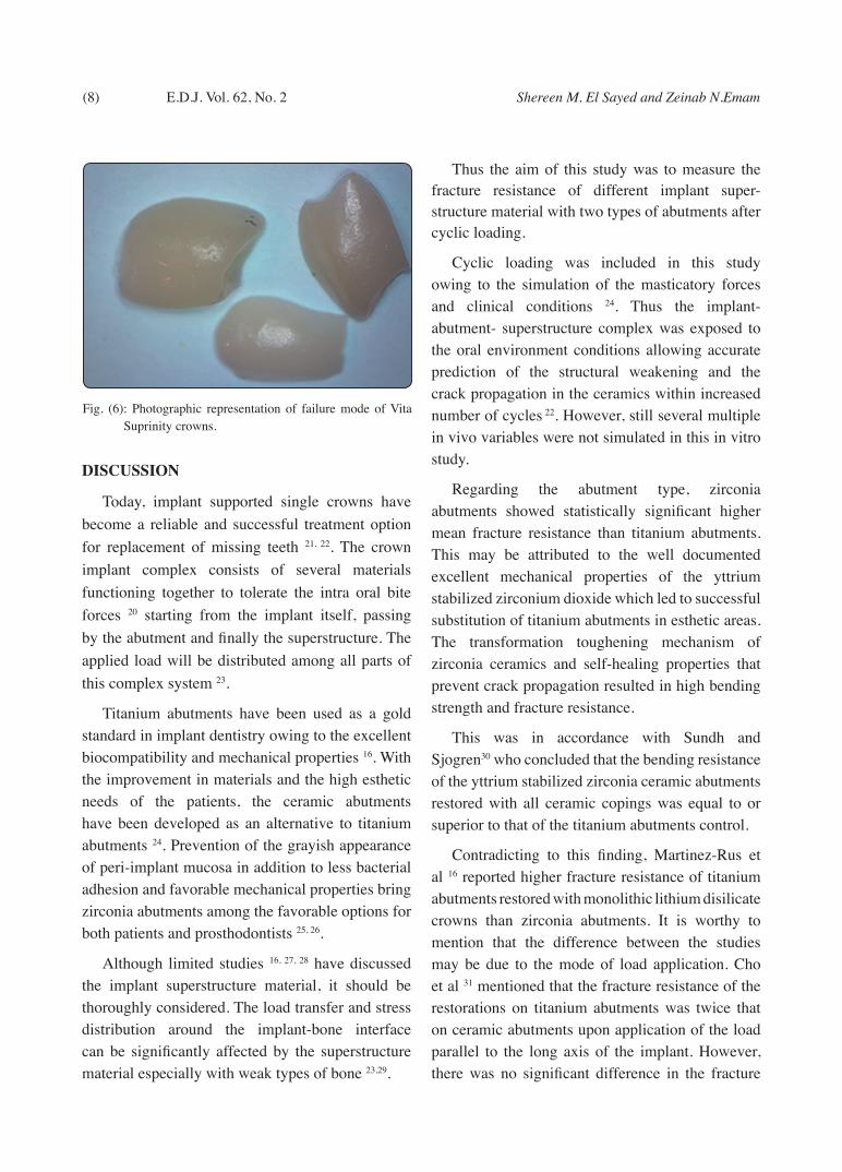
Subgroups B (e.max CAD crowns) and C (Vita Suprinity crowns) showed comparable failure modes where all the crowns revealed vertical splitting into three to four parts regardless of the abutment type. The fracture started at the margin and propagated occlusally. Fig. (4-6) showed representative sample from each subgroup.
Fig. (3): Bar chart representing mean and standard deviation values for fracture resistance of the two abutment types with each ceramic type.
Fig. (4): Photographic representation of failure mode of zirconia crowns over zirconia abutment.
Fig. (5): Photographic representation of failure mode of IPS e.max crowns over zirconia abutment.
(8) Shereen M. El Sayed and Zeinab N.EmamE.D.J. Vol. 62, No. 2
DISCUSSION
Today, implant supported single crowns have become a reliable and successful treatment option for replacement of missing teeth 21, 22. The crown implant complex consists of several materials functioning together to tolerate the intra oral bite forces 20 starting from the implant itself, passing by the abutment and finally the superstructure. The applied load will be distributed among all parts of this complex system 23.
Titanium abutments have been used as a gold standard in implant dentistry owing to the excellent biocompatibility and mechanical properties 16. With the improvement in materials and the high esthetic needs of the patients, the ceramic abutments have been developed as an alternative to titanium abutments 24. Prevention of the grayish appearance of peri-implant mucosa in addition to less bacterial adhesion and favorable mechanical properties bring zirconia abutments among the favorable options for both patients and prosthodontists 25, 26.
Although limited studies 16, 27, 28 have discussed the implant superstructure material, it should be thoroughly considered. The load transfer and stress distribution around the implant-bone interface can be significantly affected by the superstructure material especially with weak types of bone 23,29.
Thus the aim of this study was to measure the fracture resistance of different implant super-structure material with two types of abutments after cyclic loading.
Cyclic loading was included in this study owing to the simulation of the masticatory forces and clinical conditions 24. Thus the implant-abutment- superstructure complex was exposed to the oral environment conditions allowing accurate prediction of the structural weakening and the crack propagation in the ceramics within increased number of cycles 22. However, still several multiple in vivo variables were not simulated in this in vitro study.
Regarding the abutment type, zirconia abutments showed statistically significant higher mean fracture resistance than titanium abutments. This may be attributed to the well documented excellent mechanical properties of the yttrium stabilized zirconium dioxide which led to successful substitution of titanium abutments in esthetic areas. The transformation toughening mechanism of zirconia ceramics and self-healing properties that prevent crack propagation resulted in high bending strength and fracture resistance.
This was in accordance with Sundh and Sjogren30 who concluded that the bending resistance of the yttrium stabilized zirconia ceramic abutments restored with all ceramic copings was equal to or superior to that of the titanium abutments control.
Contradicting to this finding, Martinez-Rus et al 16 reported higher fracture resistance of titanium abutments restored with monolithic lithium disilicate crowns than zirconia abutments. It is worthy to mention that the difference between the studies may be due to the mode of load application. Cho et al 31 mentioned that the fracture resistance of the restorations on titanium abutments was twice that on ceramic abutments upon application of the load parallel to the long axis of the implant. However, there was no significant difference in the fracture
Fig. (6): Photographic representation of failure mode of Vita Suprinity crowns.

THE IMPACT OF IMPLANT ABUTMENT MATERIAL ON THE FRACTURE RESISTANCE (9)
resistance between restorations on both abutments when the load was applied obliquely.
This was opposed by the results of our study as the load was applied parallel to the long axis of the implant and still the zirconia abutments had higher fracture resistance than titanium abutments for all superstructure materials. This may be tentatively related to the factor of the cyclic loading.
Adhesive cementation was performed for the superstructure over the abutment for the benefit of stabilization and high retention of all ceramic crowns 32. Although many dentists still prefer the retrievability of the restorations cemented with provisional types of cements on implants 33.
Regarding the superstructure types, monolithic zirconia revealed the highest mean fracture resistance followed by e.max CAD and the Vita Suprinity showed the lowest mean fracture resistance for both types of abutments. This may be related to the difference in modulus of elasticity and fracture toughness of each material. The chemical composition and the initial fracture strength of each material play a significant role in the variation within the fracture resistance values.
Schaeffer et al 33 mentioned the importance of the choice of the abutments and its effect on the fracture resistance of the ceramic superstructure considering this complex as layered structure. Any elastic or plastic deformation of the abutments which is considered the foundation may subject the ceramic superstructure to bending forces under occlusal loads, thus decreasing its fracture resistance. They reached a conclusion that chipping of veneered ceramic could be reduced when a solid abutment and cement were used.
Moreover, previous studies 34-37 suggested that abutments with low elastic moduli were destructive to all ceramic crowns. They found that abutments with low elastic modulus bend more during masticatory simulation resulting in higher rate of
veneered ceramic chipping although differences were not significant 33. Scherrer and De Rijk 37 found that improved fracture toughness would be gained upon increasing the elastic modulus of the supporting material.
On the other hand, Martinez-Ruz et al 16 suggested that owing to the ductility of metals (Titanium abutments), bending resistance could compensate for the fracture of all ceramic superstructure and therefore less fracture resistance could be expected from zirconia abutment-ceramic compared to titanium abutment-ceramic complex.
On top of that the material of choice of the super structure is considered as important as the abutment material. Some authors suggested that when a material of low modulus of elasticity is used in the implant superstructure, it might act as a buffer zone to counteract the high stiffness of zirconia abutments leading to a better stress absorption and distribution 38, 39. In addition, the influence of cement type as an intermediate layer within these types of materials must be considered. Cements with high compressive strength used with implant super structure materials having low modulus of elasticity serve as stress buffers between super structure and implant improving the stability of the whole complex system 20.
Regarding the fractographic analysis, zirconia crowns showed cracks in most of the samples and two samples only showed complete vertical splitting into two halves. The high fracture strength and the modulus of elasticity of the monolithic zirconia ceramic material might have given an explanation to the expected limited risk of fracture. Although these properties lead to high fracture resistance values, more stress dissipation around the cervical region of the abutment and abutment-implant junction could be expected 40. However, no damage was detected on the abutments. The cracks initiated on the occlusal surface at the contact with the steel ball, although previous studies 41-43 reported tensile
(10) Shereen M. El Sayed and Zeinab N.EmamE.D.J. Vol. 62, No. 2
stress concentration at the cemented surface of all-ceramic crowns leading to initiation of cracks at this point rather than the occlusal loading point. This may be due to the use of the adhesive cementation which might have dampened the tensile stress at the bonded surface resulting in cracking at the loading point.
As regard to the e.max CAD and Vita Suprinity crowns, the fracture initiated from the cervical margin, propagated occlusally and splitted into three to four parts. This may be due to the concentration of the stresses at the margin. Thinning of the margin on the chamfer finish line of the abutments may be a contributing factor in the weakness of the lithium disilicate crowns even with the addition of the zirconia in the Vita Suprinity. This was supported by the study done by Abbate et al 44 who noticed cracks at the thin margins of all-ceramic crowns. The authors suggested that these cracks would have initiated the fracture under clinical conditions within a short period.
Finally, we can say that It is important to understand the forces applied to the whole complex, as the forces applied and absorbed on the implant superstructure will pass towards the other parts of the implant complex creating more stress distribution around the cervical part of the abutment and abutment-implant connection which in return will lead to bone resorption around the cervical parts and results in loss of the implant itself 40.
All the tested groups in this study were able to withstand the intraoral physiologic occlusal forces applied in the premolar area which is 450N 17, 45. The absence of the simulated humid environment and thermal stresses in the test design of this study is considered a limiting factor which might have affected the results as the aging effect of the materials and the degradation by water sorption were not included.
Further investigations will be needed to analyze the stress distribution mechanism on the bone around the implant system and on the implant system itself
upon using different implant-abutment-cement-superstructure combinations through the use of finite element analysis and strain gauges in a more simulated standardized thermo-mechanical fatigue conditions with increased number of cyclic loading. The numerical values of the static loading test must be correlated.
CONCLUSIONS
Based on the results of this study zirconia abutments showed better durability than titanium abutments with different all ceramic superstructure materials. Zirconia abutments with zirconia all ceramic superstructure showed the highest fracture resistance. All the tested superstructure and abutment materials can withstand the normal physiologic forces applied in the premolar area.
REFERENCES
1. Anja Z., Alexander O., Hermann P., Christoph H., Franz H., Arnold W., Irena S. Eleven-Year Follow-Up of a Prospective Study of Zirconia Implant Abutments Supporting Single All-Ceramic Crowns in Anterior and Premolar Regions. Clinical Implant Dentistry and Related Research 2014; 12:1-10
2. Yildirim M, Edelhoff D, Hanisch O, Spiekermann H. Ceramic abutments–a new era in achieving optimal esthetics in implant dentistry. Int J Periodontics Restorative Dent2000; 20:81–91.
3. Ormianer Z, Schiroli G. Maxillary single-tooth replacement utilizing a novel ceramic restorative system: results to 30 months. J Oral Implantol 2006; 32:190–199.
4. Reich S, Wichmann M, Nkenke E, Proeschel P. Clinical fit of all ceramic three-unit fixed partial dentures, generated with three different CAD/CAM systems Eur J Oral Sci 2005;113:174-9.
5. Strub JR, Rekow ED, Witkowski S. Computer-aided design and fabrication of dental restorations: Current systems and future possibilities. J Am Dent Assoc. 2006;137(9):1289-96.
6. Christensen GJ. Ceramics vs. porcelain fused to metal crowns: give your patients a choice. J Am Dent Assoc. 1994; 125:311,312, 314.

THE IMPACT OF IMPLANT ABUTMENT MATERIAL ON THE FRACTURE RESISTANCE (11)
7. Hansen PA, West LA. Allergic reaction following insertion of a Pd- Cu-Au fixed partial denture: a clinical report. J Prosthodont. 1997;6:144-148.
8. Vult Von Steyern P, Carlson P, Nilner K. All-ceramic fixed partial dentures designed according to the DC-Zirkon technique. A 2-year clinical study. J Oral Rehabil. 2005; 32:180-187.
9. Piwowarczyk A, Ottl P, Lauer HC, et al. A clinical report and overview of scientific studies and clinical procedures conducted on the 3M ESPE Lava All-Ceramic System. J Prosthodont. 2005; 14:39-45.
10. Studart AR, Filser F, Kocher P, et al. Cyclic fatigue in water of veneer-framework composites for all-ceramic dental bridges. Dent Mater. 2007;23:177-185.
11. Rismanchian M, Shafiei S, Nourbakhshian F, et al. Flexural strengths of implant-supported zirconia based bridges in posterior regions. J Adv Prosthodont. 2014;6:346-350.
12. Guess PC ,Zavanelli RA,Silva NR,Bonfante Ea, Coelho PG,Thompson Vp.Monolithic CAD/CAM lithium disilicate versus veneered Y-TZP crowns comparison of failure modes and reliability after fatigue.Int. Jprosthodont.2010,23(5) 434-42.
13. Saninno G,Germano F,Arcuri L,Bigelli E,Arcuri C.CEREC CAD/CAM chairsided system.Oral implantology 2014;57-70-7.
14. Beuer F, Stimmelmayer M, Gueth JF, Edelhoff D, Naumann M. In vitro performance of full contour zirconia single crowns. Dent Mater 2012; 28: 449-456.
15. Kokubo Y, Kano T, Tsumita M, Sakurai S , Itayama A and Fukushima S .Retention of zirconia copings on zirconia implant abutments cemented with provisional luting agents .Journal of Oral Rehabilitation 2010 37; 48–53
16. Martinez-Rus F, Ferreiroa A, Ozcan M, Bartolome J, Pradies G. Fracture resistance of crowns cemented on titanium and zirconia implant abutments: a comparison of monolithic versus manually veneered all ceramic systems. Int J Oral Maxillofac Implants 2012; 27: 1448-1455.
17. El S’adany AF, Masoud GE, Kamel MS, Korsel AM. Fracture resistance of all ceramic crowns supported by zirconia and alumina versus titanium implant abutments. Tanta Dental Journal 2013; 10(3): 103-111.
18. Jemt T, Karlsson S, Hed egard B. “Mandibular movement in young adults recorded by internally placed light-emitting diode.” J Prosthet Dent 1979 ; 42:669–73.
19. CraigRG. “Restorative Dental Materials.11th ed.” Missouri: Mosby; p.68. (2002).
20. Rohr N, Coldea A, Zitzmann N, Fischer J. Loading capacity of zirconia implant supported hybrid ceramic crowns. Dent Mater 2015; 31: e279-e288.
21. Almeida EO, Freitas AC, Bonfante EA, Marotta M, Silva N et al. Mechanical testing of implant-supported anterior crowns with different implant/abutment connections. Int J Oral Maxillofac Implants 2013; 28: 103-8.
22. Gehrke P, Johannson D, Fischer C, Stawarczyc B, Beuer F. In vitro fatigue and fracture resistance of one- and two-piece CAD/CAM zirconia implant abutments. Int J Oral Maxillofac Implants 2015; 30:546-554.
23. Soliman TA, Tamam RA, Yousief SA, El-Anwar MI. Assessment of stress distribution around implant fixture with three different crown materials. Tanta Dental Journal 2015; 12(4): 249-258.
24. Gehrke P, Dhom G, Brunner J, Wolf D, Degidi M et al. Zirconia implant abutments: fracture strength and influence of cyclic loading on retaining-screw loosening. Quint int J 2006; 37: 41-48.
25. hosseini M, Kleven E, Gotfredsen K. Fracture mode during cyclic loading of implant supported single tooth restorations. J Prosthet Dent 2012; 108: 74-83.
26. Nakamura K, Kanno T, Milleding P, Ortengren U. Zirconia as a dental implant abutment material: a systematic review. Int J Prosthodont 2010; 23: 299-309.
27. Gehrt M, Wolfart S, Rafai N, Reich S, Edelhoff D. Clinical results of lithium-disilicate crowns after up to 9 years of service. Clin Oral Investig 2013; 17: 275-84.
28. Albrecht T, Kirsten A, Kappert HF, Fischer H. Fracture load of different crown systems on zirconia implant abutments. Dent Mater 2011; 27:298-303.
29. Karl M, Rosch S, Graef F, Taylor TD, Heckmann SM. Strain situation after fixation of three-unit ceramic veneered implant superstructures. Implant Dent 2005; 14: 157-165.
30. Sundh A, Sjogren D. Astudy of the bending resistance of implant supported reinforced alumina and machined zirco-nia abutments and copies. Dent Mater 2008; 24: 611-617.
31. Cho HW, Dong JK, Jin TH, Oh SC, Lee HH et al. A study on the fracture strength of implant supported restorations using milled ceramic abutments and all ceramic crowns. Int J Prosthodont 2002; 15: 9-13.

(12) Shereen M. El Sayed and Zeinab N.EmamE.D.J. Vol. 62, No. 2
32. Tinschert J, Natt G, Mautsch W, Augthun M, Spiekermann H. Fracture resistance of lithium disilicate-, alumina-, and zirconia-based three-unit fixed partial dentures: a laboratory study. Int J Prosthodont 2001; 14(3):231-238.
33. Schafer L, Winkler C, Brandl G, Eckl S, Preis V et al. The impact of luting agent and stiffness of implant-abutments on marginal adaptation, chipping, and fracture resistance of zirconia crowns. J Mech Behav Biomed Mater 2014; 39: 279-291.
34. Kelly JR, Giordano R, Prober R, Cima MJ. Fracture surface analysis of dental ceramics: clinically failed restorations. Int J Prosthodont 1990; 3: 430-440.
35. Malament K, Socransky S. Survival of Dicor glass ceramic dental restorations over 16 years. Part III: effect of luting agent and tooth or tooth-substitute core structure. J Prosthet Dent 2001; 86(5): 511-519.
36. Rosentritt M, Plein T, Kolbeck C, Behr M, Handel G. In vitro fracture force and marginal adaptation of ceramic crowns fixed on natural and artificial teeth. Int J Prosthodont 2000; 13(5): 387-391.
37. Scherrer SS, De Rijk WG. The fracture resistance of all-ceramic crowns on supporting structures with different elastic moduli. Int J Prosthodont 1993; 6: 462-7.
38. Coldea A, Swain MV, Theil N. Hertzian contact response and damage tolerance of dental ceramics. J Mech Behav Biomed Mater 2014; 34: 124-133.
39. Christel P, Meunier A, Heller M, Torre JP, Peille CN. Mechanical properties and short-term in-vivo evaluation of yttrium oxide partially stabilized zirconia. J Biomed Mater Res 1989; 23: 45-61.
40. De Kok P, Kleverlaan CJ, De Jager N, Kuijs R, Feilzer AJ. Mechanical performance of implant supported posterior crowns. J Prosthet Dent 2015; 114: 59-66.
41. Campos RE, Soares CJ, Quagliatto PS, Soares PV, De Oliviera Jr OB et al. In vitro sudy of fracture load and fracture pattern of ceramic crowns: a finite element and fractography analysis. J Prosthodont 2011; 20:447-455.
42. Dong XD, Darvell BW. Stress distribution and failure mode of dental ceramic structures under hertzian indentation. Dent Mater 2003; 19: 542-551.
43. Scherrer SS, Quinn GD, Quinn JB. Fracographic failure analysis of a Procera AllCeram crown using stereo and scanning electron microscopy. Dent Mater 2008; 24: 1107-1113.
44. Abbate MF, Tjan AH, Fox WM. Comparison of the marginal fit of various ceramic crown systems. J Prosthet Dent 1989; 61:527–31.
45. Inan O, Secilmis A, Eraslan O. Effect of pontic framework design on the fracture resistance of implant supported all ceramic fixed partial dentures. J Appl Oral Sci 2009; 17(5): 533-8.