the immunoglobulin superfamily protein izumo is required for sperm to fuse with eggs
TRANSCRIPT

Received 13 September 2004; accepted 4 January 2005; doi:10.1038/nature03318.
1. Tabata, T. & Takei, Y. Morphogens, their identification and regulation. Development 131, 703–712
(2004).
2. St Johnston, R. D. & Gelbart, W. M. Decapentaplegic transcripts are localized along the dorsal-ventral
axis of the Drosophila embryo. EMBO J. 6, 2785–2791 (1987).
3. Arora, K., Levine, M. S. & O’Connor, M. B. The screw gene encodes a ubiquitously expressedmember
of the TGF-b family required for specification of dorsal cell fates in theDrosophila embryo.Genes Dev.
8, 2588–2601 (1994).
4. Dorfman, R. & Shilo, B. Z. Biphasic activation of the BMP pathway patterns theDrosophila embryonic
dorsal region. Development 128, 965–972 (2001).
5. Irish, V. F. & Gelbart, W. M. The decapentaplegic gene is required for dorsal-ventral patterning of the
Drosophila embryo. Genes Dev. 1, 868–879 (1987).
6. Rushlow, C., Colosimo, P. F., Lin, M. C., Xu, M. & Kirov, N. Transcriptional regulation of the
Drosophila gene zen by competing Smad and Brinker inputs. Genes Dev. 15, 340–351 (2001).
7. Ross, J. J. et al. Twisted gastrulation is a conserved extracellular BMP antagonist.Nature 410, 479–483
(2001).
8. Ferguson, E. L. & Anderson, K. V. Decapentaplegic acts as a morphogen to organize dorsal-ventral
pattern in the Drosophila embryo. Cell 71, 451–461 (1992).
9. Brummel, T. J. et al.Characterization and relationship of Dpp receptors encoded by the saxophone and
thick veins genes in Drosophila. Cell 78, 251–261 (1994).
10. Penton, A. et al. Identification of two bone morphogenetic protein type I receptors inDrosophila and
evidence that Brk25D is a decapentaplegic receptor. Cell 78, 239–250 (1994).
11. Letsou, A. et al. DrosophilaDpp signaling is mediated by the punt gene product: a dual ligand-binding
type II receptor of the TGF-b receptor family. Cell 80, 899–908 (1995).
12. Teleman, A. A. & Cohen, S.M. Dpp gradient formation in theDrosophilawing imaginal disc.Cell 103,
971–980 (2000).
13. Kosman, D. & Small, S. Concentration-dependent patterning by an ectopic expression domain of the
Drosophila gap gene knirps. Development 124, 1343–1354 (1997).
14. Decotto, E. & Ferguson, E. L. A positive role for Short gastrulation in modulating BMP signaling
during dorsoventral patterning in the Drosophila embryo. Development 128, 3831–3841 (2001).
15. Eldar, A. et al. Robustness of the BMP morphogen gradient in Drosophila embryonic patterning.
Nature 419, 304–308 (2002).
16. Shimmi, O. & O’Connor, M. B. Physical properties of Tld, Sog, Tsg and Dpp protein interactions are
predicted to help create a sharp boundary in Bmp signals during dorsoventral patterning of the
Drosophila embryo. Development 130, 4673–4682 (2003).
17. Holley, S. A. et al. The Xenopus dorsalizing factor Noggin ventralizes Drosophila embryos by
preventing DPP from activating its receptor. Cell 86, 607–617 (1996).
18. Ashe, H. L. & Levine, M. Local inhibition and long-range enhancement of Dpp signal transduction by
Sog. Nature 398, 427–431 (1999).
19. Marques, G. et al. Production of a DPP activity gradient in the early Drosophila embryo through the
opposing actions of the SOG and TLD proteins. Cell 91, 417–426 (1997).
20. Oelgeschlager, M. et al. The pro-BMPactivity of Twisted gastrulation is independent of BMP binding.
Development 130, 4047–4056 (2003).
21. Neul, J. L. & Ferguson, E. L. Spatially restricted activation of the SAX receptor by SCWmodulates
DPP/TKV signaling in Drosophila dorsal-ventral patterning. Cell 95, 483–494 (1998).
22. Nguyen, M., Park, S., Marques, G. & Arora, K. Interpretation of a BMP activity gradient inDrosophila
embryos depends on synergistic signaling by two type I receptors, SAX and TKV. Cell 95, 495–506
(1998).
23. Shimmi, O., Umulis,D.,Othmer, H.&O’Connor,M. B. Facilitated transport of a Dpp/Scwheterodimer
by Sog/Tsg leads to robust patterning of the Drosophila blastoderm embryo. Cell (in the press).
24. Mason, E. D., Williams, S., Grotendorst, G. R. & Marsh, J. L. Combinatorial signaling by Twisted
Gastrulation and Decapentaplegic. Mech. Dev. 64, 61–75 (1997).
25. Wisotzkey, R. G. et al. Medea is a Drosophila Smad4 homolog that is differentially required to
potentiate DPP responses. Development 125, 1433–1445 (1998).
26. Rushlow, C., Doyle, H., Hoey, T. & Levine,M.Molecular characterization of the zerknullt region of the
Antennapedia gene complex in Drosophila. Genes Dev. 1, 1268–1279 (1987).
27. Ferrell, J. E. Self-perpetuating states in signal transduction: positive feedback, double-negative
feedback and bistability. Curr. Opin. Cell Biol. 14, 140–148 (2002).
28. Lai, K., Robertson, M. J. & Schaffer, D. V. The Sonic hedgehog signaling system as a bistable genetic
switch. Biophys. J. 86, 2748–2757 (2004).
29. Stein, D., Roth, S., Vogelsang, E. & Nusslein-Volhard, C. The polarity of the dorsoventral axis in the
Drosophila embryo is defined by an extracellular signal. Cell 65, 725–735 (1991).
30. Little, S. C. &Mullins, M. C. Twisted gastrulation promotes BMP signaling in zebrafish dorsal-ventral
axial patterning. Development 131, 5825–5835 (2004).
31. Xie, J. & Fisher, S. Twisted gastrulation enhances BMP signaling through chordin dependent and
independent mechanisms. Development 132, 383–391 (2005).
Supplementary Information accompanies the paper on www.nature.com/nature.
Acknowledgements We thank N. Dostatni, M. Levine and M. Markstein for fly stocks; S. Cohen,
M. Levine and M. Markstein for plasmids; P. ten Dijke and C. Heldin for antibodies; V. Bindokas
for assistance with confocal microscopy; S. Lemke and M. Markstein for discussions; D. Bishop,
M. O. Casanueva, C. Li, A. Mahowald, J. Malamy,M.Markstein, V. Prince and J. Staley for helpful
comments on the manuscript, and M. O’Connor for sharing unpublished data and reagents.
Preliminary data on the second function of Tsg were obtained by E. Decotto. Funding was
provided by grants to E.L.F. from the NIH and from the Human Frontiers Science program.
Competing interests statement The authors declare that they have no competing financial
interests.
Correspondence and requests for materials should be addressed E.L.F.
..............................................................
The immunoglobulin superfamilyprotein Izumo is required forsperm to fuse with eggsNaokazu Inoue1, Masahito Ikawa2, Ayako Isotani1,3 & Masaru Okabe1,2,3
1Genome Information ResearchCenter, 2Research Institute forMicrobial Diseases,3Faculty of Pharmaceutical Sciences, Osaka University, Yamadaoka 3-1, Suita,Osaka 565-0871, Japan.............................................................................................................................................................................
Representing the 60 trillion cells that build a human body, asperm and an egg meet, recognize each other, and fuse to form anew generation of life. The factors involved in this importantmembrane fusion event, fertilization, have been sought for a longtime1. Recently, CD9 on the egg membrane was found to beessential for fusion2–4, but sperm-related fusion factors remainunknown. Here, by using a fusion-inhibiting monoclonalantibody5 and gene cloning, we identify a mouse sperm fusion-related antigen and show that the antigen is a novel immuno-globulin superfamily protein. We have termed the gene Izumoand produced a gene-disrupted mouse line. Izumo2/2 mice werehealthy but males were sterile. They produced normal-lookingsperm that bound to and penetrated the zona pellucida but wereincapable of fusing with eggs. Human sperm also contain Izumoand addition of the antibody against human Izumo left the spermunable to fuse with zona-free hamster eggs.
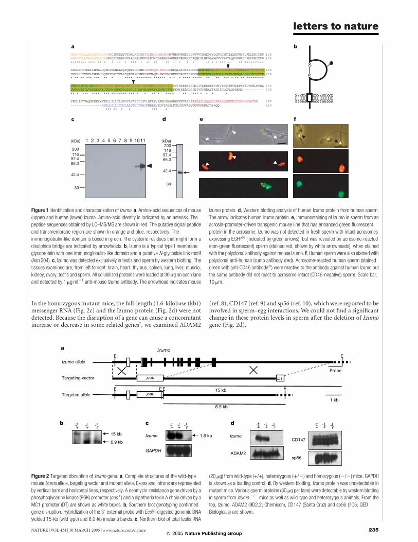
To identify factors involved in sperm–egg fusion, we used amonoclonal antibody, OBF13, against mouse sperm that specificallyinhibits the fusion process5. The antigen was identified by separ-ation of the crude extracts from mouse sperm by two-dimensionalgel electrophoresis and subsequent immunoblotting with themonoclonal antibody. We termed the antigen ‘Izumo’ after aJapanese shrine dedicated to marriage. The identified spot wasanalysed by liquid chromatography tandem mass spectrometry(LC–MS/MS), and ten peptides that were 100% identical to a partof the sequence listed in the RIKEN full-length database (NationalCentre for Biotechnology Information (NCBI) accession numberXM_133424) were found. The registered DNA sequence was con-firmed by sequencing after polymerase chain reaction with reversetranscription (RT–PCR) with total RNA prepared from the testis. Ahuman homologue was found as an unverified gene in the NCBIdatabase (accession number BC034769). The gene encodes a novelimmunoglobulin superfamily (IgSF), type I membrane protein withan extracellular immunoglobulin domain that contains one puta-tive glycosylation site (Fig. 1a, b). Mouse Izumo was shown to be atestis (sperm)-specific 56.4-kDa antigen by western blotting with apolyclonal antibody raised against recombinant mouse Izumo(Fig. 1c). Izumo was also detectable as a 37.2-kDa protein bywestern blotting of human spermwith anti-human Izumo antibody(Fig. 1d). Izumo was not detectable on the surface of fresh sperm.Coinciding with the fact that mammalian sperm are incapable offertilizing eggs when ejaculated and that fertilization occurs onlyafter an exocytotic process called the acrosome reaction, bothmouse and human Izumo became detectable on sperm surfaceonly after the acrosome reaction (Fig. 1e, f). This would probably bebecause Izumo is not localized on plasma membrane of freshspermatozoa but is hidden under plasma membrane and accessibleafter the acrosome reaction, as occurs with CD46 on mouse sperm6.
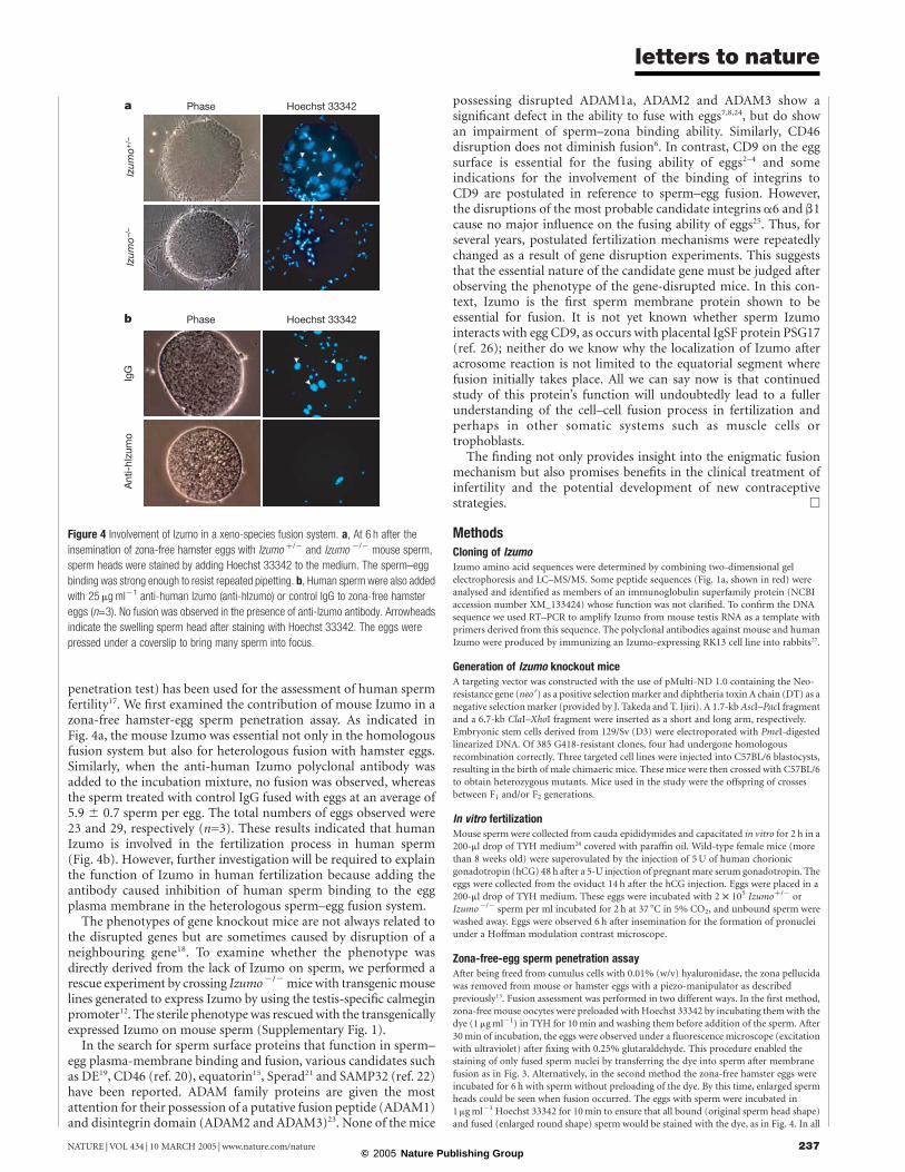
To address the physiological role of Izumo in vivo we generatedIzumo-deficient mice by homologous recombination. An Izumo-targeting construct was designed to replace exons 2–10 with aneomycin-resistant gene (neor) (Fig. 2a). Both the targeting eventin D3 embryonic stem cells and the germline transmission oftargeted genes were confirmed by Southern blot analysis (Fig. 2b).
letters to nature
NATURE | VOL 434 | 10 MARCH 2005 | www.nature.com/nature234© 2005 Nature Publishing Group
In the homozygous mutant mice, the full-length (1.6-kilobase (kb))messenger RNA (Fig. 2c) and the Izumo protein (Fig. 2d) were notdetected. Because the disruption of a gene can cause a concomitantincrease or decrease in some related genes7, we examined ADAM2
(ref. 8), CD147 (ref. 9) and sp56 (ref. 10), which were reported to beinvolved in sperm–egg interactions. We could not find a significantchange in these protein levels in sperm after the deletion of Izumogene (Fig. 2d).
Figure 1 Identification and characterization of Izumo. a, Amino-acid sequences of mouse
(upper) and human (lower) Izumo. Amino-acid identity is indicated by an asterisk. The
peptide sequences obtained by LC–MS/MS are shown in red. The putative signal peptide
and transmembrane region are shown in orange and blue, respectively. The
immunoglobulin-like domain is boxed in green. The cysteine residues that might form a
disulphide bridge are indicated by arrowheads. b, Izumo is a typical type I membrane
glycoprotein with one immunoglobulin-like domain and a putative N-glycoside link motif
(Asn 204). c, Izumo was detected exclusively in testis and sperm by western blotting. The
tissues examined are, from left to right: brain, heart, thymus, spleen, lung, liver, muscle,
kidney, ovary, testis and sperm. All solubilized proteins were loaded at 30 mg on each lane
and detected by 1mgml21 anti-mouse Izumo antibody. The arrowhead indicates mouse
Izumo protein. d, Western blotting analysis of human Izumo protein from human sperm.
The arrow indicates human Izumo protein. e, Immunostaining of Izumo in sperm from an
acrosin-promoter-driven transgenic mouse line that has enhanced green fluorescent
protein in the acrosome. Izumo was not detected in fresh sperm with intact acrosomes
expressing EGFP30 (indicated by green arrows), but was revealed on acrosome-reacted
(non-green fluorescent) sperm (stained red, shown by white arrowheads), when stained
with the polyclonal antibody against mouse Izumo. f, Human sperm were also stained with
polyclonal anti-human Izumo antibody (red). Acrosome-reacted human sperm (stained
green with anti-CD46 antibody31) were reactive to the antibody against human Izumo but
the same antibody did not react to acrosome-intact (CD46-negative) sperm. Scale bar,
10 mm.
Figure 2 Targeted disruption of Izumo gene. a, Complete structures of the wild-type
mouse Izumo allele, targeting vector and mutant allele. Exons and introns are represented
by vertical bars and horizontal lines, respectively. A neomycin-resistance gene driven by a
phosphoglycerine kinase (PGK) promoter (neo r ) and a diphtheria toxin A chain driven by a
MC1 promoter (DT) are shown as white boxes. b, Southern blot genotyping confirmed
gene disruption. Hybridization of the 30external probe with EcoRI-digested genomic DNA
yielded 15-kb (wild type) and 6.9-kb (mutant) bands. c, Northern blot of total testis RNA
(20mg) from wild-type (+/+), heterozygous (þ/2) and homozygous (2/2) mice. GAPDH
is shown as a loading control. d, By western blotting, Izumo protein was undetectable in
mutant mice. Various sperm proteins (30mg per lane) were detectable by western blotting
in sperm from Izumo 2/2 mice as well as wild-type and heterozygous animals. From the
top, Izumo, ADAM2 (9D2.2; Chemicon), CD147 (Santa Cruz) and sp56 (7C5; QED
Biologicals) are shown.
letters to nature
NATURE |VOL 434 | 10 MARCH 2005 | www.nature.com/nature 235© 2005 Nature Publishing Group

Intercrosses between heterozygous F1 mice yielded offspring thatsegregated in a mendelian distribution: 43 wild-type, 92 hetero-zygous and 47 homozygous mutant weaning pups. Izumo2/2
mutant mice were healthy and showed no overt developmentalabnormalities. Izumo2/2 females demonstrated normal fecundity.Izumoþ/2 males also showed normal fertilizing ability (Fig. 3a).However, Izumo2/2 males were sterile despite normal matingbehaviour and ejaculation, with normal vaginal plug formations.After observation of 28 plugs, nine pairs of Izumo2/2 male andwild-type females were kept for another 4 months but no pregnan-cies were observed (Fig. 3a). In at least four different cases of geneknockouts that resulted in male sterility attributed to impairedzona-binding ability, the sperm also failed to migrate into theoviduct7,8,11,12. However, disruption of Izumo did not cause anydefect in sperm migration into the oviduct (data not shown, andthere was no reduction of sperm motility in Izumo2/2 sperm;motility was measured 120min after incubation by computer-aidedsperm analysis (CASA; mean ^ s.e.m. ¼ 81.7 ^ 7.7% in Izumoþ/2
sperm and 77 ^ 8.9% in Izumo2/2 sperm)). The sterile nature ofIzumo2/2 sperm was shown in the in vitro fertilization system(Fig. 3b, c, and Supplementary Movie 1). The impaired fertilizationstep undoubtedly followed zona penetration because spermpenetrated the zona pellucida and accumulated in the perivitellinespace of the eggs (Fig. 3d).Syngamy can be considered to occur to two stages: binding of the
sperm plasma membrane to that of the egg, and actual membranefusion. Izumo2/2 sperm were capable of binding to the plasmamembranes of eggs whose zona pellucida had been mechanicallyremoved13(Fig. 3e, f). In this system, the Izumoþ/2 sperm incubatedfor 2 and 6 h fused to eggs in approximate ratios of 4.5 and 6 spermper egg, respectively, but no Izumo2/2 sperm fused with eggs.
Sperm cannot fuse with eggs unless the former have undergonethe acrosome reaction14. To verify the acrosomal status of Izumo2/2
sperm, we stained the sperm accumulated in perivitelline spaceswith the MN9 monoclonal antibody, which immunoreacts only tothe equatorial segment of acrosome-reacted sperm15. The stainingindicated that the Izumo2/2 sperm had undergone the acrosomereaction (Fig. 3d) but failed to fuse with eggs.
Because no offspring were fathered by Izumo2/2 male mice, itwas unclear whether the defect was limited to fusion or extended tolater developmental stages. To address this question, we usedintracytoplasmic sperm injection (ICSI) to insert Izumo2/2
sperm directly into the cytoplasm of wild-type eggs and bypassthe fusion step16. Eggs injected with Izumo2/2 sperm were success-fully activated and the fertilized eggs were transplanted into theoviducts of pseudopregnant females. The eggs implanted normallyand the resulting embryos developed appropriately to term withrates similar to those of heterozygous mice (Table 1).
Sperm–egg fusion is known to be less species-specific thansperm–zona interaction. For example, human sperm cannotpenetrate the hamster zona pellucida but they can fuse with zona-free hamster eggs, and this system (zona-free hamster-egg sperm
Figure 3 Male infertility caused by Izumo disruption. a, Fecundity of Izumo þ/2 and
Izumo 2/2 males and Izumo 2/2 females. The numbers in parentheses indicate the
numbers of mating pairs. b, c, In vitro fertilization of sperm from Izumo þ/2 and
Izumo 2/2 mice. Unlike Izumo þ/2, the eggs inseminated with Izumo 2/2 sperm had
many sperm on their zona pellucida, owing to the failure of sperm–egg fusion that
probably leads to the absence of zona-reaction to lessen the sperm-binding ability of the
zona pellucida. The error bars in b are not visible (n ¼ 5). d, Upper panel, accumulation of
many sperm in the perivitelline space of the eggs recovered from the females mated with
Izumo 2/2 males. Lower panel, sperm in perivitelline space labelled with acrosome-
reacted, sperm-specific monoclonal antibody MN9 (ref. 15). e, Average numbers of fused
sperm observed 2 and 6 h after insemination (n ¼ 5). f, Fused sperm stained by Hoechst
33342 preloaded into the egg. The arrowheads show the fused sperm. Errors bars in a
and e are s.e.m.
Table 1 Development of eggs after ICSI with Izumo2/2 sperm
Sperm No. ofeggs used
No. of eggssurvivingafter ICSI
No. of eggsdeveloping totwo-cell stage
No. ofpups born
.............................................................................................................................................................................
Izumo2/2 95 59 42 (71%)* 12 (29%)†Izumoþ/2 85 54 43 (80%) 6 (14%).............................................................................................................................................................................
*Percentages are based on numbers of eggs surviving after ICSI.†All offspring from Izumo2/2 sperm were confirmed to possess the Izumo-null allele.
letters to nature
NATURE | VOL 434 | 10 MARCH 2005 | www.nature.com/nature236© 2005 Nature Publishing Group
penetration test) has been used for the assessment of human spermfertility17. We first examined the contribution of mouse Izumo in azona-free hamster-egg sperm penetration assay. As indicated inFig. 4a, the mouse Izumo was essential not only in the homologousfusion system but also for heterologous fusion with hamster eggs.Similarly, when the anti-human Izumo polyclonal antibody wasadded to the incubation mixture, no fusion was observed, whereasthe sperm treated with control IgG fused with eggs at an average of5.9 ^ 0.7 sperm per egg. The total numbers of eggs observed were23 and 29, respectively (n=3). These results indicated that humanIzumo is involved in the fertilization process in human sperm(Fig. 4b). However, further investigation will be required to explainthe function of Izumo in human fertilization because adding theantibody caused inhibition of human sperm binding to the eggplasma membrane in the heterologous sperm–egg fusion system.
The phenotypes of gene knockout mice are not always related tothe disrupted genes but are sometimes caused by disruption of aneighbouring gene18. To examine whether the phenotype wasdirectly derived from the lack of Izumo on sperm, we performed arescue experiment by crossing Izumo2/2mice with transgenicmouselines generated to express Izumo by using the testis-specific calmeginpromoter12. The sterile phenotypewas rescuedwith the transgenicallyexpressed Izumo on mouse sperm (Supplementary Fig. 1).
In the search for sperm surface proteins that function in sperm–egg plasma-membrane binding and fusion, various candidates suchas DE19, CD46 (ref. 20), equatorin15, Sperad21 and SAMP32 (ref. 22)have been reported. ADAM family proteins are given the mostattention for their possession of a putative fusion peptide (ADAM1)and disintegrin domain (ADAM2 and ADAM3)23. None of the mice
possessing disrupted ADAM1a, ADAM2 and ADAM3 show asignificant defect in the ability to fuse with eggs7,8,24, but do showan impairment of sperm–zona binding ability. Similarly, CD46disruption does not diminish fusion6. In contrast, CD9 on the eggsurface is essential for the fusing ability of eggs2–4 and someindications for the involvement of the binding of integrins toCD9 are postulated in reference to sperm–egg fusion. However,the disruptions of the most probable candidate integrins a6 and b1cause no major influence on the fusing ability of eggs25. Thus, forseveral years, postulated fertilization mechanisms were repeatedlychanged as a result of gene disruption experiments. This suggeststhat the essential nature of the candidate gene must be judged afterobserving the phenotype of the gene-disrupted mice. In this con-text, Izumo is the first sperm membrane protein shown to beessential for fusion. It is not yet known whether sperm Izumointeracts with egg CD9, as occurs with placental IgSF protein PSG17(ref. 26); neither do we know why the localization of Izumo afteracrosome reaction is not limited to the equatorial segment wherefusion initially takes place. All we can say now is that continuedstudy of this protein’s function will undoubtedly lead to a fullerunderstanding of the cell–cell fusion process in fertilization andperhaps in other somatic systems such as muscle cells ortrophoblasts.The finding not only provides insight into the enigmatic fusion
mechanism but also promises benefits in the clinical treatment ofinfertility and the potential development of new contraceptivestrategies. A
MethodsCloning of IzumoIzumo amino acid sequences were determined by combining two-dimensional gelelectrophoresis and LC–MS/MS. Some peptide sequences (Fig. 1a, shown in red) wereanalysed and identified as members of an immunoglobulin superfamily protein (NCBIaccession number XM_133424) whose function was not clarified. To confirm the DNAsequence we used RT–PCR to amplify Izumo from mouse testis RNA as a template withprimers derived from this sequence. The polyclonal antibodies against mouse and humanIzumo were produced by immunizing an Izumo-expressing RK13 cell line into rabbits27.
Generation of Izumo knockout miceA targeting vector was constructed with the use of pMulti-ND 1.0 containing the Neo-resistance gene (neor) as a positive selectionmarker and diphtheria toxin A chain (DT) as anegative selectionmarker (provided by J. Takeda and T. Ijiri). A 1.7-kb AscI–PacI fragmentand a 6.7-kb ClaI–XhoI fragment were inserted as a short and long arm, respectively.Embryonic stem cells derived from 129/Sv (D3) were electroporated with PmeI-digestedlinearized DNA. Of 385 G418-resistant clones, four had undergone homologousrecombination correctly. Three targeted cell lines were injected into C57BL/6 blastocysts,resulting in the birth of male chimaeric mice. These mice were then crossed with C57BL/6to obtain heterozygous mutants. Mice used in the study were the offspring of crossesbetween F1 and/or F2 generations.
In vitro fertilizationMouse sperm were collected from cauda epididymides and capacitated in vitro for 2 h in a200-ml drop of TYH medium28 covered with paraffin oil. Wild-type female mice (morethan 8 weeks old) were superovulated by the injection of 5U of human chorionicgonadotropin (hCG) 48 h after a 5-U injection of pregnantmare serum gonadotropin. Theeggs were collected from the oviduct 14 h after the hCG injection. Eggs were placed in a200-ml drop of TYH medium. These eggs were incubated with 2 £ 105 Izumoþ/2 orIzumo2/2 sperm per ml incubated for 2 h at 37 8C in 5% CO2, and unbound sperm werewashed away. Eggs were observed 6 h after insemination for the formation of pronucleiunder a Hoffman modulation contrast microscope.
Zona-free-egg sperm penetration assayAfter being freed from cumulus cells with 0.01% (w/v) hyaluronidase, the zona pellucidawas removed from mouse or hamster eggs with a piezo-manipulator as describedpreviously13. Fusion assessment was performed in two different ways. In the first method,zona-free mouse oocytes were preloaded with Hoechst 33342 by incubating themwith thedye (1 mgml21) in TYH for 10min and washing them before addition of the sperm. After30min of incubation, the eggs were observed under a fluorescence microscope (excitationwith ultraviolet) after fixing with 0.25% glutaraldehyde. This procedure enabled thestaining of only fused sperm nuclei by transferring the dye into sperm after membranefusion as in Fig. 3. Alternatively, in the second method the zona-free hamster eggs wereincubated for 6 h with sperm without preloading of the dye. By this time, enlarged spermheads could be seen when fusion occurred. The eggs with sperm were incubated in1mgml21 Hoechst 33342 for 10min to ensure that all bound (original sperm head shape)and fused (enlarged round shape) sperm would be stained with the dye, as in Fig. 4. In all
Figure 4 Involvement of Izumo in a xeno-species fusion system. a, At 6 h after the
insemination of zona-free hamster eggs with Izumo þ/2 and Izumo 2/2 mouse sperm,
sperm heads were stained by adding Hoechst 33342 to the medium. The sperm–egg
binding was strong enough to resist repeated pipetting. b, Human sperm were also added
with 25 mgml21 anti-human Izumo (anti-hIzumo) or control IgG to zona-free hamster
eggs (n=3). No fusion was observed in the presence of anti-Izumo antibody. Arrowheads
indicate the swelling sperm head after staining with Hoechst 33342. The eggs were
pressed under a coverslip to bring many sperm into focus.
letters to nature
NATURE |VOL 434 | 10 MARCH 2005 | www.nature.com/nature 237© 2005 Nature Publishing Group

experiments the human sperm were collected from liquefied semen by the swim-upmethod and incubated for 6 h before addition to eggs. We used BWWmedium29
containing 35mgml21 human serum albumin (HSA) for the human sperm experiment.
Received 12 October 2004; accepted 17 January 2005; doi:10.1038/nature03362.
1. Stein, K. K., Primakoff, P. & Myles, D. Sperm–egg fusion: events at the plasma membrane. J. Cell Sci.
117, 6269–6274 (2004).
2. Miyado, K. et al. Requirement of CD9 on the egg plasma membrane for fertilization. Science 287,
321–324 (2000).
3. LeNaour, F., Rubinstein, E., Jasmin, C., Prenant,M. &Boucheix, C. Severely reduced female fertility in
CD9-deficient mice. Science 287, 319–321 (2000).
4. Kaji, K. et al. The gamete fusion process is defective in eggs of CD9-deficient mice. Nature Genet. 24,
279–282 (2000).
5. Okabe, M. et al. Capacitation-related changes in antigen distribution on mouse sperm heads and its
relation to fertilization rate in vitro. J. Reprod. Immunol. 11, 91–100 (1987).
6. Inoue, N. et al. Disruption of mouse CD46 causes an accelerated spontaneous acrosome reaction in
sperm. Mol. Cell. Biol. 23, 2614–2622 (2003).
7. Nishimura, H., Kim, E., Nakanishi, T. & Baba, T. Possible function of the ADAM1a/ADAM2 fertilin
complex in the appearance of ADAM3 on the sperm surface. J. Biol. Chem. 279, 34957–34962 (2004).
8. Cho, C. et al. Fertilization defects in sperm from mice lacking fertilin beta. Science 281, 1857–1859
(1998).
9. Saxena, D. K., Oh-Oka, T., Kadomatsu, K., Muramatsu, T. & Toshimori, K. Behaviour of a sperm
surface transmembrane glycoprotein basigin during epididymal maturation and its role in
fertilization in mice. Reproduction 123, 435–444 (2002).
10. Bookbinder, L. H., Cheng, A. & Bleil, J. D. Tissue- and species-specific expression of sp56, a mouse
sperm fertilization protein. Science 269, 86–89 (1995).
11. Hagaman, J. R. et al. Angiotensin-converting enzyme and male fertility. Proc. Natl Acad. Sci. USA 95,
2552–2557 (1998).
12. Ikawa, M. et al. Calmegin is required for fertilin alpha/beta heterodimerization and sperm fertility.
Dev. Biol. 240, 254–261 (2001).
13. Yamagata, K. et al. Sperm from the calmegin-deficient mouse have normal abilities for binding and
fusion to the egg plasma membrane. Dev. Biol. 250, 348–357 (2002).
14. Yanagimachi, R. Mammalian Fertilization (eds Knobil, E & Neill, J. D.) (Raven, New York, 1994).
15. Manandhar, G. & Toshimori, K. Exposure of sperm head equatorin after acrosome reaction and its
fate after fertilization in mice. Biol. Reprod. 65, 1425–1436 (2001).
16. Kimura, Y. & Yanagimachi, R. Intracytoplasmic sperm injection in the mouse. Biol. Reprod. 52,
709–720 (1995).
17. Yanagimachi, R., Yanagimachi, H. & Rogers, B. J. The use of zona-free animal ova as a test-system for
the assessment of the fertilizing capacity of human spermatozoa. Biol. Reprod. 15, 471–476 (1976).
18. Olson, E. N., Arnold, H. H., Rigby, P. W. &Wold, B. J. Know your neighbors: three phenotypes in null
mutants of the myogenic bHLH gene MRF4. Cell 85, 1–4 (1996).
19. Rochwerger, L., Cohen, D. J. & Cuasnicu, P. S. Mammalian sperm–egg fusion: the rat egg has
complementary sites for a sperm protein that mediates gamete fusion. Dev. Biol. 153, 83–90 (1992).
20. Anderson, D. J., Abbott, A. F. & Jack, R. M. The role of complement component C3b and its receptors
in sperm–oocyte interaction. Proc. Natl Acad. Sci. USA 90, 10051–10055 (1993).
21. Ilayperuma, I. Identification of the 48-kDa G11 protein from guinea pig testes as sperad. J. Exp. Zool.
293, 617–623 (2002).
22. Hao, Z. et al. SAMP32, a testis-specific, isoantigenic sperm acrosomal membrane-associated protein.
Biol. Reprod. 66, 735–744 (2002).
23. Blobel, C. P. et al. A potential fusion peptide and an integrin ligand domain in a protein active in
sperm–egg fusion. Nature 356, 248–252 (1992).
24. Nishimura, H., Cho, C., Branciforte, D. R., Myles, D. G. & Primakoff, P. Analysis of loss of adhesive
function in sperm lacking cyritestin or fertilin beta. Dev. Biol. 233, 204–213 (2001).
25. He, Z. Y. et al.None of the integrins known to be present on the mouse egg or to be ADAM receptors
are essential for sperm–egg binding and fusion. Dev. Biol. 254, 226–237 (2003).
26. Ellerman, D. A., Ha, C., Primakoff, P., Myles, D. G. & Dveksler, G. S. Direct binding of the ligand
PSG17 to CD9 requires a CD9 site essential for sperm–egg fusion.Mol. Biol. Cell 14, 5098–5103 (2003).
27. Inoue, N. et al. A novel chicken membrane-associated complement regulatory protein: molecular
cloning and functional characterization. J. Immunol. 166, 424–431 (2001).
28. Toyoda, Y., Yokoyama, M. & Hoshi, T. Studies on the fertilization of mouse egg in vitro. Jpn. J. Anim.
Reprod. 16, 147–151 (1971).
29. Overstreet, J. W., Yanagimachi, R., Katz, D. F., Hayashi, K. & Hanson, F. W. Penetration of human
spermatozoa into the human zona pellucida and the zona-free hamster egg: a study of fertile donors
and infertile patients. Fertil. Steril. 33, 534–542 (1980).
30. Nakanishi, T. et al. Real-time observation of acrosomal dispersal from mouse sperm using GFP as a
marker protein. FEBS Lett. 449, 277–283 (1999).
31. Okabe, M. et al. A human sperm antigen possibly involved in binding and/or fusion with zona-free
hamster eggs. Fertil. Steril. 54, 1121–1126 (1990).
Supplementary Information accompanies the paper on www.nature.com/nature.
Acknowledgements We thank K. Toshimori for providing anti-MN9 antibody; K. Yamagata for
discussions; G. L. Gerton and S.Moss for critically reviewing the draft; and Y.Maruyama,A. Kawai
and Y. Koreeda for technical assistance with gene disruption. This work was supported by grant-
in-aid for Scientific Research and the 21st Century COE program from theMinistry of Education,
Culture, Sports, Science, and Technology of Japan.
Competing interests statement The authors declare that they have no competing financial
interests.
Correspondence and requests for materials should be addressed to M.O.
(e-mail: [email protected]). Sequences have been deposited with GenBank under
accession numbers AB195681 for mouse Izumo, AB195682 for human Izumo and AB195683 for
rat Izumo.
..............................................................
Agonist/endogenous peptide–MHCheterodimers drive T cell activationand sensitivityMichelle Krogsgaard1, Qi-jing Li1, Cenk Sumen1*, Johannes B. Huppa1,2,Morgan Huse1 & Mark M. Davis1,2
1The Department of Microbiology and Immunology, and 2Howard HughesMedical Institute, Stanford University School of Medicine, Stanford, California94305, USA
* Present address: Center for Blood Research, Harvard Medical School, Boston, Massachusetts 02115,
USA
.............................................................................................................................................................................
ab T lymphocytes are able to detect even a single peptide–majorhistocompatibility complex (MHC) on the surface of an antigen-presenting cell1,2. This is despite clear evidence, at least withCD41 T cells, that monomeric ligands are not stimulatory3,4. Inan effort to understand how this remarkable sensitivity isachieved, we constructed soluble peptide–MHC heterodimersin which one peptide is an agonist and the other is one of thelarge number of endogenous peptide–MHCs displayed by pre-senting cells. We found that some specific combinations of theseheterodimers can stimulate specific T cells in a CD4-dependentmanner. This activation is severely impaired if the CD4-bindingsite on the agonist ligand is ablated, but the same mutation on anendogenous ligand has no effect. These data correlate well withanalyses of lipid bilayers and cells presenting these ligands, andindicate that the basic unit of helper T cell activation is aheterodimer of agonist peptide– and endogenous peptide–MHC complexes, stabilized by CD4.
It has been known for some time that ab T-cell antigen receptor(TCR)-bearing T cells are selected for weak or rare interactions withsome of the thousands of self-peptide–MHC (pMHC) complexes(that is, endogenous peptide–MHC complexes) presented in thethymus and also require the presence of these complexes in theperiphery to survive5–7. However, the role of self-peptide–MHCcomplexes in the activation of mature T cells is controversial.Although there is some evidence to suggest that endogenouspeptides or their mimics enhance CD4þ helper T cell activation8,and that T cell responsiveness declines rapidly in the absence ofendogenous pMHC contact7, others working with CD8þ T cellshave found no effect9. Recently, we have found that there is asubstantial (,20%) recruitment of endogenous peptide–MHCmolecules into the immunological synapse1,8. This seems to bedriven by weak interactions with TCRs. This phenomenon, togetherwith data showing that even a single agonist peptide–MHC complexcan initiate T cell activation1,2 has led to the suggestion of a‘pseudodimer’ model of T cell activation, in which agonist andendogenous peptide–MHC complexes, stabilized by CD4, arecrucial intermediates for triggering CD4þ T lymphocytes1.
To test this model, we used the well-characterized moth cyto-chrome c (MCC) system in which specific T cells bearing the 5C.C7TCR recognize MCC bound to the murine MHC class II moleculeI-Ek (ref. 10). We looked at the T cell–antigen-presenting cell (APC)synapse and measured the accumulation of MCC and its variants aswell as a spectrum of self-peptide ligands previously found to beassociated with I-Ek (refs 8, 11, 12; Fig. 1a). In these assays, we usedin vitro-stimulated lymph node cells isolated from 5C.C7-ab TCRtransgenic mice10. These T cell blasts weremixed with APCs that hadbeen loaded with a strong agonist variant of MCC (K5), and thepeptide being analysed was labelled with Cy3. The mixture wasimaged by time-lapse three-dimensional (3D) fluorescencemicroscopy. Figure 1b shows representative images of pMHCaccumulation at the T cell–APC interface. Consistent with previous
letters to nature
NATURE | VOL 434 | 10 MARCH 2005 | www.nature.com/nature238© 2005 Nature Publishing Group