the immune geography of iga induction and function aj macpherson, kd mccoy, f-e johansen and p...
TRANSCRIPT

The immune geography of
IgA induction and function
AJ Macpherson, KD McCoy, F-E Johansen and P Brandtzaeg
Mucosal Immunology (Nature Publishing Group)Volume 1, Number 1, January 2008
Review presented by
Semrah Kati12/08/2008
overview
general introduction
Ig A basics
Ig A secretion
function of sIg A
class switch
B-cell homing
example

general introduction
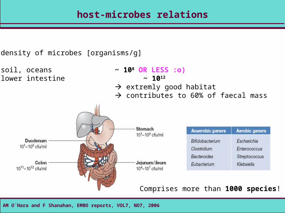
host-microbes relations
density of microbes [organisms/g]
soil, oceans ~ 108 OR LESS :o)lower intestine ~ 1012
extremly good habitat contributes to 60% of faecal mass
AM O´Hara and F Shanahan, EMBO reports, VOL7, NO7, 2006
Comprises more than 1000 species!
K Suzuki et al., seminars in immunology, review, 20071: (latin) cum mensa – (engl.) sharing a tableAM O´Hara and F Shanahan, EMBO reports, VOL7, NO7, 2006
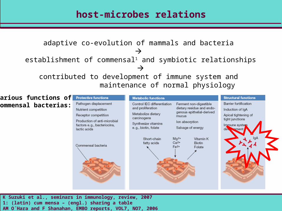
adaptive co-evolution of mammals and bacteria
establishment of commensal1 and symbiotic relationships
contributed to development of immune system and maintenance of normal physiology
various functions of commensal bacterias:
host-microbes relations
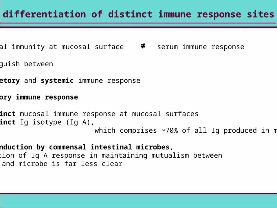
humoral immunity at mucosal surface ≠ serum immune response
distinguish between
secretory and systemic immune response
secretory immune response
distinct mucosal immune response at mucosal surfacesdistinct Ig isotype (Ig A),
which comprises ~70% of all Ig produced in mammals
Ig A induction by commensal intestinal microbes, function of Ig A response in maintaining mutualism between host and microbe is far less clear
differentiation of distinct immune response sites

Ig A basics
playing critical role in mucosal immunity
found in mucous secretions, including tears, saliva, intestinal juice, colostrum, vaginal fluid and secretions from the prostate and respiratory epithelium;additionally found in small amounts in the blood
functional activity
mainly neutralisation of pathogens and exotoxins, poorly activates the complement system, weakly opsonises
distribution
mainly transported through epithelium as dimer; somethimes diffusion to extravascular areas as monomeraverage serum concentration: 2,1 mg/ml
(Ig G1: 9 mg/ml, Ig E: 0,00003 mg/ml)
immune globulin A (IgA)- basics
CA Janeway et al., Immunobiology, 5th Ed., 2002, Spektrum
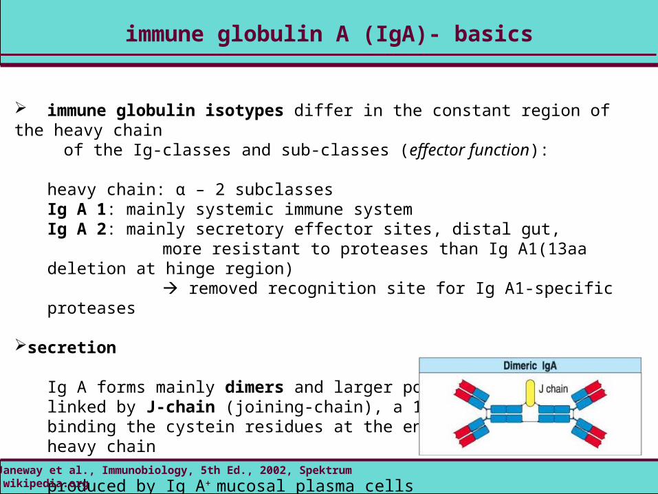
immune globulin isotypes differ in the constant region of the heavy chain of the Ig-classes and sub-classes (effector function):
heavy chain: α – 2 subclassesIg A 1: mainly systemic immune systemIg A 2: mainly secretory effector sites, distal gut,
more resistant to proteases than Ig A1(13aa deletion at hinge region) removed recognition site for Ig A1-specific proteases
secretion
Ig A forms mainly dimers and larger polymers (pIgA)linked by J-chain (joining-chain), a 15 kDa polypeptide binding the cystein residues at the end of the constant heavy chain
produced by Ig A+ mucosal plasma cells higher antigen avidity
immune globulin A (IgA)- basics
CA Janeway et al., Immunobiology, 5th Ed., 2002, Spektrumwww.wikipedia.org

Ig A secretion
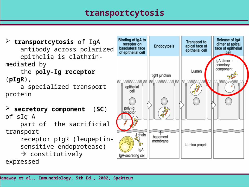
transportcytosis of IgA antibody across polarized epithelia is clathrin-mediated by the poly-Ig receptor (pIgR), a specialized transport protein
secretory component (SC) of sIg A part of the sacrificial transport receptor pIgR (leupeptin- sensitive endoprotease) constitutively expressed
transportcytosis
CA Janeway et al., Immunobiology, 5th Ed., 2002, Spektrum

secretory component (SC)
stabilizes sIg A in harsh intestinal environment and gives mucophilic propertiesfree SC exhibits scavenger properties with respect to enteric pathogens
pathogenicity
selective Ig A immunodeficiency higher sIgM production (hyper-IgM-Syndrom) sIgM less stable than sIgA, because no covalent binding of SC sIgM compensation less consistent in airways than in gut (higher susceptibility to infections in respiratory tract) mild phenotype
secretory component and pathogenicity
A Phalipon and B Corthesy, Trends Immunol., 2003www.wikipedia.org
sIg A
function of sIg A
function of Ig A in different systems – non-pathogens
intestinal bacteria 1014 – human cells in the body 1013
Ig A highly induced
sIg A protection mechanisms far less clear
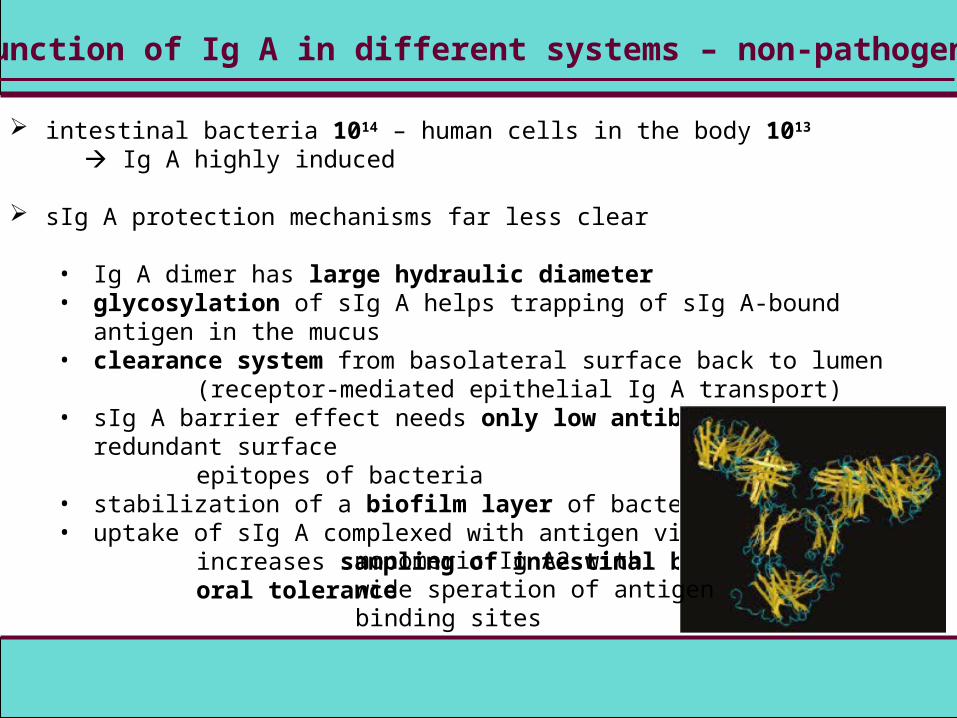
• Ig A dimer has large hydraulic diameter• glycosylation of sIg A helps trapping of sIg A-bound antigen in the mucus• clearance system from basolateral surface back to lumen
(receptor-mediated epithelial Ig A transport)• sIg A barrier effect needs only low antibody affinities to redundant surface
epitopes of bacteria• stabilization of a biofilm layer of bacteria• uptake of sIg A complexed with antigen via M cells
increases sampling of intestinal bacteria, oral tolerance
monomeric Ig A2 withwide speration of antigen binding sites
function of Ig A in different systems – microbial pathogens
sIg A protection properties, „first line of defense“
• toxin-neutralizing sIg A• inhibit early invasion and horizontal fecal-oral spread of pathogens• redundant role • compensation by antibodies of other isotypes (Ig M and Ig G) or by innate
immune mechanisms

class switch
Ig A class switch recombination (CSR) induction
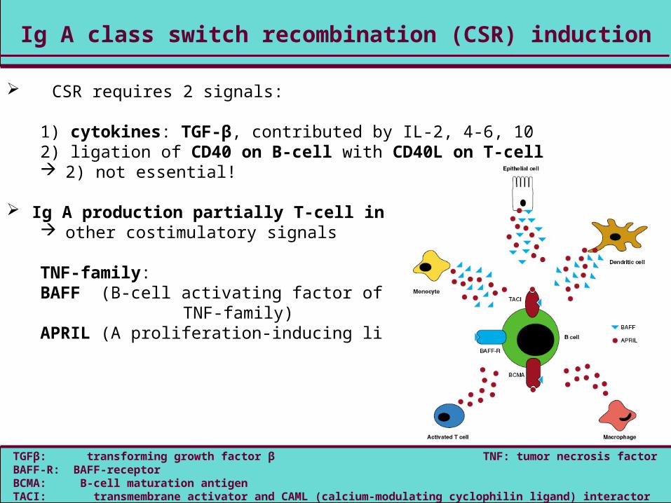
CSR requires 2 signals:
1) cytokines: TGF-β, contributed by IL-2, 4-6, 102) ligation of CD40 on B-cell with CD40L on T-cell 2) not essential!
Ig A production partially T-cell independent other costimulatory signals TNF-family:BAFF (B-cell activating factor of the
TNF-family)APRIL (A proliferation-inducing ligand)
TGFβ: transforming growth factor β TNF: tumor necrosis factorBAFF-R: BAFF-receptorBCMA: B-cell maturation antigenTACI: transmembrane activator and CAML (calcium-modulating cyclophilin ligand) interactor
some CSR induction investigations
APRIL-TACI interaction necessary for Ig A induction
(redundancy of Ig A CSR next to CD40-CD40L requirements?)
intestinal DC from PP and mesenteric lymph nodes secrete permanently RA
synergizes with IL- 5, 6 to induction of Ig A production in B-cells
induction of small intestine homing receptor CCR9
DC: dendritic cellsPP: Peyer´s patchesRA: retinoic acid
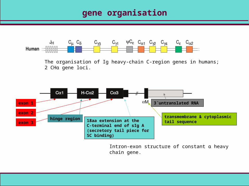
gene organisation
The organisation of Ig heavy-chain C-region genes in humans; 2 CHα gene loci.
Intron-exon structure of constant α heavy chain gene.
exon 1
exon 2
exon 3hinge region 18aa extension at the
C-terminal end of sIg A(secretory tail piece for SC binding)
transmembrane & cytoplasmic tail sequence
3´untranslated RNA
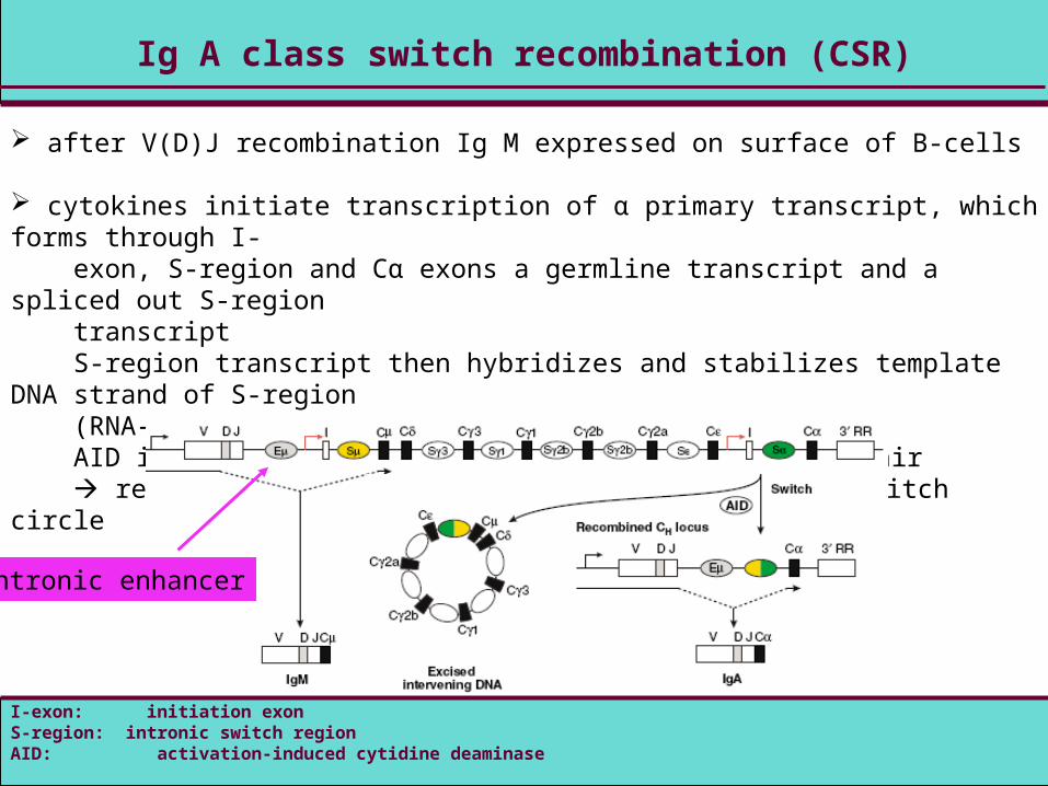
Ig A class switch recombination (CSR)
after V(D)J recombination Ig M expressed on surface of B-cells
cytokines initiate transcription of α primary transcript, which forms through I- exon, S-region and Cα exons a germline transcript and a spliced out S-region transcript S-region transcript then hybridizes and stabilizes template DNA strand of S-region (RNA-DNA-hybrid) AID introduces dsDNA breaks at S-region and DNA repair rearranged heavy chain constant region and DNA switch circle
I-exon: initiation exonS-region: intronic switch regionAID: activation-induced cytidine deaminase
intronic enhancer
B-cell homing
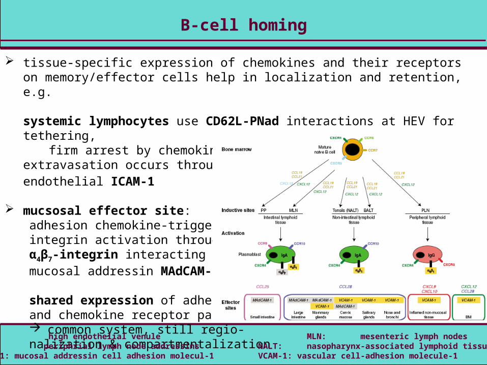
B-cell homing
tissue-specific expression of chemokines and their receptors on memory/effector cells help in localization and retention, e.g.
systemic lymphocytes use CD62L-PNad interactions at HEV for tethering, firm arrest by chemokine-triggered integrin activation, extravasation occurs through
αLβ2-integrin interaction with endothelial ICAM-1
mucsosal effector site:adhesion chemokine-triggeredintegrin activation through α4β7-integrin interacting withmucosal addressin MAdCAM-1
shared expression of adhesionand chemokine receptor pairs common system, still regio-nalization & compartmentalization
HEV: high endothelial venule MLN: mesenteric lymph nodesPNad: periphrial lymph node addressins NALT: nasopharynx-associated lymphoid tissueMAdCAM-1: mucosal addressin cell adhesion molecul-1 VCAM-1: vascular cell-adhesion molecule-1
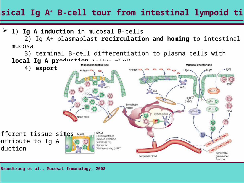
classical Ig A+ B-cell tour from intestinal lympoid tissue
1) Ig A induction in mucosal B-cells 2) Ig A+ plasmablast recirculation and homing to intestinal mucosa 3) terminal B-cell differentiation to plasma cells with local Ig A production (after ~17d) 4) export of Ig A through intestinal epithelial cell layer
P Brandtzaeg et al., Mucosal Immunology, 2008
different tissue sitescontribute to Ig Ainduction

example
PP: Peyer´s patchesB Corthésy, Journal of Immunology, 2007DC: dendritic cells
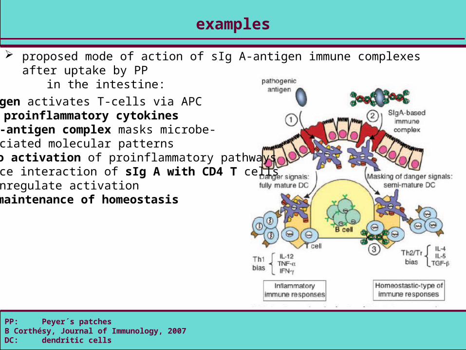
examples
proposed mode of action of sIg A-antigen immune complexes after uptake by PP in the intestine:
1) pathogen activates T-cells via APC proinflammatory cytokines2) sIg A-antigen complex masks microbe- associated molecular patterns no activation of proinflammatory pathways3) surface interaction of sIg A with CD4 T cells downregulate activation maintenance of homeostasis

Thanks for your attention!
Any questions?