the ice nucleation ability of one of the most abundant
TRANSCRIPT

Atmos. Chem. Phys., 11, 1191–1201, 2011www.atmos-chem-phys.net/11/1191/2011/doi:10.5194/acp-11-1191-2011© Author(s) 2011. CC Attribution 3.0 License.
AtmosphericChemistry
and Physics
The ice nucleation ability of one of the most abundant types offungal spores found in the atmosphere
R. Iannone1, D. I. Chernoff1, A. Pringle2, S. T. Martin 3, and A. K. Bertram 1
1Department of Chemistry, University of British Columbia, Vancouver, British Columbia, V6T 1Z1, Canada2Department of Organismic and Evolutionary Biology, Harvard University, Cambridge, Massachusetts, USA3School of Engineering and Applied Sciences & Department of Earth and Planetary Sciences, Harvard University,Cambridge, Massachusetts, USA
Received: 11 September 2010 – Published in Atmos. Chem. Phys. Discuss.: 21 October 2010Revised: 1 February 2011 – Accepted: 1 February 2011 – Published: 11 February 2011
Abstract. Recent atmospheric measurements show that bi-ological particles are a potentially important class of ice nu-clei. Types of biological particles that may be good ice nucleiinclude bacteria, pollen and fungal spores. We studied theice nucleation properties of water droplets containing fun-gal spores from the genusCladosporium, one of the mostabundant types of spores found in the atmosphere. For wa-ter droplets containing aCladosporiumspore surface area of∼217 µm2 (equivalent to∼5 spores with average diametersof 3.2 µm ), 1% of the droplets froze by−28.5◦C and 10%froze by –30.1◦C. However, there was a strong dependenceon freezing temperature with the spore surface area ofCla-dosporiumwithin a given droplet. Mean freezing tempera-tures for droplets containing 1–5 spores are expected to beapproximately−35.1± 2.3◦C (1σ S. D.). Atmospheric icenucleation on spores ofCladosporium sp., or other sporeswith similar surface properties, thus do not appear to ex-plain recent atmospheric measurements showing that biolog-ical particles participate as atmospheric ice nuclei. The poorice nucleation ability ofCladosporium sp.may be attributedto the surface which is coated with hydrophobins (a classof hydrophobic proteins that appear to be widespread in fila-mentous fungi). Given the ubiquity of hydrophobins on sporesurfaces, the current study may be applicable to many fungalspecies of atmospheric importance.
Correspondence to:A. K. Bertram([email protected])
1 Introduction
Ice nucleation can occur in the atmosphere by either ho-mogeneous nucleation or heterogeneous nucleation. Het-erogeneous nucleation involves solid or partially solid par-ticles, called ice nuclei (IN), that have the potential to mod-ify climate by changing the formation conditions and proper-ties of ice clouds and mixed-phase clouds (Baker and Peter,2008; Hegg and Baker, 2009; Lohmann and Hoose, 2009).At temperatures greater than approximately−38◦C, freez-ing occurs initially by heterogeneous nucleation on ice nu-clei. Possible atmospheric ice nuclei include mineral dust(DeMott et al., 2003; Hung et al., 2003; Pruppacher andKlett, 1997), soot (Andreae and Rosenfeld, 2008; Karcheret al., 2007), crystalline salts (Abbatt et al., 2006; Martin,1998; Shilling et al., 2006; Wise et al., 2010; Zuberi et al.,2002), and biological aerosol particles (Christner et al., 2008;Mohler et al., 2007).
Several recent reviews have highlighted the need to quan-tify the number and source of biological ice nuclei in the at-mosphere (Ariya et al., 2009; DeMott and Prenni, 2010; Mar-tin et al., 2010; Mohler et al., 2007; Szyrmer and Zawadzki,1997). In addition, it has been suggested that the most im-portant carbonaceous particles that may be acting as ice nu-clei above−15◦C may be biological particles (DeMott andPrenni, 2010). If biological ice nuclei are abundant in the at-mosphere they can influence the hydrological cycle and mayplay an important role in precipitation (Sands et al., 1982;Morris et al., 2004).
Recent field measurements of IN have highlighted the po-tential importance of biological particles in ice cloud for-mation in the atmosphere (Pratt et al., 2009; Prenni et al.,2009). Pratt et al. (2009) observed that ice residuals col-lected in situ from cloud particles (from−31◦C to −34◦C)
Published by Copernicus Publications on behalf of the European Geosciences Union.
1192 R. Iannone et al.: The ice nucleation ability of one of the most abundant types of fungal spores
contain a significant fraction of biological material. Anotherrecent study concerning biological IN has demonstrated that,for pristine conditions over the Amazon rainforest, the levelof IN can be predicted though measurements of a combina-tion of mineral dust and biological particles (Prenni et al.,2009). To account for their data they had assumed that thesampled biological particles could induce ice nucleation withan efficiency of∼0.2 for temperatures between−18◦C and−31◦C. At temperatures above approximately−25◦C bio-logical IN appeared to dominate over mineral dust IN.
Types of biological particles that can act as IN include bac-teria, pollen and fungal spores. The most well-characterizedbiological IN species is the bacterium Pseudomonas syringae(P. syringae). This bacterium is an efficient IN species thatis associated with onsets for ice formation as high as−2◦C(Mohler et al., 2007; Lindow et al., 1989; Maki et al., 1974;Morris et al., 2004; Vali et al., 1976). The ice nucleatingability of pollen, relatively large bioaerosol particles with adiameter range of 10–100 µm , has been addressed in severalstudies (Diehl et al., 2001, 2002; von Blohn et al., 2005).Freezing temperatures for 4 types of pollen particles in theimmersion freezing mode ranged from−13.5◦C to−21.5◦C(von Blohn et al., 2005). However, the fraction of pollengrains that are effective IN have not yet been determined.
Concerning the IN ability of fungi, a study by Pouleur etal. (1992) had examined the IN capability of suspensionsof 20 fungal species at temperatures as low as−10.0◦C.However, the IN ability of fungal spores was not specificallydetermined.
Recent modelling studies have begun to explore the effectof biological ice nucleation on cloud microphysics, dynam-ics and precipitation (Diehl et al., 2006; Hoose et al., 2010;Phillips et al., 2009). However, due to the limited under-standing of the ice nucleation properties of biological parti-cles, such modelling studies often have significant uncertain-ties. More studies on the ice nucleation properties of biologi-cal particles are needed, especially focusing on the abundanttypes of biological particles found in the atmosphere.
Spores, which are the reproductive units of fungi, are liber-ated into the air and are dispersed by air currents into the at-mosphere. The amount of fungal spores in the atmosphere issubject to the time of day and meteorological factors (Gilbertand Reynolds, 2005; Hirst, 1953; Kurkela, 1997; Stennettand Beggs, 2004; Stepalska and Wołek, 2009). The av-erage number of fungal spores in the continental bound-ary layer air is on the order of 103–104 m−3 (Elbert et al.,2007). As a result, they are of sufficient quantity for theirconsideration in ice cloud formation. To determine theiratmospheric relevance, knowledge of their activation spec-trum (i.e., proportion of active IN against temperature andice supersaturation) is needed. Fungal spores can also betransported over vast distances. By convection, they canreach high altitudes in the troposphere (Fulton, 1966; Fultonand Mitchell, 1966; Gregory, 1978; Heise and Heise, 1948;Hirst et al., 1967a, b; Pady and Kelly, 1953; Proctor, 1935;
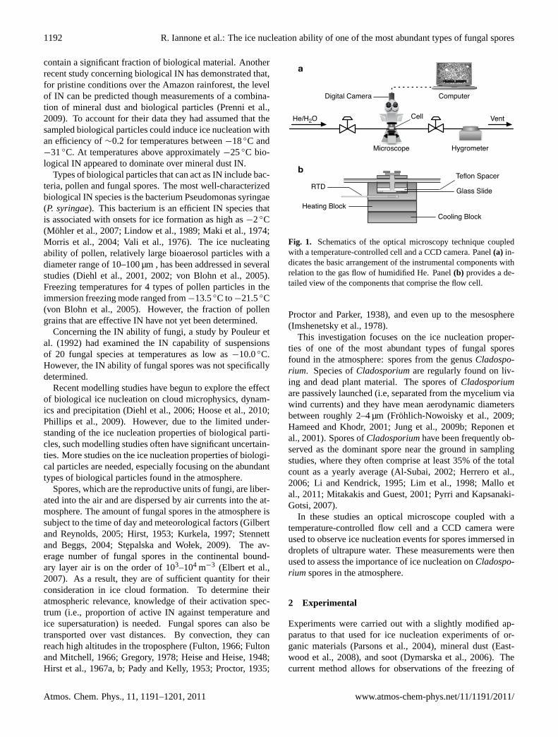
Figure 1 (Cladosporium Paper)
a
b
Cooling BlockHeating Block
RTD Glass Slide
Teflon Spacer
Cell
HygrometerMicroscope
Computer
Vent
Digital Camera
He/H2O
Fig. 1. Schematics of the optical microscopy technique coupled with a temperature-controlled cell and a CCD camera. Panel (a) indicates the basic arrangement of the instrumental components with relation to the gas flow of humidified He. Panel (b) provides a detailed view of the components that comprise the flow cell.
27
Fig. 1. Schematics of the optical microscopy technique coupledwith a temperature-controlled cell and a CCD camera. Panel(a) in-dicates the basic arrangement of the instrumental components withrelation to the gas flow of humidified He. Panel(b) provides a de-tailed view of the components that comprise the flow cell.
Proctor and Parker, 1938), and even up to the mesosphere(Imshenetsky et al., 1978).
This investigation focuses on the ice nucleation proper-ties of one of the most abundant types of fungal sporesfound in the atmosphere: spores from the genusCladospo-rium. Species ofCladosporiumare regularly found on liv-ing and dead plant material. The spores ofCladosporiumare passively launched (i.e, separated from the mycelium viawind currents) and they have mean aerodynamic diametersbetween roughly 2–4 µm (Frohlich-Nowoisky et al., 2009;Hameed and Khodr, 2001; Jung et al., 2009b; Reponen etal., 2001). Spores ofCladosporiumhave been frequently ob-served as the dominant spore near the ground in samplingstudies, where they often comprise at least 35% of the totalcount as a yearly average (Al-Subai, 2002; Herrero et al.,2006; Li and Kendrick, 1995; Lim et al., 1998; Mallo etal., 2011; Mitakakis and Guest, 2001; Pyrri and Kapsanaki-Gotsi, 2007).
In these studies an optical microscope coupled with atemperature-controlled flow cell and a CCD camera wereused to observe ice nucleation events for spores immersed indroplets of ultrapure water. These measurements were thenused to assess the importance of ice nucleation onCladospo-rium spores in the atmosphere.
2 Experimental
Experiments were carried out with a slightly modified ap-paratus to that used for ice nucleation experiments of or-ganic materials (Parsons et al., 2004), mineral dust (East-wood et al., 2008), and soot (Dymarska et al., 2006). Thecurrent method allows for observations of the freezing of
Atmos. Chem. Phys., 11, 1191–1201, 2011 www.atmos-chem-phys.net/11/1191/2011/

R. Iannone et al.: The ice nucleation ability of one of the most abundant types of fungal spores 1193
spore-containing water droplets. Additionally, the amountof fungal material present in the droplets was directly deter-mined through the analysis of acquired video after evaporat-ing the drops, providing insight on the relationship betweenthe spore surface area and the heterogeneous freezing tem-perature.
2.1 Freezing experiments and apparatus
The apparatus used in the freezing measurements consistedof an optical microscope (Zeiss Axiolab A) coupled to aflow cell wherein the relative humidity (RH) could be ac-curately controlled and the temperature could be decreasedor increased at a fixed rate ranging from 0.1–5.0◦C min−1.Only one cooling rate, 5 K min−1, was used due to experi-mental constraints. Higher cooling rates were not possiblewith the current setup and lower cooling rates resulted in sig-nificant mass transfer between unfrozen and frozen droplets.A schematic of the flow cell is shown in Fig. 1. The opticalmicroscope was equipped with a 10× objective and a SonyXC-ST50 digital video camera, itself fitted with a 0.4× re-duction lens. The temperature of the flow cell was controlledusing a combination of a refrigerating circulator (ULT-95,Thermo Neslab) and an electrical heater supported by a tem-perature controller. Cell temperature was monitored throughthe use of a Pt-100 resistance temperature detector (RTD).
The bottom surface of the flow cell, which supported thespores during the freezing experiments, consisted of a pre-siliconized glass cover slide (Hampton Scientific, Hartfield,PA) that served as a hydrophobic substrate for reducing theprobability of ice nucleation directly on the surface. It waspreviously demonstrated that these types of slides do not in-duce heterogeneous freezing of supercooled water droplets(Koop et al., 1998).
The experimental protocol for the freezing experimentsconsisted of the following steps.Cladosporiumspores weredeposited on the bottom surface of the flow cell using theprocedure described in Sect. 2.3. Next, the temperature ofthe flow cell was decreased to 2◦C and the dew point in theflow cell was set to approximately 3–5◦C using a humidifiedflow of ultra high purity (UHP) He (99.999%, Praxair). Thisresulted in the nucleation and growth of water droplets on thebottom surface of the flow cell and on the spores. Dropletswere allowed to grow to 200–300 µm in diameter. The gasflow was then stopped and the flow cell was isolated by clos-ing the valves immediately upstream and downstream of thecell. Next, the temperature in the flow cell was decreasedat a rate of 5◦C min−1 until a temperature of−40◦C wasreached. During this process, digital video was captured at aframe rate of 30 fps (frames per second) by use of the CCDcamera. Images were acquired from the digital video andanalysed to determine the freezing spectrum, defined as thefraction of droplets frozen as a function of temperature. Af-ter freezing, the temperature was increased to 2◦C and thedroplets were exposed to a flow of dry He gas to fully evapo-
rate the water, leaving only the spore inclusions on the slide.Images were also acquired during this procedure to deter-mine the amount of fungal material contained in each droplet.
Figure 2 provides images of a droplet in a heterogeneousnucleation experiment at various conditions during an exper-iment. Panel a shows a droplet containing inclusions at thebeginning of the freezing experiment. Panels b and c showimages of the same droplets after freezing. Panel d shows animage of the remaining fungal spore inclusions after evapo-rating the droplet.
Experiments were also performed using ultrapure waterdroplets without spores. This provided a means to determinewhether the hydrophobic substrate supporting the particlesinfluenced the freezing results. In these experiments, purewater droplets were condensed from the vapour phase ontothe hydrophobic surface and their freezing temperatures weredetermined using the same procedure as outlined above forheterogeneous freezing.
2.2 Temperature and cooling rate calibration
The RTD was calibrated against the melting points of ultra-pure water (0.0◦C) and n-decane (−29.7◦C). Melting pointdeterminations indicated a +0.16◦C offset for n-decane anda +0.07◦C offset for water. The reported freezing tempera-tures were corrected for bias using a fit function based on thisoffset data.
2.3 Preparation and collection of spores ofCladosporium sp.
Cladosporiumwas obtained from existing stock at the Cana-dian Centre for the Culture of Microorganisms (CCCM)(Department of Botany, University of British Columbia,Canada). Colonies ofCladosporiumwere grown on potatodextrose agar in plastic Petri dishes. The cultures were in-cubated for a minimum of 3 weeks at 26◦C before any ex-periments were carried out. Preparation and collection ofspores were conducted in a Class II biological safety cabinetto avoid contamination of the samples by dust and to preventthe release of spores into the laboratory environment.
Spores were harvested from fungal colonies and depositedon hydrophobic slides using a spore dispenser (i.e., an RHmeter and a flow cell) and an impactor (Fig. 3). A scalpelwas used to excise portions of theCladosporiumculturesfrom the Petri dish which were then transferred to a steril-ized borosilicate glass flow tube. The tube was placed insidethe flow cell and the spore dispenser was then hermeticallysealed.
Inside the flow cell, UHP N2 gas (99.999%, Praxair) waspassed over the fungal cultures to dislodge and aerosolize thespores. A flow rate of 5× 103 cm3 min−1 was maintainedduring a 1 h collection time. A 120 cm3 Supelcarb HC Hy-drocarbon Trap (Sigma-Aldrich) minimized organic contam-inants within the 300 L of N2 admitted to the system during
www.atmos-chem-phys.net/11/1191/2011/ Atmos. Chem. Phys., 11, 1191–1201, 2011

1194 R. Iannone et al.: The ice nucleation ability of one of the most abundant types of fungal spores
HeterogeneousNucleation
–20ºC
–30ºC
–40ºC
Inclusions at 2ºC
Figure 2 (Cladosporium Paper)
128 µm2
82 µm2
100 µm
a
b
c
d
28
Fig. 2. Images from a heterogeneous nucleation experiment. Im-age(a) shows a liquid water droplet (cell temperature of−20.0◦C)containingCladosporiumspore inclusions (not visible) where nofreezing has yet occurred. Images (b andc) (cell temperatures of−30.0◦C and−40.0◦C, respectively) show the droplet after freez-ing. Image(d) depicts the remaining inclusions (with a total area of210 µm2) upon evaporating the droplets at 2.0◦C and passing dryHe gas through the cell. For reference, the droplet boundary is pro-vided as a black outline and the individual areas of the inclusionsare given.
spore collection. Experiments used either dry N2 or a humid-ified flow of N2 (RH∼35%). Impaction of the spores ontothe hydrophobic glass substrate occurred as the N2 carriergas exited a borosilicate glass impactor tube.
An Olympus IX70 inverted microscope equipped with a40× objective was used to determine the morphology of theparticles. A 20× objective was used to determine the sizedistributions of the collected spores.
3 Results and discussion
3.1 Morphology and size distribution of collected spores
Individual spores often appeared to be either roughly circu-lar or, alternatively, lemon-shaped. Figure 4 provides severalimages ofCladosporium sp.spores at 40× magnification us-ing the Olympus IX70 microscope. The morphology of thesespores and their aspect ratios are consistent with results re-ported in the literature (Reponen et al., 1997; Schubert et al.,2007). Approximately 90% of the spore dispersal units (i.e.,particles dislodged from the fungal mycelium) contained 1spore (Fig. 4a–c), and the remaining 10% contained 2 ormore spores (Fig. 4d, e). Observations in the atmosphere atground level have shown that a significant fraction of the col-lected spore dispersal units contain 2 or more spores (Davies,1957; Harvey, 1967; Hyde and Williams, 1953).
In addition to characterising the morphology of the spores,we also determined the size distribution of individual sporesto further confirm that the collected material was actu-ally spores and also to provide an estimate of the size ofspores used in our studies. Volume equivalent diameters(Dvolume) for each individual spore were calculated usingimages recorded at 20× magnification with the IX70 micro-scope and the formulaDvolume=
3√
W2 ·L, whereW repre-sents the maximum width orthogonal to the lengthL (Repo-nen et al., 2001). Any dispersal unit that contained 2 or morespores (i.e., Fig. 4d, e) was not included in these calculations.The results from these calculations are shown in Fig. 5. In-dividual spores had a mean volume equivalent diameter of3.2± 1.0 µm (1σ S. D.). This mean diameter is consistentwith reported diameters for spores ofCladosporium(Carlileet al., 2001; Frohlich-Nowoisky et al., 2009; Hameed andKhodr, 2001; Jung et al., 2009a; Reponen et al., 1997). Themean diameter may be an upper limit to the true mean size,because in our experiments particles having diameters lessthan approximately 1 µm were not resolved with the micro-scope. The mean size corresponds to single spores collectedwith the impactor. In the freezing experiments an average of4.8 spores (i.e., spore units) was contained in each droplet(cf. Sect. 3.3).
Atmos. Chem. Phys., 11, 1191–1201, 2011 www.atmos-chem-phys.net/11/1191/2011/

R. Iannone et al.: The ice nucleation ability of one of the most abundant types of fungal spores 1195
Figure 3 (Cladosporium Paper)
1.8 mm
Detail of Impactor
Glass SlideMetal Stage
2 mm
Glass Slide
Flow Cell
Flow TubeContainingFungal Colonies
AdjustableInjector Tube Adjustable
Impactor Tube
5 SLPM
N2/H2OR.H.
Meter
Spore Dispenser
Impactor
Metal Stage
SapphireWindow
Cooling Block
Detail of Flow Cell
Heating Block
RTD
10× Lens
Glass SlideTeflon SpacerHumidified He
HygrometerMicroscope
Computer
Vent
Digital Camera
He/H2O Cell
a b
Fig. 3. Schematics for the collection of fungal spores using a spore dispenser and an impactor. Panel (a) depicts the arrangement of the of the spore dispenser and impactor. Panel (b) provides a detailed view of the impactor, where the impactor tube’s aperture size and the distance from glass slide are given. Excised portions of Cladosporium cultures were placed inside the borosilicate glass flow tube. The tube was sealed inside the flow cell where a ¼″ O.D. stainless steel tube with bent tip directed a flow of N2 (~5×103 cm3 min–1 for 1 h) at the cultures. Experiments either used dry or humidified (~35% relative humidity, or RH) N2 gas.
30
Fig. 3. Schematics for the collection of fungal spores using a spore dispenser and an impactor. Panel(a) depicts the arrangement of theof the spore dispenser and impactor. Panel(b) provides a detailed view of the impactor, where the impactor tube’s aperture size and thedistance from glass slide are given. Excised portions ofCladosporiumcultures were placed inside the borosilicate glass flow tube. The tubewas sealed inside the flow cell where a1/4 O. D. stainless steel tube with bent tip directed a flow of N2 (∼5× 103 cm3 min−1 for 1 h) at thecultures. Experiments either used dry or humidified (∼35% relative humidity, or RH) N2 gas.
3.2 Pure water droplets
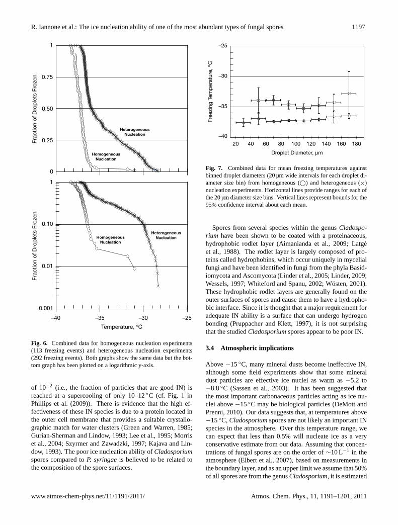
A total of 113 individual homogeneous freezing events wasobserved for pure water droplets without spores. Figure 6provides a summary of the freezing results. As shown, 50%of the droplets had frozen at approximately−37◦C, which iswithin the range of temperatures expected when consideringclassical homogeneous nucleation theory. In Fig. 7, the meanhomogeneous freezing data is plotted as a function of dropletsize (binned in 20 µm diameter bins). From the plotted data,a small dependence of freezing temperature on droplet sizewas observed as expected from classical nucleation theory(Pruppacher and Klett, 1997). The mean freezing tempera-tures for all the size ranges studied are within 0.5–2.0◦C ofthe predicted homogeneous freezing temperatures using ho-mogeneous nucleation rates and equations presented in Prup-pacher (1995) and Pruppacher and Klett (1997). In addi-tion, the measured freezing temperatures are in good agree-ment with freezing temperatures reported in other studiesfor which unsupported droplets (i.e., aerosol droplets) wereused. Wood et al. (2002), for example, reported averagefreezing temperatures of−37.0◦C to −37.3◦C for dropletdiameters of 40–66 µm . The reasonable agreement betweenthe predicted homogeneous freezing temperatures, previoushomogeneous freezing experiments, and the results of thisstudy illustrates that the hydrophobic surface supporting theparticles does not significantly influence the freezing temper-atures. This observation is consistent with previous measure-ments using similar supports (Bertram et al., 2000; Koop etal., 1998).
3.3 Heterogeneous nucleation
Nine freezing experiments were carried out for waterdroplets containing fungal spores ofCladosporium, and 16–43 droplets were analysed for freezing in each experiment.
Only droplets that presented clear visual evidence for freez-ing and that contained spores were considered for the het-erogeneous nucleation analyses. A total of 292 individualheterogeneous freezing events was observed. In these ex-periments the average surface area for the spore inclusionswas 217 µm2 (σ = 172 µm2). Assuming that one spore cor-responds to a diameter of 3.2 µm (as presented in Sect. 3.1)then, on average, each droplet in these experiments contained4.8 spores.
The results from the heterogeneous freezing experimentsare summarized in Fig. 6. As seen, the heterogeneous freez-ing results are warmer than the pure water freezing exper-iments, where 1% of the droplets heterogeneously froze at−28.5◦C and 10% froze at−30.1◦C. It is interesting to notethat the ice nucleation ability ofCladosporiumis similar tokaolinite in the immersion mode (Murray et al., 2010; Luondet al., 2010). Kaolinite, an abundant mineral in the atmo-sphere, is often considered a good ice nucleus.
In Fig. 7, the heterogeneous freezing results are plotted asa function of droplet diameter in the same manner as for thefreezing experiments of pure water droplets. This plot illus-trates that the heterogeneous freezing results are statisticallydifferent from the pure water case and, hence, the fungalspores are acting as ice nuclei. The difference between ho-mogeneous and heterogeneous freezing ranges from∼3◦Cto ∼5◦C.
Another way to analyse and present the heterogeneousfreezing data is to do so as a function of inclusion size (i.e.,surface area) contained within each droplet undergoing het-erogeneous nucleation. A summary of the heterogeneousdata binned according to inclusion area is provided in Fig. 8.Included for comparison are the homogeneous data (zero sur-face area). Included as a secondary x-axis is the number ofspores in each droplet, assuming an average spore diame-ter of 3.2 µm for the size of an individual spore. The gen-eral trend observed in Fig. 8 is an increase in the median
www.atmos-chem-phys.net/11/1191/2011/ Atmos. Chem. Phys., 11, 1191–1201, 2011

1196 R. Iannone et al.: The ice nucleation ability of one of the most abundant types of fungal spores
Each image is 20 µm × 20 µm
Figure 4 (Cladosporium Paper)
a
A = 8.6 µm2
L = 4.4 µm, W = 2.3 µm
b
A = 14.4 µm2
L = 6.1 µm, W = 3.0 µm
d
A = 18.9 µm2
L = 7.7 µm, W = 2.2 µm
c
A = 12.0 µm2
L = 4.4 µm, W = 2.9 µm
e
A = 26.4 µm2
L = 9.3 µm, W = 2.5 µm
Two Spore Units
One Spore Unit
Fig. 4. Images of spores of Cladosporium collected on a glass slide and observed with the Olympus IX70 microscope at 40× magnification. Panels (a–c) show dispersal units for Cladosporium containing one spore, whereas panels (d and e) depict observations of two or more spores (comprising 10% of the total observations during size studies at the 20× magnification level). Each image measures 20 µm × 20 µm. Measurement values for area (A), length (L), and width (W) are provided in each panel.
31
Fig. 4. Images of spores ofCladosporiumcollected on a glass slideand observed with the Olympus IX70 microscope at 40× magnifi-cation. Panels (a–c) show dispersal units forCladosporiumcontain-ing one spore, whereas panels (d and e) depict observations of twoor more spores (comprising 10% of the total observations duringsize studies at the 20× magnification level). Each image measures20 µm× 20 µm . Measurement values for area(A), length(L), andwidth (W) are provided in each panel.
freezing temperature as the surface area of the inclusion in-creases per droplet (i.e., as the number of spores per dropletincreases). This trend is consistent with previous heteroge-neous freezing experiments in which the freezing tempera-ture depended on the surface area of the heterogeneous icenuclei (Archuleta et al., 2005; Hung et al., 2003; Kanji et al.,2008; Marcolli et al., 2007; Phillips et al., 2008). The me-dian heterogeneous freezing temperature for the smallest sizebin (10–200 µm2) is approximately 3◦C warmer compared tothe homogeneous nucleation data. The dependence of spore
Figure 5 (Cladosporium Paper)
Fra
ctio
n o
f F
un
gal S
po
res
Projected Diameter(Dproj), µm
0.30
0.20
0.10
01 2 3 4 5 6 7 8 9
Fra
ctio
n o
f F
un
gal S
po
res
Volume EquivalentDiameter (Dvolume), µm
0.30
0.20
0.10
01 2 3 4 5 6
Fig. 5. Distribution of volume equivalent diameters (Dvolume) for spores of Cladosporium observed with the Olympus IX70 microscope at 20× magnification. Only spore areas for one spore unit were included in calculations of Dvolume. Data are centered on integer values for Dvolume. The mean volume equivalent diameter is 3.2±1.0 µm (1σ S.D.).
32
Fig. 5. Distribution of volume equivalent diameters (Dvolume) forspores ofCladosporiumobserved with the Olympus IX70 micro-scope at 20× magnification. Only spore areas for one spore unitwere included in calculations ofDvolume. Data are centered on in-teger values forDvolume. The mean volume equivalent diameter is3.2± 1.0 µm (1σ S. D.).
inclusions on freezing temperatures has a pronounced effectespecially for>8 spores droplet−1 at which point the averagedifference in supercooling between homogeneous nucleationand heterogeneous nucleation is 8◦C.
There is a paucity of studies on the heterogeneous freezingof fungal spores for which comparisons may be made to thisstudy. One study examined the IN ability of a wide varietyof fungal genera, including 14 species of the genusFusar-ium (Pouleur et al., 1992). That study reported high freez-ing temperatures for the genusFusarium(up to−2.5◦C). Acaveat, however, is that the experiments were performed ondroplets containing a suspension of fungal material and thisdoes not provide specific results on the ice nucleating abil-ity of fungal spores. Furthermore, the dependence on surfacearea was not addressed. Similarly, several ice nucleation ex-periments were carried out with species of lichens (Ashworthand Kieft, 1992; Kieft, 1988; Kieft and Ahmadjian, 1989;Kieft and Ruscetti, 1990), which are symbiotic organismswherein fungi and algae and/or cyanobacteria form a singlebiological entity (Nash, 2008). In one particular study,Rhi-zoplaca chrysoleuca, a species of lichen, was found to inducefreezing at approximately−4◦C (Kieft and Ruscetti, 1990).However, the ice nucleation properties of spores from lichenswere not addressed.
Compared to biological particles such as the bacteriumP. syringae, Cladosporiumis not an effective IN. For ex-ample, strains ofP. syringaeinduce freezing at temperaturesas high as−2◦C (Lindow et al., 1989; Maki et al., 1974;Morris et al., 2004a) and for some strains a freezing fraction
Atmos. Chem. Phys., 11, 1191–1201, 2011 www.atmos-chem-phys.net/11/1191/2011/
R. Iannone et al.: The ice nucleation ability of one of the most abundant types of fungal spores 1197
1
0.50
0.25
0
Fra
ctio
n o
f D
rop
lets
Fro
zen
HomogeneousNucleation
HeterogeneousNucleation
0.75
1
0.10
0.01
0.001
Fra
ctio
n o
f D
rop
lets
Fro
zen
Temperature, ºC
–25–30–40 –35
HomogeneousNucleation
HeterogeneousNucleation
Figu
re 6
(Cla
dosp
oriu
m P
aper
)
Fig. 6. Combined data for homogeneous nucleation experiments (113 freezing events) and heterogeneous nucleation experiments (292 freezing events). Both graphs show the same data but the bottom graph has been plotted on a logarithmic y-axis.
33
Fig. 6. Combined data for homogeneous nucleation experiments(113 freezing events) and heterogeneous nucleation experiments(292 freezing events). Both graphs show the same data but the bot-tom graph has been plotted on a logarithmic y-axis.
of 10−2 (i.e., the fraction of particles that are good IN) isreached at a supercooling of only 10–12◦C (cf. Fig. 1 inPhillips et al. (2009)). There is evidence that the high ef-fectiveness of these IN species is due to a protein located inthe outer cell membrane that provides a suitable crystallo-graphic match for water clusters (Green and Warren, 1985;Gurian-Sherman and Lindow, 1993; Lee et al., 1995; Morriset al., 2004; Szyrmer and Zawadzki, 1997; Kajava and Lin-dow, 1993). The poor ice nucleation ability ofCladosporiumspores compared toP. syringaeis believed to be related tothe composition of the spore surfaces.
Figure 7 (Cladosporium Paper)
–25
–30
–35
–40
Fre
ezi
ng
Tem
pera
ture
, ºC
Droplet Diameter, µm
20 40 60 80 100 120 140 160 180
Fig. 7. Combined data for mean freezing temperatures againstbinned droplet diameters (20 µm wide intervals for each droplet di-ameter size bin) from homogeneous (©) and heterogeneous (×)nucleation experiments. Horizontal lines provide ranges for each ofthe 20 µm diameter size bins. Vertical lines represent bounds for the95% confidence interval about each mean.
Spores from several species within the genusCladospo-rium have been shown to be coated with a proteinaceous,hydrophobic rodlet layer (Aimanianda et al., 2009; Latgeet al., 1988). The rodlet layer is largely composed of pro-teins called hydrophobins, which occur uniquely in mycelialfungi and have been identified in fungi from the phyla Basid-iomycota and Ascomycota (Linder et al., 2005; Linder, 2009;Wessels, 1997; Whiteford and Spanu, 2002; Wosten, 2001).These hydrophobic rodlet layers are generally found on theouter surfaces of spores and cause them to have a hydropho-bic interface. Since it is thought that a major requirement foradequate IN ability is a surface that can undergo hydrogenbonding (Pruppacher and Klett, 1997), it is not surprisingthat the studiedCladosporiumspores appear to be poor IN.
3.4 Atmospheric implications
Above −15◦C, many mineral dusts become ineffective IN,although some field experiments show that some mineraldust particles are effective ice nuclei as warm as−5.2 to−8.8◦C (Sassen et al., 2003). It has been suggested thatthe most important carbonaceous particles acting as ice nu-clei above−15◦C may be biological particles (DeMott andPrenni, 2010). Our data suggests that, at temperatures above−15◦C, Cladosporiumspores are not likely an important INspecies in the atmosphere. Over this temperature range, wecan expect that less than 0.5% will nucleate ice as a veryconservative estimate from our data. Assuming that concen-trations of fungal spores are on the order of∼10 L−1 in theatmosphere (Elbert et al., 2007), based on measurements inthe boundary layer, and as an upper limit we assume that 50%of all spores are from the genusCladosporium, it is estimated
www.atmos-chem-phys.net/11/1191/2011/ Atmos. Chem. Phys., 11, 1191–1201, 2011

1198 R. Iannone et al.: The ice nucleation ability of one of the most abundant types of fungal spores
Figure 8 (Cladosporium Paper)
1.0–5.0
–25
–30
–35
–40
Fre
ezi
ng
Tem
pera
ture
, ºC
010–200
200–400
400–600
600–800
800–1000
Total Inclusion Area per Droplet, µm2
05.0–7.1
7.1–8.7
8.7–10.1
10.1–11.3
Number of Cladosporium Spores per Droplet
Fig. 8. Box plot showing a five-number statistical summary for homogeneous nucleation experiments (leftmost box) and binned data from all heterogeneous nucleation experiments. Each box represents temperatures as maximum and minimum values, and the 1st, 2nd (median), and 3rd quartiles. Freezing data are distributed into 200 µm2 bins that represent the total area of all observable inclusions per frozen droplet (bottom x-axis). The average uncertainty in the inclusion area is ±28 µm2. Assuming that all inclusions are spores with a mean volume equivalent diameter of 3.2 µm, the top x-axis provides the numbers of Cladosporium spores corresponding to each area bin.
35
Fig. 8. Box plot showing a five-number statistical summary for ho-mogeneous nucleation experiments (leftmost box) and binned datafrom all heterogeneous nucleation experiments. Each box repre-sents temperatures as maximum and minimum values, and the 1st,2nd (median), and 3rd quartiles. Freezing data are distributed into200 µm2 bins that represent the total area of all observable inclu-sions per frozen droplet (bottom x-axis). The average uncertaintyin the inclusion area is±28 µm2. Assuming that all inclusions arespores with a mean volume equivalent diameter of 3.2µm, the topx-axis provides the numbers ofCladosporiumspores correspondingto each area bin.
that the number of IN fromCladosporiumspores is signifi-cantly less than∼0.025 L−1. This value is a factor of approx-imately 4 to 800 smaller than the number of IN observed inthe atmosphere at temperatures around−15◦C (DeMott etal., 2010).
At low temperatures (i.e.,−25◦C to −35◦C) spores ofCladosporiumand other similar spores may compete withother active IN (e.g., mineral dust). Modelling studies arerequired to assess the importance ofCladosporiumin thistemperature range. As mentioned above,Cladosporiumhas asimilar ice nucleation ability in the immersion mode to kaoli-nite, which is an abundant mineral dust in the atmosphere(Murray et al., 2010; Luond et al., 2010).
A recent study concerning biological IN in the wet seasonover the Amazon rainforest has demonstrated that the levelof atmospheric IN can be predicted through measurementsof a combination of mineral dust and biological particles(Prenni et al., 2009). To explain their data, Prenni et al. hadassumed that the sampled biological particles could induceice nucleation with an efficiency of∼0.2 for temperaturesbetween−18 and−31◦C. At temperatures above approx-
imately −25◦C, biological particles appeared to dominate.The size range of IN measured by Prenni et al. (2009) was≤1.3 µm in aerodynamic diameter. Some species of fungican produce spores in this size range, but the fraction ofCla-dosporiumspores in this size range is very small. In addi-tion, for Cladosporium, less than 0.5% of the droplets wereobserved to freeze at temperatures above -25◦C accordingto Fig. 6. Hence,Cladosporiumspores cannot explain theobservations by Prenni et al. (2009) Some other type of bio-logical material must have been active as ice nuclei in thesestudies.
In a recent study by Pratt et al. (2009), ice residuals col-lected in situ from cloud particles at−31◦C to −34◦C con-tained a significant fraction of biological material. Therewas, however, a notable size cutoff: ice residual particles>700 nm were not admitted to their MS instrument (for iden-tification of biological markers within individual particles; atotal of 46 particles were examined). The number of intactspores with aerodynamic diameters less than 700 nm in theatmosphere is likely very small (Frohlich-Nowoisky et al.,2009; Hameed and Khodr, 2001; Jung et al., 2009a; Reponenet al., 2001). Hence, some other biological material, besidesintact fungal spores, was likely responsible for the observa-tions by Pratt et al. (2009).
4 Summary and conclusions
Given the lack of published studies on the heterogeneousfreezing of fungal spores, cloud modelling calculations in-corporating the effect of fungal spores have relied on as-sumptions. We focused on one of the most abundant typesof fungal spores found in the atmosphere: spores from thegenusCladosporium. The onsets for heterogeneous freez-ing of pure water droplets containing spores ofCladosporiumoccurred as high as−28.4◦C. However, there was a strongdependence between the freezing temperature and the totalspore surface area ofCladosporiumfor a given droplet. Assuch, mean freezing temperatures for droplets containing 1–5 spores are expected to be approximately−35.1±2.3◦C.Our result suggests that fungal spores are ineffective IN attemperatures warmer than−15◦C. Assuming that the con-centration of all types of fungal spores in the atmosphereis ∼10 L−1 and that 50% of these spores are ofCladospo-rium, the number of IN fromCladosporiumspores is esti-mated as∼0.025 L−1. The poor ice nucleation ability ofCla-dosporiumspores compared to the bacterial INP. syringaecan be rationalized on the basis of the spore surface ofCla-dosporium, which is coated with hydrophobins (a class of hy-drophobic proteins that appear to be widespread in filamen-tous fungi). By comparison, the surface ofP. syringaeis be-lieved to contain a protein that provides a hydrogen-bondinglattice match to ice.
Spores ofCladosporiummay, nevertheless, compete withother active IN such as mineral dust at temperatures from
Atmos. Chem. Phys., 11, 1191–1201, 2011 www.atmos-chem-phys.net/11/1191/2011/

R. Iannone et al.: The ice nucleation ability of one of the most abundant types of fungal spores 1199
−25◦C to−35◦C. A detailed modelling study is required toexamine their impacts over this temperature range. The con-clusions in this paper are based on fungal spores obtainedfrom one species of fungi. For further generalization, stud-ies on other types of fungal spores are required. However,it is interesting to note that hydrophobins are thought to beubiquitous in filamentous fungi, rendering the bulk of speciesas unlikely candidates for effective IN based on our currentunderstanding of heterogeneous ice nucleation.
Acknowledgements.This research was supported financiallyby the Natural Sciences and Engineering Research Council ofCanada (NSERC) and the Canada Research Chairs Program. Theauthors thank E. Polishchuk and J. Chen of the Biological ServicesLaboratory (UBC Department of Chemistry) for their invaluableassistance.
Edited by: R. Krejci
References
Abbatt, J. P. D., Benz, S., Cziczo, D. J., Kanji, Z. A., Lohmann, U.,and Mohler, O.: Solid ammonium sulfate aerosols as ice nuclei:a pathway for cirrus cloud formation, Science, 313, 1770–1773,2006.
Aimanianda, V., Bayry, J., Bozza, S., Kniemeyer, O., Perruccio, K.,Elluru, S. R., Clavaud, C., Paris, S., Brakhage, A. A., Kaveri,S. V., Romani, L., and Latge, J.-P.: Surface hydrophobin pre-vents immune recognition of airborne fungal spores, Nature, 460,1117–1121, 2009.
Al-Subai, A. A. T.: Air-borne fungi at Doha, Qatar, Aerobiologia,18, 175–183, 2002.
Andreae, M. O. and Rosenfeld, D.: Aerosol-cloud-precipitationinteractions, Part 1. The nature and sources of cloud-activeaerosols, Earth-Sci. Rev., 89, 13–41, 2008.
Archuleta, C. M., DeMott, P. J., and Kreidenweis, S. M.: Ice nu-cleation by surrogates for atmospheric mineral dust and mineraldust/sulfate particles at cirrus temperatures, Atmos. Chem. Phys.,5, 2617–2634,doi:10.5194/acp-5-2617-2005, 2005.
Ariya, P. A., Sun, J., Eltouny, N., Hudson, E., Hayes, C. T., andKos, G.: Physical and chemical characterization of bioaerosolsimplications for nucleation processes, Int. Rev. Phys. Chem., 28,1–32,doi:10.1080/01442350802597438, 2009.
Ashworth, E. N. and Kieft, T. L.: Measurement of ice nucleation inlichens using thermal analysis, Cryobiology, 29, 400–406, 1992.
Baker, M. B. and Peter, T.: Small-scale cloud processes and climate,Nature, 451, 299–300, 2008.
Bertram, A. K., Koop, T., Molina, L. T., and Molina, M. J.: Iceformation in (NH4)2SO4-H2O particles, J. Phys. Chem. A, 104,584–588,doi:10.1021/jp9931197, 2000.
Carlile, M. J., Watkinson, S. C., and Gooday, G. W.: The Fungi,2nd ed., Academic Press, London, UK, 608 pp., 2001.
Christner, B. C., Morris, C. E., Foreman, C. M., Cai, R., and Sands,D. C.: Ubiquity of biological ice nucleators in snowfall, Science,319, 1214–1214,doi:10.1126/science.1149757, 2008.
Davies, R. R.: A study of air-borneCladosporium, T. Brit. Mycol.Soc., 40, 409–414, 1957.
DeMott, P. J., Cziczo, D. J., Prenni, A. J., Murphy, D. M., Krei-denweis, S. M., Thomson, D. S., Borys, R., and Rogers, D. C.:
Measurements of the concentration and composition of nuclei forcirrus formation, P. Natl. Acad. Sci. USA, 100, 14655–14660,doi:10.1073/pnas.2532677100, 2003.
DeMott, P. J. and Prenni, A. J.: New directions: need for defin-ing the numbers and sources of biological aerosols acting as icenuclei, Atmos. Environ., 44, 1944–1945, 2010.
DeMott, P. J., Prenni, A. J., Liu, X., Kreidenweis, S. M., Petters, M.D., Twohy, C. H., Richardson, M. S., Eidhammer, T., and Rogers,D. C.: Predicting global atmospheric ice nuclei distributions andtheir impacts on climate, P. Natl. Acad. Sci. USA, 107, 11217–11222, 2010.
Diehl, K., Quick, C., Matthias-Maser, S., Mitra, S., and Jaenicke,R.: The ice nucleating ability of pollen – Part I: Laboratory stud-ies in deposition and condensation freezing modes, Atmos. Res.,58, 75–87,doi:10.1016/S0169-8095(01)00091-6, 2001.
Diehl, K., Matthias-Maser, S., Jaenicke, R., and Mitra, S.: The icenucleating ability of pollen – Part II: Laboratory studies in im-mersion and contact freezing modes, Atmos. Res., 61, 125133,doi:10.1016/S0169-8095(01)00132-6, 2002.
Diehl, K., Simmel, M., and Wurzler, S.: Numerical sensitivity stud-ies on the impact of aerosol properties and drop freezing modeson the glaciation, microphysics, and dynamics of clouds, J. Geo-phys. Res., 111, D07202,doi:10.1029/2005JD005884, 2006.
Dymarska, M., Murray, B. J., Sun, L., Eastwood, M. L., Knopf,D. A., and Bertram, A. K.: Deposition ice nucleation on sootat temperatures relevant for the lower troposphere, J. Geophys.Res., 111, D04204,doi:10.1029/2005JD006627, 2006.
Eastwood, M. L., Cremel, S., Gehrke, C., Girard, E., and Bertram,A. K.: Ice nucleation on mineral dust particles: onset condi-tions, nucleation rates and contact angles, J. Geophys. Res., 113,D22203,doi:10.1029/2008JD010639, 2008.
Elbert, W., Taylor, P. E., Andreae, M. O., and Poschl, U.: Contribu-tion of fungi to primary biogenic aerosols in the atmosphere: wetand dry discharged spores, carbohydrates, and inorganic ions, At-mos. Chem. Phys., 7, 4569–4588,doi:10.5194/acp-7-4569-2007,2007.
Frohlich-Nowoisky, J., Pickersgill, D. A., Despres, V. R.,and Poschl, U.: High diversity of fungi in air particu-late matter, P. Natl. Acad. Sci. USA, 106, 12814–12819,doi:10.1073/pnas.0811003106, 2009.
Fulton, J. D.: Microorganisms of the upper atmosphere. III. Re-lationship between altitude and micropopulation, Appl. Micro-biol., 14, 237–240, 1996.
Fulton, J. D. and Mitchell, R. B.: Microorganisms of the upper at-mosphere. II. Microorganisms in two types of air masses at 690meters over a city, Appl. Microbiol., 14, 232–236, 1966.
Gilbert, G. S. and Reynolds, D. R.: Nocturnal fungi: Airbornespores in the canopy and understory of a tropical rain forest,Biotropica, 37, 462–464, 2005.
Green, R. L. and Warren, G. J.: Physical and functional repetitionin a bacterial ice nucleation gene, Nature, 317, 645–648, 1985.
Gregory, P. H.: Distribution of airborne pollen and spores and theirlong distance transport, Pure Appl. Geophys., 116, 309–315,1978.
Gurian-Sherman, D. and Lindow, S. E.: Bacterial ice nucleation:Significance and molecular basis, FASEB J., 7, 1338–1343,1993.
Hameed, A. A. A., and Khodr, M. I.: Suspended particulates andbioaerosols emitted from an agricultural non-point source, J. En-
www.atmos-chem-phys.net/11/1191/2011/ Atmos. Chem. Phys., 11, 1191–1201, 2011

1200 R. Iannone et al.: The ice nucleation ability of one of the most abundant types of fungal spores
viron. Monit., 3, 206–209, 2001.Harvey, R.; Air-spora studies at Cardiff: I.Cladosporium, Trans.
Brit. Mycol. Soc., 50, 479–495, 1967.Hegg, D. A. and Baker, M. B.: Nucleation in the atmosphere, Rep.
Prog. Phys., 72, 056801,doi:10.1088/0034-4885/72/5/056801,2009.
Heise, H. A. and Heise, E. R.: The distribution of ragweed pollenand alternaria spores in the upper atmosphere, J. Allergy, 19,403–407, 1948.
Herrero, A. D., Ruiz, S. S., Bustillo, M. G., and Morales, P. C.:Study of airborne fungal spores in Madrid, Spain, Aerobiologia,22, 135–142, 2006.
Hirst, J. M.: Changes in atmospheric spore content: diurnal peri-odicity and the effects of weather, Trans. Brit. Mycol. Soc., 36,375–393, 1953.
Hirst, J. M., Stedman, O. J., and Hogg, W. H.: Long-distance sporetransport: methods of measurement, vertical spore profiles andthe detection of immigrant spores, J. Gen. Microbiol., 48, 329–355, 1967a.
Hirst, J. M., Stedman, O. J., and Hurst, G. W.: Long-distance sporetransport: vertical sections of spore clouds over the sea, J. Gen.Microbiol., 48, 357–377, 1967b.
Hoose, C., Kristjansson, J. E., and Burrows, S. M.: How importantis biological ice nucleation in clouds on a global scale?, Environ.Res. Lett., 5, 024009,doi:10.1088/1748-9326/5/2/024009, 2010.
Hung, H.-M., Malinowski, A., and Martin, S. T.: Kinetics of hetero-geneous ice nucleation on the surfaces of mineral dust cores in-serted into aqueous ammonium sulfate particles, J. Phys. Chem.A, 107, 1296–1306, 2003.
Hyde, H. A. and Williams, D. A.: The incidence ofCladosporiumherbarumin the outdoor air at Cardiff, 1949–1950, T. Brit. My-col. Soc., 36, 260–266, 1953.
Imshenetsky, A. A., Lysenko, S. V., and Kazakov, G. A.: Upperboundary of the biosphere, Appl. Environ. Microb., 35, 1–5,1978.
Jung, J. H., Lee, C. H., Lee, J. E., Lee, J. H., Kim, S. S., and Lee, B.U.: Design and characterization of a fungal bioaerosol generatorusing multi-orifice air jets and a rotating substrate, J. AerosolSci., 40, 72–80,doi:10.1016/j.jaerosci.2008.09.002, 2009a.
Jung, J. H., Lee, J. E., Lee, C. H., Kim, S. S., and Lee, B. U.:Treatment of fungal bioaerosols by a high-temperature, short-time process in a continuous-flow system, Appl. Environ. Mi-crob., 75, 2742–2749,doi:10.1128/AEM.01790-08, 2009b.
Kajava, A. V. and Lindow, S. E.: A model of the three-dimensionalstructure of ice nucleation proteins, J. Mol. Biol., 232, 709–717,1993.
Kanji, Z. A., Florea, O., and Abbatt, J. P. D.: Ice formation via de-position nucleation on mineral dust and organics: dependence ofonset relative humidity on total particulate surface area, Environ.Res. Lett., 3, 025004,doi:10.1088/1748-9326/3/2/025004, 2008.
Karcher, B., Mohler, O., DeMott, P. J., Pechtl, S., and Yu, F.: In-sights into the role of soot aerosols in cirrus cloud formation, At-mos. Chem. Phys., 7, 4203–4227,doi:10.5194/acp-7-4203-2007,2007.
Kelly, C. D. and Pady, S. M.: Microbiological studies of air oversome nonarctic regions of Canada, Can. J. Botany, 31, 90–106,1953.
Kieft, T. L.: Ice nucleation activity in lichens, Appl. Environ. Mi-crob., 54, 1678–1681, 1988.
Kieft, T. L. and Ahmadjian, V.: Biological ice nucleation activity inlichen mycobionts and photobionts, Lichenologist, 21, 355–362,1989.
Kieft, T. L. and Ruscetti, T.: Characterization of biological ice nu-clei from a lichen, J. Bacteriol., 172, 3519–3523, 1990.
Koop, T., Ng, H., Molina, L. T., and Molina, M. J.: A new opticaltechnique to study aerosol phase transitions: The nucleation ofice from H2SO4 aerosols, J. Phys. Chem. A, 102, 8924–8931,1998.
Kurkela, T.: The number ofCladosporiumconidia in the air in dif-ferent weather conditions, Grana, 36, 54–61, 1997.
Latge, J.-P., Bouziane, H., and Diaquin, M.: Ultrastructure andcomposition of the conidial wall ofCladosporium cladospori-oides, Can. J. Microbiol., 34, 1325–1329, 1988.
Lee, R. E., Warren, G. J., and Gusta, L. V. (eds.): Biological icenucleation and its applications, American Phytopathological So-ciety, St. Paul, MN, ISBN 978-0-89054-172-2, 370 pp., 1995.
Li, D.-W. and Kendrick, B.: A year-round outdoor aeromycologicalstudy in Waterloo, Ontario, Canada, Grana, 34, 199–207, 1995.
Lim, S. H., Chew, F. T., Dali, S. D. B. M., Tan, H. T. W., Lee, B.W., and Tan, T. K.: Outdoor airborne fungal spores in Singapore,Grana, 37, 246–252, 1998.
Linder, M. B.: Hydrophobins: proteins that self assem-ble at interfaces, Curr. Opin. Colloid In., 14, 356–363,doi:10.1016/j.cocis.2009.04.001, 2009.
Linder, M. B., Szilvay, G. R., Nakari-Setala, T., and Penttila, M.E.: Hydrophobins: the protein-amphiphiles of filamentous fungi,FEMS Microbiol. Rev., 29, 877–896, 2005.
Lindow, S. E., Lahue, E., Govindarajan, A. G., Panopoulos, N. J.,and Gies, D.: Localization of ice nucleation activity and the iceCgene product inPseudomonas syringaeand Escherichia coli,Mol. Plant Microbe In., 2, 262–272, 1989.
Lohmann, U. and Hoose, C.: Sensitivity studies of different aerosolindirect effects in mixed-phase clouds, Atmos. Chem. Phys., 9,8917–8934,doi:10.5194/acp-9-8917-2009, 2009.
Luond, F., Stetzer, O., Welti, A., and Lohmann, U.: Experimentalstudy on the ice nucleation ability of size-selected kaolinite par-ticles in the immersion mode, J. Geophys. Res., 115, D14201,doi:10.1029/2009JD01295, 2010.
Maki, L. R., Galyan, E. L., Chang-Chien, M.-M., and Caldwell,D. R.: Ice nucleation induced byPseudomonas syringae, Appl.Microbiol., 28, 456–459, 1974.
Mallo, A., Nitiu, D., and Gardella Sambeth, M.: Airborne fun-gal spore content in the atmosphere of the city of La Plata, Ar-gentina, Aerobiologia, 27(1), 77–84doi:10.1007/s10453-10010-19172-10450, 2011.
Marcolli, C., Gedamke, S., Peter, T., and Zobrist, B.: Efficiency ofimmersion mode ice nucleation on surrogates of mineral dust,Atmos. Chem. Phys., 7, 5081–5091,doi:10.5194/acp-7-5081-2007, 2007.
Martin, S. T.: Phase transformations of the ternary system(NH4)2SO4-H2SO4-H2O and the implications for cirrus cloudformation, Geophys. Res. Lett., 25, 1657–1660, 1998.
Martin, S. T., Andreae, M. O., Artaxo, P., Baumgardner, D., Chen,Q., Goldstein, A. H., Guenther, A., Heald, C. L., Mayol-Bracero,O. L., McMurry, P. H., Pauliquevis, T., Poschl, U., Prather, K.A., Roberts, G. C., Saleska, S. R., Silva Dias, M. A., Spracklen,D. V., Swietlicki, E., and Trebs, I.: Sources and propertiesof Amazonian aerosol particles, Rev. Geophys., 48, RG2002,
Atmos. Chem. Phys., 11, 1191–1201, 2011 www.atmos-chem-phys.net/11/1191/2011/

R. Iannone et al.: The ice nucleation ability of one of the most abundant types of fungal spores 1201
doi:10.1029/2008RG000280, 2010.Mitakakis, T. Z. and Guest, D. I.: A fungal spore calendar for the
atmosphere of Melbourne, Australia, for the year 1993, Aerobi-ologia, 17, 171–176, 2001.
Mohler, O., DeMott, P. J., Vali, G., and Levin, Z.: Microbiology andatmospheric processes: the role of biological particles in cloudphysics, Biogeosciences, 4, 1059–1071,doi:10.5194/bg-4-1059-2007, 2007.
Morris, C. E., Georgakopoulos, D., and Sands, D.: Ice nucleationactive bacteria and their potential role in precipitation, J. Phys IV,121, 87–103,doi:10.1051/jp4:2004121004, 2004.
Murray, B. J., Wilson, T. W., Broadley, S. L., and Wills, R. H.:Heterogeneous freezing of water droplets containing kaoliniteand montmorillonite particles, Atmos. Chem. Phys. Discuss., 10,9695–9729, 2010,http://www.atmos-chem-phys-discuss.net/10/9695/2010/.
Nash III, T. H.: Lichen Biology, 2nd ed., Cambridge UniversityPress, New York, 496 pp., 2008.
Pady, S. M. and Kelly, C. D.: Numbers of fungi and bacteria intransatlantic air, Science, 117, 607–609, 1953.
Parsons, M. T., Mak, J., Lipetz, S. R., and Bertram, A. K.: Deliques-cence of malonic, succinic, glutaric, and adipic acid particles, J.Geophys. Res., 109, D06212,doi:10.1029/2003JD004075, 2004.
Phillips, V. T. J., DeMott, P. J., and Andronache, C.: An empiri-cal parameterization of heterogeneous ice nucleation for multi-ple chemical species of aerosol, J. Atmos. Sci., 65, 2757–2783,2008.
Phillips, V. T. J., Andronache, C., Christner, B., Morris, C. E.,Sands, D. C., Bansemer, A., Lauer, A., McNaughton, C., andSeman, C.: Potential impacts from biological aerosols on en-sembles of continental clouds simulated numerically, Biogeo-sciences, 6, 987–1014,doi:10.5194/bg-6-987-2009, 2009.
Pouleur, S., Richard, C., Martin, J.-G., and Antouin, H.: Ice nu-cleation activity inFusarium acuminatumand Fusarium ave-naceum, Appl. Environ. Microb., 58, 2960–2964, 1992.
Pratt, K. A., DeMott, P. J., French, J. R., Wang, Z., Westphal, D. L.,Heymsfield, A. J., Twohy, C. H., Prenni, A. J., and Prather, K.A.: In situ detection of biological particles in cloud ice-crystals,Nat. Geosci., 2, 397–400,doi:10.1038/NGEO521, 2009.
Prenni, A. J., Petters, M. D., Kreidenweis, S. M., Heald, C. L.,Martin, S. T., Artaxo, P., Garland, R. M., Wollny, A. G., andPoschl, U.: Relative roles of biogenic emissions and Saharandust as ice nuclei in the Amazon basin, Nat. Geosci., 2, 401–404,doi:10.1038/NGEO517, 2009.
Proctor, B. E.: The microbiology of the upper air. II, J. Bacteriol.,30, 363–375, 1935.
Proctor, B. E. and Parker, B. W.: Microbiology of the upper air.III. An Improved apparatus and technique for upper air investi-gations, J. Bacteriol., 36, 175–185, 1938.
Pruppacher, H. R.: A new look at homogeneous ice nucleation insupercooled water drops, J. Atmos. Sci., 52, 1924–1933, 1995.
Pruppacher, H. R. and Klett, J. D.: Microphysics of Clouds andPrecipitation, Kluwer Academic Publishers, Dordrecht, 954 pp.,1997.
Pyrri, I. and Kapsanaki-Gotsi, E.: A comparative study on the air-borne fungi in Athens, Greece, by viable and non-viable sam-pling methods, Aerobiologia, 23, 3–15, 2007.
Reponen, T., Willeke, K., Ulevicius, V., Grinshpun, S. A., and Don-nelly, J.: Techniques for dispersion of microorganisms into air,
Aerosol Sci. Tech., 27, 405–421, 1997.Reponen, T., Grinshpun, S. A., Conwell, K. L., Wiest, J., and An-
derson, M.: Aerodynamic versus physical size of spores: mea-surement and implication for respiratory deposition, Grana, 40,119–125, 2001.
Sands, D. C., Langhans, V. E., Scharen, A. L., and de Smet, G.:The association between bacteria and rain and possible resultantmeteorological implications, J. Hungarian Meteorol. Serv., 86,148–152, 1982.
Sassen, K., DeMott, P. J., Prospero, J. M., and Poellot, M.R.: Saharan dust storms and indirect aerosol effects onclouds: CRYSTAL-FACE results, Geophys. Res. Lett., 30, 1633,doi:10.1029/2003GL017371, 2003.
Schubert, K., Groenewald, J. Z., Braun, U., Dijksterhuis, J., Starink,M., Hill, C. F., Zalar, P., de Hoog, G. S., and Crous, P. W.: Biodi-versity in theCladosporiumherbarium complex (Davidiellaceae,Capnodiales), with standardisation of methods forCladosporiumtaxonomy and diagnostics, Stud. Mycol., 58, 105–156, 2007.
Shilling, J. E., Fortin, T. J., and Tolbert, M. A.: Depositional ice nu-cleation on crystalline organic and inorganic solids, J. Geophys.Res., 111, D12204,doi:10.1029/2005JD006664, 2006.
Stennett, P. J. and Beggs, P. J.:Alternaria spores in the atmosphereof Sydney, Australia, andrelationships with meteorological fac-tors, Int. J. Biometeorol., 49, 98–105, 2004.
Stepalska, D., and Wołek, J.: Intradiurnal periodicity of fun-gal spore concentrations (Alternaria, Botrytis, Cladosporium,Didymella, Ganoderma) in Cracow, Poland, Aerobiologia, 25,333–340, 2009.
Szyrmer, W. and Zawadzki, I.: Biogenic and anthropogenic sourcesof ice-forming nuclei: a review, B. Am. Meteorol. Soc., 78, 209–228, 1997.
Vali, G., Christensen, M., Fresh, R. W., Galyan, E. L., Maki, L. R.,and Schnell, R. C.: Biogenic ice nuclei. Part II: Bacterial sources,J. Atmos. Sci., 33, 1565–1570, 1976.
von Blohn, N., Mitra, S., Diehl, K., and Borrmann, S.: The ice nu-cleating ability of pollen. Part III: New laboratory studies in im-mersion and contact freezing modes including more pollen types,Atmos. Res., 78, 182–189,doi:10.1016/j.atmosres.2005.03.008,2005.
Wessels, J. G. H.: Hydrophobins: proteins that change the nature ofthe fungal surface, Adv. Microb. Physiol., 38, 1–45, 1997.
Whiteford, J. R. and Spanu, P. D.: Hydrophobins and the interac-tions between fungi and plants, Mol. Plant Pathol., 3, 391–400,2002.
Wise, M. E., Baustian, K. J., and Tolbert, M. A.: Internally mixedsulfate and organic particles as potential ice nuclei in the tropicaltropopause region, P. Natl. Acad. Sci. USA, 107, 6693–6698,2010.
Wood, S., Baker, M., and Swanson, B.: Instrument for studies ofhomogeneous and heterogeneous ice nucleation in free-fallingsupercooled water droplets, Rev. Sci. Instrum., 73, 3988–3996,doi:10.1063/1.1511796, 2002.
Wosten, H. A. B.: Hydrophobins: Multipurpose proteins, Ann. Rev.Microbiol., 55, 625–646, 2001.
Zuberi, B., Bertram, A. K., Cassa, C. A., Molina, L. T., and Molina,M. J.: Heterogeneous nucleation of ice in (NH4)2SO4-H2O par-ticles with mineral dust immersions, Geophys. Res. Lett., 29,1504,doi:10.1029/2001GL014289, 2002.
www.atmos-chem-phys.net/11/1191/2011/ Atmos. Chem. Phys., 11, 1191–1201, 2011