the glaucomas of dogs and cats - in focus · the glaucomas of dogs and cats july 18th 2009 richard...
TRANSCRIPT

The Glaucomas of Dogs and Cats
July 18th 2009
Richard R Dubielzig

Definition of Glaucoma
Intraocular pressure too high for the normal
functioning of the retina and
optic nerve head

Selected Canine Glaucoma Diseases
• Goniodysgenesis (Primary Glaucoma)• Thin-walled Iridociliary Cysts
(Pigmentary Uveitis)• Melanosis

Canine Primary GlaucomaSynonyms
• Goniodysgenesis• Pectinate ligament dysplasia• Mesodermal dysgenesis• Open-angle closed-cleft glaucoma (Peiffer)• Acute angle-closure glaucoma (Miller)

What is Goniodysgenesis?
• The normal Canine Iridocorneal Angle and the Drainage System
• Goniodysgenesis: A solid sheet of iris-like uveal tissue extending from the iris base to the distorted terminus of Descemet’smembrane

Primary Glaucoma in Dogs• 28% of Cocker Spaniels in the COPLOW
collection are affected• 35% of Bassett Hounds • 62% Female

The Normal Canine Iridocorneal Angle

GoniodysgenesisNormal Pressure

Goniodysgenisis

Goniodysgenesis with Glaucoma

Why Do Dogs With Goniodysgenesis Get Glaucoma?
• Only a small % of dogs with goniodysgenesiswill develop glaucoma.
• More extensive goniodysgenisis increases the risk of glaucoma during the life of the dog.
• Epidemiological aspects of canine glaucoma• (James Wood, Epidemiological aspects of canine glaucoma, ECVO/ESVO/BrAVO, 2004.
• Once the first eye has glaucoma, the second eye is very likely to develop disease.
• But, but, but: These facts do not contribute much to answer the question.
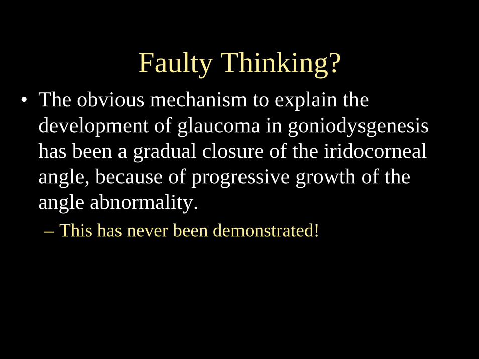
Faulty Thinking?• The obvious mechanism to explain the
development of glaucoma in goniodysgenesishas been a gradual closure of the iridocornealangle, because of progressive growth of the angle abnormality.– This has never been demonstrated!

What is Canine Primary Glaucoma?• Sudden onset of painful, red, often blind eye with very
high pressures• The response to treatment is variable, but severe cases
are blind from the start• Very poor success rate with any treatment protocols
tried• Females affected more than males• Enucleation is a common outcome
– When dealing with the second eye, enucleation is often chosen very early (24 hours from the first signs of disease)

Human Primary Angle Closure Glaucoma as a Potential Model for
Canine Primary Glaucoma• Affects Eskimos and Asians with greater frequency• Hyperopic eyes, smaller eyes• Large lens• Shallow anterior chamber• Women affected 3x more than men• Pathophysiology
– Contact of the pupillary margins with the lens (pupillary block)– Pressure gradient between the anterior and posterior chambers– Forward bowing of the peripheral iris closes the angle– Treated successfully by laser iridotomy

Normal Glaucoma Glaucoma
The Relationship of Canine Primary Glaucoma to Pupillary Block
Thanks to Dr Paul Miller

Before Latanoprost After Latanoprost
Miosis and the Iris Profile

What can pathology say about the early changes leading to outflow obstruction in
canine primary glaucoma?• Evidence supports the idea that the pupillary
margins of the iris rub against the lens• Evidence that pigment dispersion may play a role• Acute inflammation is seen in the iridocorneal
angle and the limbus soon after the start of clinical signs
• Evidence of gradual atrophy of the corneoscleraltrabecular meshwork

Evidence of Pupillary Iris Rubbing against the LensLeading to Pigment Dispersion
andEvidence of Acute Inflammation Playing a Role in the
Iridocorneal Angle and the Limbus
Work done by Chris Reilly

Upper Angle
Lower Angle
Pigment Dispersion

30 hour GlaucomaSuppurative Inflammation

Pigment Dispersion in Primary Glaucoma
• Distinguish superior from inferior angle by pigment alone– 1 to 3 Days: 92%– 4 to 7 Days: 95%– Chronic: 79%
• Cells Stripped from Iris– 1 to 3 Days: 43%– 4 to 7 Days: 75%– Chronic: 55%
• Pigmented Cells in the Angle– 1 to 3 Days: 64%– 4 to 7 Days: 95%– Chronic: 50%
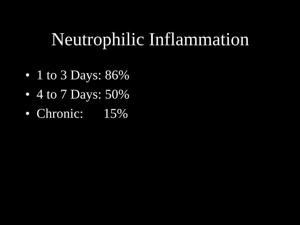
Neutrophilic Inflammation
• 1 to 3 Days: 86%• 4 to 7 Days: 50%• Chronic: 15%

One Day Trabecular MeshworkAtrophied
Evidence of gradual atrophy of the corneoscleral trabecular meshwork
Cocker Spaniel: NormotensiveGoniodysgenesis
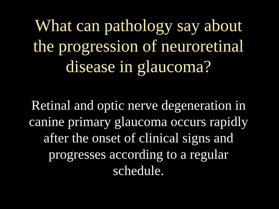
What can pathology say about the progression of neuroretinal
disease in glaucoma?
Retinal and optic nerve degeneration in canine primary glaucoma occurs rapidly
after the onset of clinical signs and progresses according to a regular
schedule.

Effects of Canine Primary Glaucoma on the Optic Nerve
and the Retina
Two day glaucoma, Canine

Early and Later Optic Nerve and Retina
Kerry Ketring images

One Day Glaucoma“Red Dead” Ganglion Cells
The Retina in Primary Glaucoma

“Red Dead”Ganglion Cells

Ganglion Cell Counts
0
10
20
30
40
50
60
70
80
ControlsN=10
Pre-GlaucomaN=2
1 Day N= 5 2 Day N=7 3 Day N=5 4-5 Days N=7 7 Days N=7
GC UpGC Down

“Red Dead” Ganglion CellsRead/Dead GC
0
1
2
3
4
5
6
7
8
9
10
Controls N=10 Pre-GlaucomaN=2
1 Day N= 5 2 Day N=7 3 Day N=5 4-5 Days N=7
Read/Dead GC

Retinal Thickness
0
50
100
150
200
250
300
ControlsN=10
Pre-Glaucoma
N=2
1 Day N= 5 2 Day N=7 3 Day N=5 4-5 DaysN=7
7 Days N=7
Superior RetinaInferior Retina

Retinal CD 18+ Phagocytes

Retinal MHC2+ Phagocytes

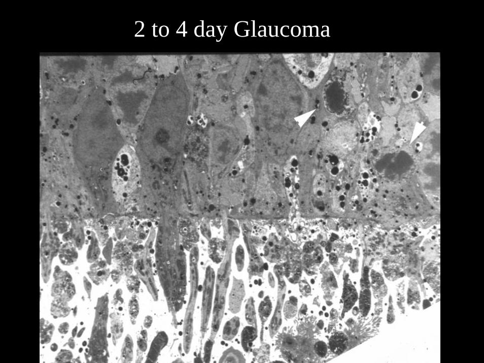
2 to 4 day Glaucoma (Canine)

Four Day Glaucoma
2 to 4 day Glaucoma

TUNEL for Apoptosis
TUNEL+ not seenbefore day 2/3
2 to 4 day Glaucoma

30 hour Glaucoma
The Optic Nerve in Primary Glaucoma

3 day Glaucoma

Optic Nerve 2 to 4 Days

4 day Optic Nerve HeadPhagocytosis/Malacia

5 day Canine Glaucoma

5 day Canine Glaucoma

Chronic Glaucoma

Optic Nerve CD18+ Phagocytes

CD18 on 4-5 day Glaucoma Optic Nerve

Optic Nerve MHC2+ Phagocytes

Schnabel's Cavernous Optic Atrophy

Pre-Glaucoma: The Second Eye
The Up Side The Down Side

The Second Eye
Up Down
Atrophy of the Corneoscleral Trabecular Meshwork

A New Paradigm in the Pathogenesis of Canine Primary Glaucoma associated with
Goniodysgenesis:1. The angle abnormality, along with growth of
the lens, causes contact between the lens capsule and the pupillary margin of the iris.
2. Pigment epithelial cells rub off the pupillarymargin of the iris.
3. Pigment in the angle causes atrophy/necrosis of trabecular-lining cells in the corneoscelraltrabecular meshwork.

A New Paradigm, continued
4. This leads to increased pressure in the anterior chamber, pushing the iris against the lens.
5. Now there is a vicious cycle, which leads to an explosive pressure rise that stops perfusion of the optic nerve and retina.

The Canine Glaucoma Diseases
• Goniodysgenesis (Primary Glaucoma)• Thin-walled Iridociliary Cysts (Pigmentary
Uveitis)• Melanosis
Deehr AJ, Dubielzig RR (1998). A histopathological study of iridociliary cysts and glaucoma in Golden Retrievers. Vet.Ophthal. 1: 153-158

Thin-walled Iridociliary Cysts (Pigmentary uveitis)
• 108 cases in Golden Retriever dogs• Clinically considered inflammatory• Histologic features
– Thin-walled cysts– Posterior synechia– Retrocorneal membrane– PIFM– Minimal inflammation– Pigmented cyst fragments– Pigment dispersion

Thin-walled Cysts in Golden Retriever

Thin-walled Cysts

Thin-walled Cysts (Pigmentary Uveitis))
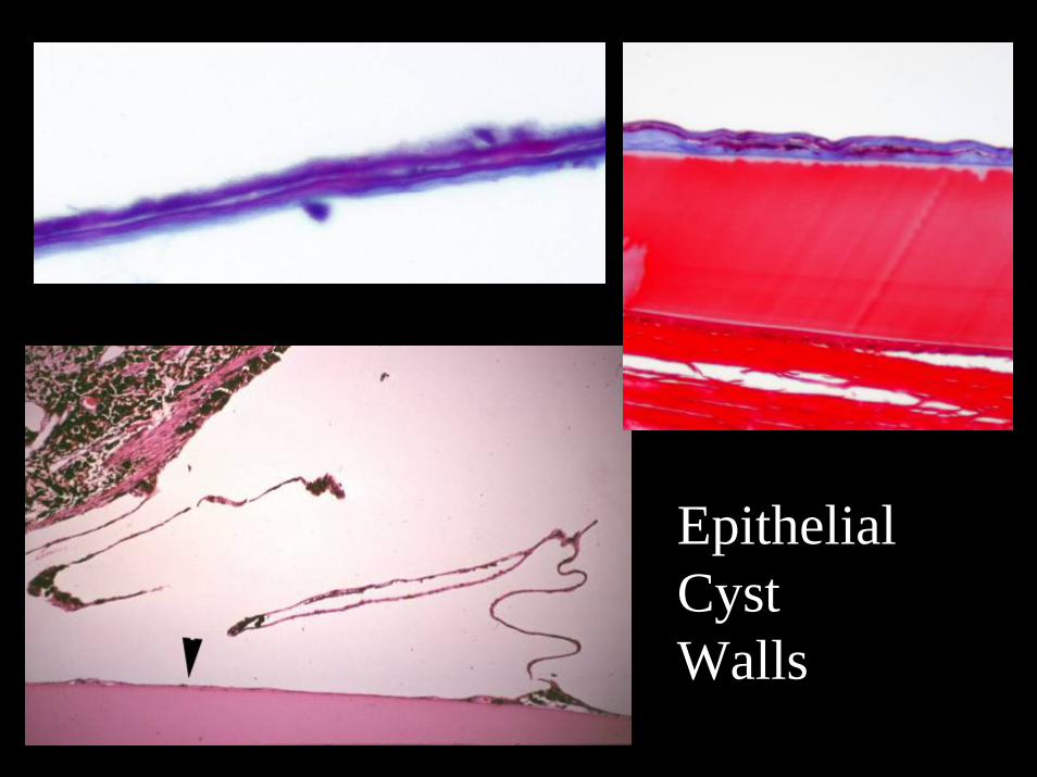
EpithelialCystWalls

Pre-iridal Fibrovascular Membrane
Retrocorneal membrane and Doubling of Descemet’s

The Canine Glaucoma Diseases
• Goniodysgenesis (Primary Glaucoma)• Thin-walled Iridociliary Cysts (Pigmentary
Uveitis)• Melanosis
Petersen-Jones SM, Mentzer AL, Dubielzig RR, Render JA, Steficek BA, Kiupel M (2008) Ocular melanosis in the Cairn terrier: histopathologic description of the condition, and the immunohistological and ultrastructural characterization of the characteristic pigment-laden cells. Vet Ophthalmol. 11(4): 260-268

Canine Ocular Melanosis

Ocular Melanosis

Canine Ocular Melanosis195 Cases
• Cairn Terrier…48• Labrador retriever…26• Boxer…19• Golden Retriever…. 9• Boston Terrier …8• Dachshund….6

Ocular Melanosis

Ocular Melanosis

Selected Glaucoma Diseases in Cats
• Lymphoplasmacytic Uveitis• Angle Recession (Contusion)• Aqueous Misdirect Syndrome• Open Angle Glaucoma

Feline Lymphoplasmacytic Uveitis
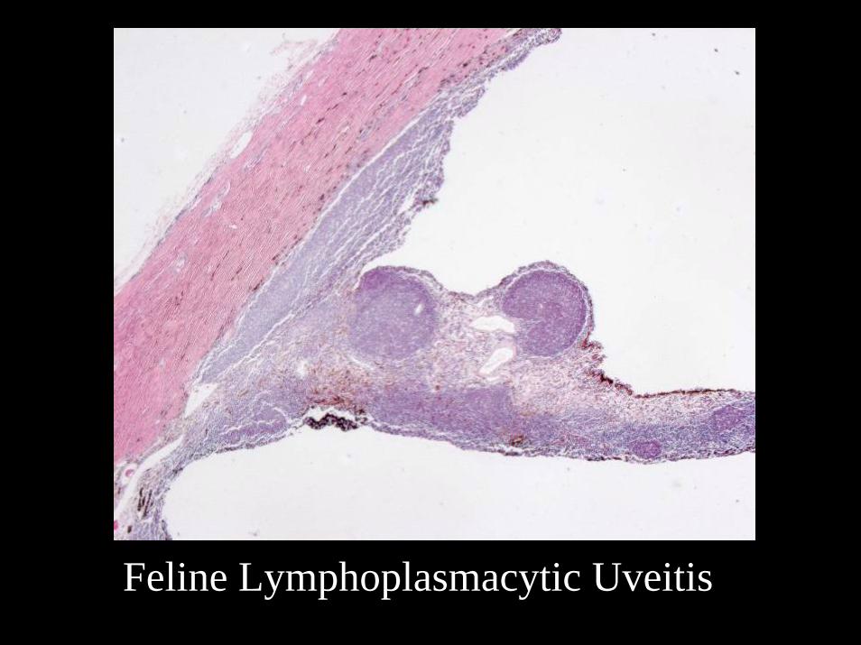
Feline Lymphoplasmacytic Uveitis

Feline Lymphoplasmacytic Uveitis

Select Glaucoma Diseases in Cats
• Lymphoplasmacytic Uveitis• Angle Recession (Contusion)• Aqueous Misdirect Syndrome• Open Angle Glaucoma

Angle Recession

Angle Recession

*
Angle Recession

Acute Cyclodialysis
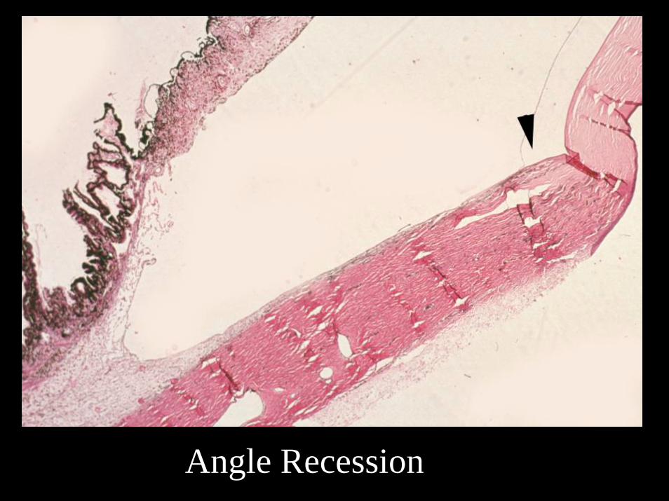
Angle Recession

Normal Feline Iridocorneal Angle
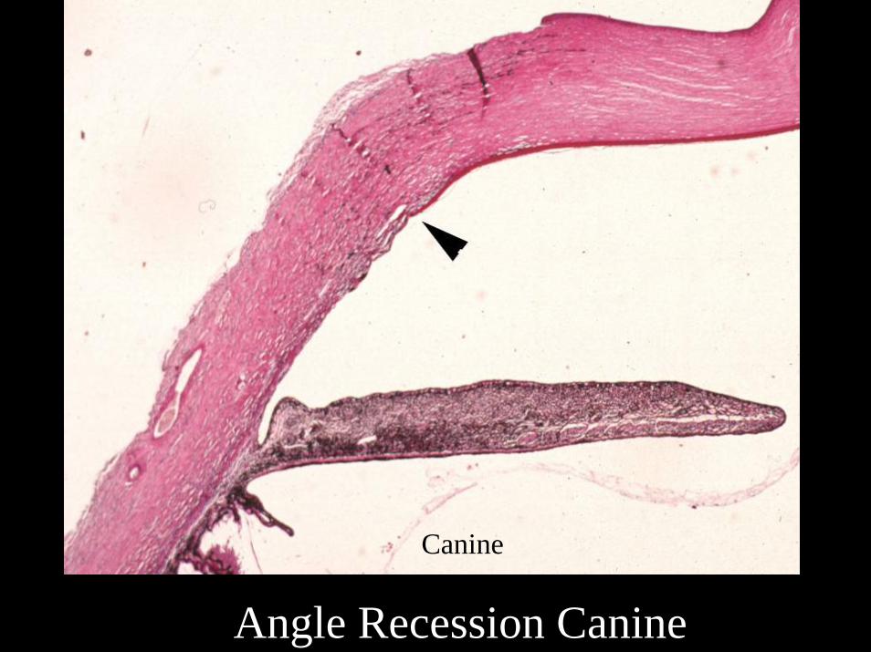
Canine
Angle Recession Canine

Angle Recession

Select Glaucoma Diseases in Cats
• Lymphoplasmacytic Uveitis• Angle Recession (Contusion)• Aqueous Misdirect Syndrome• Open Angle Glaucoma
Czederpiltz JMC, La Croix NC, van der Woerdt A, Bentley E, Dubielzig RR, Murphy CJ, Miller PE. (2005) Putative aqueous humor misdirection syndrome as a cause of glaucoma in cats: 32 cases (1997-2003). J.Am. Vet. Med. Assoc. 227: 1434-1441

Aqueous Mis-direct Syndrome

Aqueous Mis-direct Syndrome

Select Glaucoma Diseases in Cats
• Lymphoplasmacytic Uveitis• Angle Recession (Contusion)• Aqueous Misdirect Syndrome• Open Angle Glaucoma
Jacobi S, Dubielzig RR. (2008) Feline primary open angle glaucoma. Vet Ophthalmol. 11: 162-165

Feline Open Angle Glaucoma12 cases