the genetic principles of the development of the systemic lymphatic vessels in the mammalian embryo....
TRANSCRIPT

THE GENETIC PRINCIPLES OF THE DEVELOPMEST OF THE SYSTEMIC LYMPHATIC VESSELS IN
THE M-IIMMALLUV EMBRYO
PRELIMINARY COMMUNICATION
GEO. S. HUNTINGTON
Piono the Simtornical Laboratory of Columbia University
THIRTY-FOUR FIGURES'
In 1906, at the 22nd session of the Association of American Anatomists, McC'lure and I presented a joint communication on the development of the main lymphatic channels in embryos of the domestic cat, in their relation to the venous system.1 In this preliminary paper we held that the lymphatic vessels of the entire manimalian body are formed by the confluence of perivenous mesodermal spaces, developed, as separate anlages, outside the intima of the early venous channels, but not communicating with the same: except eventually at certain definite points of lym- phat ico-venous junction which are secondarily formed. This view pronounces for the ontogenesis of lymphatic endothelial cells, lining the separate mesodermal spaces, independently of the pre- existing haemal vascular endothelium. The mesodermal inter- cellular spaces, thus forming the fundaments of the future lym- phatic vessels, are in no sense d&vatives from the embryonic veins, although closely associated with them topographically, and eventually replacing the same.
At the time of the publication of the paper quoted, embodying an outline of these views of manimalian lymphatic ontogenesis,
Cost of illustrations met by t.he author. G. S. Huntington and C. P. W. McClure. The development of the main lyniph
channels of the cat in their relation t o the venous syst,em. z 4 n ~ . Jour. A 7 6 0 l . , 1-01.
6 , 1907. Ahstr. AXAT. REC.. rol. 1. pp. 3G41.
THF. .4KATOYICAl . R l X i > l I l ) , 1.01,. 4 , KO. 11

400 GEO. S. HUNTINGTON
McClure and I were not aware of the fact that the mammalian jugular lymph sacs afford, in the typical mammalian organiza- tion, in so far as the same is definitely determined at present, the sole or chief portals of entry of the entire systemic lymphatic circulation into the veins.
We consequent'ly failed to recognize correctly the true mor- phological type of the adult mammalian lymphatico-venous con- nections in our earlier preliminary paper, and hence regarded them, at that time, as the direct secondary junctions of the inde- pendently developed systemic lymphatic vessels with the veins.
The real significance of the adult lymphatico-venous connections was only subsequently recognized by us in the course of a detailed joint investigation of the area involved. A preliminary account of our studies on the development of the jugular lymph sacs in the embryo of the cat, was presented at the 23rd session of the Association of American Anatomists held at Chicago in December, 1907, and published in the Proceedings of that meeting.3 The details of this investigation, with full critical analysis of all the main developmental stages, in an extensive series of cat embryos, and illustrations of the reconstructions of all important and repre- sentative periods, are published in the April number of the Amer- ican ,Journal of Anatomy of this year.*
After the completion of our joint work on the development of the mammalian jugular lymph sac, I published, in 1907,5 a genetic interpretation of the development of the mammalian lymphatic system, as a whole, in which I regarded the same as the final product of the union of two genetically different and very unequal components :
The entire extensive system of the lymphatic vessels of the adult, including the thoracic and right lymphatic ducts and their tributaries, is formed by the confluence of numerous peri-
1.
Geo. S. Huntington and C. l?. W. McClure. The anatomy and development of the jugular lymph sacs in the domestic cat. ANAT. REC., vol. 2, pp. 1-18, May, 1908.
American Journul of Ariatomy, vol. 10, pp. 177-311, April, 1910.
malian lymphatic system. ANAT. KEC., vol. 2, pp. 19-45, May, 1908. ti G . S. Huntington. The genetic interpretation of the development of the mam-

SYSTEMIC LYMPHATIC VESSELS 40 1
venous and extra intimal intercellular mesodermal spaces, in the sense previously defined. These primary anlages of the future systemic lymphatic vessels are, from ,,heir inception, lined by a lymphatic vascular endothelium, whidh is not derived from t!he lmwzal vascular endothelium , but which develops indeeendently of the same.
The lymphatic channels, formed by the subsequent confluence of these originally discrete and separate mesodermal spaces, fol- low in large part the embryonic veins closely, but they are neither derived from them, nor do they communicate with them, ex- cept at the definite points at which the rudimentary mamma- lian type of a lymphabico-venous heart is developed.
This structure develops, as the jugular lymph sac of the typical mammal, directly irom the perivenous capillary reticulum of the early pre- and post-cardinal veins, adjacent to, and includ- ing, their point of confluence to form the duct of Cuvier.
This mammalian jugular lymph sac, the rudimentary homologue of one of the more highly organized veno-lymphatic hearts of the lower vertebrates, arises directly from the veins. Subsc- quently, after evacuation of its blood contents, i t apparently separates for a short period completely from the same, and fin'ally establishes two sets of permanent connections :
(a ) With the independently formed systemic lymphatic chan- nels of the entire body in the majority of the mammalian types carefully determined up to the present date.
( b ) Secondary connections with the venous system, re-entering the same a t one or more typical and constant points, and thus forming the link which eventually unites the mammalian lym- phatic and venous systems, developed primarily independently of each other.
Thus the investigation of mammalian lymphatic development divides itself naturally, in accordance with the postulates of the genetic theory above defined, into three separate and distinct iiiain parts:
The development and adult anatomy of the jugular lymph
2.
1. ';a(%.

402 GEO. S . HUNTINGTON
2. The development and adult anatomy of the general sys- temic lymphatic vessels.
3. The mode of union with each other of the two components just enumerated, and the resulting establishment of a continuous centripetal lymphatic vascular system, with definite and constant terminals in the venous trunks.
The first of these problems, involving the ontogenetic history of the mammalian jugular lymph sacs, having been established in detail by the joint investigations of McClure and myself above quoted, I intend to follow in similar detail the second and third postulates of the theory of mammalian lymphatic development just outlined, and to prove that, in the composite organization of the final adult lymphatic system, the jugular lymph-sacs, of direct venous oriqin, constitute the links eventually uniting the haemal vascular system of the mammal with the systemic lym- phatic vessels, which latter develop independently of the veins, by the confluence of numerous intercellular perivenous mesen- chynial spaces. The embryonic veins, along and around which the earliest anlages of the systemic lymphatic channels develop, appear as evanescent and temporary components of the embry- onic haemal vascular system. They are not carried into the definite and tJipical adult venous organization, but they afford, in reference t o the correlated lymphatic system, by their sepa- ration from the permanent venous channels, and their consequent collapse and atrophy, a series of lines of less resistance in the embryonic body, which paths of easiest progress are utilized by the growing lymphatic vessels. In this way the histological picture of a gradual replacement of an early embryonic vein by a succeeding secondary “ perivenous ” or I ‘ extra-intimal ’’ lymphatic vascular channel is obtained, through the confluence of numerous inesenchymal spaces, surrounding, and eventually replacing, the decadent embryonic veins, but in no sense genetically derived from the latter.
In other words, and in order awin to reiterate emphatically the conception of mammalian systemic lymphatic development which I have consistently upheld since my first expression of opinion on the subject, I desire to repeat my conviction that all

SYSTEMIC LYMPHATIC‘ VESSELS 40.3
systemic lymphatic vessels of the mammalian embryo, including the thoracic and right lymphatic ducts and their tributaries, are neither in their genesis continuous centrifugal “buds ” or “sprouts” from sacs of venous origin, wherever situated, nor “multiple out- growths ” or “veno-lymphatic anlages,” derived from embryonic veins, such “outgrowths” separating subsequently from the veins, and then fusing into continuous and connected lymphatic channels. The systemic lymphatic vessels of the mammalian embryo, as dis- tinguished from the jugular, or reno-caval lymph-sacs, or from any other adult lymphatico-venous junctions of equivalent value, are, on the contrary, in my estimation, from their first ontogenetic inception, structurally and genetically independent of the haemal vascular system. Their endo thelial lining is not derived from the pre-existing embryonic blood vascular endothelium. The multiple independent perivenous spaces forming the anlages of the future systemic lymphatic channels join to form progressively increasing links of longer channel segments, destined in the normal course of development, to become united into a continuous lymphatic vascular system. This lymphatic system finally attains, in the average and typical mammalian forms, one or more permanent connections with the definite venous system through the portals furnished by the rudimentary lymphatico-venous hearts or lymph sacs. The most prevalent mammalian type of this secondarily acquired lymphatico-venous connection is furnished by the jugular lymph sacs, as outlined in the publications already quoted. While this form of lymphatico-venous junction in the adult is by far the most prevalent type encountered in the mammalian series,e there is no reason why, in certain mammalian groups, other points of veno-lymphatic communication, inherited, in these specialized types phylogenetically by selection from the available line of mu1 tiple pre-mammalian lymphatico-venous hearts, should not be carried into the adult organization as permanent portals of entry of the lymphatic into the venous ~ y s t e m . ~ The post-caval
li C . F. W. 3IcClure and C . 1’. Silvestcr. A coinpartitive study of the lymphatico- venoiis communications in adrllt mammals. -1S.iT. REC.. col. 3, pp. 534-561, 1!)00.
The pliylogcnetic relations of t h e lymphatic and blood- vnscnl:ir systems in vertehratt~s;.
G. S. Huntington. -ls . irr. REC.. vol. 4. no. 1 , Jnniitry. 1910.

404 GEO. S. Ht-'NTINGTON
and reno-caval lyniphatico-venous connections recently denion- strated by C. F. Silvesters of Princeton University as uniformly found in the entire group of South American primates, and the intermediate correlated conditions found by myself in Macropus rufus, are readily and correctly interpreted on this basis.
The present communication is intended as an outline of the develo,pment of the mammalian systemic lymphatic vessels, in order to demonstrate what I believe to be the uniform, constant and consistent ontogenetic principle underlying their formation. I have been impressed by the fact that the histological pictures furnished by ungulate, rodent and marsupial embryos are, in reference to the development of the systemic lymphatic channels, relatively obscure and indefinite, when compared with the clear- cut and well-defined conditions encountered uniformly in the aeluroid carnivore. In describing, therefore, in this preliminary account the genetic principle which I believe governs the develop- ment of all mammalian systemic lymphatic channels, as distin- guished from the lymph hearts of venous origin, I have confined my illustrations to the embryos of the cat, and have selected cer- tain portions of the thoracic ducts of this animal in the critical stages, as concrete examples of the developmental processes occurring in zill other regions of the embryo, as will be fully demon- strated in the complete publications to follow. With the ontogene- sis of the systemic lymphatic channels definitely established in this form, it is not difficult to determine, by comparison, the presence of corresponding tbypical developmental conditions in embryos of the pig, rat and oppossum. But in none of these latter forms are the typical genetic stages as clearly marked', and the tissues as definitely differentiated as in the cat.
The right and left thoracic ducts develop in cat embryos of between 11 mm. and 16 mm. crown-rump measure. Prior to the 11 mm. stage no anlages of any portions of the future ducts are observable. In the average 16 mm. embryo the separate anlages have usually united into continuous lymphatic channels, which
8 Twmty-fifth Session of the Association of American Anatomists, Boston, December 28, 29 and 30, 1909.

SYSTEMIC LYMPHATIC VESSELS 405
are connected through the jugular lymph sacs with the syrstemic veins.
I believe that the adult thoracic ducts of the cat are developed by fusion of three distinct and separate regional segments. Each of these segments is in turn formed by confluence of a number of originally discrete adages, which develop independently of the venous system as extra-intimal or perivenous mesenchymal spaces in the sense previously defined ( 2 , 5 ) . These spaces are applied to, or surround, the walls of the embryonic veins of the lower cervical and mediastinal region. The three main divisions, thus formed independently of the venous system, unite with each other to form the channels of the left and right thoracic ducts, and these channels gain their point of entrance into the systemic veins by uniting with a process of the jugular lymph sacs (thoracic duct approach) derived from their dorsal aspect, just cephalad to the common jugular approach.
The ontogenetic history of the ducts may therefore be con- sidered under four headings, viz. :
1. The ‘(Thoracic duct approach” of the jugular lymph sac, forming the terminal of the adult duct on each side
2. The pre-azygos segment. This includes two distinct and separate channels: (a) The ventral mediastinal or broncho-mediastinal lymphatic
trunk, which drains the ventral mediastinum, viz., the pericardial, tracheal, bronchial, lateral esophageal and thymic areas.
This lymphatic channel, associated with the pulmonary arteries, develops through confluence of a large number of separate and independent extra-intimal lymphatic spaces following and sur- rounding the embryonic venous plexuses of the ventral medias- tinum. The chain formed by these spaces eventually unites, directly or indirectly, with the similar chain forming the anlage of the pre-azygos segment of the thoracic duct.
The pre-azygos segment of the thoracic ducf includes the portion of the main channel from the point of its entrance into the jugular lymph sac, through the thoracic duct approach of the latter, caudad to its intersection with the dorsal surface of the aortic arch.
(b)

406 GEO. S . HUNTINGTON
In the adult, animal this segment forms the relatively long por- tion which ascends cephqlo-sinistrad from the point where the duct parts company with the right azygos vein. under cover of the aortic arch, and the vertical portion of the left subclavian artery, dorsal to the vertebral vein and to the left innominate confluence, to its junction with the jugular lymph sac. In this part of its course the t.horacic duct receives the lymphatic return from the ventral mediastinum through channels which join it to the ventral mediastinal trunk as just defined. The pre-aaygos segment of the main duct is again formed in the embryo by confluence of independent mesenchymal spaces around and along the preverte- bra1 and dorsal mediastinal venous plexuses of the embryo.
The a q g o s segment comprises the portions of the thoracic ducts caudal to the level of the aortic arch. It develops, again independently, as the result of fusion of a number of extra-intimal mesenchymal spaces closely applied to the ventral surface of the azygos veins, and of their ventro-medial tributaries, or surround- ing the latter.
4. The post-azygos segment, through which the thoracic ducts establish their connection with the Receptaculum and the system of the abdominal lymphatics.
The purpose of the present paper is to employ the facts ascer- tained in regard to the development of the two thoracic ducts as a concrete illustration of the genetic principles underlying the formation of all systemic lymphatic organization.
For this purpose the right and left ducts will be regarded as bilateral equivalents, as they actually are in ceptain stages. As a matter of fact the right channel in the main azygos region is the first portion to differentiate clearly and offers the best illus- tration of lymphatic histogenesis in the earlier and critical stages.
Inasmuch as the development of the post-azygos segment of both ducts is intimately connected with that of the principal ab- dominal lymphatic channels, and hence requires for its elucida- tion a detailed consideration of these structures, I will confine my illustrations in the present paper to the development of the two main anterior segments, viz., the pre-azygos and the azygos portions of the entire duct, with the distinct understanding that
3 .

SYSTEMIC LYMPH.%TIC VESSELS 407
identical ontogenetic processes a.re responsible for t,hedevelopment not onlyof the post-azygos segments of the ducts and the mesen- teric lymph sacs? but for all other systemic lymphatic channels of the entire body.
I. PRE-AZYGOS SEGMENT O F THE THORACIC DUCT
A . Ventral or broncho-mediasfinal trunk
The area in which this lymphatic channel develops, is shown topographically in fig. 1, a transverse section of the upper thoracic region in a 12 mm. embryo (series 78, slide 5, section 9.) The lym- phatic anlages arise in the mesenchyme between the pulmonary arteries (10) ventrally, the coeloiii laterally, the precardinal veins (3, S), vagi (22), trachea (9) and aorta (7) dorsally. This area is indicated by the 2 in fig. 1.
In the earlier stages (embryos betw-een l lmm. and 14mni.) an extensive ventro-medial capillary network obtains along and between the main venous lines of the right and left sides, involving the caudal part of the internal jugular, the common jugular and innominate veins.
Now, if the ventral portion of this venous plexus is followea caudad into the upper thoracic region, the following observations can be made in stages of the proper length, and adequately fixed and stained:
In embryos between I1 and 12mm. only venous capillaries are found, in the majority of cases.
In 13 mm. embryos certain of the venous radicles entering into this plexus are partly surrounded and enveloped by inde- pendently developed extra-intinla1 lymphatic spaces, the first anlages of the future ventral mediast inal lymphatic channel.
Fig. 2 shows a section of this region in a 13 mm. embryo (series 107, slide 9, sect.ion 40).
Between left pulmonary artery (10) and aorta (7) are branches of the ventral mediastinal plexus. One of these (4) is partially surrounded by a lymphatic anlage ( 5 ) , but the process of replace- ment is in its earliest phases.
(1)
(2)

408 GEO. S . HIJNTIXGTOK
(3) In the 13.5 mm. embryo the full and convincing proof of the extra-intimal derivation of this channel is given.
Fig. 3 shows a transverse section of the upper thoracic region of a 13.5 mm. embryo (series 189, slide 8, section 36). Just ventro- mesad of the left vagus nerve and its encircling vein is a venous radicle (4) almost completely surrounded by an extra-intimal lym- phatic space (5) in the process of replacing the atrophying vein with which it is so closely associated. The corresponding structures are seen on the right side (4, 5 ) .
Fig. 3A shows the extra-intimal lymphatic space and the con- tained vein on the left side of this section in a higher maghi- fication ( x 300). It will be seen that the lymphatic space nearly envelops the venule. The latter, if followed cephalad and caudad, is found separated from the functional venous channels. It appears collapsed and shrunken, and contains only a few degen- erating erythrocytes. We are dealing here with a further advance in the conditions found in the immediately preceding 13 mm. stage. (Fig. 2, series 107, slide 9, section 40). The venous core of the earlier lymphatic anlage is in process of further recession and degeneration, as the perivenous lymphatic space enlarges and more and more completely replaces the antecedent venous channel upon and around which it develops. On the right sideof fig. 3 (series 189, slide 8, section 36), the section has cut the cor- responding vein and the enveloping extra-intimal space at right angles, so that the central kernel of the shrinking vein (4), still con- taining ti few red blood cells, is nearly surrounded by the replacing extra-intimal lymphatic (5). The vein, or rather its remnant, bears a relation to the perivenous replacing lymphatic which is exactly the same as that of a collapsed inner tube to the enveloping shoe of a pneumatic tire. The inner skin of the shoe and the rim of the wheel represent the lymphatic intimal endothelium. The space between them and the collapsed inner tube is the lumen of the future ventral mediastinal lymphatic channel. The inner tube itself is the embryonic vein upon which the secondary lymphatic channel is built. In the course of further development it disintegrates and disappears, leaving a clear lumen to the lymphatic channel which thus secondarily replaces it.
Usually the replacing lymphatic begins as an extra-intimal chan-

SYSTEMIC LYMPHATIC VESSELS 409
nel partially surrounding the embryonic vein which it is destined to replace. This leads in the course of further development to an expansion of the lymphatic space not concentric with the axial line of the shrinking vein. The remnant of the vein retires to a point on the intimal circumference of the new lymphatic channel and appears to project into the latter.
The resulting histological pictures are hence in many cases quite analogous to the appearance of a inesonephric glomerulus in its relation to the lumen of a Wolffian tubule. Of course, as in the case of this illustration, a section, for example, in the axis of the line A-B will divide the shrinking vein and the enveloping lym- phatic in such a way as to produce the following picture:
4
4
This, however, is exceptional. This is not a haphazard process, observed only occasionally,
in a limited number of embryos, and then only in single sections, or, at most, in a few successive sections. In any average embryo of the proper length the samestructures appear in the same situa- tion and in identical relationship to the embryonic environment. It is often possible to follow the forming lyniphatic with its atrophied vein kernel for long distances, and in different embryos of the same crown rump measure the constant repetition of identical histological pictures is remarkable.

There are, of course, individual cases of variation, in which systemic lymphatic development is either more advanced or more retarded than is normal for the average run of embryos in a given stage. But if a very large number of embryos of each typical period are examined and compared the average stage of extra- intimal lymphatic development attained by the majority of indi- viduals in each period is remarkably constant and uniform. I shall have occasion, in the complete publication, to refer again in detail to the question of chronological embryonic variation.
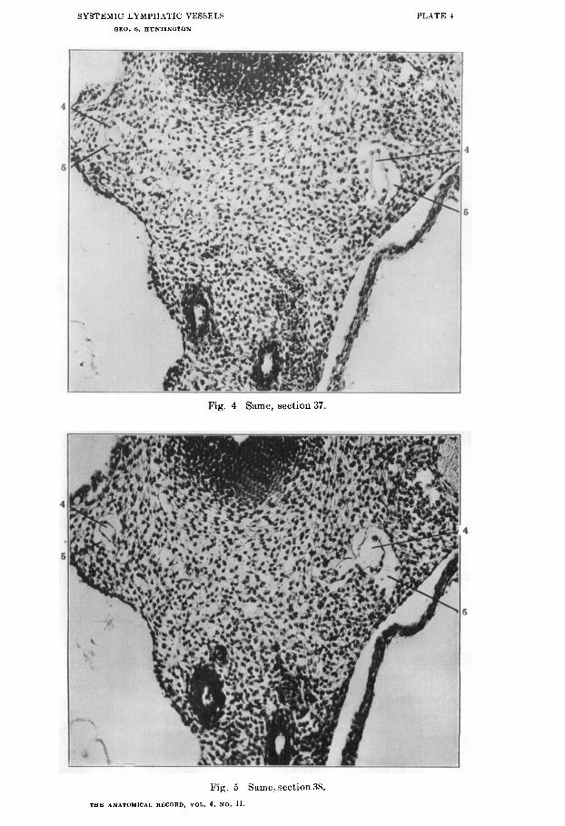
The existence of the perivenous lyniphatic spaces in this and other regions of the embryo has been PO often denied by recent contributors to the subject, or, if admitted, explained in every possible way except on the basis of the correct interpretation, that I publish in this paper a series of micro-photographs of five successive sections through the pretracheal mediastinal regionof a 13.5 cat embryo (series 189, slide 8, sections 36 to 40) (figs. 3 to 7).
Fig. 3, above described, shows the general topographical area involved. Figs. 4 to 7 are cut down to economize space.
In all five figures the atrophying vein kernel (4) and the replac- ing lymphatic anlage surrounding the same (5) have been cut obliquely on the left side of the embryo, and hence give longer stretches of the structures (4 and 5) involved. On the right side the plane of section is more at right angles to both the venous core and the enveloping lymphatic space in the first four figures. In fig. 7 the lymphatic space of the right side terminates in charac- teristic fashion blindly and the atrophied vein merges impercepti- bly into the surrounding mesenchyme. The remnants of par- tially degenerated erythrocytes in the lumen of the atrophied venous core are especially clearly seen in all the sections on the left side.
Of course the photographs, and especially the reduced repro- ductions, offer far less striking hishological pictures than the stained and cliff erentiated slide, although they sufficiently well demonstrate the actual conditions.
In the illustrations only a few of the niore marked areas of lymphatic replacement of decadent venules are indicated by the

SYSTEMIC LYMPHATIC VESSELS 411
leaders 5 and 4 respectively. Numerous other smaller areas of identical significance are seen on close examination in adjacent parts of the field.
In the succeeding 14 mm. stage the ontogenetic process just outlined is, in the average embryo of this measure, fully developed.
Fig. 8 shows a section of a 14 mm. embryo in this region (series 214, slide 13, section 13). Comparison with fig. 3 will show the existence of the identical relations between the same decadent vein and the replacing extra-intimal lymphatic on both right and left sides. The embryos are cut approximately in the same plane and hence the resulting pictures are almost identical.
Figs. 9, 10, 11, and 12 show corresponding sections of the same embryo further caudad.
In fig. 9 three areas are indicated by leaders in which the atro- phied vein (4) is in relation with the enveloping and replacing extra-intimal lymphatic anlage (5 ) . In the succeeding section (fig. 10) the two tlorsal areas have practically become confluent, and the tortuous and collapsed endothelial bag representing the rem- nant of the decadent venule (4) can be followed for some distance. The ventral area in fig. 9 offers only an indistinct central venous core (4), surrounded by the lymphatic anlage (5). In the FUC-
ceeding section (fig. lo), however, the unmistakable relationship and significance of the two spaces is clearly revealed.
The two successive sections of the same slide of this embryo, shown in figs. 11 and 12, give remarkably distinct histological pictures of lymphatic ontogenesis, and also show the gradual increase in the area of the lymphatic perivenous compartment as compared with the contained venous remnant. In both sections a few red blood cells are still to be noticed within the lumen of the latter.
Finally, in another 14 nim. embryo (figs. 13 and 14, series 212, slide 10, sections 5 and 6) conditions identical with the preceding are well shown on both sides of two successive sections. The same decadent venules (4) and the associated enveloping perivenous lymphatic anlages (5) are found in the typical situation between trachea, aorta and vagi dorsad and the pulmonary arteries ven- trad.

412 GEO. S. HUNTINGTON
Fig. 14 likewise offers the explanation of the fact that the aver- age 14 or 14.5 mm. embryo affords the clearest and most distinct pictures of systemic lymphatic ontogenesis. In these stages the decadent vein (4): detached from the functional venous channels, is still relatively large, while the perivenous lymphatic space (5) has also markedly increased in size as compared with the 13 mm. stage. The two structures, taken together, form therefore striking histological objects in the field. Subsequently, with the further degeneration and final complete elimination of the venous kernel, and the condensation of the perivenous lymphatic space into a defi- nite lymphatic channel, the lumen of the latter appears relatively smaller. Thus in two successive sections of a 15 mm. embryo (series 216, slide 10, sections 32 and33,figs. 15 and 16) theidentical lymphatic anlage (5 in figs. 15 and 16) can readily be traced, but appears now as a wide channel with clear lumen. The central venous core, so prominent in the earlier stages (13, 13.5 and 14 mm.) has either disappeared entirely, or is merely indicated by insignificant remnants (4). The same conditions, with further condensation of the mesenchyme, and consequent further reduc- tion of the lymphatic lumen, are encountered in the 15.5 and 16 mm. stages (fig. 17, series 215, slide 14, section 13, 15.5 inm. and fig. 18, series 230, slide 12, section 25, 16 mm.)
No impartial observer can mistake the significance of the con- ditions here shown. Every stage of the process can be followed in detail. The behavior of the decadent embryonic vein, and its relation to the enveloping extra-intimal lymphatic channel, are absolutely demonstrated. The endothelium of the shrinking vein has no share in furniyhing the independent lymphatic endothelium of the replacing mesenchymal space, and nowhere, in the entire process, is there the faintest suggestion of an “out- bud” or of a “splitting off” from the circumference of an other- wise valid embryonic vein of “ lymphatic” or ‘‘ veno-lymphatic ” anlages.
The conditions here described are definite ontogenetic facts remarkably constant in every embryo of the proper age. They cannot be disregarded in proniulgating theories of mammalian lymphatic development. The only conclusion which seems to

SYSTEMIC LYMPH.lTIC VESSELS 413
me to be warranted by actual observation is that certain embry- onic veins form, during the process of their atrophy and final elim- ination from the definite venous organization, the supporting lines along which certain of the perivenous extra-intimal lymphatic anlages first develop. The initial development of lymphatic spaces, is, however, by no means confined to the immediate envir- onment of a degenerating embryonic vein. The same field which demonstrates the histogenetic processes above dedcribed in the development of the extra-intimal lymphatic spaces surrounding a decadent vein will, at the same time, show numerous equivalent lymphatic spaces developing independently of antecedent veins as enlarging intercellular mesenchymal clefts.
These early lymphatic anlages, formed independently of ante- cedent embryonic venous capillaries, are smaller and offer less striking pictures, than those which develop in association with an atrophying vein, and which hence reach a greater size at a rela- tively early period. They are more difficult to differentiate, but their existence can on close examination be absolutely determined, and their connection with the larger perivenous lymphatic spaces can be established.
The fact that numerous early embryonic venous channels, large and small, atrophy and disappear during the normal course of subsequent development, appears to afford a more favorable field for the greater development of the adjacent mesenchymal intercellular spaces, so that these enlarge more rapidly, as the correlated vein recedes. This relationship appears, however, to be based exclusively on the physical and mechanical advantages which the abandoned and shrinking primary venous line affords to the adjacent mesenchymal spaces for more rapid enlargement in the sense of replacing the disappearing vein and occupying secondarily the space formerly filled by the haernal channel. This is evidently an important factor in determining the size and extent of the final lymphatic channel rectulting from the con- fluence of the originally separate and independent perivenous anlages. ('onsequently, in the adult, the largest and best defined systemic lymphatic vessels either accompany reduced adult rem- nants of a relatively larger embryonic venous channel, or, in case

414 (;EN. S. HUNTINGTON
of the latter’s entire default, topographically replace the same. Now, while this relation manifests itself strikingly in many parts of the body, it is quite evident that the development of lym- phatic channels occurs in other parts independently of preceding veins, by the confluence of independent intercellular rnesenchymal spaces.
In judging regarding the genetic principles underlying mamma- lian systemic lymphatic development it is absolutely necessary clearly and correctly to value the relations above detailed between degenerating early embryonic venous channels and the systemic lymphatic anlages developed in association with them and des- tined to eventually replace them more or less completely topo- graphically. 1 can readily see why certain recent contributions to the subject assume that the well defined lymphatic channels of a later stage are the direct derivatives of the equally well defined venous plexuses of earlier embryos, since they cover each other mutually absolutely in the topographical sense. Such an assumption is, however, in my opinion, faulty, because it is based on insufficient or inaccurate observation, and fails in correctly interpreting the genetic factors responsible for the topographical replacement of an earlier vein by a later lymphatic channel.
Again, a careful consideration of the facts above detailed, must inevitably lead to the conviction that the real developmental processes active in systemic lymphatic ontogenesis can never be deiermined by injection of embryos however successful. A glance at the preceding illustrations will show that a successful injection of the embryonic venous system might very well, before complete detachment has occurred, fill from the permanent haemal channels the still large and patent portions of the venous plexus already for the most part surrounded by the extra-intinial lymphatic anlages. Such a preparation would lead the observer to conclude that the line of the future lymphatic channels was still altogether venous. He would have no means of determining the co-existing true lymphatic anlages, nor could these be demonstrated by a simultaneous lymphatic injection, because, at this period, they are isolated segments of the future lymphatic chain, not yet in communication with each other, or with theveins through the jugu-
SYSTEMIC LYMPHATIC VESSELS 415
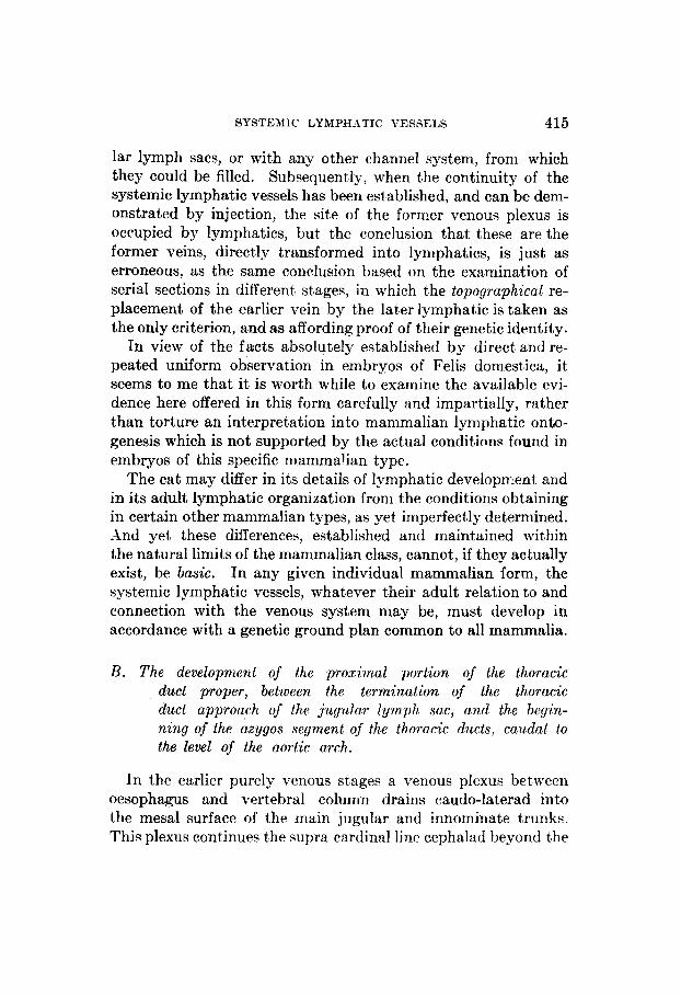
lar lymph sacs, or with any other channel system, from which they could be filled. Subsequently, when the continuity of the systemic lymphatic vessels has been established, and can be dem- onstrated by injection, the site of the former venous plexus is occupied by lymphatics, but the conclusion that these are the former veins, directly transformed into lymphatics, is just as erroneous, as the same conclusion based on the examination of serial sections in different stages, in which the topographical re- placement of the earlier vein by the later lymphatic is taken as the only criterion, and as affording proof of their genetic identity.
In view of the facts absolutely established by direct and re- peated uniform observation in embryos of Felis domestica, it seems to me that it is worth while to examine the available evi- dence here offered in this form carefully and impartially, rather than torture an interpretation into mammalian lymphatic onto- genesis which is not supported by the actual Conditions found in embryos of this specific mammalian type.
The cat may differ in its details of lymphatic developn-lent and in its adult lymphatic organization from the conditions obtaining in certain other mammalian types, as yet imperfectly determined. A h d yet these differences, established and maintained within the natural limits of the mammalian class, cannot, if they actually exist, be basic. In any given individual mammalian form, the systemic lymphatic vessels, whatever their adult relation to and connection with the venous system may be, must develop in accordance with a genetic ground plan common to all rnammalia.
B. The development of the proximal portion of the thoracic duct proper, between the termination of the thoracic duct approach of the jugular lymph sac, and the begin- ning of the axygos segment of the thoracic ducts, caudal to the level of the aortic arch.
In the earlier purely venous stages a venous plexus between oesophagus and vertebral column drains caudo-laterad into the mesa1 surface of the main jugular and innominate trunks. This plexus continues the supra cardinal line cephalad beyond the

416 GEO. S. HUNTINGTON
level of the aaygos-Cuvierian junction. The terminals of this plexus are frequently joined by dorsal somatic venous tributaries near their entrance into the n:ain vein.
Some of the elements of this early embryonic prevertebral venous plexus are secondarily replaced by perivenous or extra- intimal lymphatic spaces in exactly the same way as in the devel- opment of the ventral mediastinal duct. The resulting, originally separate, extra-intim a1 lymphatic anlages, having replaced the venule along and around which they developed, m i t e with each other and form the pre-azygos segment of the thoracic duct, between the thoracic duct approach of the jugular lymph sac and the level of the aortic arch, at which the azygos portion of the thoracic ducts begins.
The general area in which this development proceeds, is indi- cated in the topographical fig. 1, by the letter Y .
The embryonic stages between 13.5 and 15.5 mm. furnish abun- dant evidence of this genetic process. Figs. 19 and 20 show two sections of a 14 mm. embryo (series 210, slide 9, sections 23 and 26) in the prevertebral area of the upper thoracic region. The anlage of the pre-azygos segment of the thoracic duct (5 ) is seen on the left side of the interval betwhen oesophagus and the preverte- bra1 plexus (17) and sympathetic nerve(1). The sections show the identical characters previously noted in the development of the broncho-mediastinal trunk, but both the decadent central venous core of the anlage (4) and the perivenous lymphatic space (5) are larger and better developed.
These pictures are again constant in embryos of the appropriate stages. The lymphatic adage can be accurately traced from its indefinite beginning among the perivenous mesenchymal inter- cellular clefts through a number of successive sections to its similar distal termination in the same intercellular plexus. Following the sections from this point caudad through a varying intervening area in which no distinct lymphatic channel appears, the same line will sooner or later reveal the repetition of the same process, and the formation of another link in the still disjointed chain of primitive lymphatic anlages, eventually destined to unite into the continuous-channel of the pre-azygos segment of the thoracic duct.

STSTE\IIC LY A1 PH.lTIC VESSELS 41'7
11. THE AZPGOS SKGXIENT OF THE THORACIC: DUCT
This main part of the thoracic duct develops by the confluence of the extra-intimal lymphatic anlages, which begin to appear in the 12.5 mm. embryo, are clearly marked in the 13 and 13.5 nim. embryo, increase in the 14 nim. stage, become confluent tb form longer segments in the 1.5 and 15.5 mm. embryos, and finally unite into the bilateral and continuous channels of the tho- racic ducts in the average 16 mm. embryo, although instances are not rare in which the complete continuity of the thoracic ducts is not attained until a later stage. These extra-intimal 1-mphatic anlages develop in close association with the ventral aspect of the azygos veins and their ventral branches, but are from the begin- ning genetically distinct and independent of the same.
In the earlier and purely venous stages, the azygos veins receive, in addition to the terminals of the supracardinal plexus, larger dorsal somatic tributaries from the body walls and from the inte- rior of the vertebral canal, and smaller ventromedial branches which drain the periaortic space close to the wall of the main arterial vessel. When these ventral azygos tributaries appear they occupy in general the position described by McClure as charac- teristic for the cardinal collateral plexus of the Marsupalia.s
The ventro-medial azygos tributary plexus is found in the purely venous condition, before any perivenous lymphatic devel- opment associated with it has begun in this region, in embryos of 11 and 12 mm. (Fig. 21, series 213, slide 11, section 29, 11 mm ; fig. 22, series 21i, slide 11, section 27, 12 mm.). The plexus occu- pies the area ventral to the intersegmental aortic branches and the sympathetic nerves, between the aorta and the main azygos trunks.
Later, in 13.5 to 14 mm. embryos, portions of this early plexus appear detached in certain areasof the sub-azygos region from the main venous trunks. In many cases the line of the obliterated connection can still be traced for a time as a strand of differentiated mesoderm, and the separated elements of the azygos plexus still
C . F. R. McClure. Theanatomyand development of the post-cava in Didel- phis marsupialis. Am. Jotcr. Anat., vol. 5, 1906.

418 GEO. S. HUNTINGTON
contain frequently red blood cells in the earlier stages. The lym- phatic anlages of the thoracic ducts form along and around these degenerating elements of the azygos plexus, as extra-intimal or perivenous spaces, in exactly the sake manner as above described for the regions further cephalad.
The recognition of this reduced ventro-medial tributary system of the azygos veins is of the greatest importance to the correct interpretation of the mammalian thoracic duct development. S o t only do the extra-intimal lymphatic anlages of the azygos segments of the duct form along and around these venules, but in the shme way the anterior part of the mesenteric lymphatic network of the abdomen has its origin in the extra-intimal lym- phatic spaces which develop around the caudal continuation of the ventral plexus in front and along the sides of the abdominal aorta, in the root of the dorsal mesogastrium. These perivenous lymphatic spaces subsequently unite to form the receptaciilum and establish, on one hand, connections with the independently developed intestinal lymphatic channels, and, on the other, with the thmacic duct.
McClure, in a paper published in 1908,lO on the development of the thoracic ducts in the cat, very clearly described and figured this secondary and evanescent line of the venous capillary plexus along the innominate and azygos veins which forms the basis for the subsequent development of the main segments of the tho- racic duct. I can completelyconfirni the accuracyof his obse va- tions on this structure, which he for the first time mapped out and demonstrated completely. I am obliged to differ from him, as shown in the preceding pages, in reference to theinterpretation of the rBle taken by the temporary venous plexus in the develop- ment of the thoracic ducts. I cannot regard the ducts as arising directly from the detached venous elements of the plexus, but believe, as here shown, that these elements are secondarily replaced by independent extra-intimal lymphatic spaces, which then join to form t,he continuous channels of the thoracic ducts.
l o C. F. W. McClure. ducts in thc domestic ca t .
Tlic developmcnt of the thoracic ant1 right lymphatic -4nrrt dlvz. . Bd. 32, no.;. 21 and 22, 1908.

SYSTEMIC LYMPHATIC VESSELS 419
I am quite convinced that, in determining definitely questions as intricate as are the relations between developing haemal and lymphatic channelsin the mammalian embryo, avery large nhmber of individual examples of each stage are absolutely necessary. I feel that if McClure had had at his command the amount of mate- rial on which this communication is based, his conclusions would have coincided with those here expressed, and he would not have assigned to the thoracic ducts a genetic origin differ- rent from that which we upheld for all systemic lymphatic devel- opment in the mammalian embryo in our first joint publica- tion on the subject in 1906 (2), and which, with the exception of the thoracic ducts, he still regards as the fundamental basis of systemic lymphatic development.
It is necessary to exercise great care in the critical stages in order correctly to distinguish between the degenerating vessels of the plexus and the extra-intimal lymphatic anlages replacing them, and to compare results obtained from a number of embryos of the same stage. If this is done there can remain no doubt that the azygos segments of the two thoracic ducts in the embryos of the cat develop by confluence of extra-intimal perivenous lym- phatic spaces. These anlages appear at first as isolated spaces, either surrounding the retreating veins or closely applied to part of their circumference and subsequently to the ventral wall of the main azygos trunks, usually laterad to the points where the ventral plexus connects with the main azygos channel. Thus, compare the micro-photographs of series 34 and 214, figs 23 to 32. In the succeeding stages these numerous separate lymphatic nnlages coalesce into longer continuous channel-segments. I t is again noteworthy that in stages between 13.5 mm. and 14 mm., the still separate and distinct lymphatic anlages are relatively larger and more clearly evident than in the subsequent (15 mm. to 15.5 mm.) stages in which they have more extensively joined to form longer links of the system. Finally, in the 16 nun. embryo, where usually all the qeparate segments are assembled into the continuous channel of the thoracic duct, additional new mesen- chymal spaces are added and thus a second and permanent increase in size and caliber of the latter appears to begin, which

420 GEO. 6. HUNTINGTOlrl
can be traced in the subsequent stages as occurring in correlation with the reducticnof the adjacent azygos trunks. Themammalian systemic lymphatic vessels seem to thus share with the embryonic veins this tendency towards apparently excessive diffuse plexi- form developnient in their respective early genetic stages. Sub- sequently the definite channel, lymphatic or hzemal, seems to concentrate along static lines, as a vessel of relatively smaller caliber, out of the antecedent more generalized plexus, and from this stage onfurther growth centres on the definite vessel replacing the earlier diffuse plexus.
In a 14 mm. embryo (series 34, Princeton Embryological C'ol- lection, slide 31) the main azygos trunks have increased in size, have approached the dorso-lateral circumference of the aorta more closely, and the interazygos anastomosis has developed.
The ventro-medial plexus is, however, still present in the typical position-Wigs. 23and 24,4; series 34, slide 31, sections 18 and 19.)
Further cephalad (slide 28 of the same embryo), the beginning extra-intimal replacement of this plexus by the lymphatic anlages of the thoracic duct is encountered (figs. 25 and 26, series 34, slide 28, sections 19 and 20). The venule (figs. 25 and '26, 4), still containing a few red blood cells, is almost completely detached from the definite azygos venous channel, although its original continuity with the same can $ti11 be traced by a strand of dif- ferentiated mesenchyme representing the obliterated channel of corrmunication. This central detached venous kernel (4) is surrounded by the extra-intimal lymphatic space (5 ) .
Figs. 27 to 30 show successive sections from two slides of another 14 mm. embryo (series 214), in which the process of lymphatic replacement of the azygos plexus is further advanced. All trace of the original connection of the central venous core (4) with the aeygos systera is lost in these sections. The detached and aban- doned venule is entirely empty and forms a partially collapsed endothelial tube surrounded, as before, by the perivenous lyni- phatic anlage of the thoracic duct (5). The four figures show this development both at the level of the inter-segmental arteries (figs. 27 and 28) and in the intervals between these aortic branches (figs. 29 and 30). The figures published here are not isolated sec-
SYSTEMIC LYMPHATIC VESSELS 42 1
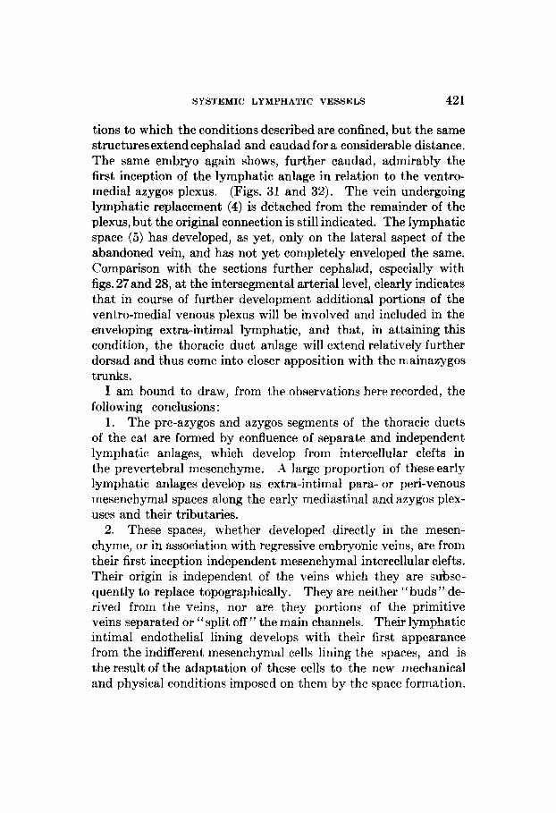
tions to which the conditions described are confined, but the same structuresextendcephalad and caudad for a considerable distance. The same embryo again shows, further caudad, admirably the first inception of the lymphatic anlage in relation to the ventro- medial azygos plexus. (Figs. 31 and 32). The vein undergoing lymphatic replacement (4) is detached from the remainder of the plexus, but the original connection is still indicated. The lymphatic space (5 ) has developed, as yet, only on the lateral aspect of the abandoned vein, and has not yet completely enveloped the same. Comparison with the sections further cephalad, especially with figs. 27and 28, at the intersegmental arterial level, clearly indicates that in course of further development additional portions of the ventro-medial venous plexus will be involved and included in the enveloping extra-intimal lymphatic, and that, in attaining this condition, the thoracic duct anlage will extend relatively further dorsad and thus come into closer apposition with the IX ainazygos trunks.
I am bound to draw, from the observations here recorded, the following conclusions :
1. The pre-azygos and azygos segments of the thoracic ducts of the cat are formed by confluence of separate and independent lymphatic anlages, which develop from intercellular clefts in the prevertebral mesenchyme. large proportion of these early lymphatic anlages develop as extra-intimal para- or peri-venous mesenchymal spaces along the early mediastinal and azygos plex- uses and their tributaries.
2. These spaces, whether developed directly in the mesen- chyme, or in association with regressive embryonic veins, are from their first inception independent mesenchymal intercellular clefts. Their origin is independent of the veins which they are subse- quently to replace topographically. They are neither “buds ” de- rived from the veins, nor are they portions of the primitive veins separated or “split off” the main channels. Their lymphatic intimal endothelial lining develops with their first appearance from the indifferent mesenchynial cells lining the spaces, and is the result of the adaptation of these cells to the new mechanical and physical conditions imposed on them by the space formation.

422 GEO. S. HUNTINGTOK
The lymphatic endothelium does not arise by “sprouting,” or otherwise, from the pre-existing haemal vascular endothelium of the early embryonic veins.
As a matter of fact, in place of being derived from the endothe- lium of the blood channels, the intima of the degeneratingveincan in hundreds of observations be followed through its stages of dis- integration, partial reversion to indifferent mesenchymal cells, and final complete elimination, within the lumen of the extra- intimal lymphatic channel partly or completely enveloping the venous rudiment. Nowhere is there the slightest indicatio of “budding” or sprouting,’’ or of any other active process on the part of the degenerating haemal endothelium.
The above named individual segrrents of the thoracic and right lymphatic ducts, thus formed through confluence of a large number of separate and independently developed mesenchymal and perivenous anlages, finally unite with each other to form a continuous bilateral channel, which secondarily effects a junction with the tharacic duct approach of the jugular lymph sac, through which the general lymphatic system gains its entrance into the venous system.
DS and aeygos segments, and in the area of the tributary ventral mediastinal trunk, offer the most striking and convincing evidence of the truth of the extra-intimal theory of systemic lymphatic development in this mammalian embryo, in the relation exhibited by the first perivenous lymphatic anlages to early embryonic venous channels which they surround and subsequently replace.
For this reason I have selected the thoracic dmts as representa- tive systemic lymphatic channels, whose developmental history will serve as a concrete illustration of the genetic principles ex- pressed in this communication. Many of the details of the thoracic duct development are here designedly not considered, although they are of great importance and significance.
These questions can be much more clearly and coniprehen- sively studied in their relation to the adult anatomy of the ducts and their mode of union with the system of the abdominal lym-
3.
4. The thoracic ducts, especially in their pre-a2

SYSTEMIC LYMPH.4TIC VESSELS 423
phatics. They will be considered in detail in a more extensive memoir on mammalian lymphatic development to be presently published.
As above stated, the development of parts of the thoracic ducts is introduced in this paper solely for the purpose of affording a concrete illustration of the general principles underlying the devel- opment of all the systemic lymphatic channels in the particular mammalian embryo (Felis domestica) here considered. The same principles obtain in systemic lymphatic genesis in all mammalian types which I have had the opportunity of examining, but the embryos of the cat offer by far the most conclusive, consistent and striking evidence.
The early independent genetic history of the spaces, which I have above described as the first anlages of the thoracic duct channels in the embryos of the cat, and the fact that in subsequent stages they appear consistently and in every possible combination as extra-intimal or perivenous mesenchymal spaces, following and surrounding the branches of the prevertebral, ventral medias- tinal and ventro-medial azygos venous plexuses, excludes to my mind, the possibility of considering them as direct derivatives from the venous plexuses, or as so-called “venous outgrowths ” of the in- nominate and main azygos veins, subsequently detached from the parent trunks. The actual conditions observed and here described are too obvious and constant to admit of any doubt. They can be verified by any observer on sufficient material of the proper stages. I think it is time for investigators engaged in solving the problem of mammalian lymphatic development to abandon superficial lines of comparison and generalization, based often on isolated and insufficient observations, or, as in the injection experiments, on methods which, from the nature of the problem, are utterly inadequate and almost barbarous. Results obtained from observa- tions of this kind are, at their best, misleading, when dealing with a genetic question as delicately balanced as is the relation between developing haenial and lymphatic channels in the mamma!ian embryo.
5.
EXPLANATION O F FIGURES
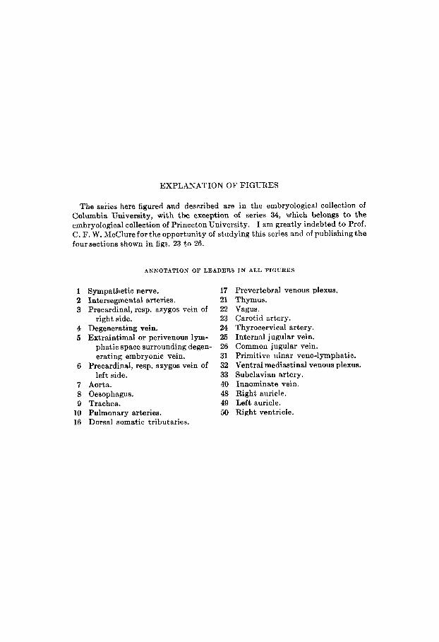
The series here figured and described are in the embryological collection of Columbia University, with the exception of series 34, which belongs to the embryological collection of Princeton University. I am greatly indebted t o Prof. C. F. W. McClure for the opportunity of studying this series and of publishing the four sections shown in figs. 23 t o 26.
ANNOTATION OF LEADERS IN ALL FIGURES
1 Sympathetic nerve. 2 Intersegmental arteries. 3 Precardinal, resp. azygos vein of
4 Degenerating vein. 5 Extraintimal or perivenous lym-
phatic space surrounding degen- erating embryonic vein.
left side.
right side.
6
7 Aorta. 8 Oesophagus. 9 Trachea.
Precardinal, resp. azygos vein of
10 Pulmonary arteries. 16 Dorsal somatic tributaries.
17 21 22 23 24 25 26 31 32 33 40 48 49 50
Prevertebral venous plexus. Thymus. Vagus. Carotid artery. Thyrocervical artery. Internal jugular vein. Common jugular vein. Primitive ulnar veno-lymphatic. Ventral mediastinal venous plexus. Subclavian artery. Innominate vein. Right auricle. Left auricle. Right ventricle.

Fig. 1 Trnnsvrrsc srctinn nf nntc~rinr iliorncic region of 12 IIIIII . cat ~ I I I ~ J Y ~ O
(series 78, slirlc 5, sect ion 9, X 30).
TIP. ~ ~ ~ ~ n \ i i r & i . R w o r t n . vni 1. un I I

Fig. 2 Transverse scctioii of aiitcrior tliorucic region of 13 I I I I I I . cat cmhryo (scrics 107, s l i t lcs 9, sectioii 40, X 725).

SYSTEMIC LYMPIIITIC VESSELS OEO. 8. HUNTINOTON
Fig. 4 Same, section 37.
Fig. 5 Same, section 38.
T E E ANATOMICAL RECORD, VOL. 4, N O . 11.
PLATE 4

~~
SYSTEMIC I,Y.\IPHATIC VESSELS CEO. 8. EUNTlNCTON
Fig. G Samc, section 39.
Fig. 7 Same, section M. THE ANATOMICAL RECORD, V O I . 4, NO. 11.
PLATE 5


PL.\'I'l: 7

SYSTI'; \ I IC LYMPFTA'L'IC VESSELS CEO. 8. HUTNTINOTOS
Fig 11. Same, section 21.
Fig. 12 Snmc, section 22.

PLATE !I
Fig. 13 Transverse section of anterior thoracic region of R 14 mm. cat embryo, (scrics 212, slide 10, section 5 , X225).
Fig 14 Same. section 6.

l’ l . . \’ i’ i , Ill
Fig. 15 Transverse section of :interior thoracic region of a 15 mm. cat einhryo, (series 216, sliile 10, section 33, x ‘L’L5).

Fig. 17 ' I ' rons~~cr~esectiori of anterior ttiorncic region of n 13.6 mm. cnt embryo (srrirs 21.5, slidc 11, section 1 3 , X 22,j).

SYSTEMIC LYMPHATIC VESSELS OEO. 0. BIJNTINUTON
PLATE 12
Fig. 19 Transverse section of anterior thoracic prevertebral area of a 14 mm. cat embryo, (series 214, slide 9, section 23, x 225).

SYSTEMIC LYMPHATIC VESSELS QEO. 8. EUTNTINQTON
PLATE 13
Fig. 21 Transverse section of middle thoracic region of :I 11 nim. cat embryo, (series 213, slide 11, section 20, X225).
Fig. 22 Transvcrsc scction of middle thoracic region of a 12 mm. cat embryo, (series 217, slide 11, section 27, X 226).

PLATE: 11
I2ig. 23 ‘I’r:rri,-versc sect ion of initldle thoracic region of n 14 mm. cat riiil)r\ n, (series 34, I’riuceton University Eriibryological Collection, ,dlidc 31, scct ion 18, x 2‘2.5).


S;YS'L'E.\IIC LYMPHATIC VESPE1,S QEO. 5 . HUNTINOTON
Fig. 27 Transverse section of middle thoracic rcgion of n 1 + I I I I I I . cat embryo (scrics 214, slide 15, section 10, X22.5).
Fig. 2.9 Pam?, sect ion 11
TEE ANATOMIC.AL ILECOIID, VOL. 4 . NO. 1 1 .

