the future of mitral valve repair - · · 2015-05-03the future of mitral valve repair ... •...
TRANSCRIPT

The future of mitral valve repairThe future of mitral valve repair
Dr. Ali AburummanQAHI KHMC
Al Riyadh ‐ 2011

Lancet 1957


The future of Mitral valve repairThe future of Mitral valve repair
• Mitral valve diseased eitherMitral valve diseased either• Stenosis
i i• Regurgitation• Combine pathology

Mitral StenosisMitral Stenosis
• EtiologyEtiology– Rheumatic Degenerative (calcification)– Degenerative (calcification)
– Congenital (parachute MV)t i fl t– post‐inflammatory,
– metabolic syndromes O h– Others

33--D Echo of Mitral StenosisD Echo of Mitral Stenosis33--D Echo of Mitral StenosisD Echo of Mitral Stenosis
LA viewLA view LV viewLV view

33--D measurement of Mitral valve areaD measurement of Mitral valve area
LA iLA i LV iLV iLA viewLA view LV viewLV view
MVA= MVA= 00..914 914 cmcm22

Real Time TTE of MS
A B C
LALALALA
D E F G

T t t f it l St iTreatment of mitral Stenosis
1. Close mitral valvotomy (CMV)… not used.2.Open mitral valvotomy (OMV) rarely used.3.Percutaneous Balloon mitral valvotomy
(PBMV) commonly used .4. Mitral valve replacement more frequent
surgical option

Indications for percutaneous valvuloplasty of MS
• Class IClass I– Symptomatic patients (NYHA II, III, or IV), moderate or severe MS (MVA ≤ 1 5) and valvemoderate or severe MS (MVA ≤ 1.5) and valve morphology favorable for percutaneous balloon valvotomy in the absence of left atrial thrombus or moderate to severe MR

• Class IIaClass IIa– Asymptomatic patients with moderate or severe MS and valve morphology favorable for percutaneous balloon valvotomy who have pulmonary hypertension (PAP > 50 at rest or 60 with exercise) in the absence of LA thrombus or moderate to severe MR
– Patients with NYHA III‐IV symptoms, moderate or severe MS and a nonpliable calcified valve who are at high risk for
i th b f LA th b d/ MRsurgery in the absence of LA thrombus or mod/sev MR

• Class IIbClass IIb– Asymptomatic patients, moderate or severe MS and valve morphology favorable for percutaneousand valve morphology favorable for percutaneous balloon valvotomy who have new onset of AF in the absence of left atrial thrombus or mod/sev MR.
– Patients in NYHA III‐IV, mod or sev MS, and a nonpliable calcified valve who are low‐risk candidates for surgery
• Relative contraindicationsRelative contraindications– Left atrial thrombus
• TEE frequently performed prior to the procedure toTEE frequently performed prior to the procedure to rule out thrombus
• According to Palacios and Vahanian, no consensus regarding thrombus localized in L atrial appendage.
– Limit to patients with contraindications to surgery or those with urgent need for intervetiong
– Significant MR (3+ to 4+)

• Last month I face with 78 year old patientLast month I face with 78 year old patient• Present with calcific mitral Stenosis 1.8cm
i h l i• With LAD lesion • Is their any contraindication to try BMV +Stent to LAD

Immediate outcomes of percutaneous valvuloplasty of MV
• The immediate results of percutaneous mitral valvotomy are similar to those of surgical mitral commissurotomy
– Mean MVA doubles (from 1.0 cm2 to 2.0 cm2) – 50% to 60% reduction in transmitral gradient. – Overall, 80% to 95% procedural success (MVA >1.5 cm2 and a decrease in LA pressure to <18 mm Hg).
• Most common acute complications• Most common acute complications – severe MR 2% to 10% – residual ASD
• Large ASD (>1.5:1 L‐>R shunt) in <12% with the double balloon technique and <5% with the Inoue balloon technique. q
• Smaller ASD detected by TEE in larger numbers of patients. • Less frequent complications
– perforation of LV (0.5% ‐ 4.0%)– embolic events (0.5% ‐ 3%)– MI (0.3% ‐ 0.5%).
• Mortality – 1% to 2% – < 1% with increasing experience in selected patients.
Long‐term results of percutaneous valvuloplasty of MS
• Survival rate 60 % ‐ 90 % over 3‐7 yrs• Restenosis rate 40% after 7 yrs.• Randomized trials comparing percutaneous approach with both closed
and open surgical commissurotomy consisted mainly of younger patients p g y y y g pwith favorable morphology.– No significant difference in acute hemodynamic results or complication rate.– No difference in clinical improvement or exercise time in early follow‐upp y p– More favorable hemodynamics and symptomatic results with percutaneous
approach than closed commissurotomy and equivalent results with open commissurotomy.
• MR stable or decreaswes slightly.• ASD likely to close later in majority of cases.

• Percutaneous BMV is the procedure of choice in patients who have symptomatic, hemodynamically severe stenosis with an echocardiographic score of 8 or less and without left atrial thrombus
• if the score > 8 the durability well be low and in favor of replacement


Treatment of mitral stenosisTreatment of mitral stenosis
• All of us agree BMV is treatment of choiceAll of us agree BMV is treatment of choice • It replace CMV & OMV in favorable cases
ll h i id i i b f h• All the time we said repair is better for the patient especially female at child bearing age, & id f i& we avoid warfarine
• But the durability of OMV was query if it not suitable for BMV >>>> replacement

• What about mitral regurgitation• What about mitral regurgitation
Carpentier Classification of Mitral Regurgitation
• Type Leaflet , Mobility & Cause
• I Normal Annular dilatation Leaflet perforation• II Excessive Chordal elongation or rupture Papillary muscle
elongation or ruptureelongation or rupture• III Restricted • a. Restricted opening;
C i l f i l fl d h d l hi k i• Commissural fusion, leaflet and chordal thickening• b. Restricted closure; Excess tension on chordae during systole

Modified Carpentier Classification of lMitral Regurgitation
• Leaflet Motion Descriptionl l dil i• Ia Normal Annular dilatation
• Ib Leaflet perforation• IIa Increased Chordal elongationg• IIb Chordal rupture• IIc Papillary muscle infarction or scarring• IId Papillary muscle ruptureIId Papillary muscle rupture• IIIa Restricted Commissural or chordal fusion and• shortening
IIIb L fl h i b d ki i• IIIb Leaflet tethering by dyskinetic or• aneurysmal LV segments• IV Variable Dynamic papillary muscle dysfunction

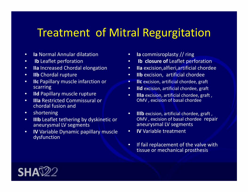
Treatment of Mitral RegurgitationTreatment of Mitral Regurgitation• Ia Normal Annular dilatation
Ib L fl t f ti• Ia commisroplasty // ring
Ib l f L fl t f ti• Ib Leaflet perforation• IIa Increased Chordal elongation• IIb Chordal rupture• IIc Papillary muscle infarction or
• Ib closure of Leaflet perforation• IIa excision,alfieri,artificial chordee• IIb excision, artificial chordee• IIc excision, artificial chordee, graftp y
scarring• IId Papillary muscle rupture• IIIa Restricted Commissural or
chordal fusion and
g• IId excision, artificial chordee, graft• IIIa excision, artificial chordee, graft ,
OMV , excision of basal chordee
• shortening• IIIb Leaflet tethering by dyskinetic or
aneurysmal LV segments• IV Variable Dynamic papillary muscle
• IIIb excision, artificial chordee, graft , OMV , excision of basal chordee repair aneurysmal LV segments
• IV Variable treatmentIV Variable Dynamic papillary muscle dysfunction
IV Variable treatment
• If fail replacement of the valve with tissue or mechanical prosthesis

The future of Mitral valve repairThe future of Mitral valve repair
Surgical weaponsSurgical weapons 1. Commisserotomy ±splitting of chordee and muscle 2. Annulus ring, annuloplastyg, p y3. Excision and plication± sliding4. Closure of perforation5. Excision of 2ndary chordee6. Alfieri stitch7. Artificial chordee8. Grafting pericardial, tricuspid..etc9 Remodeling of the ventricle9. Remodeling of the ventricle
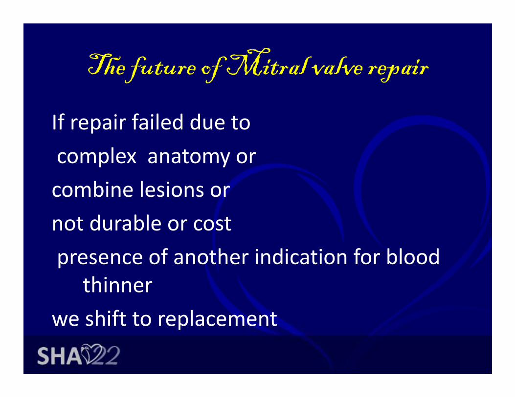
The future of Mitral valve repairThe future of Mitral valve repair
If repair failed due toIf repair failed due to complex anatomy orcombine lesions or not durable or costnot durable or costpresence of another indication for blood
thinner we shift to replacementwe shift to replacement

• “ Most often the entire valve appears Most often the entire valve appears normal;… There is little to fix, yet the valve leaks… the valve is structurally ynormal; it need not be replaced, but currently we do not know how to fix it…”
• - L. Henry Edmunds Jr. 1997 y(Cardiac Surgery in the Adult)
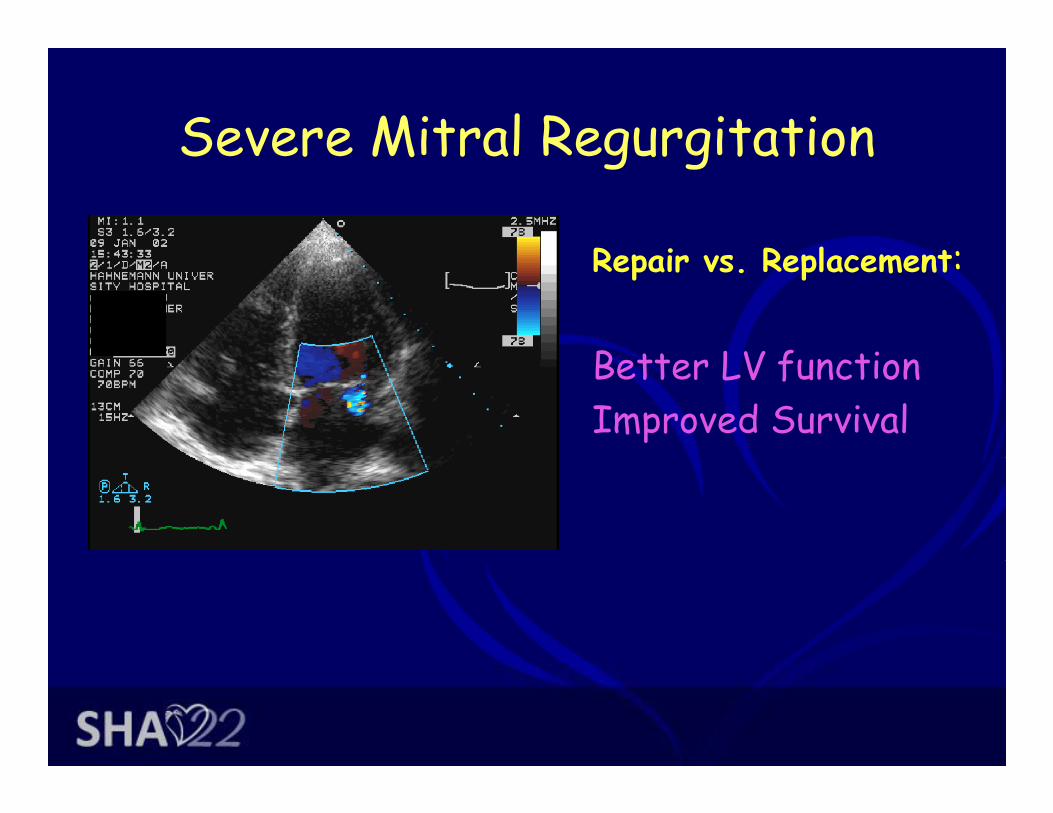
Severe Mitral RegurgitationSevere Mitral Regurgitation
R i R l t:Repair vs. Replacement:
B tt LV f tiBetter LV functionImproved Survival
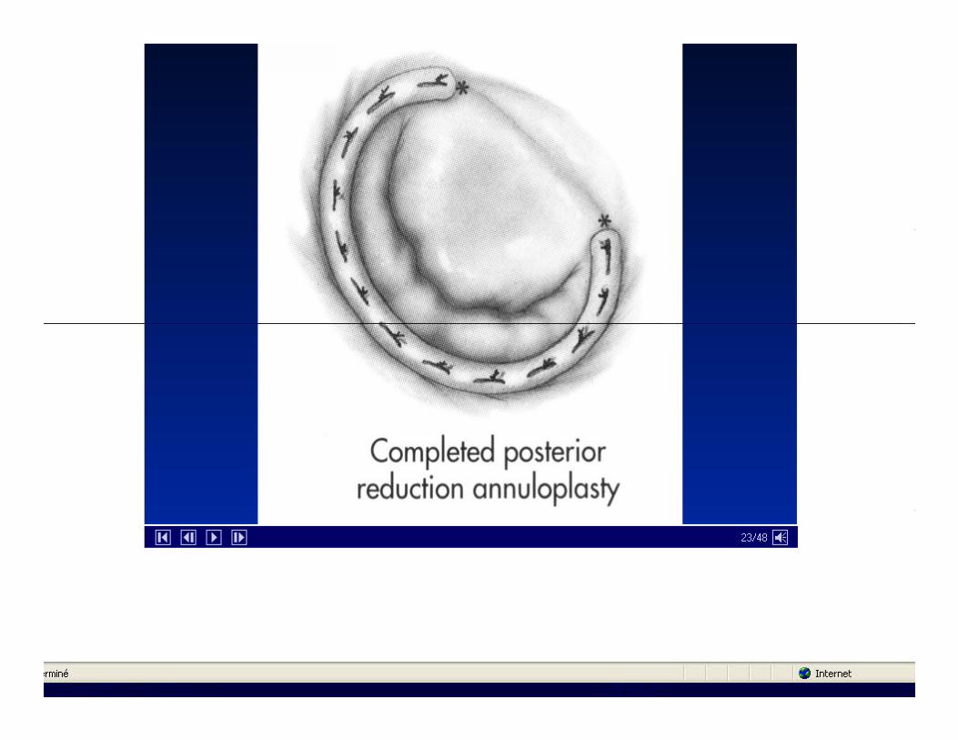


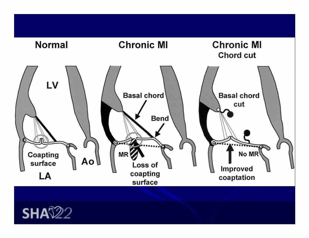
• (A) Posterior leaflet ( )endocarditis with P2 segment prolapse. (B) P2 segment is resected (C)segment is resected. (C) Compression sutures are placed along the posterior
( )annulus. (D) Sliding plasty of segments P1 and P3 is performed. p


• LeafletLeaflet perforation (anterior leaflet) treated by autologous
i di lpericardial patch


Coronary Revasc & IMRCoronary Revasc. & IMR
Gold Standard
– Ischemic MR grade 3-4
Carpentier type IIIb dysfunction– Carpentier type IIIb dysfunction– Reduction annuloplasty

Adams et al. Ann ThoracSurg 2006;;82::2096‐2101
• Fig 1. (a) Placement of one or more simple Gore‐Tex sutures in the papillary muscle head is easy p p y yto perform after posterior leaflet resection because of enhanced exposure. (b) Typical appearance of a myxomatous mitral valve after posterior leaflet quadrangular resection and sliding valvuloplasty. Poor leaflet apposition is present in all leaflet segments and segmentalpresent in all leaflet segments, and segmental anterior leaflet prolapse can be localized only by height comparison with the normal reference point (usually P1). (c) After ring annuloplasty symmetric leaflet apposition limits valve incompetence to the prolapsing anterior leaflet segment (d) After annuloplasty both arms ofsegment. (d) After annuloplasty both arms of the Gore‐Tex suture are passed through the margin of the prolapsing segment. Optimal artificial chord height is determined by intermittently testing valve competency by injecting saline into the ventricle. (e) Completed
frepair with artificial chord in place. A symmetric line of apposition results in valve competence.


Intervention mitral valve repairIntervention mitral valve repair• percutaneous • �� Coronary sinus annuloplasty
• • Edwards Monarc• Mitral Repair• Approaches
• • Edwards Monarc• • Cardiac Dimensions Carillon• • Viacor Shape Changing Rods• • St. Jude Annulus Reshaping• �� Direct annuloplasty• �� Direct annuloplasty• • Mitralign Suture‐Based Plication• • Guided Delivery Anchor‐Cinch Plication• • QuantumCor RF Annulus Remodeling• • MiCardia variable size ring• • MiCardia variable size ring• �� Leaflet repair• • EValve Mitraclip• • Edwards Mobius stitch• �� Chamber + annular remodeling• �� Chamber + annular remodeling• • Myocor iCoapsys• • Ample PS3

Figure 3. Coronary sinus annuloplasty devices.
Masson J , Webb J G Circ Cardiovasc Interv 2009;2:140-146
Copyright © American Heart Association

Figure 1. A, Mitraclip device in its open position.
Masson J , Webb J G Circ Cardiovasc Interv 2009;2:140-146
Copyright © American Heart Association
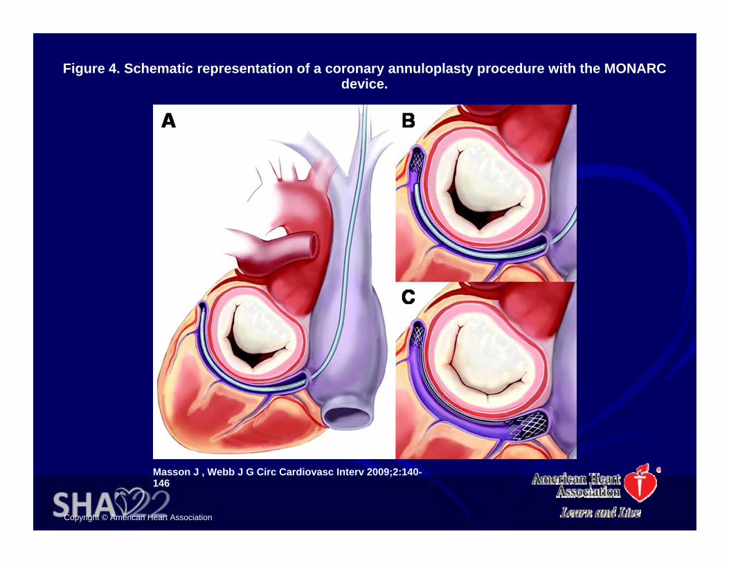
Figure 4. Schematic representation of a coronary annuloplasty procedure with the MONARC device.
Masson J , Webb J G Circ Cardiovasc Interv 2009;2:140-146
Copyright © American Heart Association

Figure 6. Direct annular plication concept.
Masson J , Webb J G Circ Cardiovasc Interv 2009;2:140-146
Copyright © American Heart Association

Figure 5. Schematic representation of the atrial remodeling PS3 device (A) and the left ventricular remodeling iCoapsys device (B).
Masson J , Webb J G Circ Cardiovasc Interv 2009;2:140-146
Copyright © American Heart Association

Figure 7. Direct annular plication in an ovine model using the Accucinch device.
Masson J , Webb J G Circ Cardiovasc Interv 2009;2:140-146
Copyright © American Heart Association

The future of mitral valve surgeryThe future of mitral valve surgery
• Even Para leakingEven Para leaking prosthetic valve device was available

E t T th tEven percutaneous Trans catheter mitral valve implantation p

Figure 8. Transcatheter mitral valve implantation.
Masson J , Webb J G Circ Cardiovasc Interv 2009;2:140-146
Copyright © American Heart Association

Mitral valve repair in JordanMitral valve repair in Jordan
• During the period between 1980 and 1990 anDuring the period between 1980 and 1990 an 3000 case of open heart surgery was done in Jordan 30% was valvular heart surgery 70% of them CMV or OMV
• During the period between 1990 and 2000 an 15000 case of open heart surgery was done in Jordan10% was valvular heart surgery but less h 1% OMV h d BMVthan 1% was OMV, why we started BMV

Mitral valve repair in JordanMitral valve repair in Jordan
• During the period between 2000 and 2010 anDuring the period between 2000 and 2010 an 30000 case of open heart surgery was done in Jordan10% was valvular heart surgery butJordan10% was valvular heart surgery but repair procedure more than 40% of mitral valve surgeryvalve surgery

Mitral valve repair in JordanMitral valve repair in Jordan
• Most common annular ring IMRMost common annular ring … IMR• Posterior leaflet remolding.
ifi i l h d• Artificial chordee• Alfieri stitch• Excision of 2ndary chordee• These cover 95% of repair processThese cover 95% of repair process


The future of mitral valve surgeryThe future of mitral valve surgery
• Our old enemies became new friendsOur old enemies became new friends• Infection and adhesions
• Infective endocarditis if medical treat failed may need surgical interference
• And of course redo surgery g y


The future of mitral valve surgeryThe future of mitral valve surgery
• So I think we had to review our classification of repairable mitral valve and non repairable taken in mind the following points
• 1 durabilty of repair1.durabilty of repair.• 2. the need of blood thinner .• 3. Combine pathologyp gy• 4. Age ,sex, patient IQ …..etc • 5.possibility of percutaneous repair.• 6. cost.• 7. trained surgeon

The future of mitral valve surgeryThe future of mitral valve surgery
• For exampleFor example • Central ischemic mitral regurge with CABG grade II III IVgrade II –III‐‐ IV
• Revascularization alone may improve MR b l II d if f il d ibelow II+ and if failed percutaneous ring
•

The future of mitral valve surgeryThe future of mitral valve surgery
• The durability of repair with rheumatic mitralThe durability of repair with rheumatic mitral valve is low
• So percutaneous procedure may bridge the• So percutaneous procedure may bridge the patient for few years until resolve the reason for MVRfor MVR

The future of mitral valve surgeryThe future of mitral valve surgery
• The usage of new generation of BioprostheticThe usage of new generation of Bioprosthetic valves with long life and no need for blood thinner omit the need of the low durablethinner omit the need of the low durable repair

• St. Jude Medical's newSt. Jude Medical s new pericardial aortic stented tissue valve, the Trifecta, has been granted CE Mark of
l b thapproval by the European authorities

SJM's Aortic Trifecta ValveSJM s Aortic Trifecta Valve• The next‐generation tissue valve has
a tri leaflet stented pericardial design• The valve's titanium stent, which
provides a fatigue resistant frame toa tri‐leaflet stented pericardial design which offers excellent hemodynamic performance, or nearly unobstructed blood flow, in order to mimic as closely as possible the flow of a
provides a fatigue resistant frame to support the valve within a patient's heart, is covered with pericardial tissue to allow tissue‐to‐tissue contact when the leaflets open and y p
natural, healthy heart. The unique valve design includes leaflets manufactured from pericardial tissue attached to the exterior of the valve stent which open more fully and
pclose, which reduces the amount of wear and deterioration. Additional attributes contributing to the Trifecta valve's durability include proprietary tissue fixation and St Jude Medical'sstent which open more fully and
efficiently to perform like a natural heart valve.
tissue fixation and St. Jude Medical s patented Linx(TM) AC Technology, an anticalcification treatment designed to reduce tissue mineralization (hardening).( g)
The future of mitral valve surgeryThe future of mitral valve surgery
• May conclusion of primary lectures wasMay conclusion of primary lectures was • Mitral valve repair was done by senior surgeon for fit patient
under optimal conditions as TEE testing with acceptance of near normal results keeping the geometry of the heart as normal
• while in replacement a junior surgeon do the surgery for non healthy valve under non optimal conditions as no need TEE testing and ignorance of the geometry of the heart by cuttingtesting and ignorance of the geometry of the heart by cutting all of the chordee
conclusionconclusion
• We had to review the timing &the best method ofWe had to review the timing &the best method of repair taking in consideration the durability of repair, the need of blood thinner, the pathology, Age ,sex, patient IQ, possibility of percutaneous repair & the cost.
• We had to focus the new generation of trained surgeon certain repair method
d f ll h h l d l d l b• And filling the other in especial medical video library for the history
