the expression ofsurface antigens three trophoblastic
TRANSCRIPT
The expression of surface antigens on three trophoblastictissues in the mouse
Jan CarterDepartment ofBiochemistry, Trinity College, Dublin 2, Ireland
Summary. The expression of paternal and maternal antigens on three types of mouse
trophoblast cultured in vitro has been examined with the mixed haemadsorption test.Primary trophoblastic giant cells outgrown from blastocysts and placental trophoblastfrom 16-day pregnancies expressed antigens of both sources at all stages in culture.Secondary trophoblastic giant cells outgrown from the tip of 8-day ectoplacental cones
did not give a positive reaction until they had outgrown for 3\p=n-\5days. Microdensito-metry after Feulgen staining showed that the cells which gave a positive mixed haemad-sorption response were polyploid. Incubation of blastocysts and ectoplacental conetissue with progesterone, oestradiol-17\g=b\and hCG did not affect the results.
Introduction
The cells of the trophoblast are generally accepted as being involved in the proposed fetal-maternalbarrier which permits maternal acceptance of the fetus (Edwards & Coombs, 1974). Their role is notfully understood ; it has been suggested that there is some non-cellular barrier associated with thetrophoblast layers (Kirby, Billington, Bradbury & Goldstein, 1964; Currie & Bagshawe, 1967)which renders them antigenically inert. Experiments using neuraminidase-treated ectoplacentalcone trophoblast (Currie, Van Doorninck & Bagshawe, 1968) supported this view, but Simmons,Lipschultz, Rios & Ray (1971) were unable to repeat these results. Ectoplacental cone grafts are notrejected unless the host is presensitized (Simmons & Russell, 1967) and do not presensitize hosts toallogeneic skin grafts (Searle, Jenkinson & Johnson, 1975). Hulka & Mohr (1968), however, demon¬strated that ectoplacental cone could immunize the host. Whole placenta can induce hypersensitivityto paternal strain alloantigens and placental tissue grafts have been shown to be susceptible torejection (Uhr & Anderson, 1962; Schlesinger, 1964).
The confusing nature of the evidence for the antigenicity of trophoblast may be due in part to thedifficulties of separating trophoblast from other tissue elements. In the mouse it is possible to isolatetrophoblast from 3 different stages of development and to grow it in vitro: the stages are (1) primarygiant cells outgrown from the blastocyst, (2) secondary giant cells from the ectoplacental cone, and(3) placental trophoblast. Paternal and maternal antigens have been demonstrated on the primarytrophoblastic giant cells of the mouse by the mixed haemadsorption technique (Carter, 1976b; Sellens,1977). Billington, Jenkinson, Searle & Sellens (1977) showed that H-2 and non-H-2 antigens were
expressed on 5-day outgrowths of ectoplacental cone and placental trophoblast of 12-15-day-preg-nant mice.
In the present study, paternal and maternal antigens on the three types of mouse trophoblast,cultured in vitro, were examined by the mixed haemadsorption technique
Materials and Methods
The inbred mice used were of the C57/BL10/ScSn (H-2b) and CBA/Fa(H-2k) strains and are sub¬sequently referred to as C57 or CBA animals. Ovulation was spontaneous or induced at 4 weeks ofageby intraperitoneal injections of 5 i.u. PMSG (Folligon: Intervet, Cambs) followed by 5 i.u. hCG(Chorulon: Intervet, Cambs) 48 h later, i.e. 12 h before the middle of the subsequent dark periodDownloaded from Bioscientifica.com at 12/15/2021 01:29:27AM
via free access

(Gates, 1971). Females were caged with males during this dark period and checked for vaginal plugsthe following morning. The day of finding a vaginal plug was designated Day 1.
Trophoblast tissuePrimary giant cell trophoblast from blastocysts was obtained on Day 4 by flushing uteri of super-
ovulating females with phosphate-buffered saline (PBS). Acid-washed sterile coverslips were placedin sterile tissue-culture dishes and the blastocysts were cultured in small drops of Snow's medium(Ansell & Snow, 1975) supplemented with 10% fetal calf serum (FCS: Gibco Bio-Cult, Glasgow)under light liquid paraffin. The blastocysts were incubated at 37°C in an atmosphere of 5 % C02 inair, controlled by a C02 monitor (Gow-Mac Instruments, Shannon, Ireland). After 2-3 days inculture the blastocyst has usually hatched, attached and begun to outgrow : this stage was designated'ao'. Approximately 1 day later outgrowth was complete and the culture was designated '.
Secondary giant cells from ectoplacental cone trophoblast were obtained by dissection ofembryosfrom their deciduas on Day 8 of pregnancy (naturally ovulating females). A small piece of tissue was
cut from the tip of each ectoplacental cone and only those pieces which were thought to be smallenough to exclude any cells from the ectoplacental core (Johnson, 1975) were cultured in TrowellsT-8 medium (Gibco Bio-Cult) supplemented with 10% FCS. Medium was changed after 48 h.Other culture conditions were identical to those described for blastocyst culture. During the first24 h, the ectoplacental cone tissue attached and a few layers of secondary giant cells outgrew ; thiswas designated 'ao' (see PI. 1, Fig. 1). The radius of the outgrowth continued to increase and laterstages were designated ' (see PI. 1, Fig. 1).
Placental trophoblast from 16-day-pregnant mice was outgrown in Trowells T-8 medium in thesame way, taking approximately lxl mm pieces of spongiotrophoblast. These were cultured for1 or 4 days.
Hormones used in the culture medium (see 'Results') were progesterone and oestradiol-17ß(Sigma, St. Louis, U.S.A.) and hCG (Chorulon ; Intervet, Cambs).Antisera
An antiserum to a mixture of male and female CBA or C57 spleen cells was raised in C57 or CBAmice respectively. Approximately half a donor spleen in PBS was given by intraperitoneal injectionevery week for 5-10 weeks. Test bleeds were made to determine the cytotoxicity of the antisera againsttarget lymphocytes.
An antiserum to sheep red blood cells was raised in -strain mice. Recipients were given 0-2 mlof a 1 % suspension of sheep RBCs intraperitoneally each week for 7-10 weeks. The serum was
collected and stored at —20°C. Rabbit anti-mouse IgG was obtained from Cappell Laboratories,Downington, U.S.A.
Mixed haemadsorption test
The test used was that of Fagraeus, Espmark & Jonsson (1965). Optimal conditions for thesensitization of sheep RBCs were established in a chessboard experiment with RBCs sensitized withdifferent concentrations of anti-sheep RBC and anti-mouse Ig serum (Tachibana, Worst & Klein,1970). Fresh erythrocytes were washed three times in PBS, incubated in a 1:1000 dilution of mouse
anti-sheep RBC serum followed by a 1:100 dilution of rabbit anti-mouse IgG serum and finallydiluted to 4 % (v/v) and stored at 4°C in PBS for periods up to 1 week. Incubations were carriedout at 37CC for 1 h and there were three washes with PBS between each step in the procedure.
To prepare the outgrowths for the test, oil was removed from the coverslips by rinsing in PBS at37°C and they were then incubated for 1 h at 37°C with a 1:160 dilution of anti-CBA serum or a
1 :40 dilution of anti-C57 serum. The sera were not fractionated or heat-inactivated. After rinsingwith PBS the cultures were layered with sensitized sheep RBCs (1 %, v/v) for a further hour at room
temperature. Unattached erythrocytes were washed off with PBS and the amount of attachment wasestimated by using phase-contrast microscopy. The positive controls for the antisera were on culturesof epidermis from 16-day fetuses. The blastocysts were scored according to the percentage of targetcells (i.e. trophoblast and inner cell mass of the outgrowth) to which the sensitized RBCs were
Downloaded from Bioscientifica.com at 12/15/2021 01:29:27AMvia free access
attached. The ectoplacental cone and placental outgrowths were scored according to the percentageof target cells of the outermost layer of the outgrowth to which the sensitized RBCs were attached.The score was expressed on the following scale: 40 (100% attachment), 3-5 (90%), 30 (50-80%),2-0 (25-50%), 1-0 (10-25%), 0-5 (1-10%), 0 (0%) (Carter, 1976b). There was no adherence of sensi¬tized sheep RBCs in the absence ofantisera.
HistologyPermanent mounts of outgrowths on coverslips were made by fixation for 1 h in Bouin's fluid,
followed by staining with haematoxylin and eosin and mounting with neutral glycerine jelly (Difco,West Molesey, Surrey).Microdensitometry
Ectoplacental cone tissue was obtained from the outermost layer ofcells of 2- or 4-day outgrowths.Imprints of the cut surface of liver were used as control tissue. Nuclear DNA in all cells was demon¬strated by the Feulgen reaction with the following schedule : fixation in ethanol : glacial aceticacid (3 :1 v/v) for 10 min, hydrolysis for 40 min in 5 n-HCI at room temperature, two rinses in distilledwater, Schiff 's reagent for 2 h, three sulphite rinses of 10 min each. The coverslips were then washedin running tap water and mounted in glycerine jelly.
A microdensitometer (M-85 : Vickers Instruments Ltd, York) was used for measurement of theintensity of Feulgen staining and integrated scans were carried out at 565 nm. All readings were
corrected for background absorption. A standard of 70-80 liver nuclei was used for evaluating thebasic DNA content of the trophoplast cells. Liver nuclei are diploid, tetraploid or octoploid andcontain 2C, 4C or 8C units of DNA. The haploid amount of DNA is C and by using the mean DNAvalue of the 2C liver nuclei as a standard, the frequency distribution of the trophoblast cells could beplotted on a logarithmic scale.
Results
Ectoplacental cone trophoblastThe expression of surface antigens on cells of the ectoplacental cone after 1-5 days of culture is
shown in Table 1. All CBA CBA outgrowths incubated with anti-C57 serum and C57 C57outgrowths incubated with anti-CBA serum gave negative scores.
Table 1. Expression of CBA and C57 cell surface antigens on ectoplacental cone outgrowths (no. in parentheses)after mixed haemadsorption (mean score + s.e.m.)
Anti-CBA serum Anti-C57 serum
Days in--Parent mating culture 'ao' ' 'ao' *©'
CBA CBA 1 0 (14) 2-5 ± 0-5 (2) NE NE2 0(3) 3-11+0-2(9) NE 0(6)
3-5 0-17 + 0-17(3) 3-65 ± 0-15 (20) NE 0(4)
C57 C57 2 NE 0 (5) 0 (12) NE5 NE 0(5) NE 2-54 ±0-18 (14)
NE = not examined.
All ' outgrowths of CBA CBA ectoplacental cones incubated with anti-CBA serum gavescores exceeding 2-5 and all 'ao' outgrowths were negative (PI. 1, Fig. 2) with one exception, a 4-day'ao' outgrowth which gave a score of 0-5. The same response was shown for C57 C57 ectoplacentalcone outgrowths incubated with anti-C57 serum. The outer layer of cells of a 4-day ' outgrowth
Downloaded from Bioscientifica.com at 12/15/2021 01:29:27AMvia free access
10
6
2
6
2
14
10
6
2
10
6
2
6
2
12
10
6
2
sia I~
- (6) nn^y^n -1-ì.-1-1-r"—r V&-P-. (c)
Mhrl.1 Vl·^ —"n-r
J1
- -1-1-rL- (d)
il
l£{(e)
nnim1 Y I II 1 l I I/ i
on
iDûd nr~*— -1-1-r2 4 8 16 32 64 128 256 512
C value
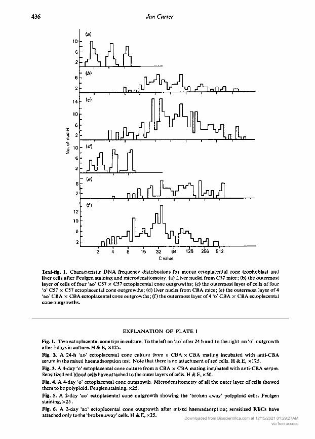
Text-fig. 1. Characteristic DNA frequency distributions for mouse ectoplacental cone trophoblast andliver cells after Feulgen staining and microdensitometry. (a) Liver nuclei from C57 mice; (b) the outermostlayer of cells of four 'ao' C57 C57 ectoplacental cone outgrowths; (c) the outermost layer of cells of four ' C57 C57 ectoplacental cone outgrowths; (d) liver nuclei from CBA mice; (e) the outermost layer of 4'ao' CBA CBA ectoplacental cone outgrowths ; (f) the outermost layer of4 ' CBA CBA ectoplacentalcone outgrowths.
EXPLANATION OF PLATE 1
Fig. 1. Two ectoplacental cone tips in culture. To the left an 'ao' after 24 h and to the right an ' outgrowthafter 3 days in culture. H &E, 125.Fig. 2. A 24-h 'ao' ectoplacental cone culture from a CBA CBA mating incubated with anti-CBAserum in the mixed haemadsorption test. Note that there is no attachment of red cells. & , 125.Fig. 3. A 4-day ' ectoplacental cone culture from a CBA CBA mating incubated with anti-CBA serum.
Sensitized red blood cells have attached to the outer layers ofcells. & E, x50.Fig. 4. A 4-day ' ectoplacental cone outgrowth. Microdensitometry of all the outer layer of cells showedthem to be polyploid. Feuglen staining, x25.Fig. 5. A 2-day 'ao' ectoplacental cone outgrowth showing the 'broken away' polyploid cells. Feulgenstaining, x25.Fig. 6. A 2-day 'ao' ectoplacental cone outgrowth after mixed haemadsorption; sensitized RBCs haveattached only to the 'broken away' cells. & E, x25.
Downloaded from Bioscientifica.com at 12/15/2021 01:29:27AMvia free access
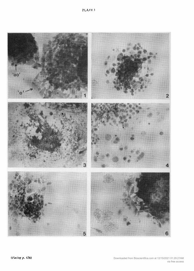
PLATE 1
Downloaded from Bioscientifica.com at 12/15/2021 01:29:27AMvia free access
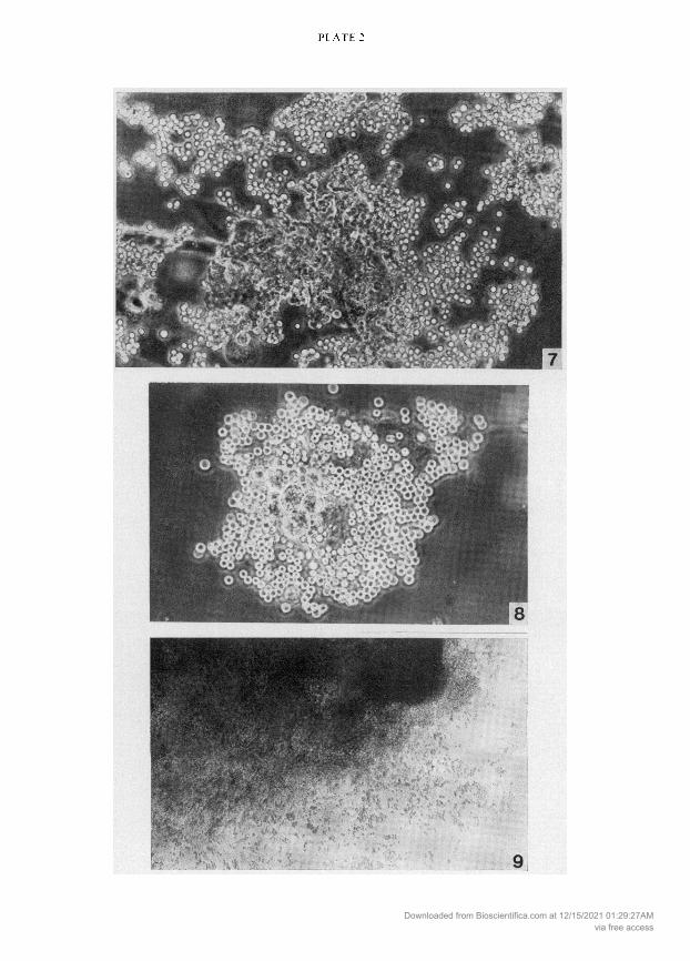
PLATE 2
Downloaded from Bioscientifica.com at 12/15/2021 01:29:27AMvia free access
(PI. 1, Fig. 3) was given a score of40, although the first layers of secondary trophoblastic cells and theoriginal ectoplacental cone were poorly labelled. Red cells became trapped in the cellular matrix ofthe initial tissue and after 4 days in culture there was some red cell attachment to the whole outgrowth.The embryonic cells retained the large size typical of trophoblast cells, and microdensitometryshowed that all the cells of the outer layer in 'ao' and ' outgrowths were polyploid (see Text-fig. 1).The average amount of nuclear DNA for CBA 'ao' and ' and C57 'ao' cells was between 128 and 256times the haploid amount ofDNA measured in control cells of liver from the two strains and between64 and 128 times for C57 ' cells. The average number of cells in the outermost layer of the 'ao' and ' outgrowths measured was 250 ±1-9 (n = 8) and 58-9 ± 4-7 (n = 8) respectively. PI. 1, Fig. 4shows an ' outgrowth after Feulgen staining. After 1-2 days in culture the 'ao' outgrowths some¬
times produced cells which grew rapidly away from the main mass of tissue but were still connectedby cytoplasmic bridges (PI. 1, Fig. 5). The two large cells to the right of PI. 1, Fig. 5 had 256 and 64times the haploid amount of DNA. Although the 'ao' outgrowth did not express antigens, the sensi¬tized RBCs did attach to these 'broken-away' cells (see PI. 1, Fig. 6).
Blastocyst andplacental trophoblastThe mixed haemadsorption test for maternal and paternal antigens on cultures of blastocyst
outgrowths was positive at both the 'ao' and ' stages of culture. Red cells attached to all cells of theculture, i.e. inner cell mass and trophoblast cells (PI. 2, Figs 7 and 8). Control 'ao' or ' outgrowths ofCBA CBA matings after incubation with anti-C57 serum showed no attachment of red cells.The same results were obtained with all outgrowths of C57 CBA and C57 C57 matings afterincubation with appropriate antisera.
Red cell attachment occurred over the whole surface of 1- and 4-day placental cultures (Table 2:PI. 2, Fig. 9).
Table 2. Expression of CBA and C57 cell surface antigens on blastocyst and placental cultures after mixed haemad¬sorption (mean score + s.e.m., no. of outgrowths in parentheses)
Blastocyst Placenta
Parent mating Antiserum 'ao' outgrowth ' outgrowth 1-day culture 4-day culture
CBA X CBA Anti-CBA 3-86 + 009 (7) 3-80 ± 006 (34) 3-67 ± 006 (5) 400Anti-C57 NE 0(20) 0(3) NE
C57XCBA Anti-CBA 3-25 + 0-14(4) 2-89 ± 016 (9) 3-68 + 0-05(3) 3-67 + 0-05(4)Anti-C57 NE 3 00(6) 3-29 ± 013 (4) 3-24 ± 008 (6)
C57 X C57 Anti-CBA NE 0 (10) 0 (3) NEAnti-C57 3-06 + 0-15(8) 3-86 ±0-14 (4) 308 ±012 (6) NE
NE = not examined.
EXPLANATION OF PLATE 2
Fig. 7. A 4-day ' blastocyst outgrowth from a C57 CBA mating after incubation with anti-CBA serum
The outgrowth was given a score of 30 in the mixed haemadsorption test. Phase contrast, x355.
Fig. 8. A 2-day 'ao' blastocyst outgrowth from a CBA CBA mating after incubation with anti-CBAserum. The RBCs on the inner cell mass are in another focal plane. Phase contrast, 300.
Fig. 9. A 1-day placental outgrowth from a C57 C57 mating after incubation with anti-C57 serum.
Note the RBC attachment throughout the culture. & , 60.Downloaded from Bioscientifica.com at 12/15/2021 01:29:27AM
via free access

Hormone incubationWhen CBA CBA blastocysts and ectoplacental cones were incubated with medium containing
10 or 2-5 µg progesterone/ml, 100 µg or 25 ng oestradiol-17ß/ml or 10 µg progesterone +25 ngoestradiol-17ß/ml there was no difference in the amount of red cell attachment or to the area to whichthe cells attached. All groups gave average scores exceeding 30 when incubated with anti-CBAserum and negative scores when incubated with anti-C57 serum. Incubation with 0-1,1-0 or 10-0 i.u.hCG/ml culture medium also had no effect.
Discussion
Primary trophoblastic giant cells express maternal and paternal antigens (Carter, 1976b), and thepresent results show that these antigens are expressed at all stages of outgrowth of the blastocyst.However, in the mixed haemadsorption assay, there was no attachment of sensitized sheep RBCs tothe surface of secondary trophoblast cells after incubation with antiserum to paternal or maternalstrains. After 3-5 days in culture, the ectoplacental cone tissue has produced several layers of second¬ary giant cells, the outermost of which express antigens. When secondary giant cells of the ecto¬placental cone are first put into culture, antigens are presumably either absent (Simmons, Cruse &McKay, 1967) or masked, perhaps by a mucopolysaccharide (Kirby et al, 1964). The antigenic sitesexpressed when secondary giant cells are cultured for 3-5 days are strain-specific for C57 or CBA andof maternal and paternal origin. They could include either major (H-2) or minor (H-3 or H-6)histocompatibility antigens. Billington et al (1977) used the mixed haemadsorption techniqueto show that H-2 and non-H-2 antigens were expressed on the cell surface of outgrowths for 7-day -strain ectoplacental cones cultured for 5 days and these observations agree with the data presentedhere for cultures ofthat age.
The expression of cell surface antigens is dependent on growth conditions. Cikes & Klein (1972)showed that the antigens on cultured mouse lymphoma cells were subject to considerable physiolo¬gical variation : there was a decrease in antigen expression during rapid cell growth and in old culturesand expression was dependent on cell cycle stage with maximum expression during Gl. The secondarygiant cells which express antigens in the present experiments are the older cells in culture and showincreased antigen expression. Although there was no measurable antigen on these cells during thefirst 24-48 h in culture, there was positive expression of primary giant cells and placental trophoblastat this time. It is unlikely, therefore, that the differential expression of antigens shown here has thesame basis as the observations ofCikes & Klein (1972).
The ectoplacental cone trophoblast used in the present experiments was taken 7 days afterdetection of a vaginal plug. Bernard, Ripoche & Bennett (1977), using the same invasive giant cells of -strain mice and immunoperoxidase labelling, observed that they were the only cells during embryodevelopment to lack immunoglobulins. The cells of the 5-day embryo and the 10-day trophoblast are
surrounded by immunoglobulins. Therefore the failure of Bernard-e7 al (1977) to detect immuno¬globulins and the data presented here suggest a cell surface change in ectoplacental cone cells whichprevents the attachment of immunoglobulin molecules.
Ferguson & Palm (1977) were not able to demonstrate antigens on the trophoblast giant cells ofthe 12-13-day rat placenta, but antigens were detectable on cytotrophoblast and fibroblasts.In the present study of the mouse placenta it was not possible to isolate trophoblast giant cells fromother cell types to determine whether they are labelled in the mixed haemadsorption test.
Progesterone, oestradiol and hCG have been shown to be immunosuppressive in some systems(Monroe, 1971; Wattman, Bürde & Berias, 1971; Contractor & Davies, 1973; Martin, Spicer &Smythe, 1974; Carter, 1976a). In this study, progesterone and oestradiol were used at concentrationsknown to have an effect on cell division of uterine tissue in vitro (Grant, 1973; Carter & McLaren,1975), but there was no change in the amount of labelling or area to which the sensitized RBCsattached to cells ofectoplacental cones or primary giant cells.
The present investigation shows that, in the mouse, antigenic expression on the secondarytrophoblast cells of the ectoplacental cone differs from that on the primary trophoblast of the blasto-Downloaded from Bioscientifica.com at 12/15/2021 01:29:27AM
via free access
cyst and the trophoblast of the placenta, suggesting that different mechanisms are involved in theprotection of the fetus at the various stages of trophoblast development.
I thank Dr A. McLaren for her helpful criticism and advice and Mr J. McPartlin for help with themicrodensitometry. I am grateful to the Irish Medical Research Council for financial support.
References
Ansell, J.D. & Snow, M.H.L. (1975) The developmentof trophoblast in vitro from blastocysts containingvarying amounts of inner cell mass. /. Embryol. exp.Morph. 33,177-185.
Bernard, O., Ripoche, M.A. & Bennett, D. (1977)Distribution of maternal immunoglobulins in themouse uterus and embryo in the days after implan¬tation./, exp. Med. 145,58-75.
Billington, W.D., Jenkinson, E.J., Searle, R.F. &Sellens, M.H. (1977) Alloantigen expression duringearly embryogenesis and placental ontogeny in themouse; immunoperoxidase and mixed hemadsorp-tion studies. Transplantation Proc. 9, 1371-1377.
Carter, J. (1976a) The effect ofprogesterone, oestradioland HCG on cell-mediated immunity in pregnantmice./. Reprod. Fert. 46,211-216.
Carter, J. (1976b) Expression of maternal and paternalantigens on trophoblast. Nature, Lond. 262, 292-293.
Carter, J. & McLaren, A. (1975) The effect of oestro¬gen and progesterone on the incorporation of tri¬tiated thymidine in mouse uteri in vitro. J. Reprod.Fert. 42,439^145.
Cikes, M. & Klein, G. (1972) Quantitative studies ofantigen expression in cultured murine lymphomacells. I. Cell surface antigens in "asynchronous"cultures. /. natn. Cancer Inst. 49, 1599-1606.
Contractor, S.F. & Davies, H. (1973) Effect of humanchorionic somatomammotrophin and humanchorionic gonadotrophin on phytohaemagglutinin-induced lymphocyte transformation. Nature, NewBiol. 243,284-286.
Currie, G.A. & Bagshawe, K.D. (1967) The maskingof antigens on trophoblast and cancer cells. Lancet i.708-710.
Currie, G.A., Van Doorninck, W. & Bagshawe, K.D.(1968) Effect of neuraminidase on the immuno-genicity of early mouse trophoblast. Nature, Lond.219,191-192.
Edwards, R.G. & Coombs, R.R.A. (1974) Immuno¬logical interactions between mother and fetus. InClinical Aspects of Immunology, pp. 561-597. EdsP. G. H. Grell, R. R. A. Coombs & P. J. Lachmann.Blackwell, Oxford.
Fagraeus, ., Espmark, J.A. & Jonsson, J. (1965)Mixed haemadsorption: a mixed antiglobulinreaction applied to antigens on a glass surface.Immunology 9,161-175.
Ferguson, F.G. & Palm, J. (1977) Reactivity of ratplacental cells with alloantisera. /. Embryol. exp.Morph. 39,195-202.
Gates, A.H. (1971) Maximising yield and developmentaluniformity of eggs. In Methods in Mammalian
Embryology, pp. 64-75. Ed. J. C. Daniel, Jr. FreemanPress, San Francisco.
Grant, P. (1973) The effect of progesterone and oestra¬diol on immature mouse uteri maintained as organcultures./. Endocr.SI, 171-174.
Hulka, J.F. & Mohr, K. (1968) Trophoblast anti-genicity demonstrated by altered challenge of graftsurvival. Science, N. Y. 161,696-698.
Johnson, M.H. (1975) Antigens of peri-implantationtrophoblast. In Immunobiology of the Trophoblast,pp. 87-100. Eds R. G. Edwards, C. W. S. Howe &M. H. Johnson. Cambridge University Press.
Kirby, D.R.S., Billington, W.D., Bradbury, S. &Goldstein, D. (1964) Antigen barrier of the mouse
placenta. Nature, Lond. 204,548-549.Martin, B.J., Spicer, S.S. & Smythe, N.M. (1974)
Cytochemical studies of the maternal surface of thesyncytiotrophoblast of human early and termplacenta. Anat. Ree. 178,769-786.
Monroe, J.S. (1971) Progesteroids as immunosup-pressive agents. /. Reticuloendothelial Soc. 9, 361-365.
Schlesinger, J. (1964) Serologie studies of embryonicand trophoblastic tissues of the mouse. Immunology93,255-263.
Searle, R.F., Jenkinson, E.J. & Johnson, M.H. (1975)Immunogenicity of mouse trophoblast and em¬
bryonic sac. Nature, Lond. 255, 719-720.Sellens, M.H. (1977) Antigen expression on early
mouse trophoblast. Nature, Lond. 269, 60-61.Simmons, R.L. & Russell, P.S. (1967) Xenogeneic
antigens in mouse trophoblast. Transplantation 5,85-88.
Simmons, R.L., Cruse, V. & McKay, D.G. (1977)The immunologie problem of pregnancy. II. Ultra-structure of isogeneic and allogeneic trophoblastictransplants. Am. J. Obstet Gynec. 97, 218-230.
Simmons, R.L., Lipschultz, M.L., Ríos, A. & Ray,R.K. (1971) Failure of neuraminidase to unmaskhistocompatibility antigens. Nature, New Biol. 213,111-112.
Tachibana, T., Worst, P. & Klein, E. (1970) Detectionof cell surface antigens on monolayer cultures. II.The application of M.H.A. on a microscale. Immuno¬logy 19,809-816.
Uhr, J.W., & Anderson, S.G. (1962) The placenta as a
homotransplant. Nature, Lond. 194, 1292-1293.Wattman, S.R., Bürde, R.M. & Berias, J. (1971)
Prevention of corneal homograft rejection byestrogens. Transplantation 11, 194-196.
Received 2 May 1978
Downloaded from Bioscientifica.com at 12/15/2021 01:29:27AMvia free access