the expression of aquaporin 1 and 5 in uterine leiomyomata ... · ihc staining revealed aqp1, aqp5...
TRANSCRIPT

Vol. 12, No. 1 81
Copyright © 2012 by the Society for Biology of Reproduction
SHORT COMMUNICATION
The expression of aquaporin 1 and 5 in uterine leiomyomata in premenopausal women:
a preliminary studyMariusz T. Skowronski1,2, Leszek Frackowiak3,4, Agnieszka Skowronska5
2Department of Animal Physiology, University of Warmia and Mazury, Olsztyn; 3Department of Public Health and Epidemiology, University
of Warmia and Mazury, Olsztyn; 4Department of Oncology, MSWiA Hospital in Olsztyn; 5Department of Human Physiology,
University of Warmia and Mazury, Olsztyn, Poland
Received: 26 February 2011; accepted: 15 November 2011
SUMMARY
The aim of this study was to investigate the expression of aquaporins (AQPs) in uterine tissues of premenopausal women. We demonstrated for the first time the expression of AQP1, AQP5 and AQP9 in uterine leiomyomata and in the adjacent normal endometrium and myometrium. The expression of AQP1 and 5 was higher in leiomyomata than in unaffected uteri. AQP9 was expressed only in the unaffected endometrium. It is possible that AQP1 and AQP5 contribute to the formation of leiomyomata in premenopausal women. Reproductive Biology 2012 12 1: 81-89.Key words: aquaporins, uterine leiomyomata, immunolocalization, protein expression
1 Corresponding author: University of Warmia and Mazury in Olsztyn, Department of Animal Physiology, Oczapowskiego 1A, 10-718 Olsztyn, Poland; e-mail: [email protected]

Aquaporins in uterine leiomyomata82
INTRODUCTION
Uterine leiomyomata (fibroids) are the most common type of benign tu-mors observed in at least 20% of reproductive-age women and in 40-50% of women older than 40 [1]. The development of leiomyomata is considered to be estrogen dependent due to fibroids’ ability to grow during pregnancy and shrink during menopause, after ovariectomy or gonadoliberin (GnRH) agonist therapy [11, 14]. The pathophysiology of fibroids is poorly under-stood. The recent discovery of aquaporins’ involvement in numerous types of tumors, especially those arising from organs where water permeability plays an important role [4], inspired us to study aquaporin (AQP) involve-ment in the development of uterine leiomyomata.
Aquaporins are transmembrane water-channel proteins that play a cru-cial role in transcellular and transepithelial water movement [4]. At least 13 AQP isoforms were found in mammals (AQP0-AQP12). Aquaporin 1, AQP2, AQP4, AQP5 and AQP8 are primarily water-selective, whereas AQP3, AQP7, AQP9 and AQP10 also transport glycerol and other small solutes [13]. The objective of this study was to investigate the cellular and subcellular expression of AQP1, AQP5 and AQP9 in uterine leiomyomata and in the ad-jacent normal endometrium and myometrium.
MATERIALS AND METHODS
Samples of uterine leiomyomata and the adjacent myometrium and endo-metrium were collected from five premenopausal women aged 43 to 45 during the proliferative phase. The patients were undergoing hysterectomies at the Polyclinic of the MSWiA Hospital in Olsztyn, and they were exam-ined by the same histopathologist. None of the patients had received any medical treatment for fibroids at least three months before surgery. Tissue samples were collected from each patient for Western blot (WB) and im-munohistochemical (IHC) analysis [12]. The experiment was carried out upon the approval of the Local Ethics Committee. Written informed consent for publication of this case report was obtained from patients.

Skowronski et al 83
Tissue samples were directly placed in an ice-cold dissection buffer [12]. Briefly, the samples were homogenized and centrifuged. The isolated proteins were loaded onto 12.5% polyacrylamide gel. Then, the proteins were separated by electrophoresis and electro-transferred to nitrocel-lulose membranes (Hybond ECL, Amersham Pharmacia Biotech, UK) for one h at 100 V. The membranes were rinsed and incubated overnight at 5°C with anti-AQPs or β-actin antibodies. The antibodies to AQP1, AQP5 and AQP9 had previously been characterized [12]. Afterwards, the membranes were rinsed and incubated with horseradish peroxidase-conjugated goat anti-rabbit IgG secondary antibody (Dako A/S, Denmark) in PBS-T for 1 h. After rinsing with PBS-T, antibody-antigen reaction sites were visualized in an enhanced chemiluminescence (ECL) system and exposed to photographic film (Hyperfilm ECL, Amersham Pharmacia Biotech, UK). Densitometric analysis of AQP1 and AQP5 protein levels were normalized against β-actin (anti-β-actin antibody: A2066, Sigma, St. Louis, MO, USA) and by means of GelScan for Windows v. 1.45 software (Kucharczyk, Poland).
In addition to WB, an indirect IHC staining was used to examine AQP protein expression [12]. Briefly, paraffin-embedded tissue sections
were dewaxed, rehydrated and blocked by 0.5% H2O2. Nonspecific bind-ing of IgG was prevented by incubating the sections in 50 mM NH4Cl
for 30 min, followed by blocking in PBS supplemented with 1% BSA, 0.05% saponin and 0.2% gelatin. The sections were rinsed and incu-bated overnight at 4°C with primary antibodies. Then, the sections were rinsed and incubated with secondary antibody. Labeling was visualized
by 0.05% 3,3 diaminobenzidine tetrahydrochloride (DAB). Microscopic observations were performed under a Leica DMRE light microscope (Heidelberg, Germany).
Several positive controls were performed in IHC and WB analysis. AQP1 protein was noted in the apical and basolateral plasma membrane domains in proximal tubule cells of the human kidney (IHC: fig. 1G; [7, 13]). AQP5 protein was found in the apical plasma membrane of acinar cells in the hu-man salivary gland (IHC: fig. 1H; [3]). AQP9 staining was observed on the si-nusoidal surfaces of hepatocyte plates in the human liver (IHC: fig. 1I; [12]).
Aquaporins in uterine leiomyomata84
A B C
D E F
G H I

Skowronski et al 85
Moreover, AQP1 and AQP5 were determined in homogenates of the human kidneys (WB: fig. 2C) and human salivary glands (WB: fig. 2F). Negative controls for all IHC and WB tests were performed without primary anti-bodies to the respective AQPs, and they did not show any staining (data not shown). Data were analyzed by one-way ANOVA and LSD post hoc tests. The results were expressed as mean values±SEM from five independent observations and considered to be statistically significant at p<0.05.
RESULTS AND DISCUSSION
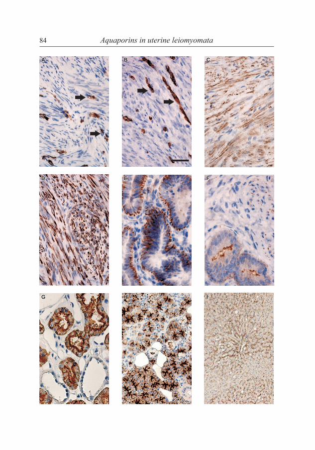
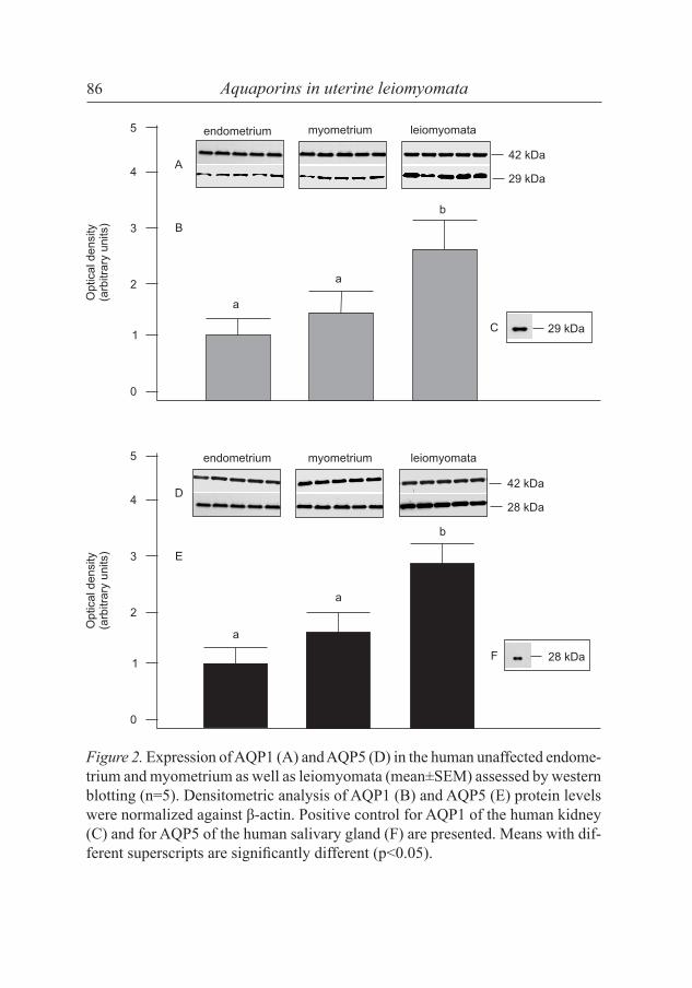
IHC staining revealed AQP1, AQP5 and AQP9 protein expression in the uterus of premenopausal women (fig. 1). AQP1 protein was demonstrated in unaffected uteri (endometrium and myometrium) as well as leiomyomata (fig. 2A) by WB. This finding was extended by IHC which showed the presence of AQP limmuno-reactivity in the capillary endothelium of both unaffected uteri (fig. 1A) and leio-myomata (fig. 1B). AQP1 expression did not differ between unaffected endome-trium and myometrium (fig. 2B). Significantly higher AQP1 level (p<0.05) was found in leiomyomata than in control endometrium and myometrium (fig. 2B).
It is possible that high expression of AQP1 in leiomyomata contributes to abnormal vascular growth and permeability. Vascular expression of AQP1 was found to be significantly lower in endometrium of menorrhagia patients than in control individuals. This suggests that AQP1 is involved in abnormal
Figure 1. Immunolocalization of AQP1, AQP5 and AQP9 in human uterine tissues determined by immunohistochemistry. AQP1 protein was found in the capillary endothelium of unaffected uteri (A) and leiomyomata (B). Arrows indicate localiza-tion of the vessels. AQP5 protein was detected in the plasma membrane of smooth muscle cells in unaffected uteri (C) and leiomyomata (D) as well as in the baso-lateral plasma membrane domains of glandular epithelial cells (E). AQP9 protein was demonstrated in the apical plasma membrane domains in glandular epithelial cells of unaffected uteri (F). AQP1, AQP5 and AQP9 immunostaining presented as positive controls was found, respectively, in human kidney cortex (G), salivary gland (H) and human liver (I); the bar = 50 µm.
Aquaporins in uterine leiomyomata86
Figure 2. Expression of AQP1 (A) and AQP5 (D) in the human unaffected endome-trium and myometrium as well as leiomyomata (mean±SEM) assessed by western blotting (n=5). Densitometric analysis of AQP1 (B) and AQP5 (E) protein levels were normalized against β-actin. Positive control for AQP1 of the human kidney (C) and for AQP5 of the human salivary gland (F) are presented. Means with dif-ferent superscripts are significantly different (p<0.05).

Skowronski et al 87
endometrial vascular growth and permeability [8]. Moreover, AQP1 mRNA and protein expression was reported to be higher in ovarian cancer and bor-derline tumors than in normal ovary and ovarian benign tumors, suggesting a AQP1 role in epithelial ovarian malignancies [15]. Recently, in addition to its canonical function as a water-permeable channel, AQP1 was reported to function as a O2 transporter and O2 homeostasis controller [2], features, which may be important for cancer development.
Aquaporin 5 protein was demonstrated in unaffected human uteri and leio-myomata (fig. 2D) by WB. As for AQP1, this finding was also confirmed by IHC which showed that AQP5 was localized in the plasma membrane of smooth muscle cells in unaffected uteri (fig. 1C) and leiomyomata (fig. 1D) as well as in the basolateral plasma membrane domains of glandular epithelial cells (fig. 1E). The expression of AQP5 tended to be higher (p=0.08) in the unaffected myometrium than endometrium (fig. 2E). Moreover, the AQP5 expression in uterine leiomyomata was higher (p<0.05) in comparison to those of the con-trol endometrium and myometrium (fig. 2E). To date, the presence of AQP5 in uterine epithelia has been shown in ovariectomized rats [5], pregnant rats [6], mice during implantation [10] and in the myometrium of the porcine uterus [13].
We did not demonstrate the AQP9 expression in unaffected myometrium and leiomyomata, but demonstrated its presence in the apical membrane of the epithelial endometrium (fig. 1F). AQP9 is an aquaglyceroporin, and as such is permeable not only to water, but also to glycerol, urea and other non-electrolytes. Lindsay and Murphy [6] first reported AQP9 expression in the apical plasma membrane of the glandular epithelium of rat uteri. Our group recently detected AQP9 protein in the glandular epithelium and the lu-minal epithelium of porcine uteri [13]. The protein was also observed in the nuclei, the cytoplasm and the cell membrane of human granulosa cells [9]. It is of interest that AQP9 mRNA expression was significantly lower in granulosa cells in women with the polycystic ovary syndrome than in control individuals, and it was significantly correlated with the testoster-one level, sex hormone-binding globulin level and the free androgen index in the follicular fluid [9].
To our knowledge, we demonstrated for the first time the expression of AQPs in the leiomyomata and unaffected endometrium and myometrium

Aquaporins in uterine leiomyomata88
of premenopausal women. Moreover, the expression of AQP1 and 5 was higher in leiomyomata than in unaffected uteri. In addition, AQP9 was expressed only in the unaffected endometrium. The role of the proteins ap-pears to not be limited to water transport for normal endometrial functioning such as secretion and implantation, AQP1 and AQP5 may also contribute to the development of leiomyomata, angiogenesis and tumor growth.
ACKNOWLEDGMENTS
This research was supported by the Polish Ministry of Science and Higher Education (grant numbers N N308 0042 33 and 0206.0805). The authors would like to thank Professor Jadwiga Przała for her comments and sug-gestions and Janina Bukowska for technical support.
REFERENCES
1. Buttram VC, Reiter RC 1981 Uterine leiomyomata: etiology, symptomatology and management. Fertility and Sterility 36 433-445.
2. Echevarría M, Muñoz-Cabello AM, Sánchez-Silva R, Toledo-Aral JJ, López-Barneo J 2007 Development of cytosolic hypoxia and hypoxia-inducible factor stabilization are facilitated by aquaporin-1 expression. The Journal of Biological Chemistry 282 30207-30215.
3. Gresz V, Kwon TH, Hurley PT, Varga G, Zelles T, Nielsen S, Case RM, Steward MC 2001 Identification and localization of aquaporin water channels in human salivary glands. American Journal of Physiology Gastrointestinal and Liver Physi-ology 281 247-254.
4. King IS, Agre P 1996 Pathophysiology of the aquaporin water channels. Annual Review of Physiology 58 619-648.
5. Lindsay LA, Murphy CR 2006 Redistribution of aquaporins 1 and 5 in the rat uterus is dependent on progesterone: a study with light and electron microscopy. Reproduction 131 369-378.
6. Lindsay LA, Murphy CR 2007 Aquaporins are upregulated in glandural epithelium at the time of implantation in the rat. Journal of Molecular Histology 38 87-95.
7. Maunsbach AB, Marples D, Chin E, Ning G, Bondy C, Agre P, Nielsen S 1997 Aquaporin-1 water channel expression in human kidney. Journal of the American Society of Nephrology 8 1-14.
Skowronski et al 89
8. Mints M, Hildenbrand A, Lalitkumar LP, Andersson S, Nielsen S, Gemzell-Danielsson K, Stavreus-Evers A 2007 Expression of aquaporin-1 in endometrial blood vessels in menorrhagia. Internal Journal of Molecular Medicine 19 407-411.
9. Qu F, Wang F-F, Lu X-E, Dong M-Y, Shena JZ, Lv P-P, Ding GL, Shi BW, Zhang D, Huang HF 2010 Altered aquaporin expression in women with polycystic ovary syndrome: hyperandrogenism in follicular fluid inhibits aquaporin-9 in granulosa cells through the phosphatidylinositol 3-kinase pathway. Human Reproduction 25 1441-1450.
10. Richard C, Gao J, Brown N, Reese J 2003 Aquaporin water channel genes are differentially expressed and regulated by ovarian steroid hormones during the pe-riimplantation period in the mouse. Endocrinology 144 1533-1541.
11. Robby SJ, Bentley RC, Anderson MC 2000 Pathology and pathophysiology of uterine smooth muscle tumors. Environmental Health Perspectives 108 779-784.
12. Skowronski MT, Kwon TH, Nielsen S 2009 Immunolocalization of aquaporin 1, 5 and 9 in the female pig reproductive system. Journal of Histochemistry and Cy-tochemistry 57 61-67.
13. Skowronski MT 2010 Distribution and quantative changes in amounts of aquaporin 1, 5 and 9 in the pig uterus during the estrous cycle and early pregnancy. Reproduc-tive Biology and Endocrinology 8 109.
14. Stewart EA 2001 Uterine fibroids. Lancet 357 293-298.15. Yang JH, Shi YF, Cheng Q, Qian YL 2005 Protein and mRNA expression of aqua-
porin-1 in epithelial ovarian tumors and its clinic significance. Zhonghua Fu Chan Ke Za Zhi 40 623-626.