the evolution of insulin glargine and its continuing ... · the evolution of insulin glargine and...
TRANSCRIPT

REVIEW ARTICLE
The Evolution of Insulin Glargine and its ContinuingContribution to Diabetes Care
Rolf Hilgenfeld • Gerhard Seipke • Harald Berchtold •
David R. Owens
� The Author(s) 2014. This article is published with open access at Springerlink.com
Abstract The epoch-making discovery of insulin her-
alded a new dawn in the management of diabetes. How-
ever, the earliest, unmodified soluble insulin preparations
were limited by their short duration of action, necessitating
multiple daily injections. Initial attempts to protract the
duration of action of insulin involved the use of various
additives, including vasoconstrictor substances, which met
with limited success. The subsequent elucidation of the
chemical and three-dimensional structure of insulin and its
chemical synthesis and biosynthesis allowed modification
of the insulin molecule itself, resulting in insulin analogs
that are designed to mimic normal endogenous insulin
secretion during both fasting and prandial conditions.
Insulin glargine was the first once-daily, long-acting insulin
analog to be introduced into clinical practice more than
10 years ago and is specifically designed to provide basal
insulin requirements. It has a prolonged duration of action
and no distinct insulin peak, making it suitable for once-
daily administration and reducing the risk of nocturnal
hypoglycemia that is seen with intermediate-acting insu-
lins. Insulin glargine can be used in combination with
prandial insulin preparations and non-insulin anti-diabetic
agents according to individual requirements.
1 Introduction
1.1 The Pre-Insulin Era
Prior to the 1920s, the prognosis for people with insulin-
requiring diabetes mellitus was very poor, with limited
treatment options being available and high resultant mor-
bidity and mortality, particularly in children and young
adults [1]. Generally, confirmation of the diagnosis of
diabetes in this ‘pre-insulin’ era meant eventual coma and
subsequent death, often within 2 years of diagnosis [2]. At
this time, physicians had to manage the disease through
dietary modification alone, with some affected individuals
being restricted to a diet with an almost negligible carbo-
hydrate intake in a bid to control blood glucose levels [3–
5]. In such circumstances, the benefit of such ‘starvation’
diets, involving repeated fasting and prolonged under-
nourishment, was relatively short-lived, providing only a
modest extension of life [4]. Furthermore, there was little
or no evidence to support longer-term efficacy benefits of
undernourishment therapy, which was accompanied by a
risk of infection, inanition, and poor quality of life [4].
However, such an approach would have improved the
metabolic status of those with ‘non-insulin-dependent’
diabetes, which was not recognized as a separate entity
until the mid-1930s and so this differentiation between
Electronic supplementary material The online version of thisarticle (doi:10.1007/s40265-014-0226-4) contains supplementarymaterial, which is available to authorized users.
R. Hilgenfeld (&)
Institute of Biochemistry, Center for Structural and Cell Biology
in Medicine and Center for Brain, Behavior and Metabolism,
University of Lubeck, Ratzeburger Allee 160, 23538 Lubeck,
Germany
e-mail: [email protected]
R. Hilgenfeld
Shanghai Institute of Materia Medica, Shanghai, China
G. Seipke � H. Berchtold
Sanofi-Aventis Deutschland GmbH, Frankfurt am Main,
Germany
D. R. Owens
Diabetes Research Group, Institute of Life Sciences College of
Medicine, Swansea University, Swansea, Wales
Drugs
DOI 10.1007/s40265-014-0226-4

diabetes types could not be used in treatment decisions at
the time [6]. Indeed, dietary intake continues to be a
mainstay of the management of type 2 diabetes mellitus
(T2DM), taking on increased importance as obesity has
become an increasing problem worldwide.
The magnitude of the discovery of insulin must
therefore be viewed in the context of a disease that
resulted in rapid deterioration and death. The eventual
isolation and purification of insulin in a series of rela-
tively crude experiments in the early 1920s heralded a
new dawn for the management of diabetes, providing
hope for the first time to all those people suffering from
this debilitating disease. The pivotal discovery of insulin
at the University of Toronto built on the work of earlier
scientists, culminating in the development of an effective
pancreatic extract (Fig. 1).
1.2 The Discovery of Insulin
Diabetes has been known about for millennia, with the first
known description of symptoms allied to diabetes and
suggested treatment written circa 1500 BC in the Ebers
papyrus from ancient Egypt [1, 7]. Circa 230 BC, Apol-
lonius of Memphis first used the term ‘diabetes’; however,
its cause and the organ responsible for this condition were
not elucidated until more than two millennia later (in the
late nineteenth century) [1, 7].
In 1889, in the laboratory of Oscar Minkowski and
Joseph von Mering, it was observed that a dog developed
diabetes, with all of the characteristic symptoms of the
disease, after total pancreatectomy [8]. This finally con-
firmed the central role of the pancreas in the etiopatho-
genesis of diabetes. Later in 1893, Eduoard Hedon further
Fig. 1 A timeline highlighting
significant contributions to the
development of insulin as a
therapeutic agent for the
treatment of diabetes and the
development of modified
insulins with prolonged times of
action
R. Hilgenfeld et al.

demonstrated that, in animals, complete removal of the
pancreas was required for the development of diabetes [9].
Hedon observed that grafting a small piece of pancreatic
tissue under the skin after total pancreatectomy alleviated
diabetes, but it promptly returned on removal of the tissue.
This was also demonstrated independently by Minkowski
and, in 1893, led Gustave-Eduoard Laguesse to suggest that
the small clusters of ductless cells within the pancreas—
that had been described by Paul Langerhans in his doctoral
thesis, and that he named the Islets of Langerhans—could
be the source of the substance involved in glucose control
[5, 10]. This association between the islet cells and diabetes
was confirmed in 1901 by Eugene Opie who connected the
degeneration of the islet cells to the appearance of diabetes
[1, 11, 12]. Subsequently, numerous attempts were made to
isolate the glucose-lowering substance, with varying suc-
cess, by a number of investigators, including Georg Lud-
wig Zuzler (alcohol and saline extraction) [13], Nicolas
Paulesco (ice-cold water) [14], Ernest Lyman Scott (acid
and ethanol extraction) [15], Israel Kleiner and Samuel
James Meltzer (water and saline dilution) [16], John Ren-
nie and Thomas Fraser (boiling water and weak acetic acid)
[17], and CP Kimball and John Murlin (acid and alcohol
extraction) [18], among others [1, 5, 19, 20].
In 1907, Zuzler removed the pancreas from a dog,
extracted the pancreas with alcohol and injected the same
dog with this extract, which he called ‘acomatol’. He
observed that it reduced the amount of glycosuria and
raised the pH of the blood [1]. Soon after, in 1908, he used
the extract to revive a subject who was in diabetic coma.
However, the treatment produced severe complications,
which were ascribed to toxic impurities in the extract. The
person later died when the supply of the extract ran out;
nevertheless, Zuzler continued his attempts to produce a
pure extract for a further few years and, in 1911, Hoffman-
La Roche assisted Zuzler’s creation of an experimental
laboratory [5, 13, 19, 21]. On 28 May 1912, acomatol was
granted a patent (Patent 1027790) [21].
In 1916, Paulesco injected a diabetic dog with a pan-
creatic extract, extracted with ice-cold water, and observed
that this led to the death of the dog from hypoglycemia,
with its blood glucose levels falling from 140 to 26 mg%
[1, 5, 14]. In 1921, Paulesco presented papers at meetings
of the Romanian Society of Biology on his experiments in
dogs (21 April, 19 May, and 23 June), and these results
were published in France on 23 July 1921 [14, 21]. These
demonstrated that his pancreatic extract, ‘pancreine’,
reduced blood sugar, ketones, urea, and urine in both
normal and depancreatized dogs [22–25]. Further details of
this work were published in France on 31 August 1921 and,
on 10 April 1922, he filed a patent application with the
Romanian Government for pancreine [21, 23]. The publi-
cation of these results was delayed owing to the First
World War, and this also meant that there was a hiatus
during which his research was effectively put on hold,
delaying his progress. In 1919, Kleiner reported on his
work, whereby he extracted freshly ground dog pancreas
with salted distilled water; in all 16 reported experiments,
the extract caused a temporary decrease in the blood sugar
levels of depancreatized dogs [16, 20]. However, these
decreases in blood sugar levels were accompanied by mild
toxic symptoms—most commonly, elevated temperature.
1.3 The Successful Extraction and Use of Insulin
to Treat Human Diabetes
On 17 May 1921, Banting and Best began working together
in Professor JR Macleod’s laboratory at the University of
Toronto and, within 6 months, had succeeded in both
extracting insulin and demonstrating that their crude
extract reduced blood glucose in pancreatectomized dogs
[26]. Over the course of the next 2 years (1921–1922),
Frederick Banting, Charles Best, and John James Rickard
Macleod, with the invaluable assistance of the chemist
James Collip, achieved an improved extract of insulin from
animal pancreata and successfully administered the extract
to individuals with diabetes mellitus [26, 27]. Their origi-
nal process involved ligating the pancreatic duct, therefore
destroying the exocrine pancreas, and isolating the endo-
crine-producing islet of Langerhans from whole pancreas
followed by acid-ethanol extraction [28]. This approach
was adopted as Banting thought that this would yield a
purer extract, free from trypsin, which would degrade the
active principle. However, owing to the labor-intensive
surgery needed to ligate the pancreas, production of the
extract was slow and other approaches were attempted.
Initially, they used secretin-exhausted glands for the
extraction, which had been produced by slow injection of
secretin over 4 h until the flow of pancreatic fluid through a
cannula placed in the pancreatic duct stopped [20]. This
was also extremely labor intensive and, on 6 December
1921, they extracted fetal-calf pancreas with slightly acidic
95 % alcohol, and the extract successfully lowered blood
glucose levels. Finally, on 11 December 1921, they per-
formed the extraction on whole adult cow pancreas, and
this extract reduced a depancreatized dog’s blood sugar
from 0.460 to 0.180 % in 3 h [20, 29]. This discovery that
insulin could be extracted from whole pancreas was a
major step towards its successful use in treating diabetes, as
the extract was available from a cheap and readily avail-
able source material.
Having demonstrated that their extract reduced blood
glucose levels in dogs, they moved on to human trials. The
first administration to a person with diabetes, 14-year-old
Leonard Thompson, occurred in January 1922 [20, 27, 30].
This resulted in a reduction of blood sugar levels from
Evolution of Insulin Glargine

0.440 to 0.320 %, as well as a drop in the 24-h excretion of
glucose from 91.5 to 84 g [20, 27]. However, no clinical
benefit was observed and severe local reactions, including
abscesses, were observed. This extract was described as a
murky, light-brown liquid, and Collip subsequently pro-
vided an improved extract of greater purity, which was
tested on 23 January 1922 on Leonard Thompson [20, 27].
Frequent injections over the first 24 h of treatment resulted
in immediate improvement, with blood sugar levels drop-
ping from 0.520 to 0.120, and glucose excretion from 71.1
to 8.7 g, and the elimination of ketonuria, accompanied by
an associated symptomatic improvement [1, 20, 27, 30].
This purer and more consistent extract did not result in
such severe injection-site reactions, highlighting the
importance of obtaining as pure an extract as possible.
Subsequently, the patent for the production of insulin was
given to the University of Toronto by Banting, Best, and
Collip.
The discovery of insulin represented a significant
moment in medical history, with Banting and Macleod
being awarded the 1923 Nobel Prize in Physiology or
Medicine for the discovery of insulin. Banting shared his
prize money with Best, and Macleod shared his with Col-
lip; however, there is still controversy over the awarding of
this Nobel Prize [20, 21]. The availability of insulin meant
that people with insulin-requiring diabetes could now sur-
vive and successfully manage their disease.
2 Large-Scale Production of Insulin
The life-giving properties of insulin led to great demand
worldwide, and therefore a need for improved production
and purification techniques. Connaught Laboratories, a
predecessor company of Sanofi, initiated large-scale pro-
duction of insulin, which enabled additional but limited
clinical testing [31]. However, there were difficulties in
scaling up the production of insulin, including a period
when they were unable to produce an extract with a similar
potency to that originally investigated, as well as obtaining
reduced yields. This reduced yield and potency was related
to the use of heating to evaporate the alcohol following
extraction, which destroyed some of the insulin present. By
modifying this step to an evaporation technique involving a
milder warm air current, insulin production could continue
[20]. Nonetheless, Connaught Laboratories were unable to
produce enough insulin to meet clinical demand and so
they entered into collaboration with Eli Lilly to develop
larger-scale production techniques; this enabled them to
escalate production to meet global demand [1].
The insulin being produced at this time was inconsistent,
with wide batch-to-batch variation in potency [20]. This
meant that people being treated had to be closely
monitored and, between October and December 1922,
George Walden, Eli Lilly’s chief chemist, developed a
purification technique that enabled the production of
insulin at a higher purity and with reduced batch-to-batch
variation (10 % compared with 25 % with the previous
technique) [20]. He found that insulin precipitated from the
extract under mildly acidic conditions. By adjusting the
solution to insulin’s isoelectric point, insulin of much
greater purity could be obtained [20]. Another group, led
by Phillip Shaffer, discovered the isoelectric precipitation
method of purification at a similar time independently of
Eli Lilly, which resulted in Eli Lilly Company accepting a
non-exclusive licensing contract for the production of
insulin from the University of Toronto. This important step
meant that other companies could also produce insulin,
enabling rapid, widespread, large-scale production of
insulin [31]. By 1925, there were 12 different pharma-
ceutical companies producing insulin, which emphasized
the enormous global demand that existed for insulin to treat
diabetes [1].
3 Development of Synthetic Human Insulin
The first insulin preparations were porcine- and/or bovine-
based. It was not until the 1980s that semi-synthetic human
insulin became clinically available [32–34]. Human insulin
had been available in small quantities since the 1960s; it
was extracted from human cadaveric pancreases [1, 35–40]
and was used as reference material in insulin radioimmu-
noassay or physicochemical identity tests [1, 41]. This
human insulin was also used clinically in a limited manner
for skin-testing of insulin-allergic individuals [42], phar-
macokinetic studies [43–45], and short-term clinical stud-
ies [1, 46]. Owing to the limited availability, efforts were
undertaken to produce a synthetic version of human insulin
in the belief that human insulin was preferable to animal
insulin. The first total chemical synthesis of human insulin
was performed in 1974 by Sieber and his co-workers [47],
and this was shown to be biologically equivalent to the
natural hormone [48]. However, this method comprised
several hundred reactions and was too costly for wide-
spread use. Therefore, alternative approaches for the syn-
thesis of insulin were examined, with many groups
focusing on the conversion of porcine to human insulin [1].
The first successful semi-synthesis was carried out by
Obermeier and Geiger in 1976, but the overall yield was
very low (*6 to 10 %) [49]. The breakthrough that
enabled the large-scale production of human insulin was
the discovery that the hydrolytic reaction normally per-
formed by proteases could be reversed by carrying out the
reaction in a mixture of water and organic solvent, thus
enabling the formation of peptide bonds [50]. Several
R. Hilgenfeld et al.
methods were developed involving enzymatic transforma-
tions, including the direct conversion of porcine insulin to
human insulin ester via transpeptidation, as discussed by
Markussen et al. [33]. This process produced the ester in a
yield of 97 % and this mixture was purified to meet the
specifications of mono-component insulin [1, 33].
The introduction of recombinant DNA technology meant
that, by the end of the 1980s, most human insulin was pro-
duced biosynthetically [17]. These recombinant insulins were
produced using either Escherichia coli or yeast (Saccharo-
myces cerevisiae). In the first recombinant human insulins, the
A and B chains were produced separately and then combined
to produce insulin [28]. Subsequently, it has also been pre-
pared by the biosynthetic production of human proinsulin,
either within the expressing cell or excreted from it, which is
then converted enzymatically to human insulin [1, 17, 51].
3.1 Insulin Modification
The goal of exogenous insulin therapy is to mimic normal
endogenous insulin secretion, which adapts to fasting and
prandial conditions. When insulin therapy was first intro-
duced for clinical use, it was available only as a short-acting
formulation requiring multiple daily injections. This meant
that, from the outset, there was a drive to develop new for-
mulations of insulin and explore different routes of admin-
istration to make insulin treatment easier for people to
manage and tolerate. Early attempts were made to admin-
ister insulin by alternative routes. Enteral administration met
with very little success, as insulin is destroyed by enzymes in
the stomach and small intestine, and there is no appreciable
absorption from the large intestine [52]. However, this
approach has seen resurgence, with several phase II trials of
oral insulin therapy ongoing. Other routes of administra-
tion—such as inhalation—were also deemed to be unsuc-
cessful due to poor bioavailability [52]. Consequently,
attention was redirected to protracting the absorption of
subcutaneously administered insulin in order to reduce the
number of daily injections required. This led to the devel-
opment of insulin preparations with prolonged effect,
described as ‘intermediate-’ and ‘long-acting’ insulin for-
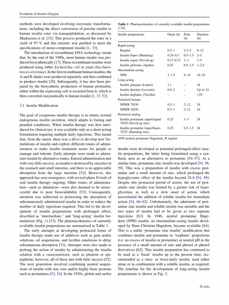
mulations (Fig. 1) [53]. The pharmacokinetics of currently
available insulin preparations are summarized in Table 1.
The early attempts at developing protracted forms of
insulin therapy made use of additives such as gum arabic
solutions, oil suspensions, and lecithin emulsions to delay
subcutaneous absorption [52]. Attempts were also made to
prolong the action of insulin by administering the insulin
solution with a vasoconstrictor, such as pituitrin or epi-
nephrine; however, all of these met with little success [52].
The next generation involved combining neutral suspen-
sions of insulin with zinc ions and/or highly basic proteins
such as protamines [52, 54]. In the 1930s, globin and surfen
insulin were developed as potential prolonged-effect insu-
lin preparations, the latter being formulated using a syn-
thetic urea as an alternative to protamine [54–57]. At a
similar time, protamine zinc insulin was developed [54, 58,
59]. This was a preparation of insulin with excess prot-
amine and a small amount of zinc, which prolonged the
hypoglycemic effect of the insulin beyond 24 h [54, 58].
Despite this protracted period of action, the use of prot-
amine zinc insulin was limited by a greater risk of hypo-
glycemia, as well as a slow onset of action, which
necessitated the addition of soluble insulin for immediate
action [54, 60–62]. Unfortunately, the admixture of prot-
amine zinc insulin and soluble insulin was unstable and the
two types of insulin had to be given as two separate
injections [63]. In 1946, neutral protamine Hage-
dorn (NPH) insulin, an intermediate-acting insulin devel-
oped by Hans Christian Hagedorn, became available [64].
This is a stable ‘protamine zinc insulin’ modification that
combines insulin and protamine in ‘isophane’ proportions
(i.e. no excess of insulin or protamine) at neutral pH in the
presence of a small amount of zinc and phenol or phenol
derivatives [65]. This insulin preparation has continued to
be used as a ‘basal’ insulin up to the present time, rec-
ommended as a once- or twice-daily insulin, used either
alone or in combination with a soluble insulin, as required.
The timeline for the development of long-acting insulin
preparations is shown in Fig. 2.
Table 1 Pharmacokinetics of currently available insulin preparations
[130]
Insulin preparations Onset (h) Peak
(h)
Duration
(h)
Rapid-acting
Regular 0.5–1 2.5–5 8–12
Insulin lispro (Humalog) 0.25–0.5 0.5–1.5 2–5
Insulin aspart (NovoLog) 0.17–0.33 1–3 3–5
Insulin glulisine (Apidra) 0.25 0.5–1.5 1–2.5
Intermediate-acting
NPH 1–1.5 6–14 16–24
Long-acting
Insulin glargine (Lantus) 1.1 – 24
Insulin detemir (Levemir) 0.8–2 – Up to 24
Insulin degludec (Tresiba) – – [25
Premixed human
NPH/R 70/30 0.5–1 2–12 24
NPH/R 50/50 0.5–1 2–12 24
Premixed analog
Insulin protamine aspart/aspart
70/30 (NovoLog mix)
0.25 1–3 24
Insulin protamine lispro/lispro
75/25 (Humalog mix)
0.25 0.5–1.5 24
NPH neutral protamine Hagedorn, R regular
Evolution of Insulin Glargine

Fig
.2
Ati
mel
ine
hig
hli
gh
tin
gsi
gn
ifica
nt
con
trib
uti
on
sto
the
dev
elo
pm
ent
of
mo
difi
edin
suli
ns
wit
hp
rolo
ng
edti
mes
of
acti
on
R. Hilgenfeld et al.
Early protracted animal insulins developed by Hallas-
Møller and Schlichtkrull capitalized on the varying solu-
bilities of their components (i.e. porcine and/or bovine
insulins) at physiological pH. The lente family of insulins
(semilente, lente, and ultralente) were created by com-
plexing neutral insulin suspensions with small amounts of
zinc ions, in the absence of any added foreign proteins or
synthetic compounds [54, 66]. This provided a spectrum of
time–action characteristics. The original lente insulin,
which had an intermediate timing of action similar to that
of NPH insulin, comprised a 30:70 mixture of amorphous
porcine insulin and crystalline bovine insulin particles
[67, 68]. Bovine ultralente insulin formed fairly large
crystals (30 lm) that remained in a subcutaneous depot for
a number of days, resulting in a duration of action similar
to that of protamine zinc insulin, enabling once-daily
administration [54].
The remainder of this review discusses insulin analog
preparations that are designed to possess a protracted
action, with a focus on insulin glargine, which was the first
‘long-acting’ insulin and was approved for clinical use in
2000.
3.2 Long-Acting Insulin Preparations: Mimicking
Basal Insulin Physiology
The main role of basal insulin secretion is to limit hepatic
glucose production and lipolysis in the fasting state, par-
ticularly overnight, without impairing glucose availability
for brain function [69]. However, older basal insulin
preparations, e.g. NPH and lente insulins, are acknowl-
edged to be associated with a number of limitations, such
as variable absorption with notable inter- and intra-indi-
vidual variation, and discernible peak plasma concentra-
tions after subcutaneous injection, thus increasing the risk
of hypoglycemia (in particular, nocturnal hypoglycemia).
Therefore, individuals treated with NPH insulin before the
evening meal or before bed may be at an increased risk of
fasting hyperglycemia. In addition, due its activity of less
than 24 h duration, a second dose in the morning is often
required. For example, even the longest-acting preparation,
human ultralente, with a peak insulin level at 10–14 h post-
injection, did not always provide adequate basal coverage
with once-daily administration at the lower dose levels [51,
58].
Therefore, in an attempt to avoid the shortcomings of
conventional basal insulin therapies, long-acting basal
insulin analogs were developed. To date, there have been
two main protraction strategies used: (1) modification of
the insulin molecule to achieve a low solubility at physi-
ological pH, e.g. insulin glargine; (2) the addition of a
fatty-acid chain of variable length to the insulin molecule,
which can bind to albumin, forming a circulating depot
from which the insulin analog is slowly released, e.g. the
insulins detemir and degludec. More recently, a third
strategy is being explored that involves the pegylation of
insulin, e.g. LY2605541 (insulin peglispro), which is cur-
rently undergoing extensive clinical evaluation [70, 71].
3.3 Early Analogs: Modified Chemical Structures
The elucidation of the chemical structure of animal insulin
by Frederick Sanger and his group [72, 73] and, subse-
quently, human insulin by Nicol and Smith [35], and the
determination of its three-dimensional structure by means
of X-ray crystallography by Dorothy Hodgkin and col-
leagues [74], as well as by the Chinese Insulin Group,
helped to reveal the relationship between proinsulin and
insulin and the spatial arrangement of the insulin molecules
within the hexamers (Fig. 3). These discoveries paved the
way for the synthesis of insulin and the eventual devel-
opment of new forms of rapid- and protracted-acting
insulin preparations based on alterations to the structure of
the insulin molecule itself (insulin analogs).
The human insulin molecule is a polypeptide with a
molecular mass of 5,808 Daltons, comprising an A and a B
chain connected by two disulphide bridges (Fig. 4a). By
changing the amino-acid sequence in such a way that it does
not prevent the interaction with either the insulin receptor or
insulin-like growth-factor receptor (i.e. protein engineering),
the ‘absorption kinetics’ of the insulin molecule can be altered
[75]. This process has been widely and successfully used in the
creation of short-acting analogs, working on the principle that
hexamer stability in the subcutaneous depot could be
decreased by alterations to structure or charge, leading to an
increased dissociation rate of the hexamers into dimers and
monomers at the site of injection, thereby enhancing the
absorption of insulin into the systemic circulation [76].
During the 1980s, initial attempts at creating long-acting
insulin analogs involved the addition of positive charges to
the insulin molecule, either by removing carboxylates (Glu,
Asp), or by the introduction of lysine or arginine using
single-chain insulin precursors [54, 77–80]. Early efforts
by Novo Nordisk involved changing GluB27 to arginine and
replacing the terminal carboxylate of the B chain by an
amide (ThrB30–NH2) [77–79]; further structural modifica-
tions created NovoSol Basal, a GlyA21ArgB27ThrB30 insulin
amide [81]. Although NovoSol Basal achieved prolonged
absorption compared with ultralente, it required double the
dose for comparable glycemic control. NovoSol Basal also
had low intra-individual variability, but high inter-indi-
vidual variability. This agent failed in clinical testing in
1989, and this was thought to be due to subcutaneous
crystal formation and degradation of the drug in the sub-
cutaneous depot by significant macrophage infiltration,
leading to reduced bioavailability [75].
Evolution of Insulin Glargine

A di-arginine (ArgB31ArgB32) preparation (Fig. 4b) [82,
83] was obtained by trypsin cleavage of the biosynthetic
precursor, proinsulin [84, 85]. As these two arginines are
present in proinsulin, linking the B chain to C peptide, and
are cleaved off during its metabolism, retaining them was
seen as a logical approach to the development of a long-
acting insulin [86]. Patents on this structural innovation
were filed in 1983 and 1984 by Hoechst AG, a predecessor
company of Sanofi. Initially, the simple concept behind
these structural modifications was that they would cause a
shift in the isoelectric point of the insulin analog from 5.4
towards a neutral pH, consequently lowering solubility at
physiological pH. The injection of such a preparation
would then result in amorphous precipitation and possibly
crystallization in the subcutaneous tissue, leading to
delayed absorption into the circulation [75]. Early results
confirmed the efficacy of ArgB31ArgB32 insulin in rabbit
models; although, in canine models after subcutaneous
injection, this effect did not show any benefit over NPH
insulin (Fig. 5a) and, consequently, development of this
formulation was discontinued.
Subsequent investigation, in animal models, of subcu-
taneous injection of acidic solutions with varying zinc
concentrations revealed that the lowest total potency of
20 lg/mL was the ideal concentration for ArgB31ArgB32
insulin crystallization in vitro, and may indicate a change
in the morphology of the subcutaneous precipitate from
amorphous to crystalline (Fig. 5b).
Findings from studies of long-acting insulin analogs
demonstrate that, even in cases such as NovoSol Basal and
ArgB31ArgB32 insulin, which have similar solubility pro-
files, variations in chemical structure can produce markedly
different pharmacokinetic profiles and pharmacodynamic
outcomes in vivo. This is highlighted by a study of
NovoSol Basal in dogs, which reported lower total blood
glucose-lowering properties than for insulin glargine
(Fig. 5c). In addition, the failure of analogs such as ArgB0
to exhibit prolonged glucose-lowering activity in spite of
an increased isoelectric point suggested that merely
increasing the isoelectric point was too simple a concept to
achieve prolonged activity; rather, the impact of the three-
dimensional structure seemed to play a major role.
Fig. 3 The three-dimensional
structures of proinsulin (a),
human insulin monomer (b),
dimer (c), and hexamer (d). In
b–d: green, A chain(s);
magenta: B chain(s). In a, the
C-peptide within proinsulin is
indicated in blue. a Adapted
from Yang et al. [128]; b, c and
d Adapted from Smith et al.
[129]
R. Hilgenfeld et al.

Fig. 4 Schematic structures of human insulin and insulin analogs (a), ArgB31ArgB32 insulin analog (b), and insulin glargine
(GlyA21ArgB31ArgB32) (c)
Evolution of Insulin Glargine

It was at this point that structural biologists entered the
stage. Initially, attention was focused on the role of phenol
in stabilization (dimer-to-dimer interaction) of the insulin
hexamer and monoclinic insulin crystals [87, 88]. Phenol
or its derivatives were introduced as a bacteriostatic agent
and preservative in insulin preparations; however, it was
subsequently realized that phenol was critical in stabilizing
the dimer–dimer interactions within the hexamer, thereby
protracting the action of insulin [76, 88, 89]. Subsequent
crystallographic analyses of the long-acting insulins
focused on the contact areas between insulin hexamers in
phenol-containing monoclinic crystals. It could be shown
that the addition of extra arginine residues at the C-ter-
minal of the B-chain leads to an increase in the number of
polar interactions between hexamers in the crystals, con-
comitant with a higher packing density of the crystals and a
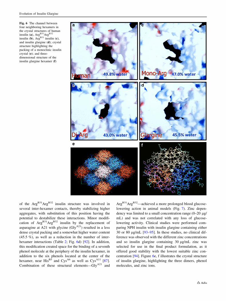
reduced water content. Thus, phenol-containing, mono-
clinic crystals of human insulin comprise 49.8 % solvent
(Fig. 6a). Attachment of two arginine residues at the
C-terminus of the B chain, as in ArgB31ArgB32 insulin,
introduces many additional hydrogen-bonding and salt-
bridge interactions between neighboring hexamers, leading
to a shrinkage of the unit cell of the monoclinic crystals
with a lower water content (43 %), and a higher packing
density (Table 2; Fig. 6b). The addition of a single arginine
residue at the C-terminus of the B chain (ArgB31 insulin)
resulted in a water content and packing density between
that seen with human insulin and ArgB31ArgB32 insulin
(Table 2; Fig. 6c). The activity of ArgB31 insulin is pro-
tracted by about 2 h compared with human insulin. A
correlation between crystal packing density and duration of
activity was observed, suggesting that stable crystal for-
mation at physiological pH was important for the protrac-
tion of time–action. In the case of ArgB31ArgB32 insulin,
pre-formed crystals with their tight packing are subject to
very slow solubilization following subcutaneous injection,
and this may ultimately explain the loss of activity fol-
lowing injection of solutions with high crystallization
tendencies. Thus, although low solubility at physiological
pH is necessary for prolonged duration of insulin action, it
is not sufficient alone: attention must also be paid to the
degree of inter-hexamer interaction and crystal stability
against dissolution. It is also of interest that even
uncharged residues that are capable of making extra
interactions and/or reducing the solubility of the insulin
derivative (such as phenylalanine in both positions B31 and
B32) were found to lead to a prolonged activity profile;
thus, the effect did not only depend on a shift of the iso-
electric point. Even in the case of PheB31PheB32 insulin, the
correlation between reduced water content of the crystals
(46.9 %) and the duration of activity was found to hold
true.
4 Development of Insulin Glargine (Lantus�)
Subsequent steps were taken by Hoechst AG to improve on
the structure of the ArgB31ArgB32 insulin analog to main-
tain a low solubility at physiological pH in order to achieve
a more prolonged bioavailability, and many analogs were
made that contained a range of different modifications [90,
91]. X-ray crystallographic data indicated that position A21
Fig. 5 (a) Blood glucose profiles after subcutaneous injection of
either crystalline or amorphous ArgB31ArgB32 insulin (0.3 IE/kg) in
dogs (n = 6); (b) Blood-glucose profiles with ArgB31ArgB32 insulin
(0.3 IU/kg) in dogs (n = 6) according to varying zinc concentrations;
(c) Variations in blood-glucose profiles between NovoSol Basal and
insulin glargine (pH 4 and 80 lg/ml zinc; 0.3 IU/kg) in dogs (n = 6)
R. Hilgenfeld et al.
of the ArgB31ArgB32 insulin structure was involved in
several inter-hexamer contacts, thereby stabilizing higher
aggregates, with substitution of this position having the
potential to destabilize these interactions. Minor modifi-
cation of ArgB31ArgB32 insulin by the replacement of
asparagine at A21 with glycine (GlyA21) resulted in a less
dense crystal packing and a somewhat higher water content
(45.5 %), as well as a reduction in the number of inter-
hexamer interactions (Table 2; Fig. 6d) [92]. In addition,
this modification created space for the binding of a seventh
phenol molecule at the periphery of the insulin hexamer, in
addition to the six phenols located at the center of the
hexamer, near HisB5 and CysA6 as well as CysA11 [87].
Combination of these structural elements—GlyA21 and
ArgB31ArgB32—achieved a more prolonged blood glucose-
lowering action in animal models (Fig. 7). Zinc depen-
dency was limited to a small concentration range (0–20 lg/
mL) and was not correlated with any loss of glucose-
lowering activity. Clinical studies were performed com-
paring NPH insulin with insulin glargine containing either
30 or 80 lg/mL [93–95]. In these studies, no clinical dif-
ference was observed with the different zinc concentrations
and so insulin glargine containing 30 lg/mL zinc was
selected for use in the final product formulation, as it
offered good stability with the lowest suitable zinc con-
centration [94]. Figure 6e, f illustrates the crystal structure
of insulin glargine, highlighting the three dimers, phenol
molecules, and zinc ions.
Fig. 6 The channel between
four neighboring hexamers in
the crystal structures of human
insulin (a), ArgB31ArgB32
insulin (b), ArgB31 insulin (c),
and insulin glargine (d); crystal
structure highlighting the
packing of a monoclinic insulin
crystal (e); and three-
dimensional structure of the
insulin glargine hexamer (f)
Evolution of Insulin Glargine

As a result of its chemical structure, this insulin analog
is less soluble at neutral pH than human insulin and pre-
cipitates in the subcutaneous tissue post-injection, slowing
its absorption and extending its duration of action [87]. The
structural properties of insulin glargine mean that it is
soluble in acidic solutions (pH 4) and does not require re-
suspension prior to injection, unlike NPH insulin. This
need for resuspension was the predominant cause of the
increased variability in the time–action characteristics of
NPH insulin [96]. Furthermore, insulin glargine functions
essentially as a ‘prodrug’ in the subcutaneous tissue, with
the majority of activity from its metabolites.
Following subcutaneous injection, insulin glargine is
rapidly metabolized into its two main active metabolites:
M1 (GlyA21) and M2 (GlyA21, des-ThrB30) [97], with little
or no glargine molecule being detected in the systemic
circulation. The M1 metabolite accounts for approximately
90 % of the available daily plasma insulin [97], and its
release from the poorly soluble parent compound is the
primary mechanism, resulting in the pharmacokinetic
characteristics and consequent pharmacodynamic effect
with the long-acting time–action profile observed with
insulin glargine treatment [98–100]. Steady state is attained
within a few days [101]. Importantly, adverse events,
injection-site reactions, and antibody formation with insu-
lin glargine were found to be comparable with NPH [75].
A patent for the GlyA21ArgB31ArgB32 insulin analog, i.e.
insulin glargine (Fig. 4c), was filed in 1988, and a New
Drug Application was made in the USA and Europe in
April 1999. Following an extensive clinical trial program,
insulin glargine was approved by the US FDA and the
European Medicines Agency for once-daily subcutaneous
administration for the treatment of type 1 diabetes mellitus
(T1DM) and T2DM in the year 2000.
5 Clinical Experience with Insulin Glargine
The recent joint recommendations from the American
Diabetes Association (ADA) and the European Association
for the Study of Diabetes (EASD) highlight the importance
of basal insulin therapy in people with T2DM. These
guidelines recommend the individualization of care and the
progressive intensification of therapy until glycemic targets
(glycated hemoglobin [HbA1c] \7.0 %) are met [102].
NPH insulin is still an effective and valuable intermediate-
acting insulin. However, the clinical need for an effective
long-acting agent to reduce the number of injections
required and lower the risk of hypoglycemia, whilst striv-
ing to achieve near normoglycemia led to the rapid adop-
tion of long-acting insulin analogs.
Clinical studies have demonstrated that, compared with
NPH, glargine has a more prolonged duration of action of
up to 24 h due to a slower and more delayed absorption
from the subcutaneous tissue, reduced variability, and a
relatively consistent, peakless concentration–time profile,
thus reducing the risk of hypoglycemia [58, 103–105].
Table 2 Packing density and inter-hexamer contacts in monoclinic
crystals of insulin and insulin analogs
Insulin Crystal
packing
density
(Da/A3)
Water
content
(%)
Hydrogen
bonds (n)
Salt
bridges
(n)
Human insulin 0.40 50 15 5
ArgB31 0.43 47 n.d. n.d.
ArgB31ArgB32 0.46 43 24 6
GlyA21ArgB31ArgB32
(Insulin glargine)
0.44 45.5 18 4
The packing density is given as molecular mass of the protein (in
Dalton) divided by the volume of the asymmetric unit of the crystal
that contains this protein (in A3). Water or additives are not included
in this parameter. A higher packing density correlates with more
interactions between the protein molecules and with a lower water
content
n.d. Not determined
Fig. 7 The contributions of both GlyA21 (a) and ArgB31ArgB32
(b) modifications to the overall glucose-lowering effects of insulin
glargine (pH 4, 40 IU/ml, 80 lg/ml zinc, dogs [n = 6], 0.3 IU/kg s.c.)
R. Hilgenfeld et al.

Glargine was the first once-daily, long-acting insulin ana-
log to be introduced into clinical practice, and it has now
been in clinical use for more than 10 years [54].
It has been suggested that insulin analogs may be
associated with an increased risk of cancer compared with
human insulin, owing to enhanced affinities for the insulin
receptor or the insulin-like growth factor receptor [97].
However, the di-arginyl molecules in insulin glargine,
which increase binding to the insulin-like growth factor
receptor in vitro, are not present in the glargine M1
metabolite, and the metabolic and mitogenic characteristics
of both the M1 and M2 metabolites have been shown to be
essentially similar to those of human insulin [97, 106].
Indeed, large epidemiological studies indicate that insulin
glargine does not have any independent carcinogenic
effects at therapeutic doses [107–109]. This is strongly
supported by the ORIGIN (Outcome Reduction with Initial
Glargine INtervention) study of 12,537 people with early
T2DM or pre-diabetes, which included cancer incidence as
a secondary outcome [110]. This represents the longest
randomized controlled study of insulin therapy, extending
over a median period of 6.2 years, with no increase seen in
the incidence of all cancers combined, any organ-specific
cancer (including breast, lung, colon, prostate, and mela-
noma), or cancer in the glargine group compared with the
standard care group.
In people with T1DM, glargine offers improved con-
venience, with only once-daily administration, and flexi-
bility as to timing of injection (morning, pre-dinner, or pre-
bedtime) [111]. In people with T2DM, glargine offers both
increased safety (reduced risk of nocturnal hypoglycemia)
and convenience (once-daily administration) when
attempting to reach the target HbA1c level of 7.0 % and
below, which is achieved in more than 50 % of subjects
[112].
The efficacy and safety of insulin glargine in both
people with T1DM and those with T2DM have been
demonstrated in a number of key randomized controlled
clinical studies. An overview of some of the key trials is
presented in Supplementary Table 1. Importantly, insulin
glargine can be used successfully with other oral and par-
enteral agents in the treatment of T2DM; for example, in
combination with prandial insulin or prandial glucagon-
like peptide (GLP)-1 receptor agonists as part of a basal-
bolus therapy [54, 113, 114]. This offers a new option for
the intensification of treatment of people with T2DM who
are not reaching glycemic targets despite receiving basal
insulin therapy. Currently, treatment is usually intensified
by the addition of prandial insulin, either as premixed
insulin or as separate injections. However, this increases
the risk of hypoglycemia and weight gain, side effects not
observed with GLP-1 receptor agonists, which have a low
risk of hypoglycemia and either a neutral effect on weight
or cause weight loss [115–118].
Insulin analogs tend to be associated with higher initial
medication costs than NPH and for this reason there is
debate as to whether they offer value for money in clinical
practice [119]. Cost-effectiveness analyses have demon-
strated that the initial expenditure associated with insulin
analogs is offset by reductions in the incidences of hypo-
glycemia associated with their use [120–122]. Other stud-
ies indicate that there may be no such cost reductions
[123]. While cost may be a consideration, it is only one of
several important factors that need to be considered when
deciding the most appropriate treatment regimen for
patients with diabetes. Blood glucose control, tolerability,
adverse events, patient adherence to treatment, and quality
of life are all essential considerations. Lower incidences of
nocturnal and severe hypoglycemia [124] and improve-
ments in patient adherence and quality of life have been
reported with use of insulin analogs due to the need for
fewer injections [125].
6 Conclusions
The discovery of insulin heralded a new dawn for people
with diabetes, with significant gains in both life expectancy
and quality of life. The ultimate goal of insulin therapy is to
mimic the physiological secretion of insulin to accommo-
date both fasting and prandial requirements, and advances
in protein engineering have enabled the development of
insulin analogs that mimic both basal and prandial
requirements.
The structural characteristics underlying the physiolog-
ical properties of insulin glargine define its clinical effec-
tiveness. Data indicate that low solubility at physiological
pH is a prerequisite, but this alone is not sufficient for a
basal insulin analog. Instead, factors determining success-
ful prolonged and continuous delivery of the analog are
likely to include a balanced number of inter-hexamer
interactions and moderate crystal stability.
Insulin glargine has demonstrated efficacy and consis-
tent safety in numerous large randomized clinical studies,
supporting its use as basal insulin therapy for the treatment
of diabetes, in line with ADA/EASD recommendations.
Insulin glargine continues to achieve real success in the
clinical setting, providing important benefits to people with
diabetes. Importantly, as glargine can be used in combi-
nation with other insulin and non-insulin antidiabetic
agents, it has a central role to play in the tailoring of
treatment on an individual basis, which is recognized as the
most appropriate approach to the effective management of
diabetes.
Evolution of Insulin Glargine

In conclusion, the development and introduction of
long-acting insulin analogs represented a dramatic step
forward in diabetes care, fulfilling the clinical need for a
basal insulin analog (which was hinted at by NPH insulin
almost half a century previously). Insulin glargine now
represents a reference basal insulin against which future
developments in long-acting insulin analogs are measured
[70, 126, 127].
Acknowledgments The authors are grateful to all members of the
‘‘New Insulins’’ team of former Hoechst AG. They thank Dr. Naoki
Sakai (University of Lubeck) for help with Fig. 3. RH acknowledges
support by the Chinese Academy of Sciences through a Visiting Pro-
fessorship for Senior International Scientists (Grant no. 2010T1S6).
Editorial support for this article was provided by Alexander Jones,
Ph.D., from Medicus International, London, and funded by Sanofi.
Conflict of interest DO has received lecture fees and honoraria
from Sanofi and Roche Diagnostics. GS and HB are employees of
Sanofi. RH declares no conflict of interest.
Dedication The authors would like to dedicate this manuscript to
the late Professor Geiger and the late Dr. Obermeier who were pio-
neers in the development of human insulin and insulin pumps.
The contents of this article and opinions expressed within are those
of the authors, and it was the decision of the authors to submit the
manuscript for publication. The authors conceived and critically
reviewed the manuscript, including input into every stage of the
development of the manuscript, and approved the final version for
submission.
Open Access This article is distributed under the terms of the
Creative Commons Attribution Noncommercial License which per-
mits any noncommercial use, distribution, and reproduction in any
medium, provided the original author(s) and the source are credited.
References
1. Owens DR. Human insulin. UK, Europe, USA: MTP Press;
1986.
2. Bliss M. The discovery of insulin: the inside story. Publ Am Inst
Hist Pharm. 1997;16:93–9.
3. Joslin EP. The treatment of diabetes mellitus. Can Med Assoc J.
1916;6(8):673–84.
4. Mazur A. Why were ‘‘starvation diets’’ promoted for diabetes in
the pre-insulin period? Nutr J. 2011;10:23. doi:10.1186/1475-
2891-10-23.
5. Zajac J, Shrestha A, Patel P, Poretsky L. The main events in the
history of diabetes mellitus. In: Poretsky L, editor. Principles of
diabetes mellitus. New York: Springer Science ? Business
Media; 2010. p. 3–16.
6. Himsworth HP. Diabetes mellitus: Its differentiation into insu-
lin-sensitive and insulin-insensitive types. Lancet.
1936;227(5864):127–30.
7. Papaspyros NS. Introduction. In: Verlag GT, editor. The history
of diabetes mellitus. Stuttgart: Thieme; 1964. p. 1–10.
8. von Mering J, Minkowski O. Diabetes mellitus nach Pankre-
asexstirpation. Arch Exp Path Pharmakol. 1890;26:371–87.
9. Hedon E. Sur la consommation du sucre chez la chien ap-
res l’extirpation du pancreas. Arch Physiol Normal Pathol Vth
Series. 1893;5:154–63.
10. Minkowski O. Historical development of the theory of pancre-
atic diabetes by Oscar Minkowski, 1929: introduction and
translation by Rachmiel Levine. Diabetes. 1989;38(1):1–6.
11. Opie EL. The relation Oe diabetes mellitus to lesions of the
Pancreas. Hyaline degeneration of the Islands Oe Langerhans.
J Exp Med. 1901;5(5):527–40.
12. Opie EL. On the relation of chronic interstitial pancreatitis to the
Islands of Langerhans and to diabetes melutus. J Exp Med.
1901;5(4):397–428.
13. Zulzer G. Ueber Versuche einer specifischen Fermenttherapie
des Diabetes. Zeitschrift fur die experimentelle Pathologie und
Therapie. 1908;5(2):307–18.
14. Paulesco NC. Action de l’extrait pancreatique injecte dans le
sang, chez un animal diabetique. CR Seanc Soc Biol (Paris).
1921;85:555–9.
15. Scott EL. On the influence of intravenous injections of an extract
of the pancreas on experimental pancreatic diabetes. Am J
Physiol. 1912;29:306–10.
16. Kleiner IS. The action of intravenous injections of pancreas
emulsions in experimental diabetes. J Biol Chem.
1919;40:153–70.
17. Rennie J, Fraser T. The islets of Langerhans in relation to dia-
betes. Biochem J. 1907;2(1–2):7–19.
18. Kimball CP, Murlin JR. Aqueous extracts of pancreas: III. Some
precipitation reactions of insulin. J Biol Chem. 1923;58:337–46.
19. Lasker SP, McLachlan CS, Wang L, Ali SMK, Jelinek HF.
Discovery, treatment and management of diabetes. J Diabetol.
2010;1(1).
20. Rosenfeld L. Insulin: discovery and controversy. Clin Chem.
2002;48(12):2270–88.
21. de Leiva A, Brugues E, de Leiva-Perez A. The discovery of
insulin: continued controversies after ninety years. Endocrino-
logia y nutricion : organo de la Sociedad Espanola de Endo-
crinologia y Nutricion. 2011;58(9):449–56. doi:10.1016/j.
endonu.2011.10.001.
22. Paulesco NC. Traitement du diabete. La Presse Medicale; 1924.
23. Paulesco NC. Recherches sur le role du pancreas dans l’assim-
ilation nutritive. Arch Int Physiol. 1921;17:85–109.
24. Paulesco NC. Quelques reactions chimiques et physiques ap-
pliquees a l’extrait aqueux du pancreas pour le debarrasser des
substances proteiques en exces. Arch Int Physiol.
1923;21:71–85.
25. Paulesco NC. Divers procedes pour entroduire l’extrait pam-
creatique dans l’organisme d’un animal diabetique. Arch Int
Physiol. 1923;21:215–38.
26. Banting FG, Best CH. The internal secretion of the pancreas.
J Lab Clin Med. 1922;7:251–66.
27. Banting FG, Best CH, Collip JB, Campbell WR, Fletcher AA.
Pancreatic extracts in the treatment of diabetes mellitus. Can
Med Assoc J. 1922;12(3):141–6.
28. Barron M. The relation of the islets of Langerhans to diabetes
with special reference to cases of pancreatic lithiasis. Surg
Gynecol Obstet. 1920;31:437–48.
29. Banting FG, Best CH. Pancreatic extracts. J Lab Clin Med.
1922;7:464–72.
30. Chamoun D, Choi D, Tavares AB, Udoff LC, Levitas E, Resnick
CE, et al. Regulation of granulosa cell-derived insulin-like
growth factor binding proteins (IGFBPs): role for protein
kinase-C in the pre- and posttranslational modulation of IGFBP-
4 and IGFBP-5. Biol Reprod. 2002;67(3):1003–12.
31. von Horn H, Hwa V, Rosenfeld RG, Hall K, Teh BT, Tally M,
et al. Altered expression of low affinity insulin-like growth
factor binding protein related proteins in hepatoblastoma. Int J
Mol Med. 2002;9(6):645–9.
32. Garber AJ, Davidson JA, Krosnick A, Beaser RS, Anderson JH
Jr. Impact of transfer from animal-source insulins to
R. Hilgenfeld et al.

biosynthetic human insulin (rDNA E coli) in patients with dia-
betes mellitus. Clin Ther. 1991;13(5):627–36.
33. Markussen J, Damgaard U, Jorgensen KH, Sorensen E, Thim L.
Human monocomponent insulin. Chemistry and characteristics.
Acta medica Scandinavica Supplementum. 1983;671:99–105.
34. Thim L, Hansen MT, Sorensen AR. Secretion of human insulin
by a transformed yeast cell. FEBS Lett. 1987;212(2):307–12
(pii: 0014-5793(87)81366-2).
35. Nicol DS, Smith LF. Amino-acid sequence of human insulin.
Nature. 1960;187:483–5.
36. Mirsky IA, Jinks R, Perisutti G. The Isolation and Crystalliza-
tion of Human Insulin. J Clin Investig. 1963;42:1869–72.
doi:10.1172/JCI104871.
37. Smith LF. Isolation of insulin from pancreatic extracts using
carboxymethyl and diethylaminoethyl celluloses. Biochimica et
biophysica acta. 1964;82:231–6.
38. Kimmel JR, Pollock HG. Studies of human insulin from non-
diabetic and diabetic pancreas. Diabetes. 1967;16(10):687–94.
39. Brunfeldt K, Deckert T, Thomsen J. Human crystalline insulin
from non-diabetic and diabetic patients. Acta endocrinologica.
1969;60(3):543–9.
40. Shapcott D, O’Brien D. A method for the isolation of insulin
from single human pancreas. Diabetes. 1970;19(11):831–6.
41. World Health Organization. WHO Technical Report Series 565.
Geneva1975.
42. Kreines K. The use of various insulins in insulin allergy. Arch
Intern Med (Chicago). 1965;116:167–71.
43. Akre PR, Kirtley WR, Galloway JA. Comparative hypoglycemic
response of diabetic subjects to human insulin or structurally
similar insulins of animal source. Diabetes. 1964;13:135–43.
44. Orskov H, Christensen NJ. Plasma disappearance rate of injec-
ted human insulin in juvenile diabetic, maturity-onset diabetic
and nondiabetic subjects. Diabetes. 1969;18(10):653–9.
45. Sonksen PH, Tompkins CV, Srivastava MC, Nabarro JD. A
comparative study on the metabolism of human insulin and
porcine proinsulin in man. Clin Sci Mol Med.
1973;45(5):633–54.
46. Deckert T, Andersen OO, Grundahl E, Kerp L. Isoimmunization
of man by recrystallized human insulin. Diabetologia.
1972;8(5):358–61.
47. Sieber P, Kamber B, Hartmann A, Johl A, Riniker B, Rittel W.
Totalsynthese von Humaninsulin unter gezielter Bildung der
Disulfidbindungen. Vorlaufige Mitteilung. Helvetica Chimica
Acta. 1974;57(8):2617–21.
48. Marki F, Albrecht W. Biological activity of synthetic human
insulin. Diabetologia. 1977;13(4):293–5.
49. Obermeier R, Geiger R. A new semisynthesis of human insulin.
Hoppe-Seyler’s Zeitschrift fur physiologische Chemie.
1976;357(6):759–67.
50. Homandberg GA, Mattis JA, Laskowski M Jr. Synthesis of
peptide bonds by proteinases. Addition of organic cosolvents
shifts peptide bond equilibria toward synthesis. Biochemistry.
1978;17(24):5220–7.
51. Owens DR, Vora JP, Heding LG, Luzio S, Ryder RE, Atiea J,
et al. Human, porcine and bovine ultralente insulin: subcutane-
ous administration in normal man. Diabetic Med: J Br Diabet
Assoc. 1986;3(4):326–9.
52. Best CH. Prolongation of insulin action. Ohio J Sci.
1937;37(6):362–77.
53. Schlichtkrull J, Pingel M, Heding LG. Insulin preparations with
prolonged effect. In: Hasselblatt A, Bruchhausen FV, editors.
Handbook of experimental pharmacology. Berlin, Heidelberg,
New York: Springer; 1975. p. 729–77.
54. Owens DR. Insulin preparations with prolonged effect. Diabetes
Technol Ther. 2011;13(Suppl 1):S5–14. doi:10.1089/dia.2011.
0068.
55. Bauman L. Clinical experience with globin insulin. Am J Med
Sci. 1939;198(4):475–81.
56. Reiner L, Searle DS, Lang EH. On the hypoglycemic activity of
globin insulin. J Pharmacol Exp Ther. 1939;67:330–40.
57. Umber F, Stoerring FK, Foellmer W. Erfolge mit einem ne-
uartigen Depot Insulin ohne Protaminzusatz (Surfen-Insulin).
Klin Woch. 1938;17:443–6.
58. Lepore M, Pampanelli S, Fanelli C, Porcellati F, Bartocci L, Di
Vincenzo A, et al. Pharmacokinetics and pharmacodynamics of
subcutaneous injection of long-acting human insulin analog
glargine, NPH insulin, and ultralente human insulin and con-
tinuous subcutaneous infusion of insulin lispro. Diabetes.
2000;49(12):2142–8.
59. Chikama T, Nakamura M, Nishida T. Up-regulation of integrin
alpha5 by a C-terminus four-amino-acid sequence of substance
P (phenylalanine-glycine-leucine-methionine- amide) synergis-
tically with insulin-like growth factor-1 in SV-40 transformed
human corneal epithelial cells. Biochem Biophys Res Commun.
1999;255(3):692–7.
60. Fujita-Yamaguchi Y, Hawke DH, Shively JE, Choi S. Partial
amino acid sequence analyses of human placental insulin
receptor. Protein Seq Data Anal. 1987;1(1):3–6.
61. Matsumoto S, Isogai A, Suzuki A. N-terminal amino acid
sequence of an insect neurohormone, melanization and reddish
coloration hormone (MRCH): heterogeneity and sequence
homology with human insulin-like growth factor II. FEBS Lett.
1985;189(1):115–8.
62. Bell SC, Keyte JW. N-terminal amino acid sequence of human
pregnancy-associated endometrial alpha 1-globulin, an endo-
metrial insulin-like growth factor (IGF) binding protein–evi-
dence for two small molecular weight IGF binding proteins.
Endocrinology. 1988;123(2):1202–4.
63. Rinderknecht E, Humbel RE. The amino acid sequence of
human insulin-like growth factor I and its structural homology
with proinsulin. J Biol Chem. 1978;253(8):2769–76.
64. Krayenbuhl C, Rosenberg T. Crystalline protamine insulin. Rep
Steno Mem Hosp Nord Insulinlab. 1946;1:60–73.
65. Hagedorn HC. Protamine insulinate: (section of therapeutics and
pharmacology). Proc R Soc Med. 1937;30(6):805–14.
66. Hallas-Moller K, Jersild M, Petersen K, Schlichtkrull J. The
lente insulins, insulin-zinc suspensions. Danish Med Bull.
1954;1(5):132–42.
67. Whitehouse FW, Lowrie WL, Redfern E, Bryan JB. The lente
insulin triad, with emphasis on the use of ‘‘lente combinations’’.
Ann Intern Med. 1961;55:894–902.
68. Galloway JA, Bressler R. Insulin treatment in diabetes. Med
Clin N Am. 1978;62(4):663–80.
69. White JR Jr., Campbell RK, Hirsch I. Insulin analogues: new
agents for improving glycemic control. Postgrad Med.
1997;101(2):58–60, 3–5, 70.
70. Bergenstal RM, Rosenstock J, Arakaki RF, Prince MJ, Qu Y,
Sinha VP, et al. A randomized, controlled study of once-daily
LY2605541, a novel long-acting basal insulin, versus insulin
glargine in basal insulin-treated patients with type 2 diabetes.
Diabetes Care. 2012;35(11):2140–7. doi:10.2337/dc12-0060.
71. Rosenstock J, Bergenstal RM, Blevins TC, Morrow LA, Prince
MJ, Qu Y, et al. Better glycemic control and weight loss with the
novel long-acting basal insulin LY2605541 compared with insu-
lin glargine in type 1 diabetes: a randomized, crossover study.
Diabetes Care. 2013;36(3):522–8. doi:10.2337/dc12-0067.
72. Sanger F, Tuppy H. The amino-acid sequence in the phenylal-
anyl chain of insulin. 2. The investigation of peptides from
enzymic hydrolysates. Biochem J. 1951;49(4):481–90.
73. Sanger F, Tuppy H. The amino-acid sequence in the phenylal-
anyl chain of insulin. I. The identification of lower peptides from
partial hydrolysates. Biochem J. 1951;49(4):463–81.
Evolution of Insulin Glargine

74. Adams MJ, Blundell TL, Dodson EJ, Dodson GG, Vijayan M,
Bakar EN, et al. Structure of rhombohedral 2 zinc insulin
crystals. Nature. 1969;224(5218):491–5.
75. Vajo Z, Duckworth WC. Genetically engineered insulin analogs:
diabetes in the new millenium. Pharmacol Rev. 2000;52(1):1–9.
76. Brange J, Ribel U, Hansen JF, Dodson G, Hansen MT, Havelund
S, et al. Monomeric insulins obtained by protein engineering and
their medical implications. Nature. 1988;333(6174):679–82.
doi:10.1038/333679a0.
77. Markussen J, Diers I, Engesgaard A, Hansen MT, Hougaard P,
Langkjaer L, et al. Soluble, prolonged-acting insulin derivatives.
II. Degree of protraction and crystallizability of insulins
substituted in positions A17, B8, B13, B27 and B30. Protein
Eng. 1987;1(3):215–23.
78. Markussen J, Diers I, Hougaard P, Langkjaer L, Norris K, Snel
L, et al. Soluble, prolonged-acting insulin derivatives. III.
Degree of protraction, crystallizability and chemical stability of
insulins substituted in positions A21, B13, B23, B27 and B30.
Protein Eng. 1988;2(2):157–66.
79. Markussen J, Hougaard P, Ribel U, Sorensen AR, Sorensen E.
Soluble, prolonged-acting insulin derivatives. I. Degree of pro-
traction and crystallizability of insulins substituted in the termini
of the B-chain. Protein Eng. 1987;1(3):205–13.
80. Geiger R. Chemie des Insulins. Chemiker Zeitung. 1976;100:
111–29.
81. Jorgensen S, Vaag A, Langkjaer L, Hougaard P, Markussen J.
NovoSol Basal: pharmacokinetics of a novel soluble long acting
insulin analogue. Bmj. 1989;299(6696):415–9.
82. Zeuzem S, Stahl E, Jungmann E, Zoltobrocki M, Schoffling K,
Caspary WF. In vitro activity of biosynthetic human diarginy-
linsulin. Diabetologia. 1990;33(2):65–71.
83. Monti LD, Poma R, Caumo A, Stefani I, Picardi A, Sandoli EP,
et al. Intravenous infusion of diarginylinsulin, an insulin analogue:
effects on glucose turnover and lipid levels in insulin-treated type II
diabetic patients. Metab: Clin Exp. 1992;41(5):540–4.
84. Kemmler W, Peterson JD, Steiner DF. Studies on the conversion
of proinsulin to insulin. I. Conversion in vitro with trypsin and
carboxypeptidase B. J Biol Chem. 1971;246(22):6786–91.
85. Grau U. Inventor Hoechst Aktiengesellschaft, assignee. Phar-
maceutical agent for the treatment of diabetes mellitus United
States1984.
86. Rhodes CJ. Processing of the insulin molecule. In: LeRoith D,
Taylor SI, Olefsky JM, editors. Diabetes mellitus: a fundamental
and clinical text. 3rd ed. London: Lippincott Williams & Wil-
kins; 2004.
87. Berchtold H, Hilgenfeld R. Binding of phenol to R6 insulin
hexamers. Biopolymers. 1999;51(2):165–72. doi:10.1002/
(SICI)1097-0282(1999)51:2\165:AID-BIP6[3.0.CO;2-X.
88. Derewenda U, Derewenda Z, Dodson EJ, Dodson GG, Reynolds
CD, Smith GD, et al. Phenol stabilizes more helix in a new
symmetrical zinc insulin hexamer. Nature. 1989;338(6216):
594–6. doi:10.1038/338594a0.
89. Brange J, Skelbaek-Pedersen B, Lankjaer L, Damgaar U, Ego H,
Havelund S, et al. Galenics of insulin preparations. In: Berger
M, editor. Subcutaneous insulin therapy. Berlin, Heidelberg:
Springer; 1985.
90. Seipke G, Geisen K, Neubauer H-P, Pittius C, Rosskamp R,
Schwabe D. New insulin preparations with prolonged action
profiles: A21-modified arginine insulins [abstract]. Diabetolo-
gia. 1992;35(Suppl. 1):A4.
91. Seipke G, Berchtold H, Geisen K, Hilgenfeld R, Rosskamp R.
HOE 901: a new insulin with prolonged action [abstract]. Eur J
Endocrinol. 1995;132(Suppl. 1):25.
92. Hilgenfeld R, Sicker T, Dorschug M, Obermeier R, Geisen K,
Seipke G, et al. Controlling insulin bioavailability by crystal
contact engineering. Diabetologia. 1992;35(Supplement):A193.
93. Pieber TR, Eugene-Jolchine I, Derobert E. Efficacy and safety of
HOE 901 versus NPH insulin in patients with type 1 diabetes.
The European Study Group of HOE 901 in type 1 diabetes.
Diabetes Care. 2000;23(2):157–62.
94. HOE 901/2004 Study Investigators Group. Safety and efficacy
of insulin glargine (HOE 901) versus NPH insulin in combina-
tion with oral treatment in Type 2 diabetic patients. Diabet Med:
J Br Diabet Assoc. 2003;20(7):545–51.
95. Rosenstock J, Park G, Zimmerman J, Group, USIGTDI. Basal
insulin glargine (HOE 901) versus NPH insulin in patients with
type 1 diabetes on multiple daily insulin regimens. U.S. Insulin
Glargine (HOE 901) Type 1 Diabetes Investigator Group. Dia-
betes Care. 2000;23(8):1137–42.
96. Owens DR, Coates PA, Luzio SD, Tinbergen JP, Kurzhals R.
Pharmacokinetics of 125I-labeled insulin glargine (HOE 901) in
healthy men: comparison with NPH insulin and the influence of
different subcutaneous injection sites. Diabetes Care.
2000;23(6):813–9.
97. Owens DR. Optimizing treatment strategies with insulin glar-
gine in Type 2 diabetes. Expert Rev Endocrinol Metab.
2012;7(4):377–93.
98. Bolli GB, Frick A, Schmidt R, Eisenblaetter T, Becker R.
Plasma concentrations of insulin glargine and its metabolites
after SC injection of glargine in subjects with type 1 diabetes.
ADA 71st Scientific Sessions; June 24–28, 2011; San Diego,
CA. Abstract 71-OR2011.
99. Lucidi P, Portcellati F, Rossetti P, Candeloro P, Andreoli AM,
Frick A et al. Metabolism of insulin glargine after subcutaneous
injection of therapeutic dose in type 2 diabetes mellitus. ADA
71st Scientific Sessions; June 24–28, 2011; San Diego, CA.
Abstract 1092-P2011.
100. Werner U, Schmidt R, Blair E, Renna SM, Tennagels N. The
molecular mechanism of insulin glargine metabolism in vivo.
ADA 72nd Scientific Sessions; June 8–12, 2012; Philadelphia,
PA. Abstract 1645-P2012.
101. Porcellati F, Rossetti P, Ricci NB, Pampanelli S, Torlone E,
Campos SH, et al. Pharmacokinetics and pharmacodynamics of
the long-acting insulin analog glargine after 1 week of use com-
pared with its first administration in subjects with type 1 diabetes.
Diabetes Care. 2007;30(5):1261–3. doi:10.2337/dc06-2208.
102. Inzucchi SE, Bergenstal RM, Buse JB, Diamant M, Ferrannini
E, Nauck M, et al. Management of hyperglycemia in type 2
diabetes: a patient-centered approach: position statement of the
American Diabetes Association (ADA) and the European
Association for the Study of Diabetes (EASD). Diabetes Care.
2012;35(6):1364–79. doi:10.2337/dc12-0413.
103. Heinemann L, Linkeschova R, Rave K, Hompesch B, Sedlak M,
Heise T. Time-action profile of the long-acting insulin analog
insulin glargine (HOE901) in comparison with those of NPH
insulin and placebo. Diabetes Care. 2000;23(5):644–9.
104. Ratner RE, Hirsch IB, Neifing JL, Garg SK, Mecca TE, Wilson
CA. Less hypoglycemia with insulin glargine in intensive
insulin therapy for type 1 diabetes. U.S. Study Group of Insulin
Glargine in Type 1 Diabetes. Diabetes Care. 2000;23(5):639–43.
105. Home P. Insulin glargine: the first clinically useful extended-
acting insulin in half a century? Expert Opin Investig Drugs.
1999;8(3):307–14. doi:10.1517/13543784.8.3.307.
106. Sommerfeld MR, Muller G, Tschank G, Seipke G, Habermann
P, Kurrle R, et al. In vitro metabolic and mitogenic signaling of
insulin glargine and its metabolites. PloS one. 2010;5(3):e9540.
doi:10.1371/journal.pone.0009540.
107. Blin P, Lassalle R, Dureau-Pournin C, Ambrosino B, Bernard
MA, Abouelfath A, et al. Insulin glargine and risk of cancer: a
cohort study in the French National Healthcare Insurance
Database. Diabetologia. 2012;55(3):644–53. doi:10.1007/
s00125-011-2429-5.
R. Hilgenfeld et al.

108. Owens DR. Glargine and cancer: can we now suggest closure?
Diabetes Care. 2012;35(12):2426–8. doi:10.2337/dc12-1968.
109. Home PD, Lagarenne P. Combined randomised controlled trial
experience of malignancies in studies using insulin glargine.
Diabetologia. 2009;52(12):2499–506. doi:10.1007/s00125-009-
1530-5.
110. Investigators OT, Gerstein HC, Bosch J, Dagenais GR, Diaz R,
Jung H, et al. Basal insulin and cardiovascular and other out-
comes in dysglycemia. N Engl J Med. 2012;367(4):319–28.
doi:10.1056/NEJMoa1203858.
111. Ashwell SG, Gebbie J, Home PD. Optimal timing of injection of
once-daily insulin glargine in people with Type 1 diabetes using
insulin lispro at meal-times. Diabet Med: J Br Diabet Assoc.
2006;23(1):46–52. doi:10.1111/j.1464-5491.2005.01726.x.
112. Riddle MC, Rosenstock J, Gerich J, Insulin Glargine Study I.
The treat-to-target trial: randomized addition of glargine or
human NPH insulin to oral therapy of type 2 diabetic patients.
Diabetes Care. 2003;26(11):3080–6.
113. Owens DR, Luzio SD, Sert-Langeron C, Riddle MC. Effects of
initiation and titration of a single pre-prandial dose of insulin
glulisine while continuing titrated insulin glargine in type 2
diabetes: a 6-month ‘proof-of-concept’ study. Diabetes, Obes
Metab. 2011;13(11):1020–7. doi:10.1111/j.1463-1326.2011.
01459.x.
114. Hollander P, Cooper J, Bregnhoj J, Pedersen CB. A 52-week,
multinational, open-label, parallel-group, noninferiority, treat-
to-target trial comparing insulin detemir with insulin glargine in
a basal-bolus regimen with mealtime insulin aspart in patients
with type 2 diabetes. Clin Ther. 2008;30(11):1976–87. doi:10.
1016/j.clinthera.2008.11.001.
115. Meier JJ. GLP-1 receptor agonists for individualized treatment
of type 2 diabetes mellitus. Nat Rev Endocrinol.
2012;8(12):728–42. doi:10.1038/nrendo.2012.140.
116. Riddle M, Home P, Marre M, Niemoeller E, Ping L, Rosenstock
J. Efficacy and safety of once-daily lixisenatide in Type 2 dia-
betes insufficiently controlled with basal insulin ± metformin:
GetGoal-L study. Diabetes. 2012;61(Supplement 1):983-P
(A251).
117. Rosenstock J, Forst T, Aronson R, Sau-que-reyna L, Souhami E,
Ping L et al. Efficacy and safety of once-daily lixisenatide added
on to titrated glargine plus oral agents in Type 2 diabetes:
GetGoal-Duo 1 Study. Diabetes. 2012;61(Supplement 1):62-OR
(A18).
118. Seino Y, Min KW, Niemoeller E, Takami A, Investigators EG-
LAS. Randomized, double-blind, placebo-controlled trial of the
once-daily GLP-1 receptor agonist lixisenatide in Asian patients
with type 2 diabetes insufficiently controlled on basal insulin
with or without a sulfonylurea (GetGoal-L-Asia). Diabetes,
Obes Metab. 2012;14(10):910–7. doi:10.1111/j.1463-1326.
2012.01618.x.
119. Petznick A. Insulin management of Type 2 diabetes mellitus.
Am Fam Physician. 2011;84:183–90.
120. Brandle M, Azoulay M, Greiner RA. Cost-effectiveness and
cost-utility of insulin glargine compares with NPH insulin nased
on a 10-year simulation of long-term complications with the
Diabetes Mellitus Model in patients with type 2 diabetes in
Switzerland. Int J Clin Pharmacol Ther. 2007;45:203–20.
121. Pfohl M, Schadlich PK, Dippel FW, Koltermann KC. Health
economic evaluation of insulin glargine vs NPH insulin in
intensified conventional therapy for type 1 diabetes in Germany.
J Med Econ. 2012;15(Suppl 2):14–27.
122. Gordon J, Evans M, McEwan P, Bain S, Vora J. Evaluation of
insulin use and value for money in Type 2 diabetes in the United
Kingdom. Diabetes Ther. 2013;4:51–66.
123. Cameron CG, Bennett HA. Cost-effectiveness of insulin ana-
logues for diabetes mellitus. CMAJ. 2009;180:400–7.
124. Monami M, Marchionni N, Mannucci E. Long-acting insulin
analogues vs. NPH human insulin in type 1 diabetes. A meta-
analysis. Diab Obes Metab. 2009;11:372–8.
125. Wang L, Wei W, Miao R, et al. Real-world outcomes of US
employees with type 2 diabetes mellitus treated with insulin
glargine or neutral protamine Hagedorn insulin: a comparative
retrospective database study. BMJ Open. 2013;3:e002348.
126. Garber AJ, King AB, Del Prato S, Sreenan S, Balci MK, Munoz-
Torres M, et al. Insulin degludec, an ultra-longacting basal
insulin, versus insulin glargine in basal-bolus treatment with
mealtime insulin aspart in type 2 diabetes (BEGIN Basal-Bolus
Type 2): a phase 3, randomised, open-label, treat-to-target non-
inferiority trial. Lancet. 2012;379(9825):1498–507. doi:10.
1016/S0140-6736(12)60205-0.
127. Heller S, Buse J, Fisher M, Garg S, Marre M, Merker L, et al.
Insulin degludec, an ultra-longacting basal insulin, versus insulin
glargine in basal-bolus treatment with mealtime insulin aspart in
type 1 diabetes (BEGIN Basal-Bolus Type 1): a phase 3, ran-
domised, open-label, treat-to-target non-inferiority trial. Lancet.
2012;379(9825):1489–97. doi:10.1016/S0140-6736(12)60204-9.
128. Yang Y, Hua QX, Liu J, Shimizu EH, Choquette MH, Mackin
RB, et al. Solution structure of proinsulin: connecting domain
flexibility and prohormone processing. J Biol Chem.
2010;285(11):7847–51. doi:10.1074/jbc.C109.084921
129. Smith GD, Pangborn WA, Blessing RH. The structure of T6
human insulin at 1.0 A resolution. Acta Crystallogr D Biol
Crystallogr. 2003;59:474–82.
130. Takiya L, Dougherty T. Pharmacist’s guide to insulin prepara-
tions: a comprehensive review. Last accessed August 2012.
https://secure.pharmacytimes.com/lessons/200510-03.asp.
Evolution of Insulin Glargine