the essential lower back exam - home - stfm connect
TRANSCRIPT
The Essential
Lower Back Exam
Judith A. Furlong, M.D.,
Cathee McGonigle, D.O. &
Rob Rutherford, MD
STFM National Convention 2011 New Orleans

Objectives
Brief review of the anatomy of the back,
(hip and pelvis in Handout)
Provide a systematic approach to
examine the lower back, hip, and pelvis

Overview
60-70% of the adult population has
been affected by low back pain at some
time in their lives
Up to 15-30% may be affected at one
time
Back pain is a common cause of
absenteeism from work in employees
between the ages of 30 and 60 years

In 1988, an estimated 149 million work
days were missed by about 22.4 million
people (17.6% of all U.S. workers) due
to low back pain
Most people have complete resolution
of their symptoms in 30 days; others
have recurrence or symptoms become
chronic

Persistance of symptoms should trigger
a more extensive work-up for a cause
Keep in mind that litigation and
Worker’s Compensation claims may
significantly affect a person’s pain
behavior and impact their motivation to
return to work rapidly

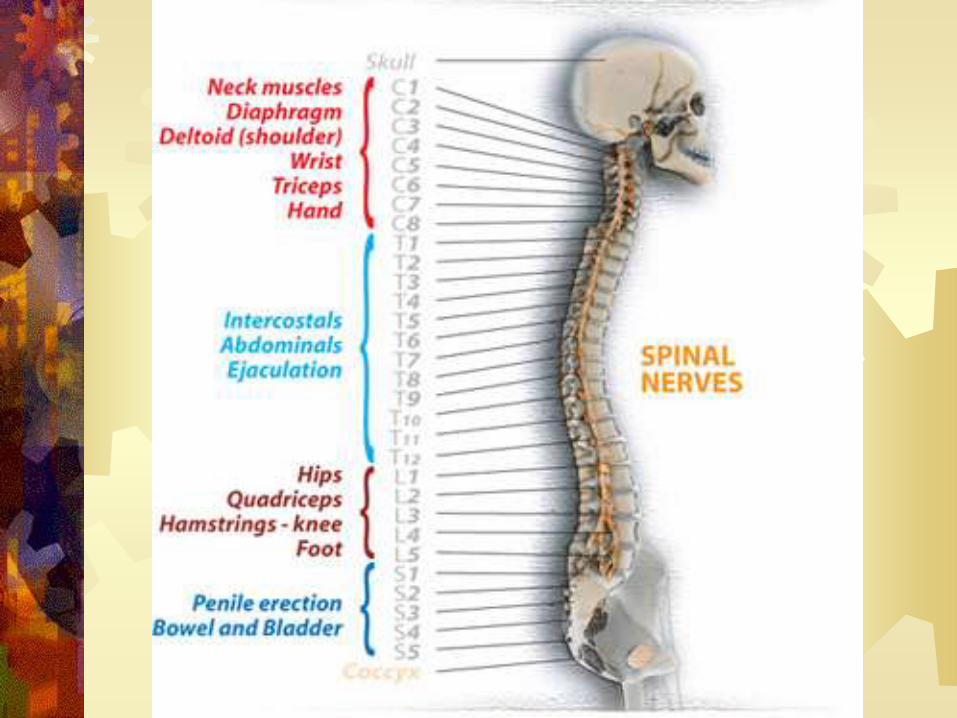
Brief Review of
Anatomy

Lumbar Spine


Muscles


Differential Diagnosis
Inflammation
Infection
Degenerative disorders
Neoplasms (primary and metastatic)
Trauma
Metabolic disorders
Developmental defects
Neurologic disorders
Referred pain
Psychological problems
Cauda equina syndrome
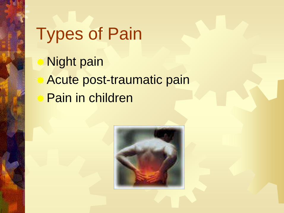
Types of Pain
Night pain
Acute post-traumatic pain
Pain in children

Obtaining the History
Location, duration, degree, and disability
Determine the mechanism of injury or
overuse
Assess pain severity
Establish the location of the pain
Prior Surgery

Red Flags
Cancer history
Recent infection
(urinary or skin)
Immuno-
compromised state
Trauma
Use of
corticosteroids
Progressive
Neurological Deficit
Bowel or Bladder
changes
Abdominal Pain
Fever
Saddle
Anesthesia
Night Pain
Cauda Equina
Syndrome

Physical Exam

Lumbar Spine, Hip, and Pelvis
Physical Exam
Remove clothing to expose the entire back
Systematic Approach
Inspection
Palpation
ROM
Neuro-vascular Exam
Special Tests
Related areas

Inspection
Observation
Gait, signs of injury
Asymmetry
Pelvic Obliquity or Leg Length Discrepancy
Edema
Ecchymosis
Redness
Deformity
Scoliosis
Abnormal Hair Growth

Palpation
Spinous processes
Paraspinous muscles
Sacroiliac joints
Tip of coccyx
Percuss firmly over posterior spine
Assess for unequal leg length

Palpation of Patient
Osteopathic Criteria - TART
Tissue texture changes
Asymmetry
Restriction of motion
Tenderness (palpation)

Assessing Range of Motion
Forward flexion (usually 80-90o) loads
the discs and therefore can increase
disc pain
Observe this posture from the rear to
evaluate for possibility of scoliosis
Extension (usually 20-30o) loads the
facets and can therefore increase facet
pain

Lateral bending (20 to 30o each
direction) exacerbates pain from muscle
strain by muscle stretching
Rotation or twisting (30 to 40o each
direction) also stretches muscles and
exacerbates pain due to muscle strain

Range of Motion

Strength Testing
Heel walking (anterior tibial muscles –
L4)
Toe walking (gastroc-soleus muscles –
S1)
Resisted great toe dorsiflexion – L5

Focused Neurologic Exam
Deep tendon reflexes – knee and ankle
jerk
Straight leg raise
Evaluate for ankle clonus – positive
response suggests an upper motor
neuron lesion eg. proximal cord
compression
Crossed straight-leg raise test
Rectal exam

Sensory Distribution
Normal Exam
Video by Rob Rutherford, MD

Special Testing - Adults
Thomas
Trendelenburg
Stork Test
Leg Length
Hamstrings
Quadriceps
Ely’s Test
Ober
Straight Leg Raise
FABER
FADIR
Fulcrum
Pelvic Rock Test
Flexion
Seated and Standing
Spring Test

Thomas Test
Test: Iliopsoas, Rectus Femoris,
TFL, ITB, Flexion Contracture
Patient: Supine
Physician: Stabilize pelvis
w/hand under lumbar spine, Flex
both pt legs until lumbar flattens,
pt extends one leg
Positive Test: unable to have:
RF – 90 degrees of knee flexion;
IP – neutral angle of hip; or
TFL/ITB – 15 degrees of hip
ABD.
Hoppenfeld, Stanley, Physical Exam
of Spine and Extremities, 1976.

Trendelenburg’s Test
Test: Gluteus Medius Strength
Patient: Standing
Physician: Behind patient, ask pt to
stand on one leg, monitor pelvis –
horizontal vs. obliquity
Positive: Pelvis drops on contralateral
side of weak gluteus medius

Trendelenburg
Negative
Trendelenburg Test
Positive
Trendelenburg Test

Pelvic Obliquity
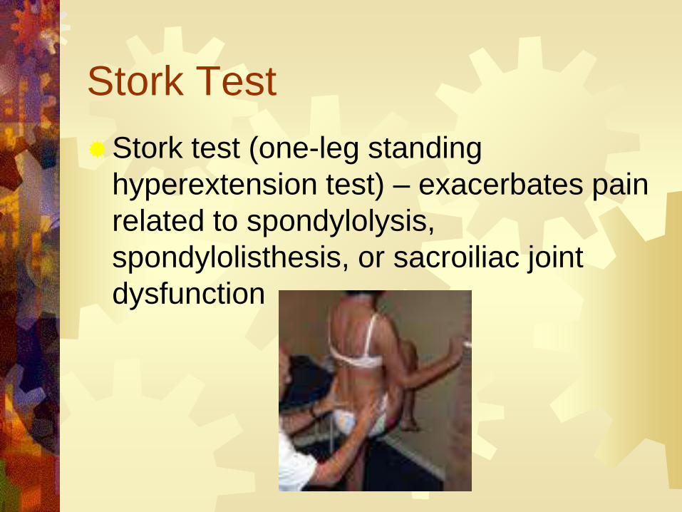
Stork Test
Stork test (one-leg standing
hyperextension test) – exacerbates pain
related to spondylolysis,
spondylolisthesis, or sacroiliac joint
dysfunction
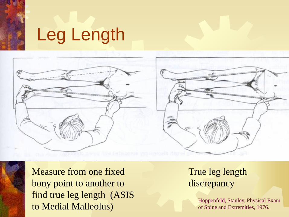
Leg Length
Measure from one fixed
bony point to another to
find true leg length (ASIS
to Medial Malleolus)
True leg length
discrepancy
Hoppenfeld, Stanley, Physical Exam
of Spine and Extremities, 1976.
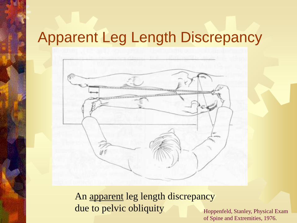
Apparent Leg Length Discrepancy
An apparent leg length discrepancy
due to pelvic obliquity Hoppenfeld, Stanley, Physical Exam
of Spine and Extremities, 1976.

True Leg Length Discrepancy
True leg length measurements are equal
despite the apparent discrepancy

Leg Length
Tibial length
discrepancy
Femoral length discrepancy

Hamstrings
Test: Hamstring
Patient: Supine
Physician: Flex pt
hip to 90, then
extend knee
Positive: Inability to
extend the knee,
Measure the
contracture
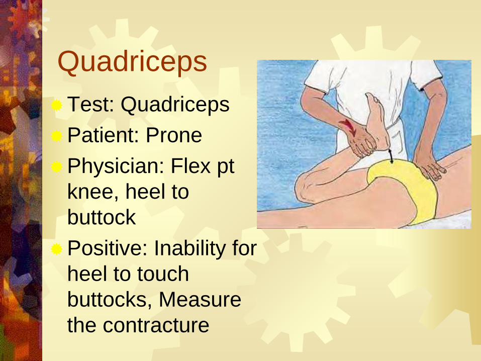
Quadriceps
Test: Quadriceps
Patient: Prone
Physician: Flex pt
knee, heel to
buttock
Positive: Inability for
heel to touch
buttocks, Measure
the contracture

Ely’s Test Test: Rectus Femoris
Patient: Prone
Physician: Flex pt
knees
Positive Test: The hip
on ipsilateral side
spontaneous flexes

Ober Test
Test: IT Band
Patient: Lateral Recumbant
Physician: Behind pt, stabilize
pelvis with one hand and
support the pt leg with other,
abduct hip and flex knee to 90,
allow leg to drop to table
Positive: Leg stays abducted
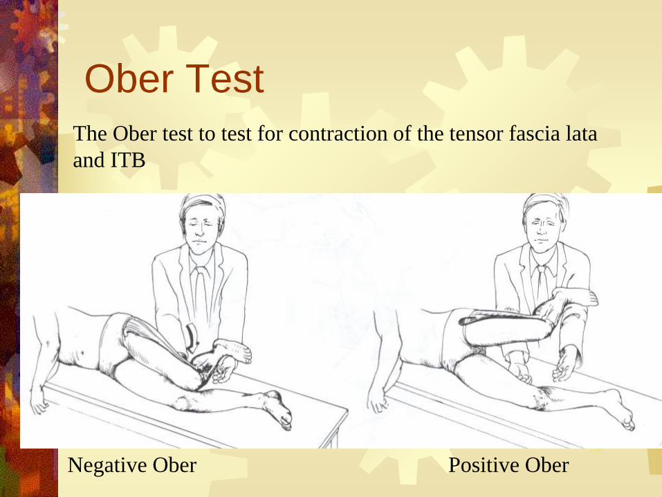
Ober Test
The Ober test to test for contraction of the tensor fascia lata
and ITB
Negative Ober Positive Ober

Straight Leg Raise
Lift the leg with knee extended while
patient is sitting or supine
Radiation of pain past the knee
suggests sciatica, frequently caused by
L5-S1 disc herniation
Ankle dorsiflexion increases sciatic pain
Ankle plantarflexion decreases sciatic
pain

Straight Leg Raise Test
http://www.musculoskeletalnetwork.com/display/article/1145622/1397911#

FABER
Test: Hip or SI Joint
Patrick Test
Flexion
Abduction
External Rotation
Patient: Supine
Physician: Cross pt one leg over other,
F, AB, ER the hip
Positive: Anterior – Hip, Posterior - SI

FADIR Test: Femoral Acetabular Impingement
FADIR
Flexion
Adduction
Internal Rotation
Patient: Supine
Physician: Beside pt, F, ADD, IR the hip
Positive: Anterior Lateral Hip Pain,
“C” Sign

Fulcrum Test
Test: Stress Fracture Femur
Patient: Seated, Knees extended
Physician – one hand under femur, other hand on top of knee, applies pressure
Positive – elicits pain in femur

Pelvic Rock Test
Test: Restriction or SD
of SI joints or pelvis
Innominate Rock Test
Patient: Supine
Physician: Both hands
over ASIS, apply
alternating forces
Positive – Firm
palpatory findings

Flexion Tests Standing
Physician seated, pt standing, Thumbs on PSIS
Pt forward flexes at waist
POSITIVE – asymmetry
Indicates: Iliosacral SD on ipsilateral side
Seated
Pt seated on stool/table, physician thumbs on PSIS, pt forward flexes
POSITIVE – asymmetry
Indicates: sacroiliac SD on ipsilateral side

Standing Flexion Test
Netterimages.com

Spring Test Test: Sacral dysfunction
Patient: Prone
Physician: Gentle
pressure
Positive: Resistance to
force
Indicates: Sacral SD –
unilateral or bilateral
backward sacrum

Related Area Exam
Lower Back
Hip
Knee

Questions?
Thank You!

References
Karageanes, S. Principles of Mannual Sports Medicine. 2005
Hoppenfeld, S. Physical Exam of the Spine and Extremities.
1976.
General Osteopathic Techniques,
http://www.eastlandpress.com/preview/las.pdf
Netter’s Sports Medicine, www.netterimages.com
Gray’s Anatomy