the epstein-barr virus lytic transactivator zta interacts...
TRANSCRIPT

JOURNAL OF VIROLOGY,0022-538X/98/$04.0010
Nov. 1998, p. 8559–8567 Vol. 72, No. 11
Copyright © 1998, American Society for Microbiology. All Rights Reserved.
The Epstein-Barr Virus Lytic Transactivator Zta Interactswith the Helicase-Primase Replication Proteins
ZHIGANG GAO,1 ANITA KRITHIVAS,1 JON E. FINAN,1 O. JOHN SEMMES,1†SIFANG ZHOU,1 YILONG WANG,1 AND S. DIANE HAYWARD1,2*
Molecular Virology Laboratories, Department of Pharmacology and Molecular Sciences1
and Department of Oncology,2 Johns Hopkins School of Medicine,Baltimore, Maryland 21205-2185
Received 13 April 1998/Accepted 2 July 1998
The Epstein-Barr virus transactivator Zta triggers lytic gene expression and is essential for replication of thelytic origin, oriLyt. Previous analysis indicated that the Zta activation domain contributed a replication-specific function. We now show that the Zta activation domain interacts with components of the EBV helicase-primase complex. The three helicase-primase proteins BBLF4 (helicase), BSLF1 (primase), and BBLF2/3(primase-associated factor) were expressed fused to the Myc epitope. When expression plasmids for BBLF4 orBBLF2/3 plus BSLF1 (primase subcomplex) were separately transfected, the proteins localized to the cyto-plasm. Interaction between Zta and the components of the helicase-primase complex was tested by examiningthe ability of Zta to alter the intracellular localization of these proteins. Cotransfection of Zta with Myc-BBLF4resulted in nuclear translocation of Myc-BBLF4; similarly, cotransfection of Zta with the primase subcomplexled to nuclear translocation of the Myc-BSLF1 and Myc-BBLF2/3 proteins. This relocalization providesevidence for an interaction between Zta and the helicase and Zta and the primase subcomplex. An affinity assayusing glutathione S-transferase–Zta fusion proteins demonstrated that Myc-BBLF4 and Myc-BBLF2/3 plusBSLF1 bound to the Zta activation domain (amino acids 1 to 133). In the nuclear relocalization assay, theamino-terminal 25 amino acids of Zta were required for efficient interaction with the primase subcomplex butnot for interaction with BBLF4. Evidence for interaction between oriLyt bound Zta and the helicase-primasecomplex was obtained in a superactivation assay using an oriLyt-chloramphenicol acetyltransferase (CAT)reporter. Zta activated expression from a CAT reporter containing the complete oriLyt region and regulatedby the oriLyt BHLF1 promoter. Cotransfection of the helicase-primase proteins, one of which was fused to aheterologous activation domain, led to Zta-dependent superactivation of CAT expression. This assay alsoprovided evidence for an interaction between the single-stranded DNA binding protein, BALF2, and theZta-tethered helicase-primase complex. The helicase-primase interaction is consistent with a role for Zta instabilizing the formation of an origin-bound replication complex.
Expression of the Zta (BZLF1, ZEBRA) transactivator in-duces lytic cycle reactivation in latently Epstein-Barr virus(EBV)-infected lymphoblastoid cell lines (15, 56, 65). Zta is abZip transcriptional transactivator that has a nonconsensusdimerization domain (10, 24) and binds as a homodimer toAP-1 sites and to related sequences called Zta response ele-ments (ZREs) (7, 10, 20, 41, 66). Zta stabilizes the formationof a DNA-bound complex containing the basal transcriptionfactors TFIID and TFIIA (14, 39), and it is most active onpromoters containing noncanonical TATA boxes that havereduced affinity for TFIID (38, 42). Both DNA binding andactivation of endogenous viral promoters are modified byphosphorylation of Zta (25, 34). In addition to regulating EBVlytic promoters, Zta modifies cellular gene expression (8, 9, 23)and has been found to interact with a variety of cellular pro-teins, including the retinoic acid receptor, NF-kB/p65, and p53(29, 54, 64, 73).
Zta has a second role in lytic cycle reactivation, serving as anessential regulatory protein for replication of the lytic origin,oriLyt (1, 21, 58, 61). EBV oriLyt is duplicated with one copy
located within the BamHI H fragment and a second copylocated in the region that is deleted in the sequenced B95-8isolate (30, 35). The origin comprises two essential elementsand one auxiliary domain (30, 60, 62). The essential BHLF1promoter and leader region and the auxiliary upstream en-hancer domain contain seven ZREs. The promoter ZREs areessential for replication, while the enhancer ZREs are dispens-able (60). The enhancer domain also contains two binding sitesfor the Rta transactivator (12, 27, 31). Rta is not essential fororiLyt replication but has a significant effect on replicationefficiency (21). In addition to Zta and Rta, oriLyt replicationrequires six EBV-encoded replication proteins that were orig-inally defined in a Challberg cotransfection replication assay(21, 22). The six core replication proteins have homologs in theother herpesviruses (17, 48, 53, 59, 71). Their functions aresufficiently conserved that the six core herpes simplex virus(HSV) proteins plus Zta and Rta can replicate EBV oriLyt(21); similarly, the six core EBV proteins plus the cytomega-lovirus ancillary proteins can replicate cytomegalovirus oriLyt(59). The core EBV proteins are the DNA polymerase (BALF5),the polymerase accessory protein (BMRF1), the single-strand-ed DNA binding protein (BALF2), the helicase (BBLF4), theprimase (BSLF1), and the primase-associated protein (BBLF2/3).Relatively few studies characterizing the core replication pro-teins have been performed. The polymerase and polymeraseaccessory protein (BMRF1) interact and together mediate pro-cessive DNA replication with strand displacement in model
* Corresponding author. Mailing address: Department of Pharma-cology and Molecular Sciences, Johns Hopkins School of Medicine,725 N. Wolfe St., Baltimore, Maryland 21205-2185. Phone: (410) 955-2548. Fax: (410) 955-8685. E-mail: [email protected].
† Present address: Department of Microbiology, University of Vir-ginia Medical School, Charlottesville, VA 22908.
8559
on Novem
ber 11, 2018 by guesthttp://jvi.asm
.org/D
ownloaded from

replication systems (33, 36, 43, 68, 70). BMRF1 binds DNAnonspecifically (32). Further, BMRF1 has been found to inter-act with the bZip domain of Zta (75) and to function indepen-dently as a transcriptional activator in transient expressionassays (52, 75). Although the mechanism of this activation hasnot been established, a region within the second essential do-main of oriLyt that is required for BMRF1 transactivation ofthe BHLF1(oriLyt) promoter has been mapped (74). BALF2has been shown to have single-stranded DNA binding proper-ties, and a role in melting hairpin structures to facilitate pro-cessive DNA replication has been postulated (18, 69). TheEBV helicase-primase proteins have not been subjected tofunctional analyses.
Attempts to determine the role of Zta in oriLyt replicationhave focused largely on the contribution of the activation do-main, and different studies have reached different conclusions.The Zta activation domain is encoded within the first exon,which comprises amino acids (aa) 1 to 167 (55). In transcrip-tion assays, the activation domain is both modular and redun-dant in that loss of individual subdomains can be compensatedfor by multimerization of the remaining domains and by mul-timerization of Zta binding sites in the target promoter (7, 13).Three studies have used EBV-positive cell lines to evaluate therequirement for Zta activation domain sequences in oriLytreplication. Schepers et al. (61) found that the activation do-main could not be substituted by the activation domain fromHSV VP16 and, using chimeric Zta-E2 and Gal4-Zta construc-tions and a modified oriLyt reporter in which the ZREs wereconverted to either E2 or Gal4 binding sites, that Zta aa 28 to103 were necessary for replication (60). Using EBV-positivecells and an unmodified oriLyt reporter, Askovic and Baumann(1) observed oriLyt replication when the Zta activation domainwas exchanged for a heterologous activation domain; in thissystem, deletions of individual regions of the activation domaindid not identify any region that was specifically required forreplication.
We have previously used a cotransfection replication assayto examine the contribution of Zta (21, 58). This assay systemis strictly defined in that EBV-negative cells are transfectedwith an oriLyt-containing plasmid, and replication is totallydependent on the cotransfected EBV replication genes. In thissystem, deletion of the Zta activation domain between aa 2 and10, aa 25 and 86, or aa 93 and 141 did not affect replication.However deletion of aa 2 to 25, and more specifically aa 13 to19, severely impaired replication efficiency, implicating a re-gion between aa 11 and 25 as serving a replication function.We have further pursued the requirement for Zta activationdomain sequences by examining interactions between Zta andthe core replication proteins. Evidence of an interaction withthe helicase-primase complex is presented. The finding that theamino terminus of the Zta activation domain is involved in thehelicase-primase interaction strongly suggests that this inter-action is functionally relevant and that Zta contributes to ori-Lyt replication, in part, by stabilizing formation of a tetheredreplication complex.
MATERIALS AND METHODS
Plasmids. The SG5 vector (Stratagene), which uses the simian virus 40 earlypromoter to drive expression, was modified by introduction into the vectorBamHI site of a sequence encoding aa 425 to 434 of the human c-myc gene tocreate pJH363. The double-stranded insert was generated by annealing theoligonucleotides 59-GATCTAAGATGGCGGAACAAAAGCTTATTTCTGAAGAAGACTTGG and 59-GATCCCAAGTCTTCTTCAGAAATAAGCTTTTGTTCCGCCATCTTA. The five replication genes for which immunological re-agents were not available were introduced into this vector. The previouslydescribed expression vectors (59) for BSLF1, BALF5, BBLF4, and BALF2 weremodified by conversion of an upstream vector XbaI site into either a Bgl II site
(BSLF1, BALF5, and BBLF4) or a BamHI site (BALF2), and DNA fragmentscontaining the open reading frames were isolated by cleavage with either BglII orBamHI and ligated into BglII-cleaved pJH363. The open reading frame forBBLF2/3 was isolated as a BamHI fragment from pEF75A (21).
Another modified SG5 vector, pJH209, contains the sequence encoding thenuclear localization signal and transcriptional activation domain (aa 424 to 487)from EBV EBNA2 introduced into the BglII site of SG5. The EBNA2 sequenceswere amplified by using PCR techniques and the primers 59-GCTAGGATCCCCAATACATGAACCGGAG and 59-GCTAAGATCTCTGGATGGAGGGGCGAGG. The six replication gene open reading frames were introduced into theBglII site of pJH209 as either BamHI or BglII fragments as described above. Theindividual replication gene expression clones are summarized in Table 1. Ex-pression of the appropriately sized proteins from each construction was con-firmed by Western blotting.
The reporter for glutathione S-transferase (GST) fused to Zta aa 1 to 133[Zta(1-133)], pDH245, was generated by cleaving pDH237 with SmaI and EcoRIto remove the DNA binding and dimerization domains; a BglII linker was addedto the blunted ends, and the DNA was cleaved with BglII and religated. TheoriLyt-chloramphenicol acetyltransferase (CAT) reporter, pDH123, has beendescribed elsewhere (40), as have the expression plasmids for Zta (pRTS21),Zta(D2-25) (pRTS68), and Zta(D13-19) (pDH285) (58).
Immunofluorescence assays. Vero cells were seeded at 8 3 104 cells per wellin two-well slide chambers. Cells were transfected with a maximum of 3 mg ofDNA by the calcium phosphate procedure. After transfection, cells were incu-bated in Dulbecco modified Eagle medium plus 10% fetal bovine serum for 16 hat 35°C in 3% CO2 and, after a medium change, for a further 24 h. Cells werewashed in 13 Tris-saline (100 mM Tris-HCl [pH 7.5]), fixed with 1% parafor-maldehyde in phosphate-buffered saline (0.144 g of KH2PO4, 9.0 g of NaCl, and0.795 g of Na2HPO4 z 7H2O per liter) for 10 min at room temperature andpermeabilized for 20 min on ice in 0.2% Triton X-100 in phosphate-bufferedsaline. Cells were incubated with primary antibody for 60 min at 37°C and withsecondary antibody at 37°C for 30 min. Antibodies used were anti-Myc mousemonoclonal (1:200) and rabbit polyclonal (1:300) (Santa Cruz BiotechnologyInc., Santa Cruz, Calif.), anti-BMRF1 monoclonal (DAKO Corp., Carpinteria,Calif.), anti-BZLF1 monoclonal (1:1,000; DAKO) and polyclonal (1:800; gift ofMarie Hardwick, Johns Hopkins School of Hygiene and Public Health) (31)antibodies; fluorescein isothiocyanate (FITC)-conjugated goat anti-mouse im-munoglobulin G (IgG; 1:100; Cappel Organon Teknika, Durham, N.C.); andFITC-conjugated donkey anti-rabbit (1:100) and rhodamine-conjugated donkeyanti-mouse (1:100) and anti-rabbit (1:100) IgG (Chemicon, Temecula, Calif.).
CAT assay. Vero cells were plated in six-well cluster dishes at 2 3 105 cells perwell 16 h before transfection with a medium change 4 h before transfection. Cellswere transfected by calcium phosphate precipitation with the oriLyt-CAT re-porter pDH123 (1 mg), Zta reporter pRTS21, pRTS68, or pDH285 (0.2 mg), andindividual replication genes (0.8 mg). Vector SG5 DNA was used to equalize theamount of DNA in each transfection to 4.8 mg. Cells were harvested 40 h aftertransfection, and CAT activity was assayed (44). CAT activity was quantitatedwith a PhosphorImager (Molecular Dynamics, Sunnyvale, Calif.).
GST affinity assay. GST and GST-Zta fusion proteins were induced by growthin medium containing 5 mM isopropyl-b-D-thiogalactopyranoside for 3 h at 30°C.Pelleted bacteria were resuspended in binding buffer (20 mM HEPES [pH 7.9],20 mM KCl, 1 mM MgCl2, 2 mM dithiothreitol, 17% glycerol) and sonicated.Cell debris was removed by centrifugation at 10,000 3 g for 10 min. The super-natant was incubated with glutathione-agarose beads (Sigma, St. Louis, Mo.) at4°C overnight and then washed three times in binding buffer. The amount ofprotein bound to the beads was determined by Coomassie blue staining ofprotein separated on a sodium dodecyl sulfate (SDS)-polyacrylamide gel. Equalamounts of each GST protein were used in the affinity assays.
293T cells in 100-mm-diameter dishes were transfected with a maximum of 15mg of DNA per dish, and cells were harvested 40 h after transfection. Cells werelysed in 500 ml of isotonic buffer (142.5 mM KCl, 5 mM MgCl2 10 mM HEPES
TABLE 1. EBV replication genes and expression plasmids
Viralgene Function Clone c-Myca E2TANLSb
BMRF1 Polymerase accessoryfactor
RTS14 DH295
BALF5 DNA polymerase RTS13 DH312 DH294BALF2 Single-stranded DNA
binding proteinRTS12 DH316 JF3
BSLF1 Primase RTS11 DH310 JF5BBLF4 Helicase RTS28 PG14 JF7BBLF2/3 Primase-associated
factorRTS25 DH318 JF6
a Protein is expressed as a fusion with an epitope (aa 425 to 434) from c-Myc.b Protein is expressed as a fusion with the nuclear localization signal and
transcriptional activation domain (aa 424 to 487) from EBV EBNA2.
8560 GAO ET AL. J. VIROL.
on Novem
ber 11, 2018 by guesthttp://jvi.asm
.org/D
ownloaded from

[pH 7.2], 1 mM EGTA [pH 8.0], 0.2% Nonidet P-40). The cell extract wasincubated with the GST fusion proteins overnight at 4°C, after which the complexwas washed five times in binding buffer. The complex was dissociated from thebeads by boiling for 5 min in 23 SDS-polyacrylamide gel electrophoresis(PAGE) loading buffer (2% SDS, 10% glycerol, 100 mM dithiothreitol, 60 mMTris [pH 6.8], 0.02% bromophenol blue), and the proteins were separated bySDS-PAGE on a 10% gel. Proteins were transferred to a nitrocellulose mem-brane (Bio-Rad, Hercules, Calif.), and the replication proteins were detected byincubation with anti-Myc antibody (1:1,000) followed by visualization by theenhanced chemiluminescence reaction (Amersham Life Science, Buckingham-shire, England).
RESULTS
Intracellular localization of the EBV helicase-primase pro-teins. BALF2, BALF5, and BMRF1 are known to individuallylocalize to the nucleus (33, 69). However, the three EBV he-licase-primase proteins have not been extensively character-ized, and their intracellular localization has not been exam-ined. As an initial step in evaluating potential interactionsbetween Zta and the core replication proteins, we generatedvectors expressing Myc-tagged replication proteins. These re-agents were used to examine the localization of the individualcore replication proteins in transfected cells (Fig. 1). As pre-viously described, the BMRF1, BALF5, and BALF2 proteinslocalized to the nucleus (exemplified by Myc-BALF2 in Fig. 1).When individually transfected, Myc-BBLF2/3 showed mixednuclear and cytoplasmic staining, Myc-BSLF1 was perinuclear,and Myc-BBLF4 localized to the cytoplasm. The vectors forthese three latter Myc-proteins express proteins in the appro-priate size range, as shown in Fig. 2. The open reading framesfor BBLF2/3, BSLF1, and BBLF4 are predicted to encode 765,874, and 811 aa, respectively, while the control DNA polymer-ase protein is predicted to contain 1,014 aa. The effect of sec-ondary modifications on the relative mobility of these proteinsis not known.
The three helicase-primase proteins encoded by HSV have
been shown to form a tripartite complex which influencesintracellular localization (6, 16). Evidence that the EBV ho-mologs also interact came from an examination of the intra-cellular localization of the helicase-primase proteins in triplytransfected cells. Cotransfection of Myc-BSLF1 in the pres-ence of BBLF2/3 and BBLF4 resulted in discrete nuclearlocalization of Myc-BSLF1. Similarly, cotransfection of Myc-BBLF2/3 with BSLF1 plus BBLF4 resulted in localization ofMyc-BBLF2/3 to the nucleus (Fig. 3). The nuclear localizationof BBLF2/3 and BSLF1 required the concurrent presence of
FIG. 1. Immunofluorescence assay showing the intracellular localization ofthe proteins of the helicase-primase complex. Vero cells were transfected withMyc-tagged BSLF1, BBLF4, and BBLF2/3 and a control, BALF2. Transfectedproteins were visualized with anti-Myc antibody and FITC-conjugated anti-mouse IgG secondary antibody.
FIG. 2. Expression of the Myc-tagged helicase-primase proteins. Expressionvectors for Myc-BBLF2/3, Myc-BBLF4, and Myc-BSLF1 and for a control rep-lication protein, Myc-BALF5, were individually transfected into Cos cells, andprotein expression was analyzed by Western blotting using anti-Myc antibodyand visualization by chemiluminescence. Positions of the helicase-primase pro-tein bands are indicated with arrowheads; positions of the 97- and 68-kDamolecular size markers are indicated on the right.
FIG. 3. Immunofluorescence assay showing the ability of BBLF4, BSLF1,and BBLF2/3 to form a tripartite complex that localizes to the nucleus in co-transfected Vero cells. Cotransfection of any two of the helicase-primase pro-teins did not confer nuclear localization. Transfected proteins were visualizedwith anti-Myc antibody and FITC-conjugated anti-mouse IgG secondary anti-body.
VOL. 72, 1998 Zta INTERACTION WITH HELICASE-PRIMASE COMPLEX 8561
on Novem
ber 11, 2018 by guesthttp://jvi.asm
.org/D
ownloaded from

all three members of the complex. Myc-BBLF2/3 in the pres-ence of BSLF1 gave a diffuse cytoplasmic pattern, and Myc-BSLF1 in the presence of BBLF2/3 was also cytoplasmic (Fig.3). Similarly, Myc-BBLF4 in the presence of either BSLF1 orBBLF2/3 remained cytoplasmic.
Zta relocates both the helicase and the primase subcomplexinto the nucleus. In contrast to the cytoplasmic localization ofthe individual members of the helicase-primase complex, Zta isa nuclear protein (Fig. 4). The differential localization of Ztaand the helicase-primase proteins provided an assay for poten-tial interactions between Zta and these proteins. Cotransfec-tion of Zta with either Myc-BBLF2/3 or Myc-BSLF1 did notchange the localization of these proteins from that observedfor the individually transfected proteins shown in Fig. 1. Myc-BBLF2/3 showed the mixed nuclear and cytoplasmic pattern,and Myc-BSLF1 remained perinuclear (Fig. 4). However, co-transfection of Zta with the combination of BSLF1 and BBLF2/3 converted the localization of these proteins from the dis-tinctly cytoplasmic pattern observed in doubly transfected cellsto a dominant nuclear pattern (Fig. 4). Cotransfection of Ztawith Myc-BBLF4 converted the strictly cytoplasmic BBLF4pattern to a strictly nuclear pattern (Fig. 4) indicative of aseparate interaction between Zta and BBLF4. In the experi-ment shown in Fig. 4, the cells were transfected with Zta andBBLF4 at a ratio of 2:1. At this ratio, some cells with cytoplas-mic staining are also visible.
Not all of the cotransfected cells expressed both proteins. Atthe 2:1 ratio for the Zta-Myc fusion protein expression plas-mids, cells expressing Zta but not the Myc-tagged proteins
were more common than those expressing only the Myc-taggedproteins. As shown by double staining (Fig. 5), all cells in whichMyc-BBLF4 and the Myc-BBLF2/3-plus-BSLF1 subcomplexwere present in the nucleus also expressed Zta. Different com-binations of antibody and fluorescence tag were used to ensurethe specificity of the results. For example, Zta was detectedwith polyclonal anti-Zta rabbit antiserum and FITC-taggedanti-rabbit antibody in the cotransfection with Myc-BBLF2/3plus BSLF1, polyclonal anti-Zta rabbit antiserum and rhoda-mine-tagged anti-rabbit antiserum in the cotransfection withMyc-BSLF1 plus BBLF2/3, and monoclonal mouse antiserumin the cotransfection with Myc-BBLF4.
BBLF4 and the BBLF2/3-plus-BSLF1 primase subcomplexinteract with the activation domain of Zta. We had previouslypresented evidence that the activation domain of Zta also con-tributed a replication-specific function. To determine whetherthe activation domain mediated the interaction between Ztaand BBLF4 and between Zta and the BSLF1-plus-BBLF2/3primase subcomplex, a GST-Zta affinity assay was performedwith GST fusion proteins containing the activation domain ofZta, GST-Zta(1-133), and control GST protein. Extracts of293T cells cotransfected with either Myc-BBLF4 or Myc-BBLF2/3 plus BSLF1 were incubated with equal amounts ofthe GST proteins bound to beads. After washing, the boundproteins were solubilized by boiling in SDS, separated on adenaturing polyacrylamide gel, and transferred to a nitrocel-lulose membrane. Reactive proteins were visualized using anti-Myc antibody and a chemiluminescence detection system (Fig.6). Myc-BBLF4 and Myc-BBLF2/3 each bound to GST-Zta(1-133). Neither protein bound significantly to the control GSTprotein. Thus, the interaction between Zta and the helicase-primase complex is mediated by the Zta activation domain.
Effect of N-terminal deletions of the Zta activation domain.We then used an immunofluorescence assay and anti-Myc an-tibody to evaluate the effect of Zta N-terminal activation do-main deletions in cotransfected cells. Zta(D2-25) and Zta(D13-19) have been previously characterized (58). The ability ofZta(D2-25) and Zta(D13-19) to relocalize cotransfected Myc-BBLF2/3 plus BSLF1 was examined in cells transfected withZta and Myc-BBLF2/3 plus BSLF1 at a ratio of 5:1. As sum-marized in Table 2, Myc-BBLF2/3 plus BSLF1 gave a complete-ly cytoplasmic signal. Cotransfection with wild-type Zta result-ed in approximately 90% of the fluorescent cells showing eithernuclear or mixed nuclear plus cytoplasmic staining. Cells scoredas nuclear staining had immunofluorescence signal only in thenucleus. Cells scored as mixed nuclear plus cytoplasmic showednuclear staining that was equal to or stronger than that in thecytoplasm. The mixed staining pattern was not seen in theabsence of Zta and presumably reflects incomplete relocaliza-tion of the Myc-tagged protein. In contrast to the effect ofwild-type Zta, between 88 and 97% of the stained cells cotrans-fected with Myc-BBLF2/3 and either Zta(D2-25) or Zta(D13-19) showed cytoplasmic fluorescence. Wild-type Zta, Zta(D2-25), and Zta(D13-19) each give a discrete nuclear pattern inthe transfected cells (reference 58 and data not shown). Thus,in cotransfected cells, deletion of amino-terminal sequences ofZta severely impaired interaction with the primase subcom-plex. Deletion of Zta N-terminal sequences had less effect onthe interaction of Zta with Myc-BBLF4. The results obtainedwith Zta and Myc-BBLF4 cotransfected at a 5:1 ratio are alsolisted in Table 2. At this ratio, wild-type Zta converted theMyc-BBLF4 staining pattern from cytoplasmic to predomi-nantly nuclear, with a small proportion of the cells showingmixed nuclear plus cytoplasmic staining. Zta(D2-25) behavedsimilarly to wild-type Zta. Zta(D13-19) was less efficient atnuclear relocation than Zta(D2-25), but a significant propor-
FIG. 4. Immunofluorescence assay demonstrating relocalization of Myc-BBLF4, Myc-BBLF2/3 plus BSLF1, and Myc-BSLF1 plus BBLF2/3 in the pres-ence of cotransfected Zta. Myc-tagged proteins were visualized with anti-Mycantibody and FITC-conjugated anti-mouse IgG secondary antibody. Zta wasdetected with mouse monoclonal antibody. The ratio of cotransfected Zta toMyc-tagged expression vector was 2:1. Both nuclear and cytoplasmic Myc-BBLF4 were detected at this ratio.
8562 GAO ET AL. J. VIROL.
on Novem
ber 11, 2018 by guesthttp://jvi.asm
.org/D
ownloaded from
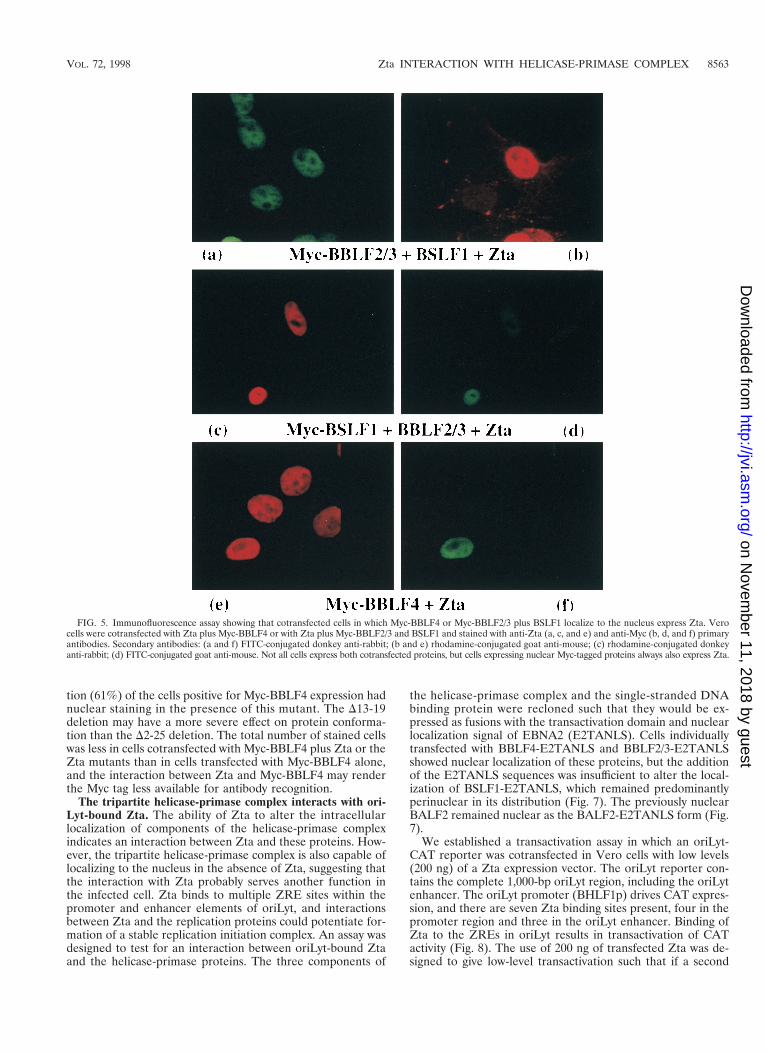
tion (61%) of the cells positive for Myc-BBLF4 expression hadnuclear staining in the presence of this mutant. The D13-19deletion may have a more severe effect on protein conforma-tion than the D2-25 deletion. The total number of stained cellswas less in cells cotransfected with Myc-BBLF4 plus Zta or theZta mutants than in cells transfected with Myc-BBLF4 alone,and the interaction between Zta and Myc-BBLF4 may renderthe Myc tag less available for antibody recognition.
The tripartite helicase-primase complex interacts with ori-Lyt-bound Zta. The ability of Zta to alter the intracellularlocalization of components of the helicase-primase complexindicates an interaction between Zta and these proteins. How-ever, the tripartite helicase-primase complex is also capable oflocalizing to the nucleus in the absence of Zta, suggesting thatthe interaction with Zta probably serves another function inthe infected cell. Zta binds to multiple ZRE sites within thepromoter and enhancer elements of oriLyt, and interactionsbetween Zta and the replication proteins could potentiate for-mation of a stable replication initiation complex. An assay wasdesigned to test for an interaction between oriLyt-bound Ztaand the helicase-primase proteins. The three components of
the helicase-primase complex and the single-stranded DNAbinding protein were recloned such that they would be ex-pressed as fusions with the transactivation domain and nuclearlocalization signal of EBNA2 (E2TANLS). Cells individuallytransfected with BBLF4-E2TANLS and BBLF2/3-E2TANLSshowed nuclear localization of these proteins, but the additionof the E2TANLS sequences was insufficient to alter the local-ization of BSLF1-E2TANLS, which remained predominantlyperinuclear in its distribution (Fig. 7). The previously nuclearBALF2 remained nuclear as the BALF2-E2TANLS form (Fig.7).
We established a transactivation assay in which an oriLyt-CAT reporter was cotransfected in Vero cells with low levels(200 ng) of a Zta expression vector. The oriLyt reporter con-tains the complete 1,000-bp oriLyt region, including the oriLytenhancer. The oriLyt promoter (BHLF1p) drives CAT expres-sion, and there are seven Zta binding sites present, four in thepromoter region and three in the oriLyt enhancer. Binding ofZta to the ZREs in oriLyt results in transactivation of CATactivity (Fig. 8). The use of 200 ng of transfected Zta was de-signed to give low-level transactivation such that if a second
FIG. 5. Immunofluorescence assay showing that cotransfected cells in which Myc-BBLF4 or Myc-BBLF2/3 plus BSLF1 localize to the nucleus express Zta. Verocells were cotransfected with Zta plus Myc-BBLF4 or with Zta plus Myc-BBLF2/3 and BSLF1 and stained with anti-Zta (a, c, and e) and anti-Myc (b, d, and f) primaryantibodies. Secondary antibodies: (a and f) FITC-conjugated donkey anti-rabbit; (b and e) rhodamine-conjugated goat anti-mouse; (c) rhodamine-conjugated donkeyanti-rabbit; (d) FITC-conjugated goat anti-mouse. Not all cells express both cotransfected proteins, but cells expressing nuclear Myc-tagged proteins always also express Zta.
VOL. 72, 1998 Zta INTERACTION WITH HELICASE-PRIMASE COMPLEX 8563
on Novem
ber 11, 2018 by guesthttp://jvi.asm
.org/D
ownloaded from

activation domain were tethered to the reporter, then theeffects of this second activator would also be detectable. Co-transfections were performed in which the cells received ori-Lyt-CAT and the E2TANLS-tagged proteins, either individ-ually or in groups and in the presence or absence of Zta. Asshown in a representative assay (Fig. 8), cotransfection ofBALF2-E2TANLS had no effect on Zta activation of the ori-Lyt-CAT reporter. Cotransfection of BBLF4-E2TANLS, BSLF1-E2TANLS, and BBLF2/3-E2TANLS plasmids individually gavelimited activation over that seen with Zta alone, while cotrans-fection of the three helicase-primase expression plasmids witheither BBLF2/3 or BSLF1 carrying the activation domain tagconsistently resulted in a fourfold activation of the reporterabove that seen with Zta alone. This activation was Zta specificand was not seen in the absence of Zta.
Interestingly, BALF2-E2TANLS, which had no effect on Ztaactivation individually, produced significant superactivation inthe presence of the untagged BBLF4, BSLF1, and BBLF2/3proteins, suggesting that BALF2 may interact with the heli-case-primase complex.
DISCUSSION
Studies on Zta transcriptional activation have provided ev-idence for Zta-mediated stabilization of a DNA-bound TFIIA-TFIID complex. In the stepwise addition model of transcrip-tion complex assembly, recruitment of TFIID and recruitmentof TFIIA represent the initial steps in the assembly of the coreinitiation complex. The role of Zta in oriLyt replication hasbeen less clear. Zta binds to multiple sites in oriLyt, includingthe four oriLyt (BHLF1) promoter ZREs that are essential fororiLyt replication (41, 60, 61). A transcriptional contribution toreplication by Zta seems likely. Removal of the Zta transcrip-tional activation domain abolishes replication activity (1, 58,60), and fusion of an additional heterologous activation do-main increases reactivation efficiency (2). As a transcriptionfactor, Zta may contribute either by disrupting nucleosomeformation and increasing the accessibility of the origin to thereplication complex or by introducing topological changes inthe DNA that facilitate replication initiation (28, 37). How-ever, in the replication assay performed with EBV-negativecells where all of the EBV genes are introduced individually onexpression plasmids, there was not a direct correlation betweenthe ability of Zta to function as a transcriptional activator andits ability to support oriLyt replication (58). As has been re-ported for the papillomavirus E2 transcriptional activator (57),mutations in the Zta activation domain revealed a separable
DNA replication function. Zta activation domain mutants suchas Zta(D2-25) and Zta(D13-19) were able to activate expres-sion from the endogenous BMRF1 promoter but were unableto provide replication function, suggesting that the amino ter-minus of the activation domain might specify an additionalreplication-specific activity. We have now presented experi-mental data indicating that amino-terminal Zta activation do-main sequences are required for efficient interaction with thecore replication proteins of the primase subcomplex.
When individually transfected, the Myc-tagged helicase(BBLF4) and primase (BSLF1) proteins localized to the cyto-plasm, while the primase-associated protein (BBLF2/3) showed amixed nuclear and cytoplasmic distribution. These three pro-teins were originally designated on the basis of their aminoacid homology with HSV replication proteins, and the EBVproteins themselves have not been functionally characterized.The immunofluorescence assays provided evidence that, like
FIG. 6. BBLF4 and BBLF2/3 plus BSLF1 interact with the Zta activationdomain, as determined by GST affinity assay in which extracts of 293T cellstransfected with Myc-BBLF4 (lanes 2, 4, and 6) or Myc-BBLF2/3 plus BSLF1(lanes 1, 3, and 5) were incubated with GST-Zta(1-133) and control GST pro-teins. Bound protein was separated by SDS-PAGE, transferred to a nitrocellu-lose membrane, and incubated with anti-Myc antibody. Reactive proteins werevisualized by chemiluminescence. The input lanes were loaded with 1/15 of theamount of extract incubated with the GST beads. The position of the 97-kDamolecular size marker is indicated on the right.
FIG. 7. Immunofluorescence assay showing the intracellular localization intransfected Vero cells of the three helicase-primase proteins and BALF2 ex-pressed as fusions with E2TANLS. Transfected proteins were visualized withanti-EBNA2 antibody and FITC-conjugated anti-mouse IgG secondary antibody.Addition of the tag sequences resulted in nuclear localization of BBLF4 andBBLF2/3 but was insufficient to relocate individually transfected BSLF1. The lo-calization of the nuclear BALF2 protein was not affected by the addition of the tag.
TABLE 2. Effect of Zta activation domain deletions onlocalization of BBLF4 and BBLF2/3 plus BSLF1
Localization (% of positive cells)a
Nucleus Nucleus 1 cytoplasm Cytoplasm
Myc-BBLF2/3 1 BSLF1 0 0 1001wt Zta 61 6 2 28 6 1 11 6 21Zta(D2-25) 7 6 4 5 6 5 88 6 21Zta(D13-19) 2 6 2 1 6 1 97 6 3
Myc-BBLF4 0 0 1001wt Zta 91 6 5 9 6 5 01Zta(D2-25) 91 6 1 9 6 1 01Zta(D13-19) 61 6 1 30 6 1 9 6 1
a Myc-tagged proteins were detected in transfected Vero cells by indirectimmunofluorescence using anti-Myc antibody and FITC-conjugated anti-mouseIg. Values are averages of two experiments.
8564 GAO ET AL. J. VIROL.
on Novem
ber 11, 2018 by guesthttp://jvi.asm
.org/D
ownloaded from

their HSV counterparts, BSLF1 and BBLF2/3 interact toform a primase subcomplex. Interaction with HSV UL8 (theBBLF2/3 homolog) leads to more efficient primer synthesis bythe primase (63, 67). When individually transfected, Myc-BBLF2/3 showed mixed nuclear and cytoplasmic staining, butwhen it was cotransfected with BSLF1, the pattern changed tothat of BSLF1 and was strictly cytoplasmic. Further, the pres-ence of Zta did not affect the localization of either BSLF1 orBBLF2/3 when they were individually cotransfected with Zta.Nuclear relocalization by Zta required the concurrent pres-ence of both BSLF1 and BBLF2/3, suggesting that interactionbetween BSLF1 and BBLF2/3 produces a conformationalchange in one or other protein. Triple transfection of BBLF4,BBLF2/3, and BSLF1 also resulted in nuclear localization ofthese proteins, indicating that they form a helicase-primasecomplex. This behavior is identical to that observed with thethree HSV helicase-primase proteins (6).
Interaction between Zta and the helicase-primase complexwas demonstrable in three different assays. The ability of Ztato independently translocate to the nucleus the cotransfectedhelicase, Myc-BBLF4, and the cotransfected primase subcom-plex indicates that Zta contacts at least two of the helicase-primase proteins. The presence of both BSLF1 and BBLF2/3was required for the primase subcomplex interaction with Zta,and which of these two proteins actually contacts Zta cannot bedetermined from the experiments described here. The HSVorigin binding protein, UL9, has been found to contact UL8,the BBLF2/3 analog (49). Although nuclear localization wasinitially used as a measure of interaction with Zta, it seems
unlikely that this is the primary replication-specific function forZta in the infected cell since the tripartite helicase-primasecomplex is itself capable of nuclear localization. Transcriptionfactors may also be involved in directly recruiting core repli-cation proteins or stabilizing the formation of the replisome(11, 19, 50). If Zta were to fulfill such a role, then the inter-action with the helicase-primase complex would likely takeplace with oriLyt-bound Zta. A superactivation assay designedto test this possibility indicated that Zta bound to an oriLyt-CAT reporter interacted with the helicase-primase proteins.Stable interaction was most clearly demonstrable in the pres-ence of the tripartite complex, which consistently gave a four-fold activation when either member of the primase subcomplexwas expressed fused to a heterologous transactivation domain.Most models of replication postulate that the initial steps inDNA synthesis involve sequence-specific recognition by an ini-tiator protein followed by localized DNA melting or unwinding(26). A number of other viruses encode origin binding proteinsthat have helicase or unwinding activity. Examples includesimian virus 40 T antigen (4), HSV UL9 (5), and papillomavi-rus E1 (72). Zta acts as the EBV oriLyt origin binding proteinand may ensure localized unwinding through the combinationof transcriptional stimulation and direct recruitment of thehelicase-primase complex.
The oriLyt-CAT superactivation assay also raised the possi-bility of an interaction between the Zta-tethered helicase-pri-mase complex and the single-stranded DNA binding proteinBALF2. Studies of HSV replication complex assembly in bothvirus-infected and DNA-transfected cells have indicated thatthe helicase primase proteins, UL5, UL8, and UL52, are need-ed for localization of the HSV single-stranded DNA bindingprotein, UL29, to prereplicative sites (45, 46, 76). Thus, theproposed HSV assembly model involves the single-strandedDNA binding protein assembling into a complex with thehelicase primase proteins, with polymerase and polymeraseaccessory protein, UL42, being recruited subsequently. Thepotential exists for multiple contacts within the replicationcomplex. Contacts between the HSV single-stranded DNAbinding protein and polymerase and the origin binding protein,UL9, have also been documented (3, 51), as have contactsbetween UL8 and polymerase (47). The information availablefor assembly of the EBV replication complex is much morelimited, but the present study provides parallels with the HSVassembly model in that initial steps in assembly seem to involvetethering of the helicase-primase complex at the origin by in-teraction with DNA-bound Zta and entry of the single-strand-ed DNA binding protein, BALF2, through interaction with thehelicase-primase proteins. A pictorial summation of the EBVdata is presented in Fig. 9.
We had previously found that Zta(D2-25) and Zta(D13-19)were unable to replicate oriLyt in a cotransfection replication
FIG. 8. Transient expression assay using Zta-dependent superactivation ofexpression from an oriLyt-CAT reporter as a measure of Zta-helicase-primaseinteraction. The results shown are representative of results obtained in threedifferent experiments. Vero cells were cotransfected with oriLyt-CAT plus orminus Zta, and the indicated individual replication genes were expressed asE2TANLS fusions (indicated by an asterisk) or groups of replication proteins,one of which was expressed as an E2TANLS fusion. Cotransfection of the threehelicase-primase proteins with one member tagged with the E2TANLS consis-tently resulted in Zta-dependent superactivation of CAT expression.
FIG. 9. Representation of the initial stages of core replication protein as-sembly as deduced from this study. OriLyt bound dimers of Zta interact throughthe activation domain with both the helicase (BBLF4) and primase subcomplex(BSLF1 plus BBLF2/3) to provide a stabilizing tether for the core replicationcomplex. The single-stranded DNA binding protein BALF2 may in turn contactthe helicase-primase complex.
VOL. 72, 1998 Zta INTERACTION WITH HELICASE-PRIMASE COMPLEX 8565
on Novem
ber 11, 2018 by guesthttp://jvi.asm
.org/D
ownloaded from

assay (58). However, in studies in other laboratories usingdifferent assay systems for oriLyt replication, replacement ormutation of the Zta activation domain had not been able touncover a replication specific contribution for this domain (1,60). While the reason for the different results cannot be as-cribed with certainty, the present study allows speculation. Inthe nuclear relocation assay, deletion of Zta aa 2 to 25 or 13 to19 had little effect on the interaction of Zta with BBLF4,although the deletions severely reduced Zta interaction withthe primase subcomplex. Since the helicase primase proteinsthemselves interact to form a tripartite complex, it seems likelythat deletion of Zta aa 2 to 25 might destabilize the interactionwith the tripartite complex by eliminating the contact point orstructural context for the BBLF2/3-plus-BSLF1 interaction butthat the complex could still be tethered to Zta at reducedaffinity through the contacts made by BBLF4. In the same vein,in the cotransfection replication assay mutation of Zta atcodons 12 and 13, Zta(m12/13), did not allow detectable rep-lication in the absence of Rta but supported oriLyt replicationquite effectively in the presence of Rta (58). This result wasinterpreted to imply that Rta may also make some contactswith the replication complex, stabilizing complex formation.Other replication systems rely on EBV-positive cells, and Rtais always present. The combination of multiple contact pointsby the helicase-primase complex and the stabilizing contribu-tion of Rta may have hindered detection of the helicase-pri-mase interaction in these systems. Another possible complica-tion for Zta mutagenesis studies is the observation that thebZip domain of Zta can interact with the polymerase accessoryfactor BMRF1 (75). Such an interaction could potentially pro-vide another tethering point for the replication complex. In thesame way that the Zta activation domain is functionally redun-dant in transcriptional activation assays, multiple contacts be-tween Zta and the core replication proteins generate func-tional redundancy for oriLyt replication.
ACKNOWLEDGMENTS
We thank Michael Delannoy for assistance with the confocal mi-croscopy, Mabel Chiu for technical assistance, and Feng Chang formanuscript preparation.
This work was funded by National Institute of Health grant RO1CA30356. S.D.H. was supported by American Cancer Society grantFRA429, and A.K. received support from NIH Anti-Cancer DrugDevelopment training grant 5 T32 CA09243.
REFERENCES
1. Askovic, S., and R. Baumann. 1997. Activation domain requirements fordisruption of Epstein-Barr virus latency by ZEBRA. J. Virol. 71:6547–6554.
2. Baumann, R., E. Grogan, M. Ptashne, and G. Miller. 1993. Changing Ep-stein-Barr viral ZEBRA protein into a more powerful activator enhances itscapacity to disrupt latency. Proc. Natl. Acad. Sci. USA 90:4436–4440.
3. Boehmer, P. E., and I. R. Lehman. 1993. Physical interaction between theherpes simplex virus 1 origin-binding protein and single-stranded DNA-binding protein ICP8. Proc. Natl. Acad. Sci. USA 90:8444–8448.
4. Borowiec, J. A., F. B. Dean, P. A. Bullock, and J. Hurwitz. 1990. Binding andunwinding—how T antigen engages the SV40 origin of DNA replication.Cell 60:181–184.
5. Bruckner, R. C., J. J. Crute, M. S. Dodson, and I. R. Lehman. 1991. Theherpes simplex virus 1 origin binding protein: a DNA helicase. J. Biol. Chem.266:2669–2674.
6. Calder, J. M., E. C. Stow, and N. D. Stow. 1992. On the cellular localizationof the components of the herpes simplex virus type 1 helicase-primasecomplex and the viral origin-binding protein. J. Gen. Virol. 73:531–538.
7. Carey, M., J. Kolman, D. A. Katz, L. Gradoville, L. Barberis, and G. Miller.1992. Transcriptional synergy by the Epstein-Barr virus transactivatorZEBRA. J. Virol. 66:4803–4813.
8. Cayrol, C., and E. K. Flemington. 1996. The Epstein-Barr virus bZIP tran-scription factor Zta causes G0/G1 cell cycle arrest through induction ofcyclin-dependent kinase inhibitors. EMBO J. 15:2748–2759.
9. Cayrol, C., and E. K. Flemington. 1995. Identification of cellular target genesof the Epstein-Barr virus transactivator Zta: activation of transforming
growth factor bigh3 (TGF-bigh3) and TGF-b 1. J. Virol. 69:4206–4212.10. Chang, Y.-N., D. L.-Y. Dong, G. S. Hayward, and S. D. Hayward. 1990. The
Epstein-Barr virus Zta transactivator: a member of the bZIP family withunique DNA-binding specificity and a dimerization domain that lacks thecharacteristic heptad leucine zipper motif. J. Virol. 64:3358–3369.
11. Chen, M., N. Mermod, and M. Horwitz. 1990. Protein-protein interactionsbetween adenovirus DNA polymerase and nuclear factor 1 mediate forma-tion of the DNA replication preinitiation complex. J. Biol. Chem. 265:18634–18642.
12. Chevallier-Greco, A., H. Gruffat, A. Manet, A. Calender, and A. Sergeant.1989. The Epstein-Barr virus (EBV) DR enhancer contains two functionallydifferent domains: domain A is constitutive and cell specific, domain B istransactivated by the EBV early protein R. J. Virol. 63:615–623.
13. Chi, T., and M. Carey. 1993. The ZEBRA activation domain: modularorganization and mechanism of action. Mol. Cell. Biol. 13:7045–7055.
14. Chi, T., P. Lieberman, K. Ellwood, and M. Carey. 1995. A general mecha-nism for transcriptional synergy by eukaryotic activators. Nature 377:254–257.
15. Countryman, J., and G. Miller. 1985. Activation of expression of latentEpstein-Barr herpesvirus after gene transfer with a small cloned subfragmentof heterogeneous viral DNA. Proc. Natl. Acad. Sci. USA 81:7632–7636.
16. Crute, J. J., T. Tsurumi, L. Zhu, S. K. Weller, P. D. Olivo, M. D. Challberg,E. S. Mocarski, and I. R. Lehman. 1989. Herpes simplex virus 1 helicase-primase: a complex of three herpes-encoded gene products. Proc. Natl.Acad. Sci. USA 86:2186–2189.
17. Davison, A. J., and P. Taylor. 1987. Genetic relations between varicella-zoster virus and Epstein-Barr virus. J. Gen. Virol. 68:1067–1079.
18. Decaussin, G., V. Leclerc, and T. Ooka. 1995. The lytic cycle of Epstein-Barrvirus in the nonproducer Raji line can be rescued by the expression of a135-kilodalton protein encoded by the BALF2 open reading frame. J. Virol.69:7309–7314.
19. Dornreiter, I., L. F. Erdile, I. U. Gilbert, D. von Winkler, T. J. Kelly, and E.Fanning. 1992. Interaction of DNA polymerase alpha-primase with cellularreplication protein A and SV40 T antigen. EMBO J. 11:769–776.
20. Farrell, P., D. Rowe, C. Rooney, and J. Kouzarides. 1989. EBV BZLF-1transactivator specifically binds to consensus AP-1 sites and is related toc-fos. EMBO J. 8:127–132.
21. Fixman, E. D., G. S. Hayward, and S. D. Hayward. 1995. Replication ofEpstein-Barr virus oriLyt: lack of a dedicated virally encoded origin-bindingprotein and dependence on Zta in cotransfection assays. J. Virol. 69:2998–3006.
22. Fixman, E. D., G. S. Hayward, and S. D. Hayward. 1992. trans-acting re-quirements for replication of Epstein-Barr virus ori-Lyt. J. Virol. 66:5030–5039.
23. Flemington, E., and S. H. Speck. 1990. Epstein-Barr virus BZLF1 transactivator induces the promoter of a cellular cognate gene, c-fos. J. Virol. 64:4549–4552.
24. Flemington, E., and S. H. Speck. 1990. Evidence for coiled-coil dimer for-mation by an Epstein-Barr virus transactivator that lacks a heptad repeat ofleucine residues. Proc. Natl. Acad. Sci. USA 87:9459–9463.
25. Francis, A. L., L. Gradoville, and G. Miller. 1997. Alteration of a singleserine in the basic domain of the Epstein-Barr virus ZEBRA protein sepa-rates its functions of transcriptional activation and disruption of latency.J. Virol. 71:3054–3061.
26. Gavin, K. A., M. Hidaka, and B. Stillman. 1995. Conserved initiator proteinsin eukaryotes. Science 270:1667–1671.
27. Gruffat, H., E. Manet, A. Rigolet, and A. Sergeant. 1990. The enhancer factorR of Epstein-Barr virus (EBV) is a sequence-specific DNA binding protein.Nucleic Acids Res. 18:6835–6843.
28. Guo, Z.-S., and M. L. DePamphilis. 1992. Specific transcription factorsstimulate simian virus 40 and polyomavirus origins of DNA replication. Mol.Cell. Biol. 12:2514–2524.
29. Gutsch, D. E., E. A. Holley-Guthrie, Q. Zhang, B. Stein, M. A. Blanar, A. S.Baldwin, and S. C. Kenney. 1994. The bZip transactivator of Epstein-Barrvirus, BZLF1, functionally and physically interacts with the p65 subunit ofNF-k B. Mol. Cell. Biol. 14:1939–1948.
30. Hammerschmidt, W., and B. Sugden. 1988. Identification and characteriza-tion of oriLyt, a lytic origin of DNA replication of Epstein-Barr virus. Cell55:427–433.
31. Hardwick, J. M., P. M. Lieberman, and S. D. Hayward. 1988. A new Epstein-Barr virus transactivator, R, induces expression of a cytoplasmic early anti-gen. J. Virol. 62:2274–2284.
32. Kiehl, A., and D. I. Dorsky. 1995. Bipartite DNA-binding region of theEpstein-Barr virus BMRF1 product essential for DNA polymerase accessoryfunction. J. Virol. 69:1669–1677.
33. Kiehl, A., and D. I. Dorsky. 1991. Corperation of EBV DNA polymerase andEA-D(BMFR1) in vitro, and colocalization in nuclei of infected cells. Virol-ogy 184:330–340.
34. Kolman, J. L., N. Taylor, D. R. Marshak, and G. Miller. 1993. Serine-173 ofthe Epstein-Barr virus ZEBRA protein is required for DNA binding and isa target for casein kinase II phosphorylation. Proc. Natl. Acad. Sci. USA 90:10115–10119.
8566 GAO ET AL. J. VIROL.
on Novem
ber 11, 2018 by guesthttp://jvi.asm
.org/D
ownloaded from

35. Laux, G., U. K. Freese, and G. W. Bornkamm. 1985. Structure and evolutionof two related transcription units of Epstein-Barr virus carrying small tandemrepeats. J. Virol. 56:987–995.
36. Li, J.-S., B.-S. Zhou, G. E. Dutschman, S. P. Grill, R.-S. Tan, and Y.-C.Cheng. 1987. Association of Epstein-Barr virus early antigen diffuse compo-nent and virus-specified DNA polymerase activity. J. Virol. 61:2947–2949.
37. Li, R., and M. R. Botchan. 1994. Acidic transcription factors alleviate nu-cleosome-mediated repression of DNA replication of bovine papillomavirustype 1. Proc. Natl. Acad. Sci. USA 91:7051–7055.
38. Lieberman, P. 1994. Identification of functional targets of the Zta transcrip-tional activator by formation of stable preinitiation complex intermediates.Mol. Cell. Biol. 14:8365–8375.
39. Lieberman, P. M., and A. J. Berk. 1994. A mechanism for TAFs in tran-scriptional activation: activation domain enhancement of TFIID-TFIIA-pro-moter DNA complex formation. Genes Dev. 8:995–1006.
40. Lieberman, P. M., J. M. Hardwick, and S. D. Hayward. 1989. Responsive-ness of the Epstein-Barr virus NotI repeat promoter to the Z transactivatoris mediated in a cell-type-specific manner by two independent signal regions.J. Virol. 63:3040–3050.
41. Lieberman, P. M., J. M. Hardwick, J. Sample, G. S. Hayward, and S. D.Hayward. 1990. The Zta transactivator involved in induction of lytic cyclegene expression in Epstein-Barr virus-infected lymphocytes binds to bothAP-1 and ZRE sites in target promoter and enhancer regions. J. Virol. 64:1143–1155.
42. Lieberman, P. M., J. Ozer, and D. B. Gursel. 1997. Requirement for tran-scription factor IIA (TDFIIA)-TFIID recruitment by an activator dependson promoter structure and template competition. Mol. Cell. Biol. 17:6624–6632.
43. Lin, J.-C., N. D. Sista, F. Besencon, J. Kamine, and J. S. Pagano. 1991.Identification and functional characterization of Epstein-Barr virus DNApolymerase by in vitro transcription-translation of a cloned gene. J. Virol. 65:2728–2731.
44. Ling, P. D., J. J. Ryon, and S. D. Hayward. 1993. EBNA-2 of herpesviruspapio diverges significantly from the type A and type B EBNA-2 proteins ofEpstein-Barr virus but retains an efficient transactivation domain with aconserved hydrophobic motif. J. Virol. 67:2990–3003.
45. Liptak, L. M., S. L. Uprichard, and D. M. Knipe. 1996. Functional order ofassembly of herpes simplex virus DNA replication proteins into prereplica-tive site structures. J. Virol. 70:1759–1767.
46. Lukonis, C. J., J. Burkham, and S. K. Weller. 1997. Herpes simplex virustype 1 prereplicative sites are a heterogeneous population: only a subset arelikely to be precursors to replication compartments. J. Virol. 71:4771–4781.
47. Marsden, H. S., G. W. McLean, E. C. Barnard, G. J. Francis, K. MacEach-ran, M. Murphy, G. McVey, A. Cross, A. P. Abbotts, and N. D. Stow. 1997.The catalytic subunit of the DNA polymerase of herpes simplex virus type 1interacts specifically with the C terminus of the UL8 component of the viralhelicase-primase complex. J. Virol. 71:6390–6397.
48. McGeoch, D. J., M. A. Dalrymple, A. Dolan, D. McNab, L. J. Perry, P.Taylor, and M. D. Challberg. 1988. Structures of herpes virus type 1 genesrequired for replication of virus DNA. J. Virol. 62:444–453.
49. McLean, G. W., A. P. Abbotts, M. E. Parry, H. S. Marsden, and N. D. Stow.1994. The herpes simplex virus type 1 origin-binding protein interacts spe-cifically with the viral UL8 protein. J. Gen. Virol. 75:2699–2706.
50. Mohr, I. J., R. Clark, S. Sun, E. J. Androphy, P. MacPherson, and M. R.Botchan. 1991. Targeting the E1 replication protein to the papillomavirusorigin of replication by complex formation with the E2 transactivator. Sci-ence 250:1694–1699.
51. O’Donnell, M. E., P. Elias, B. E. Funnell, and I. R. Lehman. 1987. Interac-tion between the DNA polymerase and single-stranded DNA-binding pro-tein (infected cell protein 8) of herpes simplex virus. J. Biol. Chem. 262:4260–4266.
52. Oguro, M. O., N. Shimizu, Y. Ono, and K. Takada. 1987. Both the rightwardand the leftward open reading frames within the BamHI M DNA fragmentof Epstein-Barr virus act as trans-activators of gene expression. J. Virol. 61:3310–3313.
53. Pari, G. S., and D. G. Anders. 1993. Eleven loci encoding trans-acting factorsare required for transient complementation of human cytomegalovirus ori-Lyt-dependent DNA replication. J. Virol. 67:6979–6988.
54. Pfitzner, E., P. Becker, A. Rolke, and R. Schule. 1995. Functional anatago-nism between the retinoic acid receptor and the viral transactivator BZLF1is mediated by protein-protein interactions. Proc. Natl. Acad. Sci. USA 92:12265–12269.
55. Rooney, C. M., D. T. Rowe, T. Ragot, and P. J. Farrell. 1989. The splicedBZLF1 gene of Epstein-Barr virus (EBV) transactivates an early EBV pro-
moter and induces the virus productive cycle. J. Virol. 63:3109–3116.56. Rooney, C., N. Taylor, J. Countryman, H. Jenson, J. Kolman, and G. Miller.
1988. Genome rearrangements activate the Epstein-Barr virus gene whoseproduct disrupts latency. Proc. Natl. Acad. Sci. USA 85:9801–9805.
57. Sakai, H., T. Yasugi, J. D. Benson, J. J. Dowhanick, and P. M. Howley. 1996.Targeted mutagenesis of the human papillomavirus type 16 E2 transactiva-tion domain reveals separable transcriptional activation and DNA replica-tion functions. J. Virol. 70:1602–1611.
58. Sarisky, R. T., Z. Gao, P. M. Lieberman, E. D. Fixman, G. S. Hayward, andS. D. Hayward. 1996. A replication function associated with the activationdomain of the Epstein-Barr virus Zta transactivator. J. Virol. 70:8340–8347.
59. Sarisky, R. T., and G. S. Hayward. 1996. Evidence that the UL84 geneproduct of human cytomegalovirus is essential for promoting oriLyt-depen-dent DNA replication and formation of replication compartments in cotrans-fection assays. J. Virol. 70:7398–7413.
60. Schepers, A., D. Pich, and W. Hammerschmidt. 1996. Activation of oriLyt,the lytic origin of DNA replication of Epstein-Barr virus, by BZLF1. Virol-ogy 220:367–376.
61. Schepers, A., D. Pich, and W. Hammerschmidt. 1993. A transcription factorwith homology to the AP-1 family links RNA transcription and DNA repli-cation in the lytic cycle of Epstein-Barr virus. EMBO J. 12:3921–3929.
62. Schepers, A., D. Pich, J. Mankertz, and W. Hammerschmidt. 1993. cis-actingelements in the lytic origin of DNA replication of Epstein-Barr virus. J. Vi-rol. 67:4237–4245.
63. Sherman, G., J. Gottlieb, and M. D. Challberg. 1992. The UL8 subunit of theherpes simplex virus helicase-primase complex is required for efficientprimer utilization. J. Virol. 66:4884–4892.
64. Sista, N. D., C. Barry, K. Sampson, and J. Pagano. 1995. Physical andfunctional interaction of the Epstein-Barr virus BZLF1 transactivator withthe retinoic acid receptors RAR alpha and RXR alpha. Nucleic Acids Res.23:1729–1736.
65. Takada, K., N. Shimizu, S. Sakuma, and Y. Ono. 1986. trans-activation of thelatent Epstein-Barr virus (EBV) genome after transfection of the EBV DNAfragment. J. Virol. 57:1016–1022.
66. Taylor, N., E. Flemington, J. L. Kolman, R. P. Baumann, S. H. Speck, andG. Miller. 1991. ZEBRA and a Fos-GCN4 chimeric protein differ in theirDNA-binding specificities for sites in the Epstein-Barr virus BZLF1 pro-moter. J. Virol. 65:4033–4041.
67. Tenney, D. J., W. W. Hurlburt, P. A. Micheletti, M. Bifano, and R. K.Hamatake. 1994. The UL8 component of the herpes simplex virus helicase-primase complex stimulates primer synthesis by a subassembly of the UL5and UL52 components. J. Biol. Chem. 269:5030–5035.
68. Tsurumi, T., T. Daikoku, R. Kurachi, and Y. Nishiyama. 1993. Functionalinteraction between Epstein-Barr virus DNA polymerase catalytic subunitand its accessory subunit in vitro. J. Virol. 67:7648–7653.
69. Tsurumi, T., A. Kobayashi, K. Tamai, H. Yamada, T. Daikoku, Y. Ya-mashita, and Y. Nishiyama. 1996. Epstein-Barr virus single-stranded DNA-binding protein: purification, characterization, and action on DNA synthesisby the viral DNA polymerase. Virology 222:352–364.
70. Tsurumi, T., H. Yamada, T. Daikoku, Y. Yamashita, and Y. Nishiyama. 1997.Strand displacement associated DNA synthesis catalyzed by the Epstein-Barr virus DNA polymerase. Biochem. Biophys. Res. Commun. 238:33–38.
71. Wu, C. A., N. J. Nelson, D. J. McGeoch, and M. D. Challberg. 1988. Iden-tification of herpes simplex virus type 1 genes required for origin-dependentDNA synthesis. J. Virol. 62:435–443.
72. Yang, L., I. Mohr, E. Fouts, D. A. Lim, M. Nohaile, and M. Botchan. 1993.The E1 protein of the papillomavirus BPV-1 is an ATP dependent DNAhelicase. Proc. Natl. Acad. Sci. USA 90:5086–5090.
73. Zhang, Q., D. Gutsch, and S. Kenney. 1994. Functional and physical inter-action between p53 and BZLF1: implications for Epstein-Barr virus latency.Mol. Cell. Biol. 14:1929–1938.
74. Zhang, Q., E. Holley-Guthrie, J. Q. Ge, D. Dorsky, and S. Kenney. 1997. TheEpstein-Barr virus (EBV) DNA polymerase accessory protein, BMRF1,activates the essential downstream component of the EBV oriLyt. Virology230:22–34.
75. Zhang, Q., Y. Hong, D. Dorsky, E. Holley-Guthrie, S. Zalani, N. A. Elshiekh,A. Kiehl, T. Le, and S. Kenney. 1996. Functional and physical interactionsbetween the Epstein-Barr virus (EBV) proteins BZLF1 and BMRF1: effectson EBV transcription and lytic replication. J. Virol. 70:5131–5142.
76. Zhong, L., and G. S. Hayward. 1997. Assembly of complete, functionallyactive herpes simplex virus DNA replication compartments and recruitmentof associated viral and cellular proteins in transient cotransfection assays.J. Virol. 71:3146–3160.
VOL. 72, 1998 Zta INTERACTION WITH HELICASE-PRIMASE COMPLEX 8567
on Novem
ber 11, 2018 by guesthttp://jvi.asm
.org/D
ownloaded from