the efficacy of cholesterol-based carriers in drug delivery
TRANSCRIPT
molecules
Review
The Efficacy of Cholesterol-Based Carriers inDrug Delivery
Ngonidzashe Ruwizhi and Blessing Atim Aderibigbe *
Department of Chemistry, University of Fort Hare, Alice Campus, Eastern Cape 5700, South Africa;[email protected]* Correspondence: [email protected]
Academic Editor: Qun WangReceived: 4 July 2020; Accepted: 6 August 2020; Published: 22 September 2020
�����������������
Abstract: Several researchers have reported the use of cholesterol-based carriers in drug delivery.The presence of cholesterol in cell membranes and its wide distribution in the body has led to itbeing used in preparing carriers for the delivery of a variety of therapeutic agents such as anticancer,antimalarials and antivirals. These cholesterol-based carriers were designed as micelles, nanoparticles,copolymers, liposomes, etc. and their routes of administration include oral, intravenous andtransdermal. The biocompatibility, good bioavailability and biological activity of cholesterol-basedcarriers make them potent prodrugs. Several in vitro and in vivo studies revealed cholesterol-basedcarriers potentials in delivering bioactive agents. In this manuscript, a critical review of the efficacyof cholesterol-based carriers is reported.
Keywords: cholesterol; anticancer; antibacterial; antiviral; wound dressing; drug delivery system
1. Introduction
Drug delivery, which in principle is administering a pharmaceutical compound to achieve atherapeutic effect, continues to gain ground due to the continual rise in the cases of drug resistance [1].Critical subjects that need careful examination when it comes to drug delivery include the route ofdrug delivery and the design of the drug delivery system. Drug delivery routes usually used includeintravenous, oral, nasal, [2] pulmonary [3], buccal [4], transdermal [5], etc. A drug delivery systemcan be any device or formulation enabling the introduction of a therapeutic compound into the body.An ideal drug delivery system improves the efficacy and safety of the drug by controlling the timeneeded for the drug to reach its target organ and the rate of drug release at the site [1].
Nanoparticles [6], micelles [7], liposomes [8], niosomes [9] and polymer–drug conjugates [10]are some of the commonly reported drug delivery systems. The nature of the target organ and thedistance from the site of administration determines the type of system that will be used for drugdelivery. Furthermore, cholesterol, an important component in cell membranes [11–14], has beenutilized in the development of drug delivery systems. Its efficacy as a major component in drugdelivery systems has been reported by several researchers. This review report in vitro and in vivooutcomes of cholesterol-based carriers designed for delivering drugs.
2. Cholesterol and Its Function as a Drug Carrier
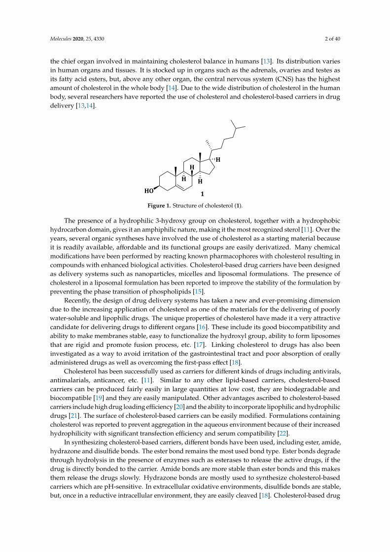
Cholesterol (Figure 1) (1) is a fatty substance or a lipid-type molecule that is essential in the humanbody and also a very important component in the structure of cell membranes [11]. It is a precursorof hormone production [12] (e.g., estrogen and testosterone) and it is used to produce bile, which isessential in aiding digestion. It is required for the function and fluidity maintenance of our nervecells. It is manufactured in all mammalian cells via the mevalonate pathway but the liver remains
Molecules 2020, 25, 4330; doi:10.3390/molecules25184330 www.mdpi.com/journal/molecules
Molecules 2020, 25, 4330 2 of 40
the chief organ involved in maintaining cholesterol balance in humans [13]. Its distribution variesin human organs and tissues. It is stocked up in organs such as the adrenals, ovaries and testes asits fatty acid esters, but, above any other organ, the central nervous system (CNS) has the highestamount of cholesterol in the whole body [14]. Due to the wide distribution of cholesterol in the humanbody, several researchers have reported the use of cholesterol and cholesterol-based carriers in drugdelivery [13,14].
Molecules 2020, 25, x FOR PEER REVIEW 2 of 41
remains the chief organ involved in maintaining cholesterol balance in humans [13]. Its distribution varies in human organs and tissues. It is stocked up in organs such as the adrenals, ovaries and testes as its fatty acid esters, but, above any other organ, the central nervous system (CNS) has the highest amount of cholesterol in the whole body [14]. Due to the wide distribution of cholesterol in the human body, several researchers have reported the use of cholesterol and cholesterol-based carriers in drug delivery [13,14].
HO
H H
HH
1
Figure 1. Structure of cholesterol (1).
The presence of a hydrophilic 3-hydroxy group on cholesterol, together with a hydrophobic hydrocarbon domain, gives it an amphiphilic nature, making it the most recognized sterol [11]. Over the years, several organic syntheses have involved the use of cholesterol as a starting material because it is readily available, affordable and its functional groups are easily derivatized. Many chemical modifications have been performed by reacting known pharmacophores with cholesterol resulting in compounds with enhanced biological activities. Cholesterol-based drug carriers have been designed as delivery systems such as nanoparticles, micelles and liposomal formulations. The presence of cholesterol in a liposomal formulation has been reported to improve the stability of the formulation by preventing the phase transition of phospholipids [15].
Recently, the design of drug delivery systems has taken a new and ever-promising dimension due to the increasing application of cholesterol as one of the materials for the delivering of poorly water-soluble and lipophilic drugs. The unique properties of cholesterol have made it a very attractive candidate for delivering drugs to different organs [16]. These include its good biocompatibility and ability to make membranes stable, easy to functionalize the hydroxyl group, ability to form liposomes that are rigid and promote fusion process, etc. [17]. Linking cholesterol to drugs has also been investigated as a way to avoid irritation of the gastrointestinal tract and poor absorption of orally administered drugs as well as overcoming the first-pass effect [18].
Cholesterol has been successfully used as carriers for different kinds of drugs including antivirals, antimalarials, anticancer, etc. [11]. Similar to any other lipid-based carriers, cholesterol-based carriers can be produced fairly easily in large quantities at low cost, they are biodegradable and biocompatible [19] and they are easily manipulated. Other advantages ascribed to cholesterol-based carriers include high drug loading efficiency [20] and the ability to incorporate lipophilic and hydrophilic drugs [21]. The surface of cholesterol-based carriers can be easily modified. Formulations containing cholesterol was reported to prevent aggregation in the aqueous environment because of their increased hydrophilicity with significant transfection efficiency and serum compatibility [22].
In synthesizing cholesterol-based carriers, different bonds have been used, including ester, amide, hydrazone and disulfide bonds. The ester bond remains the most used bond type. Ester bonds degrade through hydrolysis in the presence of enzymes such as esterases to release the active drugs, if the drug is directly bonded to the carrier. Amide bonds are more stable than ester bonds and this makes them release the drugs slowly. Hydrazone bonds are mostly used to synthesize cholesterol-based carriers which are pH-sensitive. In extracellular oxidative environments, disulfide bonds are stable, but, once in a reductive intracellular environment, they are easily cleaved [18]. Cholesterol-based drug delivery systems such as niosomes do not have a chemical reaction between the non-ionic
Figure 1. Structure of cholesterol (1).
The presence of a hydrophilic 3-hydroxy group on cholesterol, together with a hydrophobichydrocarbon domain, gives it an amphiphilic nature, making it the most recognized sterol [11]. Over theyears, several organic syntheses have involved the use of cholesterol as a starting material becauseit is readily available, affordable and its functional groups are easily derivatized. Many chemicalmodifications have been performed by reacting known pharmacophores with cholesterol resulting incompounds with enhanced biological activities. Cholesterol-based drug carriers have been designedas delivery systems such as nanoparticles, micelles and liposomal formulations. The presence ofcholesterol in a liposomal formulation has been reported to improve the stability of the formulation bypreventing the phase transition of phospholipids [15].
Recently, the design of drug delivery systems has taken a new and ever-promising dimensiondue to the increasing application of cholesterol as one of the materials for the delivering of poorlywater-soluble and lipophilic drugs. The unique properties of cholesterol have made it a very attractivecandidate for delivering drugs to different organs [16]. These include its good biocompatibility andability to make membranes stable, easy to functionalize the hydroxyl group, ability to form liposomesthat are rigid and promote fusion process, etc. [17]. Linking cholesterol to drugs has also beeninvestigated as a way to avoid irritation of the gastrointestinal tract and poor absorption of orallyadministered drugs as well as overcoming the first-pass effect [18].
Cholesterol has been successfully used as carriers for different kinds of drugs including antivirals,antimalarials, anticancer, etc. [11]. Similar to any other lipid-based carriers, cholesterol-basedcarriers can be produced fairly easily in large quantities at low cost, they are biodegradable andbiocompatible [19] and they are easily manipulated. Other advantages ascribed to cholesterol-basedcarriers include high drug loading efficiency [20] and the ability to incorporate lipophilic and hydrophilicdrugs [21]. The surface of cholesterol-based carriers can be easily modified. Formulations containingcholesterol was reported to prevent aggregation in the aqueous environment because of their increasedhydrophilicity with significant transfection efficiency and serum compatibility [22].
In synthesizing cholesterol-based carriers, different bonds have been used, including ester, amide,hydrazone and disulfide bonds. The ester bond remains the most used bond type. Ester bonds degradethrough hydrolysis in the presence of enzymes such as esterases to release the active drugs, if thedrug is directly bonded to the carrier. Amide bonds are more stable than ester bonds and this makesthem release the drugs slowly. Hydrazone bonds are mostly used to synthesize cholesterol-basedcarriers which are pH-sensitive. In extracellular oxidative environments, disulfide bonds are stable,but, once in a reductive intracellular environment, they are easily cleaved [18]. Cholesterol-based drug
Molecules 2020, 25, 4330 3 of 40
delivery systems such as niosomes do not have a chemical reaction between the non-ionic surfactantsand cholesterol but the components are rather mixed in ratios to give dispersions and gels [23,24].
In 2018, Albuquerque et al. reported recent advances in the synthesis and applications ofcholesterol-based compounds. In 2014, a review article reported synthetic cholesterol-based compoundsthrough oxidation reactions, substitution on the hydroxyl group, addition on the C5-C6 double bondand functionalizing C-H and C-C bonds. This review is focused on research work which was publishedbetween 2015 and 2018 but not reported in the 2018 review paper as well as the latest (2019 and 2020)reports on cholesterol-based carriers in drug delivery.
3. Application of Cholesterol-Based Carriers in Anticancer Drug Delivery
In 2018, the World Health Organization (WHO) reported 18.1 million new cancer cases and9.6 million cancer-related deaths making cancer one of the leading cause of death [25]. The high numberof people who die from cancer has prompted researchers to develop several classes of antitumoragents [26]. The effectiveness of most of the currently used anticancer agents is severely limited bydrug resistance. Most of the anticancer drugs fail due to drug resistance, thereby making the patientsto succumb to the disease [27].
Cholesterol-based carriers have been used in targeted anticancer drug delivery [28]. It accumulatesin the ovarian tissue where it is used for the synthesis of sex hormones. This has led to the use ofcholesteryl drug conjugates for targeting of the ovary. Research has shown that patients with ovariantumors had an eightfold uptake of labeled cholesteryl oleate by the tumorous cells compared to thenormal ones [13]. Normal cells and cancer cells have differences in substrate uptake. Due to theirhigh uncontrolled growth rate, tumors require more nutrients, which results in the overexpression ofvarious receptors such as the folate, growth factor, transferrin and Low-Density Lipoproteins (LDL)receptors. Investigations on drug delivery systems targeting these receptors have been reported bysome researchers [29].
3.1. Micelles
Polymeric micelles are promising drug delivery systems which have attracted considerable interestdue to their enhanced solubility in water and prolonged blood circulation [30,31]. Incorporationof cholesterol to any drug delivery system improves transportation across the cell membrane.Drugs encapsulated in these micelles can be delivered to specific tissues, prolonging the half-life of theincorporated drug in the blood and achieving a controlled drug release mechanism without exertingsevere toxic side effects. Bioactive agents which are incorporated into the core of the polymeric micellesinclude charged compounds, lipophilic substances and metal complexes. Micelles are structurallystable even at low concentrations [32].
The use of small interfering RNA (siRNA) is a potential therapeutic approach to treat cancer.It acts by silencing genes that contribute to drug resistance during chemotherapy. Its successful therapyis dependent on the use of delivery vehicles due to its biological instability and inefficient cellularuptake [33]. Delivering siRNA to tumor sites is a challenge because it degrades rapidly by nucleasesand it is poorly translocated due to its high negative charge. Chemical modification of siRNA hasbeen investigated to overcome its limitations, but it is still characterized by increased non-specificbinding and high toxicity. On the other hand, curcumin has long been known to be a hydrophobicchemotherapeutic cancer agent [34]. Muddineti et al. developed a curcumin loaded chitosan-cholesterolmicellar system as a potential drug–siRNA carrier for cancer combination therapy [35].
Cellular uptake studies were done by visualizing A549 cells treated with siRNA and curcuminloaded chitosan-cholesterol micelles (C-CCM/siRNA) under a fluorescence microscope. The uptakeof the micelles and their accumulation in the cytosol of A549 cells was rapid within 30 min.Thirty minutes after C-CCM and siRNA treatment of A549 cells, the Geo mean fluorescence ofA549 was 14,259 ± 246 and 19,153 ± 357, respectively, and it increased to 2245 ± 142 and 3645 ± 168,respectively, after 120 min [35]. The results revealed that the drug uptake from the micelle system

Molecules 2020, 25, 4330 4 of 40
was dependent on time. Both fluorescence results showed that the drug-loaded micelles exhibitedcellular uptake which was time-dependent. The siRNA/curcumin loaded micelles formulation wasstable at 4 ◦C over a period of one month. The results revealed that micelles prepared from chitosanand cholesterol are promising systems for combination therapy in cancer treatment [35]. However,no cytotoxicity study was investigated to fully understand the micellar system.
α-Tocopherol, a form of vitamin E and α-tocopheryl succinate (a-TOS) have also been recentlystudied as a drug delivery vehicle resulting from their ability to solubilize hydrophobic drugs [36].α-TOS induce apoptosis in various cancer cells via membrane destabilization and it is a potentialmulti-drug resistance inhibitor in cancer therapy. Muddineti et al. developed a drug delivery systemcomposed of vitamin E, a lipophilic core of cholesterol and a hydrophilic corona of polyethyleneglycol (PEG), covalently bonded by a trifunctional linker, lysine (Scheme 1) (2), which was loaded withcurcumin [37]. Micelles 2 loaded with curcumin (C-CVM) had a bigger hydrophobic compartmentsize varying in the range of 162.2–175.8 nm compared to polyethylene glycol amine phosphatidylethanolamine micelles (PPM) (20.4–24.4 nm). Micelles 2 displayed high drug encapsulation efficienciesof between 97.2% and 98.6% [37].
Molecules 2020, 25, x FOR PEER REVIEW 4 of 41
a period of one month. The results revealed that micelles prepared from chitosan and cholesterol are promising systems for combination therapy in cancer treatment [35]. However, no cytotoxicity study was investigated to fully understand the micellar system.
α-Tocopherol, a form of vitamin E and α-tocopheryl succinate (a-TOS) have also been recently studied as a drug delivery vehicle resulting from their ability to solubilize hydrophobic drugs [36]. α-TOS induce apoptosis in various cancer cells via membrane destabilization and it is a potential multi-drug resistance inhibitor in cancer therapy. Muddineti et al. developed a drug delivery system composed of vitamin E, a lipophilic core of cholesterol and a hydrophilic corona of polyethylene glycol (PEG), covalently bonded by a trifunctional linker, lysine (Scheme 1) (2), which was loaded with curcumin [37]. Micelles 2 loaded with curcumin (C-CVM) had a bigger hydrophobic compartment size varying in the range of 162.2–175.8 nm compared to polyethylene glycol amine phosphatidyl ethanolamine micelles (PPM) (20.4–24.4 nm). Micelles 2 displayed high drug encapsulation efficiencies of between 97.2% and 98.6% [37].
It exhibited reduced cell viability, good cellular uptake and sustained drug release in B16F10 and MDAMB-231 cell lines. A low hemolysis activity between 1.10% and 2.81%, which is below the acceptable range of less than 5%, was reported in the cholesterol vitamin E-based polymer, indicating the safe use of the formulation for in vivo studies. Cellular uptake studies showed that the micelles, 2 and C-PPM were taken up in dose- and time-dependent manner as the fluorescence was brighter after 4 h when compared to an hour after administration of the formulation [37]. Both micelles, 2 and C-PPM, showed no significant difference in the cellular uptake which is due to the presence of PEG-based polymeric corona. In vitro cytotoxicity evaluation on the free curcumin, CVM, PPM and micelles 2 and C-PPM in B16F10 and MDA-MB-231 cell lines revealed that PPM showed no cytotoxic effect on both cell lines with nearly 100% cell survival. However, the presence of α-TOS led to CVM showing significant cytotoxic effect [37].
BocHN
NH
O
OHFmoc
(i) - (v)
O O NH
NH
OO
HN
OChol
O
O
OO
2
Scheme 1. Synthesis of the cholesterol-based amphiphilic polymer (2). Reagents and conditions: (i) PEG5K-Amine, EDCl, NHS, TEA; (ii) 4N HCl/Dioxane, 24 h, r.t; (iii) alpha-Tocopherol succinate, EDCl, NHS, TEA, 24 h, r.t.; (iv) piperidine, 4 h, r.t.; and (v) cholesterol chloroformate, TEA, 4 h, r.t.
Cytotoxic study profiles of C-PPM, free curcumin and micelles 2 for 6 h followed by incubation for 24 h were 48.7%, 58.2% and 42.8, respectively, in B16F10 cells and 74.2%, 59.6% and 58.2%, respectively, in MDA-MB-231 cells. The cell viabilities demonstrated by CVM and PPM (at a concentration of 50 µg/mL) were 42.4% and 91.7%, respectively, in B16F10, 45.2% and 92.3% in MDA-MB-231 cells, respectively. The results showed significant cytotoxicity induced by micelles 2 and this was due to the synergism between α-TOS-conjugated CVM and curcumin. Growth inhibition results showed that the cellular uptake of micelles 2 into the tumor spheroids inhibited cell proliferation
Scheme 1. Synthesis of the cholesterol-based amphiphilic polymer (2). Reagents and conditions:(i) PEG5K-Amine, EDCl, NHS, TEA; (ii) 4N HCl/Dioxane, 24 h, r.t; (iii) alpha-Tocopherol succinate,EDCl, NHS, TEA, 24 h, r.t.; (iv) piperidine, 4 h, r.t.; and (v) cholesterol chloroformate, TEA, 4 h, r.t.
It exhibited reduced cell viability, good cellular uptake and sustained drug release in B16F10and MDAMB-231 cell lines. A low hemolysis activity between 1.10% and 2.81%, which is below theacceptable range of less than 5%, was reported in the cholesterol vitamin E-based polymer, indicatingthe safe use of the formulation for in vivo studies. Cellular uptake studies showed that the micelles,2 and C-PPM were taken up in dose- and time-dependent manner as the fluorescence was brighter after4 h when compared to an hour after administration of the formulation [37]. Both micelles, 2 and C-PPM,showed no significant difference in the cellular uptake which is due to the presence of PEG-basedpolymeric corona. In vitro cytotoxicity evaluation on the free curcumin, CVM, PPM and micelles 2and C-PPM in B16F10 and MDA-MB-231 cell lines revealed that PPM showed no cytotoxic effect onboth cell lines with nearly 100% cell survival. However, the presence of α-TOS led to CVM showingsignificant cytotoxic effect [37].
Cytotoxic study profiles of C-PPM, free curcumin and micelles 2 for 6 h followed by incubation for24 h were 48.7%, 58.2% and 42.8, respectively, in B16F10 cells and 74.2%, 59.6% and 58.2%, respectively,in MDA-MB-231 cells. The cell viabilities demonstrated by CVM and PPM (at a concentration of50 µg/mL) were 42.4% and 91.7%, respectively, in B16F10, 45.2% and 92.3% in MDA-MB-231 cells,
Molecules 2020, 25, 4330 5 of 40
respectively. The results showed significant cytotoxicity induced by micelles 2 and this was due to thesynergism between α-TOS-conjugated CVM and curcumin. Growth inhibition results showed thatthe cellular uptake of micelles 2 into the tumor spheroids inhibited cell proliferation resulting in anenhanced therapeutic effect. The developed micelles 2 are potential therapeutics for the treatment ofdrug-resistant tumors [37].
Sarkar et al. synthesized cholesterol-based hydrazone (CBH) tethered tiny amphiphiles withdifferent carbonyl residues (Figure 2) such as benzaldehyde (3a), p-dimethylaminobenzaldehyde (3b)and benzophenone (3c). Compound 3a was the most stable of the three and its drug loading efficiencyof doxorubicin (DOX) was 57%. Drug release studies of the DOX-loaded compound 3a showed thatbelow the pH of 6.5, the drug-loaded vesicles released DOX better than at pH 7.0–8.0, possibly due tothe hydrazone bond cleavage [38]. The release pattern is important since most tumor cells have boththeir intracellular and extracellular pH below 6.5 [17], meaning the anticancer drug is released right atthe site of action enhancing its efficacy.
Molecules 2020, 25, x FOR PEER REVIEW 5 of 41
resulting in an enhanced therapeutic effect. The developed micelles 2 are potential therapeutics for the treatment of drug-resistant tumors [37].
Sarkar et al. synthesized cholesterol-based hydrazone (CBH) tethered tiny amphiphiles with different carbonyl residues (Figure 2) such as benzaldehyde (3a), p-dimethylaminobenzaldehyde (3b) and benzophenone (3c). Compound 3a was the most stable of the three and its drug loading efficiency of doxorubicin (DOX) was 57%. Drug release studies of the DOX-loaded compound 3a showed tha,t below the pH of 6.5, the drug-loaded vesicles released DOX better than at pH 7.0–8.0, possibly due to the hydrazone bond cleavage [38]. The release pattern is important since most tumor cells have both their intracellular and extracellular pH below 6.5 [17], meaning the anticancer drug is released right at the site of action enhancing its efficacy.
O
NH
OO
HN O
CholO
NH
NR 3a-c
R:
NR:
3a
3b
3c R:
Figure 2. Chemical structures of cholesterol-based hydrazone tethered amphiphiles, compounds 3a–c.
3.2. Nanoparticles
Liposomes have been used as a drug delivery system but suffer from drawbacks such as high leakage of the encapsulated drugs, high production cost, fast clearance from circulation and short stability [39]. Heidari et al. prepared titania nanotubes (TNTs) in which 5-fluorouracil, a chemotherapeutic drug, was filled into the cylindrical empty space. Liposomes made of soy lecithin and cholesterol (Scheme 2) were used to coat the drug-loaded TNTs capping the nanotube hole to prevent drug leakage. The drug loading was 100% in the nanotubes. The rate of release of 5-fluorouracil was influenced by the liposomal layers coating on the surface of the nanotubes. At TNT concentrations of <300 µg/mL and between 300 and 1500 µg/mL, about 90% and 80% of the HeLa cells were alive, respectively [40]. The highest toxicity was noted above 3000 µg/mL [41]. Cytotoxicity studies revealed that 5-fluorouracil loaded with compound 4 had a decreased IC50 of 250 µg/mL when compared to about 470 µg/mL for the free 5-fluorouracil. Compound 4 displayed enhanced cellular internalization with effective delivery of 5-fluorouracil into the cells [40].
Figure 2. Chemical structures of cholesterol-based hydrazone tethered amphiphiles, compounds 3a–c.
3.2. Nanoparticles
Liposomes have been used as a drug delivery system but suffer from drawbacks such ashigh leakage of the encapsulated drugs, high production cost, fast clearance from circulation andshort stability [39]. Heidari et al. prepared titania nanotubes (TNTs) in which 5-fluorouracil,a chemotherapeutic drug, was filled into the cylindrical empty space. Liposomes made of soylecithin and cholesterol (Scheme 2) were used to coat the drug-loaded TNTs capping the nanotubehole to prevent drug leakage. The drug loading was 100% in the nanotubes. The rate of release of5-fluorouracil was influenced by the liposomal layers coating on the surface of the nanotubes. At TNTconcentrations of <300 µg/mL and between 300 and 1500 µg/mL, about 90% and 80% of the HeLacells were alive, respectively [40]. The highest toxicity was noted above 3000 µg/mL [41]. Cytotoxicitystudies revealed that 5-fluorouracil loaded with compound 4 had a decreased IC50 of 250 µg/mL whencompared to about 470 µg/mL for the free 5-fluorouracil. Compound 4 displayed enhanced cellularinternalization with effective delivery of 5-fluorouracil into the cells [40].
DOX can be readily loaded into DNA nanoparticles for drug delivery [42]. Choi et al. synthesizedamphiphilic DNA-cholesterol/DNA-peptide hybrid duplex that assembled into DNA nanoparticles(c-DNA-p nanoparticles) in an aqueous solution. The binding efficiency studies were done by analyzingthe fluorescent bands of free DOX and DOX/c-DNA-p complexes on 2% agarose gel electrophoresis.That 0.5 mM of the nanoparticles fully complexed with 3.5 mM DOX revealed the absence of freeDOX, indicating a simple and efficient method of loading DOX into the nanoparticles. DOX wasfully bound to c-DNA-p nanoparticles at neutral pH 7.4 but the drug began to dissociate from theDOX/c-DNA-p complex at acidic pH of 6.5. At pH 5.0, most of the DOX dissociated from the complex

Molecules 2020, 25, 4330 6 of 40
thus indicating the lowering of the binding affinity between DOX and the c-DNA-p nanoparticleswhich could facilitate cytosolic DOX release [43].Molecules 2020, 25, x FOR PEER REVIEW 6 of 41
O O
O
OO+ Chol + HO
OH
PEG
P
N
14 7
7
O
O
O O
O
OO
P
N
14 7
7
O
O(i)
HOO
H
Chol
4
Scheme 2. Synthetic route for liposomes containing lecithin, PEG and cholesterol (4). Reagents and conditions: (i) soy lecithin, chol, ethanol, stir, 20 min, PEG, stir, 24 h, 25 °C.
DOX can be readily loaded into DNA nanoparticles for drug delivery [42]. Choi et al. synthesized amphiphilic DNA-cholesterol/DNA-peptide hybrid duplex that assembled into DNA nanoparticles (c-DNA-p nanoparticles) in an aqueous solution. The binding efficiency studies were done by analyzing the fluorescent bands of free DOX and DOX/c-DNA-p complexes on 2% agarose gel electrophoresis. That 0.5 mM of the nanoparticles fully complexed with 3.5 mM DOX revealed the absence of free DOX, indicating a simple and efficient method of loading DOX into the nanoparticles. DOX was fully bound to c-DNA-p nanoparticles at neutral pH 7.4 but the drug began to dissociate from the DOX/c-DNA-p complex at acidic pH of 6.5. At pH 5.0, most of the DOX dissociated from the complex thus indicating the lowering of the binding affinity between DOX and the c-DNA-p nanoparticles which could facilitate cytosolic DOX release [43].
Nanoparticle instability and limited drug loading capacity in most cholesterol-containing polymeric nanoparticles lead to premature drug release into the plasma before reaching the tumor sites. Gonzalez-Fajardo et al. designed and synthesized a drug vehicle, P(NBCh9-bNBPEG), with several cholesteric and PEG side chains as hydrophobic and hydrophilic blocks, respectively. The brush-like architecture had the ability to form stable nanoparticles for the encapsulation of hydrophobic drugs [44]. DOX was loaded into the P(NBCh9-b-NBPEG) nanoparticles (DOX-NPs) and in vivo studies were performed in a xenograft mouse tumor model. There was a significantly reduced tumor growth without side effects when the mice were treated with DOX-NPs. The low release of DOX from the nanoparticles in the blood as well as the reduced accumulation in the major organs like liver, spleen and heart revealed the improved safety profile of DOX-NPs when compared to the free drugs. The results revealed the potential of P(NBCh9-b-NBPEG) nanoparticles for the delivery of hydrophobic drugs with reduced toxicity [44].
Nguyen et al. synthesized a novel amphiphilic cholesterol-based block polymer comprising of polymethacrylate with cholesterol and PEG having reducible disulfide bonds (PC5MA-SS-PEO) (Figure 3) (5) as a nanoparticulate redox-sensitive delivery system. DOX was encapsulated to the self-assembled nanoparticles and the drug loading was 18% w/w with 95% drug encapsulation efficiency. The DOX-loaded nanoparticles exhibited excellent stability and readily released DOX in dithiothreitol reductive conditions. Notably, the reducible nanoparticles 5 rapidly released DOX inside the tumor cells thereby inducing higher toxicity when compared to the non-reducible PC5MA-PEO-thioester nanoparticles. The in vivo images taken from A549 tumor-bearing severe combined immunodeficiency mice showed that the nanoparticles 5 accumulated preferably in the tumor tissues
Scheme 2. Synthetic route for liposomes containing lecithin, PEG and cholesterol (4). Reagents andconditions: (i) soy lecithin, chol, ethanol, stir, 20 min, PEG, stir, 24 h, 25 ◦C.
Nanoparticle instability and limited drug loading capacity in most cholesterol-containingpolymeric nanoparticles lead to premature drug release into the plasma before reaching the tumor sites.Gonzalez-Fajardo et al. designed and synthesized a drug vehicle, P(NBCh9-bNBPEG), with severalcholesteric and PEG side chains as hydrophobic and hydrophilic blocks, respectively. The brush-likearchitecture had the ability to form stable nanoparticles for the encapsulation of hydrophobic drugs [44].DOX was loaded into the P(NBCh9-b-NBPEG) nanoparticles (DOX-NPs) and in vivo studies wereperformed in a xenograft mouse tumor model. There was a significantly reduced tumor growthwithout side effects when the mice were treated with DOX-NPs. The low release of DOX from thenanoparticles in the blood as well as the reduced accumulation in the major organs like liver, spleen andheart revealed the improved safety profile of DOX-NPs when compared to the free drugs. The resultsrevealed the potential of P(NBCh9-b-NBPEG) nanoparticles for the delivery of hydrophobic drugswith reduced toxicity [44].
Nguyen et al. synthesized a novel amphiphilic cholesterol-based block polymer comprisingof polymethacrylate with cholesterol and PEG having reducible disulfide bonds (PC5MA-SS-PEO)(Figure 3) (5) as a nanoparticulate redox-sensitive delivery system. DOX was encapsulated to theself-assembled nanoparticles and the drug loading was 18% w/w with 95% drug encapsulation efficiency.The DOX-loaded nanoparticles exhibited excellent stability and readily released DOX in dithiothreitolreductive conditions. Notably, the reducible nanoparticles 5 rapidly released DOX inside the tumorcells thereby inducing higher toxicity when compared to the non-reducible PC5MA-PEO-thioesternanoparticles. The in vivo images taken from A549 tumor-bearing severe combined immunodeficiencymice showed that the nanoparticles 5 accumulated preferably in the tumor tissues when comparedto the healthy cells/tissues revealing that they are promising delivery systems for the delivery ofanticancer drugs with reduced toxicity [45].

Molecules 2020, 25, 4330 7 of 40
Molecules 2020, 25, x FOR PEER REVIEW 7 of 41
when compared to the healthy cells/tissues revealing that they are promising delivery systems for the delivery of anticancer drugs with reduced toxicity [45].
O O S S O S S C12H25
O
O O O
O
O
Chol
S
x
y
z
5
Figure 3. Structure of cholesterol-based block copolymer with disulfide linkage (5).
Abraxane is a nanoparticle composite of paclitaxel (PTX) and human serum albumin (HSA), approved for cancer [46,47]. The poor colloidal stability of abraxane in the blood has been found not to improve PTX serum half-life [48,49]. To circumvent the aforementioned problem, Battogtokh et al. synthesized cholesteryl bovine serum albumin (Chol-BSA) (Scheme 3) nanoparticles (6) as PTX carriers. When loaded with PTX, nanoparticles 6 showed a 94.8% drug loading efficiency and 37.9% drug loading capacity making the formulation a potential drug carrier than its closest competitors [50]. The nanoparticles had an optimum size of 150 nm and a negative charge surface making them stable in aqueous medium [51]. The release of PTX was two-fold slower from PTX-loaded nanoparticle 6 when compared to PTX-BSA over a period of 72h at 37 °C, showing the sustained drug release from nanoparticles 6. When incubated with B16F10 cells, there was 2- and 0.5-fold increase in PTX uptake from the PTX-loaded nanoparticles 6 when compared to PTX-Cre/EtOH and PTX–BSA, respectively. After incubating PTX-loaded nanoparticles 6 with MCF-7 cells for 1 h, the uptake of the nanoparticles was reported to be 3.5- and 1.4-fold higher when compared to PTX-Cre/EtOH and PTX–BSA, respectively. These findings revealed the capability of the PTX-loaded nanoparticles 6 in delivering more drugs to the cells [50].
N
O
O
O O N
O
O
O
Chol
N,N-disuccinimidyl Carbonate
NO
OO
O
ONHS-Chol
BSA
NH2
NH2H2N
HN O
NH
O NH
O
OO
O
(i)
Chol
Chol
Chol
Chol
(ii)
6
Scheme 3. Synthesis of cholesteryl bovine serum albumin nanoparticles (6): Reagents and conditions: (i) THF, DSC, DMAP, 24 h, rt under argon; and (ii) THF, stir, 12 h.
Figure 3. Structure of cholesterol-based block copolymer with disulfide linkage (5).
Abraxane is a nanoparticle composite of paclitaxel (PTX) and human serum albumin (HSA),approved for cancer [46,47]. The poor colloidal stability of abraxane in the blood has been found notto improve PTX serum half-life [48,49]. To circumvent the aforementioned problem, Battogtokh etal. synthesized cholesteryl bovine serum albumin (Chol-BSA) (Scheme 3) nanoparticles (6) as PTXcarriers. When loaded with PTX, nanoparticles 6 showed a 94.8% drug loading efficiency and 37.9%drug loading capacity making the formulation a potential drug carrier than its closest competitors [50].The nanoparticles had an optimum size of 150 nm and a negative charge surface making them stablein aqueous medium [51]. The release of PTX was two-fold slower from PTX-loaded nanoparticle6 when compared to PTX-BSA over a period of 72h at 37 ◦C, showing the sustained drug releasefrom nanoparticles 6. When incubated with B16F10 cells, there was 2- and 0.5-fold increase inPTX uptake from the PTX-loaded nanoparticles 6 when compared to PTX-Cre/EtOH and PTX–BSA,respectively. After incubating PTX-loaded nanoparticles 6 with MCF-7 cells for 1 h, the uptake ofthe nanoparticles was reported to be 3.5- and 1.4-fold higher when compared to PTX-Cre/EtOH andPTX–BSA, respectively. These findings revealed the capability of the PTX-loaded nanoparticles 6 indelivering more drugs to the cells [50].
Molecules 2020, 25, x FOR PEER REVIEW 7 of 41
when compared to the healthy cells/tissues revealing that they are promising delivery systems for the delivery of anticancer drugs with reduced toxicity [45].
O O S S O S S C12H25
O
O O O
O
O
Chol
S
x
y
z
5
Figure 3. Structure of cholesterol-based block copolymer with disulfide linkage (5).
Abraxane is a nanoparticle composite of paclitaxel (PTX) and human serum albumin (HSA), approved for cancer [46,47]. The poor colloidal stability of abraxane in the blood has been found not to improve PTX serum half-life [48,49]. To circumvent the aforementioned problem, Battogtokh et al. synthesized cholesteryl bovine serum albumin (Chol-BSA) (Scheme 3) nanoparticles (6) as PTX carriers. When loaded with PTX, nanoparticles 6 showed a 94.8% drug loading efficiency and 37.9% drug loading capacity making the formulation a potential drug carrier than its closest competitors [50]. The nanoparticles had an optimum size of 150 nm and a negative charge surface making them stable in aqueous medium [51]. The release of PTX was two-fold slower from PTX-loaded nanoparticle 6 when compared to PTX-BSA over a period of 72h at 37 °C, showing the sustained drug release from nanoparticles 6. When incubated with B16F10 cells, there was 2- and 0.5-fold increase in PTX uptake from the PTX-loaded nanoparticles 6 when compared to PTX-Cre/EtOH and PTX–BSA, respectively. After incubating PTX-loaded nanoparticles 6 with MCF-7 cells for 1 h, the uptake of the nanoparticles was reported to be 3.5- and 1.4-fold higher when compared to PTX-Cre/EtOH and PTX–BSA, respectively. These findings revealed the capability of the PTX-loaded nanoparticles 6 in delivering more drugs to the cells [50].
N
O
O
O O N
O
O
O
Chol
N,N-disuccinimidyl Carbonate
NO
OO
O
ONHS-Chol
BSA
NH2
NH2H2N
HN O
NH
O NH
O
OO
O
(i)
Chol
Chol
Chol
Chol
(ii)
6
Scheme 3. Synthesis of cholesteryl bovine serum albumin nanoparticles (6): Reagents and conditions: (i) THF, DSC, DMAP, 24 h, rt under argon; and (ii) THF, stir, 12 h.
Scheme 3. Synthesis of cholesteryl bovine serum albumin nanoparticles (6): Reagents and conditions:(i) THF, DSC, DMAP, 24 h, rt under argon; and (ii) THF, stir, 12 h.
Cytotoxicity studies revealed that B16F10 and MCF-7 cells treated with 10 and 100 nM PTX-loadednanoparticles 6 for 48 h exhibited lower cell viability when compared to the same cell lines that were
Molecules 2020, 25, 4330 8 of 40
treated with PTX-Cre/EtOH and PTX–BSA. No cytotoxic effect was displayed by nanoparticles 6.In vivo antitumor studies showed that the tumor volume in mouse model was smaller than that ofmice administered with saline and other groups. Tumor volume of PTX-loaded nanoparticles 6 wasreported to be 46.94% at day 8 after administration, compared to 78.23% and 77.35% of PTX-Cre/EtOHand PTX–BSA, respectively. The results indicated that PTX anti-tumor effect can be enhanced usingnanoparticles 6 as a nanocarrier [50].
Tamoxifen (TMX) is a drug used to treat estrogen receptor and progesterone receptor breastcancer [52]. To improve its cell penetration, entrapment and pharmacokinetics, Mazumdar et al.developed mPEG-b-(CB-{g-chol}-co-LA), a self-assembling cholesterol grafted lipopolymer, synthesizedfrom poly(ethyleneglycol)-block-2-methyl-2 carboxylpropylenecarboxylic acid-co-poly (L-lactide)[mPEG-b-(CB-{g-COOH}-co-LA)] copolymer followed by the incorporation of cholesterol throughcarbodiimide coupling. TMX release studies were done at pH 7.4 and 5.6, with the latter representingthe acidic tumor medium. After 96 h at pH 7.4, 72% of TMX was released from [mPEG-b-(CB-{g-COOH}-co-LA)] while 55% was released from methoxy–poly (ethylene glycol)-poly(D,L-Lactide) (mPEG-PLA)(used for comparison). After 96 h of incubation at pH 5.6, about 97% of TMX was released from bothnanoparticles [53].
The TMX loaded mPEG-b-(CB-{g-chol}-coLA) lipopolymeric nanoparticles displayed highercellular uptake efficiency and improved IC50 values of 22.2 µM in the breast cancer cell lines in 4T1and 18.8 µM in MCF-7 compared to IC50 values of 27.6 µM and 23.5 µM, respectively, for the free TMX.The results also showed that at the above IC50 values, TMX loaded lipopolymeric nanoparticles inducedcell death and arrested cell cycle at G0/G1 phase at the same rate as the free TMX. The pharmacokineticsresults showed approximately 2.5- and 2.7-fold increase in the half-life and mean residence time,respectively, of TMX after incorporating it into lipopolymeric nanoparticles. The findings indicatethat mPEG-b-(CB-{gchol}-co-LA) lipopolymeric nanoparticles are useful for the internalization andenhanced residence time in breast cancer cell lines with reduced drug dose and side effects [53].
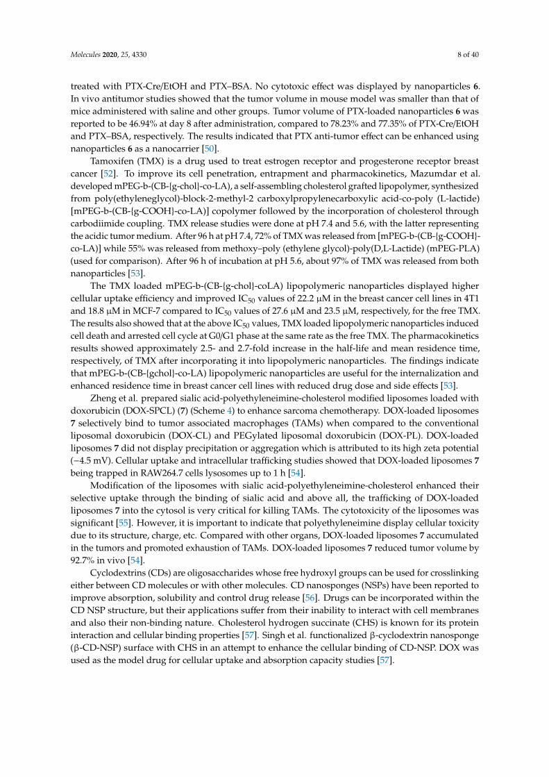
Zheng et al. prepared sialic acid-polyethyleneimine-cholesterol modified liposomes loaded withdoxorubicin (DOX-SPCL) (7) (Scheme 4) to enhance sarcoma chemotherapy. DOX-loaded liposomes7 selectively bind to tumor associated macrophages (TAMs) when compared to the conventionalliposomal doxorubicin (DOX-CL) and PEGylated liposomal doxorubicin (DOX-PL). DOX-loadedliposomes 7 did not display precipitation or aggregation which is attributed to its high zeta potential(−4.5 mV). Cellular uptake and intracellular trafficking studies showed that DOX-loaded liposomes 7being trapped in RAW264.7 cells lysosomes up to 1 h [54].
Modification of the liposomes with sialic acid-polyethyleneimine-cholesterol enhanced theirselective uptake through the binding of sialic acid and above all, the trafficking of DOX-loadedliposomes 7 into the cytosol is very critical for killing TAMs. The cytotoxicity of the liposomes wassignificant [55]. However, it is important to indicate that polyethyleneimine display cellular toxicitydue to its structure, charge, etc. Compared with other organs, DOX-loaded liposomes 7 accumulatedin the tumors and promoted exhaustion of TAMs. DOX-loaded liposomes 7 reduced tumor volume by92.7% in vivo [54].
Cyclodextrins (CDs) are oligosaccharides whose free hydroxyl groups can be used for crosslinkingeither between CD molecules or with other molecules. CD nanosponges (NSPs) have been reported toimprove absorption, solubility and control drug release [56]. Drugs can be incorporated within theCD NSP structure, but their applications suffer from their inability to interact with cell membranesand also their non-binding nature. Cholesterol hydrogen succinate (CHS) is known for its proteininteraction and cellular binding properties [57]. Singh et al. functionalized β-cyclodextrin nanosponge(β-CD-NSP) surface with CHS in an attempt to enhance the cellular binding of CD-NSP. DOX wasused as the model drug for cellular uptake and absorption capacity studies [57].
Molecules 2020, 25, 4330 9 of 40Molecules 2020, 25, x FOR PEER REVIEW 9 of 41
NNH2
NH
NN
HN
NNH2H2N
NH
NH2
n
Polyethylenimine (PEI)
Cl OChol
O
NNH2
NH
NN
HN
NNHH2N
NH
NH2
n
Chol-chloroformate(CH)
OChol
O
PEI-CH
(i)
O OH
OHO
HO
HOOHOH
HN
OSialic acid (SA)
NNH
NH
NN
HN
NNHHN
NH
HN
n
OChol
O
O OH
O
HO
HOOHOH
HN
O
O
OHO
NHOHOH
OHOH
O
O
HO O
OHNH
O
OHOHOH
(ii)
7
Scheme 4. Synthesis of SA-PEI-CH conjugates (7). Reagents and conditions: (i) TEA, DCM; and (ii) EDC/NHS, Formamide, stir, 0.5 h, stir, 12 h under nitrogen.
Modification of the liposomes with sialic acid-polyethyleneimine-cholesterol enhanced their selective uptake through the binding of sialic acid and above all, the trafficking of DOX-loaded liposomes 7 into the cytosol is very critical for killing TAMs. The cytotoxicity of the liposomes was significant [55]. However, it is important to indicate that polyethyleneimine display cellular toxicity due to its structure, charge, etc. Compared with other organs, DOX-loaded liposomes 7 accumulated in the tumors and promoted exhaustion of TAMs. DOX-loaded liposomes 7 reduced tumor volume by 92.7% in vivo [54].
Cyclodextrins (CDs) are oligosaccharides whose free hydroxyl groups can be used for crosslinking either between CD molecules or with other molecules. CD nanosponges (NSPs) have been reported to improve absorption, solubility and control drug release [56]. Drugs can be incorporated within the CD NSP structure, but their applications suffer from their inability to interact with cell membranes and also their non-binding nature. Cholesterol hydrogen succinate (CHS) is known for its protein interaction and cellular binding properties [57]. Singh et al. functionalized β-cyclodextrin nanosponge (β-CD-NSP) surface with CHS in an attempt to enhance the cellular binding of CD-NSP. DOX was used as the model drug for cellular uptake and absorption capacity studies [57].
β-CD-NSP-CHS exhibited a 5.8% higher drug adsorption capacity when compared to β-CD-NSP. The enhanced drug adsorption is attributed to the hydrophobic nature of the surface. The DOX release profiles at pH 6.8 and 1.2 of β-CD-NSP-DOX and β-CD-NSP-CHS-DOX were the same, suggesting that CHS grafting did not change the release pattern. The drug release from both
Scheme 4. Synthesis of SA-PEI-CH conjugates (7). Reagents and conditions: (i) TEA, DCM; and (ii)EDC/NHS, Formamide, stir, 0.5 h, stir, 12 h under nitrogen.
β-CD-NSP-CHS exhibited a 5.8% higher drug adsorption capacity when compared to β-CD-NSP.The enhanced drug adsorption is attributed to the hydrophobic nature of the surface. The DOX releaseprofiles at pH 6.8 and 1.2 of β-CD-NSP-DOX and β-CD-NSP-CHS-DOX were the same, suggestingthat CHS grafting did not change the release pattern. The drug release from both nanosponges at pH1.2 was fast with over 82% of the drug released within 15 min when compared to phosphate buffer atpH 6.8 in which less than 30% of the drug diffused from the NSP [57].
Cytotoxicity studies revealed that β-CD-NSP-CHS exerted no cytotoxic effects in HeLa cells evenat a high sample concentration of 100 µg/mL. The surface-modified β-CD-NSP was biocompatible andsafe in drug delivery. Laser scanning confocal microscopic images showed high DOX accumulationin HeLa cells incubated with DOX-loaded β-CD-NSP-CHS samples when compared to HeLa cellstreated with free DOX. This finding indicate that CHS played an important role in enhancing the drugpenetration and cell internalization [58]. The surface modified CD-NSP is useful in the delivering ofsmall low water-soluble drugs thereby improving both their solubility and bioavailability [57].
Mallick et al. prepared liposomes from cholesterol-phenylalanine-arginine-phenylalanine-lysine(Chol-FRFK) sequence and 1,2-dioleoyl-sn-glycero-3-phoshphoethanolamine (DOPE) was used toformulate (Chol-FRFK/D liposomes) for delivery of an anticancer drug, Antimycin A (AMA), to themitochondria. The Chol-FRFK/D liposomes had a higher cellular uptake and mitochondrial targetingcompared to the two controls, DQAsomes and DOTAP/DOPE (D/D) liposomes. Treating A549 cellswith Chol-FRFK/D liposomes showed an IC50 of 10 µM compared to an IC50 of 50 µM for free AMA [15].Drug encapsulation in the Chol-FRFK/D liposomes was 100% since AMA could be accommodatedboth in the lipid bilayer membrane and the inside of the core. The Chol-FRFK/D-AMA formulation
Molecules 2020, 25, 4330 10 of 40
was very cytotoxic that it caused mitochondria-mediated apoptosis in A549 cells. The Chol-FRFK/Dliposomes are a promising anticancer delivery system for anticancer therapy [15].
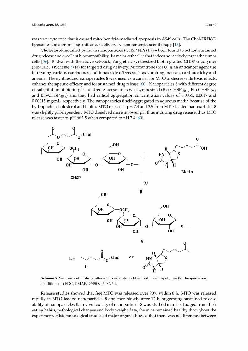
Cholesterol-modified pullulan nanoparticles (CHSP NPs) have been found to exhibit sustaineddrug release and excellent biocompatibility. Its major setback is that it does not actively target the tumorcells [59]. To deal with the above set-back, Yang et al. synthesized biotin grafted CHSP copolymer(Bio-CHSP) (Scheme 5) (8) for targeted drug delivery. Mitoxantrone (MTO) is an anticancer agent usein treating various carcinomas and it has side effects such as vomiting, nausea, cardiotoxicity andanemia. The synthesized nanoparticles 8 was used as a carrier for MTO to decrease its toxic effects,enhance therapeutic efficacy and for sustained drug release [60]. Nanoparticles 8 with different degreeof substitution of biotin per hundred glucose units was synthesized (Bio-CHSP-20.1, Bio-CHSP-29.2
and Bio-CHSP-38.9) and they had critical aggregation concentration values of 0.0055, 0.0017 and0.00015 mg/mL, respectively. The nanoparticles 8 self-aggregated in aqueous media because of thehydrophobic cholesterol and biotin. MTO release at pH 7.4 and 3.5 from MTO-loaded nanoparticles 8was slightly pH-dependent. MTO dissolved more in lower pH thus inducing drug release, thus MTOrelease was faster in pH of 3.5 when compared to pH 7.4 [60].Molecules 2020, 25, x FOR PEER REVIEW 11 of 41
O
OH
OCH2
O OChol
O O
OHO
OH
OHO
OHOH
OHO
OH
O
CHSP
HN
NH
SH
HO
OH
O
Biotin
(i)
O
OH
OCH2OHO
OH
OHO
OHOH
OHO
OH
O
OR
OChol
O
O
or HN
NH
SH
HO
O
R =
8
Scheme 5. Synthesis of Biotin grafted- Cholesterol-modified pullulan co-polymer (8). Reagents and
conditions: (i) EDC, DMAP, DMSO, 45 °C, 5d.
Release studies showed that free MTO was released over 90% within 8 h. MTO was released rapidly in MTO-loaded nanoparticles 8 and then slowly after 12 h, suggesting sustained release ability of nanoparticles 8. In vivo toxicity of nanoparticles 8 was studied in mice. Judged from their eating habits, pathological changes and body weight data, the mice remained healthy throughout the experiment. Histopathological studies of major organs showed that there was no difference between the control and those administered with nanoparticles 8. This means that the nanoparticles did not induce acute toxicity after intravenous administration at a dose of 200 mg/kg. Together with the other in vitro results, nanoparticles 8 was biocompatible and safe for hydrophobic drug delivery [60].
Xu et al. developed novel cholesterol/imidazole modified oxidized-starch (Cho-Imi-OS) (Scheme 6) (9) nanoparticles as a pH-sensitive and neutrally charged tumor-targeted drug delivery system, using curcumin as a model drug. The curcumin-loaded nanoparticles 9 drug loading and entrapment efficiencies were 4.16% and 17.84%, respectively. Curcumin was released more quickly at pH 5.5 (65% cumulative release) than at pH 7.4 (38% cumulative release) after 68 h. The faster cumulative release at pH 5.5 is very important since the pH in tumor cells endosomes is between 5.0 and 6.0, meaning most of the curcumin is released right at the tumor site for effective therapy [61].
Scheme 5. Synthesis of Biotin grafted- Cholesterol-modified pullulan co-polymer (8). Reagents andconditions: (i) EDC, DMAP, DMSO, 45 ◦C, 5d.
Release studies showed that free MTO was released over 90% within 8 h. MTO was releasedrapidly in MTO-loaded nanoparticles 8 and then slowly after 12 h, suggesting sustained releaseability of nanoparticles 8. In vivo toxicity of nanoparticles 8 was studied in mice. Judged from theireating habits, pathological changes and body weight data, the mice remained healthy throughout theexperiment. Histopathological studies of major organs showed that there was no difference between

Molecules 2020, 25, 4330 11 of 40
the control and those administered with nanoparticles 8. This means that the nanoparticles did notinduce acute toxicity after intravenous administration at a dose of 200 mg/kg. Together with the otherin vitro results, nanoparticles 8 was biocompatible and safe for hydrophobic drug delivery [60].
Xu et al. developed novel cholesterol/imidazole modified oxidized-starch (Cho-Imi-OS) (Scheme 6)(9) nanoparticles as a pH-sensitive and neutrally charged tumor-targeted drug delivery system,using curcumin as a model drug. The curcumin-loaded nanoparticles 9 drug loading and entrapmentefficiencies were 4.16% and 17.84%, respectively. Curcumin was released more quickly at pH 5.5(65% cumulative release) than at pH 7.4 (38% cumulative release) after 68 h. The faster cumulativerelease at pH 5.5 is very important since the pH in tumor cells endosomes is between 5.0 and 6.0,meaning most of the curcumin is released right at the tumor site for effective therapy [61].Molecules 2020, 25, x FOR PEER REVIEW 12 of 41
O
O
OH
OHOH O
O
OHOH O
O
IO
OH
OHOH
starch
(i) O
O
OH
OHOH O
O
O
O
O
OH
OHOH
OO
0xidized-starch (OS)
ClChol
O
O
O
O
OOH O
OO
O
O
OHOH
N
NN
N
Chol
Chol
Chol
(ii)
9 Scheme 6. Synthetic route for Cho-Imi-OS (9). Reagents and conditions: (i) NaIO4, 3 h, 37 °C, dark; and (ii) Imidazole, 100 °C, 1 h, stir.
The nanoparticles without a drug and curcumin-loaded nanoparticles 9 showed hemolysis ratios of 1% and 2%, respectively, which are all within the standard level of 5%. A549 cells effective uptake of curcumin-loaded nanoparticles 9, when compared to free curcumin, was attributed to the nano-size of the curcumin-loaded nanoparticles 9. Curcumin-loaded nanoparticles 9 exhibited an IC50 of 4.2 µg/mL, and the nanoparticles not loaded with drug maintained cell viability of more than 85% [61].
Wu et al. prepared liposomes with hydrogenated soybean phospholipids (HSPC), 1,2-distearoyl-sn-glycero-3-phosphoethanolamine-N-[methoxy(polyethylene glycol)-2000] (DSPE-PEG2000) and varying amounts of cholesterol (Table 1) and loaded them with doxorubicin. The rate of drug release decreased as the cholesterol content in the liposomes increased. Liposomal cellular uptake in BxPC-3 and HPaSteC cells decreased with increase in the liposomal cholesterol content. The authors reported a decrease in the membrane rigidity of the liposomes as the cholesterol content increased. The aforementioned findings suggest that when the liposomal membranes are less rigid, there is an increase in surface contact with the cells resulting in a prolonged cellular uptake [62].
Table 1. Characterization of liposomes with different cholesterol content.
Preparation Composition (Molar
Ratio) LC (%) of
DOX Diffusivity
(µm2/s) Ratio of
Diffusivity Lip1 97.5:-:2.5 10.6 0.14 1 Lip2 77.5:20.0:2.5 10.8 0.75 5.4 Lip3 67.5:30.0:2.2 11.0 1.55 11.1 Lip4 57.5:40.0:2.5 11.1 1.13 8.1 Lip5 47.5:50.0:2.5 1 0.9 0.35 2.5 Lip6 56.3:38.4:5.3 11.0 1.72 12.2 LC (%), drug loading content; -, not applicable. Compositions of the liposomes were HSPC:cholesterol:DSPE-PEG2000.
Scheme 6. Synthetic route for Cho-Imi-OS (9). Reagents and conditions: (i) NaIO4, 3 h, 37 ◦C, dark;and (ii) Imidazole, 100 ◦C, 1 h, stir.
The nanoparticles without a drug and curcumin-loaded nanoparticles 9 showed hemolysis ratiosof 1% and 2%, respectively, which are all within the standard level of 5%. A549 cells effective uptake ofcurcumin-loaded nanoparticles 9, when compared to free curcumin, was attributed to the nano-sizeof the curcumin-loaded nanoparticles 9. Curcumin-loaded nanoparticles 9 exhibited an IC50 of 4.2µg/mL, and the nanoparticles not loaded with drug maintained cell viability of more than 85% [61].
Wu et al. prepared liposomes with hydrogenated soybean phospholipids (HSPC), 1,2-distearoyl-sn-glycero-3-phosphoethanolamine-N-[methoxy(polyethylene glycol)-2000] (DSPE-PEG2000) and varyingamounts of cholesterol (Table 1) and loaded them with doxorubicin. The rate of drug release decreasedas the cholesterol content in the liposomes increased. Liposomal cellular uptake in BxPC-3 and HPaSteCcells decreased with increase in the liposomal cholesterol content. The authors reported a decreasein the membrane rigidity of the liposomes as the cholesterol content increased. The aforementionedfindings suggest that when the liposomal membranes are less rigid, there is an increase in surfacecontact with the cells resulting in a prolonged cellular uptake [62].

Molecules 2020, 25, 4330 12 of 40
Table 1. Characterization of liposomes with different cholesterol content.
Preparation Composition(Molar Ratio) LC (%) of DOX Diffusivity
(µm2/s)Ratio of
Diffusivity
Lip1 97.5:-:2.5 10.6 0.14 1Lip2 77.5:20.0:2.5 10.8 0.75 5.4Lip3 67.5:30.0:2.2 11.0 1.55 11.1Lip4 57.5:40.0:2.5 11.1 1.13 8.1Lip5 47.5:50.0:2.5 1 0.9 0.35 2.5Lip6 56.3:38.4:5.3 11.0 1.72 12.2
LC (%), drug loading content; -, not applicable. Compositions of the liposomes were HSPC:cholesterol:DSPE-PEG2000.
Lip3 and Lip2 displayed moderate rigidity inhibiting tumor spheroid growth and exhibited hightumor penetration with excellent tumor growth inhibition in vivo. The results showed that moderateliposomal rigidity resulted in better diffusivity making cholesterol-tuned liposomes a promising systemfor delivery drugs to tumor cells [62].
Qui et al. prepared and patented a method for preparing liposome comprising of a phospholipidand cholesterol-based compound to deliver chloroquine (CQ) and doxorubicin (DOX). Liposomes wereprepared from 500 mg phosphatidylcholine and 100 mg cholesterol and had an average size of 129 nm.These liposomes were loaded with 20 mg of CQ and DOX (1:1 w/w) and had a drug encapsulation of96% and 97% and drug loading of 1.5% and 1.6% for CQ and DOX, respectively [63].
Cytotoxicity tests were performed using free drugs (CQ and DOX) as controls and no significantdifference between free doxorubicin and drug-loaded liposomes was observed. Both displayed anIC50 = 0.4 µg/mL (Table 2) against human breast cancer cells (MCF-7). However, the drug-loadedliposomes were more potent on DOX-resistant MCF-7 cells (MCF-7/ADR) with IC50 = 3.3 µg/mL whencompared to free DOX with IC50 = 13.7 µg/mL. Free CQ was less effective when compared to thedrug-loaded liposomes on MCF-7 and MCF-7/ADR. The results followed a similar trend when theabove formulations were used on human promyelocytic leukemia cells (HL60) and DOX-resistancehuman promyelocytic leukemia cell (HL60/ADR), as shown in Table 2. The results showed the potentialability of the DOX and CQ-loaded liposomes in overcoming mediated multidrug resistance [63].
Table 2. IC50 (µg/mL) values for free DOX, free CQ and drug-loaded liposomes on the differentcell lines.
MCF-7 MCF-7/ADR HL60 HL60/ADR
Free DOX 0.4 15.7 0.4 13.6Free CQ 18.1 23.3 17.3 11.2
Drug loaded liposomes 0.4 3.3 0.4 1.5
3.3. Copolymers
Polymeric nanocarriers transport loaded drugs to the tumor cells [64]. They suffer from severalbarriers during transportation such as inability to diffuse through the cell membrane [65]. The biologicalpH stimulus can be used to facilitate drug release in acid-labile polymeric nanocarriers. Polymericnanocarriers need to be constructed with strong bonds that can maintain an integrated structure [66,67].
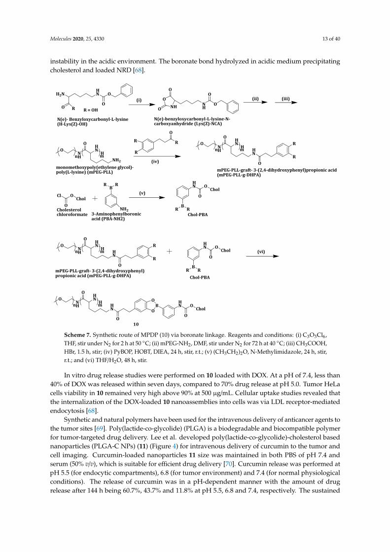
Yang et al. constructed a pH-responsive polymeric drug carrier by reversibly attachingphenylboronic acid-modified cholesterol (Chol-PBA) to catechol-pending methoxypoly (ethylene glycol)-block-poly(L-lysine) (Scheme 7). The pH-dependent metastability of monomethoxypoly(ethylene glycol)-poly(L-lysine)-graft-3-(2,4-dihydroxyphenyl)propionic acid (mPEG-PLL-g-DHPA)/Chol-PBA (MPDP)(10) nanoassemblies was evaluated using Nile Red Dye (NRD) at a pH of 7.4 and 5.0 over a period of24 h. No color changes were detected in the solution at pH 7.4. However, at pH 5.0, red deposits wereobserved with the solution color fading and ultimately becoming pale yellow. The co-polymer, 10 didnot change size at pH 7.4 but increased sharply at pH 5.0 from 196.2 to 954.8 nm, indicating its structural
Molecules 2020, 25, 4330 13 of 40
instability in the acidic environment. The boronate bond hydrolyzed in acidic medium precipitatingcholesterol and loaded NRD [68].Molecules 2020, 25, x FOR PEER REVIEW 14 of 41
H2NHN O
O RO
R = OH
(i)NH
O
OO
NH
O
O
(ii) (iii)
ONH
HN
HNH2
O
n m(iv)
R
O
R
R
ONH
HN
H HN
O
n m
O
R
R
Cl OChol
O
BRR
NH2
(v)
BR R
HN O
CholO
ONH
HN
H HN
O
n m
O
R
R
BR R
HN O
CholO
(vi)
ONH
HN
H HN
O
n m
O
OB
O HN O
CholO
N(e)- Benzyloxycarbonyl-L-lysine (H-Lys(Z)-OH)
N(e)-benzyloxycarbonyl-L-lysine-N- carboxyanhydride (Lys(Z)-NCA)
monomethoxypoly(ethylene glycol)-poly(L-lysine) (mPEG-PLL) mPEG-PLL-graft- 3-(2,4-dihydroxyphenyl)propionic acid
(mPEG-PLL-g-DHPA)
Cholesterolchloroformate 3-Aminophenylboronic
acid (PBA-NH2)Chol-PBA
Chol-PBAmPEG-PLL-graft- 3-(2,4-dihydroxyphenyl)propionic acid (mPEG-PLL-g-DHPA)
10 Scheme 7. Synthetic route of MPDP (10) via boronate linkage. Reagents and conditions: (i) C3O3Cl6, THF, stir under N2 for 2 h at 50 °C; (ii) mPEG-NH2, DMF, stir under N2 for 72 h at 40 °C; (iii) CH3COOH, HBr, 1.5 h, stir; (iv) PyBOP, HOBT, DIEA, 24 h, stir, r.t.; (v) (CH3CH2)2O, N-Methylimidazole, 24 h, stir, r.t.; and (vi) THF/H2O, 48 h, stir.
In vitro drug release studies were performed on 10 loaded with DOX. At a pH of 7.4, less than 40% of DOX was released within seven days, compared to 70% drug release at pH 5.0. Tumor HeLa cells viability in 10 remained very high above 90% at 500 µg/mL. Cellular uptake studies revealed that the internalization of the DOX-loaded 10 nanoassemblies into cells was via LDL receptor-mediated endocytosis [68].
Synthetic and natural polymers have been used for the intravenous delivery of anticancer agents to the tumor sites [69]. Poly(lactide-co-glycolide) (PLGA) is a biodegradable and biocompatible polymer for tumor-targeted drug delivery. Lee et al. developed poly(lactide-co-glycolide)-cholesterol based nanoparticles (PLGA-C NPs) (11) (Figure 4) for intravenous delivery of curcumin to the tumor and cell imaging. Curcumin-loaded nanoparticles 11 size was maintained in both PBS of pH 7.4 and serum (50% v/v), which is suitable for efficient drug delivery [70]. Curcumin release was performed at pH 5.5 (for endocytic compartments), 6.8 (for tumor environment) and 7.4 (for normal physiological conditions). The release of curcumin was in a pH-dependent manner with the amount of drug release after 144 h being 60.7%, 43.7% and 11.8% at pH 5.5, 6.8 and 7.4, respectively. The sustained and pH-dependent drug release mechanism of the drug-loaded nanoparticles influenced its drug tumor-targeting capability [70].
Scheme 7. Synthetic route of MPDP (10) via boronate linkage. Reagents and conditions: (i) C3O3Cl6,THF, stir under N2 for 2 h at 50 ◦C; (ii) mPEG-NH2, DMF, stir under N2 for 72 h at 40 ◦C; (iii) CH3COOH,HBr, 1.5 h, stir; (iv) PyBOP, HOBT, DIEA, 24 h, stir, r.t.; (v) (CH3CH2)2O, N-Methylimidazole, 24 h, stir,r.t.; and (vi) THF/H2O, 48 h, stir.
In vitro drug release studies were performed on 10 loaded with DOX. At a pH of 7.4, less than40% of DOX was released within seven days, compared to 70% drug release at pH 5.0. Tumor HeLacells viability in 10 remained very high above 90% at 500 µg/mL. Cellular uptake studies revealed thatthe internalization of the DOX-loaded 10 nanoassemblies into cells was via LDL receptor-mediatedendocytosis [68].
Synthetic and natural polymers have been used for the intravenous delivery of anticancer agents tothe tumor sites [69]. Poly(lactide-co-glycolide) (PLGA) is a biodegradable and biocompatible polymerfor tumor-targeted drug delivery. Lee et al. developed poly(lactide-co-glycolide)-cholesterol basednanoparticles (PLGA-C NPs) (11) (Figure 4) for intravenous delivery of curcumin to the tumor andcell imaging. Curcumin-loaded nanoparticles 11 size was maintained in both PBS of pH 7.4 andserum (50% v/v), which is suitable for efficient drug delivery [70]. Curcumin release was performed atpH 5.5 (for endocytic compartments), 6.8 (for tumor environment) and 7.4 (for normal physiologicalconditions). The release of curcumin was in a pH-dependent manner with the amount of drugrelease after 144 h being 60.7%, 43.7% and 11.8% at pH 5.5, 6.8 and 7.4, respectively. The sustained

Molecules 2020, 25, 4330 14 of 40
and pH-dependent drug release mechanism of the drug-loaded nanoparticles influenced its drugtumor-targeting capability [70].Molecules 2020, 25, x FOR PEER REVIEW 15 of 41
O
OOChol
OO
H
11
Figure 4. Chemical structure of PLGA-C (11).
The cytotoxicity of the nanoparticles was evaluated in Hep-2 cells derived from human laryngeal carcinoma. Both PLGA NPs and the nanoparticles 11 showed no cytotoxic effects making them safe formulation for intravenous drug delivery. In vitro cellular uptake was assessed in Hep-2 cells by encapsulating Dil, a hydrophobic fluorescent dye, into the NPs. The nanoparticles 11 cellular accumulation efficiency was 1.6-fold higher when compared to the nanoparticles when incubated for 4 h. After 24 h incubation, the cellular distribution of the nanoparticles done by CLSM showed stronger fluorescence intensity in 11 when compared to PLGA NPs. In vivo tumor targeting of 11 was shown by Near-infrared fluorescence (NIRF) images of Hep-2 tumor-xenografted mouse model. The results revealed the usefulness of 11 in targeting tumor cells [70].
Incorporation of PEG into liposome surfaces is reported to increase drug bioavailability and the circulation time [71]. PEGylation of liposomes suffers from drawbacks such as loss of response in low pH environment and liposomal cellular uptake hindrance [72,73]. Poly(2-ethyl-2-oxazoline) (PEtOz), a long-chain macromolecule polymer, is biocompatible, flexible and low cost and has been reported as a suitable pH-sensitive drug delivery vehicle [74,75]. Similar to PEG, PEtOz increases liposome stability in vitro and in vivo, thus it can be used as a PEG substitute [76]. Xu et al. constructed pH-sensitive liposomes from poly (2-ethyl-2-oxazoline) cholesterol hemisuccinate (PEtOz-CHEMS) (Scheme 8) (12) and encapsulated DOX, an anticancer drug. DOX-loaded liposomes 12 displayed slow DOX release when compared to the conventional DOX liposomes (CL-DOX), CHEMS modified DOX liposomes (CH-DOX) at pH 7.4. DOX release increased rapidly as the pH decreased and more than 90% was released within 4 h at pH 5.4. This revealed the significant change in release kinetics at low pH and the stability of the formulation at normal physiological pH, after PEtOzylation of liposomes [77].
N NOH
Et EtO O
19
HOO
CholO
O
(i)N N
O
Et EtO O
19 OChol
O
O12
Scheme 8. Synthetic route of PEtOz-CHEMS (12). Reagents and conditions: (i) DCC, DMAP, DCM, r.t, 10 h.
Cellular uptake studies after incubating A375 cells with the different liposomes showed that DOX-loaded compound 12 liposomes exhibited higher fluorescence intensity at pH 6.5 when compared to pH 7.4. PEtOzylation increased the cellular uptake of the liposomes with a maximum DOX accumulation, and, at pH 7.4, DOX-loaded liposomes 12 exhibited cellular uptake higher than that of CH-DOX, CL-DOX and PEG-DOX liposomes. CLSM analysis of A375 cells incubated with the different liposomes showed that PEtOz-DOX liposomes exhibited enhanced cellular uptake, and DOX release into the cytoplasm for PEtOzylated liposomes. A375 cells inhibition increased when incubated by the various liposomes at 37 °C under different pH. However, DOX-loaded liposomes 12 had higher cytotoxic effect at pH 6.4 when compared to pH 7.4 while CH-DOX, CL-DOX and PEG-DOX liposomes showed no difference in the inhibitions even at the different pH mediums. The results revealed the potential of PEtOz in modifying liposomes for drug delivery [77].
Figure 4. Chemical structure of PLGA-C (11).
The cytotoxicity of the nanoparticles was evaluated in Hep-2 cells derived from human laryngealcarcinoma. Both PLGA NPs and the nanoparticles 11 showed no cytotoxic effects making themsafe formulation for intravenous drug delivery. In vitro cellular uptake was assessed in Hep-2 cellsby encapsulating Dil, a hydrophobic fluorescent dye, into the NPs. The nanoparticles 11 cellularaccumulation efficiency was 1.6-fold higher when compared to the nanoparticles when incubated for4 h. After 24 h incubation, the cellular distribution of the nanoparticles done by CLSM showed strongerfluorescence intensity in 11 when compared to PLGA NPs. In vivo tumor targeting of 11 was shownby Near-infrared fluorescence (NIRF) images of Hep-2 tumor-xenografted mouse model. The resultsrevealed the usefulness of 11 in targeting tumor cells [70].
Incorporation of PEG into liposome surfaces is reported to increase drug bioavailability and thecirculation time [71]. PEGylation of liposomes suffers from drawbacks such as loss of response in lowpH environment and liposomal cellular uptake hindrance [72,73]. Poly(2-ethyl-2-oxazoline) (PEtOz),a long-chain macromolecule polymer, is biocompatible, flexible and low cost and has been reported as asuitable pH-sensitive drug delivery vehicle [74,75]. Similar to PEG, PEtOz increases liposome stabilityin vitro and in vivo, thus it can be used as a PEG substitute [76]. Xu et al. constructed pH-sensitiveliposomes from poly (2-ethyl-2-oxazoline) cholesterol hemisuccinate (PEtOz-CHEMS) (Scheme 8) (12)and encapsulated DOX, an anticancer drug. DOX-loaded liposomes 12 displayed slow DOX releasewhen compared to the conventional DOX liposomes (CL-DOX), CHEMS modified DOX liposomes(CH-DOX) at pH 7.4. DOX release increased rapidly as the pH decreased and more than 90% wasreleased within 4 h at pH 5.4. This revealed the significant change in release kinetics at low pH and thestability of the formulation at normal physiological pH, after PEtOzylation of liposomes [77].
Molecules 2020, 25, x FOR PEER REVIEW 15 of 41
O
OOChol
OO
H
11
Figure 4. Chemical structure of PLGA-C (11).
The cytotoxicity of the nanoparticles was evaluated in Hep-2 cells derived from human laryngeal carcinoma. Both PLGA NPs and the nanoparticles 11 showed no cytotoxic effects making them safe formulation for intravenous drug delivery. In vitro cellular uptake was assessed in Hep-2 cells by encapsulating Dil, a hydrophobic fluorescent dye, into the NPs. The nanoparticles 11 cellular accumulation efficiency was 1.6-fold higher when compared to the nanoparticles when incubated for 4 h. After 24 h incubation, the cellular distribution of the nanoparticles done by CLSM showed stronger fluorescence intensity in 11 when compared to PLGA NPs. In vivo tumor targeting of 11 was shown by Near-infrared fluorescence (NIRF) images of Hep-2 tumor-xenografted mouse model. The results revealed the usefulness of 11 in targeting tumor cells [70].
Incorporation of PEG into liposome surfaces is reported to increase drug bioavailability and the circulation time [71]. PEGylation of liposomes suffers from drawbacks such as loss of response in low pH environment and liposomal cellular uptake hindrance [72,73]. Poly(2-ethyl-2-oxazoline) (PEtOz), a long-chain macromolecule polymer, is biocompatible, flexible and low cost and has been reported as a suitable pH-sensitive drug delivery vehicle [74,75]. Similar to PEG, PEtOz increases liposome stability in vitro and in vivo, thus it can be used as a PEG substitute [76]. Xu et al. constructed pH-sensitive liposomes from poly (2-ethyl-2-oxazoline) cholesterol hemisuccinate (PEtOz-CHEMS) (Scheme 8) (12) and encapsulated DOX, an anticancer drug. DOX-loaded liposomes 12 displayed slow DOX release when compared to the conventional DOX liposomes (CL-DOX), CHEMS modified DOX liposomes (CH-DOX) at pH 7.4. DOX release increased rapidly as the pH decreased and more than 90% was released within 4 h at pH 5.4. This revealed the significant change in release kinetics at low pH and the stability of the formulation at normal physiological pH, after PEtOzylation of liposomes [77].
N NOH
Et EtO O
19
HOO
CholO
O
(i)N N
O
Et EtO O
19 OChol
O
O12
Scheme 8. Synthetic route of PEtOz-CHEMS (12). Reagents and conditions: (i) DCC, DMAP, DCM, r.t, 10 h.
Cellular uptake studies after incubating A375 cells with the different liposomes showed that DOX-loaded compound 12 liposomes exhibited higher fluorescence intensity at pH 6.5 when compared to pH 7.4. PEtOzylation increased the cellular uptake of the liposomes with a maximum DOX accumulation, and, at pH 7.4, DOX-loaded liposomes 12 exhibited cellular uptake higher than that of CH-DOX, CL-DOX and PEG-DOX liposomes. CLSM analysis of A375 cells incubated with the different liposomes showed that PEtOz-DOX liposomes exhibited enhanced cellular uptake, and DOX release into the cytoplasm for PEtOzylated liposomes. A375 cells inhibition increased when incubated by the various liposomes at 37 °C under different pH. However, DOX-loaded liposomes 12 had higher cytotoxic effect at pH 6.4 when compared to pH 7.4 while CH-DOX, CL-DOX and PEG-DOX liposomes showed no difference in the inhibitions even at the different pH mediums. The results revealed the potential of PEtOz in modifying liposomes for drug delivery [77].
Scheme 8. Synthetic route of PEtOz-CHEMS (12). Reagents and conditions: (i) DCC, DMAP, DCM, r.t, 10 h.
Cellular uptake studies after incubating A375 cells with the different liposomes showed thatDOX-loaded compound 12 liposomes exhibited higher fluorescence intensity at pH 6.5 when comparedto pH 7.4. PEtOzylation increased the cellular uptake of the liposomes with a maximum DOXaccumulation, and, at pH 7.4, DOX-loaded liposomes 12 exhibited cellular uptake higher than that ofCH-DOX, CL-DOX and PEG-DOX liposomes. CLSM analysis of A375 cells incubated with the differentliposomes showed that PEtOz-DOX liposomes exhibited enhanced cellular uptake, and DOX releaseinto the cytoplasm for PEtOzylated liposomes. A375 cells inhibition increased when incubated bythe various liposomes at 37 ◦C under different pH. However, DOX-loaded liposomes 12 had highercytotoxic effect at pH 6.4 when compared to pH 7.4 while CH-DOX, CL-DOX and PEG-DOX liposomesshowed no difference in the inhibitions even at the different pH mediums. The results revealed thepotential of PEtOz in modifying liposomes for drug delivery [77].

Molecules 2020, 25, 4330 15 of 40
The pH around and inside tumor cells is different from that of normal ones. The extracellular pHaround cancer cells is reported to be weakly acidic around 6.5–7.0, while, inside tumor cells, the pHis between 5.0 and 6.5 [78]. Researchers have focused on developing pH-triggered drug deliveryvehicles that are stable at normal physiological pH of 7.4 and can release drugs rapidly in the weaklyacidic environment [79,80]. In their quest for a novel hydrophobic pH-sensitive drug carrier withlower toxicity and higher efficiency, Yang et al. designed and synthesized three triblock copolymerspoly(ethylene glycol) methyl ether-b-peptide-g-cholesterol (mPEG-b-P-g-Chol) (Scheme 9) (13a–c),which self-assembled into micelles in an aqueous medium. The peptides functioned as the pH-sensitivepart and the internal core comprising of cholesterol offered space for drug loading. DOX was loadedas the model drug [81]. The three polymers (mPEG-P1-Chol 13a, mPEG-P2-Chol 13b and mPEG-P3Chol 13c, which differed only in shape, being linear, y-shaped and fork-shaped, respectively) showedvery low CMC values revealing that the micelles could remain intact even in extremely dilute volumeof the body’s systemic circulation. The three DOX-loaded polymers exhibited larger particle sizes at alower pH of 5.0 when compared to pH 7.4. The DOX loading in the three copolymers 13a–c micelleswas about 15.7%, 20.2% and 23.1% in weight, respectively, thus drug loading content and entrapmentefficiency increased with polymer complexity [81].
Molecules 2020, 25, x FOR PEER REVIEW 16 of 41
The pH around and inside tumor cells is different from that of normal ones. The extracellular pH around cancer cells is reported to be weakly acidic around 6.5–7.0, while, inside tumor cells, the pH is between 5.0 and 6.5 [78]. Researchers have focused on developing pH-triggered drug delivery vehicles that are stable at normal physiological pH of 7.4 and can release drugs rapidly in the weakly acidic environment [79,80]. In their quest for a novel hydrophobic pH-sensitive drug carrier with lower toxicity and higher efficiency, Yang et al. designed and synthesized three triblock copolymers poly(ethylene glycol) methyl ether-b-peptide-g-cholesterol (mPEG-b-P-g-Chol) (Scheme 9) (13a–c), which self-assembled into micelles in an aqueous medium. The peptides functioned as the pH-sensitive part and the internal core comprising of cholesterol offered space for drug loading. DOX was loaded as the model drug [81]. The three polymers (mPEG-P1-Chol 13a, mPEG-P2-Chol 13b and mPEG-P3 Chol 13c, which differed only in shape, being linear, y-shaped and fork-shaped, respectively) showed very low CMC values revealing that the micelles could remain intact even in extremely dilute volume of the body’s systemic circulation. The three DOX-loaded polymers exhibited larger particle sizes at a lower pH of 5.0 when compared to pH 7.4. The DOX loading in the three copolymers 13a–c micelles was about 15.7%, 20.2% and 23.1% in weight, respectively, thus drug loading content and entrapment efficiency increased with polymer complexity [81].
HOHN
NH
NH2
SH
O
HS
O
O
N
NH
24 Cl OChol
O (i)
HOHN
NH
HN
SH
O
HS
O
O
N
NH
24
OChol
O
(ii)
HN
NH
HN
SH
O
HS
O
O
N
NH
24
OChol
OHN
OO
43
P1-Chol
13
Scheme 9. Synthetic route of mPEG-P1-Chol (13). Reagents and conditions: (i) DMF, Triethyamine (TEA), rt, 72 h; and (ii) DMF, EDC-NHS, rt, 24 h.
In vitro drug release of the three polymers in solutions of pH 7.4 and 5.0 showed that DOX release was pH dependent. After 24 h, only about 35%, 30% and 26% of DOX was released from micelles 13a–c, respectively. The three polymers exhibited DOX release of about 90%, 84% and 80%, respectively, in 148 h. The enhanced positive charge on micelles surface at pH 5.0 caused the histidine moieties to experience electrostatic repulsion increasing the swelling and drug release. At 400 mg/L polymer concentration, cell viability against NIH 3T3 cells was 97.7%, 95.5% and 95.4%, and 96.8%, 95.9% and 95.0% against HepG2 cells, for 13a–c, respectively [81]. The DOX-loaded micelles exhibited lower cytotoxicity which increased slowly in lower concentrations suggesting a slow and time-controlled DOX release from the micelles. Although the free DOX-HCl showed the lowest cell viability, it was toxic to the tumor and the normal cells. The slight slowing down of the inhibition effect of DOX-loaded micelles 13 revealed their capability to protect the drug. The results showed the potential of 13 in delivering anticancer drugs [81].
Scheme 9. Synthetic route of mPEG-P1-Chol (13). Reagents and conditions: (i) DMF, Triethyamine(TEA), rt, 72 h; and (ii) DMF, EDC-NHS, rt, 24 h.
In vitro drug release of the three polymers in solutions of pH 7.4 and 5.0 showed that DOX releasewas pH dependent. After 24 h, only about 35%, 30% and 26% of DOX was released from micelles 13a–c,respectively. The three polymers exhibited DOX release of about 90%, 84% and 80%, respectively,in 148 h. The enhanced positive charge on micelles surface at pH 5.0 caused the histidine moietiesto experience electrostatic repulsion increasing the swelling and drug release. At 400 mg/L polymerconcentration, cell viability against NIH 3T3 cells was 97.7%, 95.5% and 95.4%, and 96.8%, 95.9% and95.0% against HepG2 cells, for 13a–c, respectively [81]. The DOX-loaded micelles exhibited lowercytotoxicity which increased slowly in lower concentrations suggesting a slow and time-controlledDOX release from the micelles. Although the free DOX-HCl showed the lowest cell viability, it wastoxic to the tumor and the normal cells. The slight slowing down of the inhibition effect of DOX-loadedmicelles 13 revealed their capability to protect the drug. The results showed the potential of 13 indelivering anticancer drugs [81].
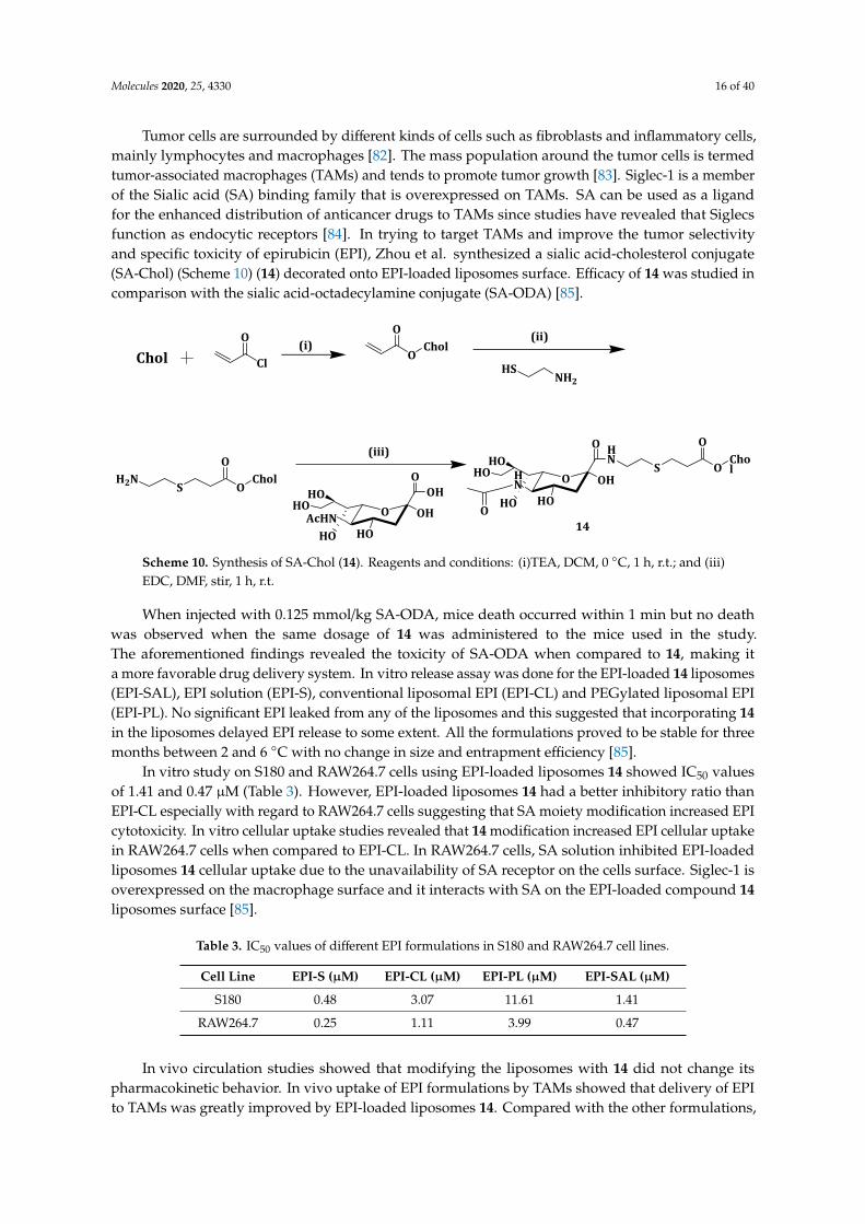
Molecules 2020, 25, 4330 16 of 40
Tumor cells are surrounded by different kinds of cells such as fibroblasts and inflammatory cells,mainly lymphocytes and macrophages [82]. The mass population around the tumor cells is termedtumor-associated macrophages (TAMs) and tends to promote tumor growth [83]. Siglec-1 is a memberof the Sialic acid (SA) binding family that is overexpressed on TAMs. SA can be used as a ligandfor the enhanced distribution of anticancer drugs to TAMs since studies have revealed that Siglecsfunction as endocytic receptors [84]. In trying to target TAMs and improve the tumor selectivityand specific toxicity of epirubicin (EPI), Zhou et al. synthesized a sialic acid-cholesterol conjugate(SA-Chol) (Scheme 10) (14) decorated onto EPI-loaded liposomes surface. Efficacy of 14 was studied incomparison with the sialic acid-octadecylamine conjugate (SA-ODA) [85].
Molecules 2020, 25, x FOR PEER REVIEW 17 of 41
Tumor cells are surrounded by different kinds of cells such as fibroblasts and inflammatory cells, mainly lymphocytes and macrophages [82]. The mass population around the tumor cells is termed tumor-associated macrophages (TAMs) and tends to promote tumor growth [83]. Siglec-1 is a member of the Sialic acid (SA) binding family that is overexpressed on TAMs. SA can be used as a ligand for the enhanced distribution of anticancer drugs to TAMs since studies have revealed that Siglecs function as endocytic receptors [84]. In trying to target TAMs and improve the tumor selectivity and specific toxicity of epirubicin (EPI), Zhou et al. synthesized a sialic acid-cholesterol conjugate (SA-Chol) (Scheme 10) (14) decorated onto EPI-loaded liposomes surface. Efficacy of 14 was studied in comparison with the sialic acid-octadecylamine conjugate (SA-ODA) [85].
When injected with 0.125 mmol/kg SA-ODA, mice death occurred within 1 min but no death was observed when the same dosage of 14 was administered to the mice used in the study. The aforementioned findings revealed the toxicity of SA-ODA when compared to 14, making it a more favorable drug delivery system. In vitro release assay was done for the EPI-loaded 14 liposomes (EPI-SAL), EPI solution (EPI-S), conventional liposomal EPI (EPI-CL) and PEGylated liposomal EPI (EPI-PL). No significant EPI leaked from any of the liposomes and this suggested that incorporating 14 in the liposomes delayed EPI release to some extent. All the formulations proved to be stable for three months between 2 and 6 °C with no change in size and entrapment efficiency [85].
Chol Cl
OO
OChol(i)
HSNH2
(ii)
H2NS O
CholO
O OH
HO
HO
OOH
HOAcHN
HOO OH
HO
HN
HO
O HN
HO
HO
O
S OChol
O(iii)
14
Scheme 10. Synthesis of SA-Chol (14). Reagents and conditions: (i)TEA, DCM, 0 °C, 1 h, r.t.; and (iii) EDC, DMF, stir, 1 h, r.t.
In vitro study on S180 and RAW264.7 cells using EPI-loaded liposomes 14 showed IC50 values of 1.41 and 0.47 µM (Table 3). However, EPI-loaded liposomes 14 had a better inhibitory ratio than EPI-CL especially with regard to RAW264.7 cells suggesting that SA moiety modification increased EPI cytotoxicity. In vitro cellular uptake studies revealed that 14 modification increased EPI cellular uptake in RAW264.7 cells when compared to EPI-CL. In RAW264.7 cells, SA solution inhibited EPI-loaded liposomes 14 cellular uptake due to the unavailability of SA receptor on the cells surface. Siglec-1 is overexpressed on the macrophage surface and it interacts with SA on the EPI-loaded compound 14 liposomes surface [85].
Table 3. IC50 values of different EPI formulations in S180 and RAW264.7 cell lines.
Cell Line EPI-S (µM) EPI-CL (µM) EPI-PL (µM) EPI-SAL (µM) S180 0.48 3.07 11.61 1.41
RAW264.7 0.25 1.11 3.99 0.47
In vivo circulation studies showed that modifying the liposomes with 14 did not change its pharmacokinetic behavior. In vivo uptake of EPI formulations by TAMs showed that delivery of EPI to TAMs was greatly improved by EPI-loaded liposomes 14. Compared with the other formulations, tumor-bearing mice injected with EPI-loaded liposomes 14 lived longer up to 60 days, denoting lower toxicity of EPI-loaded the liposomes. TAMs were identified by CD68 cells and fewer of them were
Scheme 10. Synthesis of SA-Chol (14). Reagents and conditions: (i)TEA, DCM, 0 ◦C, 1 h, r.t.; and (iii)EDC, DMF, stir, 1 h, r.t.
When injected with 0.125 mmol/kg SA-ODA, mice death occurred within 1 min but no deathwas observed when the same dosage of 14 was administered to the mice used in the study.The aforementioned findings revealed the toxicity of SA-ODA when compared to 14, making ita more favorable drug delivery system. In vitro release assay was done for the EPI-loaded 14 liposomes(EPI-SAL), EPI solution (EPI-S), conventional liposomal EPI (EPI-CL) and PEGylated liposomal EPI(EPI-PL). No significant EPI leaked from any of the liposomes and this suggested that incorporating 14in the liposomes delayed EPI release to some extent. All the formulations proved to be stable for threemonths between 2 and 6 ◦C with no change in size and entrapment efficiency [85].
In vitro study on S180 and RAW264.7 cells using EPI-loaded liposomes 14 showed IC50 valuesof 1.41 and 0.47 µM (Table 3). However, EPI-loaded liposomes 14 had a better inhibitory ratio thanEPI-CL especially with regard to RAW264.7 cells suggesting that SA moiety modification increased EPIcytotoxicity. In vitro cellular uptake studies revealed that 14 modification increased EPI cellular uptakein RAW264.7 cells when compared to EPI-CL. In RAW264.7 cells, SA solution inhibited EPI-loadedliposomes 14 cellular uptake due to the unavailability of SA receptor on the cells surface. Siglec-1 isoverexpressed on the macrophage surface and it interacts with SA on the EPI-loaded compound 14liposomes surface [85].
Table 3. IC50 values of different EPI formulations in S180 and RAW264.7 cell lines.
Cell Line EPI-S (µM) EPI-CL (µM) EPI-PL (µM) EPI-SAL (µM)
S180 0.48 3.07 11.61 1.41
RAW264.7 0.25 1.11 3.99 0.47
In vivo circulation studies showed that modifying the liposomes with 14 did not change itspharmacokinetic behavior. In vivo uptake of EPI formulations by TAMs showed that delivery of EPIto TAMs was greatly improved by EPI-loaded liposomes 14. Compared with the other formulations,
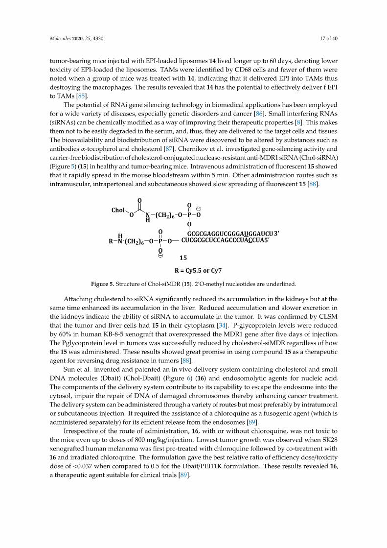
Molecules 2020, 25, 4330 17 of 40
tumor-bearing mice injected with EPI-loaded liposomes 14 lived longer up to 60 days, denoting lowertoxicity of EPI-loaded the liposomes. TAMs were identified by CD68 cells and fewer of them werenoted when a group of mice was treated with 14, indicating that it delivered EPI into TAMs thusdestroying the macrophages. The results revealed that 14 has the potential to effectively deliver f EPIto TAMs [85].
The potential of RNAi gene silencing technology in biomedical applications has been employedfor a wide variety of diseases, especially genetic disorders and cancer [86]. Small interfering RNAs(siRNAs) can be chemically modified as a way of improving their therapeutic properties [8]. This makesthem not to be easily degraded in the serum, and, thus, they are delivered to the target cells and tissues.The bioavailability and biodistribution of siRNA were discovered to be altered by substances such asantibodies α-tocopherol and cholesterol [87]. Chernikov et al. investigated gene-silencing activity andcarrier-free biodistribution of cholesterol-conjugated nuclease-resistant anti-MDR1 siRNA (Chol-siRNA)(Figure 5) (15) in healthy and tumor-bearing mice. Intravenous administration of fluorescent 15 showedthat it rapidly spread in the mouse bloodstream within 5 min. Other administration routes such asintramuscular, intrapertoneal and subcutaneous showed slow spreading of fluorescent 15 [88].
Molecules 2020, 25, x FOR PEER REVIEW 18 of 41
noted when a group of mice was treated with 14, indicating that it delivered EPI into TAMs thus destroying the macrophages. The results revealed that 14 has the potential to effectively deliver f EPI to TAMs [85].
The potential of RNAi gene silencing technology in biomedical applications has been employed for a wide variety of diseases, especially genetic disorders and cancer [86]. Small interfering RNAs (siRNAs) can be chemically modified as a way of improving their therapeutic properties [8]. This makes them not to be easily degraded in the serum, and, thus, they are delivered to the target cells and tissues. The bioavailability and biodistribution of siRNA were discovered to be altered by substances such as antibodies α-tocopherol and cholesterol [87]. Chernikov et al. investigated gene-silencing activity and carrier-free biodistribution of cholesterol-conjugated nuclease-resistant anti-MDR1 siRNA (Chol-siRNA) (Figure 5) (15) in healthy and tumor-bearing mice. Intravenous administration of fluorescent 15 showed that it rapidly spread in the mouse bloodstream within 5 min. Other administration routes such as intramuscular, intrapertoneal and subcutaneous showed slow spreading of fluorescent 15 [88].
CholO N
H
O
(CH2)6 O POGCGCGAGGUCGGGAUGGAUCU
OO
CUCGCGCUCCAGCCCUACCUA3'
5'OPO
O(CH2)6
OHNR
15
R = Cy5.5 or Cy7
Figure 5. Structure of Chol-siMDR (15). 2’O-methyl nucleotides are underlined.
Attaching cholesterol to siRNA significantly reduced its accumulation in the kidneys but at the same time enhanced its accumulation in the liver. Reduced accumulation and slower excretion in the kidneys indicate the ability of siRNA to accumulate in the tumor. It was confirmed by CLSM that the tumor and liver cells had 15 in their cytoplasm [34]. P-glycoprotein levels were reduced by 60% in human KB-8-5 xenograft that overexpressed the MDR1 gene after five days of injection. The Pglycoprotein level in tumors was successfully reduced by cholesterol-siMDR regardless of how the 15 was administered. These results showed great promise in using compound 15 as a therapeutic agent for reversing drug resistance in tumors [88].
Sun et al. invented and patented an in vivo delivery system containing cholesterol and small DNA molecules (Dbait) (Chol-Dbait) (Figure 6) (16) and endosomolytic agents for nucleic acid. The components of the delivery system contribute to its capability to escape the endosome into the cytosol, impair the repair of DNA of damaged chromosomes thereby enhancing cancer treatment. The delivery system can be administered through a variety of routes but most preferably by intratumoral or subcutaneous injection. It required the assistance of a chloroquine as a fusogenic agent (which is administered separately) for its efficient release from the endosomes [89].
Figure 5. Structure of Chol-siMDR (15). 2’O-methyl nucleotides are underlined.
Attaching cholesterol to siRNA significantly reduced its accumulation in the kidneys but at thesame time enhanced its accumulation in the liver. Reduced accumulation and slower excretion inthe kidneys indicate the ability of siRNA to accumulate in the tumor. It was confirmed by CLSMthat the tumor and liver cells had 15 in their cytoplasm [34]. P-glycoprotein levels were reducedby 60% in human KB-8-5 xenograft that overexpressed the MDR1 gene after five days of injection.The Pglycoprotein level in tumors was successfully reduced by cholesterol-siMDR regardless of howthe 15 was administered. These results showed great promise in using compound 15 as a therapeuticagent for reversing drug resistance in tumors [88].
Sun et al. invented and patented an in vivo delivery system containing cholesterol and smallDNA molecules (Dbait) (Chol-Dbait) (Figure 6) (16) and endosomolytic agents for nucleic acid.The components of the delivery system contribute to its capability to escape the endosome into thecytosol, impair the repair of DNA of damaged chromosomes thereby enhancing cancer treatment.The delivery system can be administered through a variety of routes but most preferably by intratumoralor subcutaneous injection. It required the assistance of a chloroquine as a fusogenic agent (which isadministered separately) for its efficient release from the endosomes [89].
Irrespective of the route of administration, 16, with or without chloroquine, was not toxic tothe mice even up to doses of 800 mg/kg/injection. Lowest tumor growth was observed when SK28xenografted human melanoma was first pre-treated with chloroquine followed by co-treatment with16 and irradiated chloroquine. The formulation gave the best relative ratio of efficiency dose/toxicitydose of <0.037 when compared to 0.5 for the Dbait/PEI11K formulation. These results revealed 16,a therapeutic agent suitable for clinical trials [89].
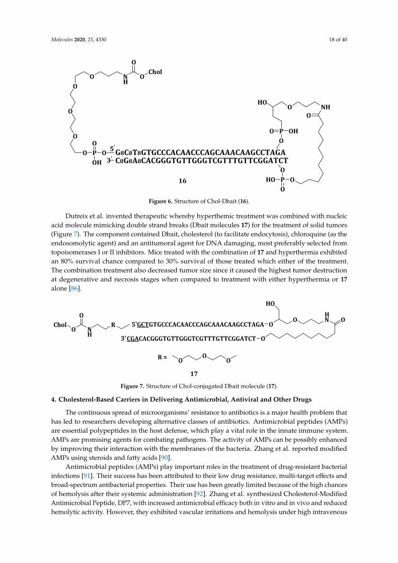
Molecules 2020, 25, 4330 18 of 40Molecules 2020, 25, x FOR PEER REVIEW 19 of 41
HOO NH
PO
OPHOO
O
OHO
O
G8C8T8GTGCCCACAACCCAGCAAACAAGCCTAGAC8G8A8CACGGGTGTTGGGTCGTTTGTTCGGATCT
OPOOH
OO
O
OO N
HO
CholO
5'
3'
16
Figure 6. Structure of Chol-Dbait (16).
Irrespective of the route of administration, 16, with or without chloroquine, was not toxic to the mice even up to doses of 800 mg/kg/injection. Lowest tumor growth was observed when SK28 xenografted human melanoma was first pre-treated with chloroquine followed by co-treatment with 16 and irradiated chloroquine. The formulation gave the best relative ratio of efficiency dose/toxicity dose of <0.037 when compared to 0.5 for the Dbait/PEI11K formulation. These results revealed 16, a therapeutic agent suitable for clinical trials [89].
Dutreix et al. invented therapeutic whereby hyperthemic treatment was combined with nucleic acid molecule mimicking double strand breaks (Dbait molecules 17) for the treatment of solid tumors (Figure 7). The component contained Dbait, cholesterol (to facilitate endocytosis), chloroquine (as the endosomolytic agent) and an antitumoral agent for DNA damaging, most preferably selected from topoisomerases I or II inhibitors. Mice treated with the combination of 17 and hyperthermia exhibited an 80% survival chance compared to 30% survival of those treated which either of the treatment. The combination treatment also decreased tumor size since it caused the highest tumor destruction at degenerative and necrosis stages when compared to treatment with either hyperthermia or 17 alone [86].
CholO N
HR 5'GCTGTGCCCACAACCCAGCAAACAAGCCTAGA O
OHN O
OCGACACGGGTGTTGGGTCGTTTGTTCGGATCT3'
HO
O
OO
OR =
17 Figure 7. Structure of Chol-conjugated Dbait molecule (17).
4. Cholesterol-Based Carriers in Delivering Antimicrobial, Antiviral and Other Drugs
The continuous spread of microorganisms’ resistance to antibiotics is a major health problem that has led to researchers developing alternative classes of antibiotics. Antimicrobial peptides (AMPs) are essential polypeptides in the host defense, which play a vital role in the innate immune system. AMPs are promising agents for combating pathogens. The activity of AMPs can be possibly enhanced by improving their interaction with the membranes of the bacteria. Zhang et al. reported modified AMPs using steroids and fatty acids [90].
Figure 6. Structure of Chol-Dbait (16).
Dutreix et al. invented therapeutic whereby hyperthemic treatment was combined with nucleicacid molecule mimicking double strand breaks (Dbait molecules 17) for the treatment of solid tumors(Figure 7). The component contained Dbait, cholesterol (to facilitate endocytosis), chloroquine (as theendosomolytic agent) and an antitumoral agent for DNA damaging, most preferably selected fromtopoisomerases I or II inhibitors. Mice treated with the combination of 17 and hyperthermia exhibitedan 80% survival chance compared to 30% survival of those treated which either of the treatment.The combination treatment also decreased tumor size since it caused the highest tumor destructionat degenerative and necrosis stages when compared to treatment with either hyperthermia or 17alone [86].
Molecules 2020, 25, x FOR PEER REVIEW 19 of 41
HOO NH
PO
OPHOO
O
OHO
O
G8C8T8GTGCCCACAACCCAGCAAACAAGCCTAGAC8G8A8CACGGGTGTTGGGTCGTTTGTTCGGATCT
OPOOH
OO
O
OO N
HO
CholO
5'
3'
16
Figure 6. Structure of Chol-Dbait (16).
Irrespective of the route of administration, 16, with or without chloroquine, was not toxic to the mice even up to doses of 800 mg/kg/injection. Lowest tumor growth was observed when SK28 xenografted human melanoma was first pre-treated with chloroquine followed by co-treatment with 16 and irradiated chloroquine. The formulation gave the best relative ratio of efficiency dose/toxicity dose of <0.037 when compared to 0.5 for the Dbait/PEI11K formulation. These results revealed 16, a therapeutic agent suitable for clinical trials [89].
Dutreix et al. invented therapeutic whereby hyperthemic treatment was combined with nucleic acid molecule mimicking double strand breaks (Dbait molecules 17) for the treatment of solid tumors (Figure 7). The component contained Dbait, cholesterol (to facilitate endocytosis), chloroquine (as the endosomolytic agent) and an antitumoral agent for DNA damaging, most preferably selected from topoisomerases I or II inhibitors. Mice treated with the combination of 17 and hyperthermia exhibited an 80% survival chance compared to 30% survival of those treated which either of the treatment. The combination treatment also decreased tumor size since it caused the highest tumor destruction at degenerative and necrosis stages when compared to treatment with either hyperthermia or 17 alone [86].
CholO N
HR 5'GCTGTGCCCACAACCCAGCAAACAAGCCTAGA O
OHN O
OCGACACGGGTGTTGGGTCGTTTGTTCGGATCT3'
HO
O
OO
OR =
17 Figure 7. Structure of Chol-conjugated Dbait molecule (17).
4. Cholesterol-Based Carriers in Delivering Antimicrobial, Antiviral and Other Drugs
The continuous spread of microorganisms’ resistance to antibiotics is a major health problem that has led to researchers developing alternative classes of antibiotics. Antimicrobial peptides (AMPs) are essential polypeptides in the host defense, which play a vital role in the innate immune system. AMPs are promising agents for combating pathogens. The activity of AMPs can be possibly enhanced by improving their interaction with the membranes of the bacteria. Zhang et al. reported modified AMPs using steroids and fatty acids [90].
Figure 7. Structure of Chol-conjugated Dbait molecule (17).
4. Cholesterol-Based Carriers in Delivering Antimicrobial, Antiviral and Other Drugs
The continuous spread of microorganisms’ resistance to antibiotics is a major health problem thathas led to researchers developing alternative classes of antibiotics. Antimicrobial peptides (AMPs)are essential polypeptides in the host defense, which play a vital role in the innate immune system.AMPs are promising agents for combating pathogens. The activity of AMPs can be possibly enhancedby improving their interaction with the membranes of the bacteria. Zhang et al. reported modifiedAMPs using steroids and fatty acids [90].
Antimicrobial peptides (AMPs) play important roles in the treatment of drug-resistant bacterialinfections [91]. Their success has been attributed to their low drug resistance, multi-target effects andbroad-spectrum antibacterial properties. Their use has been greatly limited because of the high chancesof hemolysis after their systemic administration [92]. Zhang et al. synthesized Cholesterol-ModifiedAntimicrobial Peptide, DP7, with increased antimicrobial efficacy both in vitro and in vivo and reducedhemolytic activity. However, they exhibited vascular irritations and hemolysis under high intravenous

Molecules 2020, 25, 4330 19 of 40
dosage of DP7 [90]. To reduce the hemolytic capacity of DP7, it was conjugated with cholesterol(DP7-C). DP7-C exhibited a CMC of about 3.5 and 50 µg/mL in water and culture medium, respectively,suggesting its ability to self-assemble into micelles. DP7 was more toxic than DP7-C because all themice injected intravenously with DP7 at a dosage 20 mg/kg died within 10 min while no fatalities werenoted up to 144 h in the mice injected with DP7-C even at a dose of 80 mg/kg. The in vitro and in vivostudies further showed that the DP7-C micelles exhibited lower hemolytic activity when compared toDP7 [90].
The suppression of bacterial infection by DP7-C micelles was evaluated in zebrafish embryosinfected with Pseudomonas aeruginosa strain 284 ATCC10145-GFP. The infected zebrafish embryostreated with DP7-C micelles showed that the micelles inhibited ATCC10145-GFP amplification in atime-dependent manner. A defensive immune reaction was stimulated in mice administered withDP7-C micelles in vivo. The results showed that DP7-C can significantly treat bacterial infectionwithout adverse side effects [90].
Villamil et al. conjugated a polymer of Methoxy-Polyethylene glycol and ε-caprolactone(mPEG-b-PCL) with cholesterol (mPEG-b-PCL-Chol) (Scheme 11) (18) in an attempt to attenuatethe nephrotoxicity, increase loading capacity and aqueous solubility and encapsulation efficiency ofAmphotericin B (AmB). The formulation, 18 had 60% drug encapsulation efficiency (%EE) comparedwith 28% and 42% of mPEG-b-PCL and mPEG-Chol, respectively (Table 4). The above values showedthe role played by cholesterol in increasing the encapsulation of AmB in the polymeric micelles [93].
Molecules 2020, 25, x FOR PEER REVIEW 20 of 41
Antimicrobial peptides (AMPs) play important roles in the treatment of drug-resistant bacterial infections [91]. Their success has been attributed to their low drug resistance, multi-target effects and broad-spectrum antibacterial properties. Their use has been greatly limited because of the high chances of hemolysis after their systemic administration [92]. Zhang et al. synthesized Cholesterol-Modified Antimicrobial Peptide, DP7, with increased antimicrobial efficacy both in vitro and in vivo and reduced hemolytic activity. However, they exhibited vascular irritations and hemolysis under high intravenous dosage of DP7 [90]. To reduce the hemolytic capacity of DP7, it was conjugated with cholesterol (DP7-C). DP7-C exhibited a CMC of about 3.5 and 50 µg/mL in water and culture medium, respectively, suggesting its ability to self-assemble into micelles. DP7 was more toxic than DP7-C because all the mice injected intravenously with DP7 at a dosage 20 mg/kg died within 10 min while no fatalities were noted up to 144 h in the mice injected with DP7-C even at a dose of 80 mg/kg. The in vitro and in vivo studies further showed that the DP7-C micelles exhibited lower hemolytic activity when compared to DP7 [90].
The suppression of bacterial infection by DP7-C micelles was evaluated in zebrafish embryos infected with Pseudomonas aeruginosa strain 284 ATCC10145-GFP. The infected zebrafish embryos treated with DP7-C micelles showed that the micelles inhibited ATCC10145-GFP amplification in a time-dependent manner. A defensive immune reaction was stimulated in mice administered with DP7-C micelles in vivo. The results showed that DP7-C can significantly treat bacterial infection without adverse side effects [90].
Villamil et al. conjugated a polymer of Methoxy-Polyethylene glycol and ε-caprolactone (mPEG-b-PCL) with cholesterol (mPEG-b-PCL-Chol) (Scheme 11) (18) in an attempt to attenuate the nephrotoxicity, increase loading capacity and aqueous solubility and encapsulation efficiency of Amphotericin B (AmB). The formulation, 18 had 60% drug encapsulation efficiency (%EE) compared with 28% and 42% of mPEG-b-PCL and mPEG-Chol, respectively (Table 4). The above values showed the role played by cholesterol in increasing the encapsulation of AmB in the polymeric micelles [93].
OOH
n
m-PEG
O
O
e-Caprolactone(e-CL)
(i)O
O n OH
O m
mPEG-b-PCL
Cl OChol
OmPEG-b-PCL (ii) O
O n OO m
Chol
O
18 Scheme 11. Synthesis of mPEG-b-PCL-Chol (18). Reagents and conditions: (i) Sn(EH)2/Toluene, 110 °C, 24 h; and (ii) N(CH2CH3)3, 40–60 °C, 48 h.
Micelles 18 drug release rate was slow, and this is attributed to the decreased critical micelle concentration, hydrophobic micelle core and more favorable AmB interactions. Micelles 18 not loaded with drug showed in vitro non-toxicity to fibroblast L929 cells with 80% cell survival at a dosage of 120 mg/L. AmB-loaded micelles 18 showed a noticeable increase in hemolysis. Nevertheless, it was 4 times less toxic than Fungizone, a standard fungicide [93].
Scheme 11. Synthesis of mPEG-b-PCL-Chol (18). Reagents and conditions: (i) Sn(EH)2/Toluene, 110 ◦C,24 h; and (ii) N(CH2CH3)3, 40–60 ◦C, 48 h.
Table 4. Encapsulation efficiency and Minimum Inhibition Concentration (MIC) of the three formulations.
Formulation %EE MIC (µg/mol)
mPEG-b-PCL-Chol (18) 60 0.46mPEG-b-PCL 28 0.46mPEG-Chol 42 0.93Fungizone - 0.23
Micelles 18 drug release rate was slow, and this is attributed to the decreased critical micelleconcentration, hydrophobic micelle core and more favorable AmB interactions. Micelles 18 not loadedwith drug showed in vitro non-toxicity to fibroblast L929 cells with 80% cell survival at a dosage of120 mg/L. AmB-loaded micelles 18 showed a noticeable increase in hemolysis. Nevertheless, it was4 times less toxic than Fungizone, a standard fungicide [93].
Antifungal study was performed against C. albicans SC5314 and AmB loaded micelles 18 wasmore effective when compared to mPEG-Chol due to the release profile of AmB (Table 4). In vivoresults from G. mellonella infected with a C. albicans SC5314 strain and then treated with the formulationand Fungizone showed that compound 18 loaded with AmB exhibited a survival of 80% after 7 days

Molecules 2020, 25, 4330 20 of 40
while all the non-treated larvae died [93]. The results showed that 18 did not only improved theencapsulation efficiency of AmB, but also increased its controlled release from the micelles, making it apromising drug delivery system for AmB.
Other diseases have been studied using cholesterol-based systems. Idiopathic pulmonary fibrosis(IPF) is a deadly disease with its patients having a mean survival of only 2–4 years. Signs and symptomsof IPF include injury of the alveolar epithelial cells, fibroblasts hyperplasia and accumulation ofinflammatory cells. This results in extracellular matrix, including proteoglycans and fibrillar collagensto be deposited. Research on antimalarial drugs such as chloroquine (CQ) and hydroxychloroquine(HCQ) has shown that they exert anti-inflammatory activity by the down-regulating pro-inflammatorycytokines. HCQ effectively inhibits fibroblasts proliferation and suppresses metabolic activities infibrotic skin diseases [94].
Liu et al. synthesized cholesterol-modified hydroxychloroquine (Figure 8) (19). HCQ inducesapoptosis in human dermal fibroblasts at concentrations of 1–10 µM. Compound 19 exhibited lowtoxicity against lung fibroblasts and inhibited cell proliferation at 10 µM, but it induced apoptosis at ahigher concentration of 50 µM when compared to the unconjugated HCQ [94].
Molecules 2020, 25, x FOR PEER REVIEW 21 of 41
Table 4. Encapsulation efficiency and Minimum Inhibition Concentration (MIC) of the three formulations.
Formulation %EE MIC (µg/mol) mPEG-b-PCL-Chol (18) 60 0.46
mPEG-b-PCL 28 0.46 mPEG-Chol 42 0.93 Fungizone - 0.23
Antifungal study was performed against C. albicans SC5314 and AmB loaded micelles 18 was more effective when compared to mPEG-Chol due to the release profile of AmB (Table 4). In vivo results from G. mellonella infected with a C. albicans SC5314 strain and then treated with the formulation and Fungizone showed that compound 18 loaded with AmB exhibited a survival of 80% after 7 days while all the non-treated larvae died [93]. The results showed that 18 did not only improved the encapsulation efficiency of AmB, but also increased its controlled release from the micelles, making it a promising drug delivery system for AmB.
Other diseases have been studied using cholesterol-based systems. Idiopathic pulmonary fibrosis (IPF) is a deadly disease with its patients having a mean survival of only 2–4 years. Signs and symptoms of IPF include injury of the alveolar epithelial cells, fibroblasts hyperplasia and accumulation of inflammatory cells. This results in extracellular matrix, including proteoglycans and fibrillar collagens to be deposited. Research on antimalarial drugs such as chloroquine (CQ) and hydroxychloroquine (HCQ) has shown that they exert anti-inflammatory activity by the down-regulating pro-inflammatory cytokines. HCQ effectively inhibits fibroblasts proliferation and suppresses metabolic activities in fibrotic skin diseases [94].
Liu et al. synthesized cholesterol-modified hydroxychloroquine (Figure 8) (19). HCQ induces apoptosis in human dermal fibroblasts at concentrations of 1–10 µM. Compound 19 exhibited low toxicity against lung fibroblasts and inhibited cell proliferation at 10 µM, but it induced apoptosis at a higher concentration of 50 µM when compared to the unconjugated HCQ [94].
NCl
HN N O
O
O
O Chol
19 Figure 8. Cholesterol-modified HCQ (19).
Compound 19 hindered early pulmonary inflammation. It also inhibited the CTGF/ERK signaling pathway which is effective against pulmonary fibrosis induced by bleomycin. The results revealed the capability of compound 19 to prevent bleomycin induced pulmonary fibrosis, revealing its anti-fibrotic activity useful for the treatment of inflammation caused by pulmonary fibrosis [94].
Viruses are intracellular parasites that depend on the host cells for life. A virus contains a nucleic acid genome, a protein capsid and a lipid envelope. The properties of the lipids (sphingolipids, glycerophospholipids and sterols) of an animal membrane affect the viral entry [95]. Hepatitis C virus (HCV) is a serious health condition due to its lack of an effective cure. It is the root cause of liver diseases such as cirrhosis, hepatocellular carcinoma and chronic hepatitis [96]. After the introduction of protease inhibitors to the interferon/ribavirin therapy, which showed improved virological activity, there was a need for better HCV antivirals because the inhibitors posed unwanted side effects and in only about 50% of the patients did the therapy led to a sustained antiviral response [97].
Pessi et al. prepared and patented a series of contiguous amino acids belonging to the HR2 domain of Type 1 viral fusogenic protein on an enveloped virus which they attached to the C-terminal of cholesterol (Scheme 12) (20) for viral fusion inhibition. The role played by the cholesterol moiety
Figure 8. Cholesterol-modified HCQ (19).
Compound 19 hindered early pulmonary inflammation. It also inhibited the CTGF/ERK signalingpathway which is effective against pulmonary fibrosis induced by bleomycin. The results revealed thecapability of compound 19 to prevent bleomycin induced pulmonary fibrosis, revealing its anti-fibroticactivity useful for the treatment of inflammation caused by pulmonary fibrosis [94].
Viruses are intracellular parasites that depend on the host cells for life. A virus contains a nucleicacid genome, a protein capsid and a lipid envelope. The properties of the lipids (sphingolipids,glycerophospholipids and sterols) of an animal membrane affect the viral entry [95]. Hepatitis C virus(HCV) is a serious health condition due to its lack of an effective cure. It is the root cause of liverdiseases such as cirrhosis, hepatocellular carcinoma and chronic hepatitis [96]. After the introductionof protease inhibitors to the interferon/ribavirin therapy, which showed improved virological activity,there was a need for better HCV antivirals because the inhibitors posed unwanted side effects and inonly about 50% of the patients did the therapy led to a sustained antiviral response [97].
Pessi et al. prepared and patented a series of contiguous amino acids belonging to the HR2domain of Type 1 viral fusogenic protein on an enveloped virus which they attached to the C-terminalof cholesterol (Scheme 12) (20) for viral fusion inhibition. The role played by the cholesterol moiety wasexhibited by peptide ELLELDKWASLW, which on its own had an IC50 > 100 µM, but, when derivatizedwith the C-terminal of cholesterol (Figure 9) (21), its antiviral potency improved to IC50 = 5.85 µM.Based on Table 5, the antiviral potency of C34 increased 50-fold when conjugated to cholesterol (C34 andC34-Aceta have IC50 = 205 and 270 µM, respectively, compared to IC50 = 4 for C34-Chol) [98].
Chol-C34 showed 50-fold less antiviral potency compared to underivatized C34 because of thecholesterol that was linked to the N- instead of the C-terminal. C34-Chol was found to be approximately100-fold more potent than enfuvieride, the marketed fusion inhibitor. C34-Chol was also more potentthan all the controls against other HIV-1 isolates such as BAL, NL4-3, MN-1, etc. The compound 20peptides are potent for viral fusion inhibition and also for efficacy of other therapeutic agents such asnucleoside inhibitors and limiters of HIV infection degree, which may be administered simultaneouslyor in alternating [98].
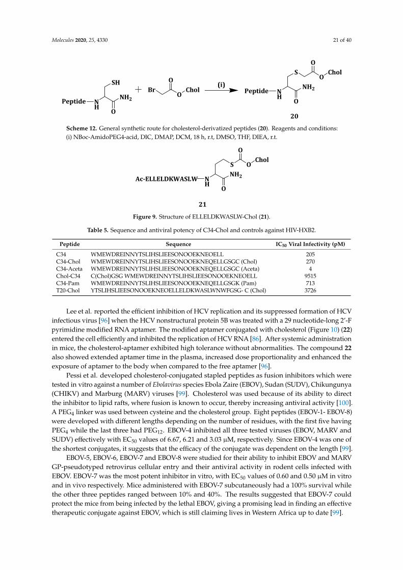
Molecules 2020, 25, 4330 21 of 40
Molecules 2020, 25, x FOR PEER REVIEW 22 of 41
was exhibited by peptide ELLELDKWASLW, which on its own had an IC50 > 100 µM, but, when derivatized with the C-terminal of cholesterol (Figure 9) (21), its antiviral potency improved to IC50 = 5.85 µM. Based on Table 5, the antiviral potency of C34 increased 50-fold when conjugated to cholesterol (C34 and C34-Aceta have IC50 = 205 and 270 µM, respectively, compared to IC50 = 4 for C34-Chol) [98].
Peptide NH
NH2
O
SHBr
OChol
O (i)Peptide N
HNH2
O
SO
CholO
20 Scheme 12. General synthetic route for cholesterol-derivatized peptides (20). Reagents and conditions: (i) NBoc-AmidoPEG4-acid, DIC, DMAP, DCM, 18 h, r.t, DMSO, THF, DIEA, r.t.
NH
NH2
S OChol
O
OAc-ELLELDKWASLW
21 Figure 9. Structure of ELLELDKWASLW-Chol (21).
Chol-C34 showed 50-fold less antiviral potency compared to underivatized C34 because of the cholesterol that was linked to the N- instead of the C-terminal. C34-Chol was found to be approximately 100-fold more potent than enfuvieride, the marketed fusion inhibitor. C34-Chol was also more potent than all the controls against other HIV-1 isolates such as BAL, NL4-3, MN-1, etc. The compound 20 peptides are potent for viral fusion inhibition and also for efficacy of other therapeutic agents such as nucleoside inhibitors and limiters of HIV infection degree, which may be administered simultaneously or in alternating [98].
Table 5. Sequence and antiviral potency of C34-Chol and controls against HIV-HXB2.
Peptide Sequence IC50 Viral Infectivity
(pM) C34 WMEWDREINNYTSLIHSLIEESONOOEKNEOELL 205 C34-Chol WMEWDREINNYTSLIHSLIEESONOOEKNEQELLGSGC (Chol) 270 C34-Aceta
WMEWDREINNYTSLIHSLIEESONOOEKNEQELLGSGC (Aceta) 4
Chol-C34 C(Chol)GSG WMEWDREINNYTSLIHSLIEESONOOEKNEOELL 9515 C34-Pam WMEWDREINNYTSLIHSLIEESONOOEKNEQELLGSGK (Pam) 713
T20-Chol YTSLIHSLIEESONOOEKNEOELLELDKWASLWNWFGSG- C (Chol)
3726
Lee et al. reported the efficient inhibition of HCV replication and its suppressed formation of HCV infectious virus [96] when the HCV nonstructural protein 5B was treated with a 29 nucleotide-long 2’-F pyrimidine modified RNA aptamer. The modified aptamer conjugated with cholesterol (Figure 10) (22) entered the cell efficiently and inhibited the replication of HCV RNA [86]. After systemic administration in mice, the cholesterol-aptamer exhibited high tolerance without abnormalities. The compound 22 also showed extended aptamer time in the plasma, increased dose proportionality and enhanced the exposure of aptamer to the body when compared to the free aptamer [96].
Scheme 12. General synthetic route for cholesterol-derivatized peptides (20). Reagents and conditions:(i) NBoc-AmidoPEG4-acid, DIC, DMAP, DCM, 18 h, r.t, DMSO, THF, DIEA, r.t.
Molecules 2020, 25, x FOR PEER REVIEW 22 of 41
was exhibited by peptide ELLELDKWASLW, which on its own had an IC50 > 100 µM, but, when derivatized with the C-terminal of cholesterol (Figure 9) (21), its antiviral potency improved to IC50 = 5.85 µM. Based on Table 5, the antiviral potency of C34 increased 50-fold when conjugated to cholesterol (C34 and C34-Aceta have IC50 = 205 and 270 µM, respectively, compared to IC50 = 4 for C34-Chol) [98].
Peptide NH
NH2
O
SHBr
OChol
O (i)Peptide N
HNH2
O
SO
CholO
20 Scheme 12. General synthetic route for cholesterol-derivatized peptides (20). Reagents and conditions: (i) NBoc-AmidoPEG4-acid, DIC, DMAP, DCM, 18 h, r.t, DMSO, THF, DIEA, r.t.
NH
NH2
S OChol
O
OAc-ELLELDKWASLW
21 Figure 9. Structure of ELLELDKWASLW-Chol (21).
Chol-C34 showed 50-fold less antiviral potency compared to underivatized C34 because of the cholesterol that was linked to the N- instead of the C-terminal. C34-Chol was found to be approximately 100-fold more potent than enfuvieride, the marketed fusion inhibitor. C34-Chol was also more potent than all the controls against other HIV-1 isolates such as BAL, NL4-3, MN-1, etc. The compound 20 peptides are potent for viral fusion inhibition and also for efficacy of other therapeutic agents such as nucleoside inhibitors and limiters of HIV infection degree, which may be administered simultaneously or in alternating [98].
Table 5. Sequence and antiviral potency of C34-Chol and controls against HIV-HXB2.
Peptide Sequence IC50 Viral Infectivity
(pM) C34 WMEWDREINNYTSLIHSLIEESONOOEKNEOELL 205 C34-Chol WMEWDREINNYTSLIHSLIEESONOOEKNEQELLGSGC (Chol) 270 C34-Aceta
WMEWDREINNYTSLIHSLIEESONOOEKNEQELLGSGC (Aceta) 4
Chol-C34 C(Chol)GSG WMEWDREINNYTSLIHSLIEESONOOEKNEOELL 9515 C34-Pam WMEWDREINNYTSLIHSLIEESONOOEKNEQELLGSGK (Pam) 713
T20-Chol YTSLIHSLIEESONOOEKNEOELLELDKWASLWNWFGSG- C (Chol)
3726
Lee et al. reported the efficient inhibition of HCV replication and its suppressed formation of HCV infectious virus [96] when the HCV nonstructural protein 5B was treated with a 29 nucleotide-long 2’-F pyrimidine modified RNA aptamer. The modified aptamer conjugated with cholesterol (Figure 10) (22) entered the cell efficiently and inhibited the replication of HCV RNA [86]. After systemic administration in mice, the cholesterol-aptamer exhibited high tolerance without abnormalities. The compound 22 also showed extended aptamer time in the plasma, increased dose proportionality and enhanced the exposure of aptamer to the body when compared to the free aptamer [96].
Figure 9. Structure of ELLELDKWASLW-Chol (21).
Table 5. Sequence and antiviral potency of C34-Chol and controls against HIV-HXB2.
Peptide Sequence IC50 Viral Infectivity (pM)
C34 WMEWDREINNYTSLIHSLIEESONOOEKNEOELL 205C34-Chol WMEWDREINNYTSLIHSLIEESONOOEKNEQELLGSGC (Chol) 270C34-Aceta WMEWDREINNYTSLIHSLIEESONOOEKNEQELLGSGC (Aceta) 4Chol-C34 C(Chol)GSG WMEWDREINNYTSLIHSLIEESONOOEKNEOELL 9515C34-Pam WMEWDREINNYTSLIHSLIEESONOOEKNEQELLGSGK (Pam) 713T20-Chol YTSLIHSLIEESONOOEKNEOELLELDKWASLWNWFGSG- C (Chol) 3726
Lee et al. reported the efficient inhibition of HCV replication and its suppressed formation of HCVinfectious virus [96] when the HCV nonstructural protein 5B was treated with a 29 nucleotide-long 2’-Fpyrimidine modified RNA aptamer. The modified aptamer conjugated with cholesterol (Figure 10) (22)entered the cell efficiently and inhibited the replication of HCV RNA [86]. After systemic administrationin mice, the cholesterol-aptamer exhibited high tolerance without abnormalities. The compound 22also showed extended aptamer time in the plasma, increased dose proportionality and enhanced theexposure of aptamer to the body when compared to the free aptamer [96].
Pessi et al. developed cholesterol-conjugated stapled peptides as fusion inhibitors which weretested in vitro against a number of Ebolavirus species Ebola Zaire (EBOV), Sudan (SUDV), Chikungunya(CHIKV) and Marburg (MARV) viruses [99]. Cholesterol was used because of its ability to directthe inhibitor to lipid rafts, where fusion is known to occur, thereby increasing antiviral activity [100].A PEG4 linker was used between cysteine and the cholesterol group. Eight peptides (EBOV-1- EBOV-8)were developed with different lengths depending on the number of residues, with the first five havingPEG4 while the last three had PEG12. EBOV-4 inhibited all three tested viruses (EBOV, MARV andSUDV) effectively with EC50 values of 6.67, 6.21 and 3.03 µM, respectively. Since EBOV-4 was one ofthe shortest conjugates, it suggests that the efficacy of the conjugate was dependent on the length [99].
EBOV-5, EBOV-6, EBOV-7 and EBOV-8 were studied for their ability to inhibit EBOV and MARVGP-pseudotyped retrovirus cellular entry and their antiviral activity in rodent cells infected withEBOV. EBOV-7 was the most potent inhibitor in vitro, with EC50 values of 0.60 and 0.50 µM in vitroand in vivo respectively. Mice administered with EBOV-7 subcutaneously had a 100% survival whilethe other three peptides ranged between 10% and 40%. The results suggested that EBOV-7 couldprotect the mice from being infected by the lethal EBOV, giving a promising lead in finding an effectivetherapeutic conjugate against EBOV, which is still claiming lives in Western Africa up to date [99].
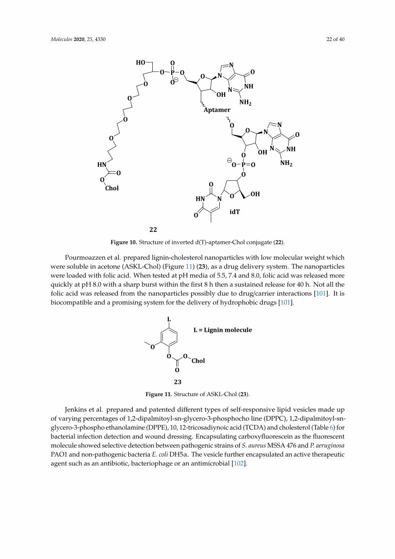
Molecules 2020, 25, 4330 22 of 40Molecules 2020, 25, x FOR PEER REVIEW 23 of 41
O
HN
O
O
O
O
O
HOP OO
O
O NN
N NHOH
NH2Aptamer
O
OO N
N
N NH
NH2
OHOPOO
ONOH
HN
O
O
O
O
O
idT
Chol
22
Figure 10. Structure of inverted d(T)-aptamer-Chol conjugate (22).
Pessi et al. developed cholesterol-conjugated stapled peptides as fusion inhibitors which were tested in vitro against a number of Ebolavirus species Ebola Zaire (EBOV), Sudan (SUDV), Chikungunya (CHIKV) and Marburg (MARV) viruses [99]. Cholesterol was used because of its ability to direct the inhibitor to lipid rafts, where fusion is known to occur, thereby increasing antiviral activity [100]. A PEG4 linker was used between cysteine and the cholesterol group. Eight peptides (EBOV-1- EBOV-8) were developed with different lengths depending on the number of residues, with the first five having PEG4 while the last three had PEG12. EBOV-4 inhibited all three tested viruses (EBOV, MARV and SUDV) effectively with EC50 values of 6.67, 6.21 and 3.03 µM, respectively. Since EBOV-4 was one of the shortest conjugates, it suggests that the efficacy of the conjugate was dependent on the length [99].
EBOV-5, EBOV-6, EBOV-7 and EBOV-8 were studied for their ability to inhibit EBOV and MARV GP-pseudotyped retrovirus cellular entry and their antiviral activity in rodent cells infected with EBOV. EBOV-7 was the most potent inhibitor in vitro, with EC50 values of 0.60 and 0.50 µM in vitro and in vivo respectively. Mice administered with EBOV-7 subcutaneously had a 100% survival while the other three peptides ranged between 10% and 40%. The results suggested that EBOV-7 could protect the mice from being infected by the lethal EBOV, giving a promising lead in finding an effective therapeutic conjugate against EBOV, which is still claiming lives in Western Africa up to date [99].
Pourmoazzen et al. prepared lignin-cholesterol nanoparticles with low molecular weight which were soluble in acetone (ASKL-Chol) (Figure 11) (23), as a drug delivery system. The nanoparticles were loaded with folic acid. When tested at pH media of 5.5, 7.4 and 8.0, folic acid was released more quickly at pH 8.0 with a sharp burst within the first 8 h then a sustained release for 40 h. Not all the folic acid was released from the nanoparticles possibly due to drug/carrier interactions [101]. It is biocompatible and a promising system for the delivery of hydrophobic drugs [101].
Figure 10. Structure of inverted d(T)-aptamer-Chol conjugate (22).
Pourmoazzen et al. prepared lignin-cholesterol nanoparticles with low molecular weight whichwere soluble in acetone (ASKL-Chol) (Figure 11) (23), as a drug delivery system. The nanoparticleswere loaded with folic acid. When tested at pH media of 5.5, 7.4 and 8.0, folic acid was released morequickly at pH 8.0 with a sharp burst within the first 8 h then a sustained release for 40 h. Not all thefolic acid was released from the nanoparticles possibly due to drug/carrier interactions [101]. It isbiocompatible and a promising system for the delivery of hydrophobic drugs [101].Molecules 2020, 25, x FOR PEER REVIEW 24 of 41
OO
L
O
OChol
L = Lignin molecule
23 Figure 11. Structure of ASKL-Chol (23).
Jenkins et al. prepared and patented different types of self-responsive lipid vesicles made up of varying percentages of 1,2-dipalmitoyl-sn-glycero-3-phosphocho line (DPPC), 1,2-dipalmitoyl-sn-glycero-3-phospho ethanolamine (DPPE), 10, 12-tricosadiynoic acid (TCDA) and cholesterol (Table 6) for bacterial infection detection and wound dressing. Encapsulating carboxyfluorescein as the fluorescent molecule showed selective detection between pathogenic strains of S. aureus MSSA 476 and P. aeruginosa PAO1 and non-pathogenic bacteria E. coli DH5α. The vesicle further encapsulated an active therapeutic agent such as an antibiotic, bacteriophage or an antimicrobial [102].
Table 6. Vesicle types, with mol % of the phospholipid/fatty acid content and average fluorescent increase in response to bacteria.
Vesicle Types and Composition Average Fluorescent Increment
Class Vesicle Types DPPC DSPC
Chole-Sterol TCDA
Hepes (Buffer) PAO1 MSSA476
A 1 88 - 10 - 37,500 115,000 75,000 2 78 - 20 - 1000 100,000 37,500 3 68 - 30 - 1000 37,500 12,500
B 4 73 - 0 25 50,000 105,000 75,000 5 63 - 10 25 37,500 102,000 76,000 6 53 - 20 25 12,500 97,000 72,000 7 43 - 30 25 1000 35,000 69,000 8 33 - 40 25 1000 13,000 73,000 9 23 - 50 25 1000 11,000 78,000
C 10 - 88 10 - 500 20,000 10,000 11 - 78 20 - 550 55,000 11,000 12 - 68 30 - 200 12,500 5000
D 13 - 73 20 5 500 60,000 35,000 14 - 68 20 10 450 50,000 40,000 15 - 63 20 15 500 42,500 62,500 16 - 58 20 20 550 52,500 85,000 17 - 53 20 25 400 35,000 37,000
E 18 - 63 10 25 200 20,000 300,000 19 - 43 30 25 300 25,000 45,000
Hepes buffer was used as the control.
The selective responses were shown by the different percentages of fluorescent responses exhibited by the different vesicle types to both PAO1 and MSSA476. For example, vesicle type 1 had a fluorescence response of about 50% to PAO1 and 80% to MSSA476 while vesicle type 2 showed 65% response to PAO1 and less than 10% for MSSA476. This was attributed to the different rates of lipids loss from the cell membranes of the bacteria. The different vesicle compositions affected membrane fluidity, exotoxins binding and cell membrane hemolysis, which all affected the sensitivity of the
Figure 11. Structure of ASKL-Chol (23).
Jenkins et al. prepared and patented different types of self-responsive lipid vesicles made upof varying percentages of 1,2-dipalmitoyl-sn-glycero-3-phosphocho line (DPPC), 1,2-dipalmitoyl-sn-glycero-3-phospho ethanolamine (DPPE), 10, 12-tricosadiynoic acid (TCDA) and cholesterol (Table 6) forbacterial infection detection and wound dressing. Encapsulating carboxyfluorescein as the fluorescentmolecule showed selective detection between pathogenic strains of S. aureus MSSA 476 and P. aeruginosaPAO1 and non-pathogenic bacteria E. coli DH5α. The vesicle further encapsulated an active therapeuticagent such as an antibiotic, bacteriophage or an antimicrobial [102].
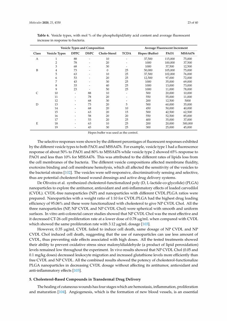
Molecules 2020, 25, 4330 23 of 40
Table 6. Vesicle types, with mol % of the phospholipid/fatty acid content and average fluorescentincrease in response to bacteria.
Vesicle Types and Composition Average Fluorescent Increment
Class Vesicle Types DPPC DSPC Chole-Sterol TCDA Hepes (Buffer) PAO1 MSSA476
A 1 88 - 10 - 37,500 115,000 75,0002 78 - 20 - 1000 100,000 37,5003 68 - 30 - 1000 37,500 12,500
B 4 73 - 0 25 50,000 105,000 75,0005 63 - 10 25 37,500 102,000 76,0006 53 - 20 25 12,500 97,000 72,0007 43 - 30 25 1000 35,000 69,0008 33 - 40 25 1000 13,000 73,0009 23 - 50 25 1000 11,000 78,000
C 10 - 88 10 - 500 20,000 10,00011 - 78 20 - 550 55,000 11,00012 - 68 30 - 200 12,500 5000
D 13 - 73 20 5 500 60,000 35,00014 - 68 20 10 450 50,000 40,00015 - 63 20 15 500 42,500 62,50016 - 58 20 20 550 52,500 85,00017 - 53 20 25 400 35,000 37,000
E 18 - 63 10 25 200 20,000 300,00019 - 43 30 25 300 25,000 45,000
Hepes buffer was used as the control.
The selective responses were shown by the different percentages of fluorescent responses exhibitedby the different vesicle types to both PAO1 and MSSA476. For example, vesicle type 1 had a fluorescenceresponse of about 50% to PAO1 and 80% to MSSA476 while vesicle type 2 showed 65% response toPAO1 and less than 10% for MSSA476. This was attributed to the different rates of lipids loss fromthe cell membranes of the bacteria. The different vesicle compositions affected membrane fluidity,exotoxins binding and cell membrane hemolysis, which all affected the sensitivity of the vesicles tothe bacterial strains [102]. The vesicles were self-responsive, discriminatively sensing and selective,thus are potential cholesterol-based wound dressings and active drug delivery systems.
De Oliveira et al. synthesized cholesterol-functionalized poly (D, L-lactide-co-glycolide) (PLGA)nanoparticles to explore the antitumor, antioxidant and anti-inflammatory effects of loaded carvedilol(CVDL). CVDL-free nanoparticles (NP) and nanoparticles with different CVDL:PLGA ratios wereprepared. Nanoparticles with a weight ratio of 1:10 for CVDL:PLGA had the highest drug loadingefficiency of 95.06% and these were functionalized with cholesterol to give NP CVDL Chol. All thethree nanoparticles (NP, NP CVDL and NP CVDL Chol) were spherical with smooth and uniformsurfaces. In vitro anti-colorectal cancer studies showed that NP CVDL Chol was the most effective andit decreased CT-26 cell proliferation rate at a lower dose of 0.78 µg/mL when compared with CVDLwhich showed the same proliferation rate with 3.12 µg/mL dosage [103].
However, 0.35 µg/mL CVDL failed to induce cell death, same dosage of NP CVDL and NPCVDL Chol induced cell death, suggesting that the use of nanoparticles can use less amount ofCVDL, thus preventing side effects associated with high doses. All the tested treatments showedtheir ability to prevent oxidative stress since malonyldialdehyde (a product of lipid peroxidation)levels remained low throughout the experiment. In vivo results showed that NP CVDL Chol (0.05 and0.1 mg/kg doses) decreased leukocyte migration and increased glutathione levels more efficiently thanfree CVDL and NP CVDL. All the combined results showed the potency of cholesterol-functionalizePLGA nanoparticles in decreasing CVDL dosage without affecting its antitumor, antioxidant andanti-inflammatory effects [103].
5. Cholesterol-Based Compounds in Transdermal Drug Delivery
The healing of cutaneous wounds has four stages which are hemostasis, inflammation, proliferationand maturation [104]. Angiogenesis, which is the formation of new blood vessels, is an essential
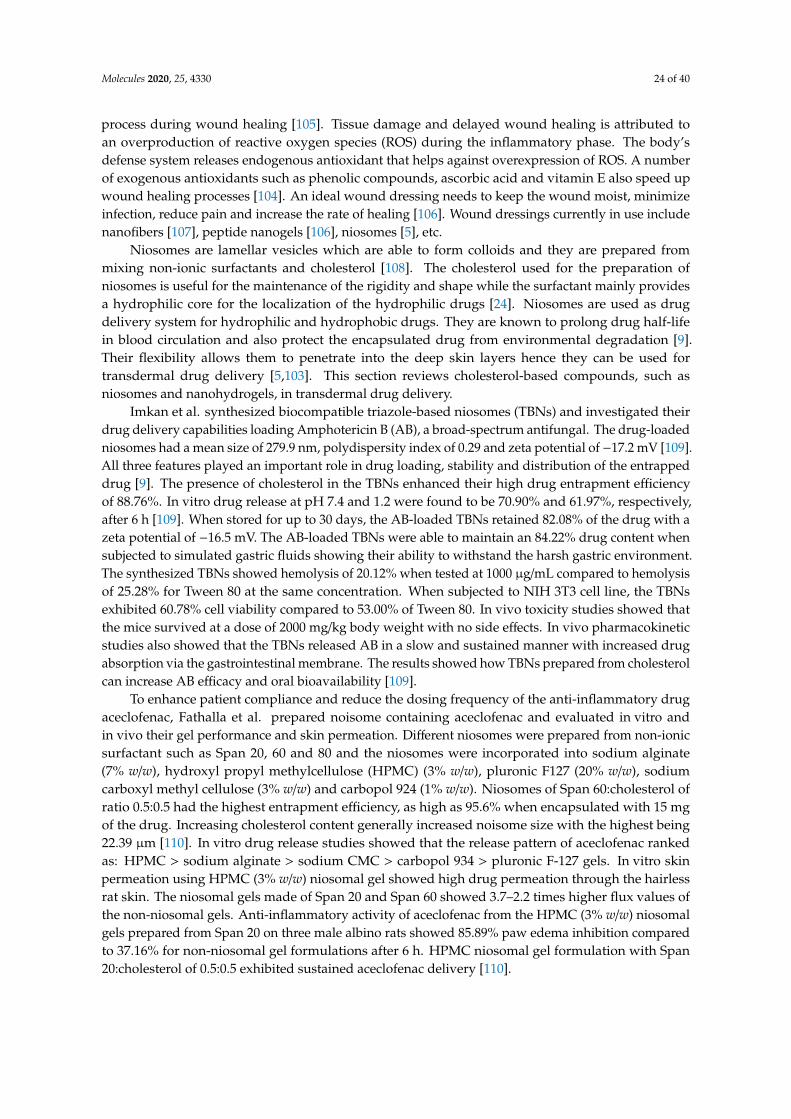
Molecules 2020, 25, 4330 24 of 40
process during wound healing [105]. Tissue damage and delayed wound healing is attributed toan overproduction of reactive oxygen species (ROS) during the inflammatory phase. The body’sdefense system releases endogenous antioxidant that helps against overexpression of ROS. A numberof exogenous antioxidants such as phenolic compounds, ascorbic acid and vitamin E also speed upwound healing processes [104]. An ideal wound dressing needs to keep the wound moist, minimizeinfection, reduce pain and increase the rate of healing [106]. Wound dressings currently in use includenanofibers [107], peptide nanogels [106], niosomes [5], etc.
Niosomes are lamellar vesicles which are able to form colloids and they are prepared frommixing non-ionic surfactants and cholesterol [108]. The cholesterol used for the preparation ofniosomes is useful for the maintenance of the rigidity and shape while the surfactant mainly providesa hydrophilic core for the localization of the hydrophilic drugs [24]. Niosomes are used as drugdelivery system for hydrophilic and hydrophobic drugs. They are known to prolong drug half-lifein blood circulation and also protect the encapsulated drug from environmental degradation [9].Their flexibility allows them to penetrate into the deep skin layers hence they can be used fortransdermal drug delivery [5,103]. This section reviews cholesterol-based compounds, such asniosomes and nanohydrogels, in transdermal drug delivery.
Imkan et al. synthesized biocompatible triazole-based niosomes (TBNs) and investigated theirdrug delivery capabilities loading Amphotericin B (AB), a broad-spectrum antifungal. The drug-loadedniosomes had a mean size of 279.9 nm, polydispersity index of 0.29 and zeta potential of −17.2 mV [109].All three features played an important role in drug loading, stability and distribution of the entrappeddrug [9]. The presence of cholesterol in the TBNs enhanced their high drug entrapment efficiencyof 88.76%. In vitro drug release at pH 7.4 and 1.2 were found to be 70.90% and 61.97%, respectively,after 6 h [109]. When stored for up to 30 days, the AB-loaded TBNs retained 82.08% of the drug with azeta potential of −16.5 mV. The AB-loaded TBNs were able to maintain an 84.22% drug content whensubjected to simulated gastric fluids showing their ability to withstand the harsh gastric environment.The synthesized TBNs showed hemolysis of 20.12% when tested at 1000 µg/mL compared to hemolysisof 25.28% for Tween 80 at the same concentration. When subjected to NIH 3T3 cell line, the TBNsexhibited 60.78% cell viability compared to 53.00% of Tween 80. In vivo toxicity studies showed thatthe mice survived at a dose of 2000 mg/kg body weight with no side effects. In vivo pharmacokineticstudies also showed that the TBNs released AB in a slow and sustained manner with increased drugabsorption via the gastrointestinal membrane. The results showed how TBNs prepared from cholesterolcan increase AB efficacy and oral bioavailability [109].
To enhance patient compliance and reduce the dosing frequency of the anti-inflammatory drugaceclofenac, Fathalla et al. prepared noisome containing aceclofenac and evaluated in vitro andin vivo their gel performance and skin permeation. Different niosomes were prepared from non-ionicsurfactant such as Span 20, 60 and 80 and the niosomes were incorporated into sodium alginate(7% w/w), hydroxyl propyl methylcellulose (HPMC) (3% w/w), pluronic F127 (20% w/w), sodiumcarboxyl methyl cellulose (3% w/w) and carbopol 924 (1% w/w). Niosomes of Span 60:cholesterol ofratio 0.5:0.5 had the highest entrapment efficiency, as high as 95.6% when encapsulated with 15 mgof the drug. Increasing cholesterol content generally increased noisome size with the highest being22.39 µm [110]. In vitro drug release studies showed that the release pattern of aceclofenac rankedas: HPMC > sodium alginate > sodium CMC > carbopol 934 > pluronic F-127 gels. In vitro skinpermeation using HPMC (3% w/w) niosomal gel showed high drug permeation through the hairlessrat skin. The niosomal gels made of Span 20 and Span 60 showed 3.7–2.2 times higher flux values ofthe non-niosomal gels. Anti-inflammatory activity of aceclofenac from the HPMC (3% w/w) niosomalgels prepared from Span 20 on three male albino rats showed 85.89% paw edema inhibition comparedto 37.16% for non-niosomal gel formulations after 6 h. HPMC niosomal gel formulation with Span20:cholesterol of 0.5:0.5 exhibited sustained aceclofenac delivery [110].
Molecules 2020, 25, 4330 25 of 40
Manconi prepared baicalin-embedded gellan-cholesterol nanohydrogels for wound healing.Baicalin sonicated nanohydrogel (BSN) and baicalin autoclaved nanohydrogel (BAN) were comparedagainst empty sonicated nanohydrogels (ESN) and empty autoclaved nanohydrogel (EAN) andcharacterized, as displayed on Table 7. The negative zeta potentials confirmed the stability of thenanohydrogels upon storage. In vitro skin delivery of baicalin showed similar drug accumulationin stratum corneum and epidermis. Treatment of 3T3 cells with hydrogen peroxide and baicalinshowed that baicalin counteracted the oxidative effect of hydrogen peroxide with a 95% cell viability,irrespective of the dosage administered (20, 10, 1 and 0.1 µg/mL). These results showed the excellentantioxidant capacity of baicalin [111].
Table 7. Mean diameter (MD), zeta potential (ZP), polydispersity index (PDI) and entrapment efficiency(EE %) of the nanohydrogels.
Nanohydrogel MD (nm) ZP (mV) PDI EE (%)
ESN 554 −30 0.45 -EAN 383 −24 0.19 -BSN 507 −25 0.49 38BAN 341 −23 0.30 35
In vivo wound healing activity of baicalin was studied by injuring the back skin of miceusing topically applied 12-O-tetradecanoylphorbol 13-acetate (TPA) followed by treating the lesionswith baicalin. The results showed that baicalin-loaded formulation healed the TPA lesions andinhibited inflammation markers more efficiently than baicalin in PBS (10 mg/mL) and evendexamethasone, a potent anti-inflammatory drug. BAN inhibited two inflammation markers,edema and myeloperoxidase (MPO), by 78% and 44%, respectively, while betamethasone creamshowed no effective inhibition. BAN also showed 100% cytokine inhibition. These results showedthe potential use of the autoclaved nanohydrogel in topical wound treatment and other skin diseasescaused by oxidative stress and inflammation [111].
Farmoudeh et al. prepared methylene blue (MB)-loaded niosomes using Span 60, Tween 60 andcholesterol in different ratios. The MB-loaded niosomes had a maximum size of 613.76 nm, a maximumentrapment efficiency of 75% and negative zeta potential values. Increasing the cholesterol to surfactantratio decreased the vesicle size. The authors used equations to prepare an optimized MB-loadedniosomal formulation with 147.8 nm vesicle size, −18.0 mV zeta potential and 63.27% entrapmentefficiency. The optimized niosomal dispersion exhibited a controlled MB release rate with 26.897%of the drug released in the first hour compared to 45.064% of the free drug. In vivo results from thewounded male Wistar rats administered the MB (niosomal gel of free) transdermally showed significantwound closure when compared to those treated with placebo gel that was still open after 14 days [104].The niosomal-gel treated wounds had thicker, organized collagen fibers with more fibroblasts and lessinflammatory infiltrate compared to thin granulation tissue, disorganized collagen fibers and highnumber of inflammatory cells exhibited in free MB-treated and control rats. Niosomal gel-treatedrats had significantly decreased malondialdehyde (MDA) and elevated Superoxide dismutase (SOD)levels only three days after surgery, but it took seven days for negative controls to reach the similarresults [104]. Since SOD is an endogenous antioxidant and MDA is an end product of lipid peroxidation,the increase of the former and decrease of the latter showed the ability of niosomal gel in acceleratingthe rate of mitochondrial cytochrome c reduction thereby reducing the production free radicals in themitochondria [112].
Aziz et al. prepared niosomes for efficient transdermal delivery of diacerein (DCN) and avoidinggastrointestinal problems associated with its oral administration. The niosomes were preparedfrom cholesterol and Span 40, Span 60, Span 80, Tween 20 or Tween 80 in varying ratios, with onlySpan 40 and Span 60 giving milky vesicular dispersion upon hydration. The optimal DCN-loadedniosomes had an entrapment efficiency of 95.63%, particle size of 436.65 nm, polydispersity index

Molecules 2020, 25, 4330 26 of 40
of 0.47 and −38.80 mV zeta potential. The optimal DCN-loaded niosomes (optimized using centralcomposite design) were prepared in hydration medium with no salt and with a lipid:surfactant ratioof 150 mg:5 parts. The optimal niosomes composition was regulated using edge activators to giveelastic vesicles for comparing with optimal niosomes [113]. The optimal niosomes enhanced the skinpermeation of DCN ex vivo with up to four times better than the drug suspension. In vivo skindeposition of DCN expressed by AUC0−10 was four folds for elastic vesicles (65.8 µg h/cm2) and twofolds for the optimized niosomes (32.7 µg h/cm2) compared to 16.7 µg h/cm2 for the drug suspension.The elastic vesicle was more effective than the optimal niosomes and improved drug retention anddeposition within the skin [114]. The optimized niosomes proved their ability to transdermally deliverDCN, avoiding problems associated with oral administration [113].
Shirsand et al. prepared niosomal gel formulations for the delivery of ketoconazole (KTZ),to improve its fungal skin treatment efficiently. Niosomes consisting of different ratios of Span 40,Span 60, Tween 60 and cholesterol were prepared (Table 8). The vesicles had a spherical shape and asmooth surface. Niosomes with the highest cholesterol content (TNS3, TNS6 and TNS9) displayedsmall particle size (Table 9). Increasing cholesterol content, increased the entrapment efficiency ofKTZ. The entrapment efficiency (EE) increase was attributed to the ability of cholesterol to close spacesbetween the bilayer membranes preventing drug leakage [115].
Table 8. Formulations of KTZ niosomes. Ratios (KTZ: surfactant: cholesterol). All ingredients weremeasure in mg. Ten milliliters of chloroform and 20 mL of PBS (pH 7.4) were added to each formulation.
Formulation CodeIngreDients
Ratios KTZ Span 40 Span 60 Tween 60 Cholesterol
TNS1 1:1:0.2 100 100 - - 20TNS2 1:1.5:0.3 100 150 - - 30TNS3 1:2:0.4 100 200 - - 40TNS4 1:1:0.2 100 - 100 - 20TNS5 1:1.5:0.3 100 - 150 - 30TNS6 1:2:0.4 100 - 200 - 40TNS7 1:1:0.2 100 - - 100 20TNS8 1:1.5:0.3 100 - - 150 30TNS9 1:2:0.4 100 - - 200 40
Table 9. Evaluations for KTZ niosomes.
Formulation Code Particle Size (µm) % EE 24 h Cumulative Drug Release (%)
TNS1 6.12 55.14 72.371TNS2 5.76 59.08 68.113TNS3 5.4 63.02 65.469TNS4 5.94 69.39 66.068TNS5 5.22 73.48 59.446TNS6 4.86 76.96 53.204TNS7 7.38 71.66 55.909TNS8 6.84 76.36 49.628TNS9 6.3 78.63 46.636
In vitro studies showed the drug release was in the range of 46.63–72.37% with the drug beingreleased by both diffusion and erosion. Though TNS1 had the highest cumulative drug release after24 h, TNS4 was found to be the best when combining vesicular size (5.94 µm), EE (69.39%) and drugrelease (66.06%). TNS4 was incorporated into a carbopol gel for further studies. TNS4 has a pH of 5.56,a drug content of 96.425 and 8370 cps viscosity. TNS4, plain gel and a marketed ointment had in vitrodrug release studies of 36.18%, 73.21% and 82.06%, respectively, after 12 h showing the sustained drugrelease for TNS4. In vitro antifungal activity studies revealed that TNS4 had the highest inhibitionzone of 25.3 mm compared to 19.6 and 23.3 mm for the ketoconazole plain gel and marketed ointment.
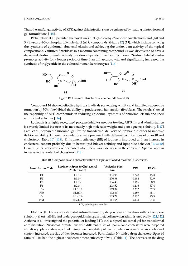
Molecules 2020, 25, 4330 27 of 40
Thus, the antifungal activity of KTZ against skin infections can be enhanced by loading it into niosomalgel formulations [115].
Ptchelintsev et al. patented the novel uses of 3’-(L-ascorbyl-2-o-phosphoryl)-cholesterol (24) and3’-(L-ascorbyl-3-o-phosphoryl)-cholesterol (APC compounds) (Figure 12) (25), which include reducingthe synthesis of epidermal abnormal elastin and achieving the antioxidant activity of the topicalcompositions. Cultured fibroblasts in a medium containing compound 24 was discovered to have adecreased elastin promoter activity in a dose-dependent manner. Compound 24 also inhibited elastinpromoter activity for a longer period of time than did ascorbic acid and significantly increased thesynthesis of triglyceride in the cultured human keratinocytes [116].
Molecules 2020, 25, x FOR PEER REVIEW 28 of 41
In vitro studies showed the drug release was in the range of 46.63–72.37% with the drug being released by both diffusion and erosion. Though TNS1 had the highest cumulative drug release after 24 h, TNS4 was found to be the best when combining vesicular size (5.94 µm), EE (69.39%) and drug release (66.06%). TNS4 was incorporated into a carbopol gel for further studies. TNS4 has a pH of 5.56, a drug content of 96.425 and 8370 cps viscosity. TNS4, plain gel and a marketed ointment had in vitro drug release studies of 36.18%, 73.21% and 82.06%, respectively, after 12 h showing the sustained drug release for TNS4. In vitro antifungal activity studies revealed that TNS4 had the highest inhibition zone of 25.3 mm compared to 19.6 and 23.3 mm for the ketoconazole plain gel and marketed ointment. Thus, the antifungal activity of KTZ against skin infections can be enhanced by loading it into niosomal gel formulations [115].
Table 9. Evaluations for KTZ niosomes.
Formulation Code Particle Size (µm) % EE 24 h Cumulative Drug Release (%) TNS1 6.12 55.14 72.371 TNS2 5.76 59.08 68.113 TNS3 5.4 63.02 65.469 TNS4 5.94 69.39 66.068 TNS5 5.22 73.48 59.446 TNS6 4.86 76.96 53.204 TNS7 7.38 71.66 55.909 TNS8 6.84 76.36 49.628 TNS9 6.3 78.63 46.636
Ptchelintsev et al. patented the novel uses of 3’-(L-ascorbyl-2-o-phosphoryl)-cholesterol (24) and 3’-(L-ascorbyl-3-o-phosphoryl)-cholesterol (APC compounds) (Figure 12) (25), which include reducing the synthesis of epidermal abnormal elastin and achieving the antioxidant activity of the topical compositions. Cultured fibroblasts in a medium containing compound 24 was discovered to have a decreased elastin promoter activity in a dose-dependent manner. Compound 24 also inhibited elastin promoter activity for a longer period of time than did ascorbic acid and significantly increased the synthesis of triglyceride in the cultured human keratinocytes [116].
PO
CholO
OH
O
HO
O
HO
HOP
OChol
O
OH
O
HO
O
HO
HO
24 25
Figure 12. Chemical structures of compounds 24 and 25.
Compound 24 showed effective hydroxyl radicals scavenging activity and inhibited superoxide formation by 50%. It exhibited the ability to produce new human skin fibroblasts. The results showed the capability of APC compounds in reducing epidermal synthesis of abnormal elastin and their antioxidant activities [116].
Lopinavir is a highly recognized protease inhibitor used for treating AIDS. Its oral administration is severely limited because of its moderately high molecular weight and poor aqueous solubility [117]. Patel et al. prepared a niosomal gel for the transdermal delivery of lopinavir in order to improve its bioavailability. Different formulations were prepared with different compositions of Span 40 and cholesterol (Table 10) [118]. Entrapment efficiency (EE) of lopinavir improved with an increase in cholesterol content probably due to better lipid bilayer stability and lipophilic behavior [119,120]. Generally, the vesicular size decreased when there was a decrease in the content of Span 40 and an increase in the content of cholesterol [118].
Figure 12. Chemical structures of compounds 24 and 25.
Compound 24 showed effective hydroxyl radicals scavenging activity and inhibited superoxideformation by 50%. It exhibited the ability to produce new human skin fibroblasts. The results showedthe capability of APC compounds in reducing epidermal synthesis of abnormal elastin and theirantioxidant activities [116].
Lopinavir is a highly recognized protease inhibitor used for treating AIDS. Its oral administrationis severely limited because of its moderately high molecular weight and poor aqueous solubility [117].Patel et al. prepared a niosomal gel for the transdermal delivery of lopinavir in order to improveits bioavailability. Different formulations were prepared with different compositions of Span 40 andcholesterol (Table 10) [118]. Entrapment efficiency (EE) of lopinavir improved with an increase incholesterol content probably due to better lipid bilayer stability and lipophilic behavior [119,120].Generally, the vesicular size decreased when there was a decrease in the content of Span 40 and anincrease in the content of cholesterol [118].
Table 10. Composition and characterization of lopinavir-loaded niosomal dispersions.
Formulation Code Lopinavir:Span 40:Cholesterol(Molar Ratio)
Vesicular Size(nm) PDI EE (%)
F1 1:0.5:- 354.94 0.228 45.3F2 1:1.0:- 276.38 0.194 52.9F3 1:1.5:- 196.45 0.165 58.9F4 1:2.0:- 203.52 0.216 57.4
F3a 1:1.3:0.2 169.34 0.212 62.5F3b 1:1.1:0.4 132.86 0.189 68.1F3c 1:0.9:0.6 105.22 0.127 75.5F3d 1:0.7:0.8 114.65 0.133 74.5
PDI, polydispersity index.
Etodolac (ETD) is a non-steroidal anti-inflammatory drug whose application suffers from poorsolubility, short half-life and undergoes quick s first pass metabolism when administered orally [121,122].Asthana et al. investigated the potential of loading ETD into a topical niosomal gel for transdermaladministration. Niosomal formulations with different ratios of Span 60 and cholesterol were preparedand dicetyl phosphate was added to improve the stability of the formulations over time. As cholesterolcontent increased, the size of the niosomes increased. Formulation N2 with a drug:cholesterol:Span 60ratio of 1:1:1 had the highest drug entrapment efficiency of 96% (Table 11). The decrease in the drug
Molecules 2020, 25, 4330 28 of 40
entrapment efficiency as the amount of cholesterol increased above the ratio of 1 suggests that acompetition of cholesterol and the drug for packing space in the bilayer [122].
Table 11. Composition molar ratios, entrapment efficiency (EE %) and 24 h cumulative drug release forthe different niosomal formulations.
Formulation Code Drug:Chol:Span 60(Molar Ratio) EE (%) Cumulative Drug
Release after 24 h
E1 1:0.5:1 85 70E2 1:1:1 96 91E3 1:1.5:1 78 87E4 1:1:0.5 87 82E5 1:1:1.5 89 60
The particle sizes of the niosomal formulations were in the range of 2–4 µm. The particlesizes increased with increase in the content of cholesterol. When the content of cholesterol islow, there is a close packing of cholesterol and nonionic surfactant with increased curvature andreduced size. When the content of cholesterol is increased, the content of surfactants decreased andthe hydrophobicity of the bilayer membrane increased, thereby increasing the vesicles radius andestablishing a thermodynamic stable form. Furthermore, the bilayer membrane displayed a rigidstructure due to cholesterol content, which results in reduced size from sonication and a bigger vesiclessize [122].
In vitro studies performed in phosphate buffer solution at pH 7.4 showed that the E2 formulationhad the highest drug release of 94.91%. Ex vivo skin permeation studies using abdominal male Albinorat skin showed drug release increased up to 97.57% in 8 h for ETD topical gel while the topicalniosomal gel only released 81.73% of its entrapped drug within the same time frame. The drug releasewas faster from the topical gel when compared to the niosomal vesicle. The results showed the potencyof niosomal gel formulations for transdermal drug delivery [122]. Alcantar et al. invented and patenteda drug delivery system composed of niosomes embedded in a polymer hydrogel for controlled drugrelease in cancerous tumors. The niosomes were prepared by combining Span 60, cholesterol anddicetyl phosphate in the ratio of 1:1:0.1 and loaded with a fluorescent dye. When immersed in water,the niosomes displayed an exponential dye release initially followed by a steady release [123].
Fetihg prepared non-ionic fluconazole-loaded niosomal gels that can be used in topical oculardrug delivery to treat fungal infections of the cornea. The niosomal gels were prepared using Span 60and cholesterol, incorporated into poloxamer 407 and chitosan gel formulations. The niosomes thatwere prepared from 2:1 surfactant:cholesterol (F2) had the highest drug entrapment efficiency of 85.35%and a superior drug release profile. Further increase in the cholesterol content decreased the drugentrapment efficiency possibly due to the regular bilayer structure disruption. F2 niosomal poloxamergel maintained its appearance and the drug content when stored for eight weeks at 4 ◦C [23].
Evaluation of the antifungal activity of the F2 niosomal formulation showed that the two testedformulations (F2 poloxamer and F2 chitosan gels) showed good inhibition zones (Table 12). F2 niosomalpoloxamer gel and F2 niosomal chitosan gel inhibited both C. albicans and C. tropicum better thanMiconazole, a commercial cream, which was used as a reference. F2 niosomal poloxamer gel had abetter minimum inhibition concentration (MIC) on P. chrysogenum (MIC = 1.9 µG/mL) when comparedto F2 niosomal chitosan gel with MIC of 2.5 µg/mL. Drug transcorneal permeation studies were similarfor both gel formulations for 2 h, but F2 poloxamer niosomal gel increased afterwards, possibly due to itbeing less viscous than F2 chitosan niosomal gel and thus having an accelerated drug flux. The resultsshowed the potential use of cholesterol-based niosomal gel formulations in topical delivery of drugsfor the treatment of fungal infections [23].
Molecules 2020, 25, 4330 29 of 40
Table 12. The antifungal activity of different fluconazole niosomal gel formulations.
Formulation Code Fungal Strains F2 NiosomalPoloxamer Gel
F2 NiosomalChitosan Gel Miconazole
Inhibition zone (mm)
C. albicans 25 18 12
C. trolicum 45 30 25
C. chrysogenum 35 30 0
MIC (µg/mL)C. albicans 15 25 30
C. trolicum 15 21 35
C. chrysogenum 1.9 2.5 Not detected
6. Cholesterol-Based Compounds for the Treatment of Dermatological Diseases
The dermal route is used to administer drugs with low melting point, low molecular weight andhigh lipophilicity. High molecular weight drugs cannot readily penetrate through the stratum corneumand the tight barriers of the epidermis [124]. Dermal drug delivery has attracted some researchersbecause it is non-invasive and improves patient compliance [125]. The skin needs to be kept healthysince it provides the first line of defense against harmful substances that penetrate into the body [126].Dermal drug delivery systems is mostly used for the treatment of skin diseases via application oftopical formulations [127]. The organized defensive membrane structure of the stratum corneumprevents sufficient drugs penetration via the skin and nanosized drug carriers have been explored toimprove the skin penetration of drugs to treat dermatological diseases [128]. This section reviews theuse of cholesterol-based compounds in dermatological diseases.
Sandoval et al. reported a case where skin lesions caused by CHILD (Congenital Hemidysplasia,Ichthyosiform erythroderma, Limb Defects) syndrome were successfully treated with topical lovastatinand cholesterol lotion. The 2-month old patient administered the topical formulation containing2% lovastatin and 2% cholesterol lotion resulted in the complete clearance of the lesions after eightweeks. The cutaneous manifestations of CHILD syndrome show the deficiency of cholesterol and toxicsteroid precursor’s accumulation. Since the replacement of cholesterol alone without blocking themetabolite production is not effective, the combination of cholesterol with lovastatin showed improvedresults [129]. This is because lovastatin inhibits endogenous cholesterol production, thereby preventingthe accumulation of toxic metabolites that may disrupt skin differentiation [130].
Khalil et al. reported the administration of topical cream composed of 10–20% glycolic acid with acombination of 2% lovastatin with 2% cholesterol to treat Autosomal Recessive Congenital Ichthyoses(ARCI). The research involved a total of 15 patients (average age 11.2 years, 10 males and 5 females)with ARCI. After two months of administration, a mild response was observed in seven patients,two patients had a good response and six patients showed very good response. After three months,only two patients still had a mild response, two patients showed a good response, nine patientsdisplayed very good response and the last two had an excellent response. Five patients appliedthe glycolic acid-lovastastin-cholesterol combination on the right side and the lovastatin-cholesterolcream on the left. The right side significantly improved compared to the left side, probably becauseof the role played by glycolic acid in allowing better skin penetration of the lovastatin-cholesterolcream. The results showed the potential of topical application of glycolic acid, 10–20% cream and acombination of 2% lovastatin and 2% cholesterol in treating patients with ARCI [130].
Atzmony et al. prepared topical cholesterol/lovastatin which was applied by five patients withdifferent types of porokeratosis, two of the patients were suffering from with porokeratosis palmaris etplantaris disseminata (PPPD), one patient with disseminated superficial actinic porokeratosis (DSAP)and two patients with linear porokeratosis (LN). The patients applied the 2% cholesterol/2% lovastatinointment twice a day for over a period of three months. After three months of treatment with theointment, the patient with DSAP had only small erythematous macules in the treated areas. The two
Molecules 2020, 25, 4330 30 of 40
PPPD patients had decreased scaling and moderately decreased erythema within four weeks oftherapy. The two LP patients also had a decrease in scale, erythema and thickness. All the patientsshowed tolerance to the therapy with no reported irritation, redness or allergic contact dermatitisat the treatment sites. The results showed that topical cholesterol/lovastatin is effective in treatingporokeratosis [131].
Ibaraki et al. prepared cationic-, neutral- and anionic-1,2-dioleoyl-sn-glycero-3-phosphocholine(DOPC)/cholesterol-based liposomes and 1,2-dioleoyl-sn-glycero-3-phosphoethanolamine (DOPE)/cholesteryl hemisuccinate (CHEMS) liposomes to investigate the effects of the surface charge andflexibility of liposomes on the dermal drug delivery efficacy. The DOPE/CHEMS liposomes showeda faster transmembrane penetration rate when compared to the DOPC/cholesterol-based liposomes.The skin irritation of the liposomes was determined with a 3D human epidermal skin model. The cationicliposomes induced more skin irritation than the rest of the liposomes, suggesting that neutral or anionicliposomes may be more suitable with the absence of skin irritation [125]. Although the cationic liposomeshad a better cellular uptake because of the electrostatic interaction with the negatively charged cellmembrane, they exhibited significantly more cytotoxicity than other liposomes, while DOPE/CHEMSliposomes were non-toxic to RAW264.7 cells. The anionic and flexible DOPE/CHEMS liposomes werefound to exhibit high intracellular uptake, did not cause skin irritation and displayed high intradermalpenetration capacity making them suitable carriers in dermal drug delivery [125].
Mahmoud et al. synthesized cholesterol-PEG decorated gold nanorods (Chol-PEG-GNR) andphospholipid (DSPE)-PEG coated GNR and investigated their preferential accumulation into the humanskin layers and their photothermal-based activity on bacteria. Chol-PEG-GNR had a better accumulationin the stratum corneum (~15.5%) than that of DSPE-PEG-GNR (~8.0%) while DSPE-PEG-GNRaccumulated better into the skin dermis (~23.5%) than Chol-PEG-GNR (~8.5%). Cholesterol andphospholipid enhanced GNR penetration through the upper layers of the skin due to their effect ofmodulating cholesterol, fatty acids and cholesteryl esters on the stratum corneum. Chol-PEG-GNRshowed a 2-log reduction (99%) in bacterial viable count at MIC value of 0.75 nM, showing goodantibacterial activity of the Chol-PEG-SH coating against S. aureus, the leading cause of soft tissue andskin infections. Chol-PEG-GNR exhibited photothermal antibacterial activity against S. aureus at asub-MIC of 0.06 nM with the generated localized heat increasing by 35 ◦C. Thus, Chol-PEG-GNR is apromising nano-therapy for the treatment of skin diseases [132].
Tsai et al. prepared elastic liposomes from cholesterol and Tween 80, which were loaded withnaringenin [133]. Naringenin is known as a potent antioxidant for the prevention of oxidativeskin damage [134]. The naringenin-loaded elastic liposomes had an average vesicle size between123.7 and 177.7 nm (except for F3 and F7) and their polydispersity index values ranged from 0.11to 0.22 (Table 13). In vitro skin permeation studies using rat skin revealed that the permeationrate and cumulative drug release after 24 h were 0.25 ± 0.1 µg/h/cm3 and 4.8 ± 2.6 µg/cm3 forsaturated aqueous solution, 0.37 ± 0.15 µg/h/cm3 and 14.4 ± 3.1 µg/cm3 for 10% Tween 80 solution and0.25 ± 0.05~0.76 ± 0.21 µg/h/cm3 and 6.4 ± 0.8 ~16.5 ± 3.4 µg/cm for elastic liposomes. The amount ofnaringenin deposited in stratum corneum, epidermis, dermis and total skin was significantly increasedfrom the elastic liposomes [133].
Tween 80 is an effective penetration enhancer [135], and its presence in the liposomes increasedthe permeation rate and the cumulative amount of the loaded drug. The elastic liposomesproduced enhancement ratios of 1.0–3.0-fold for permeation rate and 1.3–3.5-fold cumulative amount.Formulation F1 displayed the highest enhancement effect. The use of elastic liposomes as drug carriervesicles had a 1.2–1.9-fold increase on the total deposition amount (19.0 ± 3.7–30.7 ± 13.0 µg/cm3)revealing the elastic liposomes are potent systems for topical skin delivery and the transportation ofnaringenin. The highest total deposition of 30.7 ± 13.7 µg/cm3 was observed from naringenin-loadedelastic liposome with medium-level cholesterol and high-level Tween 80. The tested elastic liposomesshowed no significant edema and erythema showing their safety as a promising naringenin carrier intopical application [133].
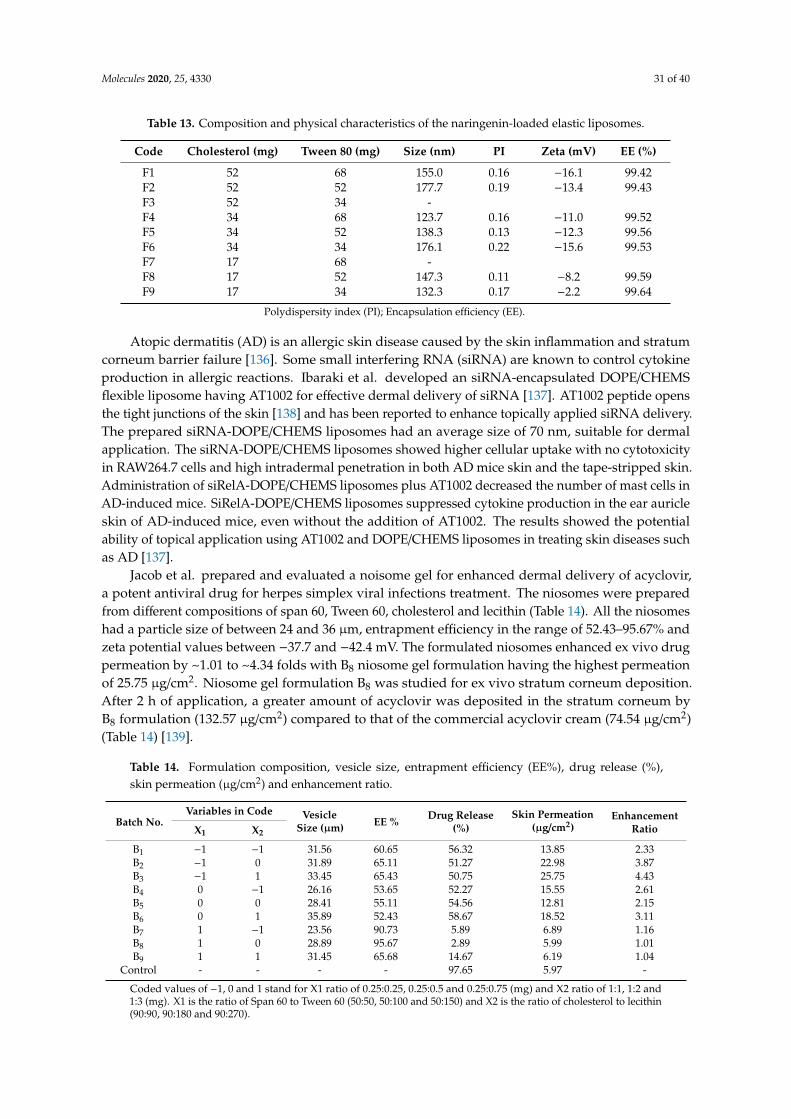
Molecules 2020, 25, 4330 31 of 40
Table 13. Composition and physical characteristics of the naringenin-loaded elastic liposomes.
Code Cholesterol (mg) Tween 80 (mg) Size (nm) PI Zeta (mV) EE (%)
F1 52 68 155.0 0.16 −16.1 99.42F2 52 52 177.7 0.19 −13.4 99.43F3 52 34 -F4 34 68 123.7 0.16 −11.0 99.52F5 34 52 138.3 0.13 −12.3 99.56F6 34 34 176.1 0.22 −15.6 99.53F7 17 68 -F8 17 52 147.3 0.11 −8.2 99.59F9 17 34 132.3 0.17 −2.2 99.64
Polydispersity index (PI); Encapsulation efficiency (EE).
Atopic dermatitis (AD) is an allergic skin disease caused by the skin inflammation and stratumcorneum barrier failure [136]. Some small interfering RNA (siRNA) are known to control cytokineproduction in allergic reactions. Ibaraki et al. developed an siRNA-encapsulated DOPE/CHEMSflexible liposome having AT1002 for effective dermal delivery of siRNA [137]. AT1002 peptide opensthe tight junctions of the skin [138] and has been reported to enhance topically applied siRNA delivery.The prepared siRNA-DOPE/CHEMS liposomes had an average size of 70 nm, suitable for dermalapplication. The siRNA-DOPE/CHEMS liposomes showed higher cellular uptake with no cytotoxicityin RAW264.7 cells and high intradermal penetration in both AD mice skin and the tape-stripped skin.Administration of siRelA-DOPE/CHEMS liposomes plus AT1002 decreased the number of mast cells inAD-induced mice. SiRelA-DOPE/CHEMS liposomes suppressed cytokine production in the ear auricleskin of AD-induced mice, even without the addition of AT1002. The results showed the potentialability of topical application using AT1002 and DOPE/CHEMS liposomes in treating skin diseases suchas AD [137].
Jacob et al. prepared and evaluated a noisome gel for enhanced dermal delivery of acyclovir,a potent antiviral drug for herpes simplex viral infections treatment. The niosomes were preparedfrom different compositions of span 60, Tween 60, cholesterol and lecithin (Table 14). All the niosomeshad a particle size of between 24 and 36 µm, entrapment efficiency in the range of 52.43–95.67% andzeta potential values between −37.7 and −42.4 mV. The formulated niosomes enhanced ex vivo drugpermeation by ~1.01 to ~4.34 folds with B8 niosome gel formulation having the highest permeationof 25.75 µg/cm2. Niosome gel formulation B8 was studied for ex vivo stratum corneum deposition.After 2 h of application, a greater amount of acyclovir was deposited in the stratum corneum byB8 formulation (132.57 µg/cm2) compared to that of the commercial acyclovir cream (74.54 µg/cm2)(Table 14) [139].
Table 14. Formulation composition, vesicle size, entrapment efficiency (EE%), drug release (%),skin permeation (µg/cm2) and enhancement ratio.
Batch No.Variables in Code Vesicle
Size (µm) EE %Drug Release
(%)Skin Permeation
(µg/cm2)Enhancement
RatioX1 X2
B1 −1 −1 31.56 60.65 56.32 13.85 2.33B2 −1 0 31.89 65.11 51.27 22.98 3.87B3 −1 1 33.45 65.43 50.75 25.75 4.43B4 0 −1 26.16 53.65 52.27 15.55 2.61B5 0 0 28.41 55.11 54.56 12.81 2.15B6 0 1 35.89 52.43 58.67 18.52 3.11B7 1 −1 23.56 90.73 5.89 6.89 1.16B8 1 0 28.89 95.67 2.89 5.99 1.01B9 1 1 31.45 65.68 14.67 6.19 1.04
Control - - - - 97.65 5.97 -
Coded values of −1, 0 and 1 stand for X1 ratio of 0.25:0.25, 0.25:0.5 and 0.25:0.75 (mg) and X2 ratio of 1:1, 1:2 and1:3 (mg). X1 is the ratio of Span 60 to Tween 60 (50:50, 50:100 and 50:150) and X2 is the ratio of cholesterol to lecithin(90:90, 90:180 and 90:270).

Molecules 2020, 25, 4330 32 of 40
Topical bioavailability studies in rabbits showed that the stratum corneum uptake of acyclovirby B8 was high while its clearance after the termination of the therapy was rapid. The formulationaccumulated in the dermis or epidermis region of the skin, facilitating drug localization which wouldenhance the therapeutic efficiency of acyclovir. The formulation did not cause any skin irritationsuch as erythema and edema in any rabbit. Stability studies showed the vesicles retained ~97.63%of acyclovir at 25 ◦C when stored for three months. The results suggest that B8 formulation can be aviable system for dermal delivery of acyclovir [139].
Wang et al. prepared Paclitaxel (PTX)-cholesterol-loaded-liposomes (PTXL) to improve the watersolubility of PTX and investigated its anti-keloid and anti-fibrotic efficacy against keloids. The PTXLaverage particle size was 101.43 nm, zeta potential of−41.63 mV and polydispersity index (PDI) of 0.244.Entrapment efficiency and loading capacity of PTXL were found to be 95.63% and 2.7%, respectively.When stored for 30 days at 4 ◦C, PTXL maintained their size, with PDI showing their stability. In vitrodrug release showed a sustained release of PTX from PTXL. Cellular uptake studies showed that PTXLwere partially taken up by human keloid fibroblasts (HKFs) through caveolin-mediated endocytosisrather than clathrin [140]. PTXL inhibited cell proliferation better than PTX when tested at thesame concentration range of 0.05–10 µg/mL. PTXL induced cell apoptosis of HKFs and arrested theHKF cell cycle in G2/M phase. Cell invasion results showed that PTXL significantly inhibited theinvasion of HKFs with invasion rates of 28.21% and 5.77% for PTX and PTXL, respectively. In vivoresults from BALB/c nude mice treated with PTXL, PTX and 5% glucose solution showed that PTXLreduced HKFs density better than both PTX and glucose solution. PTXL inhibited the AKT/GSK3βsignaling pathway, thus reducing the production of fibrogenic cytokines such as IL-6, TNF-α andTGF-β, which consequently ameliorated fibrosis progress in keloids. The results showed the potentialof PTXL in treating keloids [140].
7. Conclusions
Hybrid molecules containing cholesterol have been successfully synthesized in their broadrange. These include anticancer, antimicrobials, antimalarials and antivirals. Conjugation of drugsto cholesterol has been achieved by reacting the different functional groups on the drugs, such ascarboxylic and amines, with the hydroxyl end of cholesterol. Sometimes a linker is used betweencholesterol and the drug. Limitations such as bioavailability and toxicity have been overcome throughconjugating drugs to cholesterol. Cholesterol has been used in the design of drug delivery systemsfor dermal, transdermal, intravenous and oral drug administration. The use of cholesterol in thedesign of these systems was found to influence the particle size, stability, drug entrapment efficiency,drug release profile and cellular uptake of the systems. Although many positive outcomes have beendrawn from the conjugation of therapeutic agents to cholesterol, there is still much work that needs tobe done to make sure that they reach all advanced stage in clinical trials.
Funding: The financial assistance of the Medical Research Council (MRC), National Research Foundation (NRF),South Africa and Govan Mbeki Research Development Council, University of Fort Hare, South Africa towardsthis research is hereby acknowledged.
Acknowledgments: The financial assistance of the Medical Research Council (MRC) and National ResearchFoundation (NRF), South Africa towards this research is hereby acknowledged. The views and opinions expressedin this manuscript are those of the authors and not of MRC or NRF.
Conflicts of Interest: The authors report no conflict of interest.
Abbreviations
RNA Ribonucleic AcidEDCl 1-Ethyl-3-(3-dimethylaminopropyl)carbodiimideNHS N-HydroxysuccinimideTEA TriethylamineHCl Hydrochloric Acid

Molecules 2020, 25, 4330 33 of 40
r.t. room temperatureIC50 half maximal inhibitory concentrationDNA Dioxyribonucleic AcidTHF TetrahydrofuranDSC DichlorosilaneDMAP 4-DimethylaminopyridineDCM DichloromethaneDIC N,N′-DiisopropylcarbodiimideDMF DimethylformamidePyBOP benzotriazol-1-yl-oxytripyrrolidinophosphonium hexafluorophosphateHOBT HydroxybenzotriazoleDIEA N,N-DiisopropylethylamineCLSM Confocal Laser Scanning MicroscopyDMSO Dimethyl sulfoxideMDR Multi Drug ResistanceMTT 3-(4,5-dimethylthiazol-2-yl)-2,5-diphenyltetrazoliumbromide
References
1. Tiwari, G.; Tiwari, R.; Bannerjee, S.; Bhati, L.; Pandey, S.; Pandey, P.; Sriwastawa, B. Drug Delivery Systems:An Updated Review. Int. J. Pharm. Investig. 2012, 2, 2–11. [CrossRef]
2. Tiwari, R.; Jain, P.; Asati, S.; Haider, T.; Soni, V.; Pandey, V. State-of-Art Based Approaches for AnticancerDrug-Targeting to Nucleus. J. Drug Deliv. Sci. Technol. 2018, 48, 383–392. [CrossRef]
3. Kumar, M.N.V.R.; Muzzarelli, R.A.A.; Muzzarelli, C.; Sashiwa, H.; Domb, A.J. Chitosan Chemistry andPharmaceutical Perspectives. Chem. Rev. 2004, 104, 6017–6084. [CrossRef]
4. Arun, A.; Amrish, C.; Vijay, S.; Kamla, P. Fast Dissolving Oral Films: An Innovative Drug Delivery Systemand Dosage Form. Int. J. Chem. Tech. Res. 2010, 2, 576–583.
5. Ghafourian, T.; Nokhodchi, A.; Kaialy, W. Percutaneous Penetration Enhancers Chemical Methods in PenetrationEnhancement; Springer: Berlin/Heidelberg, Germany, 2015; pp. 208–231.
6. Dave, V.; Tak, K.; Sohgaura, A.; Gupta, A.; Sadhu, V. Lipid-Polymer Hybrid Nanoparticles: SynthesisStrategies and Biomedical Applications. J. Microbiol. Methods 2019, 160, 130–142. [CrossRef]
7. Jelonek, K.; Zajdel, A.; Wilczok, A.; Latocha, M.; Musiał-Kulik, M.; Forys, A.; Kasperczyk, J. Dual-TargetedBiodegradable Micelles for Anticancer Drug Delivery. Mater. Lett. 2019, 241, 187–189. [CrossRef]
8. Kheirolomoom, A.; Ferrara, K.W. Cholesterol Transport from Liposomal Delivery Vehicles. Biomaterials 2007,28, 4311–4320. [CrossRef] [PubMed]
9. Ge, X.; Wei, M.; He, S.; Yuan, W. Advances of Non-Ionic Surfactant Vesicles (Niosomes) and Their Applicationin Drug Delivery. Pharmaceuticals 2019, 11, 55. [CrossRef] [PubMed]
10. Aderibigbe, B.A. Design of Drug Delivery Systems Containing Artemisinin and Its Derivatives. Molecules2017, 22, 323. [CrossRef] [PubMed]
11. Albuquerque, H.M.T.; Santos, C.M.M.; Silva, A.M.S. Cholesterol-Based Compounds: Recent Advances inSynthesis and Applications. Molecules. 2019, 24, 116. [CrossRef]
12. Freitag, R. Synthetic Polymers for Biotechnology and Medicine; CRC Press: Boca Raton, FL, USA, 2003; pp. 1–171.13. Radwan, A.A.; Alanazi, F.K. Targeting cancer using cholesterol conjugates. Saudi Pharm. J. 2014, 22, 3–16.
[CrossRef] [PubMed]14. Orth, M.; Bellosta, S. Cholesterol: Its Regulation and Role in Central Nervous System Disorders. Cholesterol
2012, 2012, 292598. [CrossRef] [PubMed]15. Mallick, S.; Thi, L.; Lee, S.; Park, J.; Sig, J. Liposomes Containing Cholesterol and Mitochondria-Penetrating
Peptide (MPP) for Targeted Delivery of Antimycin A to A549 Cells. Colloids Surf. B Biointerfaces 2018,161, 356–364. [CrossRef] [PubMed]
16. Mehanna, M.; Motawaa, A.; Samaha, M. Pharmaceutical Particulate Carriers: Lipid—Based Carriers. Natl. J.Physiol. Pharm. Pharmacol. 2012, 2, 10–22.
17. Medvedeva, D.A.; Maslov, M.A.; Serikov, R.N.; Morozova, N.G.; Serebrenikova, G.A.; Sheglov, D.V.;Latyshev, A.V.; Vlassov, V.V.; Zenkova, M.A. Novel Cholesterol-Based Cationic Lipids for Gene Delivery.J. Med. Chem. 2009, 52, 6558–6568. [CrossRef]
Molecules 2020, 25, 4330 34 of 40
18. Irby, D.; Du, C.; Li, F. Lipid–Drug Conjugate for Enhancing Drug Delivery. Mol. Pharm. 2017, 14, 1325–1338.[CrossRef] [PubMed]
19. Patidar, A.; Thakur, D.S.; Kumar, P.; Verma, J. A Review on Novel Lipid Based Nanocarriers. Int. J. Pharm.Pharm. Sci. 2010, 2, 30–35.
20. Wang, Y.; Liu, L.; Jiang, Q.; Zhang, Q. Self-Aggregated Nanoparticles of Cholesterol-Modified ChitosanConjugate as a Novel Carrier of Epirubicin. Euro. Poly. J. 2007, 43, 43–51. [CrossRef]
21. Harisa, G.I.; Alanazi, F.K. Low Density Lipoprotein Bionanoparticles: From Cholesterol Transport to Deliveryof Anti-Cancer Drugs. Saudi Pharm. J. 2014, 22, 504–515. [CrossRef]
22. Chang, K.; Chang, F.; Chen, M. Developing a Novel Cholesterol-Based Nanocarrier with High TransfectionEfficiency and Serum Compatibility for Gene Therapy. J. Formos. Med. Assoc. 2019, 118, 766–775. [CrossRef]
23. Fetihg, G. Fluconazole-loaded niosomal gels as a topical ocular drug delivery system for corneal fungalinfections. J. Drug Deliv. Sci. Technol. 2016, 35, 8–15. [CrossRef]
24. Osanloo, M.; Assadpour, S.; Mehravaran, A.; Abastabar, M.; Akhtari, J. Niosome-loaded antifungal drugs asan effective nanocarrier system: A mini review. Curr. Med. Mycol. 2018, 4, 31–36. [CrossRef] [PubMed]
25. World Health Organisation. Latest Global Cancer Data. Available online: https://www.who.int/cancer/PRGlobocanFinal.pdf (accessed on 12 September 2018).
26. Zhao, Z.; Song, H.; Xie, J.; Liu, T.; Zhao, X.; Chen, X.; He, X.; Wu, S.; Zhang, Y.; Zheng, X. Research Progressin the Biological Activities of 3,4,5-Trimethoxycinnamic Acid (TMCA) Derivatives. Eur. J. Med. Chem. 2019,173, 213–227. [CrossRef] [PubMed]
27. Ling, Y.; Gao, W.J.; Ling, C.; Liu, J.; Meng, C.; Qian, J.; Liu, S.; Gan, H.; Wu, H.; Tao, J. β-Carboline andN-Hydroxycinnamamide Hybrids as Anticancer Agents for Drug-Resistant Hepatocellular Carcinoma. Eur. J.Med. Chem. 2019, 168, 515–526. [CrossRef]
28. Kumar, B.; Smita, K. Scope of Nanotechnology in Nutraceuticals; Elsevier Inc.: Amsterdam, The Netherlands, 2017.29. Shakeel, F.; Alanazi, F.K.; Raish, M.; Haq, N.; Radwan, A.A.; Alsarra, I.A. Pharmacokinetic and in Vitro
Cytotoxic Evaluation of Cholesterol-Rich Nanoemulsion of Cholesteryl-Succinyl-5- Fl Uorouracil. J. Mol. Liq.2015, 211, 164–168. [CrossRef]
30. Yao, X.; Chen, L.; Chen, X.; He, C.; Zheng, H.; Chen, X. Intercellular PH-Responsive Histidine ModifiedDextran- g -Cholesterol Micelle for Anticancer Drug Delivery. Colloids Surf. B Biointerfaces 2014, 121, 36–43.[CrossRef]
31. Laskar, P.; Samanta, S.; Kumar, S.; Dey, J. In Vitro Evaluation of PH-Sensitive Cholesterol-Containing StablePolymeric Micelles for Delivery of Camptothecin. J. Colloid Interface Sci. 2014, 430, 305–314. [CrossRef]
32. Yotsumoto, K.; Ishii, K.; Kokubo, M.; Yasuoka, S. Improvement of the Skin Penetration of HydrophobicDrugs by Polymeric Micelles. Int. J. Pharm. 2018, 553, 132–140. [CrossRef]
33. Jin, H.; Miyata, K.; Nomoto, T.; Zheng, M.; Kim, A.; Liu, X.; Cabral, H.; Christie, R.J.; Nishiyama, N.SiRNA Delivery from Triblock Copolymer Micelles with Spatially- Ordered Compartments of PEG Shell,SiRNA-Loaded Intermediate Layer, and Hydrophobic Core. Biomaterials 2014, 35, 4548–4556.
34. Senapati, S.; Mahanta, A.K.; Kumar, S.; Maiti, P. Controlled Drug Delivery Vehicles for Cancer Treatment andTheir Performance. Signal Transduct. Target. Ther. 2018, 3, 1–19. [CrossRef]
35. Muddineti, O.S.; Shah, A.; Rompicharla, S.V.; BalaramGhosh, B.; Biswas, S. Cholesterol-grafted chitosanmicelles as a nanocarrier system for drug-siRNA co-delivery to the lung cancer cells. Int. J. Biol. Markers2018, 118, 857–863. [CrossRef] [PubMed]
36. Ullah, S.; Shah, M.R.; Shoaib, M.; Imran, M.; Wadood, S.; Shah, A.; Ahmed, F.; Gul, Q.; Shah, I. HydrophilicallyModified Self-Assembling α- ocopherol Derivative as Niosomal Nanocarrier for Improving ClarithromycinOral Bioavailability. Artif. Cells Nanomed. Biotechnol. 2018, 46, 568–578. [CrossRef] [PubMed]
37. Muddineti, O.S.; Vanaparthi, A.; Rompicharla, S.V.K.; Kumari, P.; Ghosh, B.; Biswas, S. Cholesterol andVitamin E-Conjugated PEGylated Polymeric Micelles for Efficient Delivery and Enhanced Anticancer Activityof Curcumin: Evaluation in 2D Monolayers and 3D Spheroids. Artif. Cells Nanomed. Biotechnol. 2018,46, 773–786. [CrossRef] [PubMed]
38. Sarkar, D.; Choudhury, P.; Dinda, S.; Das, P.K. Vesicle Formation by Cholesterol Based Hydrazone TetheredAmphiphiles: Stimuli Responsive Dissipation of Self-Assembly. J. Colloid Interface Sci. 2018, 530, 67–77.[CrossRef] [PubMed]
Molecules 2020, 25, 4330 35 of 40
39. Akbarzadeh, A.; Rezaei-sadabady, R.; Davaran, S.; Joo, S.W.; Zarghami, N. Liposome: Classification,Preparation and Applications. Nanoscale Res. Lett. 2013, 8, 102. [CrossRef]
40. Heidari, M.; Khoee, S.; Lot, M. Synthesis of Titanium Dioxide Nanotubes with Liposomal Covers for Carryingand Extended Release of 5-FU as Anticancer Drug in the Treatment of HeLa Cells. Anal. Biochem. 2019,572, 16–24. [CrossRef]
41. Mohamed, M.S.; Torabi, A.; Paulose, M.; Kumar, D.S.; Varghese, O.K. Anodically Grown Titania NanotubeInduced Cytotoxicity Has Genotoxic Origins. Nat. Publ. Group 2017, 7, 41844. [CrossRef]
42. Bartlett, N.L.; Petroni, G.R.; Parker, B.A.; Wagner, N.D.; Gockerman, J.P.; Omura, G.A.; Canellos, G.P.;Cooper, M.R.; Johnson, J.L.; Peterson, B.A. Dose-escalated cyclophosphamide, doxorubicin, vincristine,prednisone, and etoposide (CHOPE) chemotherapy for patients with diffuse lymphoma: Cancer andLeukemia Group B studies 8852 and 8854. Cancer 2001, 92, 207–217. [CrossRef]
43. Choi, K.; Kwon, I.C.; Ahn, H.J. Self-Assembled Amphiphilic DNA-Cholesterol/DNA-Peptide Hybrid Duplexeswith Liposome-like Structure for Doxorubicin Delivery. Biomaterials 2013, 34, 4183–4190. [CrossRef]
44. Gonzalez-fajardo, L.; Mahajan, L.H.; Ndaya, D.; Hargrove, D.; Manautou, J.E.; Liang, B.T.; Chen, M.;Kasi, R.M.; Lu, X. Reduced in Vivo Toxicity of Doxorubicin by Encapsulation in Cholesterol-ContainingSelf-Assembled Nanoparticles. Pharmacol. Res. 2016, 107, 93–101. [CrossRef]
45. Nguyen, C.T.; Tran, T.H.; Amiji, M. Redox-Sensitive Nanoparticles from Amphiphilic Cholesterol-BasedBlock Copolymers for Enhanced Tumor Intracellular Release of Doxorubicin. Nanomed. Nanotech. Biol. Med.2015, 11, 2071–2082. [CrossRef]
46. Desai, N.; Trieu, V.; Damascelli, B.; Soon-shiong, P. SPARC Expression Correlates with Tumor Response toAlbumin-Bound Paclitaxel in Head and Neck Cancer Patients. Transl. Oncol. 2009, 2, 59–64. [CrossRef][PubMed]
47. Kratz, F. A Clinical Update of Using Albumin as a Drug Vehicle. J. Control. Release 2014, 190, 331–336.[CrossRef] [PubMed]
48. Sparreboom, A.; Scripture, C.D.; Trieu, V.; Williams, P.J.; De, T.; Yang, A.; Beals, B.; Figg, W.D.; Hawkins, M.;Desai, N. Comparative Preclinical and Clinical Pharmacokinetics of a Cremophor-Free, NanoparticleAlbumin-Bound Paclitaxel (ABI-007) and Paclitaxel Formulated in Cremophor (Taxol). Clin. Cancer. Res.2005, 11, 4136–4144. [CrossRef] [PubMed]
49. Gong, J.; Huo, M.; Zhou, J.; Zhang, Y.; Peng, X.; Yu, D.; Zhang, H.; Li, J. Synthesis, Characterization,Drug-Loading Capacity and Safety of Novel Octyl Modified Serum Albumin Micelles. Int. J. Pharm. 2009,376, 161–168. [CrossRef] [PubMed]
50. Battogtokh, G.; Kang, J.H.; Ko, Y.T. Long-Circulating Self-Assembled Cholesteryl Albumin NanoparticlesEnhance Tumor Accumulation of Hydrophobic Anticancer Drug. Eur. J. Pharm. Biopharm. 2015, 96, 96–105.[CrossRef] [PubMed]
51. Zhang, J.; Chen, R.; Fang, X.; Chen, F.; Wang, Y.; Chen, M. Nucleolin Targeting AS1411 Aptamer ModifiedpH-Sensitive Micelles for Enhanced Delivery and Antitumor Efficacy of Paclitaxel. Nano Res. 2015, 8, 201–218.[CrossRef]
52. Shagufta; Ahmad, I. Tamoxifen a Pioneering Drug: An Update on the Therapeutic Potential of TamoxifenDerivatives. Eur. J. Med. Chem. 2018, 143, 515–531. [CrossRef] [PubMed]
53. Mazumdar, S.; Italiya, K.S.; Sharma, S.; Chitkara, D.; Mittal, A. Effective Cellular Internalization, Cell CycleArrest and Improved Pharmacokinetics of Tamoxifen by Cholesterol Based Lipopolymeric Nanoparticles.Int. J. Pharm. 2018, 543, 96–106. [CrossRef]
54. Zheng, H.; Li, J.; Wang, M.; Luo, X.; Qiu, Q.; Hu, L.; Li, C.; Song, Y.; Deng, Y. Exhausting Tumor AssociatedMacrophages with sialic acid-polyethyleneimine-cholesterol modified liposomal doxorubicin for enhancingsarcoma chemotherapy. Int. J. Pharm. 2019, 558, 187–200. [CrossRef]
55. Seow, W.Y.; Liang, K.; Kurisawa, M.; Hauser, C.A.E. Oxidation as a Facile Strategy To Reduce the SurfaceCharge and Toxicity of Polyethyleneimine Gene Carriers. Biomacromolecules 2013, 14. [CrossRef] [PubMed]
56. Cavalli, R.; Trotta, F.; Tumiatti, W. Cyclodextrin-Based Nanosponges for Drug Delivery. J. Incl. Phenom.Macrocycl. Chem. 2006, 56, 209–213. [CrossRef]
57. Singh, P.; Ren, X.; Guo, T.; Wu, L.; Shakya, S.; He, Y. Biofunctionalization of β -Cyclodextrin NanospongesUsing Cholesterol. Carbohydr. Polym. 2018, 190, 23–30. [CrossRef] [PubMed]
Molecules 2020, 25, 4330 36 of 40
58. Chen, C.; Wang, J.; Zhao, E.; Gao, L.; Feng, Q.; Liu, X.; Zhao, Z.; Ma, X.; Hou, W.; Zhang, L. Self-AssemblyCationic Nanoparticles Based on Cholesterol-Grafted Bioreducible Poly(Amidoamine) for SiRNA Delivery.Biomaterials 2013, 34, 5303–5316. [CrossRef]
59. Tao, X.; Xie, Y.; Zhang, Q.; Qiu, X.; Yuan, L.; Wen, Y.; Li, M.; Yang, X.; Tao, T.; Xie, M. Cholesterol-ModifiedAmino-Pullulan Nanoparticles as a Drug Carrier: Comparative Study of Cholesterol-Modified CarboxyethylPullulan and Pullulan Nanoparticles. Nanomaterials 2016, 6, 1–15. [CrossRef]
60. Yang, W.; Wang, M.; Ma, L.; Li, H.; Huang, L. Synthesis and Characterization of Biotin Modified CholesterylPullulan as a Novel Anticancer Drug Carrier. Carbohydr. Polym. 2014, 99, 720–727. [CrossRef]
61. Xu, Y.; Zi, Y.; Lei, J.; Mo, X.; Shao, Z.; Wu, Y.; Tian, Y.; Li, D.; Mu, C. pH-Responsive nanoparticles basedon cholesterol/imidazole modified oxidized-starch for targeted anticancer drug delivery. Carbohydr. Polym.2020, 233, 115858. [CrossRef]
62. Wu, H.; Yu, M.; Miao, Y.; He, S.; Dai, Z.; Song, W.; Liu, Y.; Song, S.; Ahmad, E.; Wang, D. Cholesterol-TunedLiposomal Membrane Rigidity Directs Tumor Penetration and Anti-Tumor Effect. Acta Pharm. Sin. B 2018,9, 858–870. [CrossRef]
63. Qiu, L.; Yao, M. Liposome Comprising Combination of Chloroquine, Adriamycin and Preparation MethodThereof. US 2013/0189352 A1, 25 July 2013.
64. Ding, M.; Li, J.; He, X.; Song, N.; Tan, H.; Zhang, Y.; Zhou, L. Molecular Engineered Super-Nanodevices:Smart and Safe Delivery of Potent Drugs into Tumors. Adv. Mater. 2012, 24, 3639–3645. [CrossRef] [PubMed]
65. Sawant, R.R.; Torchilin, V.P. Multifunctional Nanocarriers and Intracellular Drug Delivery. Curr. Opin. SolidState Mater. Sci. 2012, 16, 269–275. [CrossRef]
66. Choi, K.Y.; Liu, G. Nanoscale Theranostic Nanoplatforms for Simultaneous Cancer Imaging and Therapy.Nanoscale 2011, 4, 330–342. [CrossRef] [PubMed]
67. Cao, Z.; Li, W.; Liu, R.; Li, X.; Li, H.; Liu, L.; Chen, Y.; Lv, C. pH- and Enzyme-Triggered Drug Release asan Important Process in the Design of Anti-Tumor Drug Delivery Systems. Biomed. Pharmacother. 2019,118, 109340. [CrossRef] [PubMed]
68. Yang, B.; Lv, Y.; Zhu, J.; Han, Y.; Jia, H.; Chen, W.; Feng, J.; Zhang, X.; Zhuo, R. A pH-ResponsiveDrug Nanovehicle Constructed by Reversible Attachment of Cholesterol to PEGylated Poly(L-Lysine) viaCatechol–Boronic Acid Ester Formation. Acta Biomater. 2014, 10, 3686–3695. [CrossRef] [PubMed]
69. Park, J.; Cho, H.; Yeol, H.; Yoon, I.; Ko, S.; Shim, J.; Cho, J.; Hyung, J.; Kim, K.; Chan, I. Hyaluronic AcidDerivative-Coated Nanohybrid Liposomes for Cancer Imaging and Drug Delivery. J. Control. Release 2014,174, 98–108. [CrossRef] [PubMed]
70. Lee, J.; Yi, S.; Park, J.; Kim, D.; Cho, H. Cholesterol-Modified Poly(Lactide-Co-Glycolide) Nanoparticles forTumor-Targeted Drug Delivery. Int. J. Pharm. 2016, 509, 483–491. [CrossRef] [PubMed]
71. Yahuafai, J.; Asai, T.; Nakamura, G.; Fukuta, T.; Siripong, P.; Hyodo, K. Suppression in Mice ofImmunosurveillance against PEGylated Liposomes by Encapsulated Doxorubicin. J. Control. Release2014, 192, 167–173. [CrossRef]
72. Wang, T.; Upponi, J.R.; Torchilin, V.P. Design of Multifunctional Non-Viral Gene Vectors to OvercomePhysiological Barriers: Dilemmas and Strategies. Int. J. Pharm. 2012, 427, 3–20. [CrossRef]
73. Tagalakis, A.D.; Kenny, G.D.; Bienemann, A.S.; Mccarthy, D.; Munye, M.M.; Taylor, H.; Wyatt, M.J.;Lythgoe, M.F.; White, E.A.; Hart, S.L. PEGylation Improves the Receptor-Mediated Transfection Efficiency ofpeptide-targeted, self-aasembling, anionic nanocomplexes. J. Control. Release 2013, 174, 177–187. [CrossRef]
74. Hoogenboom, R.; Thijs, H.M.L.; Fijten, M.W.M.; Lankvelt, B.M.V.A.N.; Schubert, U.S. One-Pot Synthesisof 2-Phenyl-2-Oxazoline-Containing Quasi-Diblock Copoly(2-Oxazoline)s under Microwave Irradiation. J.Polym. Sci. A 2006, 45, 416–422. [CrossRef]
75. Sedlacek, O.; Monnery, B.D.; Filippov, S.K.; Hoogenboom, R.; Hruby, M. Poly(2-Oxazoline)s—Are They MoreAdvantageous for Biomedical Applications Than Other Polymers ? Macromol. Rapid Commun. 2012, 33, 1648–1662.[CrossRef]
76. Xu, H.; Yu, X. The Bifunctional Liposomes Constructed by Poly(2-Ethyl-Oxazoline)- Cholesteryl MethylCarbonate: An Effectual Approach to Enhance Liposomal Circulation Time, pH-Sensitivity and Endosomalescape. Pharm. Res. 2014, 31, 3038–3050. [CrossRef] [PubMed]
77. Xu, H.; Hu, M.; Yu, X.; Li, Y.; Fu, Y.; Zhou, X.; Zhang, D.; Li, J. Design and Evaluation of pH-SensitiveLiposomes Constructed by Poly(2-Ethyl-2-Oxazoline)-Cholesterol Hemisuccinate for Doxorubicin Delivery.Eur. J. Pharm. Biopharm. 2015, 91, 66–74. [CrossRef] [PubMed]
Molecules 2020, 25, 4330 37 of 40
78. Cajot, S.; Van Butsele, K.; Paillard, A.; Passirani, C.; Garcion, E.; Benoit, J.P.; Varshney, S.K.; Jérôme, C.Smart Nanocarriers for pH-Triggered Targeting and Release of Hydrophobic Drugs. Acta Biomater. 2012,8, 4215–4223. [CrossRef]
79. Javed, M.; Ahamed, M.; Alhadlaq, H.A.; Alrokayan, S.A.; Kumar, S. Targeted Anticancer Therapy:Overexpressed Receptors and Nanotechnology. Clin. Chim. Acta 2014, 436, 78–92.
80. Yang, C.; Qiang, Y.; Xiong, T.; Zhao, B.; Dong, X.; Fang, J. Self-Assembled pH-Responsive MPEG-b-(PLA-Co-PAE) Block Copolymer Micelles for Anticancer Drug Delivery. Biomaterials 2012, 33, 6273–6283.
81. Yang, C.; Chen, Q.; Sheng, W.; Dong, X.; Zhi, C. Synthesis and Evaluation of Cholesterol-Grafted PEGylatedPeptides with pH-Triggered Property as Novel Drug Carriers for Cancer Chemotherapy. Colloids Surf.B Biointerfaces 2016, 142, 55–64.
82. Ivey, J.W.; Bonakdar, M.; Kanitkar, A.; Davalos, R.V.; Verbridge, S.S. Improving Cancer Therapies by Targetingthe Physical and Chemical Hallmarks of the Tumor Microenvironment. Cancer Lett. 2016, 380, 330–339.[CrossRef] [PubMed]
83. Dirkx, A.E.M.; Egbrink, M.G.A.; Wagstaff, J.; Griffioen, A.W. Monocyte/Macrophage Infiltration in Tumors:Modulators of Angiogenesis. J. Leukoc. Biol. 2006, 80, 1183–1195. [CrossRef]
84. Macauley, M.S.; Crocker, P.; Paulson, J.C. Siglec-Mediated Regulation of Immune Cell Function in Disease.Immunology 2014, 14, 653–666. [CrossRef]
85. Zhou, S.; Zhang, T.; Peng, B.; Luo, X.; Liu, X.; Hu, L.; Liu, Y.; Di, D.; Song, Y.; Deng, Y. Targeted Delivery ofEpirubicin to Tumor-Associated Macrophages by Sialic Acid-Cholesterol Conjugate Modified Liposomeswith Improved Antitumor Activity. Int. J. Pharm. 2017, 523, 203–216. [CrossRef]
86. Dutreix, M.; Sun, J.; Devun, F. Cancer Treatment by Combining DNA Molecules Mimicking Double StrandBreaks with Hyperthrmia. US 9,205,101 B2, 8 December 2015.
87. Sheng, R.; Luo, T.; Li, H.; Sun, J.; Wang, Z.; Cao, A. Cholesterol-Based Cationic Lipids for Gene Delivery:Contribution of Molecular Structure Factors to Physico-Chemical and Biological Properties. Colloids Surf.B Biointerfaces 2014, 116, 32–40. [CrossRef] [PubMed]
88. Chernikov, I.V.; Gladkikh, D.V.; Meschaninova, M.I.; Ven, A.G.; Zenkova, M.A.; Vlassov, V.V.;Chernolovskaya, E.L. Cholesterol-Containing Nuclease-Resistant SiRNA Accumulates in Tumors in aCarrier-Free Mode and Silences MDR1 Gene. Mol. Ther. Nucleic Acid. 2017, 6, 209–220. [CrossRef] [PubMed]
89. Sun, J.S.; Dutreix, M.; Quanz, M. Optimized In Vivo Delivery System with Endosomolytic Agents for NucleicAcid Conjugates. US 9,205,099 B2, 30 August 2016.
90. Zhang, R.; Wu, F.; Wu, L.; Tian, Y.; Zhou, B.; Zhang, X.; Huang, R.; Yu, C.; He, G.; Yang, L. Novel Self-AssembledMicelles Based on Cholesterol-Modified Antimicrobial Peptide (DP7) for Safe and Effective SystemicAdministration in Animal Models of Bacterial Infection. Antimicrob. Agents Chemother. 2018, 62, 1–27.[CrossRef] [PubMed]
91. Lakshmaiah, J.; Chen, J. Peptides Antimicrobial Peptides: Possible Anti-Infective Agents. Peptides 2015,72, 88–94. [CrossRef] [PubMed]
92. Mahlapuu, M.; Håkansson, J.; Ringstad, L.; Björn, C. Antimicrobial Peptides: An Emerging Category ofTherapeutic Agents. Front. Cell. Infect. Microbiol. 2016, 6, 194. [CrossRef] [PubMed]
93. Villamil, J.C.; Parra-giraldo, C.M.; Pérez, L.D. Enhancing the Performance of PEG-b-PCL Copolymers asPrecursors of Micellar Vehicles for Amphotericin B through Its Conjugation with Cholesterol. Colloids Surf. A2019, 572, 79–87. [CrossRef]
94. Liu, L.; Ren, J.; He, Z.; Men, K.; Mao, Y.; Ye, T.; Chen, H.; Li, L.; Xu, B.; Wei, Y. Cholesterol-ModifiedHydroxychloroquine-Loaded Nanocarriers in Bleomycin-Induced Pulmonary Fibrosis. Sci. Rep. 2017, 7, 1–11.[CrossRef]
95. Blaising, J.; Pécheur, E.I. Lipids A Key for Hepatitis C Virus Entry and a Potential Target for AntiviralStrategies. Biochimie 2013, 95, 96–102. [CrossRef]
96. Lee, C.H.; Lee, S.H.; Kim, J.H.; Noh, Y.H.; Noh, G.J.; Lee, S.W. Pharmacokinetics of a Cholesterol-ConjugatedAptamer Against the Hepatitis C Virus (HCV) NS5B Protein. Mol. Ther. Nucleic Acids. 2015, 4, e254.[CrossRef]
97. Chamoun-Emanuelli, A.M.; Pécheur, E.I.; Chen, Z. Benzhydrylpiperazine Compounds InhibitCholesterol-Dependent Cellular Entry of Hepatitis C Virus. Antivir. Res. 2014, 109, 141–148. [CrossRef]
98. Pessi, P.; Bianchi, E.; Ingallinella, P. Cholesterol Derivatives as Inhibitors of Viral Fusion. US 8,629,101 B2,14 January 2014.
Molecules 2020, 25, 4330 38 of 40
99. Pessi, A.; Bixler, S.L.; Soloveva, V.; Radoshitzky, S.; Retterer, C.; Kenny, T.; Zamani, R.; Gomba, G.;Gharabeih, D.; Wells, J.; et al. Cholesterol-Conjugated Stapled Peptides Inhibit Ebola and Marburg Viruses inVitro and In Vivo. Antivir. Res. 2019, 171, 1–10. [CrossRef] [PubMed]
100. Pessi, A. Cholesterol-Conjugated Peptide Antivirals: A Path to a Rapid Response to Emerging Viral Diseases.J. Pept. Sci. 2015, 21, 379–386. [CrossRef] [PubMed]
101. Pourmoazzen, Z.; Sadeghifar, H.; Yang, G.; Lucia, L. Cholesterol-Modified Lignin: A New Avenue for GreenNanoparticles, Meltable Materials, and Drug Delivery. Colloids Surf. B Biointerfaces 2019, 186, 1–10. [CrossRef][PubMed]
102. Jenkins, T.A.; Thet, T.N.; Mercer-Chalmers, J. Wound Dressing. U.S. Patent US9,499,852 B2, 22 November 2016.103. De Oliveira, A.L.C.S.L.; Dos Santos-silva, A.M.; Da Silva-júnior, A.A.; Garcia, V.B.; De Araujo, A.A.;
De Geus-Oei, L.F.; Chan, A.B.; Cruz, L.J.; De Afaujo Junior, R.F. Cholesterol-Functionalized Carvedilol-LoadedPLGA Nanoparticles: Anti-Inflammatory, Antioxidant and Antitumor Effects. J. Nanopart. Res. 2020, 22, 115.[CrossRef]
104. Farmoudeh, A.; Akbari, J.; Saeedi, M.; Ghasemi, M.; Asemi, N.; Nokhodchi, A. Methylene Blue-LoadedNiosome: Preparation, Physicochemical Characterization and in Vivo Wound Healing Assessment. Drug Deliv.Transl. Res. 2020, 1–14. [CrossRef] [PubMed]
105. Ibrahim, S.; Tagami, T.; Kishi, T.; Ozeki, T. Curcumin Marinosomes as Promising Nano-Drug Delivery Systemfor Lung Cancer. Int. J. Pharm. 2018, 540, 40–49. [CrossRef] [PubMed]
106. Arab, W.T.; Niyas, A.M.; Seferji, K.; Susapto, H.H.; Alexander, I.; Cima, I.; Hauser, C.A.E. Evaluation ofPeptide Nanogels for Accelerated Wound Healing in Normal Micropigs. Front Nanosci Nanotech. 2018, 4, 1–9.[CrossRef]
107. Rasool, M.; Naseer, M.I.; Malik, A.; Manan, A.; Ullah, I.; Bin, N.; Kim, M.O.; Qazi, M.H.; Kamal, M.A.; Bibi, F.Recent Developments in Nanomedicines for Management of Various Health Issues Via Metabolism andPhysico-Chemical Properties. Curr. Drug Metab. 2015, 16, 389–396. [CrossRef]
108. Moghassemi, S.; Hadjizadeh, A. Nano-Niosomes as Nanoscale Drug Delivery Systems: An IllustratedReview. J. Control. Release 2014, 185, 22–36. [CrossRef]
109. Ali, I.; Imran, M.; Saifullah, S.; Hussain, K.; Kanwal, T.; Nisar, J.; Raza, M. Synthesis of BiocompatibleTriazole Based Non-Ionic Surfactant and Its Vesicular Drug Delivery Investigation. Chem. Phys. Lipids 2020,228, 104894.
110. Fathalla, D.; Ahmed, M. In-Vitro and In-Vivo Evaluation of Niosomal Gel Containing Aceclofenac forSustained Drug Delivery. Int. J. Pharm. Sci. Res. 2014, 1, 1–11. [CrossRef] [PubMed]
111. Manconi, M.; Letizia, M.; Caddeo, C.; Cencetti, C.; Zoratto, N.; Nacher, A.; Maria, A.; Matricardi, P.Preparation of Gellan-Cholesterol Nanohydrogels Embedding Baicalin and Evaluation of Their WoundHealing Activity. Eur. J. Pharm. Biopharm. 2018, 127, 244–249. [CrossRef] [PubMed]
112. Atamna, H.; Nguyen, A.; Schultz, C.; Boyle, K.; Newberry, J.; Kato, H.; Ames, B.N. Methylene Blue DelaysCellular Senescence and Enhances Key Mitochondrial Biochemical Pathways. FASEB J. 2008, 22, 703–712.[CrossRef] [PubMed]
113. Aziz, D.E.; Abdelbary, A.A.; Elassasy, A.I. Implementing Central Composite Design for DevelopingTransdermal Diacerein-Loaded Niosomes: Ex vivo Permeation and In vivo Deposition. Curr. Drug Deliv.2018, 15, 1330–1342. [CrossRef]
114. Som, I.; Bhatia, K.; Yasir, M. Status of Surfactants as Penetration Enhancers in Transdermal Drug Delivery.J. Pharm. Bioallied Sci. 2012, 4, 2–9.
115. Shirsand, S.B.; Para, M.S.; Nagendrakumar, D.; Kanani, K.M.; Keerthy, D. Formulation and Evaluation ofKetoconazole Niosomal Gel Drug Delivery System. Int. J. Pharm. Investig. 2012, 2, 201–208. [CrossRef]
116. Ptchelintsev, D.S.; Mahwah, N.J. Ascorbyl-Phosphoryl-Cholesterol. US005951990A, 14 September 1999.117. Mahajan, H.S.; Patil, P.H. Central Composite Design-Based Optimization of Lopinavir Vitamin E-TPGS
Micelle: In Vitro Characterization and In Vivo Pharmacokinetic Study. Colloids Surf. B Biointerfaces 2020,194, 111149. [CrossRef]
118. Patel, K.K.; Kumar, P.; Thakkar, H.P. Formulation of Niosomal Gel for Enhanced Transdermal LopinavirDelivery and Its Comparative Evaluation with Ethosomal Gel. AAPS PharmSciTech 2012, 13, 1502–1510.[CrossRef]
Molecules 2020, 25, 4330 39 of 40
119. Zhang, W.; Wang, Z.; Wu, C.; Jin, Y.; Liu, X.; Wu, Z.; Liu, J. The effect of DSPE-PEG2000, Cholesterol andDrug Incorporated in Bilayer on the Formation of Discoidal Micelles. Eur. J. Pharm. Sci. 2018, 125, 74–85.[CrossRef]
120. Yeoung, J.; Youn, S.; Hun, S.; Choo, J.; Lee, E.K. Effect of Cholesterol Content on Affinity and Stability ofFactor VIII and Annexin V Binding to a Liposomal Bilayer Membrane. Chem. Phys. Lipids 2010, 163, 335–340.
121. Georgy, K.R.; Farid, R.M.; Latif, R.; Bendas, E.R. A New Design for a Chronological Release Profile of Etodolacfrom Coated Bilayer Tablets: In-Vitro and in-Vivo Assessment. J. Adv. Res. 2019, 15, 37–47. [CrossRef]
122. Asthana, G.S.; Asthana, A.; Singh, D.; Sharma, P.K. Etodolac Containing Topical Niosomal Gel: FormulationDevelopment and Evaluation. J. Drug Deliv. 2016, 1–9. [CrossRef] [PubMed]
123. Alcantar, N.; Williams, E.C.; Toomey, R. Niosome-Hydrogel Drug Delivery System. U.S. Patent US20100068264A1,18 March 2010.
124. Patzelt, A.; Cheung, W.; Jung, S.; Knorr, F.; Meinke, M.C.; Richter, H.; Rühl, E.; Yee, K.; Bich, N.; Nathalie, N.Do Nanoparticles Have a Future in Dermal Drug Delivery ? J. Control. Release 2017, 246, 174–182. [CrossRef][PubMed]
125. Ibaraki, H.; Kanazawa, T.; Oogi, C.; Takashima, T.; Seta, Y. Effects of Surface Charge and Flexibility ofLiposomes on Dermal Drug Delivery. J. Drug Deliv. Sci. Technol. 2019, 50, 155–162. [CrossRef]
126. Wischke, C.; Rühl, E.; Lendlein, A. Dermal Drug Delivery by Nanocarriers. J. Control. Release 2016, 242, 1–2.[CrossRef]
127. Hönzke, S.; Gerecke, C.; Elpelt, A.; Zhang, N.; Unbehauen, M.; Kral, V.; Fleige, E.; Paulus, F.; Haag, R.;Schäfer-korting, M. Tailored Dendritic Core-Multishell Nanocarriers for Efficient Dermal Drug Delivery:A Systematic Top-down Approach from Synthesis to Preclinical Testing. J. Control. Release 2016, 242, 50–63.[CrossRef] [PubMed]
128. Kahraman, E.; Gungor, S.; Ozisoy, Y. Potential Enhancement and Targeting Strategies of Polymeric andLipid-Based Nanocarriers in Dermal Drug Delivery. Ther. Deliv. 2017, 11, 967–985. [CrossRef]
129. Sandoval, K.R.; Machado, M.C.R.; de Oliveira, Z.N.P.; Nico, M.M.S. CHILD Syndrome: Successful Treatmentof Skin Lesions with Topical Lovastatin and Cholesterol Lotion. Ann. Bras. Dermatol. 2019, 94, 341–343.[CrossRef]
130. Khali, S.; Bardawil, T.; Saade, S.; Chedraoui, A.; Ramadan, N.; Hasbani, D.J.; Abbas, O.; Nemer, G.; Rubeiz, N.;Kurban, M. Use of Topical Glycolic Acid Plus a Lovastatin-Cholesterol Combination Cream for the Treatmentof Autosomal Recessive Congenital Ichthyoses. JAMA Dermatol. 2018, 154, 1320–1323. [CrossRef]
131. Atzmony, L.; Lim, Y.H.; Hamilton, C.; Leventhal, J.S.; Wagner, A.; Paller, A.S.; Choate, K.A. Topical Cholesterol/Lovastatin for the Treatment of Porokeratosis: A Pathogenesis-Directed Therapy. J. Am. Acad. Dermatol. 2019,82, 123–131. [CrossRef]
132. Mahmoud, N.N.; Alhusban, A.A.; Ali, J.I.; Al-bakri, A.G.; Hamed, R.; Khalil, E.A. Preferential Accumulationof Phospholipid-PEG and Cholesterol-PEG Decorated Gold Nanorods into Human Skin Layers and TheirPhotothermal-Based Antibacterial Activity. Sci. Rep. 2019, 9, 1–15. [CrossRef] [PubMed]
133. Tsai, M.; Huang, Y.; Fang, J.; Fu, Y.; Wu, P. Preparation and Characterization of Naringenin-Loaded ElasticLiposomes for Topical Application. PLoS ONE. 2015, 10, e0131026. [CrossRef] [PubMed]
134. Yin, X.; Fu, X.; Cheng, H.; Liang, L. α -Tocopherol and Naringenin in Whey Protein Isolate Particles: Partition,Antioxidant Activity, Stability and Bioaccessibility. Food Hydrocoll. 2020, 106, 105895. [CrossRef]
135. Rabiee, H.; Mahdi, S.; Shahabadi, S.; Mokhtare, A.; Rabiei, H. Enhancement in Permeation and AntifoulingProperties of PVC Ultra Filtration Membranes with Addition of Hydrophilic Surfactant Additives: Tween-20and Tween-80. Biochem. Pharmacol. 2016, 4, 4050–4061.
136. Hajar, T.; Gontijo, J.R.V.; Hanifin, J.M. New and Developing Therapies for Atopic Dermatitis. Ann. Bras.Dermatol. 2018, 93, 104–107. [CrossRef]
137. Ibaraki, H.; Kanazawa, T.; Kurano, T.; Oogi, C. Anti-RelA SiRNA-Encapsulated Flexible Liposome with TightJunction- Opening Peptide as a Non-Invasive Topical Therapeutic for Atopic Dermatitis. Biol. Pharm. Bull.2019, 42, 1216–1225. [CrossRef] [PubMed]
138. Uchida, T.; Kanazawa, T.; Takashima, Y.; Okada, H. Development of an Efficient Transdermal DeliverySystem of Small Interfering RNA Using Functional Peptides, Tat and AT-1002. Chem. Pharm. Bull. 2011,59, 196–201. [CrossRef]

Molecules 2020, 25, 4330 40 of 40
139. Jacob, S.; Nair, A.B.; Al-dhubiab, B.E. Preparation and evaluation of niosome gel containing acyclovir forenhanced dermal deposition. J. Liposome. Res. 2016, 27, 283–292. [CrossRef]
140. Wang, M.; Chen, L.; Huang, W.; Jin, M.; Wang, Q.; Gao, Z.; Jin, Z. Improving the Anti-Keloid Outcomesthrough Liposomes Loading Paclitaxel—Cholesterol Complexes. Int. J. Nanomed. 2019, 14, 1385–1400.[CrossRef]
© 2020 by the authors. Licensee MDPI, Basel, Switzerland. This article is an open accessarticle distributed under the terms and conditions of the Creative Commons Attribution(CC BY) license (http://creativecommons.org/licenses/by/4.0/).