the effect of x-rays on tissue regeneration
TRANSCRIPT

The Effect of X-Rays on Tissue RegenerationAuthor(s): Elmer G. ButlerSource: The Scientific Monthly, Vol. 39, No. 6 (Dec., 1934), pp. 511-518Published by: American Association for the Advancement of ScienceStable URL: http://www.jstor.org/stable/15833 .
Accessed: 02/05/2014 05:45
Your use of the JSTOR archive indicates your acceptance of the Terms & Conditions of Use, available at .http://www.jstor.org/page/info/about/policies/terms.jsp
.JSTOR is a not-for-profit service that helps scholars, researchers, and students discover, use, and build upon a wide range ofcontent in a trusted digital archive. We use information technology and tools to increase productivity and facilitate new formsof scholarship. For more information about JSTOR, please contact [email protected].
.
American Association for the Advancement of Science is collaborating with JSTOR to digitize, preserve andextend access to The Scientific Monthly.
http://www.jstor.org
This content downloaded from 62.122.78.81 on Fri, 2 May 2014 05:45:56 AMAll use subject to JSTOR Terms and Conditions

THIE EFFECT OF X-RAYS ON TISSUE REGENERATION By Dr. ELMER G. BUTLER
DEPARTMENT OF BIOLOGY, PRINCETON UNIVERSITY
EVER since the operator of one of the first x-ray tubes suffered the first x-ray burn it has been recognized that x-radia- tion is capable of exerting a profound effect on living tissue. Experimenters have found, however, that not all cells of an organism are equally sensitive to x-rays. Certain cells may be destroyed or may be rendered inactive by a dosage of x-rays which leaves other cells of the same organism apparently unharmed. It has been discovered that, in general, rapidly growing tissues are most sus- ceptible to x-rays; tissues which are not undergoing rapid growth changes are less susceptible. This variability in the reaction of cells and tissues to x-rays places in the hands of the biologist an unusually efficient instrument of re- search, an instrument with which he is able to render certain cells inactive and by so doing discover the importance of these cells to the organism as a whole. X-rays, for example, have been found particularly useful in the investigation of certain problems of tissue growth such as the problem of regeneration.
Many animals possess the ability of regenerating parts of the body which have been lost through injury. In the case of the common earthworm, for ex- ample, if one end of the worm be cut off then the portion of the body which re- mains is able to regenerate the lost part in approximately its original condition. Lobsters and other crustaceans can re- generate new claws to replace lost ones, fishes can regenerate fins, and amphibi- ans such as the salamanders are able to regenerate new limbs and tails.
About thirty years ago C. R. Bardeen
and F. I. Baetjer discovered that the small flatworm, Planaria, which under ordinary circumstances regenerates lost parts of the body very rapidly, loses its capacity to regenerate after it has been exposed to x-rays. More recently it has been found, through studies by W. C. Curtis, that the failure of Planaria to regenerate after exposure to x-rays is due to the fact that the x-rays destroy certain cells in the body of the worm called formative cells. Since these for- mative cells normally are the source of the new tissue which is formed during regeneration, their destruction renders the Planaria incapable of regenerating. Much the same situation prevails in some of the segmented worms in which, as R. G. Stone has shown, specialized regeneration cells are destroyed by x-rays.
In the vertebrate animals no special- ized regeneration cells, such as forma- tive cells, have been found. However, a salamander which ordinarily can readily regenerate a lost limb, loses this ability after exposure to x-rays. This fact has been repeatedly demonstrated by experi- ments carried out in my laboratory in which the commoni American salaman- der (Amblystoma ptunctatum) was used. For experimental studies on regenera- tion the limb of this animal is a con- venient structure, for the reason that under ordinary conditions it is very readily regenerated and in a young salamander regeneration of the limb takes place at a rapid rate. Within the space of three or four weeks, for ex- ample, a young salamander is able to regenerate an entirely new limb, the
511
This content downloaded from 62.122.78.81 on Fri, 2 May 2014 05:45:56 AMAll use subject to JSTOR Terms and Conditions
512 THE SCIENTIFIC MONTHLY
length of time required depending to a large extent on the age of the animal. Younger salamanders regenerate more rapidly than older ones. However, if a salamander which possesses this remark- able capacity for regeneration be given a single exposure to x-rays in proper dosage, then this capacity is completely lost.
It has been found that the age of the animal used does not modify the effect of x-rays on limb regeneration. Regen- eration is prevented as readily in an animal which possesses a fully developed limb as in a young larva in which the limb is just developing. The age of the animal or the stage of limb development, therefore, is not a factor. A salamander of any age when exposed to x-rays in proper dosage loses completely its ability to regenerate a new limb.
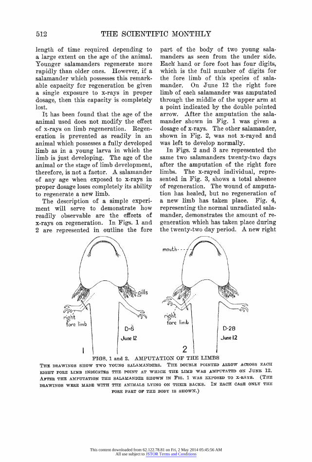
The description of a simple experi- ment will serve to demonstrate how readily observable are the effects of x-rays on regeneration. In Figs. 1 and 2 are represented in outline the fore
part of the body of two young sala- manders as seen from the under side. Eaeh hand or fore foot has four digits, which is the full number of digits for the fore limb of this species of sala- mander. On June 12 the right fore limb of each salamander was amputated through the middle of the upper arm at a point indicated by the double pointed arrow. After the amputation the sala- mander shown in Fig. 1 was given a dosage of x-rays. The other salamander, shown in Fig. 2, was not x-rayed and was left to develop normally.
In Figs. 2 and 3 are represented the same two salamanders twenty-two days after the amputation of the right fore limbs. The x-rayed individual, repre- sented in Fig. 3, shows a total absence of regeneration. The wound of amputa- tion has healed, but no regeneration of a new limb has taken place. Fig. 4, representing the normal unradiated sala- mander, demonstrates the amount of re- generation which -has taken place during the twenty-two day period. A new right
ri,ght lrght l fore lib fore llrAt
i ~~D-6 D-28 l iJune 12 2 June 12
FIGS. 1 and 2. AMPUTATION OF THE LIMBS THE DRAWINGS SHOW TWO YOUNG SALAMANDERS. THE DOUBLE POINTED ARROW ACROSS EACH
RIGHT PORE LIMB INDICATES THE POINT AT WHICH THE LIMB WAS AMPUTATED ON JUN11 12. AFTER THE AMPUTATION THE SALAMANDER SHOWN IN FIG. 1 WAS EXPOSED TO X-RAYS. (THE
DRAWINGS WERE MADE WITH THE ANIMALS LYING ON THEIR BACKS. IN EACH CASE ONLY THE
FORE PART OF THE BODY IS SHOWN.)
This content downloaded from 62.122.78.81 on Fri, 2 May 2014 05:45:56 AMAll use subject to JSTOR Terms and Conditions

EFFECT OF X-RAYS ON TISSUE REGENERATION 313
non- regenerating r limb stump reiegheatinmg
D-6 rih ibD-28
July 4 July 4
34 FIGS. 3 and 4. REGENERATING AND NON-REGENERATING LIMBS
THE SAME SALAMANDERS SHOWN IN FIGS. 1 AND 2. THESE DRAWINGS WERE MADE 22 DAYS AFTER
THE AMPUTATION OF THE RIGHT FORE LIMB OF EACH ANIMAL. No REGENERATION HAS TAKEN
PLACE IN THE X-RAYED SALAMANDER, FIG. 3. THE RIGHT FORE LIMB OF THE NORMAL UNRADIATED
SALAMANDER, FIG. 4, IS REGENERATING NICELY.
limb has already regenerated as far as the three-digit condition. A few days after this drawing was made the fourth digit appeared, and soon the right limb was again in the same condition as the unharmed left limb.
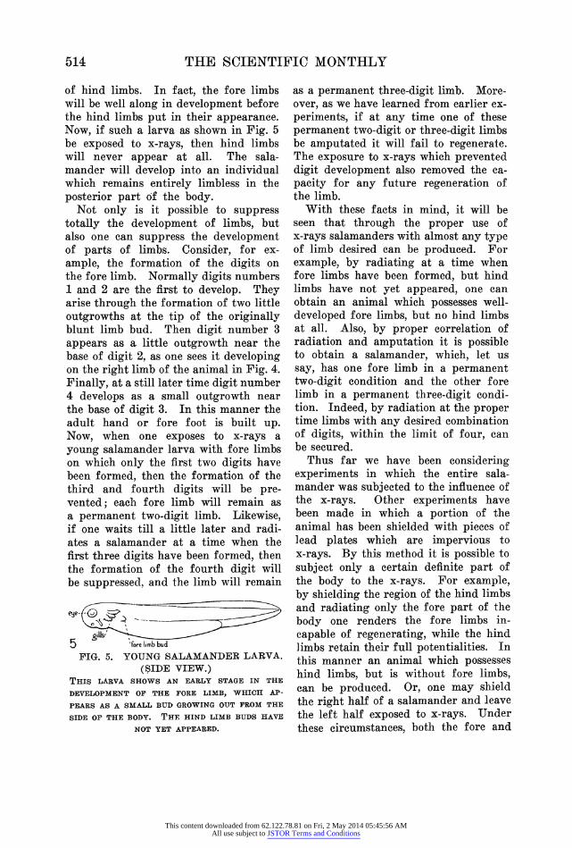
In other experiments young salaman- der larvae in which the fore limb is just beginning to develop have been used. For example, one may amputate the limb bud at a time when it has just appeared on a young salamander larva as a small blunt protrusion or bud on the side of the body wall. Such an early stage of limb development is shown on the larva represented in side view in Fig. 5. Am- putation at this early time consists sim- ply in clipping off the free end of the little limb bud. The wound heals very quickly in a young larva, such as this, and, under normal conditions, regenera- tion of the limb bud and subsequent normal limb development takes place at a rapid rate. However, if after the am- putation of the small limb bud the larva be exposed to x-rays, then the limb will
fail to develop. This experiment shows that even in this young stage of de- velopment x-radiation has completely re- moved from the animal the eapacity for limb regeneration.
Since x-radiation exerts so pronounced an effect on the regeneration of limbs, it is not surprising to discover that the normal development of limbs in young salamanders is also subject to modifica- tion by x-rays. Recently W. 0. Puckett, working in the Princeton laboratory, has made a detailed study of the influ- ence of x-rays on normal limb develop- ment. His work shows that exposure to x-rays at the proper time will com- pletely suppress normal limb develop- ment. This effect may be readily dem- onstrated by the following simple ex- periment on hind limb development. Hind limbs in the salamanders used for these studies normally develop much later than fore limbs. It will be noticed in the salamander larva shown in Fig. 5, for example, that although fore limbs are starting to develop, there is no sign
This content downloaded from 62.122.78.81 on Fri, 2 May 2014 05:45:56 AMAll use subject to JSTOR Terms and Conditions
514 THE SCIENTIFIC MONTHLY
of hind limbs. In fact, the fore limbs will be well along in development before the hind limbs put in their appearance. Now, if such a larva as shown in Fig. 5 be exposed to x-rays, then hind limbs will never appear at all. The sala- mander will develop into an individual which remains entirely limbless in the posterior part of the body.
Not only is it possible to suppress totally the development of limbs, but also one can suppress the development of parts of limbs. Consider, for ex- ample, the formation of the digits on the fore limb. Normally digits numbers 1 and 2 are the first to develop. They arise through the formation of two little outgrowths at the tip of the originally blunt limb bud. Then digit number 3 appears as a little outgrowth near the base of digit 2, as one sees it developing on the right limb of the animal in Fig. 4. Finally, at a still later time digit number 4 develops as a small outgrowth near the base of digit 3. In this manner the adult hand or fore foot is built up. Now, when one exposes to x-rays a young salamander larva with fore limbs on which only the first two digits have been formed, then the formation of the third and fourth digits will be pre- vented; each fore limb will remain as a permanent two-digit limb. Likewise, if one waits till a little later and radi- ates a salamander at a time when the first three digits have been formed, then the formation of the fourth digit will be suppressed, and the limb will remain
ege-
5 'fore limb bud FIG. 5. YOUNG SALAMANDER LARVA.
(SIDE VIEW.) THIS LARVA SHOWS AN EARLY STAGE IN THE
DEVELOPMERNT OF THE FORE LIMB, WHICH AP-
PEARS AS A SMALL BUD GROWING OUT FROM THE
SIDE OF THE BODY. THF. HIND LIMB BUDS HAVE
NOT YET APPEARED.
as a permanent three-digit limb. More- over, as we have learned from earlier ex- periments, if at any time one of these permanent two-digit or three-digit limbs be amputated it will fail to regenerate. The exposure to x-rays which prevented digit development also removed the ca- pacity for any future regeneration of the limb.
With these facts in mind, it will be seen that through the proper use of x-rays salamanders with almost any type of limb desired can be produced. For example, by radiating at a time when fore limbs have been formed, but hind limbs have not yet appeared, one can obtain an animal which possesses well- developed fore limbs, but no hind limbs at all. Also, by proper correlation of radiation and amputation it is possible to obtain a salamander, which, let us say, has one fore limb in a permanent two-digit condition and the other fore limb in a permanent three-digit condi- tion. Indeed, by radiation at the proper time limbs with any desired combination of digits, within the limit of four, can be secured.
Thus far we have been considering experiments in which the entire sala- mander was subjected to the influence of the x-rays. Other experiments have been made in which a portion of the animal has been shielded with pieces of lead plates which are impervious to x-rays. By this method it is possible to subject only a certain definite part of the body to the x-rays. For example, by shielding the region of the hind limbs and radiating only the fore part of the body one renders the fore limbs in- capable of regenerating, while the hind limbs retain their full potentialities. In this manner an animal which possesses hind limbs, but is without fore limbs, can be produced. Or, one may shield the right half of a salamander and leave the left half exposed to x-rays. Under these circumstances, both the fore and
This content downloaded from 62.122.78.81 on Fri, 2 May 2014 05:45:56 AMAll use subject to JSTOR Terms and Conditions
EFFECT OF X-RAYS ON TISSUE REGENERATION 515
hind limbs on the left side lose their capacity for regenerating, while the fore and hind limbs on the right side remain normal. These experiments demon- strate, among other things, that the ef- fect of x-rays on limb regeneration is a local effect. In other words, it is an effect which the x-rays exert directly on the cells in the limb area, and is not a general effect on the animal as a whole.
Interesting as are the foregoing ex- periments, it is of still further interest and importance to inquire into the alterations which take place within the limbs of x-rayed salamanders. What changes have the x-rays brought about in the tissue, so that it is no longer capable of regenerating? In order bet- ter to understand the changes which take place within the non-regenerating limbs of x-rayed salamanders, let us ex- amine first the manner in which new tissue is formed during normal regen- eration of the salamander limb.
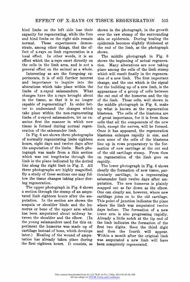
In Fig. 6 are shown three photographs of normally regenerating limbs eighteen hours, eight days and twelve days after the amputation of the limbs. Each pho- tograph was made from a thin section which was cut lengthwise through the limb in the plane indicated by the dotted line along the right limb in Fig. 2. All three photographs are highly magnified. By a study of these sections one may fol- low the tissue changes taking place dur- ing regeneration.
The upper photograph in Fig. 6 shows a section through the stump of an ampu- tated limb eighteen hours after the am- putation. In the section are shown the scapula or shoulder blade and the hu- merus or bone of the upper arm which has been amputated about midway be- tween the shoulder and the elbow. (In the young salamanders used for this ex- periment the humerus was made up of cartilage instead of bone, which develops later.) Healing of the wound of ampu- tation has already taken place during the first eighteen hours. It consists, as
shown in the photograph, in the growth over the raw stump of the surrounding skin or epidermis. During healing the epidermis becomes slightly thickened at the end of the limb, as the photograph shows.
The middle photograph in Fig. 6 shows the beginning of actual regenera- tion. Many alterations are now taking place among the cells of the limb stump which will result finally in the regenera- tion of a new limb. The first important change, and the one which is the signal for the building up of a new limb, is the appearance of a group of cells between the cut end of the humerus and the tip of the limb. These cells, well shown in the middle photograph in Fig. 6, make up what is known as the regeneration blastema. The cells of the blastema are of great importance, for it is from these cells that all the components of the new limb, except the nerves, will be formed. Once it has appeared, the regeneration blastema enlarges rapidly in size, and soon some of the cells of the blastema line up in rows preparatory to the for- mation of new cartilage at the cut end of the old cartilage stump. From then on regeneration of the limb goes on rapidly.
The lower photograph in Fig. 6 shows clearly the formation of new tissuLe, par- ticularly cartilage, in a regenerating salamander limb twelve days after am- putation. The new humerus is plainly mapped out as far down as the elbow. One can clearly see, however, where new cartilage joins on to the old cartilage. This point of junction indicates the place where the limb was amputated twelve days before. The formation of a new lower arm is also progressing rapidly. Already a little notch at the tip end of the limb indicates the formation of the first two digits. Soon the third digit and then the fourth will appear. Within a month after the original limb was amputated a new limb will have been completely regenerated.
This content downloaded from 62.122.78.81 on Fri, 2 May 2014 05:45:56 AMAll use subject to JSTOR Terms and Conditions
516 THE SCIENTIFIC MONTHLY
4
epidermis ~ merti
is HOURS ^
x0 , , .: ~~~~._ ̂
.~... ......_._..0.. : . @. _ _.X. __._. . ... .
i { < i , . .~ ~ ~ ~ ~ ~ ~ ~ ~ ~ ~ ~~~~~~~~~~~~~~~~~~~..,. U . .. ..
Aowj 4~A
eii.'i'''. -X A. | 5 T
new !a.e .old cariti.a.gE
FIG. 6. REGENERATING LIMBS THE PHOTOGRAPHS SHOW CHANGES WHICH TAKE
PLACE WITHIN THE TISSUE OF THE SALAMANDER
LIMB DURING NORMAL REGENERATION. EACH PHO-
TOGRPAPH W-AS MADE FROM A THIN SECTION CUT
LENGTHWISE THROUGH THE LIMB IN THE PLANE
INDICATED BY THE. DOTTED LINE ALONG THE RIGHT
LIMB IN FIG. 2. (ALL PHOTOGRAPHS ARE HIGHLY
MAGNIFIED). THE UPPER PHOTOGRAPH SHOWS
A LIMB STUMNP 18 HOURS AFTER LIMB AMPUTA-
TION. THE MIDDLE PHOTOGRAPH IS A REGENER-
ATING LImB 8 DAYS AFTER AMPIUTATION. THE
LOWER SHOWS REGENERATION PROGRESSING RAP-
IDLY 12 DAYS AFTER LIMB AMPUTATION.
Ilaving in mind, now, the main tissue changes which take place during the normal course of regeneration, let us turn to a study of the conditions which one finds in the tissue of the non-regen- erating limb stumps of x-rayed sala- manders. What alterations in the tissue have the x-rays brought about which render the limb incapable of regener- ating ?
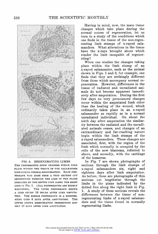
When one studies the changes taking place within the limb stump of an x-rayed salamander, such as the animal shown in Figs. 1 and 3, for example, one finds that they are strikingly different from those which accompany normal re- generation. However, differences in the tissue of radiated and unradiated ani- mals do not become apparent immedi- ately after amputation. During the first few days no very pronounced changes occur within the amputated limb other than the healing of the wound, which ordinarily takes place in an x-rayed salamander as rapidly as in a normal unradiated individual. On about the sixth day after amputation the similar- ity between the radiated and the unradi- ated animals ceases, and changes of an extraordinary and far-reaching nature begin within the limb stumps of the x-rayed salamanders. These changes are associated, first, with the region of the limb which normally is occupied by the cells of the new blastema, referred to above, and secondly, with the cartilage of the humerus.
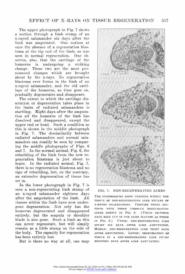
In Fig. 7 are shown photographs of sections through the limb stumps of x-rayed salamanders six, eight and eighteen days after limb amputation. As before, these are photographs of thin sections cut lengthwise through the limbs in the plane indicated by the dotted line along the right limb in Fig. 1. A study of these sections reveals the differences between the tissue of non- regenerating limbs of x-rayed salaman- ders and the tissue found in normally regenerating limbs.
This content downloaded from 62.122.78.81 on Fri, 2 May 2014 05:45:56 AMAll use subject to JSTOR Terms and Conditions
EFFECT OF X-RAYS ON TISSUE REGENERATION 517
The upper photograph in Fig. 7 shows a section throug,h a limb stump of an x-rayed salamander six days after the limb was amputated. One notices at onee the absence of a reoenerationi blas- tema at the tip enld of the liinb, as was seen in normal receneration. One ob- serves, also, that the cartilage of the hiumeruis is undergroin" a striking- change. These two are the most pro- nounced clhang,es whieh are brought about by the x-rays. No receneration blastema ever forms in the limb of ani x-rayed salamander, and the old carti- lao e of the humerus, as time goes on, gradually degenerates and disappears.
The extent to which the cartilage dis- solution or degeneration takes place in the limbs of radiated salamanders is startling. Eigrht days after the amputa- tion all the humerus of the limb has dissolved and disappeared, except the upper end or head. Suctlh a condition as this is shown in the middle photograplh in Fig. 7. The dissimilarity between r adiated salamanders and normal sala- manders can readily be seen by compar- ing the middle photographs of Figs. 6 and 7. In the normal animal, Fig. 6, the rebuilding of the limb from the new re- generation blastema is just about to begin. In the radiated animal, Fig. 7, there is no regeneration blastema and no sign of rebuilding, but, on the contrary, an extensive degeneration of tissue has set in.
In the lower photograph in Fig. 7 is seen a non-regenerating limb stump of an x-rayed salamander eighteen days after the amputation of the limb. All tissues within the limb have now under- gone degeneration. Not only has the humerus degenerated and disappeared entirely, but the scapula or shoulder blade is also gone. Such a limb as this can never regenerate, but will simply remain as a little stump on the side of the body. The capacity for regeneration has been entirely lost.
But is there no way at all, one may
6 DAYS h- ner'
4
1 DAY..
FIG. 7. NON-REGENERATING LIMBS
THE PHOTOGRAPHS SHOW CHANGES WITHIN THE
TISSUE OF NON-REGENERATING LIMIB STUMIPS OF
X-RAYED SALAMANDERS. CONIPARE THESE SEC-
TIONS WITH THOSE THROUGII REGENERATING
LIMBS SHOWN IN FIG. 6. (TTHESE SECTIONS
HIAVE BEEN CUT IN THE SAME MIANNER AS THOSE
IN FIG. 6.) UPPER: NON-REGENERATING LIMB
STUMP SIX DAYS AFTER LIMIB AMIPUTATION.
MIDDLE: NON-REGENERATING LIMB EIGHT DAYS
AFTER AMPUTATION. LOWER: DEGENERATION OF
TISSUE IN A NON-REGENERATING LIM1B STUMP
EIGHTEEN DAYS AFTER LIMB AMIIPUTATION.
This content downloaded from 62.122.78.81 on Fri, 2 May 2014 05:45:56 AMAll use subject to JSTOR Terms and Conditions
518 THE SCIENTIFIC MONTHLY
ask, by which an x-rayed salamander may regain the ability to regenerate a limb? Can not the animal in some man- ner be rehabilitated? The ability to regenerate can be restored only when a radiated animal is supplied with a new unradiated limb. This can be done by removing from a normal unradiated salamander a limb, and transplanting it to the body of an x-rayed individual. The transplanted limb may be placed in the normal limb location where it will grow, and often the salamander is able to use it in getting about nearly as well as it used its original limb. Now, if such a transplanted limb as this be am- putated, then a new one will be regen- erated. In this manner has the regen- erative ability been restored to the x-rayed animal.
From a comparison, therefore, of the regenerative capacities of normal and x-rayed salamanders several conclusions are obvious. We find that when the formation of a new blastema is sup- pressed through the use of x-rays, then regeneration is prevented. We learn, moreover, that the cells which normally
make up the regeneration blastema, and thereby enter into the formation of the new limb, come from the region of the limb itself and not from distant parts of the body, as some biologists have hereto- fore thought. The transplantation ex- periments just mentioned prove this point conclusively. We find, also, that in an x-rayed salamander the opposite of regeneration, namely, degeneration, takes place. It appears as though when tissues of the limb are prevented from regenerating, they must of necessity de- generate. There are reasons for think- ing that the extensive degeneration of the humerus in x-rayed salamanders, for example, is due to the absence of a re- generation blastema.
Experiment, when applied to a study of developmental processes, invariably brings about the formation of abnormal animals. It is through a careful analy- sis of these abnormal individuals that the experimentalist gains new insight into the factors which underlie normal development. In x-rays the biologist has an efficient instrument of research, a new method for experimental analysis.
This content downloaded from 62.122.78.81 on Fri, 2 May 2014 05:45:56 AMAll use subject to JSTOR Terms and Conditions