the effect of vertebral body tethering on spine …
TRANSCRIPT

THE EFFECT OF VERTEBRAL BODY TETHERING ON SPINE MOTION IN
ADOLESCENT IDIOPATHIC SCOLIOSIS: A PILOT STUDY
Mina Maksimovic
A thesis submitted in partial fulfillment of the requirements for the Master of Science degree in
Human Kinetics
Supervisor: Ryan B. Graham, PhD
School of Human Kinetics
Faculty of Health Sciences
University of Ottawa
© Mina Maksimovic, Ottawa, Canada, 2021

ii
Table of Contents
Table of Contents .......................................................................................................................... ii
Acknowledgements ...................................................................................................................... iv
List of Acronyms ........................................................................................................................... v
List of Figures ............................................................................................................................... vi
List of Tables ............................................................................................................................... vii
Abstract ....................................................................................................................................... viii
1.0 INTRODUCTION.............................................................................................................. 1
2.0 LITERATURE REVIEW ................................................................................................. 3
2.1 Adolescent Idiopathic Scoliosis ....................................................................................... 3
2.2 Surgical Treatment ........................................................................................................... 4
2.2.1 Posterior Spinal Fusion and Instrumentation ............................................................ 4
2.2.2 Vertebral Body Tethering ......................................................................................... 5
2.3 Spine Range of Motion .................................................................................................... 7
2.3.1 Normative Pediatrics ................................................................................................. 8
2.3.2 Adolescent Idiopathic Scoliosis ................................................................................ 8
2.3.3 Posterior Spinal Fusion and Instrumentation .......................................................... 10
2.3.4 Vertebral Body Tethering ....................................................................................... 12
3.0 PURPOSE ......................................................................................................................... 14
4.0 METHODOLOGY .......................................................................................................... 15
4.1 Ethics .............................................................................................................................. 15
4.2 Participants ..................................................................................................................... 15
4.3 Study Design .................................................................................................................. 16
4.3.1 Assent and Consent ................................................................................................. 16
4.3.2 Questionnaires......................................................................................................... 16
4.3.3 Participant Preparation and Equipment .................................................................. 17
4.3.4 Movement Protocol ................................................................................................. 18
4.4 Data Processing and Analysis ........................................................................................ 20
4.5 Statistical Analysis ......................................................................................................... 22
4.5.1 Global Angles ......................................................................................................... 22
4.5.2 Intersegmental Angles ............................................................................................ 22
5.0 RESULTS ......................................................................................................................... 24

iii
5.1 Demographics................................................................................................................. 24
5.2 Global Angles ................................................................................................................. 25
5.2.1 Forward Flexion ...................................................................................................... 25
5.2.2 Lateral Bending ....................................................................................................... 26
5.2.3 Axial Twist.............................................................................................................. 28
5.3 Intersegmental Angles .................................................................................................... 30
5.3.1 Forward Bending .................................................................................................... 30
5.3.2 Lateral Bending ....................................................................................................... 32
5.3.3 Axial Twist.............................................................................................................. 36
6.0 DISCUSSION ................................................................................................................... 38
7.0 CONCLUSION ................................................................................................................ 44
REFERENCES ............................................................................................................................ 45
APPENDIX A .............................................................................................................................. 55
APPENDIX B .............................................................................................................................. 56
APPENDIX C .............................................................................................................................. 60
APPENDIX D .............................................................................................................................. 67
APPENDIX E .............................................................................................................................. 69
APPENDIX F .............................................................................................................................. 70

iv
Acknowledgements
This work would have never been possible without the invaluable and selfless acts of
collaboration, assistance, and support from an outstanding group of people.
Thank you to our collaborators at CHEO for making this project possible. Dr. Kevin Smit,
Holly Livock and Patrick Sachsalber, it has been a privilege getting to work with you all and I
appreciate everything that you do. Thank you to my committee members, Dr. Linda McLean and
Dr. Kristi Adamo, for your time and guidance that helped me successfully complete my work. To
Dr. Shawn Beaudette; I am forever grateful for all your assistance and the time you have
contributed to this project, regardless of your own busy schedule. I also thank each participant who
took time out of their lives to be a part of this work; our sessions are something I will cherish, and
I am lucky to have met you all.
To my supervisor Dr. Ryan Graham, thank you for sharing your knowledge, for your
patience and your support during both stable and life-changing unprecedented times over the past
three years. I greatly value the independence and guidance you have provided me to help shape
this project, as well as myself. Further, thank you to every single lab member whose time has
overlapped with my own. The endless help, laughs, advice and entertainment has never gone
unappreciated. I am eternally thankful of your acceptance and friendship.
Finally, to my family and closest friends both near and abroad; I thank you for listening,
caring and always being there for me. Words cannot express my gratitude to you and how fortunate
I am to have you in my life.

v
List of Acronyms
3D Three-dimensional
ANOVA Analysis of Variance
AIS Adolescent Idiopathic Scoliosis
AP Anterior/Posterior
CHEO Children’s Hospital of Eastern Ontario
DDD Degenerative Disc Disorder
IMU Inertial Measurement Unit
IV Intervertebral
LCS Local Coordinate System
LIV Lowest Instrumented Vertebrae
ML Medio-lateral
PSF Posterior Spinal Fusion
ROM Range of Motion
SD Standard Deviation
SI Superior/Inferior
SPM Statistical Parametric Mapping
VBT Vertebral Body Tethering

vi
List of Figures
Figure 1. Qualitative explanation of the Hueter-Volkmann principle (adapted from Stokes, 2007)
......................................................................................................................................................... 6
Figure 2. Marker placement for the 57-marker intersegmental spine model ............................... 17
Figure 3. Diagram of the custom pelvis immobilization device .................................................. 19
Figure 4. Spatial reconstruction of the intersegmental spine model (from Zwambag et al., 2018)
....................................................................................................................................................... 21
Figure 5. Mean range of motion of the Control AIS, PSF and VBT groups in the thoracic, lumbar,
and total spine regions during forward flexion ............................................................................. 25
Figure 6. Mean range of motion of the Control, AIS, PSF and VBT groups in the thoracic, lumbar,
and total spine regions during left and right lateral bending ........................................................ 27
Figure 7. Mean range of motion of the Control, AIS, PSF and VBT groups in the thoracic, lumbar,
and total spine regions during left and right axial twist ............................................................... 29
Figure 8. Mean intersegmental spine range of motion of the Control, AIS, PSF and VBT groups
during forward bending................................................................................................................. 30
Figure 9. Post-hoc intersegmental spine SPM{t} values during forward bending ...................... 31
Figure 10. Mean intersegmental spine range of motion of the Control, AIS, PSF and VBT groups
during right lateral bending ........................................................................................................... 32
Figure 11. Post-hoc intersegmental spine SPM{t} values during right lateral bending .............. 33
Figure 12. Mean intersegmental spine range of motion of the Control, AIS, PSF and VBT groups
during left lateral bending ............................................................................................................. 34
Figure 13. Post-hoc intersegmental spine SPM{t} values during left lateral bending ................ 35
Figure 14. Mean intersegmental spine range of motion of the Control, AIS, PSF and VBT groups
during right axial twist. ................................................................................................................. 36
Figure 15. Mean intersegmental spine range of motion of the Control, AIS, PSF and VBT groups
during left axial twist. ................................................................................................................... 37
Figure 16. Post-hoc intersegmental spine SPM{t} values during left axial twist ........................ 37

vii
List of Tables
Table 1. Participant demographics. .............................................................................................. 24
Table 2. Surgical group details. .................................................................................................... 24
Table A1. Calibration and tracking marker placement during movement protocol. .................... 67
Table F1. Cobb angle measurements. .......................................................................................... 70

viii
Abstract
The current gold standard for the surgical treatment of adolescent idiopathic scoliosis (AIS)
is posterior spinal fusion and instrumentation (PSF); however, decreased normal spine function,
strain on unfused vertebrae and pain have been suggested as long-term complications with this
technique. Vertebral body tethering (VBT) is a novel and minimally invasive approach for
scoliosis correction that does not involve fusion and may theoretically preserve spine motion. VBT
involves the use of screws and a tether that are inserted thoracoscopically on the convexity of the
spine curvature in skeletally immature patients, allowing progressive curve correction as skeletal
maturity is achieved. No research to date has explored the effect of VBT on spine motion in AIS.
Therefore, the purpose of this thesis was to retrospectively compare global and intersegmental
spine range of motion in adolescents (9-18 years of age) without spine deformity, adolescents with
untreated AIS, adolescents having undergone PSF, and adolescents having undergone VBT to gain
insight on the effect of VBT on spine motion. Twenty participants were recruited into four groups
including Control (n = 6), untreated AIS (n = 5), post-operative PSF (n = 4) and post-operative
VBT (n = 5). Three-dimensional kinematics of the spine were collected and analyzed using an
intersegmental spine model during constrained forward flexion, right-left lateral bending, and
right-left axial twist movements. The PSF group displayed significantly lower spine ROM than
the two non-operative groups during thoracic and total left axial twist (p ≤ 0.048), whereas thoracic
and total spine range of motion during right-left lateral bending is almost equally lower in the PSF
(p ≤ 0.03) and VBT (p ≤ 0.01) groups when compared to the Control and AIS groups. These results
may suggest some preservation of spine motion in the transverse plane following VBT; however
further investigation with a greater sample size and a prospective study design is needed to support
and prove the theoretical preservation of spine motion following VBT.

1
1.0 INTRODUCTION
Adolescent idiopathic scoliosis (AIS) is a three-dimensional structural deformity of the
spine, characterized by abnormal curvature and rotation of the vertebrae that develops in children
around puberty (Nishida et al., 2017). AIS has an overall worldwide prevalence of 0.47-0.52%
and female to male ratios that range from 1.5:1 to 3:1, which increases with age (Konieczny et al.,
2013). The largest number of all musculoskeletal deformity healthcare visits are from spine
deformities in children and adolescents, where AIS is the most prevalent form of spinal deformity
and makes up 80% of all pediatric scoliosis cases (Noshchenko et al., 2015). AIS is patient-
specific; thus, a wide variety of characteristics, severities and secondary effects increases the
difficulty of determining a true cause of the spine deformity.
The aetiopathogenesis of AIS remains unclear and various demographic factors (i.e.
ethnicity, sex, age, genetics and geographic location) influence the progression of the spine
deformity (Burwell, 2003; Konieczny, 2013; Lowe et al., 2000). Furthermore, many biological
and biomechanical factors (i.e. the central nervous system, bone density, muscles, melatonin, and
skeletal framework) are being explored as possible causes for AIS (Burwell, 2003). The
multifactorial nature surrounding the cause of AIS limits the development of preventative
treatments, leaving patients with only corrective options following a diagnosis.
Current treatment options for AIS are dependent on the severity of the spine deformity.
Physiotherapy and bracing are used for curves of lesser magnitude to limit curve progression,
whereas corrective surgery is used for curves of greater magnitude. Approximately 29,000
scoliosis-related surgeries are performed on adolescents in the United States every year to correct
the deformity (Noshchenko et al., 2015). High costs are associated with the surgical treatment of
AIS and were estimated to be $1.1 billion dollars in 2012 in the United States (Vigneswaran et al.,

2
2015). Treatment type is also decided based on the location, shape and pattern of the curve, as well
as the patient’s future growth potential (BMUS, 2014).
Posterior spinal fusion and instrumentation (PSF) is the current gold standard surgical
correction for those with AIS. However, potential negative side effects of this invasive surgery
include: significant blood loss, post-operative pain, infection, pseudoarthrosis and degenerative
disc disease (DDD) (Danielsson et al., 2001; De la Garza Ramos et al., 2016). Further, a large
disadvantage of PSF is a reduction in spine range of motion (ROM), observed using motion capture
techniques (Engsberg et al., 2002; Wilk et al., 2006). Novel surgical strategies are being developed
as a result to lessen and/or eliminate these potential side-effects, such as vertebral body tethering
(VBT). VBT is a minimally-invasive and fusionless surgical technique for AIS correction
performed in skeletally immature patients (Courvoisier et al., 2015; Samdani et al., 2015).
Although the acute and long-term effects of this surgery on spine ROM have yet to be explored,
the theoretical advantage of VBT is the preservation of spine ROM due to its fusionless nature.
In order to assess the potential theoretical advantage of fusionless VBT surgery for AIS
correction, this pilot work aimed to retrospectively compare spine ROM in the following
adolescent populations: 1) those with no spine deformity; 2) those with an untreated diagnosis of
AIS; 3) those having undergone PSF; and 4) those having undergone VBT. The primary objective
was to compare spine ROM of the thoracic, lumbar, and total spine regions, while the secondary
objective was to assess spine ROM at each IV level.

3
2.0 LITERATURE REVIEW
2.1 Adolescent Idiopathic Scoliosis
Scoliosis is a three-dimensional, laterally and axially rotated curvature of the spine (Weinstein
et al., 2008). Many forms of scoliosis exist; however, the diagnosis of idiopathic scoliosis is made
when any neuromuscular or congenital causes of the deformity have been eliminated, signifying
an unknown cause for the deformity (Konieczny et al., 2013). Adolescent idiopathic scoliosis
(AIS) is the most common type of scoliosis that affects 80% of scoliosis patients and develops in
otherwise healthy children during puberty (i.e. between the ages of 9-18) (Asher & Burton, 2006).
Ponseti and Friedman (1950) classified AIS as one or two major curves in the cervical,
thoracic, and/or lumbar regions; one major curve is the most prevalent making up 70% of all AIS
cases. The most common curvature in AIS is a thoracic curvature with an apex towards the right
(25% of cases), which is defined as a convex curvature with the apex between the T2 and T11
vertebrae (Machida et al., 2018). A patient with a diagnosis of a main right thoracic AIS curvature
can experience many anatomical changes in the trunk region including: chest wall asymmetry
leading to rib prominence, elevation of the ipsilateral shoulder, thoracic translocation toward the
convexity of the curve, and waistline asymmetry (Sud & Tsirikos, 2013). Many factors influence
the onset of scoliosis resulting in a variety of different symptoms and severities, thus creating a
patient-specific profile for each diagnosis.
In 2001, Lawrence Lenke developed and presented the Lenke classification system to assist
doctors and surgeons to uniformly choose the best treatment option for each patient (Lenke et al.,
2001). The commonly used classification system defines curvatures by the curve type (there are
six defined curve types), the lumbar modifier (the location of the vertical line going through the
lumbar vertebrae) and the thoracic sagittal profile modifier (assesses thoracic kyphosis between

4
T5 and T12 vertebrae); however, this system fails to address the rotational aspect of the deformity
(Ovadia, 2013). More recently, novel technologies such as the EOS imaging system, which
provides coronal, sagittal and axial x-ray views of the spine, allow for spine reconstruction
strategies to classify and assess the spine deformity in 3D (Illés et al., 2011). Surgeons can now
more precisely consult and agree on a surgical technique that would optimize patient treatment
and post-operative quality of life.
2.2 Surgical Treatment
A growing understanding of the relationship between the heart, the chest wall, the lungs
and the 3D changes of the spine has resulted in the evolution of treatment options for scoliosis
(Hardesty et al., 2018). These improved treatment options ensure deformity control, preservation
of function, as well as improvements in psychological and quality of life aspects (Hasler, 2018).
The careful consideration of all these variables using classification systems and novel technologies
allows for patients to receive their optimal individualized care.
2.2.1 Posterior Spinal Fusion and Instrumentation
The current standard of care for the surgical treatment of AIS is posterior spinal fusion and
instrumentation (PSF) (Padhye et al., 2018). This surgical technique involves two parts: 1) spinal
fusion, and 2) instrumentation. The former involves fusing two or more vertebrae using bone grafts
and the latter utilizes various implants (e.g. plates, screws, hooks, rods, etc.) to stabilize, strengthen
and maintain alignment of the spine while fusion occurs (Deniz Olgun & Yazici, 2013). PSF may
be an effective treatment option to eliminate the spine deformity; however, many side-effects of
this surgical technique exist.
PSF has some benefits compared to other corrective surgeries (i.e. preservation of
pulmonary functions and allows the use of direct vertebral rotation), but it has been shown that the

5
execution of PSF may affect the posterior trunk musculature and the risk of infection is higher
(Deniz Olgun & Yazici, 2013). PSF may also lead to low back pain and trunk rigidity, negatively
impacting the patient’s health-related quality of life as they age (Danielsson et al., 2001; Fan et
al., 2016). Further potential long-term complications of this technique include impaired spine
function, strain on unfused vertebrae, pain and degenerative disc disease (DDD) (Danielsson,
Romberg, & Nachemson, 2006). New techniques are being introduced in hopes of minimizing
these long-term effects and, consequently, increase patient quality of life.
2.2.2 Vertebral Body Tethering
Vertebral body tethering (VBT) is a novel, fusionless and minimally invasive corrective
surgical technique executed thoracoscopically. It was first used in humans in 2010 and involves
inserting screws into the vertebrae that make up the concave curvature; this is done when the
patient is skeletally immature, so that the growth of the spine results in progressive curve correction
until skeletal maturity is achieved (Crawford & Lenke, 2010; Samdani et al., 2015).
The surgery is theoretically based on the Hueter-Volkmann principle, which states that
increasing mechanical compression of the growth plates inhibits growth rate, whereas tension
(reduced loading) stimulates it (Lalande et al., 2020; Villemure & Stokes, 2009). Vertebrae in
humans grow both via longitudinal growth, generated by these growth plates adjacent to the discs,
as well as in width by increasing the diameter of the vertebral body (Stokes, Aronsson, Dimock,
Cortright, & Beck, 2006; Stokes, Spence, Aronsson, & Kilmer, 1996). During adolescence, the
vertebrae are responsible for most of the growth of the spine and because sustained loading of
these bones modulates their growth, a combination of disc and vertebral wedging will result in
changes of its shape (Stokes & Aronsson, 2001; Stokes & Windisch, 2006). Figure 1 displays a
qualitative explanation of the Hueter-Volkmann principle as it relates to scoliosis, where the

6
reduced compression on the concave of the curve in AIS stimulates growth, while the convex side
is inhibited due to increased compression (Stokes, 2007). VBT aims to reverse the asymmetrical
distribution of compression in the spine to stimulate longitudinal growth.
Samdani et al. (2014) reported two-year results from an initial cohort of eleven patients
who had undergone VBT; they found that there was an improvement in their thoracic prominences
and the measure of their Cobb angles (a measure used to quantify the magnitude of a curve of the
spine), with no infection, neurological or instrumentation-related complications. Current research
investigating the effects of VBT on spine range of motion (ROM) is limited to computer simulation
and animal models that suggest preservation of disc health and spine motion (Aubin et al., 2018;
Cobetto et al., 2018; Newton et al., 2008a).
Vicious Cycle
Spinal
curvature
Wedging of
vertebrae
and discs
Asymmetrical
growth
Asymmetrical
loading
Figure 1. The qualitative explanation of adolescent scoliosis progression via biomechanical
growth modulation represented by the black arrows (Hueter-Volkmann principle). Red arrows
indicate the effect of VBT on spine growth using this modulation technique (adapted from Stokes,
2007).

7
2.3 Spine Range of Motion
Spine ROM is most commonly assessed by quantifying mobility of an individual during
movements in the sagittal, coronal and transverse planes (flexion/extension, lateral bending and
axial twist, respectively) (Kozanek et al., 2009). This thesis focused on movements in the thoracic
and lumbar regions of the spine and therefore, emphasis will be placed on these regions. The
lumbar spine accounts for most of the movement during flexion, the thoracic and lumbar spine
combine to account for most of the movement during lateral bending, and the thoracic spine
accounts for most of the movement during axial twisting (Nordin & Frankel, 2001). Changes in
these movement distribution patterns may arise from spine deformities, pathologies, and post-
surgical outcomes; therefore, it is important to assess any potential changes in spine motion during
movements in these planes.
ROM measurements are a standard clinical assessment during orthopaedic patient
examinations, primarily used for radiographic classification systems and in pre-operative planning
for the surgical treatment of scoliosis (Lea & Gerhardt, 1995; Lenke et al., 2001). Radiographs of
a patient’s spine yield the most accurate vertebral ROM measures during movements of interest;
however, concerns over the increased risk of lung cancer and breast cancer as a result of increased
radiation exposure prevent the completion of such studies (Bone & Hsieh, 2000; Nash et al., 1979).
The concerns arise from the high number of radiographs taken of AIS patients and the increased
sensitivity of the pediatric population towards radiation (Donaldson et al., 2007). Researchers and
medical professionals have resorted to using safer methods to assess spine ROM in clinical
populations like patients with AIS by using tools like goniometers, film digitization, optoelectronic
motion analysis, as well as newer, low-dosage x-ray systems.

8
2.3.1 Normative Pediatrics
Assessing spine ROM of the adolescent population with no spine deformity or other
existing pathologies is imperative to provide comparative data to accurately assess spine ROM in
clinical populations. Pediatric research on normative spine ROM most often looks at the influence
of age and sex on the cervical spine (Budelmann, Piekartz, & Hall, 2016; Lynch-Caris et al., 2008;
Seacrist et al., 2012), with fewer to no studies concentrating on solely the lumbar and thoracic
regions.
Early research investigating lumbar ROM using simpler methods (i.e. spondylometer,
Schober bend test) found that lumbar ROM decreased with age, while sex-related differences were
variable depending on the movement, where females often displayed greater ROM (Haley et al.,
1986; Taylor & Twomey, 1980). Similar results showing decreased lumbar ROM with increasing
age were found using a greater variety of additional methods (i.e. inclinometer, kyphometer and
BROM II) in children and adolescents (Kondratek et al., 2007; Sullivan, Dicknison, & Troup,
1994; Widhe, 2001). Assessment of total spine ROM and thoracic ROM in asymptomatic children
and adolescents is more prevalent in research comparing asymptomatic and clinical populations,
which will be further investigated with patients diagnosed with AIS.
2.3.2 Adolescent Idiopathic Scoliosis
The characterization of spine function, specifically spine motion, is essential to fully
understand the biomechanical changes caused by spine deformities such as AIS. Studies
investigating thoracolumbar and total spine motion (in the sagittal and frontal planes) in patients
with AIS compared to those without a spine deformity found either equal (Smidt et al., 1994;
Veldhuizen & Scholten, 1990) or decreased spine motion in patients with AIS. Poussa et al. (1989)
compared thoracic and lumbar spine motion in healthy controls and patients with AIS in all three

9
planes and found reduced flexion and rotation in the thoracic spine. However, the studies
mentioned above used a variety of outdated methods (i.e. tape measure techniques and
inclinometers) to evaluate spine motion in participants with heterogeneous curve magnitudes and
classifications, resulting in varying values across studies.
Galvis et al. (2016) used a magnetic sensor-based system to collect positional data at T1,
T3, T6, T10, L1, L3, and S1 vertebrae during sagittal and frontal plane bending tasks in healthy
controls and patients with AIS. The increased segmental detail of the model allowed Galvis and
colleagues (2016) to observe that patients with AIS showed no significant difference in thoracic
spine ROM in comparison to controls; however, increased spine ROM in the AIS group was noted
in spine regions directly above and below the apex of the curve.
An optical marker system model used on patients with AIS was developed by Schmid et
al. (2015) to quantify spine curvature in main thoracic AIS using reflective markers on the spinous
processes of the C7, T3, T5, T7, T9, T11 and L1-L5 vertebrae. This marker model was not used
to analyze the effect of AIS during spine movements in each of the movement planes. Instead, it
was used during gait analysis of AIS and revealed that there was increased lateral deviation in the
thoracolumbar/lumbar curvatures in the frontal plane and increased ROM in the sagittal plane,
suggesting potential compensation during gait within the spine rather than in spatio-temporal
parameters (Schmid et al., 2016). These studies indicate that a comprehensive model assessing
spine motion at more levels of the spine would be beneficial. Such a model would help determine
the effect of the spine deformity, as well as potential surgical interventions and spine compensatory
mechanisms at multiple spine levels during spine movements in patients with AIS.

10
2.3.3 Posterior Spinal Fusion and Instrumentation
The effect of PSF on spine mobility in AIS has also been explored. Kakar et al. (2018)
investigated differences in asymptomatic adolescents and patients having undergone PSF during a
stop-jump exercise. No significant differences in trunk motion patterns and jump height amplitudes
were observed, but the fused trunk region of the PSF group moved in a more synchronous manner
with the lower spine and pelvis in the frontal plane during the movement indicating lower ability
to solely use the fused region for movement. Further, Holewijn et al. (2017) explored the effect of
PSF on gait patterns pre- and post-operatively using the plug-in gait (Vicon, UK) model and
showed that while thoracic-pelvic ROM was significantly lower at higher walking speeds, there
were no other effects on spatio-temporal gait parameters. The plug-in gait model considers the
spine to be a rigid segment and does not provide insight into potential changes at each spinal level
in detail. This means that potential compensatory movement patterns in the spine at the level of
the fused or unfused vertebrae, as observed by Schmid et al. (2016), were undetectable. These
studies concentrate on post-operative spine movement during full-body dynamic tasks; however,
they show the need for a comprehensive model to assess spine ROM during dynamic movements
to better understand the effects of surgery on spine motion.
Preserving spine ROM following corrective surgery for AIS has become a great interest to
surgeons resulting in recommendations to minimize the number of levels fused to allow for a larger
distribution of functional motion across more levels of the unfused spine. Lee et al. (2013) found
that patients with L3 as the lowest instrumented vertebrae (LIV) following PSF displayed greater
loss of total spine ROM in the frontal and transverse planes compared to those with L1-L2 as their
LIV, using one marker on C7 and another on the sacrum. It is suggested that there may be an
overall decrease in spine motion following surgery and the remaining mobility is occurring in the

11
remaining segments following the distal end of the fusion (Engsberg et al., 2002; 2003). Similar
to the plug-in-gait model, this marker set does not allow for analysis of where along the spine the
motion is being distributed. Engsberg et al. (2002) prospectively evaluated changes post-
operatively at 12 and 24 months in spine ROM in patients with AIS following posterior, anterior,
or combined anteroposterior spinal fusion with markers placed on the C7, T4, T7, T10, S2
vertebrae. Post-operative results indicated a reduction in total spine ROM in the sagittal, coronal
and transverse planes, as well as in the unfused spine regions above and below the fusion
(Engsberg et al., 2002).
In a retrospective investigation using marker triads placed posteriorly at C7/T1 and
T12/L1, as well as single markers on pelvic landmarks, Wilk et al. (2006) examined spine ROM
during forward bending and right-left lateral bending in fused (with varying levels of fusion) and
unfused AIS patients and healthy controls. They found no difference between healthy controls and
the unfused group, while the fused group displayed an overall 25% loss of spine ROM across
motion planes. Patients with a fusion extending into their lumbar spine exhibited less motion in
the lumbar region than those with fusions solely in the thoracic region (Wilk et al., 2006). Both
Engsberg et al. (2002) and Wilk et al. (2006) contradicted predictions and found no compensation
of motion at the unfused levels of the spine.
Despite previous research showing an evident loss in spine ROM during simple spine
movements following PSF, this surgery will remain the standard of care for AIS correction until a
safe, novel technique is presented with the proven ability to preserve spine motion, while
successfully reducing or eliminating curve magnitude.

12
2.3.4 Vertebral Body Tethering
The effect of VBT on spine correction has been investigated via radiographic images of
patients, as well as computer-simulated models. Currently, the effect of VBT on spine ROM in
humans is unknown as no biomechanical studies have investigated this in vivo; only initial
bovine/porcine models and radiographic images of this novel surgery merely suggest preserved
spine motion.
Along with VBT, a few other fusionless techniques for curve correction (i.e. stainless-steel
and shape memory alloy staples) have indicated the ability to reduce asymmetrical loading on
vertebrae. These studies stress the importance of instrumentation parameters in fusionless
surgeries (e.g. implant positioning, initial stress set by the device) for control of curve progression
using computer simulated models and finite element modelling (Driscoll et al., 2010, 2011;
Lalonde et al., 2008). Thus, Cobetto et al. (2018) used a personalized finite element model to
assess the effects of cable tensioning and screw positioning on the tridimensional correction of
VBT and suggested that adjustment to these parameters improves correction in frontal and sagittal
planes, but not as much in the transverse plane. These models provide insight in progressing the
design and knowledge surrounding VBT to improve treatment outcomes for patients by
maintaining the anatomical integrity of the spine and consequently minimizing the existing
disadvantages of PSF.
Newton et al. (2008a) first investigated the effect of an anterolateral tether by inducing the
deformity in the coronal and sagittal planes (followed by removal of the tether) in the spine of an
immature bovine model. Biomechanically, the tethered group showed greater spine stiffness and
decreased range of motion compared to both the control group and the group where the tether was
removed. Newton et al. (2008b) further investigated the same parameters in an immature porcine

13
model at 6-months and 12-months, where they found decreased ROM in flexion, extension, right
lateral bending and particularly in left lateral bending away from the tether. While these studies
create the spine deformity rather than correct it, they provide theoretical support that tethering can
alter spine structure as a result of spinal growth in patients for scoliosis correction.
One recent study prospectively observed standard of care radiographic images from pre-
and post-operative appointments of patients having undergone VBT, where it was observed that
right side-bending analysis of post-operative radiographs displayed preservation of most of pre-
operative curve flexibility (Wong et al., 2019). This work is the only indication of preserved spine
motion in humans having undergone VBT and solely concentrates on unidirectional movement in
the frontal plane. More research is required to assess spine ROM in all planes in patients having
undergone VBT, with detailed non-invasive methods to confirm its theoretical ability to preserve
spine motion.

14
3.0 PURPOSE
This thesis is a pilot study being done prior to a larger, overarching project. The main goal
of the overarching project is to characterize spine motion in pediatric patients with adolescent
idiopathic scoliosis pre- and post-operatively to determine the impact of different surgical
treatments on vertebral spine movements.
The purpose of this pilot work was to retrospectively compare adolescent spine ROM in i)
those without spine deformity (Control), ii) those with untreated adolescent idiopathic scoliosis
(AIS), iii) patients having undergone posterior spinal fusion and instrumentation (PSF) and iv)
patients having undergone vertebral body tethering (VBT). The primary objective was to compare
ROM of global regions of the spine (i.e. total, thoracic, and lumbar regions) during forward
bending, right-left lateral bending, and right-left axial twist. The secondary objective was to
compare intersegmental spine ROM (from C7T1 through to L5S1) during the same movements.
It was hypothesized that spine ROM at the total, thoracic, lumbar and intersegmental levels
will be the same and/or interchangeable between the Control and AIS groups, who will display the
greatest spine ROM at all levels (Galvis et al., 2016; Mattson et al., 1983; Poussa et al., 1989;
Veldhuizen & Scholten, 1990). Due to previously observed large decreases in spine ROM in all
three planes of motion following PSF, it was hypothesized that the PSF group would display the
least amount of spine ROM compared to all groups (Engsberg et al., 2002; Wilk et al., 2006). It
was also hypothesized that the VBT group would show decreased spine motion in comparison to
the Control and AIS groups; however, greater spine ROM in all regions and movement planes
compared to the PSF group due to its fusionless approach. In summary, it was hypothesized that
spine ROM will follow the following pattern for all regions and planes of movement: (ROMCTRL
= ROMAIS) >* ROMVBT >* ROMPSF, where * indicates a significant difference.

15
4.0 METHODOLOGY
4.1 Ethics
Research ethics board approval was received from both the University of Ottawa (H-07-19-
4773) and the Children’s Hospital of Eastern Ontario (19/62X) prior to initiating recruitment.
4.2 Participants
Participants were recruited into four groups for this pilot study: 1) no diagnosis of AIS
(Control), 2) patients with untreated AIS (AIS), 3) patients having undergone PSF (PSF) and 4)
patients having undergone VBT (VBT). Participants in the Control group were recruited from the
pre-adolescent and adolescent population in Ottawa via posters and word of mouth. Participants
in the AIS, PSF and VBT groups were recruited from the pre-adolescent and adolescent population
via posters and word of mouth from the Children’s Hospital of Eastern Ontario (CHEO). General
inclusion criteria for all four groups stated that participants must be between the ages of 8-18, were
able to consent for participation and were able to follow instructions to perform the spine
movements in a safe and controlled manner. Participants in the Control group must not have had
a diagnosis of any form of AIS. Participants in the AIS group must have had a diagnosis of AIS
with a main right thoracic curve of >40°. Participants in the PSF and VBT groups must have been
post-operative selective right thoracic PSF patients or post-operative selective right thoracic VBT
patients, respectively, that have been cleared by their surgeon to return to full activity.
Exclusion criteria for all groups included a diagnosis of neuromuscular, congenital, or
syndromic scoliosis, major lumbar scoliosis, inability to follow instructions to perform the spine
movements in a safe and controlled manner, inability to provide informed assent/consent, lower
extremity or lumbosacral pathology that affects spine range of motion or gait, allergy to adhesives
and underlying metabolic bone disease.

16
4.3 Study Design
All participants completed a screening questionnaire (Appendix A) and were only able to
participate in the study once deemed eligible. Participants in the AIS, PSF and VBT groups that
were deemed eligible had two visits. Visit 1 took place at CHEO for approximately 3 hours and
included their routine clinical appointment. It included the collection of various demographic
patient data (age, height, weight, etc.), as well as scoliosis characteristics from radiographs
(skeletal maturity, curve pattern, flexibility, etc.). Patients were also asked by their operating
surgeon to perform basic spine range of motion movements (i.e. forward bending, extension, side
bending and rotation) as per routine clinical practice to ensure physiological ROM was possible
without causing pain, discomfort, nerve impingement or any other injuries. Visit 2 took place at
the University of Ottawa Spine and Movement Biomechanics Laboratory (E020, Lees Hall) for
approximately 3 hours to complete the movement protocol. Participants in the Control group had
only one visit where they completed the movement protocol at the University of Ottawa Spine and
Movement Biomechanics Laboratory. The remainder of the protocol will describe Visit 2 in detail.
4.3.1 Assent and Consent
Before the data collection session began, participants read and signed the participant assent
form (Appendix B) and a legal guardian signed the participant consent form (Appendix C), which
clearly described the procedure, any possible risks and the purpose of the study. Assent and consent
forms for the Control were identical; however, they omit the description of Visit 1 (CHEO
appointment).
4.3.2 Questionnaires
All participants completed the 36-Item Short Form Survey (SF-36) and the Scoliosis
Research Society Questionnaire (SRS-30) to quantify the participants’ quality of life and spine

17
health, respectively. Further, the Physical Activity Questionnaire for Children (PAQ-C) or
Physical Activity Questionnaire for Adolescents (PAQ-A) were completed depending on the
participants’ age to assess the participant’s weekly levels of physical activity. Questionnaire results
were not analyzed in this thesis.
4.3.3 Participant Preparation and Equipment
Participants were asked to change into a pair of spandex leggings and a backless shirt. After
the participant changed, participant information (i.e. sex, age, height, and weight) was gathered by
completing a Participant Demographic Form. Thoracic and thoracolumbar curvatures were also
measured using a scoliometer (Baseline, USA) by the same researcher for all participants.
Participants were then outfitted with a lower-limb cluster-based model of reflective
markers, with additional individual markers on the left and right acromion and head (Appendix
D). An additional 57 reflective kinematic, disc-shaped markers (6.5 mm in diameter) were placed
along the participant’s spine in a grid-like manner to create the intersegmental spine model; the
middle column was placed superficial to each spinous process of the C7-S1 vertebrae in standing,
while left and right marker columns were placed 3-5 cm bilaterally of the centre column (Figure
2; Zwambag, Beaudette, Gregory, & Brown, 2018). Only the intersegmental spine model was
analyzed in this work.
Figure 2. Marker placement for the 57-marker intersegmental spine model from C7 to S1 along
with three inertial measurement units placed at C7, T12 and S1.

18
The three-dimensional (3D) location of each passive reflective marker was tracked using
11 infra-red motion capture cameras (Vicon, Oxford, UK) at a sampling rate of 120 Hz. The
participant was standing facing forward with one foot on one of two side-by-side force plates (FP-
4060, Bertec, USA), collected at a sampling rate of 1200 Hz. Inertial measurement unit (IMU)
data were collected at the C7, T12 and S1 vertebrae using Mbientlab MetaMotionR IMUs (Figure
2; Mbientlab Inc., San Francisco, USA). IMU and force plate data were not analyzed in this work.
4.3.4 Movement Protocol
Once participants were instrumented with the experimental equipment, they executed a
series of spine movement tasks. Participants were instructed to stop if they experienced pain,
discomfort, or if they felt the potential for injury. Each movement was completed at a self-selected
pace.
All participants practiced each movement prior to performing the testing trials. All
participants were strapped into a custom apparatus that was used to constrain the pelvis during the
session, similar to previous research aimed at isolating spine movement (Figure 3; Graham,
Oikawa, & Ross, 2014; Granata & England, 2006). The apparatus was adjustable such that the
height of the rigid segment was adjusted appropriately for each participant. A strap was placed
across the front of the pelvis to limit translation of the pelvis in all directions while performing
constrained movements. Skalli et al., (2006) found increased post-operative pelvic ROM variation
in patients following PSF surgery, thus constrained movements were investigated to minimize
potential spine ROM differences caused by compensation at the level of the pelvis between
operative and non-operative groups.

19
Participants executed 2 sets of 1 repetition of the following movements while constrained at
the pelvis: forward flexion, right-left lateral bending, and right-left axial twist. During forward
flexion, participants maximally flexed their spine forward from a neutral standing position with
their arms following the movement pattern by extending backwards; once they felt they achieved
maximal spine flexion, they returned to their neutral standing position. During right-left lateral
bending, participants maximally bent their spine towards the right from their neutral standing
position with their arms crossed across their chest and returned to their neutral standing position
for 3 seconds; the same was then executed on the left side. During right-left axial twist, participants
maximally twisted their spine towards the right from their neutral standing position with their arms
crossed across their chest and returned to their neutral standing position for 3 seconds; the same
was then executed on the left side. More movements were completed during data collections;
however, only constrained forward flexion, right-left lateral bending and right-left axial twist were
analysed for the purpose of this thesis. The additional movement and their descriptions may be
found in Appendix E.
Figure 3. Diagram of the custom pelvis immobilization device used during the movement
protocol.

20
4.4 Data Processing and Analysis
Raw marker trajectories of all movement trials from each participant were processed using
a custom Matlab script (MathWorks, Natick, MA, USA) developed by Zwambag et al. (2018).
The centre markers at C7, T12 and S1 were re-estimated using the spatial mean of the
corresponding right and left side markers at each level due to the elevation of inertial measurement
units (IMUs) at these levels. Data were filtered with a zero-lag, 4th order, low-pass, Butterworth
filter with a cut-off frequency of 6 Hz (Zwambag et al. 2018). Each trial was time-normalized to
500 frames to account for different movement speeds between trials and participants. To further
investigate spine kinematics, local coordinate systems (LCSs) (aligned with the surface of each
participants’ back) were created for each spine level from C7 through S1 (Zwambag et al., 2018).
Each column was fitted by the anteroposterior, mediolateral, and superior/inferior
coordinate positions of each marker column with a 3D piecewise cubic polynomial (five knots and
six segments) parameterized as a function of spine level to create splines consisting of 100 evenly
spaced points. The minimum 3D Euclidean distance between each 3D raw spinous process marker
and the middle spline was taken and was used to index the location of each vertebral segment along
all three splines (left, middle and right); these three 3D splines, which consist of 300 3D points in
total, were used to create orthonormal LCSs for each vertebral segment (Beaudette et al., 2019;
Zwambag et al., 2018). The origin of each LCS was defined by evaluating the middle spline to
find the position and orientation of each vertebrae. The superior/inferior (SI) vector of each LCS
at each level was taken as the tangent of the middle spline. The anterior/posterior (AP) vectors
were taken as the cross product of the SI vectors and an intermediate vector made by taking the
difference between the left and right splines at each vertebral level; the AP vectors are thus
perpendicular to the SI vectors and connect the left and right splines. The mediolateral (ML)

21
vectors were taken as the cross product of the SI and AP vectors and are thus perpendicular to the
SI and AP vectors. Figure 3 displays the spatial reconstruction of this method. Constructing the
LCSs lined up with the surface of the back as per Zwambag et al. (2018) has shown to yield similar
relative intersegmental flexion ROM values as those from previous studies using radiographic data
(Dvorák et al., 1991; Morita et al., 2014).
All 3D rotations (i.e. C7T1 through L5S1, thoracic and lumbar, and total spine) were
computed using flexion-extension, lateral bend, and axial twist Cardan rotation sequences. 3D
spine angles of each region were then evaluated: 1) 3D intersegmental angles were obtained by
calculating the Cardan angles of C7 relative to T1 through to L5 relative to S1; 2) 3D thoracic and
lumbar spine angles were obtained by calculating the Cardan angles of C7 relative to T12 and T12
relative to S1, respectively; 3) 3D total spine angles were obtained by calculating the Cardan angles
of C7 relative to S1. This was executed for each trial of each participant.
Figure 4. Spatial reconstruction of the intersegmental spine model in the sagittal view (left),
posterolateral view (middle) and frontal view (right) during neutral standing (from Zwambag
et al., 2018). Green lines represent the superior/inferior vectors, red lines represent the
antero-posterior vectors and blue lines represent the mediolateral vectors.

22
ROM for each movement was calculated by subtracting the maximum relative angle by
the minimum relative angle of each group of participants for each intervertebral segment, as well
as the thoracic, lumbar, and total spine regions. Right and left movements for axial twist and lateral
bending were analyzed separately. Mean ROM of each participant was determined by calculating
the mean from both of their trials. Group means were then found by averaging ROM across all
participants in that group for each spine region during each movement.
4.5 Statistical Analysis
4.5.1 Global Angles
Fifteen one-way ANOVAs were utilized to determine spine ROM differences between
groups of the different levels of the spine (total, thoracic and lumbar) during each movement
(forward flexion, right lateral bending, left lateral bending, right axial twist and left axial twist).
Dependent variables included global spine regions (thoracic, lumbar and total) during forward
flexion, right-left lateral bending and right-left axial twist ROM data and independent variables
included the four groups (Control, AIS, PSF and VBT). Prior to statistical analyses, Shapiro-Wilk
tests were used to assess residuals for normality. All outlier values, assessed using boxplot
analysis, were kept in the dataset. The Kruskal-Wallis H test was used where the assumption of
normality violated the Shapiro-Wilk test (p < 0.05), and the Welch ANOVA was used where the
assumption of homogeneity of variances violated the Levene’s test (p < 0.05). A Tukey-Kramer
post hoc was used to identify specific group interactions. Statistical analyses for global angles were
performed in SPSS25 (IBM Corporation, USA) and significance level was set at α = 0.05.
4.5.2 Intersegmental Angles
Spine ROM at each vertebral level (C7T1 through L5S1) was analyzed using a statistical
parametric mapping (SPM) approach (Friston et al., 2007). SPM accommodates large

23
spatiotemporal datasets and may be used to assess multiple observed variables across a large
spatial or temporal domain. Dependent variables included spine (i.e. C7T1 through L5S1) forward
flexion, right-left lateral bending and right-left axial twist ROM data and independent variables
included the four groups (Control, AIS, PSF and VBT). SPM tests assessing parametric model
residuals against a normal distribution were used to assess normality prior to analyses. All SPM
tests displayed a normal distribution. All significant main effects were assessed using a SPM one-
way ANOVA to evaluate specific group interactions between the four groups. Similar to
Beaudette, Briar, Mavor, & Graham (2020), a scalar output statistic (i.e. SPM{F}) was calculated
for each spine level spanning C7T1 through L5S1 which quantifies the difference between mean
spine ROM values. The null hypothesis cannot be accepted nor rejected solely based on this value,
therefore a critical threshold at which 5% of curves would be expected to surpass was calculated
(Serrien, Goossens & Baeyens, 2019). A modified threshold was determined using a Bonferroni
correction for all SPM post-hoc comparisons for each movement. Following the Bonferroni
correction, all post-hoc comparisons were interpreted at an effective α = 0.0085. The null
hypothesis was rejected for all spine levels where the SPM{F} value surpasses the critical
threshold (main effect), and further specific group differences (post hoc comparisons) were
determined where curves surpass the modified threshold. All SPM analyses were performed using
open-source spm1d code (v.0.0, www.spm1d.org) in Matlab2018b (The Mathworks, Natick MA,
USA).

24
5.0 RESULTS
5.1 Demographics
Twenty adolescent (9 – 18 years of age) participants (1 male, 19 female) were recruited
into four groups for this pilot study. Detailed demographics can be found in Table 1. Details on
the time since operation and the operated levels can be found in Table 2. Further information (i.e.
Cobb angles) of the AIS and operative groups may be found in Appendix F.
Table 1. Participant demographics.
Group
N
Height
(cm)
Weight
(kg)
Age
(years)
Scoliometer (°)
Thoracic Lumbar
Mean (SD) Mean (SD) Mean (SD) Mean (SD) Mean (SD)
Control 6 167.8 (4.3) 58.3 (12.7) 15 (1.5) 0.6 (1.2) 1.3 (2.1)
AIS 5 148.6 (11.8) 40.2 (8.15) 11 (1.5) 14.2 (2.6) 7.4 (5.0)
PSF 4 163.2 (11.9) 69.6 (21.2) 16 (1.5) 7.5 (2.9) 2.0 (1.8)
VBT 5 164.9 (8.4) 55.4 (8.47) 14 (1.7) 5.3 (4.0) 2.7 (2.3)
Total 20 161.3 (11.5) 55.3 (15.76) 14 (2.4) 6.7 (5.9) 3.4 (3.9)
Note: SD = Standard deviation.
Table 2. Surgical group details.
Operative
Group
Time between surgery and
ROM testing in years (SD)
Vertebral Start
Points
Vertebral End
Points
VBT 1.5 (0.6) T5-T6 T11-L1
PSF 2.7 (0.6) T4 T12-L2
Note: SD = Standard deviation.

25
5.2 Global Angles
5.2.1 Forward Flexion
A main effect of group was observed in total spine ROM (F = 3.489, p = 0.040, η2 = 0.395,
β = 0.666; Figure 5) during forward flexion; however, the Tukey-Kramer post-hoc test displayed
no significant difference between groups. No main effect of group was observed for the thoracic
(F = 2.952, p = 0.064, η2 = 0.356, β = 0.586) and lumbar (F = 1.439, p = 0.269, η2 = 0.212, β =
0.309) regions during forward flexion (Figure 5).
Figure 5. Mean range of motion of the Control (CTRL), Adolescent Idiopathic Scoliosis (AIS),
Posterior Spinal Fusion (PSF) and Vertebral Body Tethering (VBT) groups in the thoracic, lumbar,
and total spine regions during forward flexion. Asterisks indicate significant main effect in the
spine region. Error bars indicate standard deviation.

26
5.2.2 Lateral Bending
5.2.2.1 Right Lateral Bending
The Welch ANOVA was used for any region that violated the Levene’s test. A main effect
of group was observed in thoracic ROM (Welch’s F = 16.62, p = 0.001, η2 = 0.722, β = 0.999;
Figure 6) and total spine ROM (F = 4.687, p = 0.016, η2 = 0.468, β = 803; Figure 6) during right
lateral bending. More specifically, significant differences were noted between the following
groups in the thoracic region: 1. Control (mean = 28.8°, SD = 4.60°) and PSF (mean = 9.86°, SD
= 4.08°) where p = 0.001, 2. AIS (mean = 34.4°, SD = 9.04°) and PSF (mean = 9.86°, SD = 4.08°)
where p < 0.001, and 3. AIS (mean = 34.4°, SD = 9.04°) and VBT (mean = 21.25°, SD = 5.01°)
where p = 0.016. In the total spine region, a significant difference was observed between the AIS
(mean = 46.1°, SD = 13.85°) and PSF (mean = 27.3°, SD = 5.63°) groups where p = 0.030. Data
from the lumbar region violated the Shapiro-Wilk normality test, therefore the Kruskal-Wallis H
test was used. No main effect of group was found in the lumbar (χ2 = 6.658, df = 3, p = 0.084, β =
0.638) region for right lateral bending (Figure 6).
5.2.2.2 Left Lateral Bending
A main effect of group was observed in thoracic spine ROM (F = 16.59, p < 0.001, η2 =
0.757, β = 1.000; Figure 6) and total spine ROM (F = 6.935, p = 0.003, η2 = 0.565, β = 0.937;
Figure 6) during left lateral bending. More specifically, significant differences were noted between
the following groups in the thoracic region: 1. Control (mean = 33.2°, SD = 3.40°) and PSF (mean
= 12.51°, SD = 6.25°) where p < 0.001, 2. Control (mean = 33.2°, SD = 3.40°) and VBT (mean =
19.01°, SD = 6.36°) where p = 0.001, and 3. AIS (mean = 28.0°, SD = 3.98°) and PSF (mean =
12.51°, SD = 6.25°) where p = 0.001. In the total spine region, significant differences were
observed between the following groups: 1. Control (mean = 48.9°, SD = 6.41°) and PSF (mean =

27
30.1°, SD = 10.85°) where p = 0.012, and 2. Control (mean = 48.9°, SD = 6.41°) and VBT (mean
= 30.8°, SD = 7.76°) where p = 0.010. No main effect of group was found in the lumbar (F = 3.179,
p = 0.053, η2 = 0.373, β = 0.621; Figure 6) region for left lateral bending.
Figure 6. Mean range of motion of the Control (CTRL), Adolescent Idiopathic Scoliosis (AIS),
Posterior Spinal Fusion (PSF) and Vertebral Body Tethering (VBT) groups in the thoracic, lumbar,
and total spine regions during left (negative values) and right (positive values) lateral bending.
Asterisks indicate significant main effect in the spine region. Daggers indicate statistically
significant post hoc comparisons (p < 0.05). Error bars indicate standard deviation.

28
5.2.3 Axial Twist
5.2.3.1 Right Axial Twist
No main effect of group was found in the thoracic (F = 2.091, p = 0.142, η2 = 0.282, β =
0.435; Figure 7), lumbar (F = 0.592, p = 0.629, η2 = 0.100, β = 0.146; Figure 7) and total (F =
2.416, p = 0.104, η2 = 0.312, β = 0.495; Figure 7) regions for right axial twist (Figure 7).
5.2.3.2 Left Axial Twist
The Welch ANOVA was used for any section that violated the Levene’s test. A main effect
of group in thoracic spine ROM (Welch’s F = 9.098, p = 0.005, η2 = 0.401, β = 0.653; Figure 7)
and total spine ROM (F = 3.932, p = 0.028, η2 = 0.424, β = 0.723; Figure 7) was noted during left
axial twist. A significant difference was observed in the thoracic region between the AIS (mean =
29.4°, SD = 12.25°) and PSF (mean = 9.95°, SD = 3.01°) groups where p = 0.023. Further,
significant differences were observed between the following groups in ROM of the total spine: 1.
Control (mean = 41.4°, SD = 9.50°) and PSF (mean = 18.45°, SD = 4.00°) where p = 0.028 and 2.
AIS (mean = 40.29°, SD = 13.79°) and PSF (mean = 18.45°, SD = 4.00°) where p = 0.048. No
main effect of group was found in the lumbar (F = 1.579, p = 0.233, η2 = 0.228, β = 0.336; Figure
7) region for left axial twist.

29
Figure 7. Mean range of motion of the Control (CTRL), Adolescent Idiopathic Scoliosis (AIS),
Posterior Spinal Fusion (PSF) and Vertebral Body Tethering (VBT) groups in the thoracic, lumbar,
and total spine regions during left (negative values) and right (positive values) axial twist.
Asterisks indicate significant main effect in the spine region. Daggers indicate statistically
significant post hoc comparisons (p < 0.05). Error bars indicate standard deviation.
* †
† †
*

30
5.3 Intersegmental Angles
5.3.1 Forward Bending
A significant main effect of group was observed during forward bending (L1L2, SPM{F}
= 7.209, p = 0.0307) (Figure 8); however, the post-hoc test displayed no significant difference
between groups (Figure 9).
Figure 8. Mean intersegmental spine range of motion of the Control (CTRL), Adolescent
Idiopathic Scoliosis (AIS), Posterior Spinal Fusion (PSF) and Vertebral Body Tethering (VBT)
groups during forward bending. Bolded bands and asterisks along the x-axis indicate the location
of significant main effects.

31
Figure 9. Post-hoc intersegmental spine SPM{t} values during forward bending. Comparisons
include A) CTRL vs. AIS, B) CTRL vs. PSF, C) CTRL vs. VBT, D) AIS vs. PSF, E) AIS vs. VBT
and F) PSF vs. VBT.

32
5.3.2 Lateral Bending
5.3.2.1 Right Lateral Bending
A significant main effect of group was observed during right lateral bending (T5T6-T10T11,
SPM{F} = 6.7314, p < 0.001) (Figure 10). More specifically, post-hoc comparisons show a
significant difference between the Control and PSF groups (T5T6-T9T10, SPM{t} = 5.665, p <
0.001) (Figure 11B).
Figure 10. Mean intersegmental spine range of motion of the Control (CTRL), Adolescent
Idiopathic Scoliosis (AIS), Posterior Spinal Fusion (PSF) and Vertebral Body Tethering (VBT)
groups during right lateral bending. Bolded bands and asterisks along the x-axis indicate the
location of significant main effects. Bolded line segments indicate regions and groups with
statistically significant post hoc comparisons (p < 0.0085).

33
Figure 11. Post-hoc intersegmental spine SPM{t} values during right lateral bending.
Comparisons include A) CTRL vs. AIS, B) CTRL vs. PSF, C) CTRL vs. VBT, D) AIS vs. PSF,
E) AIS vs. VBT and F) PSF vs. VBT.

34
5.3.2.2 Left Lateral Bending
A significant main effect of group was observed during left lateral bending (T4T5-L1L2,
SPM{F} = 7.047, p < 0.001; L4L5, SPM{F} = 7.047, p = 0.0391) (Figure 12). More specifically,
post-hoc comparisons show a significant difference between the Control and PSF groups (T5T6-
T9T10, SPM{t} = 5.665, p < 0.001) (Figure 13B) and the Control and VBT groups (T12L1, SPM{t}
= 5.333, p = 0.0052) (Figure 13C).
Figure 12. Mean intersegmental spine range of motion of the Control (CTRL), Adolescent
Idiopathic Scoliosis (AIS), Posterior Spinal Fusion (PSF) and Vertebral Body Tethering (VBT)
groups during left lateral bending. Bolded bands and asterisks along the x-axis indicate the location
of significant main effects. Bolded line segments indicate regions and groups with statistically
significant post-hoc comparisons (p < 0.0085).

35
Figure 13. Post-hoc intersegmental spine SPM{t} values during left lateral bending. Comparisons
include A) CTRL vs. AIS, B) CTRL vs. PSF, C) CTRL vs. VBT, D) AIS vs. PSF, E) AIS vs. VBT
and F) PSF vs. VBT.

36
5.3.3 Axial Twist
5.3.3.1 Right Axial Twist
No main effect of group was observed at any intersegmental spine level during right axial
twist (Figure 14).
Figure 14. Mean intersegmental spine range of motion of the Control (CTRL), Adolescent
Idiopathic Scoliosis (AIS), Posterior Spinal Fusion (PSF) and Vertebral Body Tethering (VBT)
groups during right axial twist.
5.3.3.2 Left Axial Twist
A significant main effect of group was observed during left axial twist (T7T8, SPM{F} =
7.230, p = 0.0441) (Figure 15); however, the post-hoc test displayed no significant difference
between groups (Figure 16).

37
Figure 15. Mean intersegmental spine range of motion of the Control (CTRL), Adolescent
Idiopathic Scoliosis (AIS), Posterior Spinal Fusion (PSF) and Vertebral Body Tethering (VBT)
groups during left axial twist. Bolded bands and asterisks along the x-axis indicate the location of
significant main effects.
Figure 16. Post-hoc intersegmental spine SPM{t} values during left axial twist. Comparisons
include A) CTRL vs. AIS, B) CTRL vs. PSF, C) CTRL vs. VBT, D) AIS vs. PSF, E) AIS vs. VBT
and F) PSF vs. VBT.

38
6.0 DISCUSSION
The purpose of this pilot work was to retrospectively compare global and intersegmental
spine ROM in adolescents without spine deformity, those with untreated AIS, patients having
undergone PSF and patients having undergone VBT. Determining if differences exist between
these groups provides insight on whether VBT, a novel approach to corrective AIS surgery,
preserves spine motion compared to the current gold standard surgery: PSF. It was hypothesized
that mean spine ROM in all global regions and at all intersegmental spine levels will display the
following pattern during all movements: (ROMCONTROL = ROMAIS) >* ROMVBT >* ROMPSF,
where * indicates a significant difference.
The Control and AIS groups did not display any differences in spine ROM in any region
during all movements. These findings reflect results by Galvis et al. (2016) and by Wilk et al.
(2006) who also found no significant difference in mean spine ROM between the two groups,
indicating that the spine curvature did not significantly inhibit spine motion in patients with AIS.
As hypothesized, the surgical groups displayed the greatest differences when compared to the
Control and AIS groups, indicating that both surgeries may cause a change in spine ROM.
The PSF groups displayed significantly lower spine ROM during right and left lateral
bending compared to both the Control and AIS groups (Figure 6), which is indicative of loss of
spine motion following this surgical corrective technique for AIS. All participants diagnosed with
AIS in this study had a main right thoracic curvature, therefore surgical instrumentation of the
VBT and PSF patients was placed primarily in the thoracic region. The thoracic region actively
participates during lateral bending, thus any instrumentation in this region may have contributed
to the lower movement in the operative groups during this movement. The lower thoracic spine
ROM in the PSF group is similar to what has been reported in previous AIS and fusion literature:

39
significantly less thoracic spine motion in post-operative patients with thoracic fusions when
compared to asymptomatic individuals and unfused patients (Wilk et al., 2006; Engsberg et al.,
2002). Wilk et al. (2006) observed an average 20° thoracic ROM difference between their unfused
and fused group during right and left lateral bending, which is what was also found in this work
(20.5°). The trunk rigidity caused by removing intersegmental motion between multiple vertebrae
via fusion most likely contributes to the repeatedly observed loss of spine motion in post-operative
PSF patients (Danielsson, Romberg & Nachemson, 2006).
Similar to the PSF group, the VBT group displayed significantly lower thoracic spine ROM
values compared to the AIS group during right lateral bending and less lateral bending compared
to the Control group (Figure 6). Unfortunately, the effect of VBT on spine ROM in humans has
yet to be explored; however, research by Newton et al. (2008a; 2008b) in bovine and porcine
models showed increased spine stiffness, as well as decreased ROM during flexion and both right
and left lateral bending after using a tether to induce scoliosis. The sudden change in growth
patterns of the spine following VBT may cause spine stiffness, resulting from changes in vertebral
morphology (as per the Hueter-Volkmann principle) and spine muscle distribution. Further, while
the tether is more flexible than the rods and fusion implemented during PSF, the results suggest
that VBT instrumentation may still cause some strain during lateral movement. Though not
observed in this study, Newton et al. (2008a) observed particularly restricted movement during
left lateral bending (moving away from the tether) in their tethered group compared to both their
control and scoliosis groups. The small sample size and retrospective methodology may have
prevented observing similar results while comparing the AIS and VBT groups during left lateral
bending in this study. A prospective analysis of a larger group of patients that undergo VBT would
have to be analyzed to make conclusive arguments.

40
Total spine ROM was significantly lower in the PSF group compared to the AIS group
during right lateral bending and compared to the Control group during left lateral bending. The
lower values of total spine ROM results reflect the findings of thoracic spine ROM during right
and left lateral bending, indicating that regional changes as a result of surgical intervention will
undoubtedly influence total spine changes in ROM. Engsberg et al. (2002) found an approximately
10-15° difference between pre-operative and post-operative PSF groups during lateral bending,
whereas Wilk et al. (2006) found an approximately 30-40° difference between the controls/unfused
groups compared to their fused groups. While ROM magnitudes vary between these studies and
this work (which showed an approximate 20° difference between the controls/unfused group and
the PSF group), the trend indicating loss of spine motion following PSF is consistent.
The VBT group also showed significantly lower total spine ROM values compared to the
Control group during left lateral bending, whereas the AIS group did not indicate this difference.
Further, the VBT and PSF groups displayed very similar values for total spine ROM during right
and left lateral bending (Figure 6). While surgery seems to affect movement of the total spine most
strongly in the frontal plane, other factors may contribute to this difference. It may be a result of
inconsistency in a post-operative period and age in the surgical groups in this work, which is
particularly important in the VBT group as participants may be at various stages in reaching
skeletal maturity. It may also be due to the slightly higher lumbar spine ROM values of the PSF
group observed during lateral bending (Figure 5), increasing total spine ROM values in the PSF
group. VBT seems to most influence lateral bending based off results in this work; however, this
needs to be explored further while minimizing confounding variables to decrease variability within
groups.

41
Contrary to right and left lateral bending, the VBT group displayed greater average total
spine ROM during both right and left axial twisting compared to the PSF group. Significant results
were as hypothesized, where the Control and AIS groups had a significantly greater total spine
ROM than the PSF group and the AIS group had significantly greater thoracic spine ROM than
the PSF group during left axial twist (Figure 7). Reduced spine motion in the PSF group was
anticipated as Engsberg et al. (2002) noted the same trend with 15-20° differences between pre-
and post-operative PSF patients during spine rotation. The thoracic region accounts for most of
the movement during axial twisting, thus any compensation of the unoperated levels of the lumbar
spine would not heavily contribute to total spine ROM. These results suggest that fusion and steel
rod instrumentation result in a greater loss of twisting ROM in the spine than does the
instrumentation of a tether; this is in line with findings from Newton et al. (2008a) who found that
tethers did not limit torsional motion in a bovine model after inducing scoliosis. More participants
and a prospective study design are needed to make conclusive arguments; however, these results
seem to show initial support for the theoretical preservation of spine motion in humans in the
transverse plane of motion via VBT when compared to PSF at the total spine level.
Further detail of regional spine ROM changes between groups can be investigated at the
intersegmental level of the spine; however, these results should be interpreted with caution due to
the model, the small sample size and unequal group sizes substantially increases variability within
groups at the vertebral level. The only significant differences noted in the intersegmental data are
between the Control and PSF groups between T5T6 and T9T10 during both right and left lateral
bending (Figure 10 and Figure 12). These vertebral levels are very central to the PSF group’s
operated levels, indicating that there may be significant reduction in spine motion in the centre of
the fused, rigid segment. The loss of movement capability of multiple vertebral segments in the

42
centre of the spine seems to limit spine motion in the operated region. However, more research is
needed on intersegmental spine kinematics in adolescents and clinical adolescent populations to
support these claims.
VBT has many known operative advantages compared to PSF including minimal trauma,
discreet incisions, and a shorter recovery period (Courvoisier et al., 2015; Samdani et al., 2015).
More research is needed to further support the predicted biomechanical advantage of VBT
compared to other surgical corrective techniques. If VBT displays significant preservation of spine
motion, results from such studies may help advance patient counselling in regarding VBT as a
scoliosis surgery option. More research is also needed on determining and understanding any long-
term effects of this surgery (i.e. pain, infection, DDD and instrumentation-related complications);
if there are less implications in comparison to the known long-term effects of PSF, children should
be screened and diagnosed as early as possible to ensure that VBT is a surgery option to optimize
patient quality of life in the long-term.
Several limitations are present in this study. Zwambag et al. (2018) showed that there is
substantial skin movement through re-digitizing the spinous processes in the lumbar and upper
thoracic regions during flexion of the spine when creating this model. Therefore, the skin artefact
associated with skin-mounted techniques, such as the one used in this thesis, unavoidably
influenced the detection of intervertebral kinematics. Unfortunately, due to the nature of the two
surgeries observed in this work, participants were not age-matched due to different skeletal
maturity requirements for PSF and VBT, potentially introducing age-related differences in spine
motion throughout puberty. Many confounding factors, including length of the deformity, number
of operated levels in the surgical groups, start and end operated vertebrae and time post-operatively
may have caused within group variability. Further, current post-hoc procedures in SPM analysis

43
are likely too simple; therefore, intersegmental results should be interpreted with caution as
validity is not guaranteed (though results error are expected to be small). Finally, the small sample
size and unequal group sizes undoubtedly influenced and limited statistical power among the
results, particularly in such clinical analyses.
This pilot work should be followed by a longitudinal multi-point prospective study
investigating spine range of motion in patients pre-operatively and post-operatively at specific time
points across all groups. A larger sample should be used to better represent the population and the
observed effect sizes presented in the results can be used in future prospective analyses to
determine required sample sizes based on study design. Testing a larger population will minimize
within-group variability and will allow for detailed assessment of the influence of surgical type on
spine ROM, as well as the influence of potential covariates (i.e., age, skeletal maturity, sex,
physical activity, etc.) on spine ROM. This will help in developing a comprehensive long-term
understanding of the effect of AIS corrective surgeries on spine motion to increase patient
treatment standards and overall patient quality of life.

44
7.0 CONCLUSION
This pilot work was the first to investigate spine ROM in the frontal, sagittal and transverse
planes of patients having undergone VBT for the treatment of AIS. It was also the first to compare
spine ROM in patients having undergone PSF, the current standard of care for AIS treatment, to
spine motion in patients having had VBT as a treatment for AIS. The results of this work support
previous findings presenting lower spine ROM in patients having undergone PSF compared to
Controls and patients with untreated AIS during lateral bending and axial twist. The results also
suggest that VBT patients display lower spine ROM compared to Controls and patients with
untreated AIS during solely lateral bending, implying preservation of spine motion in the
transverse plane following VBT. Future prospective investigation is necessary to show that VBT
has the potential to improve patient quality of life, preserve spine motion and possibly reduce long-
term complications associated with other corrective scoliosis surgeries.

45
REFERENCES
Asher, M. A., & Burton, D. C. (2006). Adolescent idiopathic scoliosis: Natural history and long -
term treatment effects. Scoliosis, 1(1), 2.
Aubin, C.-É., Clin, J., & Rawlinson, J. (2018). Biomechanical simulations of costo-vertebral and
anterior vertebral body tethers for the fusionless treatment of pediatric scoliosis. Journal
of Orthopaedic Research, 36(1), 254–264.
Beaudette, S. M., Briar, K. J., Mavor, M. P., & Graham, R. B. (2020). The effect of head and gaze
orientation on spine kinematics during forward flexion. Human Movement Science, 70,
102590.
Beaudette, S. M., Zwambag, D. P., Graham, R. B., & Brown, S. H. (2019). Discriminating
spatiotemporal movement strategies during spine flexion-extension in healthy individuals.
The Spine Journal, 19(7), 1264–1275.
Bone, M., & Hsieh, H. (2000). The risk of carcinogenesis from radiographs to pediatric
orthopaedic patients. Journal of Pediatric Orthopaedics, 20(2), 251–254.
Budelmann, K., Piekartz, H. V., & Hall, T. (2016). A normative study of cervical range of motion
measures including the flexion-rotation test in asymptomatic children: Side-to-side
variability and pain provocation. Journal of Manual & Manipulative Therapy, 24(4), 185–
191.
Burwell, R. G. (2003). Aetiology of idiopathic scoliosis: Current concepts. Pediatric
Rehabilitation, 6(3–4), 137–170.
Cobetto, N., Parent, S., & Aubin, C.-E. (2018). 3D correction over 2 years with anterior vertebral
body growth modulation: A finite element analysis of screw positioning, cable tensioning
and postoperative functional activities. Clinical Biomechanics, 51, 26–33.
46
Courvoisier, A., Eid, A., Bourgeois, E., & Griffet, J. (2015). Growth tethering devices for
idiopathic scoliosis. Expert Review of Medical Devices, 12(4), 449–456.
Crawford, C. H., & Lenke, L. G. (2010). Growth modulation by means of anterior tethering
resulting in progressive correction of juvenile idiopathic scoliosis: A case report. The
Journal of Bone and Joint Surgery-American Volume, 92(1), 202–209.
Danielsson, A. J., Wiklund, I., Pehrsson, K., & Nachemson, A. L. (2001). Health-related quality
of life in patients with adolescent idiopathic scoliosis: A matched follow-up at least 20
years after treatment with brace or surgery. European Spine Journal, 10(4), 278–288.
Danielsson, J., Romberg, L., & Nachemson, L. (2006). Spinal range of motion, muscle endurance,
and back pain and function at least 20 years after fusion or brace treatment for adolescent
idiopathic scoliosis: A case-control study. Spine, 31(3), 275–283.
De la Garza Ramos, R., Goodwin, C. R., Abu-Bonsrah, N., Jain, A., Miller, E. K., Huang, N.,
Kebaish, K. M., Sponseller, P. D., & Sciubba, D. M. (2016). Patient and operative factors
associated with complications following adolescent idiopathic scoliosis surgery: An
analysis of 36,335 patients from the Nationwide Inpatient Sample. Journal of
Neurosurgery. Pediatrics, 25(6), 730–736.
Deniz Olgun, Z., & Yazici, M. (2013). Posterior instrumentation and fusion. Journal of Children’s
Orthopaedics, 7(1), 69–76.
Donaldson, G., Stephens, G., Howard, G., Alman, G., Narayanan, G., & Wright, G. (2007).
Surgical decision making in adolescent idiopathic scoliosis. Spine, 32(14), 1526–1532.
Driscoll, M., Aubin, C.-E., Moreau, A., & Parent, S. (2010). Finite element comparison of different
growth sparring instrumentation systems for the early treatment of idiopathic scoliosis.
Studies in Health Technology and Informatics, 158, 89–94.

47
Driscoll, M., Aubin, C.-E., Moreau, A., & Parent, S. (2011). Biomechanical comparison of
fusionless growth modulation corrective techniques in pediatric scoliosis. Medical &
Biological Engineering & Computing, 49(12), 1437–1445.
Dvorák, J., Panjabi, M. M., Chang, D. G., Theiler, R., & Grob, D. (1991). Functional radiographic
diagnosis of the lumbar spine. Flexion-extension and lateral bending. Spine (Philadelphia,
Pa. 1976), 16(5), 562–571.
Engsberg, R., Lenke, G., Reitenbach, K., Hollander, W., Bridwell, H., & Blanke, H. (2002).
Prospective evaluation of trunk range of motion in adolescents with idiopathic scoliosis
undergoing spinal fusion surgery. Spine, 27(12), 1346–1354.
Engsberg, R., Lenke, G., Uhrich, L., Ross, A., & Bridwell, H. (2003). Prospective comparison of
gait and trunk range of motion in adolescents with idiopathic thoracic scoliosis undergoing
anterior or posterior spinal fusion. Spine, 28(17), 1993–2000.
Fan, H., Wang, Q., Huang, Z., Sui, W., Yang, J., Deng, Y., & Yang, J. (2016). Comparison of
functional outcome and quality of life in patients with idiopathic scoliosis treated by spinal
fusion. Medicine, 95(19).
Friston, K. J., Ashburner, J. T., Kiebel, S. J., Nichols, T. E., & Penny, W. D. (2007). Statistical
parametric mapping: The analysis of functional brain images. Elsevier.
Galvis, S., Burton, D., Barnds, B., Anderson, J., Schwend, R., Price, N., Wilson, S., & Friis, E.
(2016). The effect of scoliotic deformity on spine kinematics in adolescents. Scoliosis and
Spinal Disorders, 11.
Graham, R. B., Oikawa, L. Y., & Ross, G. B. (2014). Comparing the local dynamic stability of
trunk movements between varsity athletes with and without non-specific low back pain.
Journal of Biomechanics, 47(6), 1459–1464.

48
Granata, K. P., & England, S. A. (2006). Stability of dynamic trunk movement. Spine, 31(10),
E271–E276.
Haley, S. M., Tada, W. L., & Carmichael, E. M. (1986). Spinal mobility in young children. A
normative study. Physical Therapy, 66(11), 1697–1703.
Hardesty, C. K., Huang, R. P., El-Hawary, R., Samdani, A., Hermida, P. B., Bas, T., Balioğlu, M.
B., Gurd, D., Pawelek, J., Mccarthy, R., Zhu, F., & Luhmann, S. (2018). Early-onset
scoliosis: updated treatment techniques and results. Spine Deformity, 6(4), 467–472.
Hasler, C. C. (2018). Early-onset scoliosis: Contemporary decision-making and treatment options.
Journal of Pediatric Orthopaedics, 38, S13–S20.
Holewijn, R. M., Kingma, I., de Kleuver, M., Schimmel, J. J. P., & Keijsers, N. L. W. (2017).
Spinal fusion limits upper body range of motion during gait without inducing
compensatory mechanisms in adolescent idiopathic scoliosis patients. Gait & Posture, 57,
1–6.
Illés, T., Tunyogi-Csapó, M., & Somoskeöy, S. (2011). Breakthrough in three-dimensional
scoliosis diagnosis: Significance of horizontal plane view and vertebra vectors. European
Spine Journal, 20(1), 135–143.
Kakar, R. S., Li, Y., Brown, C. N., Kim, S.-H., Oswald, T. S., & Simpson, K. J. (2018). Spine
kinematics exhibited during the stop-jump by physically active individuals with adolescent
idiopathic scoliosis and spinal fusion. The Spine Journal, 18(1), 155–163.
Kondratek, M., Krauss, J., Stiller, C., & Olson, R. (2007). Normative values for active lumbar
range of motion in children. Pediatric Physical Therapy, 19(3), 236–244.
Konieczny, M. R., Senyurt, H., & Krauspe, R. (2013). Epidemiology of adolescent idiopathic
scoliosis. Journal of Children’s Orthopaedics, 7, 3–9.

49
Kozanek, M., Wang, S., Passias, P. G., Xia, Q., Li, G., Bono, C. M., Wood, K. B., & Li, G. (2009).
Range of motion and orientation of the lumbar facet joints in vivo. Spine, 34(19), E689.
Lalande, V., Villemure, I., Vonthron, M., Parent, S., & Aubin, C.-É. (2020). Cyclically controlled
vertebral body tethering for scoliosis: An in vivo verification in a pig model of the pressure
exerted on vertebral end plates. Spine Deformity, 8(1), 39–44.
Lalonde, N. M., Aubin, C.-E., Pannetier, R., & Villemure, I. (2008). Finite element modeling of
vertebral body stapling applied for the correction of idiopathic scoliosis: Preliminary
results. Studies in Health Technology and Informatics, 140, 111–115.
Lea, R. D., & Gerhardt, J. J. (1995). Range-of-motion measurements. Journal of Bone, 77(5), 784–
798.
Lee, M. C., Õunpuu, S., Solomito, M., Smith, B. G., & Thomson, J. D. (2013). Loss in spinal
motion from inclusion of a single midlumbar level in posterior spinal fusion for adolescent
idiopathic scoliosis. Spine, 38(22), E1405–E1410.
Lenke, G., Betz, R., Harms, H., Bridwell, H., Clements, G., Lowe, G., & Blanke, G. (2001).
Adolescent idiopathic scoliosis: a new classification to determine extent of spinal
arthrodesis. The Journal of Bone & Joint Surgery, 83(8), 1169–1181.
Lenke, L., Betz, R., Haher, T., & et al. (2001). Multisurgeon assessment of surgical decision-
making in adolescent idiopathic scoliosis: Curve classification, operative approach and
fusion levels. Spine, 26, 2347–2353.
Lynch-Caris, T., Majeske, K. D., Brelin-Fornari, J., & Nashi, S. (2008). Establishing reference
values for cervical spine range of motion in pre-pubescent children. Journal of
Biomechanics, 41(12), 2714–2719.

50
Machida, M., Weinstein, S. L., & Dubousset, J. (2018). Pathogenesis of idiopathic scoliosis.
Springer.
Mattson, G., Haderspeck‐Grib, K., Schultz, A., & Nachemson, A. (1983). Joint flexibilities in
structurally normal girls and girls with idiopathic scoliosis. Journal of Orthopaedic
Research, 1(1), 57–62.
Morita, D., Yukawa, Y., Nakashima, H., Ito, K., Yoshida, G., Machino, M., Kanbara, S., Iwase,
T., & Kato, F. (2014). Range of motion of thoracic spine in sagittal plane. European Spine
Journal, 23(3), 673–678.
Nash, C. L., Gregg, E. C., Brown, R. H., & Pillai, K. (1979). Risks of exposure to x-rays in patients
undergoing long-term treatment for scoliosis. Journal of Bone and Joint Surgery, 61(3),
371–374.
Newton, O., Farnsworth, L., Faro, D., Mahar, T., Odell, R., Mohamad, V., Breisch, V., Fricka, V.,
Upasani, V., & Amiel, V. (2008a). Spinal growth modulation with an anterolateral flexible
tether in an immature bovine model: disc health and motion preservation. Spine, 33(7),
724–733.
Newton, P. O., Upasani, V. V., Farnsworth, C. L., Oka, R., Chambers, R. C., Dwek, J., Kim, J. R.,
Perry, A., & Mahar, A. T. (2008b). Spinal growth modulation with use of a tether in an
immature porcine model. The Journal of Bone and Joint Surgery-American Volume,
90(12), 2695–2706.
Nishida, M., Nagura, T., Fujita, N., Hosogane, N., Tsuji, T., Nakamura, M., Matsumoto, M., &
Watanabe, K. (2017). Position of the major curve influences asymmetrical trunk
kinematics during gait in adolescent idiopathic scoliosis. Gait & Posture, 51, 142–148.

51
Nordin, M., & Frankel, V. H. (2001). Basic Biomechanics of the Musculoskeletal System.
Lippincott Williams & Wilkins.
Noshchenko, A., Hoffecker, L., Lindley, E. M., Burger, E. L., Cain, C. M., Patel, V. V., &
Bradford, A. P. (2015). Predictors of spine deformity progression in adolescent idiopathic
scoliosis: A systematic review with meta-analysis. World Journal of Orthopedics, 6(7),
537–558.
Ovadia, D. (2013). Classification of adolescent idiopathic scoliosis (AIS). Journal of Children’s
Orthopaedics, 7(1), 25–28.
Padhye, K., Soroceanu, A., Russell, D., & El-Hawary, R. (2018). Thoracoscopic anterior
instrumentation and fusion as a treatment for adolescent idiopathic scoliosis: A systematic
review of the literature. Spine Deformity, 6(4), 384–390.
Ponseti, I., & Friedman, B. (1950). Prognosis in idiopathic scoliosis. Journal of Bone and Joint
Surgery, 32, 381.
Poussa, M., Harkonen, H. P., & Mellin, G. (1989). Spinal mobility in adolescent girls with
idiopathic scoliosis and in structurally normal controls. Spine, 14(2), 217–219.
Samdani, A. F., Ames, R. J., Kimball, J. S., Pahys, J. M., Grewal, H., Pelletier, G. J., & Betz, R.
R. (2014). Anterior vertebral body tethering for idiopathic scoliosis: Two-year results.
Spine, 39(20), 1688–1693.
Samdani, A. F., Ames, R. J., Kimball, J. S., Pahys, J. M., Grewal, H., Pelletier, G. J., & Betz, R.
R. (2015). Anterior vertebral body tethering for immature adolescent idiopathic scoliosis:
One-year results on the first 32 patients. European Spine Journal, 24(7), 1533–1539.

52
Schmid, S., Studer, D., Hasler, C.-C., Romkes, J., Taylor, W. R., Brunner, R., & Lorenzetti, S.
(2015). Using skin markers for spinal curvature quantification in main thoracic adolescent
idiopathic scoliosis: An explorative radiographic study. PLoS ONE, 10(8), e0135689.
Schmid, S., Studer, D., Hasler, C.-C., Romkes, J., Taylor, W. R., Lorenzetti, S., & Brunner, R.
(2016). Quantifying spinal gait kinematics using an enhanced optical motion capture
approach in adolescent idiopathic scoliosis. Gait & Posture, 44, 231–237.
Seacrist, T., Saffioti, J., Balasubramanian, S., Kadlowec, J., Sterner, R., García-España, J. F.,
Arbogast, K. B., & Maltese, M. R. (2012). Passive cervical spine flexion: The effect of age
and gender. Clinical Biomechanics, 27(4), 326–333.
Serrien, B., Goossens, M., & Baeyens, J. P. (2019). Statistical parametric mapping of
biomechanical one-dimensional data with Bayesian inference. International
Biomechanics, 6(1), 9-18.
Skalli, W., Zeller, R. D., Miladi, L., Bourcereau, G., Savidan, M., Lavaste, F., & Dubousset, J.
(2006). Importance of pelvic compensation in posture and motion after posterior spinal
fusion using CD instrumentation for idiopathic scoliosis. Spine, 31(12), E359.
Smidt, G. L., Van Meter, S. E., Hartman, M. D., Messaros, S. E., Rubsam, D. L., & Anderson
Welk, K. (1994). Spine configuration and range of motion in normals and scoliotics.
Clinical Biomechanics, 9(5), 303–309.
Stokes, I. A., & Aronsson, D. D. (2001). Disc and vertebral wedging in patients with progressive
scoliosis. Journal of Spinal Disorders, 14(4), 317–322.
Stokes, I. A. F. (2007). Analysis and simulation of progressive adolescent scoliosis by
biomechanical growth modulation. European Spine Journal, 16(10), 1621–1628.

53
Stokes, I. A. F., Aronsson, D. D., Dimock, A. N., Cortright, V., & Beck, S. (2006). Endochondral
growth in growth plates of three species at two anatomical locations modulated by
mechanical compression and tension. Journal of Orthopaedic Research: Official
Publication of the Orthopaedic Research Society, 24(6), 1327–1334.
Stokes, I. A. F., & Windisch, L. (2006). Vertebral height growth predominates over intervertebral
disc height growth in adolescents with scoliosis. Spine, 31(14), 1600–1604.
Stokes, I. A., Spence, H., Aronsson, D. D., & Kilmer, N. (1996). Mechanical modulation of
vertebral body growth. Implications for scoliosis progression. Spine, 21(10), 1162–1167.
Sud, A., & Tsirikos, A. I. (2013). Current concepts and controversies on adolescent idiopathic
scoliosis: Part I. Indian Journal of Orthopaedics, 47(2), 117–128.
Sullivan, S., Dicknison, E., & Troup, D. G. (1994). The influence of age and gender on lumbar
spine sagittal plane range of motion: A study of 1126 healthy subjects. Spine, 19(6), 682–
686.
Taylor, J., & Twomey, L. (1980). Sagittal and horizontal plane movement of the human lumbar
vertebral column in cadavers and in the living. Rheumatology, 19(4), 223–232.
United States bone and joint initiative: The burden of musculoskeletal diseases in the United States
(BMUS) (Third). (2014).
Veldhuizen, A. G., & Scholten, P. J. M. (1990). Flexibility in structurally normal young females
and in young females with idiopathic scoliosis. Clinical Biomechanics, 5(2), 117–119.
Vigneswaran, H. T., Grabel, Z. J., Eberson, C. P., Palumbo, M. A., & Daniels, A. H. (2015).
Surgical treatment of adolescent idiopathic scoliosis in the United States from 1997 to
2012: An analysis of 20,346 patients. Journal of Neurosurgery. Pediatrics, 16(3), 322–
328.

54
Villemure, I., & Stokes, I. A. F. (2009). Growth plate mechanics and mechanobiology. A survey
of present understanding. Journal of Biomechanics, 42(12), 1793–1803.
Weinstein, S. L., Dolan, L. A., Cheng, J. C. Y., Danielsson, A., & Morcuende, J. A. (2008).
Adolescent idiopathic scoliosis. The Lancet, 371, 1527–1537.
Widhe, T. (2001). Spine: Posture, mobility and pain. A longitudinal study from childhood to
adolescence. European Spine Journal, 10(2), 118–123.
Wilk, A., Karol, E., Johnston, E., Colby, E., & Haideri, E. (2006). The effect of scoliosis fusion
on spinal motion: a comparison of fused and nonfused patients with idiopathic scoliosis.
Spine, 31(3), 309–314.
Wong, H.-K., Ruiz, J. N. M., Newton, P. O., & Gabriel Liu, K.-P. (2019). Non-fusion surgical
correction of thoracic idiopathic scoliosis using a novel, braided vertebral body tethering
device. JBJS Open Access, 4(4).
Zwambag, D. P., Beaudette, S. M., Gregory, D. E., & Brown, S. H. M. (2018). Development of a
novel technique to record 3d intersegmental angular kinematics during dynamic spine
movements. Annals of Biomedical Engineering, 46(2), 298–309.

55
APPENDIX A
Participant ID: ___________ Screening Date: _____________
INCLUSION CRITERIA
Yes No Participant age ranging from 8 and 18 years of age
Yes No Ability to consent for participation
Yes No Ability to comply with spinal range of motion movements
EXCLUSION CRITERIA
Yes No A diagnosis of neuromuscular, syndromic, congenital or syndromic scoliosis
Yes No Major lumbar scoliosis
Yes No Lower extremity or lumbosacral pathology that affect spinal ROM or gait
Yes No Underlying metabolic bone disease
Yes No Allergy or discomfort with adhesives on skin
Yes No Discomfort with exposing and palpation of the back (spinal region)
SPECIFIC GROUP CRITERIA – Specify which group to which the participant
belongs and ensure all additional inclusion criteria are met (YES)
A) Control group Yes No Healthy participant with no diagnosis of scoliosis
B) Non-surgical AIS Yes No Diagnosis of AIS
Yes No Main thoracic scoliosis > 40 degrees treated without surgery
C) Posterior spinal instrumentation and fusion AIS Yes No Diagnosis of AIS
Yes No Treated with thoracic posterior instrumentation and fusion
D) VBT AIS Yes No Diagnosis of AIS
Yes No Treated with Vertebral Body Tethering
Study ID
VBT Spinal Motion Study: Screening Case Report Form
VBT Spinal Motion Study: Screening Case Report Form
VBT Spinal Motion Study: Screening Case Report Form
VBT Spinal Motion Study: Screening Case Report Form

56
APPENDIX B
Assent Form
Protocol Title: Is Spinal Motion Preserved Following Vertebral Body
Tethering for Adolescent Idiopathic Scoliosis? A Pilot Study.
Investigator: Dr. Kevin Smit, MD.
Co-Investigators: Dr. Andrew Tice, MD.; Dr. James Jarvis, MD.; Dr. Ryan Graham,
PhD.; Shawn Beaudette, PhD.
Sponsor: Junior Faculty Research Grant, CHEO Department of Surgery
Grant, CHEO RI Summer Studentship Grant
Address: CHEO, Department of Orthopedics, 401 Smyth Road, Ottawa, ON K1H 8L1 Telephone Number: (613) 737-7600 Ext 2998
Why is this study being done?
We would like to invite you to be part of a research study. Research is a way to test
new ideas to see if we can do things better.
In our study, we want to compare spine movement in patients who have had surgery for
scoliosis, who have scoliosis and have not yet had surgery, and patients without
scoliosis.
Who will take part?
Children seen at CHEO for scoliosis are being asked to join this study. Also, children
who do not have scoliosis will be asked to join this study. We expect to have 24
children, join the study over the next 6 months.
The study will include 4 different groups:
1. Healthy volunteers without scoliosis
2. Patients with Scoliosis who have not yet had surgery
3. Patients with Scoliosis that have had Spinal Fusion Surgery (PSF)
4. Patients with Scoliosis that have has Vertebral Body Tethering (VBT) Surgery
What will happen during the study?
You will have two visits during this study.
During Visit 1, you will go to the CHEO Orthopedic Clinic where we will collect
information about you (such as how tall you are and how much you weigh). We will also
collect information about your scoliosis and have you answer some questions about
how you feel. Your doctor will make sure your spine moves in a safe and controlled
way.

57
During Visit 2, you will visit a lab at the University of
Ottawa where we will see how you move your back. This
visit will be scheduled within 2-5 weeks of your first
study visit. This visit will last about 2 hours. You will first
answer a few questions about how active you are. Then,
we will put little stickers on your back in the pattern that
you see in the picture on the side. These stickers are
shiny and can be seen by special cameras that will tell
us where these stickers are. We will place more shiny
markers on your feet, legs, hips and head. These
cameras will not be taking any pictures of you, it will just
capture the stickers. We will also put three sensors on your back where you can see the
black squares in the picture on the side. These sensors are little machines that help tell
us how your back moves as you do different movements.
You will do two sets of each of the movements shown below. In one set, you will be
strapped in at the hips and the other you will not be strapped in at the hips. You will be
asked to stop if any of the movements hurt or cause discomfort. In one set, we will ask
you to do each movement two times.
Forward Bending
Backward Bending
Side Bending
Side Twisting
Circumduction

58
You will sit down and stand up two times each. You will also walk in a straight line three
times.
There is a movement you can choose to do, where you will bend forward and back 35
times without stopping. You do not have to do this part if you don’t want to.
Are there good things that can happen from this study?
Sometimes good things can happen to people when they are in a study. These good
things are called “benefits.” This study will help us better understand children like you,
who may have scoliosis. It will help us understand how you move and what the best
surgery for correcting your scoliosis is, if you have it, to make sure that you can move
and play without feeling uncomfortable. That is a benefit. There are no other benefits
that we think will happen to you if you decide to join this study.
Are there bad things that can happen from this study?
There is a very small chance your back might hurt after doing the movements in this
study. You will only be allowed to do these movements if your doctor says you can do
them. There is also a chance you might get tired during one movement that you do
without stopping. This is why you can choose to do this movement. If you do not want
to, you do not have to.
Your skin might also get a little red because we will be putting small stickers and some
tape on the skin of your back. We will make sure to wipe it after if there is some
stickiness left!
What if something bad happens?
If something does go wrong, we will immediately contact your doctor to take care of you.
Stand-to-Sit
Transition
Stand-to-Sit
Transition
Sit-to-Stand
Transition
Sit-to-Stand
Transition
Walking
Walking

59
What if there is new information?
Sometimes during a study, we learn new information. We will talk to your doctors about
any new information that might be important to you.
Is this private?
We will keep your information private whether you decide to join this study or not.
Will I be paid to participate?
We will give you volunteer hours towards your middle/high school diploma for your time
and effort. If you decide to stop early, you will receive the amount of time (in hours) you
have spent participating in the study up to the point of stopping.
Can I say no?
You can choose to be a part of this study or not. You can also decide to stop being in
this study at any time once you start. Talk to your parents or your doctor if you want to
stop being in the study, and they will tell the researchers. No one will be mad at you if
you choose not to take part
What if I have questions?
Please ask us and we will do anything we can to answer your questions.
Assent form Signatures
If you agree to participate in this research study, please sign the form. I understand the
information that was explained to me and I can ask any question that I like about the
study.
Signature of
Participant
Name of Participant Date
Printed Name of Person
Who Conducted Assent
Discussion
Signature of Person Who
Conducted Assent
Discussion
Date

60
APPENDIX C
Information and consent form
Protocol Title: Is Spinal Motion Preserved Following Vertebral Body
Tethering for Adolescent Idiopathic Scoliosis? A Pilot Study.
Investigator: Dr. Kevin Smit, MD.
Co-Investigators: Dr. Andrew Tice, MD.; Dr. James Jarvis, MD.; Dr. Ryan Graham,
PhD.; Shawn Beaudette PhD.
Sponsor: Junior Faculty Research Grant, CHEO Department of Surgery
Grant, CHEO RI Summer Studentship Grant
Address: CHEO, Department of Orthopedics, 401 Smyth Road, Ottawa, ON K1H 8L1 Telephone Number: (613) 737-7600 Ext 2998
For more simplicity, the word “you”, when used in this form, means “your child”.
You are being invited to join in a research study about spine motion following corrective
surgery for adolescent idiopathic scoliosis (AIS). You are being invited to join this study
because you have or will be having surgery at the Children’s Hospital of Eastern Ontario
(CHEO) for AIS correction. Before agreeing to take part in this study, it is important that
you read and understand this document.
Taking part in this study is voluntary. Your decision to participate, or not participate in
this study will not affect the care you receive at CHEO. You are free to withdraw from
the study at any time and there will be no penalty to you.
Why is this study being done?
The purpose of this study is to compare spine movement in patients having had
vertebral body tethering (VBT) and compare their motion to patient without scoliosis,
with pre-operative untreated scoliosis and patients having posterior spinal fusion (PSF).
The study will recruit participants into the following groups:
1. Control (no scoliosis) – healthy volunteers
2. Scoliosis (pre-operative) - patients with untreated AIS undergoing PSF or VBT
3. Posterior Spinal Instrument and Fusion (PSF) - having undergone posterior spinal fusion and instrumentation (PSF) for AIS correction
4. Vertebral Body Tethering (VBT) - having undergone vertebral body tethering for AIS correction

61
This research will help doctors and researchers understand the how different surgical
techniques for AIS correction affect spine movement.
How many people will participate?
We expect to have 24 people participate, with 6 patients in each of the four groups
recruited from CHEO and the community for the non-scoliosis participants. The study is
expected to be recruiting for 6 months.
What will I have to do?
A summarized version (presented in a table) may be found in the appendix.
Visit 1: Demographic and Medical Data Collection
This visit will take place at the CHEO Orthopedic Clinic where various demographic
data (age, skeletal maturity, etc.) will be collected, as well as scoliosis characteristics
(curve pattern, Cobb angle, flexibility, etc.) and other questionnaires. This visit will follow
regular standard of care for patients being treated for AIS. The only additional thing
being asked of you is to fill-out the questionnaires during your clinic visit. You will be
cleared by your doctor to make sure you can safely complete the appropriate spine
motion exercises before visit 2.
Visit 2: Biomechanical Testing
This visit will take place at the University of Ottawa
Spine Biomechanics Laboratory located at E260G – 200
Lees Avenue. This visit will be scheduled within 2-5
weeks of Visit 1 based on your availability. A physical
activity questionnaire (PAQ-C or PAQ-A) will be
administered prior to the biomechanical testing
component (~20 min.).
The procedures will include performing various
movements involving your back (similar to how you
move your back in everyday life). You will be asked to
stop these motions if you experience any pain,
discomfort or feel like you may get injured. Sticker
sensors will be affixed with adhesive along the exposed
skin of your back, from the bottom of your neck to the bottom of your waist in a grid-like
pattern (Figure 1). Additional reflective markers will be placed on your feet, legs, pelvis
and head. Motion capture equipment will be used to track the location of these sensors
during movements (this equipment will not be taking any pictures, but simply track
where the sensors are in space). Three small sensors capturing back movement, called
inertial measurement units, will be placed at three levels of the spine to measure your
back position. The setup will take approximately 30 minutes.
Figure 1. Sensor placement layout. Circles represent the reflective sticker sensors. Squares represent each inertial measurement unit.
Figure 1. Sensor placement layout. Circles represent the reflective sticker sensors. Squares represent each inertial measurement unit.
Figure 1. Sensor placement layout. Circles represent the reflective sticker sensors. Squares represent each inertial measurement unit.
Figure 1. Sensor placement layout. Circles represent the reflective sticker sensors. Squares represent each inertial measurement unit.

62
The following movements will be completed in sets of two repetitions under a
constrained (strapped in at the hips) and unconstrained condition while standing on two
force plates.
Two repetitions of stand-to-sit transition and sit-to-stand transition will be completed with
each foot on a force plate. Three repetitions of walking in a straight line will be
completed.
There is an optional movement that may be completed where you will bend forward
continuously 35 times without stopping. This depends on your comfort level with this
movement and is therefore up to you if you would like to complete this movement. You
do not have to do these motions if you feel tired or are not comfortable proceeding.
Are there any risks to participating?
Forward Bending
Backward Bending
Side Bending
Side Twisting
Circumduction
Stand-to-Sit
Transition
Stand-to-Sit
Transition
Sit-to-Stand
Transition
Sit-to-Stand
Transition
Walking
Walking

63
Physical Risks:
Back movements including your full range of motion may pose a small risk of injury to
the spine, so these movements can only be completed once you have been cleared by
a medical doctor. Also, you may become tired during the optional repetitive forward
bending task, which is why you may choose to not complete this task.
You may be at risk of developing skin irritation or itchiness/redness as a result of skin
adhesives for the sensors placed on the back. You will be excluded from this study if
you have a history of sensitive skin. You will have your back cleaned using rubbing
alcohol to remove leftover adhesives from the skin.
Are there any benefits to participating?
If you decide to participate, you may or may not benefit from participating in this study;
however, we hope to take better care of patients in the future with the results from this
study.
Will I be paid to participate?
We will provide you with some compensation in the form of volunteer hours towards
your middle/high school diploma in recognition of your time and effort. If you withdraw,
you will receive the amount of time (in hours) you have spent participating in the study
up to the point of withdrawing.
Can I Withdraw?
You can withdraw from the study at any time without any impact to your current or future
care at CHEO. Please discuss with your investigator if you would like to withdraw. If
you withdraw your consent, the investigator will no longer collect, and disclose your
health information for the purpose of this study. Information that was already collected
may still be used by the Investigator if provided consent by the participant.
What if I get injured?
In the event that you or your child suffers injury as a direct result of participating in this
study, normal legal rules on compensation will apply. Medical care will be provided to
you or your child. By signing this consent form you are in no way waiving your legal
rights or releasing the investigator from their legal and professional responsibilities.
Will I be told about new information?
We will inform you of any new information that might change your decision to continue
to participate in this research project. We will ask you again if you still want to be in the
study.
You can receive a copy of the study results at the end of the study. We can also provide
a patient summary report if you are interested. Please let the study team know if you
would like to receive a copy.

64
What about confidentiality and privacy?
Your personal information will be kept strictly confidential except as required or
permitted by law. Any information that would indicate that a child was being harmed or
at risk of such harm, would not be kept confidential and instead be disclosed as
appropriate to the appropriate authorities.
For this study we will be collecting these personal identifiers: name, gender, and date of
birth, email address for the research purposes described in this consent form. Your
personal identifiers will be kept in a document that links this information with a study ID,
called a master list. The study ID will be used in all of the research documents instead
of your personal identifiers to protect your privacy. The master list will be stored
separately from the research data. It will be password protected and stored in a secure
location at CHEO with access restricted to the research team.
Representatives from the CHEO research Ethics Board and a quality reviewer from
CHEO research institute may look at your records at the site where these records are
held, to check that the study is following the proper laws and guidelines.
The research data produced from this study will be stored on a CHEO RedCap
database. Only members of the research team and the individuals described above will
have access to the data. Following completion of the study the research data and
master list will be kept for 7 years after the last publication of this study. They will then
be destroyed. You will not be identified in any publication or presentation of this study.
This study is taking place at multiple sites which means the research data will be shared
outside of the hospital. Representatives from The University of Ottawa Biomechanics
Lab will receive research data including your study ID, name, gender and date of birth
for data analysis and/or quality assurance. Any other personal information about you
that leaves the hospital will be coded with a study ID so that you cannot be identified by
name. This is called de-identified data. The de-identified research data will inputed into
a secure RedCap database with access to the research team from CHEO and The
University of Ottawa Biomechancs lab. A copy of the signed consent form will be
provided to you.
Is the research team benefiting from the study?
We have received funding for a medical student, master’s student and research
coordinator to contribute to running this study. The PI and any other team members are
not benefiting from this study personally, financially or any other way.
What if I have questions?
If you have any questions concerning participation in this study, or if at any time feel that
you have experienced a study-related injury or reaction to the study medication contact:
Dr. Kevin Smit 613- 737-7600 Ext.2998

65
This study has been reviewed and approved by the CHEO Research Ethics Board. The
CHEO Research Ethics Board is a committee of the hospital that includes individuals
from different professional backgrounds. The Board reviews all human research that
takes place at the hospital. Its goal is to ensure the safety of people taking part in
research. The Board’s work is not meant to replace a parent or child’s decisions and
choices that are best for them. You may contact the Research Ethics Board, for
information regarding a patient’s rights in research studies at (613) 737-7600 (3272),
although this person cannot provide any health-related information about the study.
Consent form Signatures
By signing this consent form I agree that:
• I am voluntarily agreeing to participate in this research study;
• I understand the information within this consent form;
• All of the risks and benefits of participation have been explained to me;
• All of my questions have been answered;
• I allow access to my medical records and/or personal information as described in this consent form; and
• I do not give up my legal rights by signing this form. A copy of the signed Information Sheet and/or Consent Form will be provided to me. I agree to the secondary use of my data to answer future related research questions:
Yes No Signatures ____________________________ ______________________ _________________ Signature of Participant/ PRINTED NAME Date Substitute Decision-Maker If consent is provided ____________________________ by Substitute Decision Maker: PRINTED NAME of Participant
Printed Name of Person
Who Conducted Consent
Discussion
Signature of Person Who
Conducted Consent
Discussion
Date
Use this section if a translator or impartial witness is required.
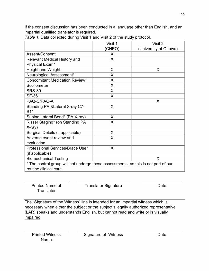
66
If the consent discussion has been conducted in a language other than English, and an
impartial qualified translator is required.
Table 1. Data collected during Visit 1 and Visit 2 of the study protocol.
Printed Name of
Translator
Translator Signature Date
The “Signature of the Witness” line is intended for an impartial witness which is
necessary when either the subject or the subject’s legally authorized representative
(LAR) speaks and understands English, but cannot read and write or is visually
impaired
Printed Witness
Name
Signature of Witness Date
Visit 1
(CHEO)
Visit 2
(University of Ottawa)
Assent/Consent X
Relevant Medical History and
Physical Exam*
X
Height and Weight X X
Neurological Assessment* X
Concomitant Medication Review* X
Scoliometer X
SRS-30 X
SF-36 X
PAQ-C/PAQ-A X
Standing PA &Lateral X-ray C7-
S1*
X
Supine Lateral Bend* (PA X-ray) X
Risser Staging* (on Standing PA
X-ray)
X
Surgical Details (if applicable) X
Adverse event review and
evaluation
X
Professional Services/Brace Use*
(if applicable)
X
Biomechanical Testing X
* The control group will not undergo these assessments, as this is not part of our
routine clinical care.

67
APPENDIX D
Table E1. Calibration and tracking marker placement during movement protocol.
Calibration Marker Placement
Head Front right
Front left
Back right
Back left
Trunk Right Acromion
Left Acromion
Left, centre and right C7
Left, centre and right T1
Left, centre and right T2
Left, centre and right T3
Left, centre and right T4
Left, centre and right T5
Left, centre and right T6
Left, centre and right T7
Left, centre and right T8
Left, centre and right T9
Left, centre and right T10
Left, centre and right T11
Left, centre and right T12
Left, centre and right L1
Left, centre and right L2
Left, centre and right L3
Left, centre and right L4
Left, centre and right L5
Sacrum Left, centre and right S1
Centre S2
Pelvis Left ASIS
Right ASIS
Left Iliac Crest
Right Iliac Crest
Thigh (Bilateral) Greater Trochanter
Lateral Femur Epicondyle
Medial Femur Epicondyle
Cluster: Middle; lateral side
Shank (Bilateral) Cluster: Middle; lateral side
Foot (Bilateral) Lateral Malleolus
Medial Malleolus
1st Metatarsophalangeal Joint
5th Metatarsophalangeal Joint
Calcaneus

68
Tracking Marker Placement
Head Front right
Front left
Back right
Back left
Trunk Right Acromion
Left Acromion
Left, centre and right C7
Left, centre and right T1
Left, centre and right T2
Left, centre and right T3
Left, centre and right T4
Left, centre and right T5
Left, centre and right T6
Left, centre and right T7
Left, centre and right T8
Left, centre and right T9
Left, centre and right T10
Left, centre and right T11
Left, centre and right T12
Left, centre and right L1
Left, centre and right L2
Left, centre and right L3
Left, centre and right L4
Left, centre and right L5
Sacrum Left, centre and right S1
Centre S2
Thigh (Bilateral) Cluster: Middle; lateral side
Shank (Bilateral) Cluster: Middle; lateral side
Foot (Bilateral) Lateral Malleolus
Medial Malleolus
1st Metatarsophalangeal Joint
5th Metatarsophalangeal Joint
Calcaneus

69
APPENDIX E
Forward flexion, right-left lateral bending, right-left axial twist and the following two movements
were all done in both a constrained and unconstrained (without the pelvis immobilizer) condition.
Two sets of one repetition were completed.
Spine Circumduction: Participants will be asked to complete a compound clockwise and counter
clockwise circumduction movement of the spine with their arms crossed against their chest. During
this movement, participants will trace the path of an imaginary circle with their heads while
bending, flexing, and twisting their spine. The movement will be completed at a self-selected pace.
Backward Bending: Participants will be asked to bend their spine backwards as far as they are
comfortable with their arms crossed over their chest. The movement will be completed at a self-
selected pace and only IMU data will be collected during this movement.
Two sets of one repetition were completed of the following movements:
Sit-to-Stand Transition: Participants will start in their natural neutral sitting posture and will
transition to their neutral standing posture. Participants will be instructed to avoid pushing off their
legs with the arms during this movement. The movement will be completed at a self-selected pace.
Stand-to-Sit Transition: Participants will start in their natural neutral standing posture and will
transition to their neutral sitting posture. Participants will be instructed to avoid bracing their arms
against their legs during this movement. The movement will be completed at a self-selected pace.
Three repetitions were completed of the following movement:
Gait: Participants will be asked to walk in a straight line for approximately 8 metres from an
indicated starting point in a natural manner to an indicated finishing point. The walk will be
completed at a self-selected pace.
As this task is more fatiguing than the other tasks, participants were given the option to complete
this additional trial:
Repeated Forward Bending (Optional): During this trial you will be strapped to a rigid support at
the waist and will be asked to bend your spine forward and backwards to touch targets placed at
shoulder and knee height. A total of 35 spine flexion-extension movements will be repeated as
one single continuous trial, paced using an auditory metronome such that each individual flexion-
extension cycle is completed over the course of 4 seconds.

70
APPENDIX F
Table F1. Cobb angle measurements (°) based on radiographic images of patients in the AIS,
PSF and VBT groups.
Note: SD = standard deviation.
Participant # Cobb Angle (°)
AIS Group
1 50.0
2 50.0
3 59.0
4 57.0
5 N/A
Mean (SD) 54.0 (4.69)
PSF Group
1 24.0
2 14.0
3 20.0
4 12.0
Mean (SD) 17.5 (5.5)
VBT Group
1 33.0
2 40.0
3 23.0
4 10.0
5 16.0
Mean (SD) 24.4 (12.2)