the early development of the nephridia in amphioxus...
TRANSCRIPT
The Early Development of the Nephridia inAmphioxus: Introduction and Part I,
Hatschek's Nephridium.By
Edwin S. Goodrich, F.R.S.
With Plates 29 and 30, and 1 Text-figure.
INTRODUCTION.
IT is now some thirty years since I first undertook the studyof 'the development of the excretory organs of A m p h i o x u s( B r a n c h i o s t o m a l a n c e o l a t u m , Pallas). When it wasdiscovered (Goodrich, 1902) that A m p h i o x u s is provided withnephridia of essentially the same structure as the solenocyte-bearing protonephridia of various Polychaetes and other Inverte-brates it became a matter of interest to find out how they aredeveloped. Yet, so far as I am aware, only one author has at-tempted to give a detailed account of their early developmentin two short papers (R. Legros, 1909 and 1910). In 1909 Idescribed the nephridia in larval stages, and since then from timeto time, as opportunity offered, I have tried to make out theirearly development and their first origin. The investigator of thisproblem is confronted with many difficulties. The material ishard to obtain, difficult to preserve satisfactorily, and parti-cularly difficult to stain well. After trying various well-knownfixatives, a mixture of corrosive-acetic and picro-nitric for em-bryonic and Bouin's fluid for larval stages were found to givethe best results. It is essential to anaesthetize the later larvaebefore fixation to prevent muscular contraction. Borax-carmineand picro-nigrosin, Heidenhain's iron-haematoxylin and eosin orlight-green, and Mann's methyl-blue and eosin are the most satis-factory stains. The cells are so small and their derivation sodifficult to make out that sections of more than about 3 or 4 p inthickness are almost useless. The observations recorded be-low on embryonic stages were made on material provided bythe Zoological Station at Naples for which I here express my

500 EDWIN S. GOODRICH
gratitude. The larval material was preserved by me at Faro, atNaples, and in Heligoland. I have also to thank the authoritiesof the Biologisches Anstalt in Heligoland for sending me pre-served larvae by post.
Eeturning now to the general problem of the homology of theexcretory organs in A m p h i o x u s already mentioned above,it may be pointed out that long ago (1895) I maintained thatthroughout the Invertebrata T r i p l o b a s t i c a two distinctorgans are found, of quite different origin and generally of dif-ferent function, the nephridium and the coelomoduct. The latterdevelops centrifugally as a more or less funnel-shaped outgrowthfrom the wall of the coelom in higher forms provided with a welldifferentiated coelomic cavity; or it develops from the genitalsac or ' gonad' in lower forms, such as Platyhelminths, in whichthe gonocoel has not yet enlarged into a coelomic ' body-cavity'.The coelomoduct, representing the original outlet for genital pro-ducts, functions as a genital duct only, except when it secondarilyacquires an excretory function. The nephridium, on the otherhand, from the first is typically excretory, is already well formedin phylogeny before the coelomic body-cavity has appeared(Platyhelminths), originates in ontogeny from more superficialrudiments (ectodermal or ectomesodermal), and in higher formsonly may come secondarily into relation with the coelom. Un-fortunately nephridia and coelomoducts, which may sometimesacquire superficial resemblances to each other, were for longconfused, and, indeed, still are often confused at the present day.This confusion is partly due to the fact that the nephridiumitself by centripetal growth may, in some annelids, penetrate intothe coelom, piercing the coelomic epithelium, and acquire asecondary opening into the coelomic cavity (Goodrich, 1895).Thus is formed from the innermost nephridial cell a nephridio-stome liable to be confused, even at the present day, with thecoelomostome or funnel-shaped opening of the coelomoduct. Inthe last century it became almost a dogma that 'nephridia'open into the coelom and are derived from its epithelium.
Coming to the Vertebrates, we find that in Craniata genitaland excretory ducts are all of the coelomoduct type deriveddirectly or indirectly from the coelomic epithelium. In spite of
NEPHRIDIA IN AMPHIOXUS 501
this they were, and still often are, taken to be nephridia. Butof true nephridia no trace has yet been found for certain in anyCraniate; they seem to have vanished entirely. On the contrary,in Cephalochorda, it is the nephridia that have survived and thecoelomoducts that have almost if not completely disappeared.This reduction is no doubt related to the shedding by theseanimals of their genital products directly by dehiscence into theatrium.
How strongly observers were influenced by the prevalent con-fusion referred to above is shown in Boveri's account of thestructure of the nephridia of A m p h i o x u s (1892); for hedescribed open funnels where no funnels exist.
Legros (1909 and 1910) claims to have shown that nephridiain A m p h i o x u s develop as funnel-shaped outgrowths fromthe coelomic epithelium which become closed off from the eoelomand acquire an opening into the atrium, or into the pharynx inthe case of Hatschek's nephridium. In adopting this interpreta-tion he also, I think, was biased by preconceived notions. Myown observations do not support his view. Although I readilyadmit that my account given below does not solve the questionof the very first origin of the nephridia, and that even thedescription of the early development is by no means complete,yet I think it may be claimed that it demonstrates clearly thatLegros was mistaken, that the nephridium does not develop asan outgrowth from the coelomic epithelium, and that it neverhas at any stage an internal opening.
Therefore, in spite of the fact that the first origin of the nephri-dium remains undiscovered, I decided to publish my results inthe hope that others more skilful or more fortunate than myselfmay solve this important problem.
DEVELOPMENT OP HATSCHEK'S NEPHRIDIUM.
Except for the paper by Legros (1910), referred to above, noaccount has been given of the early development of Hatschek'snephridium. In the previous year (Goodrich, 1909) I had de-scribed this organ in the adult A m p h i o x u s and in the larva,and dealt with the literature on the subject. Since then thestructure of the adult organ has been further considered in more

502 EDWIN S. GOODEICH
detail (Goodrich, 1933), and in a paper on "Proboscis Poresin Craniate Vertebrates" (Goodrich, 1917) I referred briefly tothe development of Hatschek's nephridium in a footnote asfollows:' This nephridium is developed neither from a mesoblastiefunnel as described by Hatschek, nor as an outgrowth fromthe second somite, as described by Legros; nor, again, from theremains of the communication of the coelomic pouch withthe gut, as alleged by MacBride, but from a little group of cellsappearing quite early just above the mouth. Several years agoI traced these cells to a stage about thirty hours old, before theopening of the mouth; but since I am unable to find out theirfirst origin, I refrained from publishing my results. They arethe cells figured recently by Smith and Newth (fig. 4, PI. 18),who are indeed correct in their surmise that they may representthe rudiment of the nephridium.'
Before describing the development of Hatschek's nephridiumit will be well to refer briefly to its structure in the adult and inlate larvae. In the adult it opens behind into the pharynx, andextends forwards along the left dorsal aorta some way beyondHatschek's pit. It is provided along its course with short blindbranches, some passing medially below the aorta and otherspassing dorso-laterally to the aorta (fig. 1, PL 29). These diverti-cula bear bunches of solenocytes extending into small chambers,isolated remnants of the coelomic cavity of the second somiteof the larva and previously continuous with it (Goodrich, 1909,1933).
In the metamorphosing larva of stage 5 of Willey, with open'secondary' right gill-slits, the nephridium is a simple tubereaching to the level of the pit, and with solenocytes along itswhole length. It now lies in a continuous chamber on the wallof which occasional flattened nuclei represent the coelomic epi-thelium (fig. 2, PL 29).
In a younger larva with six gill-slits of the first or future adultleft series, Hatschek's nephridium is seen in transverse sectionsas a short blind tube lying on the ventro-lateral wall of theposterior extension of the coelomic cavity of the second somite(fig. 3, PL 29). Some solenocytes extend forwards, and appear ina section cutting through Hatschek's pit (fig. 4, PL 29).

NEPHRIDIA IN AMPHIOXUS 503
We come now to the earlier stages obtained by the artificialfertilization of eggs at Naples. Pigs. 5 A-D, PI. 29, represent fourconsecutive transverse sections through a young larva (51 hrs.old1) in which the mouth is open but rounded, and the first gill-slit is open and functional (about stage fig. 61 of Hatschek,1881). It is a stage older than that of the diagrammatic fig. 1, ofLegros (1910) but considerably younger than that of his fig. 3.The most posterior section (fig. 5 A, PL 29) shows the openingof the nephridium into the gut just above the mouth. Herethe nephridial cells have pushed through the endodermal wall,and an incipient lumen is seen to pass up between them andappears again in the next section (fig. 5 B). In this lumen a fewcilia are already present, but it is difficult to make certain oftheir origin. At all events no fully formed solenocytes seemto be yet developed at this stage, and the nephridium endsblindly in the third section (fig. 5 c) in a solid mass of a fewcells situated between the ectoderm and endoderm above theposterior ventral region of the second mesoblastic segment.The last section passes in front of the nephridium, cutting thesecond segment, which is here indistinctly divided into dorsalsomite and ventral lateral plate (fig. 5 D). At this stage, then,the nephridium has acquired an opening through the endodermand extends forwards for a short distance between the secondand third mesoblastic segments.
In the next two younger stages (41 hrs. and 37 hrs.), when themouth has just been pierced and is a small round hole, thenephridium though smaller bears much the same relations.Usually the opening through the endodermal wall of the gut hasalready been acquired (fig. 6, PI. 29). Fig. 7, PL 29, shows thisregion in greater detail, and is taken from a specimen in whichthe opening can scarcely be said to have been formed. Evenhere cilia appear in the small lumen.
The next younger stage (30 hrs.) is perhaps the most important,for it is the youngest stage described by Legros and is the
1 As Legros (1910) remarks, the embryos of the same ages are not alwaysat exactly the same stage of growth. But the number of hours afterfertilization gives some indication of the relative state of development ofthe various embryos.
504 EDWIN S. GOODRICH
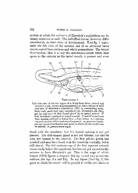
earliest at which the rudiment of Hatschek's nephridium can beclearly made out as such. The indiviflual larvae, however, differconsiderably in their state of development. Text-fig. 1 repre-sents the left view of the anterior end of an advanced larvareconstructed from sections and whole preparations. The buccaldiverticulum, that is to say the endodermal pouch which lateropens to the exterior at the larval mouth, is present and even
TEXT-FIGTJKE 1.
Left side view of anterior region of a thirty-hour larva, stained andmounted whole, drawn diagrammatically to show relation of solidrudiment of Hatschek's nephridium (UN) to surrounding parts.Body-wall, nerve cord (we), notochord (nl), Hatschek's pit (Hp),gut (g), and duct of club-shaped gland (d), in optical section. S1
first mesoblastic segment in optical section. 1S2 and S3 second andthird somites outlined in dotted line; a finer dotted line indicatescoelomic cavities of first and second segments, m, position of endo-dermal buccal diverticulum and point at which mouth would laterbe pierced, so, preoral sense-organ.
fused with the ectoderm; but the buccal opening is not yetpierced. The club-shaped gland is not yet tubular, nor has itsduct yet opened to the exterior. The first gill-slit pouch hasreached and may have fused with the ectoderm ventrally, but isstill closed. The left coelomic sac of the first segment extendstransversely below the notochord, but has not yet opened to theexterior to form Hatschek's pit. This is the stage of whichLegros (1910) figures a diagram (his fig. 1) and two transversesections (his figs. 2 a and 2 b). In my figure (Text-fig. 1) thepoint at which the mouth will be pierced is visible, and above it

NEPHRIDIA IN AMPHIOXUS 505
is seen the rudiment of Hatschek's nephridium lodged betweenmesoblastie segments 2 and 3, which are indicated by dottedlines.
Three transverse sections of this stage are drawn in fig. 8 A,B, and c, PI. 29. In the most posterior section (fig. 8 A) is seenthe buccal diverticulum of the gut which has just fused withthe ectoderm; the mouth, however, has not yet been pierced.The rudiment of Hatschek's nephridium is seen as a group offour cells surrounding a small lumen, in which cilia are develop-ing, and sending between the endoderm cells of the gut a wedge-like process reaching to a point o. Here, at a later stage, wouldbe formed the nephridiopore. The next section (fig. 8 B) showstwo nuclei in the rudiment lying between mesoblastie segment 3and 2. The most anterior section (fig. 8 c) cuts across the solidfront end of the rudiment.
Fig. 9 A, B, c, PL 30, represents a slightly younger stage. Thebuccal diverticulum is less thoroughly fused with the epidermisand resembles that figured by Legros (1910, fig. 2 b). Themost posterior section (fig. 9 A, PI. 30), taken in front of thebuccal diverticulum, cuts through the middle of the rudimentof Hatschek's nephridium. Two sections farther forward is seenits solid anterior end (fig. 9 B). The next section (not figured)shaves through its extreme tip and the hind wall of segment2; while the next and most anterior section shows only theundivided segment 2 with its continuous coelomic cavity (fig.9 c). Another section of a larva at about the same stage isdrawn in fig. 10, PI. 30.
A section through a stage when there is as yet no distinctbuccal diverticulum, but only a slight outgrowth of a few endo-dermal cells, is shown in fig. 11, PL 30. This stage is thereforeyounger than the first dealt with by Legros. It may be seen thata solid rudiment is already present between mesoblastie seg-ments 2 and 3, and has sent into the endoderm towards thepoint o a small process, which, however, has not yet reachedthe lumen of the gut.
In my batch of thirty-hour larvae, however, a few specimenscan be found at a still earlier stage of development. A transversesection of one is shown in fig. 12, PL 30. Here the future buccal

506 EDWIN S. GOODRICH
diverticulum is indicated merely by a small thickening due tothe budding off of a few endoderm cells towards the surface.No distinct group of cells forming a rounded rudiment ofHatschek's nephridium can be found, but there are seen twoor three cells against the wall of the gut in the appropriate placejust between the posterior region of segment 2, the anteriorregion of segment 3, and the gut. In fig. 13, PI. 30, representinga transverse section through a slightly earlier stage with notrace of buccal diverticulum at all, two cells flattened againstthe gut wall are again visible overlapped by the posterior regionof the second mesoblastic segment. Although these cells canoften scarcely be distinguished from the surrounding mesoblasticcells, they do not appear to belong to the wall of the secondcoelomic cavity, and I have little doubt that they represent therudiment of Hatschek's nephridium.
My next batch of embryos (24 hrs.) is considerably younger.Not only do they show no trace of buccal diverticulum, but theleft first mesoblastic segment is here a rounded sac, often still inconnexion with the end of the gut. Yet even in these one or twocells can generally be made out between the hinder wall of meso-blastic segment 2, the anterior wall of segment 3, and the endo-derm. A transverse section of such a stage is shown in fig. 14,PI. 30. There can be little doubt that these cells represent theearly rudiment of Hatschek's nephridium. They lie outside thewall of segment 2, are not derived from its epithelium; butwhence they have arisen I am unable to say.
From the account given above it will be understood that myobservations agree with those of Legros down to the stage withabout six gill-slits of the left series, where Hatschek's nephri-dium has essentially the structure depicted in his diagram (1910,fig. 5 b) of a somewhat older larva with nine gill-slits. Thenephridium is a straight canal open behind, blind in front, andwith solenocytes set along nearly its whole course. Their tubespierce the wall of the nephridial canal to reach its lumen (com-pare my figure drawn from the living larva, fig. 38, PI. 15,1909).But from such a stage back to the earliest larval stages ourresults differ radically. For, according to Legros, in earlier stagesthe nephridium is in the form of an open funnel. He begins his
NEPHEIDIA IN AMPHIOXUS 507
description of the development with the stage at which the endo-dermal buccal diverticulum has just met the epidermis andthe mouth is not yet open (his diagram fig. 1, correspondingto my stage, figs. 9, 10, and 11, PI. 30). The rudiment ofHatschek's nephridium is said to appear as a backwardly di-rected diverticulum of the 'vesicule intermediate', that regionof the still undivided left second mesoblastic segment which liesbetween the myotome above and the lateral plate below. Thisfunnel-shaped outgrowth from the posterior wall of the secondsegment is said to end blindly just above the buccal diverticulum.Of the existence of such a funnel open in front, I can find noevidence at this stage, nor at any earlier or later stage. On thecontrary, as already explained, the rudiment of the nephridiumat about this stage always has the appearance of a more or lesssolid group of cells in the same position as Legros's allegedfunnel, but it is a separate independent structure lying betweensegments 2 and 3. By careful focusing one can always make out, ingood and well-stained sections, that the anterior end of the rudi-ment is not open and is covered over by the coelomic epitheliumof segment 2.
The next older stage described by Legros is that at which thebuccal diverticulum has fused with the epidermis and the mouthis about to be pierced (his figs. 2 a, and 2 b, corresponding tomy fig. 8, PI. 29). The nephridial funnel is said to have alreadyacquired an opening into the gut, so that there would now be afree passage from the coelom of segment 2 to the lumen of thegut. Legros figures a transverse section passing in front of thealleged funnel, and another five sections farther back throughthe pharyngeal opening. But no detailed figure is given at this,or indeed at any stage, of a section through the funnel itself.
Legros appears to have studied only transverse sections whichare not really suitable for determining whether the nephridialrudiment has an anterior opening or not. To confirm my ownview that such an opening is not present I have examined longi-tudinal sections, and, though it is difficult to obtain satisfactorysections in exactly the right plane, such can occasionally befound. Figs. 15 and 16, PI. 30, represent longitudinal sectionsof two larvae at the stage when the mouth is about to be

508 EDWIN S. GOODRICH
pierced. They pass dorsally to the buccal diverticulum, and show•clearly enough the rudiment of Hatschek's nephridium as a littleindependent group of cells lodged between the second and thirdmesoblastic segments, and without any opening into the coelomof the second segment.
The solenocytes themselves, according to Legros, develop fromthe eoelomic epithelium lining the inner wall of his 'vesiculeintermediate'. He supposes that the tubes grow outwards andbackwards, and penetrate into the elongating funnel. Legrosgives a diagram purporting to show the structure of the nephri-dium at this stage (his fig. 4 c, of a five-day larva). The ' vesicleintermediaire' is now supposed to become longitudinally dividedinto an inner chamber containing the solenocytes and an outercanal formed by the original funnel and its forward extensionderived from the outer wall. At the same time the free endsof the tubes are supposed to become enclosed as the funnelcloses, and so come to pierce the wall of the completed organ.
My own observations yield no evidence whatever of this sup-posed origin of the solenocytes separate from the canal. On thecontrary, so far as I can make out, they are always derived fromthe nephridium itself, and simply grow out from it as the canallengthens. But the exact steps in the process I am not yet ableto describe in detail in the case of Hatschek's nephridium. It isclear that solenocytes can only function if their tubes are fixedin and pierce the wall of the canal. Legros's account of theformation of solenocytes with tubes and flagella complete, andof their secondary connexion with the nephridial canal, seemsto me quite unintelligible.
A summary of the results recorded in this Part I will appearshortly in Part II, now in preparation dealing with the de-velopment of the paired nephridia.DEPARTMENT OF ZOOLOGY AND COMPARATIVE ANATOMY,
UNIVERSITY MUSEUM, OXFORD.

NEPHEIDIA IN AMPHIOXUS 509
LIST OF EBFBRBNCES.
Boveri, Th. (1892).—"Nierenkanalchen des Amphioxus", 'Zool. Jahrb.Anat.', vol. 5.
Goodrich, E. S. (1895).—"On the Coelom, Genital ducts, and Nephridia",'Quart. Journ. Micr. Sci.', vol. 37.
Goodrich. E. S. (1902).—"On the structure of the Excretory organs ofAmphioxus, Part I", ibid., vol. 45.
(1909).—"On the structure of the Excretory organs of Amphioxus,Part II", ibid., vol. 54.
(1917).—"Proboscis pores in Craniate Vertebrates", ibid., vol. 62.(1933).—"Nephridia of Asymmetron and Branchiostoma compared",
ibid., vol. 75.Hatschek, B. (1881).—"Entwickl. von Amphioxus", 'Arb. Zool. Inst.
Wien', vol. 4.Legros, R. (1909) published anonymously.—"D6vel. des fentes branchiales
et des canalicules de Weiss-Boveri chez 1'Amphioxus", 'Anat. Anz.',vol. 34.
(1910).—"De l'anat. et du devel. de 1'Amphioxus", ibid., vol. 35.
EXPLANATION OF PLATES 29 and 30.
LETTERING.
a, artifact space; bd, endodermal buccal diverticulum; cnu, nucleus ofcoelomic epithelium cell; cr, cells probably representing very early rudi-ment of Hatschek's nephridium; cs, coelom of mesoblastic segment; d, ductof club-shaped gland; end, endostyle; ep, epidermis; g, endodermal wall ofgut; gr, groove from preoral pit; Hn, Hatschek's nephridium; Hp, Hat-schek's pit; lao, left dorsal aorta; mo, mouth; mp, point at which mouthwill open; no, nerve cord; nt, notochord; o, opening into gut of Hatschek'snephridium; prm, preoral muscle; pop, preoral pit; S2 and 8s, second andthird mesoblastic segments or somites; so, sense organ; sol, solenocyte.
The magnification of figs. 1, 3,4, 5 A, and 6 is given on the scale attachedto fig. 3; and that of figs. 5 B, O, D, and 7-16 is given on the scale attachedto fig. 12.
PLATE 29.Fig. 1.—Transverse section of Hatschek's nephridium in adult, showing
short dorsal and inner branches.Fig. 2.—Transverse section of Hatschek's nephridium in a late larva from
Naples with open right slits. Stage 5 of Willey.Fig. 3.—Transverse section of Hatschek's nephridium of a younger larva
from Faro, with rudiments of six right slits, passing between mouth andpreoral pit.
Fig. 4.—Transverse section of same larva passing farther forward beyondnephridial canal.

510 EDWIN S. GOODRICH
Fig. 5 A-D.—Four transverse sections of fifty-one-hour larva, A, mostposterior, cuts through opening of Hatschek's nephridium. B, secondsection farther forward, shows small lumen in nephridium. c, next section,cuts through its solid anterior end. Next but one section, D, shows coelomiccavity of second mesoblastic segment.
Fig. 6.—Transverse section of thirty-seven-hour larva cutting throughopening of Hatschek's nephridium.
Fig. 7.—Part of transverse section of thirty-seven-hour larva, slightlyyounger, in which opening of Hatschek's nephridium has not yet beenformed.
Fig. 8 A, B, C.—Consecutive transverse sections of thirty-hour larva. Inmost posterior section A are seen buccal diverticulum fused with ectoderm,and above it rudiment of Hatschek's nephridium acquiring opening intolumen of gut. In B nephridium is between segments 2 and 3. Most anteriorsection c cuts through solid end of nephridial rudiment.
PLATE 30.FIG. 9 A, B, C.—Transverse sections of thirty-hour larva with buccal
diverticulum just fused with ectoderm. Ashows solid rudiment of Hatschek'snephridium between segments 2 and 3. B, two sections farther forward, cutssolid end of nephridial rudiment. Next and most anterior section, c, cutswall and cavity of segment 2.
Fig. 10.—Transverse section of thirty-hour larva similar to that drawnin fig. 9 A and of about same stage.
Fig. 11.—Transverse section of thirty-hour larva slightly younger,showing process from solid rudiment of Hatschek's nephridium growingbetween endodermal cells towards lumen of gut.
Fig. 12.—Transverse section of younger thirty-hour larva in which smallbuccal diverticulum has not yet fused with ectoderm. Between segment 2and endoderm two cells are seen which appear to represent rudiment ofHatschek's nephridium.
Fig. 13.—Transverse section of still younger thirty-hour larva with nobuccal diverticulum. Two cells in same position as those of fig. 12 appearto represent rudiment of Hatschek's nephridium.
Fig. 14.:—Transverse section of twenty-four-hour larva at stage with notrace of buccal diverticulum, and first left mesoblastic segment a roundsac still in connexion with anterior end of gut. Two cells, apparentlyrudiment of Hatschek's nephridium, are seen between gut and segment 2.
Fig. 15.—Part of longitudinal section of thirty-hour larva showing rudi-ment of Hatschek's nephridium as solid mass of few cells between segments2 and 3. An arrow points to position of buccal diverticulum in a moreventral section.
Fig. 16.—Part of longitudinal section of another thirty-hour larva show-ing similar rudiment.

LiGoodnch dd

flGoodr/'c/i deL

53Mar.Sd. Vol. 16', MS. $L30
S? nc/nt
16