the ear - doctor 2018 - ju medicine€¦ · tympanic cavity (middle ear) is a narrow, oblique,...
TRANSCRIPT

The Ear
Dr. Heba Kalbouneh
Associate Professor of Anatomy and Histology

The ear consists of the
external ear; the middle
ear (tympanic cavity);
and the internal ear
(labyrinth), which
contains the organs of
hearing and balance.
The Ear
The internal ear converts the mechanical signals into
electrical signals to transfer information to the brain
Sound is captured by
the external ear
Mechanical signals
in the middle ear
The internal ear also contains receptors that
detect motion and position Dr. Heba Kalbouneh

External ear Middle ear
Inner ear
Dr. Heba Kalbouneh

The auricle has a characteristic shape
It collects air vibrations
It consists of a thin plate of elastic
cartilage covered by skin
External Ear
Auricle (pinna) External auditory meatus
The external auditory meatus is a
curved tube that leads from the
auricle to the tympanic membrane
It conducts sound waves from the
auricle to the tympanic membrane
Dr. Heba Kalbouneh

1
2
3
1. Helix.
2. Crus of helix
3. Auricular tubercle.
4. Antihelix.
5. Crura of antihelix.
6. Triangular fossa.
7. Scaphoid fossa.
8. Concha of auricle.
9. Tragus.
10. Antitragus.
11. Intertragic notch.
12. Lobule of auricle.
4
5
5 6
7
8 9
10
11
12
Anotia is complete absence of the external ear, and is most
likely caused by a developmental disturbance between the
seventh and eighth gestational week.
The cartilage of the auricle is
arranged in a pattern of
elevations and depressions
Prominent ears (also known as ‘bat’ ears) are
caused by the absence or inadequacy of an antihelical fold.
Dr.
Heb
a K
alb
ou
neh
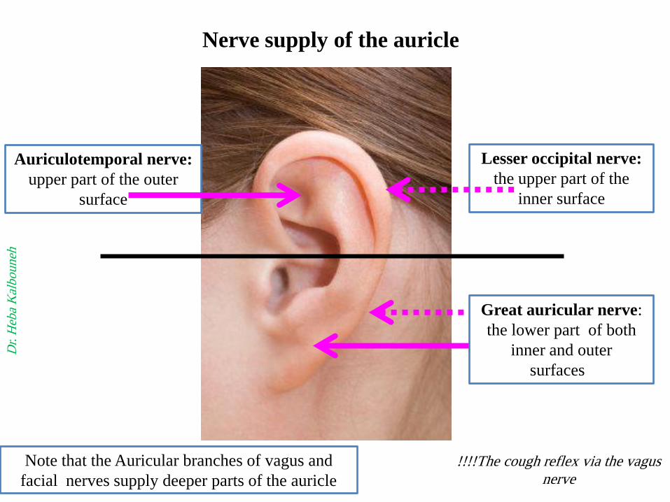
Note that the Auricular branches of vagus and
facial nerves supply deeper parts of the auricle
Auriculotemporal nerve:
upper part of the outer
surface
Great auricular nerve:
the lower part of both
inner and outer
surfaces
Lesser occipital nerve:
the upper part of the
inner surface
Nerve supply of the auricle
Dr.
Heb
a K
alb
ou
neh
!!!!The cough reflex via the vagus
nerve

The framework of the outer third of
the meatus is elastic cartilage, and the
inner two thirds is bone
The meatus is lined by skin
The outer third is provided with hairs
and sebaceous and ceruminous glands
Outer third
Cartilagenous
Inner two thirds
Bony
The external auditory meatus
Ceruminous glands are modified
sweat glands that secrete a yellowish
brown wax (cerumen=earwax)
The hairs and the wax provide a sticky
barrier that prevents the entrance of
foreign bodies
Dr. Heba Kalbouneh
Cerumen is produced in the outer third of the
cartilaginous portion of the ear canal
pH is acidic in normal healthy canals.
Contains a bactericidal enzyme.
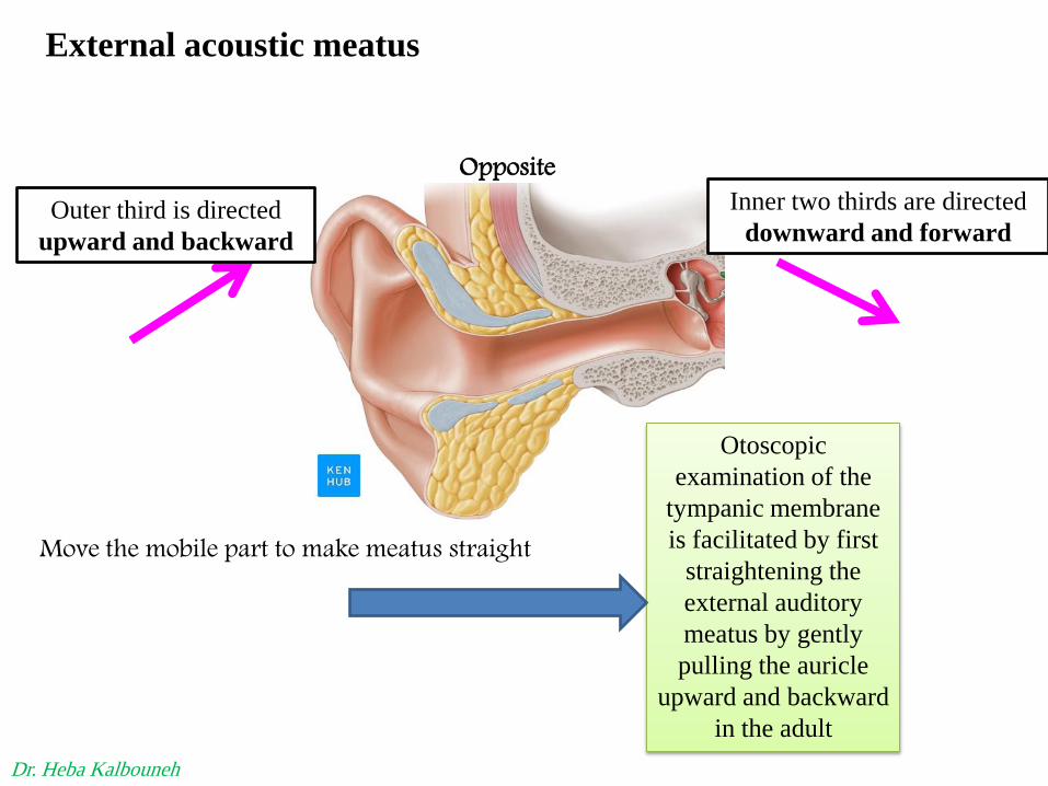
Move the mobile part to make meatus straight
Otoscopic
examination of the
tympanic membrane
is facilitated by first
straightening the
external auditory
meatus by gently
pulling the auricle
upward and backward
in the adult
Opposite
External acoustic meatus
Inner two thirds are directed
downward and forward Outer third is directed
upward and backward
Dr. Heba Kalbouneh

In the adult the external meatus is about 1 in. (2.5
cm) long and is narrowest about 0.2 in. (5 mm)
from the tympanic membrane
Dr. Heba Kalbouneh

Is formed of:
1-Outer layer:
Skin
2- Middle layer:
Fibrous tissue
3-Inner layer:
Mucous membrane
The Tympanic membrane
(ear drum)
The membrane is obliquely placed, facing downward, forward, and
laterally
Is a thin, fibrous membrane
It is concave laterally
Umbo is small
depression produced by the
tip of the handle of the
malleus
The inner surface of
tympanic membrane is
fixed to handle of Malleus
Dr.
Heb
a K
alb
ou
neh

Pars tensa
(Thick and taut)
Umbo
Handle of malleus
Remember that the middle
fibrous layer is present in the
major parts of the ear drum
which called pars tensa
However, this layer is
absent in the upper part of
the ear drum which is
called pars flaccida
(Shrapnell's membrane)
(Rivinus’ ligament)
The pars tensa and
flaccida are separated
from each other by two
folds called the anterior
and posterior malleolar
folds
Posterior
malleolar
fold
Anterior
malleolar
fold
The tympanic membrane is extremely sensitive to pain
Pars flaccida
(thin and
slack)
Malleus
(lateral process)
Cone of light
Dr.
Heb
a K
alb
ou
neh

Otoscopic Examination
Dr.
Heb
a K
alb
ou
neh
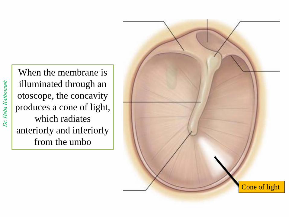
Cone of light
When the membrane is
illuminated through an
otoscope, the concavity
produces a cone of light,
which radiates
anteriorly and inferiorly
from the umbo
Dr.
Heb
a K
alb
ou
neh

Lateral
surface of
tympanic
membrane
Handle of
malleus
Cone of light
The antero-inferior quadrant of the ear drum is
called the cone of light (because it reflects the light
coming from the otoscope)
Note the tympanic
membrane is
translucent, concave
laterally
Dr.
Heb
a K
alb
ou
neh

Otitis
media
Dr.
Heb
a K
alb
ou
neh

Is an air-containing cavity in the
petrous part of the temporal bone
Is lined with mucous membrane
It contains the auditory ossicles,
whose function is to transmit the
vibrations of the tympanic membrane
(eardrum) to the inner ear
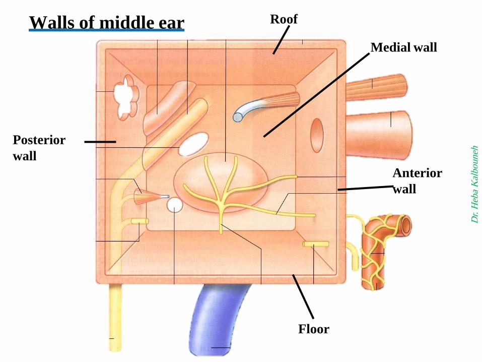
Middle Ear (Tympanic Cavity)
The middle ear has ROOF
FLOOR
ANTERIOR WALL
POSTERIOR WALL
LATERAL WALL
MEDIAL WALL
Dr.
Heb
a K
alb
ou
neh
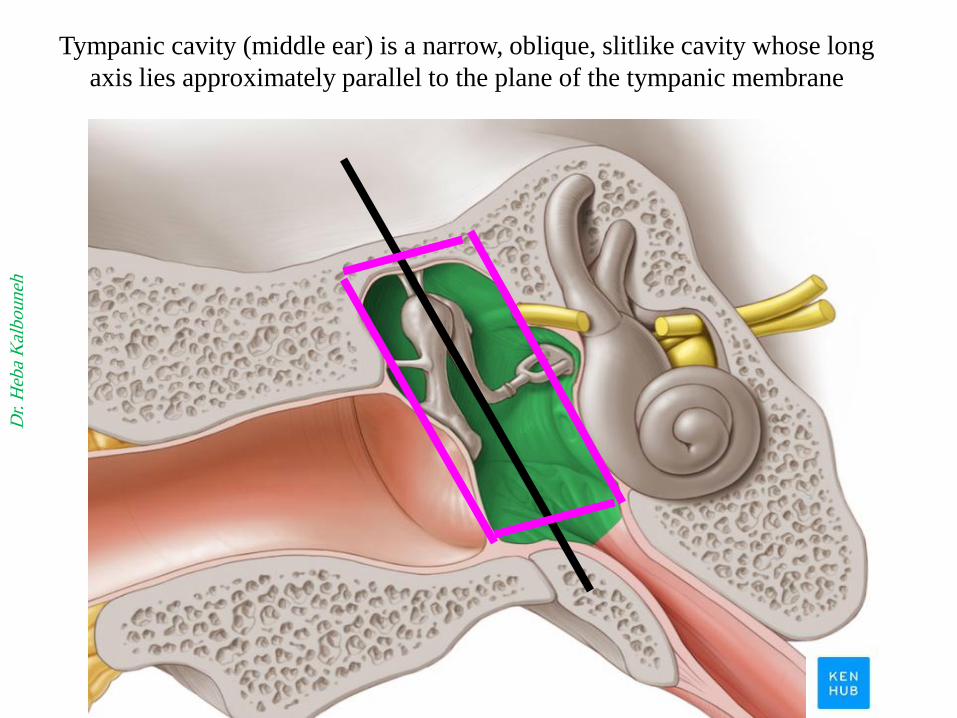
Tympanic cavity (middle ear) is a narrow, oblique, slitlike cavity whose long
axis lies approximately parallel to the plane of the tympanic membrane
Dr.
Heb
a K
alb
ou
neh
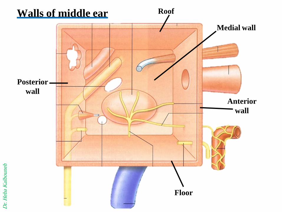
Roof
Floor
Posterior
wall
Medial wall
Walls of middle ear
Anterior
wall
Dr.
Heb
a K
alb
ou
neh

Roof
It separates the tympanic cavity
from the internal jugular vein
Is formed by tegmen tympani
(part of the petrous temporal bone)
It separates the tympanic cavity
from the meninges and the
temporal lobe of the brain in the
middle cranial fossa.
Floor
TEGMENTAL WALL
JUGULAR WALL
Dr.
Heb
a K
alb
ou
neh

Dr.
Heb
a K
alb
ou
neh
Tegmen tympani
Jugular
foramen
Petrous bone

Is formed below by a thin plate of bone that separates the tympanic
cavity from the internal carotid artery
At the upper part of the anterior wall are the openings into two canals
The lower and larger leads into the auditory tube
The upper and smaller is the entrance into the canal for the tensor tympani muscle
Anterior wall
Auditory tube
Internal carotid artery
Tensor tympani
Dr.
Heb
a K
alb
ou
neh


Opening of Eustachian
tube into nasopharynx
Dr.
Heb
a K
alb
ou
neh

EUSTACHIAN TUBE:
Pharyngo-tympanic tube
Auditory tube
Eustachian tube
It connects
the anterior wall of the
tympanic cavity to the
nasopharynx
It serves to equalize air
pressures in the tympanic
cavity and the nasopharynx
Its posterior inner third is bony
Its anterior two thirds are
cartilaginous
Bony
Cartilaginous
Dr.
Heb
a K
alb
ou
neh

Oval window:
Above and behind the
promontory, oval shaped and
closed by the base of the stapes
(Fenestra vestibuli)
Round window:
Below the posterior end of the promontory, round and closed by
the secondary tympanic membrane (Fenestra cochleae)
Medial wall Promontory is a rounded projection
(results from the underlying first turn of the
cochlea)
The horizontal part of the facial
nerve arching above the promontory
The medial wall is formed by the
lateral wall of the inner ear. Tympanic plexus
Dr.
Heb
a K
alb
ou
neh

Promontory
Cochlea
Dr.
Heb
a K
alb
ou
neh

Stapes
The base of stapes closes the
oval window of the internal
ear
Dr.
Heb
a K
alb
ou
neh

Internal acoustic meatus
Brain stem
7th
8th
Stylomastoid foramen
Dr.
Heb
a K
alb
ou
neh

Aditus
Pyramid
Vertical part of
Facial canal
Posterior wall
Chorda tympani
Dr.
Heb
a K
alb
ou
neh

1- Has in its upper part a
large, irregular opening,
the aditus to the mastoid
2-Below, a small conical
projection, the pyramid,
from its apex emerges the
tendon of the stapedius
muscle
3- The vertical part of the
facial nerve
Posterior wall
Dr.
Heb
a K
alb
ou
neh

Mastoid Antrum
The mastoid antrum lies behind the middle ear in the
petrous part of the temporal bone
It communicates with the middle ear by the aditus

The horizontal part of the facial nerve
Stylomastoid foramen

Lateral wall
The lateral wall is
largely formed by the
tympanic membrane
(ear drum)
tympanic membrane
Dr.
Heb
a K
alb
ou
neh

Chorda tympani Chorda tympani
Dr.
Heb
a K
alb
ou
neh

Anterior
Posterior

Chorda
tympani Dr.
Heb
a K
alb
ou
neh

The chorda tympani It arises from the facial nerve just above the
stylomastoid foramen
It enters the middle ear close to the
posterior border of the tympanic membrane.
It then runs forward over the tympanic
membrane and crosses the root of the handle
of the malleus
It leaves the middle ear through the
petrotympanic fissure and enters the
infratemporal fossa, where it joins
the lingual nerve
The chorda tympani contains:
1. Taste fibers from the mucous membrane
covering the anterior two thirds of the
tongue and the floor of the mouth.
2. Carries preganglionic parasympathetic
fibers to the submandibular and
sublingual glands via the
submandibular ganglion

The petrotympanic fissure
is a fissure in the temporal
bone
The chorda tympani runs
through the fissure to join
with the lingual nerve in
the infratemporal fossa
Dr. Heba Kalbouneh
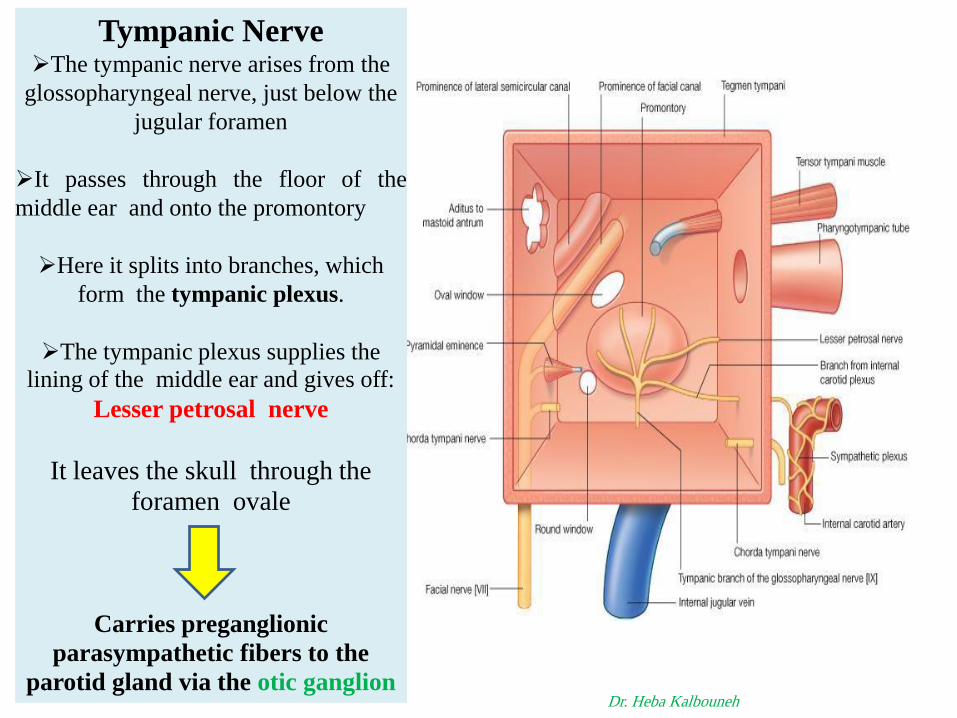
Tympanic Nerve The tympanic nerve arises from the
glossopharyngeal nerve, just below the
jugular foramen
It passes through the floor of the
middle ear and onto the promontory
Here it splits into branches, which
form the tympanic plexus.
The tympanic plexus supplies the
lining of the middle ear and gives off:
Lesser petrosal nerve
It leaves the skull through the
foramen ovale
Carries preganglionic
parasympathetic fibers to the
parotid gland via the otic ganglion Dr. Heba Kalbouneh

Foramen ovale transmits:
Mandibular nerve
Accessory meningeal artery
Lesser petrosal nerve
Emissary vein
MALE
Dr. Heba Kalbouneh

Pterygoid venous plexus in
Infratemporal fossa
Foramen ovale
Dr. Heba Kalbouneh
Emissary vein
Cavernous sinus
Roof
Floor
Posterior
wall
Medial wall
Walls of middle ear
Anterior
wall
Dr.
Heb
a K
alb
ou
neh

Roof
Tegmen tympani
Roof
Dr.
Heb
a K
alb
ou
neh

Floor
Floor
Int. jugular vein
Dr.
Heb
a K
alb
ou
neh

Tensor tympani muscle
Auditory
tube
Anterior wall
Internal carotid artery
Dr.
Heb
a K
alb
ou
neh

Promontory
Oval
window
Round
window
Horizontal part of
facial canal Medial wall
Tympanic plexus
Dr.
Heb
a K
alb
ou
neh

Aditus
Pyramid
Vertical part of
Facial canal
Posterior wall
Chorda tympani
Dr.
Heb
a K
alb
ou
neh

Infections and Otitis
Media
Through the
auditory tube
from the nasal
part of the
pharynx.
(nasopharyngitis)
Into the
mastoid
antrum
(Acute mastoiditis)
The meninges and the temporal lobe of the brain lie superiorly
(Meningitis and a cerebral abscess in the temporal lobe)
The posterior wall of the
mastoid antrum is
related to the
sigmoid venous sinus.
If the infection spreads
in this direction, a thrombosis in the
sigmoid sinus may
take place
Medial wall: A spread
of the infection in this
direction can cause a
facial nerve palsy and
labyrinthitis with
vertigo
Although rare, complications of OM are commonly encountered given
its high prevalence. Complications of OM are classified as extracranial
or intracranial. Brain abscess are commonly considered the second
most common intracranial complication of OM after meningitis


A-3 Auditory Ossicles
B-2 muscles
C-2 nerves (tympanic plexus and
chorda tympani)
D-air
CONTENTS OF THE MIDDLE EAR
.
.
- It contains the auditory
ossicles, whose function is to transmit the
vibrations of the tympanic membrane
(eardrum) to the perilymph of the internal
ear
Malleus Incus
Stapes
Dr.
Heb
a K
alb
ou
neh

Ossicles

1-The malleus is the largest ossicle and possesses head, a neck, a long process or handle,
an anterior process, and a lateral process.
its head is rounded and articulates posteriorly with the incus
The stapes has a head, a neck, two limbs, and a base
The head articulates with the long process of the incus.
The neck is narrow and receives the
insertion of the stapedius muscle. The two limbs diverge from the neck and are
attached to the oval base which closes the oval window of the internal ear
The incus possesses:
a large body and two processes:
The body articulates with the head of the malleus.
The long process articulates with the head of the stapes

Malleus
The handle is
firmly attached
to the medial
surface of the
tympanic
membrane
Handle of malleus
Dr.
Heb
a K
alb
ou
neh

Incus
Dr.
Heb
a K
alb
ou
neh

Stapes
The base of stapes closes the
oval window of the internal ear
Dr.
Heb
a K
alb
ou
neh
The Annular stapedial ligament
is a ring of fibrous tissue that
connects the base of the stapes to
the oval window of the inner ear
Calcification and hardening of the
annular ligament of the stapes
(Otosclerosis) is a common cause
of adult deafness

Muscle Nerve supply Action
Tensor tympani Mandibular division of trigeminal
nerve
Dampens down vibrations
of tympanic membrane
Stapedius Facial nerve Dampens down vibrations
of stapes
Muscles of middle ear D
r. H
eba
Kal
bo
un
eh
Anterior
Tensor tympani muscle
Stapedius muscle

Inner Ear
(labyrinth)
Dr.
Heb
a K
alb
ou
neh

Inner ear is situated in
the petrous part of the
temporal bone
Dr.
Heb
a K
alb
ou
neh
Internal acoustic meatus

Semicircular canals
Cochlea
Vestibule
The inner ear is divided into:
1- Bony labyrinth
2- Membranous labyrinth
The vestibule, the central
part of the bony labyrinth
Dr.
Heb
a K
alb
ou
neh

Bony labyrinith
Semicircular
canals
Vestibule
Cochlea
Dr.
Heb
a K
alb
ou
neh

Membranous labyrinith Semicircular
ducts
Saccule and utricle
Cochlear duct
The membranous labyrinth is lodged
within the bony labyrinth
It is filled with endolymph and
surrounded by perilymph
Dr.
Heb
a K
alb
ou
neh

Cochlear duct
The duct of the cochlea lies within
the bony cochlea
The cochlea
resembles a snail
shell
Bony
Cochlea
Dr.
Heb
a K
alb
ou
neh

Posterior semicircular canal
Dr.
Heb
a K
alb
ou
neh

Posterior semicircular duct
Lodged within the canals are the
semicircular ducts
Dr.
Heb
a K
alb
ou
neh

Superior semicircular canal
Dr.
Heb
a K
alb
ou
neh
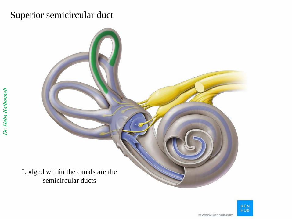
Superior semicircular duct
Lodged within the canals are the
semicircular ducts
Dr.
Heb
a K
alb
ou
neh

Lateral semicircular canal
Dr.
Heb
a K
alb
ou
neh

Lateral semicircular duct
Lodged within the canals are the
semicircular ducts
Dr.
Heb
a K
alb
ou
neh

Bony ampullae
Each canal has a swelling at one
end called the ampulla
Dr.
Heb
a K
alb
ou
neh

Membranous ampullae
Dr.
Heb
a K
alb
ou
neh
Membranous ampullae are lodged
in the bony ampullae

Utricle
Utricle and Saccule are lodged in the bony
vestibule
Dr.
Heb
a K
alb
ou
neh
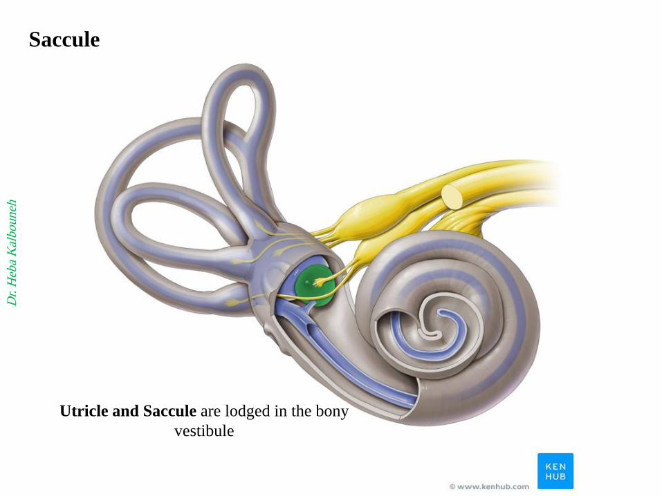
Saccule
Utricle and Saccule are lodged in the bony
vestibule
Dr.
Heb
a K
alb
ou
neh

Vestibulo-cochlear nerve
Vestibular nerve Cochlear nerve
Dr.
Heb
a K
alb
ou
neh

Vestibulo-cochlear nerve
Cochlea
Dr. Heba Kalbouneh
Vestibule and semicircular canals
The inner ear contains
Sensory receptors for
hearing and balance
Mechanoreceptors/ Hair cells
(Columnar cells)
Two maculae of the utricle
and saccule,
Three cristae ampullares in
the ampullae of each
semicircular duct
The organ of Corti in the
cochlear duct.

Vestibular nerve carries impulses from
the utricle, the saccule, and the
ampullae of the semicircular ducts
(contains the sensory receptors for
balance)
Ampullae of
semicircular canals
Utricle
Saccule Ves
tib
ule
Dr.
Heb
a K
alb
ou
neh

Cochlear nerve carries impulses
from organ of Corti in cochlea
(contains the sensory receptors
for hearing)
Cochlea
Dr.
Heb
a K
alb
ou
neh

Section through
cochlea
Scala media
(Cochlear duct)
(endolymph)
Scala vestibuli
(perilymph)
Scala tympani
(perilymph)
The cochlear duct itself forms the middle compartment,
or scala media, filled with endolymph. It is continuous with the saccule and ends at the
apex of the cochlea.
The larger scala vestibuli contains perilymph and is separated from the scala media by
the very thin vestibular membrane (Reissner membrane)
The scala tympani also contains perilymph and is separated from the scala media by
the basilar membrane

Section through
cochlea
(Scala media)
Dr.
Heb
a K
alb
ou
neh

Tectorial membrane
Organ of corti
The auditory
nerve (cochlear)
carries the
electrical signal
to the brain,
which turns it
into a sound that
we recognize
and understand
Section through
cochlea
Dr.
Heb
a K
alb
ou
neh

Modiolos
Basilar membrane
Vestibular membrane
(Reissner membrane)
1
2
3
The cochlea is about 35 mm long and makes
2¾ turns around a bony core called the
modiolus
1: Scala vestibuli
2: Scala media
(Cochlear duct)
3: Scala tympani

Tectorial
membrane
Note: Cell Bodies of cochlear
Nerve (CN VIII) located in the
Modiolus (Spiral ganglion)
The organ of Corti, or spiral
organ, where sound
vibrations of different frequencies
are detected, consists of
hair cells and other epithelial
structures supported by the
basilar membrane
Basilar
membrane
Vestibular membrane
(Reissner membrane)
Spiral
ganglion
Sensory receptor for hearing
(organ of Corti)

With the tectorial membrane removed, SEM
shows the apical plate of the rat spiral organ
through which rigid stereocilia
bundles project into endolymph

The scalae tympani and vestibuli communicate
with each other at the apex of the cochlea via a
small opening called the helicotrema
Middle ear
Scala tympani
Scala vestibuli
Scala media
Helico
trema


K + channels open
K + channels close
K +
K + K +
The auditory hair cells are located within the spiral organ
of Corti on the basilar membrane in the cochlea of the
inner ear
Stereocilia (hair bundles ) protrude from the apical surface
of the cell into the fluid-filled cochlear duct.
The inner hair cells transform the sound vibrations in the
fluids of the cochlea into electrical signals that are then
relayed via the auditory nerve to the auditory brainstem
and to the auditory cortex
The deflection of the hair-cell stereocilia opens
mechanically gated ion channels that allow positively
charged ions (primarily potassium) to enter the cell.
The influx of positive ions from the endolymph in the scala
media depolarizes the cell, resulting in a receptor potential
Note: hair cells detect movement
Damage to these hair cells results in decreased hearing
sensitivity, and because the inner ear hair cells cannot
regenerate, this damage is permanent

Transmission of sound D
r. H
eba
Kal
bo
un
eh

Sensory receptors for balance
(saccule, utricle and
semicircular ducts)
Saccule
Vestibule
Utricle
Semicircular duct
Ampulla
Horizontal
Macula
Vertical
Macula
Crista
Maculae : static balance
Cristae: kinetic balance
Problems of the vestibular
system can result in vertigo,
or dizziness, a sense of
bodily rotation and lack of
equilibrium.
Spinning the body produces
vertigo due to
overstimulation of the cristae
ampullares of the semicir
cular ducts. Overstimulation
of the maculae of the utricle
caused by repetitive changes
in linear acceleration and
directional changes can
normally lead to motion
sickness (seasickness).

Supporting
cells
Bending of
these
stereocilia
change
membrane
potential
Depolarization
Hyperpolarization
Columnar
hair cells
Gelatinous
layer
Sensory epithelium of saccule, utricle and semicircular ducts