the e1e4 protein of human papillomavirus interacts with the serine-arginine-specific protein
TRANSCRIPT

JOURNAL OF VIROLOGY, June 2007, p. 5437–5448 Vol. 81, No. 110022-538X/07/$08.00�0 doi:10.1128/JVI.02609-06Copyright © 2007, American Society for Microbiology. All Rights Reserved.
The E1�E4 Protein of Human Papillomavirus Interacts with theSerine-Arginine-Specific Protein Kinase SRPK1�
Ian Bell, Ashley Martin, and Sally Roberts*Cancer Research UK Institute for Cancer Studies, University of Birmingham, Birmingham B15 2TT, United Kingdom
Received 27 November 2006/Accepted 7 March 2007
Human papillomavirus (HPV) infections of the squamous epithelium are associated with high-levelexpression of the E1��E4 protein during the productive phase of infection. However, the precise mecha-nisms of how E1��E4 contributes to the replication cycle of the virus are poorly understood. Here, we showthat the serine-arginine (SR)-specific protein kinase SRPK1 is a novel binding partner of HPV type 1(HPV1) E1��E4. We map critical residues within an arginine-rich domain of HPV1 E1��E4, and in a regionknown to facilitate E1��E4 oligomerization, that are requisite for SRPK1 binding. In vitro kinase assaysshow that SRPK1 binding is associated with phosphorylation of an HPV1 E1��E4 polypeptide and modu-lates autophosphorylation of the kinase. We show that SRPK1 is sequestered into E4 inclusion bodies interminally differentiated cells within HPV1 warts and that colocalization between E1��E4 and SRPK1 is notdependent on additional HPV1 factors. Moreover, we also identify SRPK1 binding of E1��E4 proteins ofHPV16 and HPV18. Our findings indicate that SRPK1 binding is a conserved function of E1��E4 proteinsof diverse virus types. SRPK1 influences important biochemical processes within the cell, includingnuclear organization and RNA metabolism. While phosphorylation of HPV1 E4 by SRPK1 may directlyinfluence HPV1 E4 function during the infectious cycle, the modulation and sequestration of SRPK1 byE1��E4 may affect the ability of SRPK1 to phosphorylate its cellular targets, thereby facilitating theproductive phase of the HPV replication cycle.
Human papillomaviruses (HPVs) comprise a family of smalldouble-stranded DNA viruses that have a tropism for humanepithelial cells at skin and mucosal surfaces. To date, based ongenome sequence, over 100 HPV types have been described,and these virus types vary in both their anatomical tropism andtheir potential to induce malignant transformation (10). TheHPV replication cycle is intimately linked with the differenti-ation of the host epithelium (21). To initiate an infection, thevirus requires access, typically via a cut or abrasion, to basalcells where the virus maintains itself at a low copy number.Subsequently, infected cells undergo differentiation and mi-grate to the epithelial surface, which initiates the productivephase of virus infection, signified by genome amplification, thesynthesis of structural proteins, and assembly of infectiousprogeny. These events are orchestrated by both transcriptionaland posttranscriptional mechanisms and consequently ensurethat structural proteins are only synthesized in terminally dif-ferentiated cells (61).
High-level expression of the E4 protein accompanies theproductive phase of the HPV life cycle (40). E4 is translatedfrom spliced E1�E4 transcripts and encodes the first 5 aminoacids from the N terminus of the E1 protein fused to the E4coding sequence. The E4 open reading frame (ORF) is themost divergent ORF within the HPV family. While there issequence homology between E1�E4 proteins, this is generallyrestricted to virus types with similar pathology, and it is pre-dominantly limited to sequences at the amino and carboxy
termini of the proteins (43, 45). In natural infections, E4 isexpressed as a phosphoprotein (2, 4, 20) that assembles intooligomeric complexes (2, 12) and is further modified by re-moval of residues from the amino terminus (15, 43).
The role played by E4 in the virus life cycle is uncertain. Lossof full-length E1�E4 expression in experimental systems thatrecapitulate the infectious cycle of HPV type 16 (HPV16),HPV18, HPV31, and cottontail rabbit papillomavirus corre-lates with a defective life cycle (35, 39, 57, 57a). Collectively,these studies determined that full-length E1�E4 is necessaryfor efficient viral genome amplification and up-regulation oflate viral gene expression, which are features of the productivephase of the virus life cycle. In one system, based upon HPV16,replication of the viral genome in basal-like cells was alsocompromised in the absence of full-length E1�E4 (35). Thus,these studies implicate E1�E4 as an important regulator ofboth early and late stages of the virus life cycle. However,E1�E4 function may not be necessary for productive replica-tion of all papillomavirus types, since impairment of E1�E4synthesis from HPV11 genomes did not limit genome ampli-fication (17).
Several biological functions have been ascribed to E1�E4,and these observations include the association with keratinintermediate filaments, and in some cases, perturbation ofthese networks (14, 44); the reorganization of promyelocyticleukemia protein from intranuclear speckles (ND10 bodies)(46); the association with mitochondria (42); and the dis-ruption of normal cell division (9, 28, 34). As the precisemechanisms of how E1�E4 proteins exert these diverserange of effects are not yet fully understood (8, 29, 56), weadopted a proteomics-based approach in an effort to iden-tify HPV E1�E4-associated proteins. Here we show that the
* Corresponding author. Mailing address: Cancer Research UK In-stitute for Cancer Studies, University of Birmingham, Vincent Drive,Edgbaston, Birmingham B15 2TT, United Kingdom. Phone: 44 1214147459. Fax: 44 121 414 4486. E-mail: [email protected].
� Published ahead of print on 14 March 2007.
5437
on April 10, 2019 by guest
http://jvi.asm.org/
Dow
nloaded from

serine-arginine (SR)-specific kinase SRPK1 is a novel bind-ing partner of E1�E4 proteins from highly divergent HPV1,-16, and -18. SRPK1 belongs to a family of serine proteinkinases that phosphorylate arginine-serine (RS)-rich do-mains within a subgroup of the small non-RNP particlestermed SR proteins. SR proteins have multiple and diverseroles in mRNA metabolism (18), including the control ofpre-mRNA splicing through splice site selection, the regu-lation of the export of spliced mRNA from the nucleus, thestabilization of cytoplasmic transcripts, and promotion ofmRNA translation by ribosomes (18, 24, 25, 50). Phosphor-ylation within the RS domain differentially affects SR pro-tein activity, probably as a result of modulating protein-protein interactions and/or protein-mRNA association (32,49, 58). The release of SR proteins from distinct storagesites within the nucleus and the movement of SR proteinsthat shuttle between the nucleus and cytoplasm is under thecontrol of SRPK1 activity (5, 30). SRPK1 also phosphory-lates the lamin B receptor (LBR), an integral membraneprotein of the nuclear lamina (38). SRPK1 phosphorylationpromotes LBR binding to chromatin to facilitate nuclearenvelope organization during the cell cycle (54). SRPK1activity is therefore linked to posttranscriptional mecha-nisms of control of gene expression and nuclear organiza-tion. We provide evidence that SRPK1 binding leads tophosphorylation of an HPV1 E4 species and indicate thatthis association modulates the ability of SRPK1 to undergoautophosphorylation in vitro. Furthermore, we show thatSRPK1 is sequestered to E4-containing structures in HPV1-infected keratinocytes.
MATERIALS AND METHODS
Cell culture and transfections. 293T cells were grown in Dulbecco’s modifiedEagle medium (Invitrogen, Carlsbad, CA) supplemented with glutamine and10% fetal calf serum (Invitrogen). The simian virus 40 large T-immortalizedkeratinocyte cell line SV-JD (44), was grown in Joklick’s media (Invitrogen)supplemented with glutamine with 10% fetal calf serum.
For transfection experiments, 293T and SV-JD cells were grown so as to be80% confluent at the time of transfection. Transfections were performed usingLipofectamine 2000 reagent (Invitrogen) by following the manufacturer’s in-structions. Following overnight incubation, the cells were subsequently grown inrelevant complete media.
Bacterial expression of recombinant proteins. The HPV1, -16, and -18 E1�E4cDNAs (44) were separately cloned into pGEX-3X (Amersham, Little Chalfont,United Kingdom) so as to facilitate the expression of glutathione S-transferase(GST) E1�E4 fusion proteins. The HPV1 E1�E4 deletion mutants �44-48 and�49-53 were amplified with the primer pair 5�-GCGCGAATTCTTACACAGACCACGGGTGGATC-3� and 5�-GCGCGGATCCGCAGATAATAAAGCTCCCCAAG-3� combined with pcDNA templates that contained previously de-scribed deletions (43). The amplified sequence was cloned into appropriatelyprepared pGEX-2T. Escherichia coli strain BL21 (Stratagene, La Jolla, CA)containing the different pGEX plasmids was grown in LB media containing 50�g/ml ampicillin and 2% glucose at 37°C. Overnight cultures of 10 ml werediluted into 200 ml of LB media and incubated with shaking for 1 h. The cultureswere transferred to a 25°C incubator and induced for 2 h by the addition ofisopropyl-�-D-thiogalactopyranoside (IPTG). Cells were pelleted at 4°C and re-suspended in 4 ml of bacterial lysis (BL) buffer: phosphate-buffered saline, 1%Triton X-100 (vol/vol), and Complete protease inhibitors (Roche Applied Sci-ence, Indianapolis, IN). The bacteria were sonicated using a Microson ultrasoniccell disrupter (Misonix, Inc., NY) for three periods of about 30 s, and the lysatewas then clarified by high-speed centrifugation at 16,100 g. The soluble recom-binant GST-E1�E4 protein was immobilized on glutathione S-agarose (Sigma-Aldrich, St. Louis, MO) prior to washing in BL buffer. Soluble GST proteinswere prepared by subsequently eluting the GST protein into BL buffer contain-ing 50 mM reduced glutathione. Soluble proteins were then dialysed overnight in
25 mM Tris, pH 7.5, 200 mM NaCl, 10 mM MgCl2, and Complete proteaseinhibitors.
Polyhistidine-tagged SRPK1 was expressed in E. coli strain BL21 containingthe plasmid pRSET B-SRPK1 (59) (a kind gift of Bai-Gong Yue). E. coli wasmanipulated as described above; however, the IPTG induction was at 37°C andextended to 4 h. Subsequently E. coli was lysed using the method describedabove, and the cleared lysate was then purified on Ni-nitrilotriacetic acid-agarose(QIAGEN, Inc., CA) by mixing at 4°C for 1 h. The beads were washed six timeswith BL buffer containing 20 mM imidazole (Sigma). His-SRPK1 was eluted offthe Ni-nitrilotriacetic acid-agarose by resuspending the beads in BL buffer con-taining 250 mM imidazole, and the soluble protein was dialyzed overnight in thedialysis buffer described above.
Generation of N-Flag SRPK1. The Expand (Roche) high-fidelity, proofreadingpolymerase mix was employed in a PCR to amplify the SRPK1 ORF in a reactioncontaining IMAGE clone no. 4824261 DNA (MRC Geneservice, Cambridge,United Kingdom) together with the primer pair 5�-GCGCGAATTCCATGGAGCGGAAAGTGCTTGCGCTC-3� and 5�-GCGCGGATCCTTAGGAGTTAAGCCAAGGGTGCCG-3�. The purified PCR product was subsequently digestedwith EcoRI and BamHI and ligated into appropriately prepared pCMV-Flag4(Sigma-Aldrich) to form pCMVFlag4-SRPK1 that encodes an N-terminal Flagtag fused to SRPK1 (Flag-SRPK1).
Coprecipitations. In proteomic experiments, coprecipitations were performedusing 108 SV-JD cells per sample. Cells were lysed in 1 mM Tris-HCl, pH 7.5, 150mM NaCl, 1% NP-40, and Complete protease inhibitors (NP-40 lysis buffer), andthe lysate was cleared by centrifugation at 16,100 � g. Coprecipitations wereperformed by mixing the cleared SV-JD lysate with GST fusion proteins immo-bilized to glutathione S-agarose at 4°C for 2 h and were subsequently washed sixtimes with NP-40 lysis buffer. The samples were finally resuspended in 2�Laemmli loading buffer, boiled for 5 min, and then separated by sodium dodecylsulfate (SDS)-polyacrylamide gel electrophoresis (PAGE) on 5 to 15% gradientacrylamide gels. Silver staining was performed using the Silver Stain plus kit(Bio-Rad, Hercules, CA), and after development, specific bands were excisedand digested with trypsin (Roche) using the protocol described by Shevchenko etal. (52). Peptides were eluted in 50% acetonitrile–5% formic acid, dried, resus-pended, and analyzed on a LCQ DECA XP PLUS (Thermo Electron Corp.,Waltham, MA.) using liquid chromatography-tandem mass spectrometry (LCMS/MS). The data were searched using Turbo Sequest (Thermo ElectronCorp.).
For coprecipitation of endogenous SRPK1, 5 � 106 SV-JD cells were lysedand coprecipitations performed as described above. Coprecipitation of Flag-tagged SRPK1 involved the preparation of cell lysate from 5 � 106 293T cellstransfected with pCMVFlag4-SRPK1, and coprecipitations were performed asabove. To coprecipitate His-SRPK1, a bacterial lysate containing His-SRPK1and supplemented with 100 �g/ml RNase A (Sigma) was prepared, as describedabove, and mixed with GST fusion proteins immobilized to glutathione S-aga-rose. After 2 h of mixing, the coprecipitated complexes were washed six times inBL buffer. Samples were resolved by SDS-PAGE, transferred onto nitrocellu-lose, and analyzed by Western blotting.
Generation of HPV1 E1�E4 mutants. Previously described deletion mutants ofHPV1 E1�E4 (43) were cloned into expression plasmid pcDNA3.1 (Invitrogen).Point mutations within the HPV1 E1�E4 coding sequence were generated usingthe QuikChange site-directed mutagenesis kit (Stratagene, La Jolla, CA). HPV1E1�E4 template was used in conjunction with the primer pairs listed in Table 1.Bidirectional sequencing using a 3100 Genetic Analyzer (ABI Prism) was used toverify the sequences of mutated E1�E4 DNAs.
Immunoprecipitations. 293T cells cotransfected with pcDNA-HPV1 E4 plas-mids and pCMVFlag4-SRPK1 or an HPV1 E1-Flag expression vector (a kind giftof Saleem Khan) were harvested 24 h after transfection and lysed on ice in NP-40lysis buffer containing the Complete cocktail of protease inhibitors. Fifteenminutes later, the lysate was cleared by centrifugation at 16,100 � g. An aliquotof the lysate was removed for subsequent analysis, and the remainder of thelysate was mixed with 3 �g of rabbit anti-FLAG antibody (Sigma-Aldrich) and 50�l of a 50% slurry of protein A-Sepharose (Cancer Research UK). Immunopre-cipitations were incubated at 4°C for 2 h prior to being washed five times inNP-40 lysis buffer. Samples were resolved by SDS-PAGE, transferred ontonitrocellulose, and analyzed by Western blotting.
Western blotting. Resolved proteins were transferred electrophoretically ontoBio-Trace NT nitrocellulose membranes (Pall Life Sciences, VWR, Poole, Dor-set, United Kingdom) and blocked in 2% dried skim milk in phosphate-bufferedsaline. The mouse anti-HPV1 E1�E4 monoclonal antibody 4.37 (15) was used ata dilution of 1/250, mouse anti-Flag (Sigma-Aldrich) was used at a dilution of1/2,500, and the goat anti-mouse immunoglobulin G (IgG) conjugated to horse-
5438 BELL ET AL. J. VIROL.
on April 10, 2019 by guest
http://jvi.asm.org/
Dow
nloaded from

radish peroxidase antibody was used at a dilution of 1/3,000 (Sigma-Aldrich).Blots were developed using chemiluminescence (ECL; Amersham Pharmacia).
Immunofluorescence microscopy. Frozen sections (4 �m) of HPV1 warts werefixed in 4% paraformaldehyde for 8 min and permeabilized for 10 min in 0.2%Triton X-100. Costaining of sections using the anti-E4 monoclonal antibody 4.37and an anti-SRPK1 mouse monoclonal antibody purchased from BD Bioscienceswas performed in a humidified chamber, overnight at 4°C. Appropriate combi-nations of mouse IgG subclass-specific Alexa 488 or �594 conjugates (MolecularProbes, Inc.) were applied to the sections for 1 h at room temperature. Nucleiwere visualized by using 4�,6�-diamidino-2-phenylindole (DAPI) prior to mount-ing in ProFade (Molecular Probes, Inc.). Images were acquired using a ZeissLSM510 laser-scanning confocal microscope.
SV-JD cells were grown on glass slides and cotransfected with plasmids thatexpress HPV1 E1�E4 and Flag-SRPK1, using the transfection protocol describedabove. The cells were fixed with 4% paraformaldehyde and permeabilized inacetone (�20°C) as previously described (46). Cells were subsequently stainedfor Flag-SRPK1 with a rabbit anti-Flag antibody (Sigma-Aldrich) and HPV1E1�E4 with monoclonal antibody 4.37. Immune complexes were detected usingthe appropriate species-specific IgG-Alexa conjugates, and nuclei were counter-stained with DAPI.
In vitro kinase assays. In vitro kinase reactions were performed following thepreviously described method (38). Briefly, reaction mixtures were buffered in 25mM Tris, pH 7.5, 200 mM NaCl, 10 mM MgCl2, and 20 �M ATP and containedapproximately 200 ng His-SRPK1, 10 �Ci [�-32P]ATP, and between 1 and 25 �gof substrate protein. The reaction mixtures were incubated at 30°C for 20 min,and then reactions were stopped by the addition of 2� Laemmli loading bufferand resolved by SDS-PAGE. Gels were stained using Bio-Safe (Bio-Rad) Coo-massie stain, dried down, and exposed to autoradiography film. Band intensitieswere separately quantitated using STORM imaging on a Storm860 (GE Health-care, Waukesha, WI) combined with ImageQuant 5.0 software (GE Healthcare).
RESULTS
HPV E1�E4 proteins interact with the SR kinase SRPK1.We adopted a proteomics-based approach with the aim ofidentifying novel HPV E1�E4-associating proteins. Thus, theE1�E4 proteins derived from the cutaneous type 1 virus(HPV1) as well as the anogenital type 16 virus (HPV16) wereexpressed in bacteria as N-terminally tagged GST fusion pro-teins and purified on glutathione S-agarose. Immobilized GSTand GST-HPV E1�E4 proteins were used to coprecipitate cel-lular factors from lysates prepared from the simian virus 40large T-immortalized keratinocyte cell line SV-JD, a line pre-
viously used by us to successfully study HPV E1�E4 functions(43–46). The coprecipitations were subsequently resolved bySDS-PAGE separation prior to silver stain analysis. Theseexperiments demonstrated the presence of several bands thatwere derived from the SV-JD lysate and were only present inthe GST-HPV E1�E4 coprecipitations. One prominent bandthat had an approximate molecular mass of 100 kDa waspresent in both the HPV1 E1�E4 and HPV16 E1�E4 copre-cipitations (Fig. 1A). These bands, along with the respectiveregion from the control lane, were excised from the gel anddigested with trypsin prior to elution and analysis of peptidesby LC MS/MS (22). The outcome of these analyses indicatedthe presence of SRPK1, an SR-specific kinase, in the GST-HPV1 E1�E4 coprecipitation, while not being detected inthe GST-HPV16 E1�E4 or GST coprecipitations. The pep-tides that were sequenced from the mass spectrometry anal-ysis, in addition to the full-length SRPK1 sequence, areshown in Fig. 1B.
Having identified multiple peptides from SRPK1 in theGST-HPV1 E1�E4 coprecipitation, subsequent efforts weremade to validate this interaction in intact cells. Initially, anN-terminally Flag-tagged SRPK1 fusion protein (Flag-SRPK1)
FIG. 1. GST-HPV1 E1�E4 coprecipitates SRPK1. (A) Silver-stained 5 to 15% SDS–polyacrylamide gel of GST coprecipitationsfrom lysates prepared from SV-JD keratinocytes showing GST, GST-HPV E1�E4 fusion proteins (lower panel), and associated cellularfactors (upper panel). �, lysate present; �, lysate absent. Mass spec-trometry analysis by LC MS/MS of trypsin-digested peptides preparedfrom silver-stained bands identified SRPK1 peptides present in aprominent band (indicated by an asterisk) within the GST-HPV1E1�E4 coprecipitate. (B) The full-length amino acid sequence ofSRPK1 (gene identifier 47419936). Underlined in boldface type arethe peptides sequenced from the GST-HPV1 E1�E4 coprecipitation.
TABLE 1. Primers used for alanine-scanning mutagenesis
Substitution Primer Sequence (5�–3�)
G44A G44AF CCCAGGACAGGGCGAGGCCTCGCAGGTCCG44AR GGACCTGCGAGGCCTCGCCCTGTCCTGGG
R45A R45AF CTCCCAGGACAGGGGGGCCCCTCGCAGGTCCGACR45AR GTCGGACCTGCGAGGGGCCCCCCTGTCCTGGGAG
P46A P46AF CAGGACAGGGGGAGAGCTCGCAGGTCCGACAAAGP46AR CTTTGTCGGACCTGCGAGCTCTCCCCCTGTCCTG
R47A R47AF CAGGACAGGGGGAGGCCTGCCAGGTCCGACAAAGAC
R47AR GTCTTTGTCGGACCTGGCAGGCCTCCCCCTGTCCTGR48A R48AF GGGGGAGACCTCGAGCGTCCGACAAAGACAGCAG
R48AR CTGCTGTCTTTGTCGGACGCTCGAGGTCTCCCCCQ109A Q109AF GGGACATTCTTGCAGACTTAGACGACTTCTGC
Q109AR GTCGTCTAAGTCTGCAAGAATGTCCCTTTTGAGD110A D110AF GACATTCTTCAGGCCTTAGACGACTTCTGCAGG
D110AR GAAGTCGTCTAAGGCCTGAAGAATGTCCCTTTTGL111A L111AF ATTCTTCAAGACGCAGACGACTTCTGCAGGAAG
L111AR GCAGAAGTCGTCTGCGTCTTGAAGAATGTCCCTD112A D112AF ATTCTTCAAGATCTAGCCGACTTCTGCAGGAAGC
D112AR CCTGCAGAAGTCGGCTAGATCTTGAAGAATGTCCCD113A D113AF CAAGACTTAGACGCGTTCTGCAGGAAGCTTGGG
D113AR CTTCCTGCAGAACGCGTCTAAGTCTTGAAGAATGF114A F114AF GACTTAGACGACGCTTGCAGGAAGCTTGGGATC
F114AR AAGCTTCCTGCAAGCGTCGTCTAAGTCTTGAAG
VOL. 81, 2007 HPV E1�E4 PROTEINS ASSOCIATE WITH SRPK1 5439
on April 10, 2019 by guest
http://jvi.asm.org/
Dow
nloaded from

was coexpressed in 293T cells along with HPV1 E1�E4. Im-munoprecipitation of Flag-SRPK1 with an anti-Flag antibodydemonstrated that HPV1 E1�E4 was complexed with Flag-SRPK1 (Fig. 2). The specificity of the coassociation of HPV1E1�E4 with Flag-SRPK1 was underscored by coexpressingHPV1 E1-Flag with HPV1 E1�E4. Subsequent immunopre-cipitation showed that although the E1-Flag protein was effi-ciently immunoprecipitated by an anti-Flag antibody, noE1�E4 could be detected in the E1-Flag complex (Fig. 2).
To further validate the HPV1 E1�E4 interaction withSRPK1 and to also examine the capacity of other HPV E1�E4proteins to associate with SRPK1, a series of coprecipitationstudies were initiated. GST E1�E4 fusion proteins of HPV1,-16, and -18 were used to coprecipitate endogenous SRPK1derived from lysates prepared from SV-JD cells. In addition toGST-HPV1 E1�E4 associating with SRPK1, the GST fusionsencoding E1�E4 proteins from the high-risk anogenital typesHPV16 and HPV18 also coprecipitated SRPK1 (Fig. 3, toppanel). Also, all of the GST-HPV E1�E4 proteins coprecipi-tated Flag-SRPK1 from lysates prepared from transientlytransfected 293T cells (Fig. 3, upper middle panel).
Finally, to conclude these studies, we wanted to determinewhether we could demonstrate an interaction between SRPK1and HPV E1�E4 proteins in the absence of any other mam-malian factors. Therefore, the GST-HPV E1�E4 proteins weremixed with a lysate prepared from bacteria expressing SRPK1as a polyhistidine-tagged fusion protein (His-SRPK1). Thesestudies, shown in the lower middle panel of Fig. 3, indicate thatHis-SRPK1 associated with the GST-HPV E1�E4 fusion pro-teins while not binding to GST alone. Therefore, given that this
interaction was performed in the absence of any other eukary-otic factors, and in the presence of RNase, it is highly likelythat SRPK1 is making a direct interaction with the GST-HPVE1�E4 proteins. These experiments provide compelling evi-dence to indicate that the binding of SRPK1 is a conservedfunction of E1�E4 proteins derived from divergent HPV types.A Ponceau stain of the nitrocellulose membrane used for theendogenous SRPK1 coprecipitation indicates that comparableamounts of GST fusion proteins were used throughout thesestudies (Fig. 3, lower panel).
Mapping the domains and key amino acids within HPV1E1�E4 that mediate the association with SRPK1. To identifyHPV1 E1�E4 sequences involved in the association withSRPK1, a panel of previously described HPV1 E1�E4 dele-tion mutants (43) were coexpressed in 293T cells along withFlag-SRPK1. Cell lysates were prepared from these trans-fections, and simultaneously, the lysate was examined forthe presence of soluble E1�E4 protein, while the presence ofE1�E4 complexed with Flag-SRPK1 was determined by an
FIG. 2. HPV1 E1�E4 associates with Flag-SRPK1 in human kera-tinocytes. Western analysis of anti-Flag and isotype control immuno-precipitations from lysates prepared from 293T cells coexpressingFlag-SRPK1 and HPV1 E1�E4 or Flag-HPV1 E1 and HPV1 E1�E4.HPV1 E1�E4 was present in immunoprecipitates of Flag-SRPK1 com-plexes but not in those containing Flag-HPV1 E1, confirming thespecificity of the SRPK1-HPV1 E1�E4 association. WB, Western blot;�, present; �, absent.
FIG. 3. The E1�E4 interaction with SRPK1 is conserved betweendifferent HPV types. SRPK1 binding to E1�E4 proteins of variousHPV types was investigated by using GST-E1�E4 fusion proteins ofHPV1, HPV16, and HPV18 to coprecipitate endogenous SRPK1 de-rived from SV-JD lysates (upper panel), Flag-SRPK1 expressed in293T cells (middle upper panel), and His-SRPK1 expressed in bacteria(lower middle panel). A Ponceau-stained nitrocellulose membraneused for the SRPK1 coprecipitation demonstrates that the relativeamounts of GST and GST fusion proteins (indicated by asterisks) usedin these experiments is approximately equal (lower panel). In all cases,SRPK1 forms complexes specifically with all of the different GST-E1�E4 proteins but not GST alone. In the lower middle panel, theupper band migrating with an apparent molecular mass of over 120kDa represents the full-length His-SRPK1, while the smaller band islikely to be a breakdown product of His-SRPK1. WB, Western blot.
5440 BELL ET AL. J. VIROL.
on April 10, 2019 by guest
http://jvi.asm.org/
Dow
nloaded from

anti-Flag immunoprecipitation. The summary of these anal-yses is given in Fig. 4A, with the immunoprecipitation datashown in Fig. 4B. Loss of extreme N-terminal sequences ofE1�E4 (amino acids 2 to 15), including the keratin associa-tion motif (10LLGLL14) did not perturb the association withSRPK1. Sequences (amino acids 24 to 27) that form part ofa proline-rich motif important in a G2 arrest function of amodified form of HPV1 E1�E4 (28) also were not necessaryfor the association. However, the deletion of residues 44 to48 that form part of a bipartite arginine-rich motif (32RR33-44GRPRR48) abrogated the association with SRPK1. The
arginine dipeptide (32RR33), however, was not necessary formaintenance of the interaction. Neighboring charged resi-dues (49SDKDS53) also did not contribute to the associationwith the kinase. Sequences at the C terminus of the E1�E4protein do participate in binding SRPK1. A self-associationdomain comprising amino acids 95 to 115 lies within thisregion of the protein (1, 43). SRPK1-containing complexeswere not formed upon loss of E1�E4 residues 110 to 115,although it was not possible to evaluate the contribution ofother regions of the self-association domain because of in-adequate expression of soluble forms of the mutant pro-
FIG. 4. Mapping the domains within HPV1 E1�E4 that mediate the association with SRPK1. Anti-Flag immunoprecipitations (IP) from lysatesprepared from 293T cells coexpressing Flag-SRPK1 and mutant HPV1 E1�E4 proteins were performed to identify key residues in HPV1 E1�E4necessary for SRPK1 binding. (A) A diagrammatic summary of the data from the immunoprecipitation experiments is shown. The white domainscorrespond to regions within E1�E4 that are not required for the interaction with Flag-SRPK1, and the black domains identify residues foundnecessary for the association to occur. Alanine-scanning mutagenesis of residues in regions 44 to 48 and 109 to 114 involved in SRPK1 bindingidentified individual amino acids that are key participants in the association. �, binding; �, no binding. The relationship of the different domainsto known E1�E4 functions and sequence characteristics is also shown. (B) Western blot (WB) analysis of immunoprecipitations betweenFlag-SRPK1 and E1�E4 proteins containing various deletions. (C) Western blot analysis of immunoprecipitations between alanine point substi-tutions and Flag-SRPK1. All of these mutants aside from F114A are expressed to a similar or greater level than the wild-type E1�E4. Analysis ofthe immunoprecipitations indicate that the amino acids G44, R45, R47, D110, L111, and D113 are each required to maintain the association ofHPV1 E1�E4 with Flag-SRPK1.
VOL. 81, 2007 HPV E1�E4 PROTEINS ASSOCIATE WITH SRPK1 5441
on April 10, 2019 by guest
http://jvi.asm.org/
Dow
nloaded from
teins. Sequences at the extreme C terminus (amino acids 120to 125) that do not seem necessary for oligomerization (1)are, however, required for the association with SRPK1. Our anal-ysis of E1�E4 sequences necessary for the interaction with SRPK1has not been exhaustive, and it is possible that other regions ofE1�E4 not covered in our analysis may be involved in this asso-ciation. However, at least two regions, an arginine-rich region(amino acids 44 to 48) and the C-terminal domain (amino acids110 to 115 and 120 to 125), each abrogated the association withSRPK1 when deleted (Fig. 4A).
The E1�E4 domains implicated in SRPK1 binding were fur-ther analyzed by alanine scanning mutagenesis with the objec-tive of identifying critical amino acids that contribute to thisinteraction. Since arginine-rich regions constitute a commonfeature of E1�E4 proteins and residues 110 to 115 form aregion within HPV1 E1�E4 that possesses homology to otherE1�E4 proteins (43), the mutagenesis studies focused on thesetwo regions. The results of the immunoprecipitations sug-gested that the point mutations G44A, R45A, and R47A lo-cated within the second arginine-rich region, each failed tobind SRPK1 (Fig. 4C). Furthermore, analysis of the alaninepoint mutations incorporated within the C-terminal domainrevealed that D110A, L111A, and D113A each failed to asso-ciate with SRPK1 (Fig. 4C). In summary, individual key aminoacid residues were identified in each of the two regions impli-cated by the deletion studies to be important in the associationwith SRPK1 (Fig. 4A).
SRPK1 accumulates at inclusion bodies formed by theHPV1 E1�E4 protein. Up-regulation of E1�E4 expression inHPV infections coincides with a switch from the nonproductivephase of the life cycle to the productive phase. In HPV1 in-fections, the switch occurs immediately as cells move up fromthe basal layer and is marked by an accumulation of E4 intonumerous cytoplasmic inclusion bodies of an undefined nature(2). Nuclear E4 inclusions are observed, and these are found tobe associated with the ND10 component promyelocytic leuke-mia protein (46). To identify the cellular localization of SRPK1in HPV warts, frozen sections of HPV1 warts were costainedwith anti-E4 and anti-SRPK1 monoclonal antibodies and ex-amined by confocal microscopy. In areas of the wart showingno evidence of productive infection or E4 expression, SRPK1was shown to be present in basal and suprabasal cells, withlevels increasing as the cells became more differentiated, butwas largely absent from the most differentiated cells of thecornified layers (Fig. 5A). SRPK1 staining was predominantlylimited to the cytoplasm, an observation consistent withSRPK1 distribution in cervical keratinocytes grown in tissueculture (11). Examination of SRPK1 distribution in productiveareas of the wart that expressed E4 revealed that SRPK1 waspresent in the cytoplasm of keratinocytes in the spinous celllayer but was not associated with E4 inclusions (Fig. 5B, lowerpanel). However, in infected cells of the granular layer of thewart, SRPK1 was localized to cytoplasmic E4 inclusion bodies(Fig. 5B, upper panel). In general, both E4 and SRPK1 stainingof the inclusions is concentrated toward the periphery of theinclusion bodies and probably reflects epitope inaccessibility to-ward the center of these electron-dense structures (47). Similarproductive regions of the wart showed no evidence of nonspecificstaining following omission of the E4 and SRPK1 monoclonalantibodies from the staining protocol (data not shown).
Since transient overexpression of HPV1 E4 in monolayercultures of keratinocytes, including SV-JD cells, reproducesthe formation of E4 inclusion bodies (46), we used this systemto determine whether SRPK1 would localize to the E4 gran-ules in the absence of HPV factors. SV-JD cells were cotrans-fected with plasmids expressing full-length HPV1 E1�E4 pro-tein and Flag-SRPK1 or the Flag-SRPK1 expression vectoralone. Confocal analysis of cells costained for E4 and the Flagepitope revealed localization of SRPK1 to cytoplasmic E4,including E4 associated with inclusion structures (Fig. 5C).Flag-SRPK1 did not accumulate into inclusion bodies in theabsence of E4 (Fig. 5C).
In summary, SRPK1 kinase is sequestered to E4 cytoplasmicinclusions in cells of the upper layers of the wart, indicatingthat this is a late event in the productive cycle of the virus.Studies in transfected cells reveal that this E4 function is notdependent on additional HPV factors.
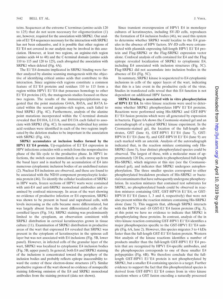
SRPK1 binding is associated with in vitro phosphorylationof HPV1 E1�E4. In vitro kinase reactions were used to deter-mine whether SRPK1 phosphorylates HPV E1�E4 proteins.Reactions were performed using His-SRPK1 and GST-HPVE1�E4 fusion proteins which were all generated by expressionin bacteria. Figure 6A shows the Coomassie-stained gel and anautoradiograph of a typical SRPK1 kinase experiment. In theCoomassie-stained gel, the location of the full-length sub-strates, GST (lane 6), GST-HPV1 E1�E4 (lane 7), GST-HPV16 E1�E4 (lane 8), and GST-HPV18 E1�E4 (lane 9) areeach marked with an asterisk. Analysis of the autoradiographindicated that, in the reaction mixture containing only His-SRPK1 (lane 5), four distinct phosphorylated species could beobserved. The largest of these, with a molecular mass of ap-proximately 120 Da, corresponds to phosphorylated full-lengthHis-SRPK1, which migrates at this size (see the Coomassie-stained gel), and indicates that SRPK1 undergoes autophos-phorylation. The three smaller species correspond to eitherphosphorylated breakdown products of His-SRPK1 or bacte-rial contaminants that have subsequently undergone phosphory-lation. In reaction mixtures that contained test substrates forSRPK1, no phosphorylated bands could be observed in reac-tion mixtures containing GST, GST-HPV16 E1�E4, or GST-HPV18 E1�E4 (lanes 1, 3 and 4, respectively) that were notalso present within the reaction mixture containing His-SRPK1alone (lane 5). This suggests that, although SRPK1 interactswith the HPV16 and -18 GST-E1�E4 fusion proteins (Fig. 3),at this point we have no evidence to indicate that SRPK1 isphosphorylating these proteins. In contrast, analysis of the invitro kinase reaction containing GST-HPV1 E1�E4 identifies amajor phosphospecies specific to the GST-HPV1 E1�E4 sam-ple (Fig. 6A, lane 2). However, this species migrates 3 to 4 kDafaster than the full-length GST-E1�E4 fusion protein. Westernblot analysis of the kinase reactions identifies a number ofproducts smaller than the full-length GST-HPV1 E1�E4 pro-tein that are recognized by HPV1 E4-specific antibodies, andthe phosphospecies corresponds to one of these smaller E4polypeptides (Fig. 6B). We therefore conclude that the full-length GST-HPV1 E1�E4 protein is not phosphorylated bySRPK1, but a smaller E4-containing species is a target. Furtherevidence to support the fact that this phosphorylated species isderived from GST-HPV1 E1�E4 comes from in vitro kinasereactions where a GST fusion encoding a naturally processed
5442 BELL ET AL. J. VIROL.
on April 10, 2019 by guest
http://jvi.asm.org/
Dow
nloaded from

N-terminal truncation (16 kDa) of the full-length 17-kDaE1�E4 protein was used as a substrate. In these studies, themigration of the major phosphospecies was only about 1 kDafaster than the phosphorylated species present in the reactionmixture containing the full-length GST-E1�E4 protein (datanot shown), thus demonstrating that truncation of HPV1E1�E4 resulted in a concomitant shift in the size of the phos-phorylated polypeptide.
Having demonstrated that SRPK1 was able to phosphorylateGST-HPV1 E1�E4, we then examined the ability of SRPK1 tophosphorylate HPV1 E1�E4 deletion mutants. Thus, the dele-tion mutants �44-48, which is unable to bind SRPK1, and�49-53, which retains SRPK1 binding, were expressed as GST
fusion proteins and used as substrates in in vitro kinase reac-tions. In these experiments, the GST substrates were titrated at1, 5, and 25 �g per kinase reaction (Fig. 7). Both HPV1 E1�E4(lanes 4 to 6) and the deletion mutant �49-53 (lanes 10 to 12)were phosphorylated by His-SRPK1. Again, the major phos-phospecies migrated slightly faster that the full-length GSTE1�E4 fusion. In contrast, the deletion mutant �44-48 was notphosphorylated by His-SRPK1 (lanes 7 to 9). Thus, with re-spect to HPV1 E1�E4, proteins which interact with SRPK1appear to be phosphorylated by SRPK1.
Further examination of the autoradiograph of the in vitrokinase reactions shown in Fig. 6 indicated that, in comparisonto reactions containing GST (lane 1), HPV16 (lane 3), or
FIG. 5. SRPK1 distribution is altered in the presence of HPV1 E1�E4. (A and B) Confocal analysis of 4-�m sections of an HPV1-induced wartcostained for E4 (red) and SRPK1 (green); nuclei were identified using DAPI (blue). (A) SRPK1 expression in regions of the wart tissue that areE4 negative and do not show evidence of productive HPV1 infection. Arrowheads indicate the basal cell layer. (B) In regions of the wart positivefor E4 expression, SRPK1 is contained within E4 inclusions present in cells of the granular layers (upper panel, examples of costained inclusionsare indicated by arrows) but not in those formed in cells of the lower (spinous) layers (bottom panel). (C) Confocal analysis of distribution of E4(red) and SRPK1 (green) in SV-JD cells cotransfected with plasmids that express HPV1 E1�E4 and Flag-SRPK1 or Flag-SRPK1 alone. Nucleiwere identified using DAPI (blue).
VOL. 81, 2007 HPV E1�E4 PROTEINS ASSOCIATE WITH SRPK1 5443
on April 10, 2019 by guest
http://jvi.asm.org/
Dow
nloaded from
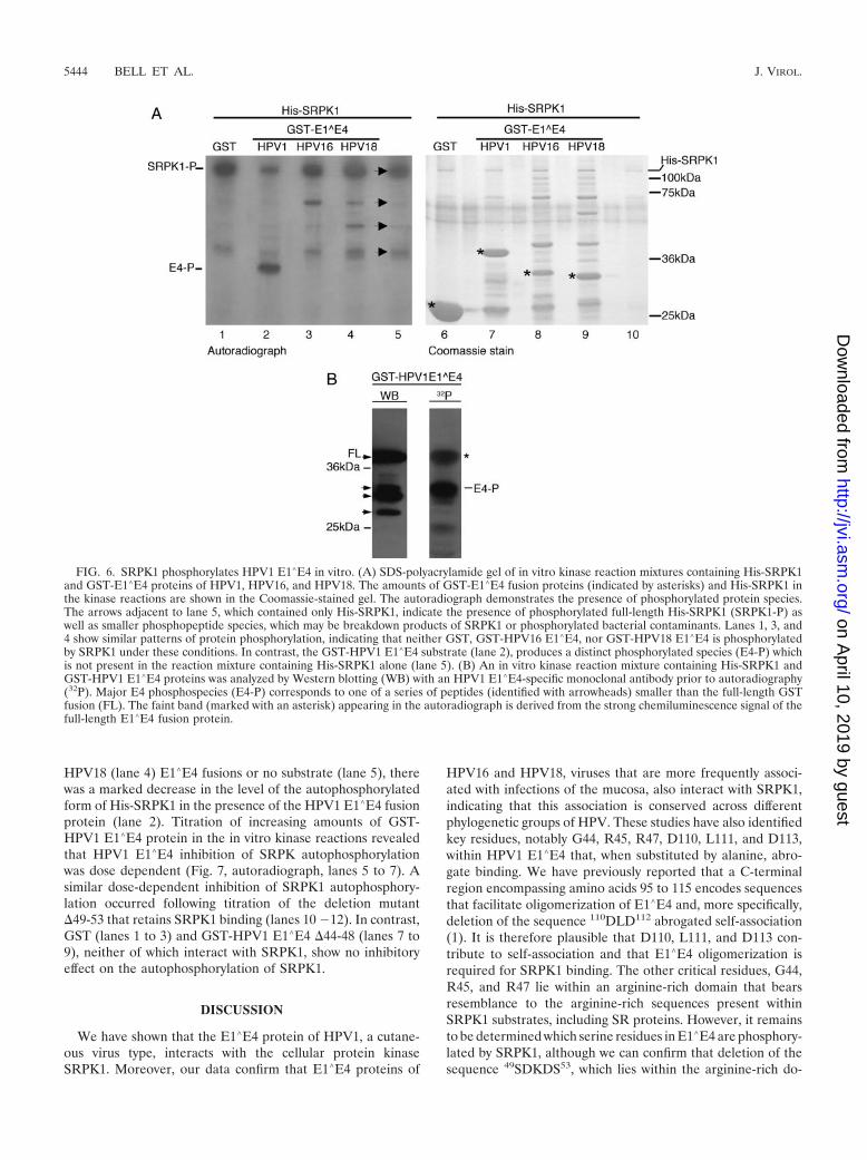
HPV18 (lane 4) E1�E4 fusions or no substrate (lane 5), therewas a marked decrease in the level of the autophosphorylatedform of His-SRPK1 in the presence of the HPV1 E1�E4 fusionprotein (lane 2). Titration of increasing amounts of GST-HPV1 E1�E4 protein in the in vitro kinase reactions revealedthat HPV1 E1�E4 inhibition of SRPK autophosphorylationwas dose dependent (Fig. 7, autoradiograph, lanes 5 to 7). Asimilar dose-dependent inhibition of SRPK1 autophosphory-lation occurred following titration of the deletion mutant�49-53 that retains SRPK1 binding (lanes 10 �12). In contrast,GST (lanes 1 to 3) and GST-HPV1 E1�E4 �44-48 (lanes 7 to9), neither of which interact with SRPK1, show no inhibitoryeffect on the autophosphorylation of SRPK1.
DISCUSSION
We have shown that the E1�E4 protein of HPV1, a cutane-ous virus type, interacts with the cellular protein kinaseSRPK1. Moreover, our data confirm that E1�E4 proteins of
HPV16 and HPV18, viruses that are more frequently associ-ated with infections of the mucosa, also interact with SRPK1,indicating that this association is conserved across differentphylogenetic groups of HPV. These studies have also identifiedkey residues, notably G44, R45, R47, D110, L111, and D113,within HPV1 E1�E4 that, when substituted by alanine, abro-gate binding. We have previously reported that a C-terminalregion encompassing amino acids 95 to 115 encodes sequencesthat facilitate oligomerization of E1�E4 and, more specifically,deletion of the sequence 110DLD112 abrogated self-association(1). It is therefore plausible that D110, L111, and D113 con-tribute to self-association and that E1�E4 oligomerization isrequired for SRPK1 binding. The other critical residues, G44,R45, and R47 lie within an arginine-rich domain that bearsresemblance to the arginine-rich sequences present withinSRPK1 substrates, including SR proteins. However, it remainsto be determined which serine residues in E1�E4 are phosphory-lated by SRPK1, although we can confirm that deletion of thesequence 49SDKDS53, which lies within the arginine-rich do-
FIG. 6. SRPK1 phosphorylates HPV1 E1�E4 in vitro. (A) SDS-polyacrylamide gel of in vitro kinase reaction mixtures containing His-SRPK1and GST-E1�E4 proteins of HPV1, HPV16, and HPV18. The amounts of GST-E1�E4 fusion proteins (indicated by asterisks) and His-SRPK1 inthe kinase reactions are shown in the Coomassie-stained gel. The autoradiograph demonstrates the presence of phosphorylated protein species.The arrows adjacent to lane 5, which contained only His-SRPK1, indicate the presence of phosphorylated full-length His-SRPK1 (SRPK1-P) aswell as smaller phosphopeptide species, which may be breakdown products of SRPK1 or phosphorylated bacterial contaminants. Lanes 1, 3, and4 show similar patterns of protein phosphorylation, indicating that neither GST, GST-HPV16 E1�E4, nor GST-HPV18 E1�E4 is phosphorylatedby SRPK1 under these conditions. In contrast, the GST-HPV1 E1�E4 substrate (lane 2), produces a distinct phosphorylated species (E4-P) whichis not present in the reaction mixture containing His-SRPK1 alone (lane 5). (B) An in vitro kinase reaction mixture containing His-SRPK1 andGST-HPV1 E1�E4 proteins was analyzed by Western blotting (WB) with an HPV1 E1�E4-specific monoclonal antibody prior to autoradiography(32P). Major E4 phosphospecies (E4-P) corresponds to one of a series of peptides (identified with arrowheads) smaller than the full-length GSTfusion (FL). The faint band (marked with an asterisk) appearing in the autoradiograph is derived from the strong chemiluminescence signal of thefull-length E1�E4 fusion protein.
5444 BELL ET AL. J. VIROL.
on April 10, 2019 by guest
http://jvi.asm.org/
Dow
nloaded from
main, does not diminish the ability of SRPK1 to bind norphosphorylate E1�E4 and suggests that other serine residuesmust be sites of SRPK1 phosphorylation. Close inspection ofthe major phosphorylated species from the in vitro kinaseexperiments indicates that it is migrating faster than the majorGST-HPV1 E1�E4 product. Loss of a proximal C-terminalsequence may enhance phosphorylation or phosphorylation ofE4 is affecting the migration of the E4 polypeptide, which ispossible given that the full-length E1�E4 protein migratesanomalously high at 17-kDa, 2.6-kDa higher than its calculatedmass of 14.4 kDa. While we have been unable to detect phos-phorylation of HPV16 and HPV18 E1�E4 proteins in vitro, wecannot rule out the possibility that these may be substrates ofSRPK1 and indeed that the GST component of these proteinsmay induce steric changes that impairs phosphorylation inthese assays. Phosphorylation is likely to be an important de-terminant of E4 function, and to date, a number of kinaseshave been implicated in E4 phosphorylation (4, 7, 20). It willthereforebeinterestingtodeterminewhetherE1�E4phosphory-lation by SRPK1 does contribute to a specific E4 function.
The oligomerization domain of HPV1 E4 displays significanthomology with the C-terminal sequences of E4 proteins de-rived from other HPV types that infect cutaneous surfaces.Within this domain, D110, L111, and D113 form part of aconserved motif (D-L-[D/E]-X-[Y/F], where X is a hydrophilicresidue) that has been shown to be necessary for oligomeriza-tion (1, 43). A similar function has been assigned to the Cterminus of HPV16 E4 (56). While sequence alignment iden-tifies homology across this region of HPV16 E4 with E4s ofother mucosal types, it is not highly conserved within the cu-taneous viruses (43, 45). The lack of homology between theoligomerization domains of cutaneous and mucosal viruseswould suggest that D110, L111, and D113 may not be involvedin direct binding of SRPK1 but that oligomerization of theprotein is perhaps necessary. With regard to the other HPV1residues necessary for SRPK1 binding (G44, R45, and R47),they are contained within a region of HPV1 E4 rich in basicamino acids (arginine, lysine, and histidine). Similar basic re-
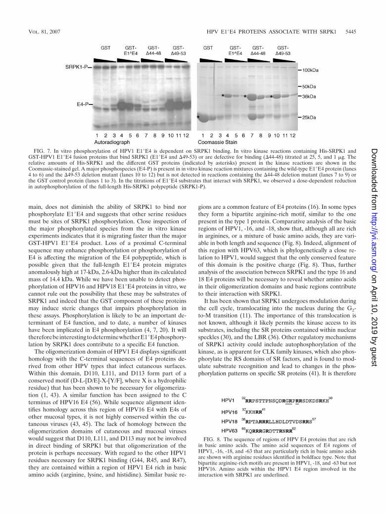
gions are a common feature of E4 proteins (16). In some typesthey form a bipartite arginine-rich motif, similar to the onepresent in the type 1 protein. Comparative analysis of the basicregions of HPV1, -16, and -18, show that, although all are richin arginines, or a mixture of basic amino acids, they are vari-able in both length and sequence (Fig. 8). Indeed, alignment ofthis region with HPV63, which is phylogenetically a close re-lation to HPV1, would suggest that the only conserved featureof this domain is the positive charge (Fig. 8). Thus, furtheranalysis of the association between SRPK1 and the type 16 and18 E4 proteins will be necessary to reveal whether amino acidsin their oligomerization domains and basic regions contributeto their interaction with SRPK1.
It has been shown that SRPK1 undergoes modulation duringthe cell cycle, translocating into the nucleus during the G2-to-M transition (11). The importance of this translocation isnot known, although it likely permits the kinase access to itssubstrates, including the SR proteins contained within nuclearspeckles (30), and the LBR (36). Other regulatory mechanismsof SRPK1 activity could include autophosphorylation of thekinase, as is apparent for CLK family kinases, which also phos-phorylate the RS domains of SR factors, and is found to mod-ulate substrate recognition and lead to changes in the phos-phorylation patterns on specific SR proteins (41). It is therefore
FIG. 7. In vitro phosphorylation of HPV1 E1�E4 is dependent on SRPK1 binding. In vitro kinase reactions containing His-SRPK1 andGST-HPV1 E1�E4 fusion proteins that bind SRPK1 (E1�E4 and �49-53) or are defective for binding (�44-48) titrated at 25, 5, and 1 �g. Therelative amounts of His-SRPK1 and the different GST proteins (indicated by asterisks) present in the kinase reactions are shown in theCoomassie-stained gel. A major phosphospecies (E4-P) is present in in vitro kinase reaction mixtures containing the wild-type E1�E4 protein (lanes4 to 6) and the �49-53 deletion mutant (lanes 10 to 12) but is not detected in reactions containing the �44-48 deletion mutant (lanes 7 to 9) orthe GST control protein (lanes 1 to 3). In the titrations of E1�E4 substrates that interact with SRPK1, we observed a dose-dependent reductionin autophosphorylation of the full-length His-SRPK1 polypeptide (SRPK1-P).
FIG. 8. The sequence of regions of HPV E4 proteins that are richin basic amino acids. The amino acid sequences of E4 regions ofHPV1, -16, -18, and -63 that are particularly rich in basic amino acidsare shown with arginine residues identified in boldface type. Note thatbipartite arginine-rich motifs are present in HPV1, -18, and -63 but notHPV16. Amino acids within the HPV1 E4 region involved in theinteraction with SRPK1 are underlined.
VOL. 81, 2007 HPV E1�E4 PROTEINS ASSOCIATE WITH SRPK1 5445
on April 10, 2019 by guest
http://jvi.asm.org/
Dow
nloaded from
of interest that, in the in vitro kinase assays, the ability ofSRPK1 to undergo autophosphorylation appeared to be inhib-ited by HPV1 E1�E4, and this inhibition was abrogated bymutations which blocked HPV1 E1�E4 binding of SRPK1.Thus, whether E1�E4 does indeed modulate SRPK1 autophos-phorylation and substrate specificity in vivo warrants furtherinvestigation.
The manipulation of SRPK1 by a virus is not without pre-cedent, indeed the herpes simplex virus type 1 encodes a pro-tein ICP27 which associates with SRPK1, relocating it to thenucleus, resulting in hypophosphorylation of SR proteins and aconsequent impairment in spliceosome assembly (51). Thisaction has the effect of inhibiting cellular pre-mRNA splicingand promotes export and expression of herpes simplex virustype 1 transcripts. There are also a number of studies thatimplicate substrates of SRPK1 in the replication cycle of vi-ruses. These studies range from human immunodeficiency vi-rus type 1 (HIV-1), where virion production is regulated by SRproteins, including ASF/SF2, SC35, and 9G8 (26), while SRp75acts to upregulate HIV-1 gene expression (19); adenovirus,where SR proteins purified from late infected cells are func-tionally inactivated (27); and vaccinia, where SR proteinsare hypophosphorylated (23). Thus, viruses appear to haveadopted mechanisms to act upon SR proteins as a means ofmodifying host gene expression and affecting efficient viralgene expression. Pertinently with respect to HPV, an SRPK1substrate, SF2/ASF, is part of a complex that associates with anegative regulatory mRNA element that is central to the post-transcriptional regulation of late gene expression in HPV16,and both expression and phosphorylation of this SR factorincrease in response to differentiation of the HPV16-infectedcells (33). In fact, numerous cis-acting elements appear tocontribute to the control of HPV late gene expression, includ-ing HPV16 (48, 60), and similar structures that are present inbovine papillomavirus transcripts are associated with multipleSR proteins (61). Further studies are required to examine thedifferentiation-specific regulation of other SR proteins and toidentify whether E1�E4 proteins, via their interaction withSRPK1, impact upon their regulation. The suggestion thatE1�E4 may modify posttranscriptional processing is not novel;indeed, HPV16 E4�E4 has previously been shown to associatewith an RNA helicase, an association that was proposed toaffect mRNA stability and ribosome biogenesis (13).
In addition to phosphorylating SR proteins, SRPK1 alsophosphorylates LBR, an inner nuclear membrane proteinwhich interacts with B-type lamins (53) and chromatin (55).The association of LBR with chromatin is cell cycle dependentand regulated by multiple kinases, including SRPK1 (54), andacts to maintain the organization of the nuclear envelope dur-ing cell division (6). Inhibition, modulation, or sequestration ofSRPK1 by E1�E4 may inhibit the attachment of LBR to chro-matin and result in the loss of nuclear envelope integrity. Sincethe sequestration of SRPK1 to HPV1 E4 inclusion bodies isrestricted to cells of the upper wart regions, and as HPV virionassembly is confined to the nucleus of these cells, destabiliza-tion of the nuclear envelope may facilitate egress of virionsfrom the nucleus. Indeed, the human polyomavirus JC virusperturbs the structure of the nuclear envelope by abrogatingthe interaction between LBR and heterochromatin and thuspromoting nuclear egress of progeny JC virus virions (37). This
possible E1�E4 function, in addition to its action on the cor-nified cell envelope (3) and the keratin cytoskeleton (14, 44)and an ability to promote apoptosis (42), may all contribute toease the passage of newly synthesized virions from the nucleusto the cytoplasm and promote their subsequent shedding fromthe upper cells of the lesion.
SRPK1 binding by E1�E4 may influence the function ofother HPV proteins. Candidate molecules such as the HPV5E2 protein, which contains a hinge region that possesses RSdomain characteristics (31), may be an SRPK1 substrate, andas such, SRPK1 may influence HPV5 E2 function by modulat-ing its ability to activate transcription/replication.
SRPK1 appears to play an important role in the regulationof some fundamental cellular processes, including the organi-zation of components of the nucleus and mRNA processing,and this may explain why it appears to be a strategically im-portant cellular target for a diverse number of viruses. Ourstudy identifies SRPK1 as a novel cellular binding partner ofE4 proteins derived from genetically diverse HPV types. Withregard to HPV1 E1�E4, we have demonstrated that the abilityof SRPK1 to undergo autophosphorylation is impaired, whileit is sequestered into E4 inclusions in terminally differentiatedcells within wart lesions. Future studies are necessary to dissectthe biochemical implications of this finding and to determinewhether this interaction has a role in the regulation of aspectsof HPV replication. The biochemistry of SRPK1 is poorlyunderstood and the role it plays in the HPV replication cycle isunknown, but novel substrates encoded by HPV and/or thehost cell may exist. Indeed this kinase may prove to be a targetfor therapeutic intervention, as has been proposed for HIV-1,where the use of small-molecule inhibitors of SRPK1 has beendemonstrated to suppress HIV-1 replication (19).
ACKNOWLEDGMENTS
We thank Saleem Khan (University of Pittsburgh) for his kind gift ofthe HPV1 E1-Flag expression vector and Bai-Gong Yue (University ofLeicester) for the generous gift of the SRPK1-His expression plasmid.We are grateful to Gillian Knight for her help in generating the HPV144-48 single-alanine substitutions and to Michele McNally for herexcellent technical assistance.
This study was supported by a Cancer Research UK ProgrammeGrant (C427/A3919) to S.R.
REFERENCES
1. Ashmole, I., P. H. Gallimore, and S. Roberts. 1998. Identification of con-served hydrophobic C-terminal residues of the human papillomavirus type 1E1E4 protein necessary for E4 oligomerisation in vivo. Virology 240:221–231.
2. Breitburd, F., O. Croissant, and G. Orth. 1987. Expression of human pap-illomavirus type-1 E4 gene products in warts, p. 115–122. In B. M. Steinberg,J. Brandsma, and L. B. Taichman (ed.), Papillomaviruses, vol. 5. Cold SpringHarbor Press, Cold Spring Harbor, NY.
3. Brown, D. R., D. Kitchin, B. Qadadri, N. Neptune, T. Batteiger, and A.Ermel. 2006. The human papillomavirus type 11 E1–E4 protein is a trans-glutaminase 3 substrate and induces abnormalities of the cornified cell en-velope. Virology 345:290–298.
4. Bryan, J. T., A. Han, K. H. Fife, and D. R. Brown. 2000. The humanpapillomavirus type 11 E1E4 protein is phosphorylated in genital epithelium.Virology 268:430–439.
5. Caceres, J. F., G. R. Screaton, and A. R. Krainer. 1998. A specific subset ofSR proteins shuttles continuously between the nucleus and the cytoplasm.Genes Dev. 12:55–66.
6. Chu, A., R. Rassadi, and U. Stochaj. 1998. Velcro in the nuclear envelope:LBR and LAPs. FEBS Lett. 441:165–169.
7. Davy, C. E., M. Ayub, D. J. Jackson, P. Das, P. McIntosh, and J. Doorbar.2006. HPV16 E1–E4 protein is phosphorylated by Cdk2/cyclin A and relo-calizes this complex to the cytoplasm. Virology 349:230–244.
5446 BELL ET AL. J. VIROL.
on April 10, 2019 by guest
http://jvi.asm.org/
Dow
nloaded from

8. Davy, C. E., D. J. Jackson, K. Raj, W. L. Peh, S. A. Southern, P. Das, R.Sorathia, P. Laskey, K. Middleton, T. Nakahara, Q. Wang, P. J. Masterson,P. F. Lambert, S. Cuthill, J. B. Millar, and J. Doorbar. 2005. Humanpapillomavirus type 16 E1 E4-induced G2 arrest is associated with cytoplas-mic retention of active Cdk1/cyclin B1 complexes. J. Virol. 79:3998–4011.
9. Davy, C. E., D. J. Jackson, Q. Wang, K. Raj, P. J. Masterson, N. F. Fenner,S. Southern, S. Cuthill, J. B. Millar, and J. Doorbar. 2002. Identification ofa G(2) arrest domain in the E1 wedge E4 protein of human papillomavirustype 16. J. Virol. 76:9806–9818.
10. de Villiers, E. M., C. Fauquet, T. R. Broker, H. U. Bernard, and H. zurHausen. 2004. Classification of papillomaviruses. Virology 324:17–27.
11. Ding, J. H., X. Y. Zhong, J. C. Hagopian, M. M. Cruz, G. Ghosh, J.Feramisco, J. A. Adams, and X. D. Fu. 2006. Regulated cellular parti-tioning of SR protein-specific kinases in mammalian cells. Mol. Biol. Cell17:876–885.
12. Doorbar, J., I. Coneron, and P. H. Gallimore. 1989. Sequence divergence yetconserved physical characteristics among the E4 proteins of cutaneous hu-man papillomaviruses. Virology 172:51–62.
13. Doorbar, J., R. C. Elston, S. Napthine, K. Raj, E. Medcalf, D. Jackson, N.Coleman, H. M. Griffin, P. Masterson, S. Stacey, Y. Mengistu, and J.Dunlop. 2000. The E1E4 protein of human papillomavirus type 16 asso-ciates with a putative RNA helicase through sequences in its C terminus.J. Virol. 74:10081–10095.
14. Doorbar, J., S. Ely, J. Sterling, C. McLean, and L. Crawford. 1991. Specificinteraction between HPV-16 E1–E4 and cytokeratins results in collapse ofthe epithelial cell intermediate filament network. Nature 352:824–827.
15. Doorbar, J., H. S. Evans, I. Coneron, L. V. Crawford, and P. H. Gallimore.1988. Analysis of HPV-1 E4 gene expression using epitope-defined antibod-ies. EMBO J. 7:825–833.
16. Doorbar, J., and G. Myers. 1996. The E4 protein, p. 58–80. In G. Myers,H. Delius, J. Icenogel, H.-C. Bernard, C. Baker, A. Halpern, and C.Wheeler (ed.), Human papillomaviruses, vol. 3. Los Alamos NationalLaboratory, Los Alamos, NM.
17. Fang, L., L. R. Budgeon, J. Doorbar, E. R. Briggs, and M. K. Howett. 2006.The human papillomavirus type 11 E1�E4 protein is not essential for viralgenome amplification. Virology 351:271–279.
18. Fu, X. D. 1995. The superfamily of arginine/serine-rich splicing factors. RNA1:663–680.
19. Fukuhara, T., T. Hosoya, S. Shimizu, K. Sumi, T. Oshiro, Y. Yoshinaka,M. Suzuki, N. Yamamoto, L. A. Herzenberg, L. A. Herzenberg, and M.Hagiwara. 2006. Utilization of host SR protein kinases and RNA-splicingmachinery during viral replication. Proc. Natl. Acad. Sci. USA103:11329–11333.
20. Grand, R. J., J. Doorbar, K. J. Smith, I. Coneron, and P. H. Gallimore. 1989.Phosphorylation of the human papillomavirus type 1 E4 proteins in vivo andin vitro. Virology 170:201–213.
21. Hebner, C. M., and L. A. Laimins. 2006. Human papillomaviruses: basicmechanisms of pathogenesis and oncogenicity. Rev. Med. Virol. 16:83–97.
22. Hillenkamp, F., M. Karas, R. C. Beavis, and B. T. Chait. 1991. Matrix-assisted laser desorption/ionization mass spectrometry of biopolymers. Anal.Chem. 63:1193A–1203A.
23. Huang, T. S., C. E. Nilsson, T. Punga, and G. Akusjarvi. 2002. Functionalinactivation of the SR family of splicing factors during a vaccinia virusinfection. EMBO Rep. 3:1088–1093.
24. Huang, Y., and J. A. Steitz. 2001. Splicing factors SRp20 and 9G8 promotethe nucleocytoplasmic export of mRNA. Mol. Cell 7:899–905.
25. Huang, Y., and J. A. Steitz. 2005. SRprises along a messenger’s journey. Mol.Cell 17:613–615.
26. Jacquenet, S., D. Decimo, D. Muriaux, and J. L. Darlix. 2005. Dual effect ofthe SR proteins ASF/SF2, SC35 and 9G8 on HIV-1 RNA splicing and virionproduction. Retrovirology 2:33.
27. Kanopka, A., O. Muhlemann, S. Petersen-Mahrt, C. Estmer, C. Ohrmalm,and G. Akusjarvi. 1998. Regulation of adenovirus alternative RNA splicingby dephosphorylation of SR proteins. Nature 393:185–187.
28. Knight, G. L., J. R. Grainger, P. H. Gallimore, and S. Roberts. 2004. Co-operation between different forms of the human papillomavirus type 1 E4protein to block cell cycle progression and cellular DNA synthesis. J. Virol.78:13920–13933.
29. Knight, G. L., A. S. Turnell, and S. Roberts. 2006. Role for Wee1 in inhi-bition of G2-to-M transition through the cooperation of distinct humanpapillomavirus type 1 E4 proteins. J. Virol. 80:7416–7426.
30. Koizumi, J., Y. Okamoto, H. Onogi, A. Mayeda, A. R. Krainer, and M.Hagiwara. 1999. The subcellular localization of SF2/ASF is regulated bydirect interaction with SR protein kinases (SRPKs). J. Biol. Chem. 274:11125–11131.
31. Lai, M. C., B. H. Teh, and W. Y. Tarn. 1999. A human papillomavirus E2transcriptional activator. The interactions with cellular splicing factors and po-tential function in pre-mRNA processing. J. Biol. Chem. 274:11832–11841.
32. Lin, S., R. Xiao, P. Sun, X. Xu, and X. D. Fu. 2005. Dephosphorylation-dependent sorting of SR splicing factors during mRNP maturation. Mol. Cell20:413–425.
33. McPhillips, M. G., T. Veerapraditsin, S. A. Cumming, D. Karali, S. G.
Milligan, W. Boner, I. M. Morgan, and S. V. Graham. 2004. SF2/ASF bindsthe human papillomavirus type 16 late RNA control element and is regulatedduring differentiation of virus-infected epithelial cells. J. Virol. 78:10598–10605.
34. Nakahara, T., A. Nishimura, M. Tanaka, T. Ueno, A. Ishimoto, and H.Sakai. 2002. Modulation of the cell division cycle by human papillomavirustype 18 E4. J. Virol. 76:10914–10920.
35. Nakahara, T., W. L. Peh, J. Doorbar, D. Lee, and P. F. Lambert. 2005.Human papillomavirus type 16 E1�E4 contributes to multiple facets of thepapillomavirus life cycle. J. Virol. 79:13150–13165.
36. Nikolakaki, E., J. Meier, G. Simos, S. D. Georgatos, and T. Giannakouros.1997. Mitotic phosphorylation of the lamin B receptor by a serine/argininekinase and p34(cdc2). J. Biol. Chem. 272:6208–6213.
37. Okada, Y., T. Suzuki, Y. Sunden, Y. Orba, S. Kose, N. Imamoto, H.Takahashi, S. Tanaka, W. W. Hall, K. Nagashima, and H. Sawa. 2005.Dissociation of heterochromatin protein 1 from lamin B receptor inducedby human polyomavirus agnoprotein: role in nuclear egress of viral par-ticles. EMBO Rep. 6:452–457.
38. Papoutsopoulou, S., E. Nikolakaki, and T. Giannakouros. 1999. SRPK1 andLBR protein kinases show identical substrate specificities. Biochem. Bio-phys. Res. Commun. 255:602–607.
39. Peh, W. L., J. L. Brandsma, N. D. Christensen, N. M. Cladel, X. Wu, and J.Doorbar. 2004. The viral E4 protein is required for the completion of thecottontail rabbit papillomavirus productive cycle in vivo. J. Virol. 78:2142–2151.
40. Peh, W. L., K. Middleton, N. Christensen, P. Nicholls, K. Egawa, K. Sotlar,J. Brandsma, A. Percival, J. Lewis, W. J. Liu, and J. Doorbar. 2002. Lifecycle heterogeneity in animal models of human papillomavirus-associateddisease. J. Virol. 76:10401–10416.
41. Prasad, J., and J. L. Manley. 2003. Regulation and substrate specificity of theSR protein kinase Clk/Sty. Mol. Cell. Biol. 23:4139–4149.
42. Raj, K., S. Berguerand, S. Southern, J. Doorbar, and P. Beard. 2004. E1�E4protein of human papillomavirus type 16 associates with mitochondria. J. Vi-rol. 78:7199–7207.
43. Roberts, S., I. Ashmole, L. J. Gibson, S. M. Rookes, G. J. Barton, and P. H.Gallimore. 1994. Mutational analysis of human papillomavirus E4 proteins:identification of structural features important in the formation of cytoplas-mic E4/cytokeratin networks in epithelial cells. J. Virol. 68:6432–6445.
44. Roberts, S., I. Ashmole, G. D. Johnson, J. W. Kreider, and P. H. Galli-more. 1993. Cutaneous and mucosal human papillomavirus E4 proteinsform intermediate filament-like structures in epithelial cells. Virology197:176–187.
45. Roberts, S., I. Ashmole, S. M. Rookes, and P. H. Gallimore. 1997. Mutationalanalysis of the human papillomavirus type 16 E1–E4 protein shows that theC terminus is dispensable for keratin cytoskeleton association but is involvedin inducing disruption of the keratin filaments. J. Virol. 71:3554–3562.
46. Roberts, S., M. L. Hillman, G. L. Knight, and P. H. Gallimore. 2003. TheND10 component promyelocytic leukemia protein relocates to human pap-illomavirus type 1 E4 intranuclear inclusion bodies in cultured keratinocytesand in warts. J. Virol. 77:673–684.
47. Rogel-Gaillard, C., G. Pehau-Arnaudet, F. Breitburd, and G. Orth. 1993.Cytopathic effect in human papillomavirus type 1-induced inclusion warts: invitro analysis of the contribution of two forms of the viral E4 protein.J. Investig. Dermatol. 101:843–851.
48. Rush, M., X. Zhao, and S. Schwartz. 2005. A splicing enhancer in the E4coding region of human papillomavirus type 16 is required for early mRNAsplicing and polyadenylation as well as inhibition of premature late geneexpression. J. Virol. 79:12002–12015.
49. Sanford, J. R., J. D. Ellis, D. Cazalla, and J. F. Caceres. 2005. Reversiblephosphorylation differentially affects nuclear and cytoplasmic functions ofsplicing factor 2/alternative splicing factor. Proc. Natl. Acad. Sci. USA 102:15042–15047.
50. Sanford, J. R., N. K. Gray, K. Beckmann, and J. F. Caceres. 2004. A novelrole for shuttling SR proteins in mRNA translation. Genes Dev. 18:755–768.
51. Sciabica, K. S., Q. J. Dai, and R. M. Sandri-Goldin. 2003. ICP27 interactswith SRPK1 to mediate HSV splicing inhibition by altering SR proteinphosphorylation. EMBO J. 22:1608–1619.
52. Shevchenko, A., M. Wilm, O. Vorm, and M. Mann. 1996. Mass spectrometricsequencing of proteins silver-stained polyacrylamide gels. Anal. Chem. 68:850–858.
53. Simos, G., and S. D. Georgatos. 1992. The inner nuclear membrane proteinp58 associates in vivo with a p58 kinase and the nuclear lamins. EMBO J.11:4027–4036.
54. Takano, M., Y. Koyama, H. Ito, S. Hoshino, H. Onogi, M. Hagiwara, K.Furukawa, and T. Horigome. 2004. Regulation of binding of lamin B recep-tor to chromatin by SR protein kinase and cdc2 kinase in Xenopus eggextracts. J. Biol. Chem. 279:13265–13271.
55. Takano, M., M. Takeuchi, H. Ito, K. Furukawa, K. Sugimoto, S. Omata,and T. Horigome. 2002. The binding of lamin B receptor to chromatin isregulated by phosphorylation in the RS region. Eur. J. Biochem. 269:943–953.
56. Wang, Q., H. Griffin, S. Southern, D. Jackson, A. Martin, P. McIntosh, C.
VOL. 81, 2007 HPV E1�E4 PROTEINS ASSOCIATE WITH SRPK1 5447
on April 10, 2019 by guest
http://jvi.asm.org/
Dow
nloaded from

Davy, P. J. Masterson, P. A. Walker, P. Laskey, M. B. Omary, and J.Doorbar. 2004. Functional analysis of the human papillomavirus type 16E1�E4 protein provides a mechanism for in vivo and in vitro keratin filamentreorganization. J. Virol. 78:821–833.
57. Wilson, R., F. Fehrmann, and L. A. Laimins. 2005. Role of the E1�E4protein in the differentiation-dependent life cycle of human papillomavirustype 31. J. Virol. 79:6732–6740.
57a.Wilson, R., G. B. Ryan, G. L. Knight, L. A. Laimins, and S. Roberts. 13February 2007, posting date. The full-length E1�E4 protein of human pap-illomavirus type 18 modulates differentiation-dependent viral DNA amplifi-cation and late gene expression. Virology. doi:10.1016/j.virol.2007.01.005.
58. Xiao, S. H., and J. L. Manley. 1998. Phosphorylation-dephosphorylation differ-entially affects activities of splicing factor ASF/SF2. EMBO J. 17:6359–6367.
59. Yue, B. G., P. Ajuh, G. Akusjarvi, A. I. Lamond, and J. P. Kreivi. 2000.Functional coexpression of serine protein kinase SRPK1 and its substrateASF/SF2 in Escherichia coli. Nucleic Acids Res. 28:E14.
60. Zhao, X., M. Rush, and S. Schwartz. 2004. Identification of an hnRNPA1-dependent splicing silencer in the human papillomavirus type 16 L1coding region that prevents premature expression of the late L1 gene. J. Vi-rol. 78:10888–10905.
61. Zheng, Z. M., and C. C. Baker. 2006. Papillomavirus genome structure,expression, and posttranscriptional regulation. Front Biosci. 11:2286–2302.
5448 BELL ET AL. J. VIROL.
on April 10, 2019 by guest
http://jvi.asm.org/
Dow
nloaded from