the drosophila copia retrotransposon contains binding sites for
TRANSCRIPT

Nucleic Acids Research, 1993, Vol. 21, No. 22 5041-5049
The Drosophila copia retrotransposon contains bindingsites for transcriptional regulation by homeoproteins
Laurent Cavarec and Thierry Heidmann*Unites de Physicochimie et Pharmacologie des Macromolecules Biologiques, CNRS U147 etINSERM U140, Institut Gustave Roussy, 39 rue Camille Desmoulins, 94805 Villejuif Cedex, France
Received August 16, Revised and Accepted October 7, 1993
ABSTRACT
We have identified in the 5' untranslated region of theDrosophila copia retrotransposon, 3' to the left LTR,a sequence for transcriptional regulation by homeo-proteins. Co-transfection assays using expressionvectors for homeoproteins and reporter vectors con-taining the lacZ gene under the control of either theentire copia LTR with 5' untranslated sequence, or aminimal heterologous promoter flanked with a 130bpfragment containing the copia untranslated region,disclosed both positive and negative modulations ofpromoter activity in Drosophila cells in culture: a 5 - 1 0fold decrease with engrailed, even-skipped and zerk-niillt in DH33 cells, and a 10 - 30 fold increase with fushitarazu and zerkniillt in Schneider II cells. In all cases,the regulatory effects were abolished with reporterplasmids deleted for a 58 bp fragment encompassingthe putative homeoprotein binding sites. Mobility shiftassays with a purified homeodomain-containing pep-tide demonstrated direct interaction with the 58bpfragment, with an affinity in the 1-10nM range asreported with the same peptide for other well charact-erized homeodomain binding regulatory sites. Foot-printing experiments with the extended LTR demon-strated protection of 'consensus' sequences, locatedwithin the 58bp fragment. These homeodomain bindingsites could be involved in the developmental regulationof the copia retrotransposon.
INTRODUCTION
Mobile genetic elements—or transposons—are 'endogenousmutagens' that have been found in all plant and animal speciesstudied so far. In Drosophila, they represent several percent ofthe genome in mass and account for up to 90% of the spontaneousmutations detected by classical genetic analysis [compiled in (1)].Transposition events are rare, but their frequency can bedramatically increased, depending on both genetic (for instancehybrid dysgenesis, presence of suppressor genes) and epigenetic('stress', heat-shock, UV irradiation) factors [reviewed in (2)].
There are several classes of transposons, but a large fractionof Drosophila mobile elements are 'retrotransposons' [reviewed
in (2, 3)], either of the retrovirus-like family—as the prototype'copia' element, or of the LINE family—as the prototype Ielement involved in the I-R hybrid dysgenesis syndrome[reviewed in (4,5)]. Their transposition is replicative and involvesreverse transcription of an intermediate RNA transcript of theelement, as now demonstrated for the yeast Tyl (6) and themammalian LAP (7) retrovirus-like elements, and for theDrosophila I LINE element (8, 9). Clearly, regulation of thelevels of transcription of these mobile elements should controltheir transposition frequency, as demonstrated for the yeast Tyland the mammalian IAP retrotransposons for which inductionof transcription at a high level results in high transpositionfrequency (6, 10).
The structure and molecular basis of transposition of severalof these elements are now well characterized, but little is knownabout the factors and host genes involved in their transcriptionalcontrol. In Drosophila, analysis of the transcripts from the wholeanimal at various stages of development disclosed defined patternsof transcription with, as a rule, only limited expression in theadult (11). The extensive analysis in (11) of the temporal patternof expression of nine retrovirus-like retrotransposons demon-strated diverse patterns among transposons, with for instance apeak of expression for second-third instar larvae for copia, Firstinstar larvae and young pupae for the 412 and mdgl elements,and embryos and larval stages for the 297 element. Interestingly,developmentally regulated expression of retrotransposons havealso been observed for some mammalian transposable elements,such as the IAP elements—which are expressed almostexclusively at the 2-cell stage of embryo development [and alsoin tumors; reviewed in (12)], or the ET (Early Transposon)element—which is also expressed only very early in development(13).
It is well established that several developmentally regulatedgenes in Drosophila (as well as in plants and vertebrates) arecontrolled by homeoproteins, which bind to DNA sequences andact as transcriptional regulators. Homeoproteins are known tobe both negative and positive regulators, and to act in associationwith each other or with other transcription factors to elicitcomplex patterns of expression, both temporally and spatially,in the developing Drosophila [reviewed in (14-18)]. Homeo-proteins contain highly conserved 60-aminoacid sequences—
* To whom correspondence should be addressed
Downloaded from https://academic.oup.com/nar/article-abstract/21/22/5041/2376659by gueston 18 February 2018

5042 Nucleic Acids Research, 1993, Vol. 21, No. 22
referred to as the homeodomains (HD)—which have been isolatedfrom several homeoproteins and were shown to be responsiblefor DNA binding activity (19-23). The structure of the HDpolypeptides has been elucidated by NMR spectroscopy andcrystallographic analysis, and discloses a helix-tum-helix structureanalogous to that of many prokaryotic transcriptional repressorsand activators (24—26). In vitro analysis of the binding to DNAof a number of homeoproteins—and of HD peptides—allowedderivation of a minimal consensus DNA binding sequence—asa 'core' ATTA sequence—the importance of which wasconfirmed by the recently determined three-dimensional structureof the Antp and engrailed homeodomain-DNA complexes(26-28). The ATTA core sequence plays a primary role indetermining the affinity of binding, with significant secondarycontributions deriving from the flanking bases (22, 23, 29, 30).
In this study, we have investigated the transcriptional propertiesof the Drosophila copia element. This prototype retrovirus-likeretrotransposon, which has been previously entirely sequenced,contains two 276 bp LTRs, a single ORF with sequencehomologies to the integrase, protease and reverse transcriptaseof vertebrate retroviruses, and a 145 bp 5' untranslated region(31). An enhancer sequence—with homologies to the SV40enhancer—had previously been identified (31, 32), which liesoutside of the LTR, in the 5' untranslated region. We report theidentification of a sequence adjacent to this enhancer, whichdisplays (i) direct interaction with a purified Antp homeodomainpeptide, as demonstrated by both footprinting experiments andmobility shift assays, and (ii) positive or negative modulation ofthe level of activity of the copia LTR promoter—as well as ofan heterologous minimal promoter—in transient co-transfectionassays in cultured cells from either D.melanogaster or D.hydei,using expression vectors for several homeoproteins.
MATERIALS AND METHODSDNA constructionsThe copia element used is the 'white apricot insertion' [lambdawa5.9 clone in (33), sequenced in (31)], that we subcloned intothe Bluescript vector (Stratagene) as a BanI fragment, leavingonly 77bp and 87bp of white DNA respectively 5' and 3' to thecopia element (pBScopia). Four oligonucleotides were synthesizedon an Applied Biosystems DNA synthesizer: Homl =(5')CAGT-CCATGCCTAATAAAC; Hom2=(5')CACAATTTATTTTCA-CAATC;Enhl=(5 ' )AAATAGCATTTTTTCACATTC;Enh2 = (5')CTGAGAAGGAAATAATTTC; they were used toPCR-amplify from the copia-containing plasmid a 130bp fragmentcontaining both the consensus homeoprotein binding sites andthe enhancer sequences (using Homl and Enh2), a 58bp fragmentcontaining the homeoprotein binding sites (using Homl andHom2), and a 72bp fragment containing the two dyad symmetryof the putative enhancer region (using Enhl and Enh2).
Construction of the copia LTR-lacZ responder plasmids. LTRlacZ: a 490bp Banl-Ddel fragment from the pBScopia plasmid(containing the entire copia LTR and 5' untranslated sequence)was inserted at the unique Sail site of pGEMnlslacZ [whichcontains a lacZ gene with nuclear location signal (34)], afterKlenow treatment of both vector and insert; the polyadenylationsequence from the hsp70 gene [as a BamHI—EcoRI fragmentfrom HZ50 (35)] was then inserted at a unique BamHI site 3'to the nlslacZ gene, after Klenow treatment of both vector andinsert. LTR AHpRE lacZ: a 360bp Banl-Apal fragment (LTR
alone) from pBScopia was inserted at the unique Sail site of apGEMnlslacZ plasmid containing the polyadenylation sequenceabove, after Klenow treatment of both vector and insert (whichreconstitutes the Sail site at the Sail-Apal junction); the 72bpfragment obtained by PCR amplification using the Enhl and Enh2primers (enhancer region) was then inserted 3' to the copia LTRat the reconstituted Sail site, after Klenow treatment of the vectorand T4 kinase treatment of the insert.
Construction of the hsp70-lacZ reporter plasmids. The hsp70-lacZresponder plasmids were constructed by inserting the copiaregulatory sequences at the unique NotI site of a plasmidcontaining a lacZ gene under control of an hsp70 enhancerlesspromoter and polyadenylation sequence [HZ50 plasmid (35)];the inserted sequences were PCR-amplified fragments, either130bp long (Homl and Enh2 primers) or 72bp long (Enhl andEnh2 primers), which were treated with T4 kinase before ligationwith the Klenow-treated vectort.
Expression vectors for homeoproteins. These were gifts from DrsK.Han and M.S.Levine, and are described in (36); they containthe coding sequence for homeoproteins inserted between theActin-5C promoter and polyadenylation signal; the control pActvector corresponds to the same plasmids, but with no insert[pAct5C-PPA in (36)].
Cells, transfections, /3-galactosidase assays and Northern blotanalysisSchneider II cells from D.melanogaster (37) and DH33 cells fromD.hydei (38) were grown in Schneider medium (GIBCO) with10% fetal calf serum (GIBCO) at 25°C. Transfections wereperformed by the calcium phosphate method (39), with 500ngand 5/ig of reporter plasmid for DH33 and Schneider II cellsrespectively (for 2.5 X10⁶ cells), and varying amounts ofexpression vector (between lOOng and 5/tg; the total amount oftransfected DNA being maintained constant with Bluescriptplasmid, Stratagene). jS-galactosidase activity was measured twodays post transfection after protein extraction as described in (40),using a spectrophotometric assay with CPRG (Boehringer) as a/3-galactosidase substrate. In standard assays, 125/il of proteinextract (0.1 mg/ml) and 4.5 mM CPRG were used for SchneiderII cells, and 5 to 50/tl (0.01 mg/ml) of protein extract with lmMCPRG for DH33 cells; /3-galactosidase is expressed as O.D. unitsper /ig of protein extract per minute. For analysis of RNA levels,transfections were performed as above, but with 107 cells, and15/ig and 30/tg of expression vector and responder plasmidrespectively. Total cellular RNAs were extracted two days post-transfection and analysed as in (8), upon electrophoresis of 10/tgaliquots on 1 % agarose-formaldehyde gels and transfer to nylonfilters (Hybond N; Amersham) in 0.15M ammonium acetate.Filters were hybridized with a 32P-labelled lacZ probe in Churchsolution (41) with final wash in 0.1 xSSC, 0.1 % SDS at 65°C.
Mobility shift assaysThe 58bp fragment amplified by PCR with the Homl and Hom2primers was radioactively labelled with y32? dATP (3000Ci/mmol; Amersham) using T4 polynucleotide kinase (Biolabs). Theradiolabelled fragment was then purified on a native poly-acrylamide gel. When indicated, isotopic dilutions (at least100-fold) were performed with unlabelled 58bp fragment whoseconcentration was determined by direct absorption measurementat 260nm. The Antp homeodomain is a 6154 g/mol purified
Downloaded from https://academic.oup.com/nar/article-abstract/21/22/5041/2376659by gueston 18 February 2018

Nucleic Acids Research, 1993, Vol. 21, No. 22 5043
peptide described in (42). Binding reactions were carried out at20 ± 1 °C for 4hr in 20/d of a solution containing 20mMTris-HCl, pH7.6, 75mM KC1, 50/xg bovine serum albumin perml, lmM dithiothreitol, and 10% glycerol, i.e. as in (21). Thesamples were then run on native 9% polyacrylamide gels at roomtemperature for 2h30 at 240V. When indicated, irrelevant 22-merdouble-stranded oligonucleotides were added in competitionassays (SP1 site:5'-GATCGATCGGGGCGGGGCGATC-3';API site: 5' CTAGTGATGAGTCAGCCGGATC-3').
DNAse I footprintingAbout 3/tg of a 578bp HindTJI fragment from the LTR-lacZresponder was end-labelled by filling in the HindlE sites witha32p d C T p w i t h m e Klenow fragment of DNA polymerase I.This fragment was then restricted with PstI to remove the 5'labelled end, and purified on a Sephadex G75 column. The probewas then run at room temperature on a native preparative 5%polyacrylamide gel and recovered by the 'crush and soak' method(39), followed by phenol extraction and ethanol precipitation.3 fmol (Ing) of this fragment was incubated with varying amounts
of Antp homeodomain in 50/tl of incubation buffer [20mMTris-HCl, pH7.6, 75mM KC1, 50/ig bovine serum albumin perml, lmM dithiothreitol, 10% glycerol, 2^g/ml poly (dl-dC).(dl-dC)], for 30 min at 25°C. Two microliters of 375U/ml DNAselwas added and incubated for 1 minute at 25 °C in the presenceof lmM MgCl2 and 0.5mM CaCl2. The reaction was stoppedupon addition of 140^1 of 'stop buffer' (190mM sodium acetate,30mM EDTA, 0.15% SDS and 65/ig/ml yeast RNA). DNA wasisolated by phenol-chloroform and chloroform extractions,followed by ethanol precipitation, and analyzed on a 8%sequencing gel. A 'G+A' reaction was performed as in (43).
RESULTSIdentification of a functional homeoprotein-responsive elementin the copia 5' untranslated regionTo test for the ability of homeoproteins to modulate transcriptionfrom the copia promoter, we used a transient co-transfection assayin Drosophila cells in culture, with constructs containing the copiapromoter linked to a readily assayable reporter gene (the lacZ
gggcccagtccatgcc TAA1 saaCA ̂ TTA AATtgTgA HIT;,
51 copia / white
LTR copia 5' untranslated * 0RF1sequence ATG
LTR-lacZ responder
hsp70-lacZ responder
+/ - HpRETATA
Figure 1. Regulatory elements in the copia LTR and 5' untranslated region, and structure of the responder plasmids for cotransfection assays with homeoproteinproducing vectors. (A) Schematic representation of the copia LTR with U3-R-U5 organization and RNA start site (+1), and of the 5' untranslated sequence withthe 22bp repeats (grey boxes) displaying dyad symmetry and homology to SV40 core enhancer and the ATTA putative 'core' sequences for homeoprotein binding(open boxes); sequences with homologies to the NP consensus are boxed with dotted lines, with perfect matches in capital letters. The 5' ends of the oligonucleotidesused for PCR amplification of the 130 bp untranslated regulatory sequence, of the 58 bp sequence containing the homeoprotein responsive element (HpRE), andof the SV40 enhancer-like sequence (ENH) are indicated with horizontal arrows. (B and C) Structure of the two 'responder' plasmids used in the cotransfectionassays with the homeoprotein-producing vectors. The copia LTR-based responder plasmid with the lacZ gene for 0-galactosidase contains the entire 130 bp untranslatedsequence deleted or not for the HpRE sequence (deletion indicated by the bracketts). The hsp7O-based responder plasmid contains a 'minimal* hsp70 promoter (enhancerlesspromoter, with TATA box and RNA start site indicated) with either the complete or the HpRE-deleted copia untranslated sequence 5' to the heterologous promoter.
Downloaded from https://academic.oup.com/nar/article-abstract/21/22/5041/2376659by gueston 18 February 2018
5044 Nucleic Acids Research, 1993, Vol. 21, No. 22
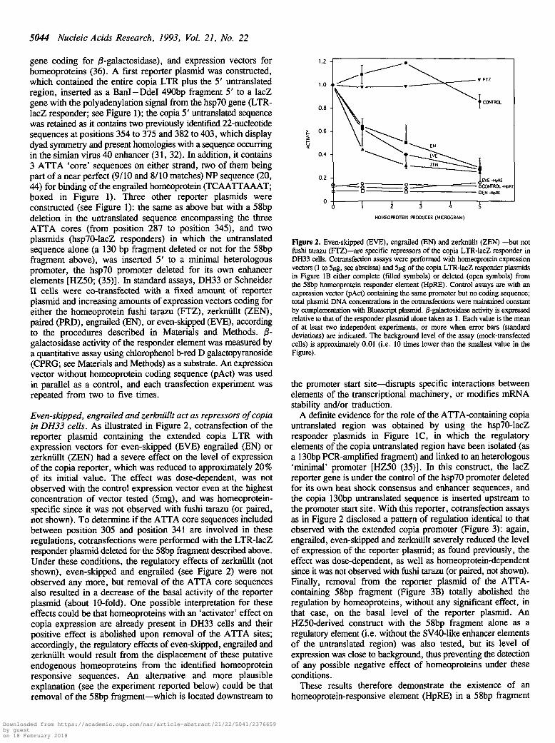
gene coding for /3-galactosidase), and expression vectors forhomeoproteins (36). A first reporter plasmid was constructed,which contained the entire copia LTR plus the 5' untranslatedregion, inserted as a Bard—Ddel 490bp fragment 5' to a lacZgene with the polyadenylation signal from the hsp70 gene (LTR-lacZ responder; see Figure 1); the copia 5' untranslated sequencewas retained as it contains two previously identified 22-nucleotidesequences at positions 354 to 375 and 382 to 403, which displaydyad symmetry and present homologies with a sequence occurringin the simian virus 40 enhancer (31, 32). In addition, it contains3 ATTA 'core' sequences on either strand, two of them beingpart of a near perfect (9/10 and 8/10 matches) NP sequence (20,44) for binding of the engrailed homeoprotein (TCAATTAAAT;boxed in Figure 1). Three other reporter plasmids wereconstructed (see Figure 1): the same as above but with a 58bpdeletion in the untranslated sequence encompassing the threeATTA cores (from position 287 to position 345), and twoplasmids (hsp70-lacZ responders) in which the untranslatedsequence alone (a 130 bp fragment deleted or not for the 58bpfragment above), was inserted 5' to a minimal heterologouspromoter, the hsp70 promoter deleted for its own enhancerelements [HZ50; (35)]. In standard assays, DH33 or SchneiderII cells were co-transfected with a fixed amount of reporterplasmid and increasing amounts of expression vectors coding foreither the homeoprotein fushi tarazu (FTZ), zerknullt (ZEN),paired (PRD), engrailed (EN), or even-skipped (EVE), accordingto the procedures described in Materials and Methods. 0-galactosidase activity of the responder element was measured bya quantitative assay using chlorophenol b-red D galactopyranoside(CPRG; see Materials and Methods) as a substrate. An expressionvector without homeoprotein coding sequence (pAct) was usedin parallel as a control, and each transfection experiment wasrepeated from two to five times.
Even-skipped, engrailed and zerknullt act as repressors of copiain DH33 cells. As illustrated in Figure 2, cotransfection of thereporter plasmid containing the extended copia LTR withexpression vectors for even-skipped (EVE) engrailed (EN) orzerknullt (ZEN) had a severe effect on me level of expressionof the copia reporter, which was reduced to approximately 20%of its initial value. The effect was dose-dependent, was notobserved with the control expression vector even at the highestconcentration of vector tested (5mg), and was homeoprotein-specific since it was not observed with fushi tarazu (or paired,not shown). To determine if the ATTA core sequences includedbetween position 305 and position 341 are involved in theseregulations, cotransfections were performed with the LTR-lacZresponder plasmid deleted for the 58bp fragment described above.Under these conditions, the regulatory effects of zerknullt (notshown), even-skipped and engrailed (see Figure 2) were notobserved any more, but removal of the ATTA core sequencesalso resulted in a decrease of the basal activity of the reporterplasmid (about 10-fold). One possible interpretation for theseeffects could be that homeoproteins with an 'activator' effect oncopia expression are already present in DH33 cells and theirpositive effect is abolished upon removal of the ATTA sites;accordingly, the regulatory effects of even-skipped, engrailed andzerknullt would result from the displacement of these putativeendogenous homeoproteins from the identified homeoproteinresponsive sequences. An alternative and more plausibleexplanation (see the experiment reported below) could be thatremoval of die 58bp fragment—which is located downstream to
1.0
GEN -HpRE
£ i 4 FHOMEOPROTEIN PRODUCER (MICROGRAM)
Figure 2. Even-skipped (EVE), engrailed (EN) and zerknullt (ZEN) —but notfushi tarazu (FTZ)—are specific repressors of the copia LTR-lacZ responder inDH33 cells. Cotransfection assays were performed with homeoprotein expressionvectors (1 to 5^g, see abscissa) and 5/tg of the copia LTR-lacZ responder plasmidsin Figure IB either complete (filled symbols) or deleted (open symbols) fromthe 58bp homeoprotein responder element (HpRE). Control assays are with anexpression vector (pAct) containing the same promoter but no coding sequence;total plasmid DNA concentrations in the cotransfections were maintained constantby complementation with Bluescript plasmid. /3-galactosidase activity is expressedrelative to that of the responder plasmid alone taken as 1. Each value is the meanof at least two independent experiments, or more when error bars (standarddeviations) are indicated. The background level of the assay (mock-transfectedcells) is approximately 0.01 (i.e. 10 times lower than the smallest value in theFigure).
the promoter start site—disrupts specific interactions betweenelements of the transcriptional machinery, or modifies mRNAstability and/or traduction.
A definite evidence for the role of the ATTA-containing copiauntranslated region was obtained by using the hsp70-lacZresponder plasmids in Figure 1C, in which the regulatoryelements of the copia untranslated region have been isolated (asa 130bp PCR-amplified fragment) and linked to an heterologous'minimal' promoter [HZ50 (35)]. In this construct, the lacZreporter gene is under the control of the hsp70 promoter deletedfor its own heat shock consensus and enhancer sequences, andthe copia 130bp untranslated sequence is inserted upstream tothe promoter start site. With this reporter, cotransfection assaysas in Figure 2 disclosed a pattern of regulation identical to thatobserved with the extended copia promoter (Figure 3): again,engrailed, even-skipped and zerknullt severely reduced the levelof expression of the reporter plasmid; as found previously, theeffect was dose-dependent, as well as homeoprotein-dependentsince it was not observed with fushi tarazu (or paired, not shown).Finally, removal from the reporter plasmid of the ATTA-containing 58bp fragment (Figure 3B) totally abolished theregulation by homeoproteins, without any significant effect, inthat case, on the basal level of the reporter plasmid. AnHZ50-derived construct with the 58bp fragment alone as aregulatory element (i.e. without the SV40-like enhancer elementsof die untranslated region) was also tested, but its level ofexpression was close to background, thus preventing the detectionof any possible negative effect of homeoproteins under dieseconditions.
These results therefore demonstrate the existence of anhomeoprotein-responsive element (HpRE) in a 58bp fragment
Downloaded from https://academic.oup.com/nar/article-abstract/21/22/5041/2376659by gueston 18 February 2018

Nucleic Acids Research, 1993, Vol. 21, No. 22 5045
B
0.8-
1 2 3 4 5
HOMEOPROTEIN PRODUCER (MICROGRAM)
D CONTROL• EVE
»HpR£ -HpRE >HpRE -HpRE
l y g Sj/g
HOMEOPROTEIN PRODUCER (MICROGRAM)
Figure 3. The copia untranslated region contains all the elements for regulation of an heterologous promoter by homeoproteins. Cotransfection assays were carriedout in DH33 cells as in Figure 2, with the hsp70-lacZ responder plasmids shown in Figure 1C; each value is the mean of at least two independent experiments,or more when error bars (standard deviations) are indicated. (A) Even-skipped, engrailed, zerknullt, but not fushi tarazu, are repressors as observed for the copiaLTR-based responder plasmid; same conditions as in Figure 2. (B) Regulation by homeoproteins of the responder plasmid depends on the presence of the copia58 bp homeoprotein responsive element (HpRE); activity of the responder plasmids (5^g) with (+HpRE) or without (-HpRE) the copia 58bp HpRE was assayedwith either 1 or 5>»g of even-skipped producing vector (dark filled) or the pAct control vector (control, open); the activity of the HpRE-containing responder plasmidwith no expression vector was taken as the common reference equal to unity.
from the copia 5' untranslated region, which can act in associationwith either the native or an heterologous promoter—de factoeither upstream or downstream to the RNA start sites.Incidentally, they also show that the copia LTR and the flanking77bp of the white gene remaining in the LTR-lacZ responderare not involved in homeoprotein regulation, since theirremoval—in the hsp70-lacZ responder—had no effect.
Fushi tarazu and zerknullt are transcriptional activators of thecopia homeoprotein responsive element in Schneider II cells. Ithas been observed [(32), and see below] that the expression ofcopia is much higher (at least 10 times) in DH33 cells (derivedfrom D.hydei, which do not contain copia elements), than inSchneider n cells (derived from D.melanogaster, which possessapprox. 100 copia elements), possibly because different sets oftranscription factors are expressed in each cell line [for instance,homeoprotein expression has never been detected in SchneiderII cells, but other transcription factors as well can be differentiallyexpressed, (45)]. Schneider II cells might therefore constitute asimple cellular model to detect direct effects of exogenoushomeoproteins, and cotransfection experiments as above wereperformed with the simplified reporter gene (hsp70-lacZresponders, see Figure 1C). As expected and at variance withtransfections of DH33 cells, 10 times more responder plasmidhad to be used to detect signals significantly above the backgroundlevel of the assay. Under these conditions, we found that fushitarazu and zerknullt (Figure 4A, and paired, not shown) had astrong positive effect on the level of expression of the reportergene: the effect was dose-dependent, with an increase of at least10-fold (see Figure 4A, B); activation was not observed with thecontrol plasmid pAct, and is homeoprotein-dependent sinceengrailed and even-skipped had no detectable effects (or a smallnegative effect, if any, for engrailed at low concentrations).Finally, cotransfection experiments with a responder from whichthe 58bp homeoprotein responsive element had been deleted, did
not show any effect of the same homeoproteins (Figure.4B).These results unambiguously confirm the role of the previouslyidentified homeoprotein-responsive element in the observedregulations. Interestingly, we observed that fushi tarazu andzerknullt were able to mediate their enhancer activity (althoughto a lesser extent) on the 58bp HpRE alone, when placed upstreamto the minimal hsp70 promoter (data not shown).
Northern blot analyses were finally performed to determinewhether the positive regulations observed in these cells actuallyreflect increases in RNA levels and to eliminate the possibilityof translational effects. Co-transfections with the responderplasmid above (30ftg) and the different expression vectors (15;tg)were performed in 107 Schneider II cells, RNAs were extracted48hrs post-transfection and Northern blots hybridized with a lacZprobe. As can be seen in Figure 4C, the level of lacZ transcriptswas enhanced when the reporter gene was co-transfected withplasmids coding for fushi tarazu and zerknullt, whereas engrailedand evenskipped vectors had no effect—or a small negativeeffect—in agreement with the data on /3-galactosidase activities.
Identification of homeodomain binding sites in the copiahomeoprotein-responsive elementCo-transfection experiments with expression vectors forhomeoproteins do not indicate whether homeoproteins act direcdyor indirectly (ie via other intermediates) at the level of theidentified homeoprotein-responsive elements in copia. To getfurther insight into the role of these elements, we therefore lookedfor the presence of homeoprotein binding sites specifically locatedin the functionally defined regulatory region. In vitro assays wereperformed (DNAse I protection experiments and mobility shiftassays) using a purified Antennapedia homeodomain (Antp HD)peptide as a specific ligand. This Antp HD is a 60-aminoacidprotein which has been previously demonstrated to specificallyinteract, in vitro, with already known homeoprotein binding sites,with an affinity in the 1-10 naaomolar range (19, 21).
Downloaded from https://academic.oup.com/nar/article-abstract/21/22/5041/2376659by gueston 18 February 2018

5046 Nucleic Acids Research, 1993, Vol. 21, No. 22
k
10-
8-
6-
4 -
2-
0-
A —
A
1
1/
^ » ZEN
- — '
^T FTZ
, m m n• EN
1 2 3 4 5
HOMEOPROTEIN PRODUCER (MICROGRAM)
B3U "
25-
20-
AC
TIV
ITY
10-
5-
1 -
o-
D CONTROLO FTZ• ZEN
r - m
c0 EVE EN fU ZEN
*HpRE -HpRE *HpRE -HpREl e g 5yg
HOMEOPROTEIN PRODUCER (MICROGRAM)
1 2 3 4 5 6
Figure 5. DNAse I footprinting analysis of the copia LTR and untranslated regionusing a 60 aminoacid homeodomain-containing polypeptide (HD). Footprintingreactions were performed on a 565bp Pstl-Hindin fragment containing the entirecopia LTR and untranslated 5' region (labelled at the 3' end of the transcribedstrand), and a 60 aminoacid homeodomain-containing polypeptide (AntennapediaHD, cf Materials and Methods). A G+A sequencing reaction was run in theadjacent lane. Amounts of Antp peptide used are as follow: lanes 1 and 7 (none),lane 2 (25nM), lane 3 (50nM), lane 4 (75nM), lane 5 (90nM), lane 6 (115nM).The sequence of the strongly protected region (I) is indicated with the ATTAcore boxed in solid lines, as well as that of the adjacent region (IT) with weakerprotection; the U3, R and U5 domains of the copia LTR are indicated.
Figure 4. Fushi tarazu and zerkniillt are specific activators of the copiahomeoprotein responsive element in Schneider II cells. (A) Cotransfection assayswere as in Figure 3, but in Schneider n cells, with 0.5^g of the hsp70-lacZresponder plasmid shown in Figure 1C; differential regulatory effects are observedin Schneider n versus DH33 cells for zerknu Ut (strong positive/strong negativeeffects), fushi tarazu (strong positive/null effects) and engrailed (nul/strong negativeeffects); control refers to pAct plasmid and activities are expressed relative tothat of the responder plasmid alone taken as unity; each value is the mean oftwo independent experiments. (B) Homeoprotein positive regulations are dependenton the presence of the copia HpRE in the responder plasmid. Same conditionsas in 4A; activities of the responder plasmids with (+HpRE) or without (—HpRE)the copia 58bp homeoprotein responsive element were assayed with 1 or 5/»gof either pAct control plasmid (control, open), fushi tarazu (grey) or zerkniillt(dark) expression vectors; the activity of the HpRE-containing respondercotransfected with 1/ig of control plasmid was taken as unity; each value is themean of two independent experiments. (C) Northern blot analysis of responderplasmid expression. 15mg of responder plasmid and 30/jg of the indicatedhomeoprotein producers were co-transfected in 107 Schneider n cells: responderalone (lanel), even-skipped (lane 2), engrailed (lane 3), fushi tarazu Qane 4),zerkniillt flane 5). Each lane corresponds to lOmg of total RNA extracted 48hrspost-transfection; the 3.9kb 'responder' transcript (upon hybridization of the blotwith a lacZ radiolabelled probe) and position of the calf 28S ribosomal RNA(r) are indicated.
DNAse I protection experiments. DNAse I protection experimentswere carried out with a 565bp Pstl—HindlE fragment from thecopia lacZ construct, which contains the complete copia LTRand untranslated sequences. End-labelled DNA (lnM) wasincubated with increasing concentrations of Antp HD in thepresence of polydl-dC, submitted to DNAse I digestion, and runon a sequencing gel as described in Materials and Methods. Asillustrated in Figure 5, a major region is protected from DNAseldigestion, namely the AATAAACAATTAAATTG sequencelocated in the 58bp homeoprotein responsive element. Thisprotected region, approximately 15bp long, is centered aroundan ATTA core embedded in the near-perfect NP sequenceindicated in Figure 1 (9/10 match, box I). Increasing HDconcentration (up to lOOnM) resulted in a weaker and incompleteprotection of the second previously noted NP-like sequencelocated just 3 ' to the previous one (8/10 match, box II). In theLTR domain, no protection can be observed even in die 3' endof the LTR where three ATTA cores are present, consistent withthe results from the co-transfection experiments on the absenceof regulatory sequences in this region. The absence of protectionof these ATTA sequences, as well as the reduced protection of
Downloaded from https://academic.oup.com/nar/article-abstract/21/22/5041/2376659by gueston 18 February 2018

Nucleic Acids Research, 1993, Vol. 21, No. 22 5047
AntpHD(nM)
0 0 4 0 8 16 3 1 6 2 12.5 25 50 100
HpRE —-—"I
1 2 3 4 5 6
2nM Antp HD 50nM Antp HD
HpRE 22-mer competitor HpRE
~0~ 50~ ~0 50~ DNA(nM) 0 25 50 250
B2
Figure 6. Mobility shift assay of Antp-homeodomain/copia-HpRE interactions.Binding of the Antp HD peptide, at the indicated concentrations, to the 58bpend-labelled copia HpRE was assayed as indicated in Materials and Methods,(upper) Increasing concentrations of Antp HD were incubated with a fixed amountof end-labelled copia HpRE and ran on a 9% polyacrylamide gel; F: free DNA,Bl and B2: bound DNA-HD complexes, (lower) Specificity of binding; end-labelled copia HpRE (0. lnM) and Antp HD at the indicated concentrations wereincubated in the presence of competitor DNA—either the unlabelled copia HpREor an unrelated 22-mer double-stranded oligonucleotide (SP1 site, same resultswith API site, cf Materials and Methods)—at the indicated concentrations.
o 1 1 10 100free DNA (nM)
Figure 7. Determination of equilibrium binding coefficients by electrophoreticmobility shift assay. Antp HD peptide at a fixed concentration of lOnM wasincubated for 4 hr in binding buffer containing increasing concentrations of labelledHpRE (0.39 to lOOnM with regular two-fold increase, from lane 1 to 9). Bound(B) and free (F) DNAs were visualized by autoradiography (upper part), anddata obtained from the titration plotted flower part); an equilibrium dissociationconstant of 4.1nM can be derived upon non linear least-square fit of the data(theoretical binding isotherm in solid line).
the second more divergent NP-like sequence above, constituteinternal controls for the specificity of the observed HD protection.
Mobility shift assay. The results above were confirmed, in aquantitative manner, by mobility shift assays. Binding of the AntpHD to the previously characterized 58bp fragment could bereadily detected by the retarded electrophoretic mobility of theend-labelled HpRE DNA fragment. Indeed, in the nanomolarrange of Antp HD concentrations, a band of retarded mobilitycould be clearly observed (Bl, see Figure 6), whose intensityincreased in a dose-dependent manner with HD concentration.This retarded band corresponds to a specific interaction of theAntp HD to saturable sites since it was competed out by an excessof unlabelled binding sequence added prior to the labelled DNA,and not by an identical amount of irrelevant unlabelled oligo-nucleotides (22-mer, see Figure 6 bottom left and Materials andMethods). In the high concentration range, a second retardedcomplex could be observed (B2, Figure 6 upper part), which isalso associated with specific interaction to saturable sites sinceit could be competed, as well as the Bl complex, by an excessof unlabelled HpRE DNA (see Figure 6, bottom right). Theequilibrium dissociation constant (Kd) for binding to the majorBl site was accurately determined from Antp homeodomainsaturation experiments by varying the concentration of the 58bpHpRE fragment, as illustrated in Figure 7; labelled DNA wasisotopically diluted (see Materials and Methods), so that DNAconcentration could be precisely determined. Accordingly, theKd was found equal to 4 nM at 20 ± 1°C (Figure 7). This value
is closely related to that in (21) for the same peptide and anoligodeoxynucleotide containing an Antp consensus binding site(1 -2 nM).
DISCUSSIONFunctional binding sites for homeoproteins in the copiaretrotransposon
We have demonstrated that the copia retrotransposon possessesbinding sites for transcriptional regulation by homeoproteins, andthis is the first report for the occurrence of such sites on a mobileelement. These binding sites are located 3' to the left LTR, inthe copia untranslated region, close to a previously identifiedenhancer element first described in (31). DNAsel footprintingexperiments and gel mobility shift assays with a purified minimalAntennapedia homeodomain protein demonstrate high affinitybinding to a nucleotide sequence with a near perfect NPconsensus, with a Kd of 4 nM in good agreement with valuespreviously measured for the same homeodomain to otherhomeoprotein binding sites [1-10 nM (19, 21)]; binding witha lower affinity was also observed to a nucleotide sequence,adjacent to the primary sequence, closely related to the consensusNP sequence. The involvement of these binding sites in copiatranscriptional regulation was demonstrated by a test usingDrosophila cells in culture, from either D.hydei or D.melano-gaster. Co-transfection experiments with expression veaors fordifferent homeoproteins and responder plasmids containing thecopia untranslated region under the control of either the copia
Downloaded from https://academic.oup.com/nar/article-abstract/21/22/5041/2376659by gueston 18 February 2018

5048 Nucleic Acids Research, 1993, Vol. 21, No. 22
LTR or an heterologous minimal promoter, disclose eitherpositive or negative regulations, whose occurrence is dependentof the presence of the HD binding site-containing fragment.Regulation is homeoprotein-dependent, concentration-dependent,and cell type-dependent, and takes place at the RNA level asshown by Northern blot analysis.
Rather paradoxically, the copia LTR has been extensively usedas a control for transfection efficacy in Drosophila cells, andparticularly in transfection assays using expression vectors forhomeoproteins and reporter plasmids [see for instance (36, 44)].In those cases, no regulatory effects of either the engrailed, even-skipped, zerkniillt, fushi tarazu or paired homeoproteins on thelevel of expression of the 'control' copia reporter had ever beenreported. This apparent discrepancies can be easily accountedfor by the difference between the constructs which had been usedand ours, as the former copia-containing constructs were deletedfrom the untranslated region and only possessed the 5'LTR strictosensu [as an EcoRI-HindlH fragment from the pCOPneoconstruction (44)].
The structural organization of the copia LTR and untranslatedsequence is reminiscent of that of the yeast Ty retroelement,where enhancer elements have also been characterized in theuntranslated region, 3' to the left LTR (46, 47). Phylogeneticanalysis have previously emphasized the close relationshipbetween these two retrotransposons (48), and recent analysis ofthe Drosophila retrovirus-like retrotransposon mdgl (49) andLINE element promoters [reviewed in (50)] have furtherevidenced that transcriptional control sequences are often locatedin these elements 3' to the RNA start site. Finally, it has beenextensively documented that homeoprotein DNA binding sitesfrom several homeotic genes can act as regulatory sequences ineither orientation, as for classical enhancers, and also when linkedto heterologous promoters [references in (14, 51)], as observedin the present study.
Diversity of the homeoprotein-mediated regulations of copiaThe complexity of the regulations mediated by homeoproteinsis illustrated by the differential effects of several homeoproteinsdepending on the cell line used. For instance, zerkniillt decreasesthe activity of the lacZ reporter gene in DH33 cells whereas itenhances its activity in Schneider II cells; fushi tarazu enhancesthe activity in Schneider II cells but has no effect in DH33 cells.Several interpretations for these differential effects can behypothesized, among which that of the presence in DH33 cellsof endogenously expressed homeoproteins that would be simplycompeted out by the products of the exogenously introducedhomeoprotein expression vectors. This interpretation, however,cannot simply account for the absence of effect of the removalof the HpRE 58bp fragment on the basal level of expression inthe hsp-lacZ responder (see Results, figure 3B). Another possiblesource of regulation diversity could be the presence of more thanone binding site for homeoprotein in the copia regulatory domain.Actually, in vitro studies with die Antp homeodomain haverevealed a major binding site, but both mobility shift assay andfoot-printing experiments have suggested possible interactions—with a significantly lower affinity for Antp HD—to a secondbinding site adjacent to the main site, and which further disclosessimilarities with the consensus NP sequence. Accordingly, thecombinatory binding of both endogenously and exogenouslyexpressed homeoproteins could result in differential effects[aldiough we did not observe, in co-transfection assays using
simultaneously two different homeoprotein expression vectors,any cooperative or synergistic effects(data not shown)]. Finally,other sites on the copia regulatory element could also be involved.For instance, we have demonstrated by mobility shift assays (45)that DH33 cell nuclear extracts contain a protein which bindsspecifically to the copia SV40-like 'enhancer' sequence, whereasSchneider II cell nuclear extracts do not. This enhancer sequenceis adjacent to the homeodomain binding sites (cf Figure 1), andthe presence or not of a factor bound to this enhancer sequence—and further possibly directly interacting with homeoproteins, asobserved for the herpes virus VP16 protein (52) or the yeast GRMprotein (53)—could severely modulate the homeoproteinresponse. It has also been demonstrated that copia gene productsare able to negatively regulate their own promoter at the levelof sites present within the LTR sensu stricto (54). It is thereforepossible that this self-regulation interferes with the homeoproteinresponse in the copia-containing Schneider n cells. Actually, wehave detected for these cells significantly weaker responses withthe copia LTR reporter than with the hsp-lacZ reporter—wherethe homeodomain binding sites are placed upstream of theheterologous hsp70 enhancerless promoter—a result consistentwith the observation in (54) that copia elements do not negativelyregulate the hsp70 promoter.
It should be finally emphasized that the diversity of theregulatory effects of homeoproteins as demonstrated in this study,is a common major feature of this class of transcription factors(see Introduction); differential effects have also been reportedfor other genes, with for instance the Drosophila dorsal mor-phogen which can mediate both activation and repression in acontext-dependent manner (55). As discussed above, thiscomplexity possibly involves multiple binding sites and factors,as well as direct protein—protein interactions with factorsinvolved in the transcription machinery [reviewed in (14); seealso (56) for evidence of multi-protein complexes between nuclearfactors and engrailed].
PerspectivesThe presence of active homeoprotein-responsive elements in thecopia retrotransposon raises several questions. A first oneconcerns their possible role in the developmental regulation ofcopia expression. Clearly now, identification of functional bindingsites should allow to devise experiments in which the effect oftheir removal or mutation can be analyzed in vivo, usingtransgenic Drosophila containing copia-driven reporter genes, inrelation with alterations in their spatial and temporal patterns ofexpression in the course of Drosophila development. Actually,preliminary experiments using transgenic Drosophila with alteredexpression for given homeoproteins—for instance Drosophilacontaining homeoprotein genes under the control of the heat-shockinducible hsp promoter—have revealed a 2—3 fold increase ofendogenous copia transcripts upon overexpression of the fushitarazu gene, when monitoring the overall copia expression bydot blot analysis, and a 'patterned' expression of a copia-drivenlacZ reporter gene under conditions of forced engrailedexpression, using an in situ assay for /3-galactosidase activity (57).Another related question concerns the generality of the observedregulations: other developmentally regulated Drosophilaretrotransposons (412, 297, gypsy) have in their untranslatedregion, close to the 5' LTR, AT-rich sequences where putativehomeoprotein binding sites can be identified. Experiments similarto those reported in this paper are now in progress with these
Downloaded from https://academic.oup.com/nar/article-abstract/21/22/5041/2376659by gueston 18 February 2018

Nucleic Acids Research, 1993, Vol. 21, No. 22 5049
elements, which should help in answering the intringuing questionof the raison d'Etre of homeoprotein binding sites inretrotransposons.
ACKNOWLEDGEMENTS
We wish to specially acknowledge Drs K.Han and M.S.Levinefor the generous gift of the homeoprotein expression vectors, andDrs A.Joliot and A.Prochiantz for that of purified Antennapediahomeodomain peptide. We are very grateful to S.Jensen forinitiating one of us to Drosophila and for helpfull discussionsand suggestions, and to M.-P.Gassama for her help in themaintenance of the fly stocks and technical assistance. Weacknowledge Drs C.Lavialle and J.F.Nicolas for critical readingof the manuscript. This work was financed by the Centre Nationalde la Recherche Scientifique (URA 147), the Institut Nationalde la Sante et de la Recherche Mddicale (U 140), the Associationpour la Recherche sur le Cancer (contract 6552 to T.H.), andthe Ligue contre le Cancer (to L.C.).
REFERENCES
1. Sankaranarayanan, K. (1988) Banbury Rep. 30, 319-336.2. Echalier, G. (1989) Advances in Virus Research 36, 33-105.3. Finnegan, D.J. (1990) Current Opinion in Cell Biology 2, 471-477.4. Finnegan, D.J. (1989) In Mobile DNA (Berg and Howe, eds), 503-517.
American Society for Microbiology Washington, D.C.5. Bucheton, A. (1990) Trends In Genetics 6, 16-21.6. Boeke, J.D., Garfinkel, D.J., Styles, C.A. and Fink, G.R. (1985) Cell 40,
491-500.7. Heidmann, O. and Heidmann, T. (1991) Cell 64, 159-170.8. Jensen, S. and Heidmann, T. (1991) EMBO J. 10, 1927-1937.9. Pelisson, A., Finnegan, D.J. and Bucheton, A. (1991) Proc. Natl. Acad.
Sci. USA 88, 4907-4910.10. Agnel, M. and Heidmann, T. unpublished data.11. Parkhurst, S.M. and Corces, V.G. (1987) EMBO J. 6, 419-424.12. Kuff, E.L. and Lueders, K.K. (1988) Adv. in Cancer Research 51, 183-276.13. Brulet, P., Condamine, H. and Jacob, F. (1985) Proc. Natl. Acad. Sci. USA
82, 2054-2058.14. Hayashi, S. and Scott, M.P. (1990) Cell 63, 883-894.15. Affolter, M., Schier, A. and Gehring, W.J. (1990) Current Opinion in Cell
Biology 2, 485-495.16. Gehring, W.J., Muller, M., Affolter, M., Percival-Smith, A., Billeter, M.,
Qian, Y.Q., Otting, G. and Wuthrich, K. (1990) Trends in Genetics 6,323-329.
17. McGinnis, W. and Krumlauf, R. (1992) Cell 68, 283-302.18. StJohnston, D. and Nusslein-Volhard, C. (1992) Cell 68, 201-219.19. Muller, M., Affolter, M., Leupin, W., Otting, G., Wuthrich, K. and Gehring,
W.J. (1988) EMBO J. 7, 4299-4304.20. Desplan, C , Theis, J. and O'Farrell, P.H. (1988) Cell 54, 1081-1090.21. Affolter, M., Percival-Smith, A., Muller, M., Leupin, W. and Gehring,
W.J. (1990) Proc. Natl. Acad. Sci. USA 87, 4093-4097.22. Ekker, S.C., Young, K.E., von Kessler, D.P. and Beachy, P.A. (1991)
EMBO J. 10, 1179-1186.23. Florence, B., Handrow, R. and Laughon, A. (1991) Mol. Cell. Biol. 11,
3613-3623.24. Otting, G., Qian, Y.Q., Muller, M., Affolter, M., Gehring, W.J. and
Wuthrich, K. (1988) EMBO J. 7, 4305-4309.25. Qian, Y.Q., Billeter, M., Otting, G., Muller, M., Gehring, W.J. and
Wuthrich, K. (1989) Cell 59, 573-580.26. Kissinger, C.R., Liu, B., Martin-Bianco, E., Komberg, T.B. and Pabo, C O .
(1990) Cell 63, 579-590.27. Otting, G., Qian, Y.Q., Billeter, M., Muller, M., Affolter, M., Gehring,
W.J. and Wuthrich, K. (1990) EMBO J. 9, 3085-3092.28. Billeter, M., Qian, Y.Q., Otting, G., Muller, M., Gehring, W.J. and
Wuthrich, K. (1990)/ Mol. Biol. 214, 183-197.29. Hanes, S.D. and Brent, R. (1991) Science 251, 426-430.30. Ekker, S.C., von Kessler, D.P. and Beachy, P.A. (1992) EMBO J. 11,
4059-4072.
31. Mount, S.M. and Rubin, G.M. (1985) Mol. Cell. Biol. 5, 1630-1638.32. Sneddon, A. and Flavell, A.J. (1989) Nucl. Acids Res. 17, 4025-4035.33. Levis, R., Bingham, P.M. and Rubin, G.M. (1982) Proc. Natl. Acad. Sci.
USA 79, 564-568.34. Bonnerot, C , Rocancourt, D., Briand, P., Grimber, G. and Nicolas, J.-F.
(1987) Proc. Natl. Acad. Sci. USA 84, 6795-6799.35. Hiromi, Y. and Gehring, W.J. (1987) Cell 50, 963-974.36. Han, K., Levine, M.S. and Manley, J.L. (1989) Cell 56, 573-583.37. Schneider, I. (1972) J. Embryol. Exp. Morphol. 27, 353-356.38. Sondermeijer, P.J.A., Derksen, J.W.M. and Lubsen, N.H. (1980) In Vitro
16, 913-914.39. Hanahan, D. (1983) J. Mol. Biol. 166, 557-580.40. Simon, J.A., Lis, J.T. and (1987) Nucl. Acids Res. 15, 2971-2988.41. Church, G.M. and Gilbert, W. (1984) Proc. Natl. Acad. Sci. USA 81,
1991-1995.42. Joliot, A., Pernelle, C , Deagostini-Bazin, H. and Prochiantz, A. (1990)
Proc. Natl. Acad. Sci. USA 88, 1864-1868.43. Maxam, A.M. and Gilbert, W. (1977) Proc. Natl. Acad. Sci. USA 74, 560.44. Jaynes, J.B. and O'Farell, P.H. (1988) Nature 336, 744-749.45. Cavarec, L., Jensen, S. and Heidmann, T. in preparation46. Fulton, A.M., Rathjen, P.D., Kingsman, S.M. and Kingsman, A.J. (1988)
Nucl. Adds Res. 16, 5439-5458.47. Farabaugh, P., Iiao, X.B., Belcourt, M., Zhao, H., Kapakos, J. and Clare,
J. (1989) Mol. Cell. Biol. 9, 4824-4834.48. Xiong, Y. and Eickbush, T. (1990) EMBO J. 9, 3353-3362.49. Arkhipova, I.R. and Dyin, Y.V. (1991) EMBO J. 10, 1169-1177.50. Martin, S.L. (1991) Current Opinion in Genetics and Development 1,
505-508.51. TenHarmsel, A., Austin, R.J., Savenelli, N. and Biggin, M.D. (1993) Mol.
Cell. Biol. 13, 2742-2752.52. Stern, S., Tanaka, M. and Herr, W. (1989) Nature 341, 624-630.53. Passmore, S., Elble, R. and Tye, B.-K. (1989) Genes & Development. 3,
921-935.54. Bryant, L.A., Brierley, C , Flavell, A.J. and Sinclair, J.H. (1991) Nucl.
Acids Res. 19, 5533-5536.55. Pan, D. and Courey, A.J. (1992) EMBO J. 11, 1837-1842.56. Gay, N.J., Poole, S. and Komberg, T. (1988) EMBO J. 7, 4291-4297.57. Cavarec, L., Jensen, S. and Heidmann, T. unpublished data.
Downloaded from https://academic.oup.com/nar/article-abstract/21/22/5041/2376659by gueston 18 February 2018