the digestive system. wall of the digestive tract a. mucosa - mucous epithelium b. submucosa -...
TRANSCRIPT

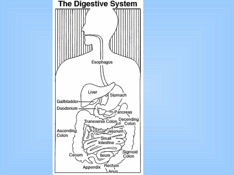
The Digestive System

WALL OF THE DIGESTIVE TRACT
A. Mucosa - mucous epithelium
B. Submucosa - connective tissue
C. Muscularis - 2 or 3 layers of smooth muscle
D. Serosa - serous membrane that covers the outside of abdominal organs ; it attaches the digestive tract to the wall of the abdominopelvic cavity by forming folds called mesenteries
The four layers of tissue that form the wall of the digestive tube is :

MOUTH
A. The mouth or oral cavity is a hollow chamber with a roof, a floor, and walls.
B. Roof - formed by hard palate and soft palate, an arch-shaped muscle separting mouth from pharynx
C. Floor - formed by tounge and its muscles ; papillae, small elevations on mucosa of tounge
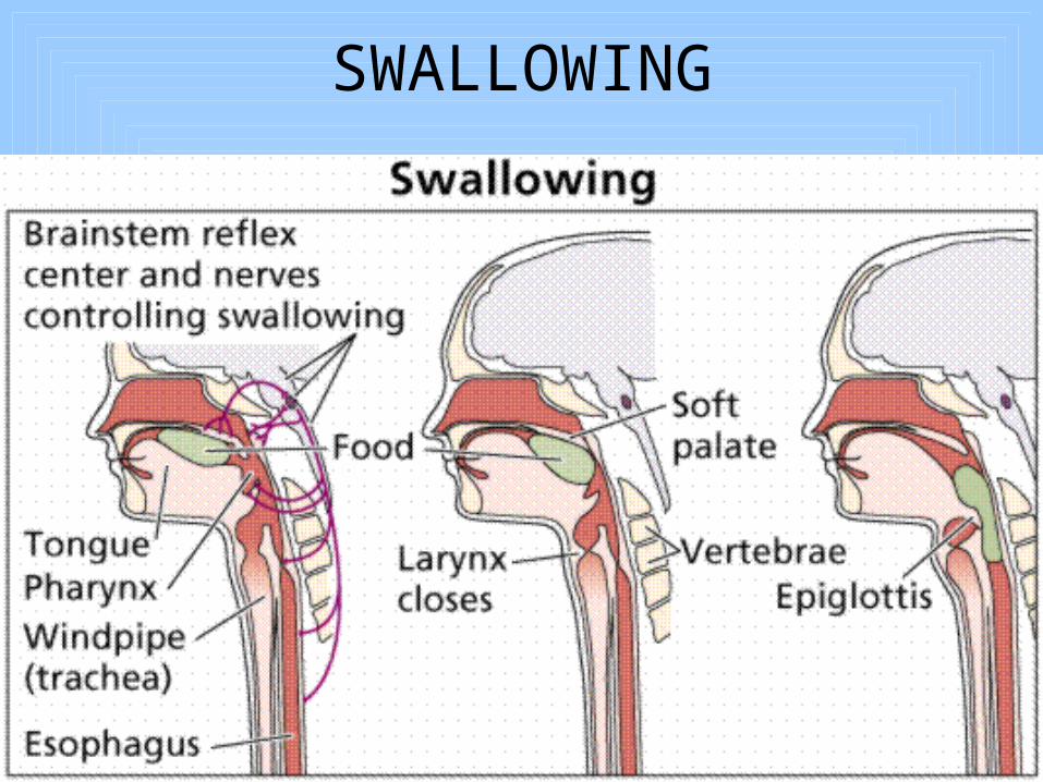
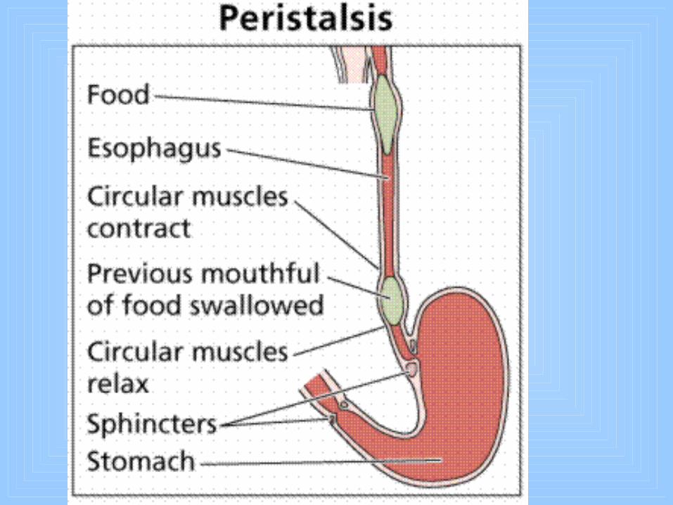
SWALLOWING

TEETHA. Names of teeth : 4 incisors, 2 canines, 4 biscuspids, and 6 tricuspids
(32 total)
Child - 4 incisor, 2 canines, 2 1st molar, 2 2nd molar, (20 total)
B. A person has a temporary set of twenty teeth and thirty-two teeth in permanent set.
C. Average age for cutting 1st tooth about six months.
D. Average age for cutting permanent teeth , six.
E. Set complete of all teeth is ages of 17 to 24 years.

Panoramic x-ray

Children teeth
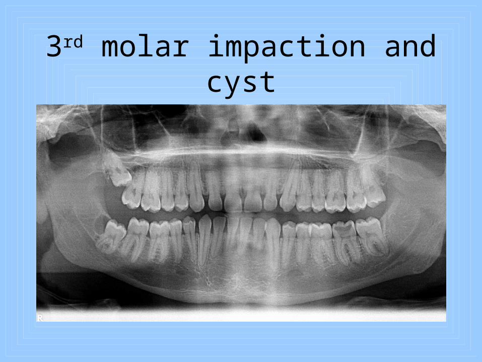
3rd molar impaction and cyst

Mandibular third molar impaction with 2nd molar
involvment

Mand. 2nd molar root canal therapy

Bite wing xay with decay
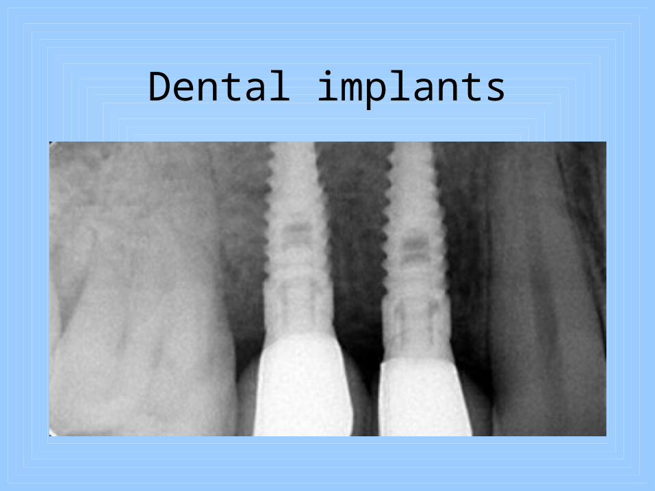
Dental implants

dental caries(decay)

SALIVARY GLANDS
A. Parotid glands - largest gland, lies just below and in front of each ear at the angle of the jaw
B. Submandibular glands - located on either side of the lingual frenulum
C. Sublingual glands - located on the floor of the mouth
The three pairs of salivary glands :

PHARYNX
A. The tubelike structure made of muscle and lined with mucous membrane.
B. Function - air must pass though the pharynx on its way to the lungs, and food must pass through it on its way to the stomach.

ESOPHAGUSA. The esophagus or food pipe, is the
muscular, mucus-lined tube that connects the pharynx with the stomach.
B. It is about 25 centimeters long
C. Function - serves as a dynamic passageway for food, pushing the food toward the stomach.

STOMACH
A. Size - expands after large meals.
B. Pylorus - lower part of stomach; pyloric sphincter muscle closes opening of pylorus into duodenum
C. Wall - many smooth muscle fibers
D. Lining - mucous membrane; many microscopic glands that secrete gastric juice and hydrochloric acid into stomach

SMALL INTESTINE
A. Size - about 7 meters long and 2 cm in diameter
B. Divisions
– Duodenum
– Jejunum
– Ileum
C. Wall - contains smooth muscle fibers that contract to produce peristlsis
D. Lining - mucous membrane

Large Intestines (Colon)
• Six feet - ascending, transverse, descending
• Appendix - Lower right - abs
• Completes digestion and absorption of nutrients
• Water and electrolyte management
• Feces production
LIVER & GALLBLADDER
A. Size - liver is the largest gland
B. Location - fills upper right section of abdominal cavity and extends over into left side
C. Liver secretes bile
D. Location of Gallbladder - undersurface of liver
E. Function - concentrates and stores bile produced in the liver

PANCREAS
A. Location - behind stomach
B. Functions 1. Pancreatic cells secrete pancreatic juice into
pancreatic ducts
2. Secrete hormones glucagon and insulin into the blood

LARGE INTESTINE
A. Divisions
1. Cecum
2. Colon - ascending, transverse, descending, and sigmoid
3. Rectum
B. Opening to exterior - anus
C. Wall - contains smooth muscle fibers that contract to produce churning.
D. Lining - mucous membrane

APPENDIX
A. Dead end tube off cecum
B. No important digestive functions in humans
C. It contains lymphatic tissue and may play a minor role in the immunologic defense mechanisms of the body.

PERITONEUM
A. Peritoneum is a serous membrane lining abdominal cavity and covering abdominal organs
1. The parietal layer of peritoneum lines abdominal cavity
2. Viscreal layer of peritoneum covers abdominal organs
3. Peritoneal space lies between parietal and visceral layers

B. Extensions - largest ones are the mesentery and greater omentum
1. Mesentery is extension of parietal peritoneum, which attaches most of small intestine to posterior abdominal wall
2. Greater omentum, or “lace apron,”hangs down from lower edge of stomach and transverse colon over intestines
DIGESTIONDigestion is changing foods so that they can
be absorbed and used by cells
A. Mechanical digestion - chewing, swallowing, and peristalis break food into tiny particles, mix them well with digestive juices, and move them along the digestive tract
B. Chemical digestion - breaks up large food molecules into compounds having smaller molecules

C. Carbohydrate digestion - mainly in small intestine
1. Pancreatic amylase - changes starches to maltose
2. Intestinal juice enzymes
a. Maltase - changes maltose to glucose
b. Sucrase - changes sucrose to glucose
c. Lactase - changes llactose to glucose
D. Protein digestion - starts in stomach, completed in small intestine
1. Gastric juice enzymes, renin and pepsin, partially digest proteins.

2. Pancreatic enzyme, trypsin, completes digestion of proteins to amino acids
3. Intestinal enzymes, peptidases, complete digestion of partially digested proteins to amino acids
E. Fat digestion - bile that contains no enzymes but emulsifies fats. Pancreatic lipase changes emulsified fats to fatty acids and glycerol in small intestine.
ABSORPTION
A. Absorption is the digested food that moves from intestine into blood or lymph
B. Where absorption occurs - foods and most water from small intestine. Some water also absorbs from large intestine